left thoracoabdominal incision
TRANSCRIPT

Left Thoracoabdominal Incision
Sudhir Sundaresan
The left thoracoabdominal incision provides excellentexposure for operations dealing with the distal esoph-
agus or proximal stomach. It is particularly useful forcomplex reoperations in this region, which are typicallyquite difficult due to the presence of significant adhesionsinvolving the stomach, diaphragm, and liver. The leftthoracoabdominal incision is indicated for (1) resection ofcarcinomas of the lower third of the esophagus or esopha-gogastric junction; (2) resection of middle third esopha-geal carcinomas, where the tumor is located below thecarina; and (3) complex esophageal repairs, notably re-operative antireflux surgery. In complex reoperations atthe esophageal hiatus or in primary repairs of massivehiatal hernias, a left thoracotomy incision alone with di-vision of the periphery of the diaphragm may be suffi-cient. However, the left thoracoabdominal incision is auseful extension of this approach to facilitate superiorexposure and safer conduct of the surgery.
The left thoracoabdominal incision can also be com-bined with a left neck incision to perform total esoph-agectomy with cervical esophagogastrostomy. Withslightly caudal extension of the lower end of the inci-sion, the left colon may be mobilized for use as areplacement conduit for the esophagus using this ap-proach. Finally, the left thoracoabdominal incisionprovides superb exposure for performance of total gas-trectomy. The incision easily facilitates Roux-en-Y re-construction to the distal esophagus.
The left thoracoabdominal incision is not an ideal ap-proach when access is needed to the esophagus at or above
the carina because the arch of the aorta obscures access tothe esophagus at this level. Relative contraindications tothis incision include a prior left thoracotomy, and priorright pneumonectomy because left lung deflation is neces-sary to achieve satisfactory exposure.
It is essential to conduct an appropriate work-up ofthe patient preoperatively. In the case of esophagealresection for cancer, the barium swallow and endos-copy are critically important to determine the upperlimit of the esophageal access necessary. Computerizedtomography is also useful for this assessment and forevaluating the extension of tumor to surrounding me-diastinal structures. Preoperative assessment for com-plex operations or reoperations for benign esophagealdisease should include a careful history, barium swal-low, endoscopy, and esophageal manometry, possiblycombined with extended pH monitoring.
Anesthetic treatment of patients undergoing left tho-racolaparotomy includes the preoperative placement of athoracic epidural catheter. This is important for postop-erative pain relief and can be retained for up to 5 days.Other routine aspects of monitoring include insertion of aradial arterial line in the right arm and Foley catheter.Either a left-sided double lumen tube, or standard endo-tracheal tube with left bronchial blocker may be used todeflate the left lung during the operation. Of note, if theleft thoracolaparotomy is to be combined with a left neckincision for total esophagectomy, the left arm will need tobe free draped. In this scenario, no intravenous or arte-rial lines should be placed in the left arm.
Operative Techniques in Thoracic and Cardiovascular Surgery, Vol 8, No 2 (May), 2003: pp 71–85 71
SURGICAL TECHNIQUE
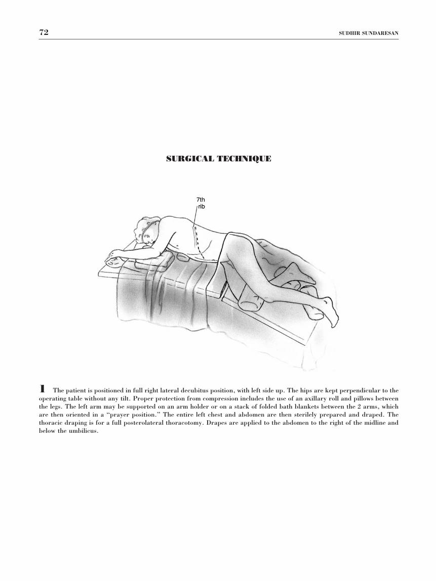
1 The patient is positioned in full right lateral decubitus position, with left side up. The hips are kept perpendicular to theoperating table without any tilt. Proper protection from compression includes the use of an axillary roll and pillows betweenthe legs. The left arm may be supported on an arm holder or on a stack of folded bath blankets between the 2 arms, whichare then oriented in a “prayer position.” The entire left chest and abdomen are then sterilely prepared and draped. Thethoracic draping is for a full posterolateral thoracotomy. Drapes are applied to the abdomen to the right of the midline andbelow the umbilicus.
72 SUDHIR SUNDARESAN
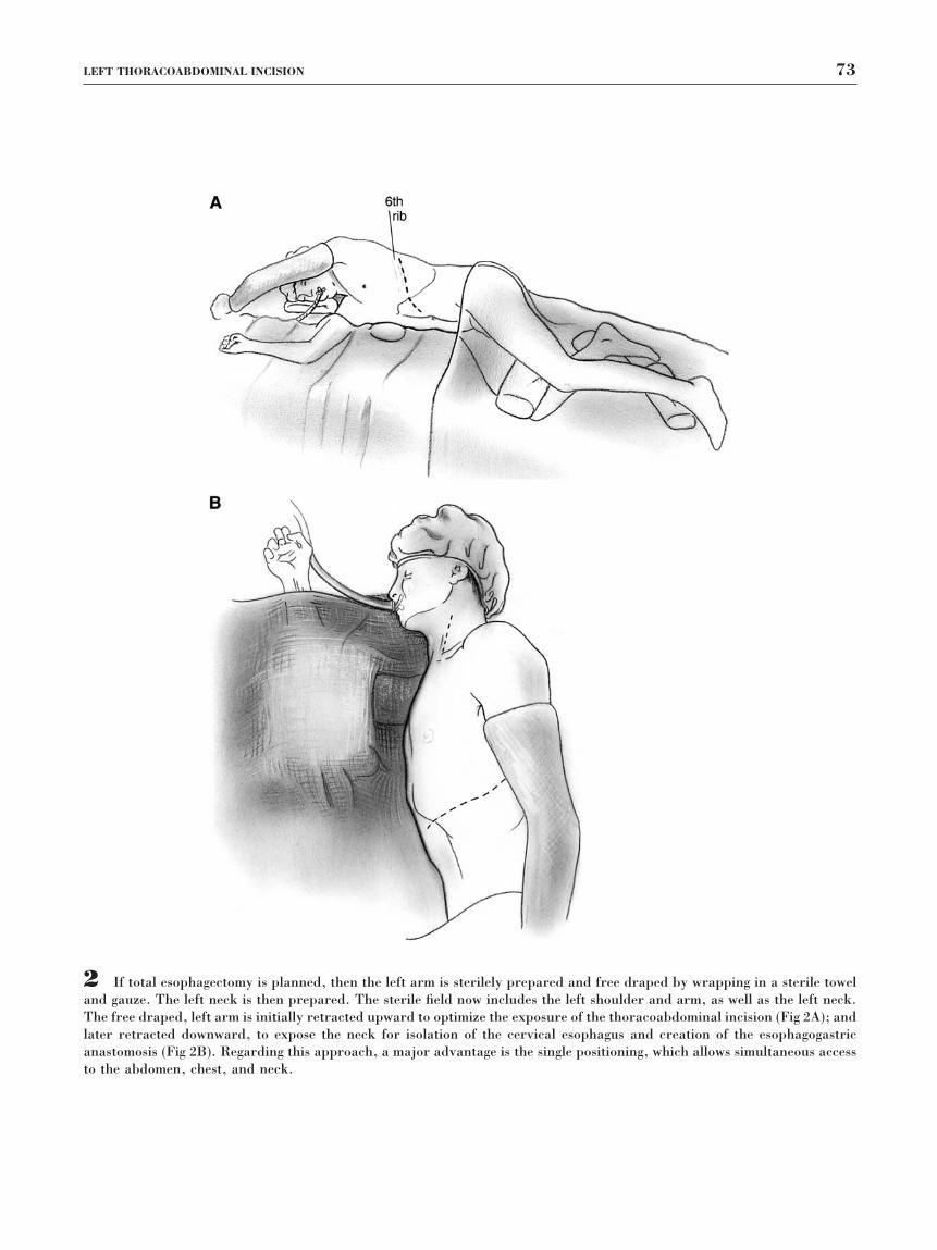
2 If total esophagectomy is planned, then the left arm is sterilely prepared and free draped by wrapping in a sterile toweland gauze. The left neck is then prepared. The sterile field now includes the left shoulder and arm, as well as the left neck.The free draped, left arm is initially retracted upward to optimize the exposure of the thoracoabdominal incision (Fig 2A); andlater retracted downward, to expose the neck for isolation of the cervical esophagus and creation of the esophagogastricanastomosis (Fig 2B). Regarding this approach, a major advantage is the single positioning, which allows simultaneous accessto the abdomen, chest, and neck.
LEFT THORACOABDOMINAL INCISION 73
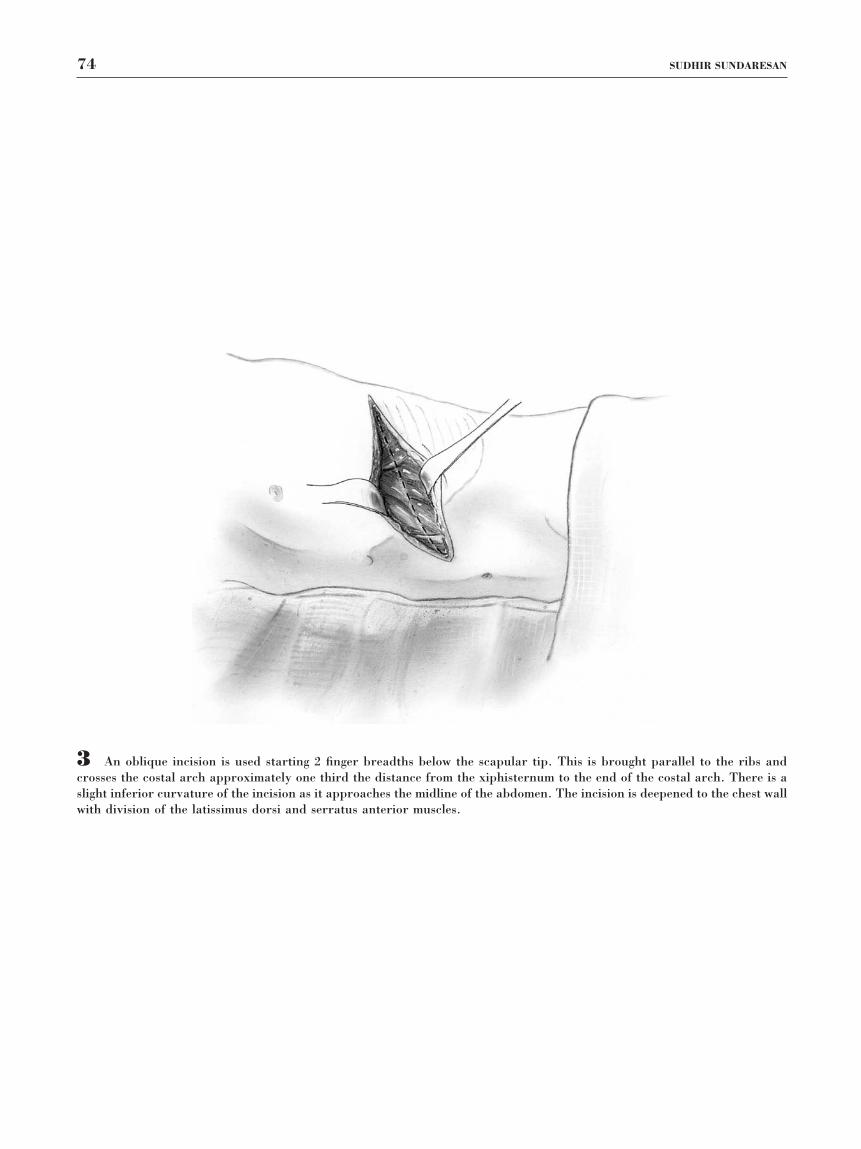
3 An oblique incision is used starting 2 finger breadths below the scapular tip. This is brought parallel to the ribs andcrosses the costal arch approximately one third the distance from the xiphisternum to the end of the costal arch. There is aslight inferior curvature of the incision as it approaches the midline of the abdomen. The incision is deepened to the chest wallwith division of the latissimus dorsi and serratus anterior muscles.
74 SUDHIR SUNDARESAN

4 The chest is entered in the seventh intercostal space by dividing the intercostal muscles flush along the upper border ofthe eighth rib. The abdomen is entered by dividing the obliques along the inferior border of the costal margin. The anteriorand posterior layers of the rectus sheath are divided, although the rectus muscle is preserved and simply retracted towardsthe abdominal midline.
LEFT THORACOABDOMINAL INCISION 75

5 A large Kelly clamp is then passed immediately deep to the costal margin. A knife is used to sharply divide the cartilageat this point. This maneuver often also divides the musculophrenic archery (ie, one of the terminal branches of the internalmammary artery). The musculophrenic artery should be securely ligated at this point.
76 SUDHIR SUNDARESAN

6 The left lung is then deflated and packed cephalad and anteriorly using moist laparotomy sponges. A series of O-silk(Ethicon, Johnson & Johnson, New Brunswick, NJ) stay sutures are then placed in the periphery of the diaphragm. Thesestay sutures orient the diaphragm properly to facilitate accurate closure later. They are also useful for retracting the dividededges of diaphragm sequentially to optimize exposure of the abdomen or the chest. Electrocautery is then used to divide thediaphragm, staying within the path outlined by the stay sutures. A 1-in attachment of diaphragm must be left inserted to thechest wall to allow the secure closure of the diaphragm at the conclusion of the operation. The total length of this phrenotomyis approximately 15 cm, and it joins the apex of the line of incision in the abdominal obliques in the shape of the letter “T.”
LEFT THORACOABDOMINAL INCISION 77

7 Upward retraction of the stay sutures provides superb access to the left upper quadrant of the abdomen. Theesophagogastric junction and entire stomach are easily accessible. The triangular ligament can be divided, allowing the leftlateral segment of the liver to be mobilized and retracted towards the right. This exposure permits the creation of either apyloroplasty or pyloromyotomy, and even Kocher maneuver if needed. The transverse colon, splenic flexure, and upperdescending colon are also well exposed. A slight caudal extension of the incision provides further exposure of the descendingcolon to permit full, left colonic mobilization for colonic interposition if desired.
78 SUDHIR SUNDARESAN
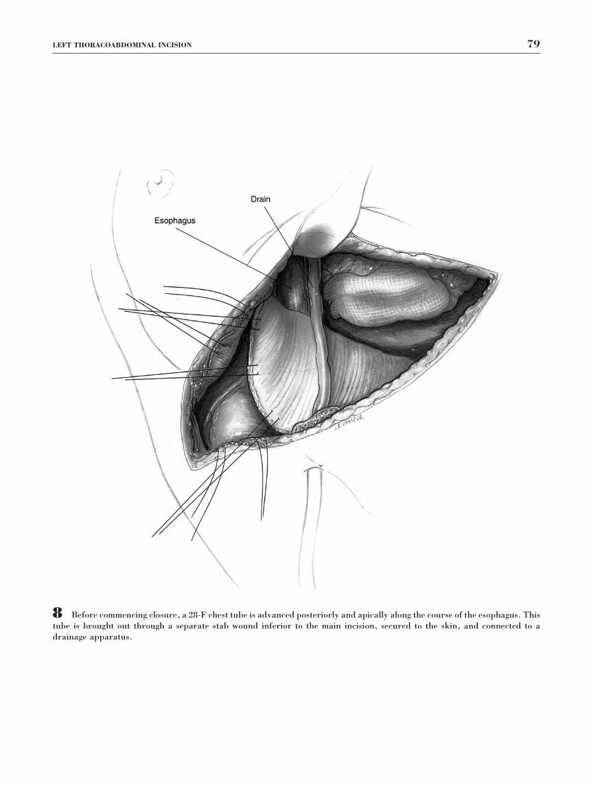
8 Before commencing closure, a 28-F chest tube is advanced posteriorly and apically along the course of the esophagus. Thistube is brought out through a separate stab wound inferior to the main incision, secured to the skin, and connected to adrainage apparatus.
LEFT THORACOABDOMINAL INCISION 79

9 Closure of the diaphragm is facili-tated using the previously placed stay su-tures. The divided margins of the dia-phragm are approximated with a series ofhorizontal mattress 0-polyprolene sutures(Fig 9A). A second layer of continuous0-polyprolene sutures is then used to ap-proximate securely the diaphragm. A U-stitch is used at the confluence of the linesof division of the diaphragm and abdom-inal obliques (see insert). The rectussheath is closed with continuous 0-poly-glactin 910 sutures in the anterior andposterior layers (Fig 9B). A continuous0-polyglactin 910 suture is used to closethe peritoneum and abdominal obliquesin one layer.
80 SUDHIR SUNDARESAN
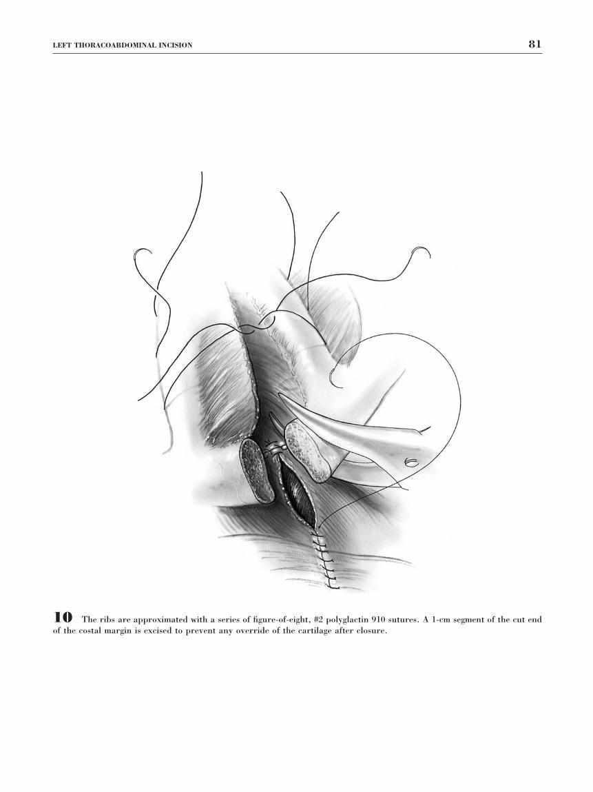
10 The ribs are approximated with a series of figure-of-eight, #2 polyglactin 910 sutures. A 1-cm segment of the cut endof the costal margin is excised to prevent any override of the cartilage after closure.
LEFT THORACOABDOMINAL INCISION 81

11 A single figure-of-eight, #1 polyglactin 910 suture is used to stabilize the cut margins of the costal arch once thepericostal sutures are tied.
82 SUDHIR SUNDARESAN

12 The fascia of the latissimus dorsi and serratus anterior muscles are approximated with continuous 0-polyglactin 910suture, respectively. Subcutaneous tissues are approximated with continuous 2-0 polyglactin 910 sutures.
13 Skin is approximated with staples. Sterile dressings are applied, and the drapes are taken down. The patient shouldbe rolled supine for reintubation with a single lumen endotracheal tube if necessary.
LEFT THORACOABDOMINAL INCISION 83
POSTOPERATIVE CAREThe immediate postoperative care depends on the mag-nitude and length of the operation. In the case of afairly straightforward operation, immediate extubationcan be accomplished. Otherwise, the author prefers toventilate electively the patient overnight with a plan forextubation the following morning. The thoracic epi-dural catheter is critical for the achievement of suffi-cient pain relief, to permit vigorous coughing, andclearing of secretions.
The chest tube is maintained on -20 cm water con-tinuous suction. Ongoing management of the chest tubedepends on the nature of the operation. In the event ofesophagogastrectomy, the chest tube typically has beensutured near the esophagogastric anastomosis. The au-thor prefers not to remove this tube until the patienthas had at least 48 hours of oral intake postoperatively.This conservative approach is performed to permitsatisfactory external drainage in the event of an anas-tomotic leak. Occasionally, small leaks are not appre-ciated on the initial contrast esophagogram but insteadbecome manifest after initiating oral intake within theensuing couple of days. Outside this scenario, the chesttube may be removed when there is no evidence of airleak and when the fluid drainage is less than 200 mLper 24 hours.
COMMENTSThe history of the left thoracoabdominal incision, itstechnical evolution, and its many clinical applicationshave been previously discussed by Heitmiller.1,2 Theleft transthoracic approach for esophagogastric resec-tion was popularized by Churchill and Sweet at theMassachusetts General Hospital more than half a cen-tury ago.3 This approach accommodated the creation ofan endothoracic esophagogastric anastomosis below theaortic arch; supra-aortic esophageal anastomosis wasalso possible but required making a second chest entryat the fourth intercostal space.4 Increasing experiencewith esophageal resection for carcinoma has led mostsurgeons to perform subtotal esophagectomy with cer-vical esophagogastric anastomosis. The advantages ofsubtotal esophagectomy for cancer include a higherlikelihood of a tumor-free esophageal margin, a betterfunctional result regarding the ease of swallowing andless tendency to gastroesophageal reflux, a lower risk ofseptic complications with an anastomotic leak, and theplacement of the anastomosis remote from mediastinallymph node stations when postoperative adjuvant ra-diotherapy is necessary. Matthews and Steel popular-ized the combination of left thoracolaparotomy incisioncombined with left neck incision for subtotal esopha-gectomy with cervical anastomosis.5
The left thoracoabdominal incision provides superbexposure for esophagogastrectomy, allows en bloc dis-
section, and permits the very precise placement of thegastric conduit. The conduit can be carefully checkedsimultaneously in the chest and abdomen, and can besutured to the pleura and peritoneum to minimize thechance of torsion or tension. Another practical advan-tage of this approach for esophageal resection is thesingle positioning and sterile field necessary to accom-plish the entire operative procedure. The advantage ofexcellent simultaneous exposure in the abdomen andchest is also extremely useful during reoperative anti-reflux surgery,6 and in total gastrectomy, where aRoux-en-Y jejunal limb originates within the abdomenbut passes through the esophageal hiatus for anastomo-sis with the distal esophagus in the lower mediastinum.
A notable drawback of the thoracoabdominal inci-sion is postoperative pain. The consequences of severe,incisional pain include lower lobe atelectasis and, lessfrequently, pneumonia. The liberal use of thoracic epi-dural catheters, along with retention of these cathetersfor up to 5 days postoperatively, has made the controlof early perioperative pain in these patients much morestraightforward. Other problems resulting from thisincision relate to the division of the diaphragm. Thepotential impairment of postoperative diaphragmaticfunction can be minimized by keeping the phrenotomyconfined to the periphery, staying within approxi-mately one inch of the chest wall attachment. Anotherpotential complication is delayed herniation of abdom-inal viscera through the site of prior diaphragm divi-sion. Once again, paying careful attention to technicaldetail during the closure of the diaphragm, as previ-ously mentioned, minimizes the potential for this prob-lem. Finally, a troublesome chrondritis at the site ofdivision of the costal arch will develop in a small mi-nority of patients. However, this is especially problem-atic if associated with suppuration, in which case reop-eration for debridement and excision of infected carti-lage becomes mandatory.
For these reasons, the author confines the use of theleft thoracoabdominal incision to those selected cases inwhich simultaneous, extensive exposure is necessarywithin the chest and abdomen. It is the author’s inci-sion of choice for total gastrectomy. It is also the mostreliable approach for reoperative antireflux surgerywhen thoracotomy and phrenotomy are insufficient,and for complex reoperations aimed at restoring esoph-ageal-gastric continuity after catastrophic complica-tions from prior surgery in this region.
REFERENCES1. Heitmiller RF: The left thoracoabdominal incision. Ann Thorac Surg
46:250-253, 19882. Heitmiller RF: Results of standard left thoracoabdominal esophagogas-
trectomy. Semin Thorac Cardiovasc Surg 4:314-319, 19923. Churchill ED, Sweet RH: Transthoracic resection of tumors of the
stomach and esophagus. Ann Surg 115:892-902, 1942
84 SUDHIR SUNDARESAN

4. Mathisen DJ: Thoracoabdominal esophagectomy for cancer of the gas-troesophageal junction. Oper Tech Thorac Cardiovasc Surg 4:186-196,1999
5. Matthews HR, Steel A: Left-sided subtotal oesophagectomy for carci-noma. Br J Surg 74:1115-1117, 1987
6. Legare JF, Henteleff HJ, Casson AG: Results of Collis gastroplasty andselective fundoplication, using a left thoracoabdominal approach, forfailed antireflux surgery. Eur J Cardiothorac Surg 21:534-540, 2002
From the University of Ottawa, Ottawa, Canada.Address reprint requests to Sudhir Sundaresan, MD, Division of Thoracic
Surgery, Ottawa Hospital-General Campus, 501 Smyth Rd, 6NW-6356, Ottawa,ON Canada K1H8L6; e-mail: [email protected].
© 2003 Elsevier Inc. All rights reserved.1522-2942/03/0802-0000$30.00/0doi:10.1053/S1522-9042(03)00030-X
LEFT THORACOABDOMINAL INCISION 85