incidence, risk factors and follow-up by aliyah - dspace
TRANSCRIPT

UNIVERSITY OF CALGARY
Plagiocephaly in Calgary Alberta Canada Incidence Risk Factors and Follow-Up
by
Aliyah Abdulrasul Mawji
A THESIS
SUBMITTED TO THE FACULTY OF GRADUATE STUDIES
IN PARTIAL FULFILMENT OF THE REQUIREMENTS FOR THE
DEGREE OF DOCTOR OF PHILOSOPHY
DEPARTMENT OF COMMUNITY HEALTH SCIENCES
CALGARY ALBERTA
SEPTEMBER 2011
copy Aliyah Abdulrasul Mawji 2011
The author of this thesis has granted the University of Calgary a non-exclusive license to reproduce and distribute copies of this thesis to users of the University of Calgary Archives
Copyright remains with the author
Theses and dissertations available in the University of Calgary Institutional Repository are solely for the purpose of private study and research They may not be copied or reproduced except as permitted by copyright laws without written authority of the copyright owner Any commercial use or re-publication is strictly prohibited
The original Partial Copyright License attesting to these terms and signed by the author of this thesis may be found in the original print version of the thesis held by the University of Calgary Archives
Please contact the University of Calgary Archives for further information E-mail uarcucalgaryca Telephone (403) 220-7271 Website httparchivesucalgaryca
Abstract
Background The Canadian Foundation for the Study of Infant Deaths (CFSID)
the Canadian Paediatric Society (CPS) and the Canadian Institute for Child Health
(CICH) released a joint statement in February 1999 recommending that infants be placed
to sleep on their backs to prevent sudden infant death syndrome (SIDS) Subsequently a
concern has been raised about a potential consequent increase in positional plagiocephaly
across Canada No literature exists on incidence of positional plagiocephaly in the
Canadian context literature on risk factors is sparse
Research Questions (a) What is the incidence of positional plagiocephaly in
infants 7-12 weeks of age in Calgary Alberta Canada (b) What are the potential risk
factors for positional plagiocephaly in infants 7-12 weeks of age in Calgary Alberta
Canada
Methods Using a prospective cohort design study participants (n=440 healthy
term infants 7 ndash 12 weeks old from well-child clinics at four community health centres in
Calgary Alberta) were assessed by two Research Nurses using Argentarsquos (2004)
Plagiocephaly Assessment Tool Parents completed a questionnaire on risk factors
Results The incidence of positional plagiocephaly was estimated to be 466
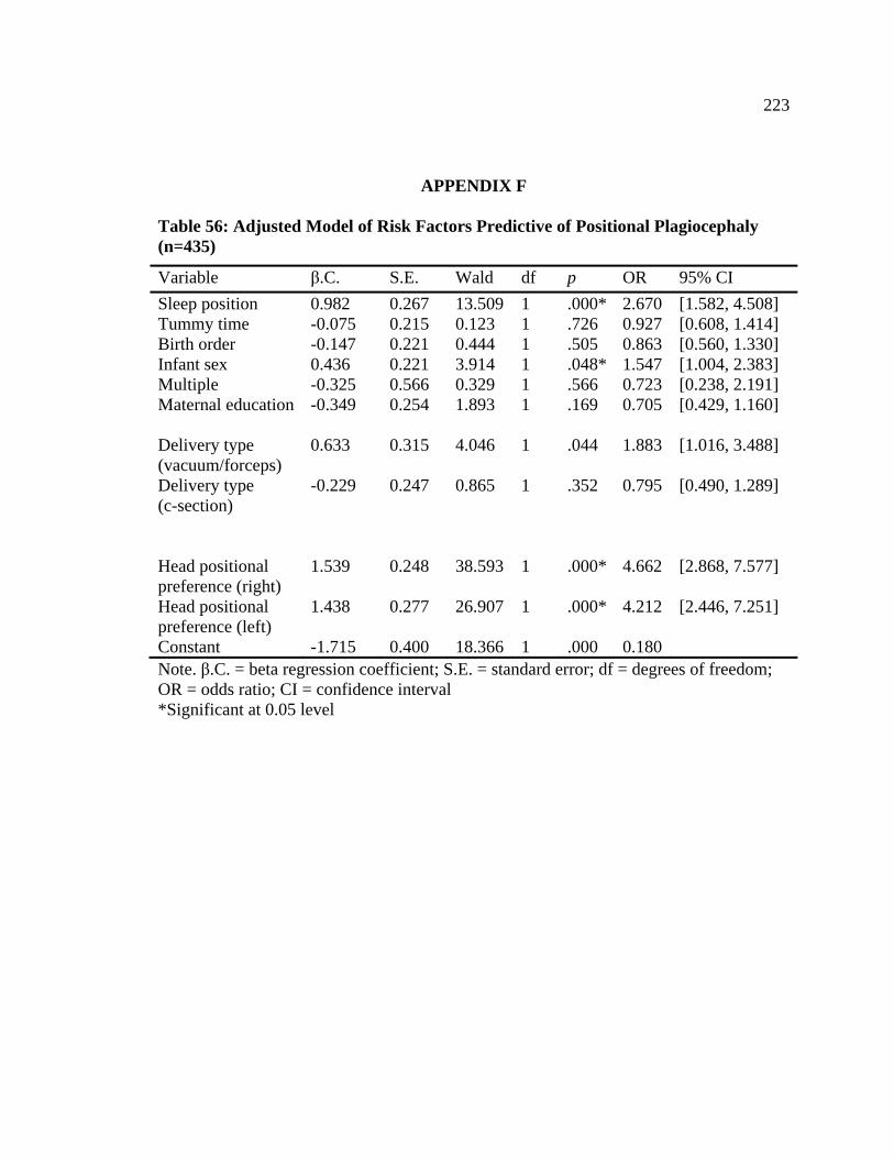
Multiple logistic regression analysis identified the following risk factors (a) right sided
head positional preference (OR 4662 p=000 CI 2868ndash7577) (b) left sided head
positional preference (OR 4212 p=000 CI 2446ndash7251) (c) supine sleep position
(OR 2670 p = 000 CI 1582ndash4508) (d) vacuumforceps assisted delivery (OR 1883
p=044 CI 1016ndash3488) and (e) male sex (OR 1547 p= 048 CI 1004ndash2383)
iii
Conclusion Given that positional plagiocephaly is a preventable condition an
incidence of 466 is very troubling Information to vary infantsrsquo head positions needs to
be communicated to parentsguardians well before the 2-month well-child clinic visit
Prevention education can be targeted to parentsguardians of those infants that are male
and infants that have had assisted deliveries This could occur in the prenatal period or
during the neonatal period by postpartum nurses and PHNs involved in postpartum care
Further studies are also required in order to better understand the role of family
physicians in identifying the condition and follow-up process of such infants
iv
Acknowledgements
I would like to thank first my supervisor Dr Ardene Robinson Vollman who
provided much guidance and support throughout my doctoral studies I am grateful for
the constructive feedback provided consistently throughout my studies and value our
conversations throughout the years I would like to acknowledge my Co-Supervisor Dr
Jennifer Hatfield who provided critical learning in terms of identifying my strengths and
building on them I am grateful to my committee members Dr Reg Sauve and Dr
Deborah McNeil who were very encouraging throughout the entire process
I would like to acknowledge Dr Tak Fung who always made time to answer my
data analysis questions with enthusiasm I would like to thank the clinicians at the Head
Shape Clinic at the Alberta Childrenrsquos Hospital Dr Keith Jorgenson Dr Heather
Graham Lori Walker Barb Mikkelsen Judith Mair and Jeanette Deere for taking the
time to teach me how to conduct plagiocephaly and torticollis assessments learning
about cranial orthosis treatment and assisting with data collection Thanks also to all of
the Public Health Nurses that assisted with data collection I am grateful to the families
that agreed to participate in this study their participation helped us learn more about
positional plagiocephaly and what we can do to prevent its development and improve
services for those with the condition in the future
I would like to thank my parents Almas and Abdulrasul Mawji who have always
encouraged me to follow my dreams and who have always supported me in every
decision I have made Last but not least I would like to thank Naushad Dosani for
knowing when to offer well-needed diversions and offering his assistance with anything I
may have needed throughout the process
v
Dedication
To my Family
vi
Table of Contents
Approval Page ii Abstract iii Acknowledgements v Dedication vi Table of Contents vii List of Tables xi List of Figures and Illustrations xv List of Definitions xvi List of Symbols Abbreviations and Nomenclature xvii
CHAPTER ONE INTRODUCTION AND LITERATURE REVIEW 1 Background1 Definition 1 Rationale for Conducting the Research 3
Incidence and risk factors 3 Health care systems and plagiocephaly in the Canadian context 4 HSCs in Canada 4 Key professions working in the area of plagiocephaly 5 Referral mechanisms 5 Assessment tools used to identify plagiocephaly and its severity 6 Perceived increase or decrease of plagiocephaly 7
Research Objectives 7 Research Questions 8 Contributions of this Research9 Organization of this Thesis 11
LITERATURE REVIEW 12 Epidemiology of Plagiocephaly 12
Incidence13 Prevalence15 Evolution in the shape of plagiocephaly 18
Assessment 19 Anthropometric measurements 20 Three-dimensional computer analysis 23 Radiographic assessments Computed tomography 27 Clinical observation assessment tools 29
Risk Factors 34 Predictors of Severity43 Plagiocephaly ndash Torticollis Co-Diagnosis 44 Prevention 49 Interventions for Plagiocephaly 50
The mechanism of active counter-positioning 51 The mechanism of orthotic helmets 52 The effectiveness of active counter-positioning vs orthotic helmet treatment54
vii
Early Recognition 57 Potential Developmental Concerns Associated with Plagiocephaly 59
CHAPTER TWO METHODS 69
What is the incidence of positional plagiocephaly in infants 7-12 weeks of age
Is Argentas (2004) Plagiocephaly Assessment Tool appropriate for use in well-
What intervention and follow-up actions do PHNs take if positional plagiocephaly is identified in healthy infants at the 2-month well-child
What intervention and follow-up actions do clinicians (physiotherapists and occupational therapists) take when healthy infants with positional plagiocephaly are referred to the infant repositioning class (IRC) after the
Rationale for Collecting Data at the 2-month Well-Child Clinic Visit vs 4-month
What is the incidence of positional plagiocephaly in infants 7-12 weeks of age
What intervention and follow-up actions do PHNs take if positional plagiocephaly is identified in healthy infants at the 2-month well-child
Research Objectives and Research Questions 69 Study Design71 Inclusion Criteria 72 Sample 72 Clinical Observation Tool74
Cost effectiveness 74 Validity 75 Reliability 77 Suitabilityfeasibility 78 Time required to complete the assessment 79 Level of expertise required for an accurate assessment 80 Level of intrusiveness81 Utility as a teaching tool81
Recruitment 82 Data Collection 83
in Calgary Alberta Canada 83 What are the potential risk factors for positional plagiocephaly in infants 7-12
weeks of age in Calgary Alberta Canada 84
child clinics 84
clinic84
2-month well-child clinic visit in Calgary Alberta Canada 86
Well-Child Clinic Visit 86 Rationale for collecting data at the 2-month well-child visit 87 Rationale for conducting the study at the 4-month well-child visit 89 Reasons for the decision to collect data the 2-month clinic 90
Data Cleaning 93 Data Analysis 94
in Calgary Alberta Canada 94 What are the potential risk factors for positional plagiocephaly in infants 7-12
weeks of age in Calgary Alberta Canada 94
clinic101
viii
What intervention and follow-up actions do clinicians (physiotherapists and occupational therapists) take when healthy infants with positional plagiocephaly are referred to the infant repositioning class (IRC) after the 2-month well-child clinic visit in Calgary Alberta Canada 101
Significance of the Study102 Ethics 102
CHAPTER THREE RESULTS 107
What is the Estimated Incidence of Positional Plagiocephaly in Infants 7-12 Weeks
Is Argentas (2004) Plagiocephaly Assessment Tool Appropriate for Use in Well-
Statistical Model of risk factors predictive of positional plagiocephaly (twins
What Intervention and Follow-Up Actions do PHNs Take if Positional Plagiocephaly is Identified in Healthy Infants at the 2-Month Well-Child
What Intervention and Follow-Up Actions do Clinicians (Physiotherapists and Occupational Therapists) Take When Healthy Infants with Positional Plagiocephaly are Referred to the Infant Repositioning Class (IRC) After the
Response Rate107 The Sample 108
of Age in Calgary Alberta Canada 109 Incidence of positional plagiocephaly (n=440) 109
Child Clinics114 Suitabilityfeasibility 114 Acceptability of the tool to the target population 114 Measures taken to ensure reliability 115 The yield of the tool 115
What are the Potential Risk Factors for Positional Plagiocephaly in Infants 7-12 Weeks of Age in Calgary Alberta Canada 116
Non-modifiable risk factors (n=440) 116 Infant risk factors 116 Maternal risk factors 120
Modifiable risk factors (n=440)123 Infant risk factors 123 Maternal risk factors 128
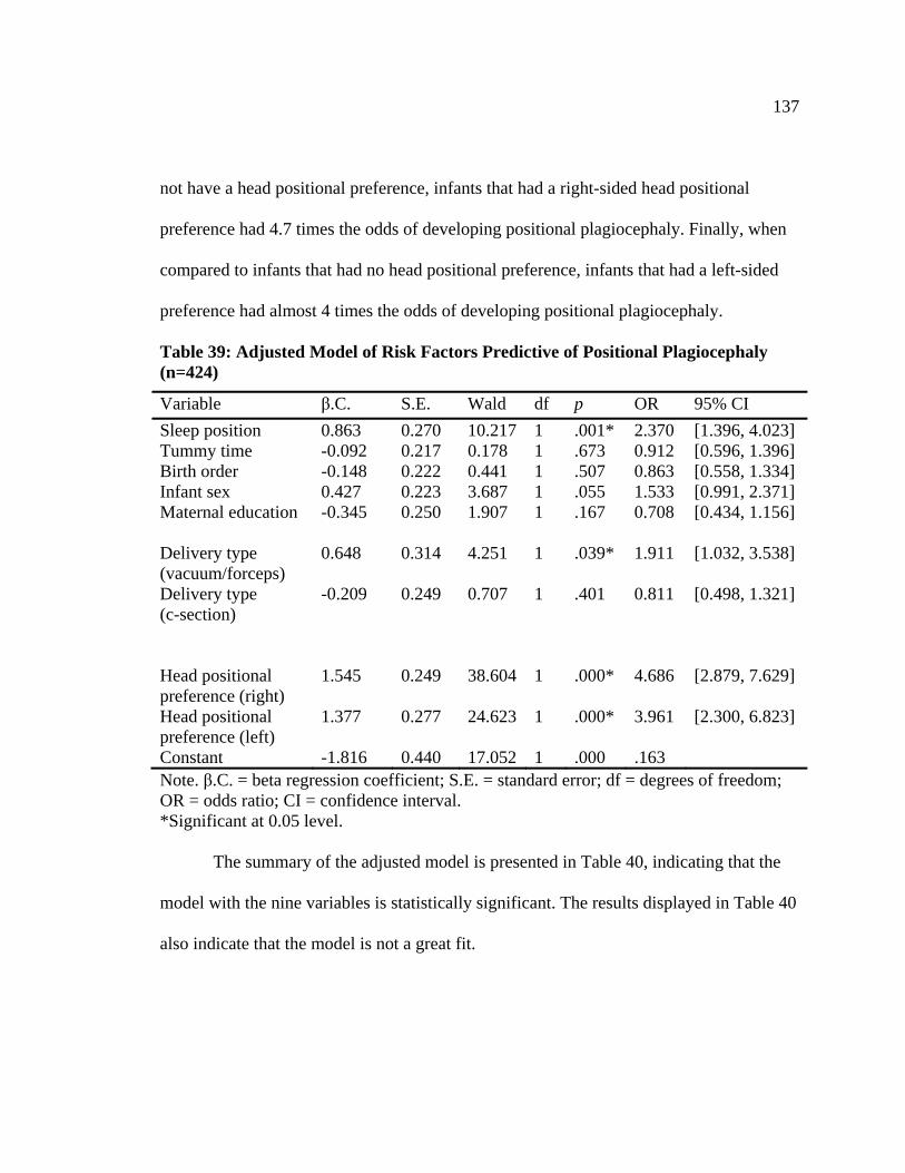
Multicollinearity 131 Statistical model of risk factors predictive of positional plagiocephaly 131
removed) 135
Clinic 139
2-Month Well-Child Clinic Visit in Calgary Alberta Canada 144 Conclusion 148
CHAPTER FOUR DISCUSSION150 Incidence of Positional Plagiocephaly 150
Comparison with other incidence studies 150 Sampling and sample size 151 Age range of study participants 153 Assessment techniques for positional plagiocephaly 155
Argentarsquos (2004) Plagiocephaly Assessment Tool 158
ix
Severity of Plagiocephaly Observed163 Accompanying Brachycephaly 164 The Model165
Risk factors predictive of positional plagiocephaly 165 Supine sleep positioning 165 Sex 166 Delivery type167 Head positional preference and the side of head that plagiocephaly was
observed 168 Non-significant variables170
Tummy time 170 Birth order and maternal age 171 Multiple gestation pregnancy 172 Maternal education 173
Contradictions Found in the Literature174 Implications for Practice Based on Incidence and Risk Factor Data 176 PHN Actions and Follow-up by the IRC at Alberta Childrenrsquos Hospital 179
Repositioning teaching 179 Referral to the IRC 180 Referral to the family physician 183 Follow-up planned at the 4-month well-child clinic visit 184
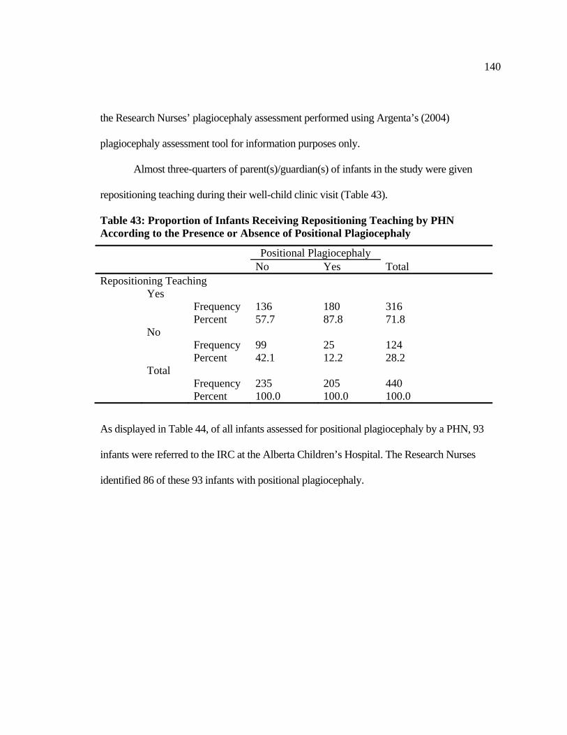
Strengths of the Present Study 184 Methods 184 Tool186 Data collection186
Limitations of the Present Study 186 Methods 186 Data collection187 Data analysis188
Directions for Future Research 189 Conclusion 190
REFERENCES 192
APPENDIX A213
APPENDIX B 214
APPENDIX C 217
APPENDIX D219
APPENDIX E 221
APPENDIX F223
APPENDIX G228
x
List of Tables
Table 1 Number of Infants that met the Inclusion Criteria at the Four CHCs 73
Table 2 Distribution of Sample across Sites 107
Table 3 Number of Infants Identified With Positional Plagiocephaly According to Type 109
Table 4 Distribution of Positional Plagiocephaly across Data Collection Sites 110
Table 5 Side of Head on which Positional Plagiocephaly was Observed According to Type 111
Table 6 Distribution of Observed Positional Plagiocephaly According to Severity and Type 112
Table 7 Distribution of Infants with Positional Plagiocephaly also Demonstrating Signs of Brachycephaly According to Type of Plagiocephaly 113
Table 8 Non Modifiable Infant Risk Factors for Positional Plagiocephaly According to Birth Order 117
Table 9 Occurrence of Positional Plagiocephaly According to Infant Birth Order 117
Table 10 Non Modifiable Infant Risk Factors for Positional Plagiocephaly According to Infant Sex 118
Table 11 Occurrence of Positional Plagiocephaly According to Infant Sex 118
Table12 Non Modifiable Infant Risk Factors for Positional Plagiocephaly According to Multiple Gestation Pregnancy 119
Table 13 Occurrence of Positional Plagiocephaly According to Multiple Gestation Pregnancies 119
Table 14 Age of Infants Participating in the Study in Months 120
Table 15 Maternal Delivery Type 120
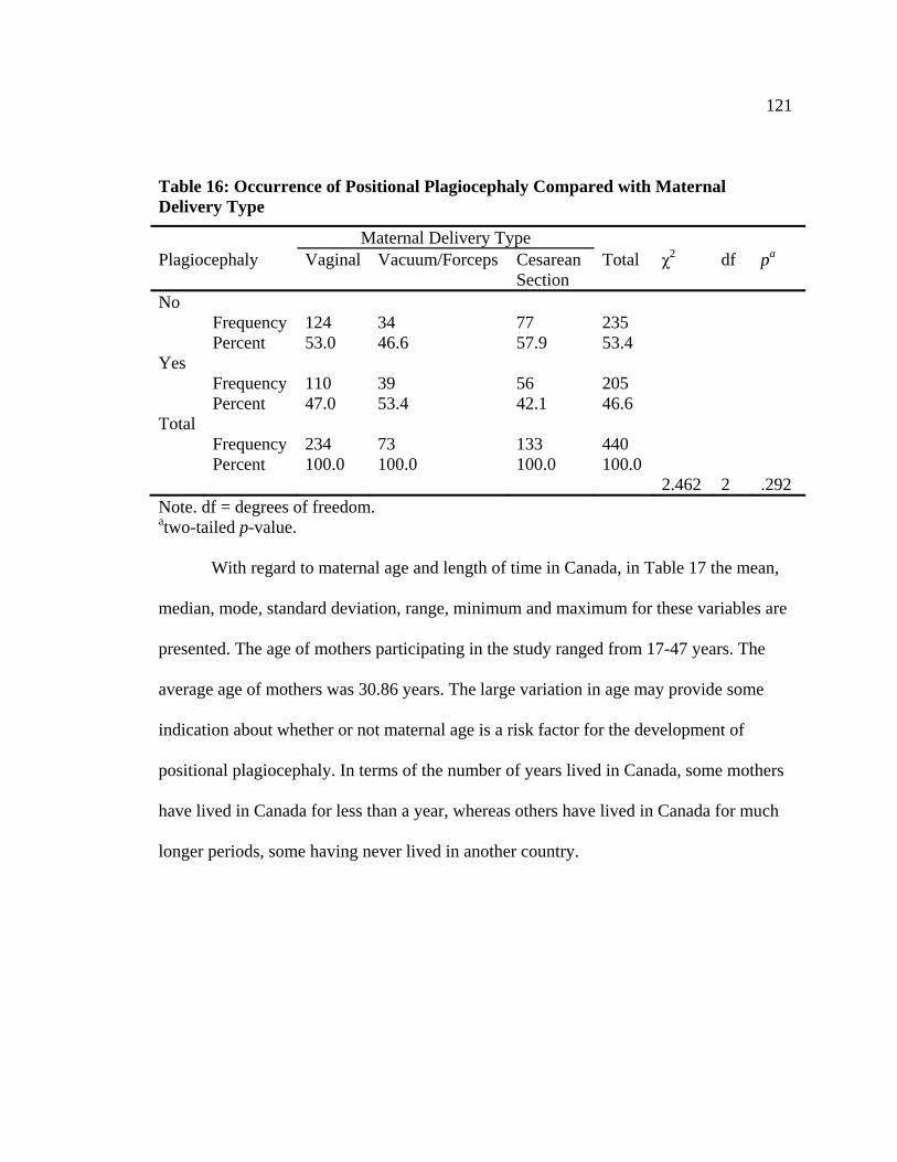
Table 16 Occurrence of Positional Plagiocephaly Compared with Maternal Delivery Type 121
Table 17 Maternal Age and Maternal Number of Years Lived in Canada 122
Table 18 Occurrence of Positional Plagiocephaly According to Maternal Age and Number of Years lived in Canada 122
xi
Table 19 t- test for Equality of Means for Maternal Age and Number of Years Lived in Canada 123
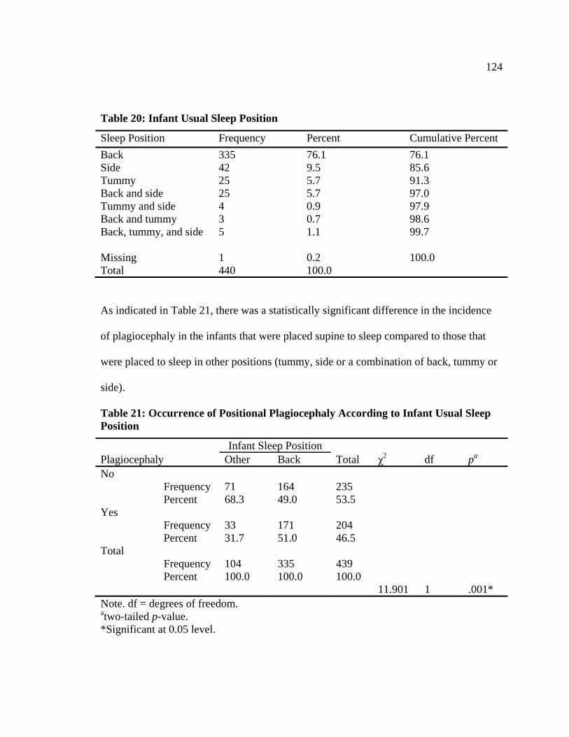
Table 20 Infant Usual Sleep Position 124
Table 21 Occurrence of Positional Plagiocephaly According to Infant Usual Sleep Position 124
Table 22 Infant Head Positional Preference While Laying Supine 125
Table 23 Occurrence of Positional Plagiocephaly According to Infant Head Positional Preference While Laying Supine 125
Table 24 Infant Usual Feeding Position 126
Table 25 Occurrence of Positional Plagiocephaly According to Infant Usual Feeding Position 127
Table 26 Infant Tummy Time Per Infant Per Day 127
Table 27 Occurrence of Positional Plagiocephaly According to Infant Tummy Time (Per Infant per Day) 128
Table 28 Highest Level of Maternal Education Completed 129
Table 29 Occurrence of Positional Plagiocephaly According to Highest Level of Maternal Education Completed 129
Table 30 Maternal Language Barrier 130
Table 31 Occurrence of Positional Plagiocephaly According to the Presence or Absence of Maternal Language Barrier 130
Table 32 Variables Demonstrating Multicollinearity 131
Table 33 Unadjusted Statistical Model of Risk Factors Predictive of Positional Plagiocephaly 132
Table 34 Adjusted Statistical Model of Risk Factors Predictive of Positional Plagiocephaly (n=435) 133
Table 35 Summary of the Adjusted Statistical Model (n=435) 134
Table 36 Contingency Table for Hosmer-Lemeshow Test 134
Table 37 Percentage of Cases Classified Correctly 134
Table 38 Unadjusted Model of Risk Factors Predictive of Positional Plagiocephaly 136
xii
Table 39 Adjusted Model of Risk Factors Predictive of Positional Plagiocephaly (n=424) 137
Table 40 Summary of the Adjusted Model (n=424) 138
Table 41 Contingency Table for Hosmer-Lemeshow Test 138
Table 42 Percentage of Cases Classified Correctly 139
Table 43 Proportion of Infants Receiving Repositioning Teaching by PHN According to the Presence or Absence of Positional Plagiocephaly 140
Table 44 Proportion of Infants Referred to the IRC by PHN According to the Presence or Absence of Positional Plagiocephaly 141
Table 45 Proportion of Infants Referred to their Family Physician by PHN according to the Presence or Absence of Positional Plagiocephaly 141
Table 46 PHN Reasons for Referral to IRC andor Family Physician 142
Table 47 Proportion of Infants that will be followed up by PHN at 4-Month Well-Child Clinic According to the Presence or Absence of Positional Plagiocephaly 142
Table 48 PHN Comments Providing Additional Information About Individual Infant Plagiocephaly Assessments 143
Table 49 Proportion of Infants with each Type of Plagiocephaly That Presented at the IRC According to IRC Clinician (Physiotherapists and Occupational Therapist) Assessment 144
Table 50 Severity of Plagiocephaly of Infants that presented at the IRC According to IRC Clinician (Physiotherapists and Occupational Therapist) Assessment 145
Table 51 Comparison of Assessment of infants assessed at both CHC and IRC 146
Table 52 Frequency of Torticollis Assessed in Infants that presented at the IRC According to IRC Clinician (Physiotherapists and Occupational Therapist) Assessment 147
Table 53 Frequency of Infants that presented at the HSC that were Referred for Physiotherapy by IRC Clinicians (Physiotherapists and Occupational Therapist) 147
Table 54 Clinical Classification of Positional Plagiocephaly 217
Table 55 Clinical Classification of Positional Plagiocephaly 221
Table 56 Adjusted Model of Risk Factors Predictive of Positional Plagiocephaly (n=435) 223
xiii
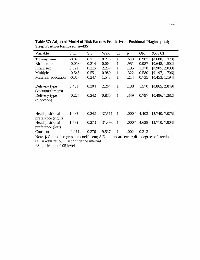
Table 57 Adjusted Model of Risk Factors Predictive of Positional Plagiocephaly Sleep Position Removed (n=435) 224
Table 58 Adjusted Model of Risk Factors Predictive of Positional Plagiocephaly Birth Order Removed (n=434) 225
Table 59 Adjusted Model of Risk Factors Predictive of Positional Plagiocephaly Maternal Education Removed (n=435) 226
Table 60 Adjusted Model of Risk Factors Predictive of Positional Plagiocephaly Head Positional Preference Removed (n=434) 227
xiv
List of Figures and Illustrations
Figure 1 Data Collection Process Diagram 83
Figure 2 The Sample 108
xv
List of Definitions
Term Definition Anthropometry The science of measuring the human body Brachycephaly A skull shape that is short in proportion to its width Craniosynostosis Premature closure of skull sutures Congential Muscular The presence of unilateral fibrosis or shortening of the SCM Torticollis muscle Euryon The extremity on either side of the head of the greatest
transverse diameter of the head Frontozygomaticus The suture between the frontal bone and the zygomatic bone Incidence The number of new health-related events in a defined
population within a specified period of time Ipsilateral Pertaining to the same side of the body Lambdoid The suture that separates the parietal and temporal bones of
the skull from the occipital bone Occipital bone A bone in the lower back part of the skull between the
parietal and temporal bones Parietal bone One of two bones that together form the roof and sides of the
skull Plagiocephaly A malformation of the skull producing the appearance of a
lopsided head Prevalence The proportion of individuals in a population that have a
disease or a condition Sternocleidomastoid One of two muscles arising from the sternum and inner part
of the clavicle Sclerosis A hardening within the nervous system especially of the
brain and spinal cord resulting from the degeneration of nervous elements such as the myelin sheath
Scoliosis A lateral curvature of the spine it usually consists of two curves the original abnormal curve and a compensatory curve in the opposite direction
Suture The line of union in an immovable articulation as those between the skull bones
Torticollis A deformity of the neck secondary to shortening of neck muscles which tilts the head to the affected side with the chin pointing to the other side
xvi
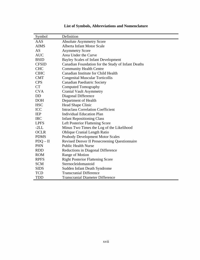
List of Symbols Abbreviations and Nomenclature
Symbol Definition AAS Absolute Asymmetry Score AIMS Alberta Infant Motor Scale AS Asymmetry Score AUC Area Under the Curve BSID Bayley Scales of Infant Development CFSID Canadian Foundation for the Study of Infant Deaths CHC Community Health Centre CIHC Canadian Institute for Child Health CMT Congenital Muscular Torticollis CPS Canadian Paediatric Society CT Computed Tomography CVA Cranial Vault Asymmetry DD Diagonal Difference DOH Department of Health HSC Head Shape Clinic ICC Intraclass Correlation Coefficient IEP Individual Education Plan IRC Infant Repositioning Class LPFS Left Posterior Flattening Score -2LL Minus Two Times the Log of the Likelihood OCLR Oblique Cranial Length Ratio PDMS Peabody Development Motor Scales PDQ ndash II Revised Denver II Presecreening Questionnaire PHN Public Health Nurse RDD Reductions in Diagonal Difference ROM Range of Motion RPFS Right Posterior Flattening Score SCM Sternocleidomastoid SIDS Sudden Infant Death Syndrome TCD Transcranial Difference TDD Transcranial Diameter Difference
xvii
1
CHAPTER ONE INTRODUCTION AND LITERATURE REVIEW
Background
In 1992 the American Academy of Paediatrics released a statement
recommending that healthy infants be placed in the supine position (ie on their backs)
to sleep (American Academy of Paediatrics Task Force on Infant Positioning and SIDS
1992) Canada followed suit in February 1999 when the Canadian Foundation for the
Study of Infant Deaths (CFSID) the Canadian Paediatric Society (CPS) and the Canadian
Institute for Child Health (CICH) released a joint statement entitled Reducing the Risk of
Sudden Infant Death Syndrome (SIDS) in Canada The current version of the statement
recommends also that all healthy infants be placed supine to sleep (Government of
Canada CPS CICH amp CFSID 2011) Evidence supported the supine sleep position to
reduce the incidence of SIDS indeed SIDS mortality in Canada decreased from 06 per
1000 in 1999 to 035 per 1000 in 2004 (Smylie amp Sauve 2009) Although the most
significant benefit of supine sleeping is reduced infant mortality it is not without
consequence The supine sleep position has been thought to contribute to an increase in
positional plagiocephaly across Canada (Dubeacute amp Flake 2003 Neufeld amp Birkett 2000)
However no literature exists on the incidence or prevalence of the condition in the
Canadian context
Definition
Plagiocephaly is defined as a malformation of the skull producing the appearance
of a lopsided head (Thomas 1997) The term plagiocephaly has been generically used to
describe distortion of the cranium that occurs from both premature fusions of the cranial
2
sutures (synostotic plagiocephaly) as well as from external moulding forces
(deformational plagiocephaly) (Littlefield amp Kelly 2004) These external forces are
associated with parentsrsquo or caregiversrsquo positioning of infants during sleep and other
activities (Collett Breiger King Cunningham amp Speltz 2005) Only recently has a clear
differentiation between these two conditions been made Various other terms have also
been used in the medical literature to refer to the deformational form of plagiocephaly
including (a) positional plagiocephaly (b) nonsynostotic plagiocephaly (c)
plagiocephaly without synostosis (d) occipital plagiocephaly (e) posterior
plagiocephaly (f) benign position moulding (g) functional lambdoid synostosis (h) skull
moulding and (i) flat head syndrome (Littlefield amp Kelly 2004) Additionally many
early studies mention cranial deformation that occurred as a consequence of congenital
muscular torticollis scoliosis neurologic issues and so on but never specifically use the
term plagiocephaly (Littlefield amp Kelly 2004) To avoid confusion since much of the
literature uses the terms positional plagiocephaly or deformational plagiocephaly in this
thesis both terms will be used interchangeably
According to Kane Mitchell Craven and Marsh (1996) infants with
plagiocephaly generally present with unilateral occipital flattening where one side of the
occiput is flattened and contralateral occipital bulging where the other side of the occiput
is rounded Infants with more severe plagiocephaly may also have asymmetric faces that
might include (a) forehead protrusion ipsilateral (same side) to the occipital flattening
(b) forehead flattening ipsilateral (same side) to the occipital rounding (c) ear
displacement where the ear on the side of the occipital flattening is located anteriorly (in
3
front of) and below when compared to the location of the other ear and (d) chin deviation
where the chin points in direction to the side opposite of the occipital flattening (Kane et
al 1996 Littlefield Kelly Pomatto amp Beals 2002) According to Najarian (1999)
plagiocephaly is of significant concern because if it is not diagnosed and treated early the
associated changes in facial features identified above can be permanent This permanent
chance in facial features may have adverse psycho-social implications for the child that
may be at increased risk for teasing and bullying during school years The helmet
approach to treat positional plagiocephaly has been proven effective and is well
documented (Bialocerkowski Vladusic amp Howell 2005 Bruner David Gage amp
Argenta 2004 de Ribaupierre et al 2007 Katzel Koltz Sbitany Emerson amp Girotto
2010 Larsen 2004 Lee et al 2008 Lima 2004 Littlefield 2004 Losee et al 2007
McGarry et al 2008 Najarian 1999 Robinson amp Proctor 2009 Teichgraeber et al
2004 Xia et al 2008)
Rationale for Conducting the Research
Incidence and risk factors
No research has been undertaken in the Canadian context to determine the
incidence of positional plagiocephaly or the risk factors contributing to its development
Clinicians working at Head Shape Clinics (HSCs) in Canada have reported anecdotally a
perception of an increased number of infants attending these specialty clinics (Dubeacute amp
Flake 2003 Neufeld amp Birkett 1999 Neufeld amp Birkett 2000) No surveillance system
exists at present in Canada to capture data on plagiocephaly
4
Health care systems and plagiocephaly in the Canadian context
In efforts to understand the magnitude of this issue in the Canadian context phone
calls were made to various childrenrsquos hospitals across Canada to identify
specialty HSCs that have been established to deal with the observed increase in
positional plagiocephaly
different types of health professions working in the area of plagiocephaly
referral mechanisms
assessment tools used to determine severity of plagiocephaly and
cliniciansrsquo perceived increase or decrease in the incidence of positional
plagiocephaly based on observations at HSCs
HSCs in Canada
Five HSCs were discovered at various Childrenrsquos Hospitals across Canada (a)
Vancouver British Columbia (b) Edmonton Alberta (c) Calgary Alberta (d)
Winnipeg Manitoba and (e) Ottawa Ontario Although messages were left at the
information desk as well as the neurology department at the Montreal Childrenrsquos
Hospital no response was received and no information exists on their website pertaining
to the presence of a HSC
Of the clinics contacted clinic days of operation ranged from one day a week to
three days a week Children diagnosed with plagiocephaly in Saskatchewan are referred
to the Edmonton clinic Patients from Northern British Columbia and the North West
Territories are referred also to the Edmonton clinic (W Beaudoin personal
communication September 18 2008) Although there is no clinic devoted to head shapes
5
in Nova Scotia New Brunswick PEI and Newfoundland infants are managed by
physiotherapists as the need arises (K Atkins personal communication September
17 2008) The HSC in Toronto closed after it was unable to keep up with the demand
As a result the management of positional plagiocephaly in the Toronto area has been
decentralized Family physicians now work with infants identified as having
deformational plagiocephaly and their families
The majority of the clinics contacted were linked to local orthotists that create
helmets as indicatedprescribed for treatment (K Atkins personal communication
September 17 2008) In Toronto referrals are made as required to orthotists at The
Hospital for Sick Children if a helmet is the indicated treatment (A George personal
communication September 18 2008 T DaSilva personal communication October 1
2008) The HSC in Calgary outsources helmet manufacturing to Orthomerica a company
based in the United States of America (US) (L Walker personal communication May
7 2008)
Key professions working in the area of plagiocephaly
Across Canada professionals from various disciplines work in the area of
plagiocephaly Most often nurses physiotherapists and occupational therapists manage
and run the clinics identified above The clinic in Calgary was the only clinic identified to
have paediatricians directly involved in the day to day assessments of infants
Referral mechanisms
Referral mechanisms vary across Canada and include family self-referrals nurse
referrals and physician referrals HSC formats vary across Canada from consultations
6
with individual families to group clinics in Vancouver where four families are seen at one
time Telehealth services are also provided out of the Vancouver clinic to areas where
plagiocephaly expertise is minimal (P Mortenson personal communication September
17 2008)
Assessment tools used to identify plagiocephaly and its severity
Although many front line health care professionals use subjective assessments to
determine the presence or absence of plagiocephaly once an infant is referred to a
specialty HSC a variety of assessment methods are used that differ across provinces
Assessment techniques identified include subjective observations manually collecting
anthropometric measurements and the use of Orthomericarsquos STARscanner Various
rating scales are also used including one produced by Cranial Technologies Inc another
US based company This scale is used by the HSC in Calgary and in Ottawa while the
HSC in Edmonton has adapted this scale and produced a ten-point scale of its own (L
Walker personal communication May 7 2008 K Dubeacute personal communication
September 17 2008 W Beaudoin personal communication September 18 2008) The
STARscanner uses 3-D technology to acquire head shape data through lasers and
cameras Although the clinic in Winnipeg uses the scanner to obtain assessment data the
Calgary clinic uses the scanner after severity has been assessed by physicians to produce
measurement data for helmet production (T Martin personal communication September
17 2008 J Mair personal communication May 21 2008) These data are e-mailed to
Orthomerica which then manufactures the corresponding helmet
7
Although effort has been made in the Canadian context to identify reliable and valid
anthropometric methods for quantifying the severity of positional plagiocephaly (Mortenson
amp Steinbok 2006) a discussion with one of the authors suggests that these methods in the
end have been proven unreliable and other more reliable means of quantifying severity are
needed (P Mortenson personal communication September 17 2008)
Perceived increase or decrease of plagiocephaly
Although no information on the incidence of plagiocephaly in Canada was found
in the literature clinicians at various HSCs across Canada report an increase over the last
few years based on observations of increased referrals ranging from 100 referrals a year
to over 1000 referrals a year (A George personal communication September 18 2008
K Atkins personal communication September 17 2008 K Dubeacute personal
communication September 17 2008 L Walker personal communication May 7 2008
P Mortenson personal communication September 17 2008 T DaSilva personal
communication October 1 2008 W Beaudoin personal communication September 18
2008) This perceived increase is considered to be an underestimation in terms of
incidence of positional plagiocephaly since clinicians at various HSCs across Canada do
not see the infants identified in the community as having the condition Rather only those
recognized by public health nurses (PHNs) or family physicians as requiring intervention
in the form of helmet treatment are referred to the clinics where they exist
Research Objectives
Since no information exists regarding the incidence of positional plagiocephaly in
the Canadian context and the literature on risk factors is limited a study is required that
8
estimates the incidence of plagiocephaly of infants 7-12 weeks of age and identifies
potential risk factors for infants aged 7-12 weeks that attend the 2-month well-child
clinic In addition since there are no agreed-upon assessment methods for positional
plagiocephaly in primary care and the fact that subjective assessments predominate a
study is required to identify and field test an appropriate assessment tool for
plagiocephaly for use in well-child clinics Furthermore in an effort to understand
patterns of care that infants with positional plagiocephaly receive a study is required to
explore how infants identified with positional plagiocephaly are followed at various
levels of the health care system A study is also required in order to explore actions of
various health care professionals working with infants identified with positional
plagiocephaly The site chosen for this study is Calgary Alberta Canada
Research Questions
The five research questions for the present study are
1 What is the incidence of positional plagiocephaly in infants 7-12 weeks of age in
Calgary Alberta Canada
2 What are the potential risk factors for positional plagiocephaly in infants 7-12
weeks of age in Calgary Alberta Canada
3 Is Argentas (2004) Plagiocephaly Assessment Tool appropriate for use in well-
child clinics
9
4 What intervention and follow-up actions do PHNs take if positional plagiocephaly
is identified in healthy infants at the 2-month well-child clinic
5 What intervention and follow-up actions do clinicians (physiotherapists and
occupational therapists) take when healthy infants with positional plagiocephaly
are referred to the infant repositioning class after the 2-month well-child clinic
visit in Calgary Alberta Canada
Contributions of this Research
The reported incidence of plagiocephaly in the literature varies widely and is
frequently based on anecdotal evidence of increase in the number of referrals to specialty
clinics (Glasgow Siddiqi Hoff amp Young Kane et al 1996 McGarry et al 2008) In the
US differences in diagnostic criteria and subjective classifications are noted as factors
contributing to the lack of clarity in prevalence (and perhaps incidence) information
available currently (Hutchison Hutchison Thompson amp Mitchell 2004) It is anticipated
that this research will identify the incidence of the condition in Calgary Alberta Canada
It is important to know the incidence of the condition to ascertain how wide spread the
issue is and if any interventions need to be implemented to prevent the development of
the condition
Second this research will identify risk factors predictive of positional
plagiocephaly in infants 7-12 weeks of age By understanding the factors associated with
the development of positional plagiocephaly it may be possible to identify potential
points for intervention to prevent positional plagiocephaly by the most appropriate health
10
professionals thus reducing morbidity due to plagiocephaly and its associated burden on
the health system Morbidity here is defined as the number of cases of the condition in
relationship to a specific population (Thomas 1997) Therefore the nature of
plagiocephaly morbidity relates to the incidence and prevalence of the condition
Third little work has been done to establish reliable and practical assessment
tools for plagiocephaly Argenta (2004) presents a five-point classification scale that is
easily reproducible cost-effective and easily understood by both families and health
professionals Spermon Spermon-Marijnen and Scholten-Peeters (2008) found Argentarsquos
five-point rating scale to be moderately reliable (k = 54) This research will use
Argentarsquos five point scale to estimate the incidence of plagiocephaly in Calgary at the
population level It is important to have a reliable tool to obtain accurate estimates of
incidence and prevalence of the condition If this tool is widely adopted it would be
possible to determine the geographic spread of positional plagiocephaly and where
geographically specific interventions need to be implemented
It must be noted that Argentarsquos tool is classified as an assessmentdiagnostic tool
and not a screening tool The purpose of assessmentdiagnostic tools is to establish the
presence or absence of a condition whereas the purpose of a screening test is to detect
potential disease indicators (Wilson amp Jungner 1968) Since plagiocephaly does not have
a disease process with accompanying changes pathophysiologically it cannot be detected
in asymptomatic infants Therefore it is appropriate that an assessmentdiagnostic tool be
used for its detection
11
Fourth this research will explore actions of various health care professionals
working with infants identified with positional plagiocephaly It is hoped that this
research will also raise the awareness of PHNs about positional plagiocephaly prevention
as well as the importance of early recognition and early intervention Finally this
research will explore how infants identified with positional plagiocephaly are followed at
various levels of the health care system
Organization of this Thesis
This thesis is organized into four chapters In chapter one the issue is introduced
and the literature is critically reviewed The research objectives and research questions
are presented in both chapters one and two In chapter two the study methods are
discussed including the study design inclusion criteria sample size data collection data
analysis and rationale for the infant age at which data were collected In chapter three the
research results are presented followed by chapter four in which the results are discussed
along with the strengths and limitations of the study suggestions for future research
implications for PHNs other health care professionals and the health system
12
LITERATURE REVIEW
A variety of topic areas must be considered in order to understand the process of
plagiocephaly development options for intervention and the importance of its prevention
This chapter will encompass content related to plagiocephaly in the following areas (a) the
epidemiology of plagiocephaly including current evidence of incidence and prevalence of
plagiocephaly in infants (b) various assessment methods currently used to identify
plagiocephaly (c) risk factors predictive of the development of positional plagiocephaly (d)
risk factors predictive of severity (e) plagiocephaly-torticollis co-diagnosis (f) prevention (g)
interventions for plagiocephaly (h) early recognition of plagiocephaly and what can be done to
prevent its development and (i) potential developmental concerns associated with
plagiocephaly
PubMed Medline and CINAHL databases were searched during the proposal
development phase and every six months subsequently until the research was concluded The
literature search spanned 2007 ndash 2010 Key terms used to search the databases included (a)
plagiocephaly (b) positional plagiocephaly (c) flat head syndrome and (d) occipital
flattening Reference lists from articles obtained from the database searches were also
examined and some publications were obtained from these lists
Epidemiology of Plagiocephaly
Recently there has been consensus that the incidence of plagiocephaly has been
increasing (Argenta David Wilson amp Bell 1996 Biggs 2003 Glasgow et al 2007 Kane et
al 1996 Littlefield Saba amp Kelly 2004 McGarry et al 2008 Streef amp Walker 2009 White
13
et al 2010) Because of different definitions of plagiocephaly and varying methods of
assessment the true incidence and prevalence of positional plagiocephaly remains uncertain
Incidence
The incidence rate for positional plagiocephaly is commonly cited as 1 in 300 live
births (Argenta 2004 Hummel amp Fortado 2005 Kane et al 1996 Liu et al 2008)
However this rate may have been taken out of context or misinterpreted from the original
source as this figure was calculated in the 1970s well before the increase in positional
plagiocephaly was observed as a result of recommendations for infant supine sleep position to
prevent SIDS (Clarren Smith amp Hanson 1979 McGarry et al 2008) The reported incidence
is based frequently on anecdotal evidence of the increase in the number of referrals to
specialty clinics (Argenta David Wilson amp Bell 1996 Biggs 2003 Glasgow et al 2007
Kane et al 1996 McGarry et al 2008 Streef amp Walker 2009 White et al 2010)
Only four population-based studies that report the incidence of positional
plagiocephaly were located Littlefield Saba et al (2004) sampled 342 infants less than 10
months old attending well-baby clinics at a single centre in Arizona US Assessments of
study participants were completed by a single physician Littlefield Saba et al (2004) used a
clinical observation tool to guide the assessment Infants were identified as having no
plagiocephaly mild moderate or severe plagiocephaly By this method Littlefield et al
identified 52 of 342 infants (152) as having some form of positional plagiocephaly While
data collection spanned January-December 1999 data were collected at a single centre
limiting the generalizability of the findings to the wider population of Arizona It must be
14
noted that the exclusion criteria the sample size calculation and the formula used to calculate
incidence were not provided
In their cross-sectional study Peitsch Keefer LaBrie and Mulliken (2002) assessed
183 healthy singleton neonates that were examined at two centres 24 ndash 72 hours after delivery
in Massachusetts US The reported incidence of plagiocephaly was 131 of those
examined Conversely Peitsch et al (2002) report the incidence of plagiocephaly in infants
from multiple births to be much higher at 56 (n=18) Infants that were less than 36 weeks
gestation and infants that required prolonged treatment in the intensive care unit were
excluded Anthropometric cranial measurements were taken using a spreading caliper Two
oblique cranial diameters were determined measuring from the supraorbital point to the
parietooccipital scalp at the point of maximal convexivity These diameters were measured
three times and the average was recorded in centimetres and used to calculate the transcranial
difference (TCD) All measurements were made by the same person with the same measuring
device lending a high degree of reliability to the data
In their study of 1001 consecutive healthy newborns 36 ndash 72 hours after delivery at a
single centre over a 6 month period in 2006 in Nice France Rubio et al (2009) report the
incidence of neonatal posterior plagiocephaly to be 31 as determined by a single
paediatrician that conducted subjective assessments of all of the study participants Infants less
than 32 weeks gestation and those requiring prolonged treatment in the intensive care unit
were excluded
Yet another hospital-based prospective cross sectional study of 102 healthy newborns
found that 61 of infants presented with head asymmetry (Stellwagen Hubbard Chambers amp
15
Jones 2008) These assessments were made via photo analysis and took place between
January-June of 2004 during the newborn physical exam before discharge from the hospital in
California US Photographs were taken by two clinicians and then analyzed by one blinded
investigator The exclusion criteria and the number of centres involved in this study were not
stated
The four studies identified above have produced varying results indicating that the
incidence of plagiocephaly ranges from 31 to 61 It is difficult to compare these results
based on differences in sampling and sample size differences in the age range of study
participants and differences in assessment techniques for positional plagiocephaly Of
significance some infants may have altered skull shape at birth which van Vlimmeren et al
(2007) have demonstrated to revert to normal early in the postnatal period In addition in their
systematic review Bialocerkowski Vladusic and Ng (2008) assert that the term positional
plagiocephaly refers to infants older than 6 weeks of age with altered skull shape Therefore
the incidence calculations provided by Peitsch et al (2002) Rubio et al (2009) and
Stellwagen et al (2008) may not be valid for population measures for the incidence of
plagiocephaly Based on the information provided above there is a clear need for a study that
captures incidence data for positional plagiocephaly The following section will discuss the
literature pertaining to the prevalence of positional plagiocephaly
Prevalence
Three population-based studies were located that reported the prevalence of
positional plagiocephaly Hutchison et al (2004) conducted a prospective cohort study in
New Zealand of 200 infants that were recruited at birth and followed up to 2 years of age
16
Of the 200 infants that were enrolled in the study 100 were seen at 6 weeks 198 (99)
were seen at 4 months 196 (98) were seen at 8 months 192 (96) were seen at 12
months and 181(905) were followed to 2 years Head shape assessments of study
participants were conducted at 6 weeks 4 months 8 months 12 months and 2 years
where each infantrsquos head was photographed using HeadsUpTM an elastic head
circumference band that is photographed from above the head The photograph is
analyzed using a custom-written software program that obtains measurements to quantify
the head shape The cephalic index and oblique cranial length ratio (OCLR) were
obtained The OCLR is the ratio of the long cross-diagonal measurement to the shorted
cross-diagonal measurement If the cephalic index was 93 or above or the OCLR was
106 or above the infant was included as a case The overall prevalence rates for
deformational plagiocephaly only of the cohort were 16 at 6 weeks 197 at 4 months
92 at 8 months 68 at 12 months and 33 at 24 months Although not explicitly
stated it appears that study participants did not receive any form of intervention allowing
researcher to track the natural history of plagiocephaly indicating that head shape
changes naturally over time From this study it appears that the severity of positional
plagiocephaly increases and peaks at 4 months of life and then begins to improve It
would have been helpful to the reader if the ethics of not offering treatment were
discussed along with the long term implications (ie emotional costs) of the infants that
may have otherwise received treatment
In their study of 7609 infants under the age of 6 months attending various infant health
care centres in The Netherlands Boere-Boonekamp and van der Linden-Kuiper (2001)
17
calculated the prevalence of asymmetric flattening of the occiput to be 99 in their
population (calculation shown) Across various infant health care centres 167 physicians took
part in the assessment of the infants A limitation of this study is that of the 7609 infants
participating in the study only 623 showed initial signs of positional preference between 1 and
6 months of age Only those infants were assessed for asymmetric flattening of the occiput
between 7ndash14 months and then again between 2ndash3 years Of the 623 infants that showed
initial signs of positional preference 259 infants were found to have asymmetric flattening of
the occiput between 7ndash14 months and 68 between 2ndash3 years Infants without positional
preference were not assessed for plagiocephaly and hence the reported prevalence of
plagiocephaly is likely to be under-estimated The authors do not discuss training that the 167
physicians may or may not have received in preparation for data collection The assessment
methods used by the 167 physicians were not stated and the large number of physicians
involved in the assessments suggests that the assessments may not have been undertaken
systematically thereby jeopardising reliability
Glasgow et al (2007) measured the transcranial diameter difference (TDD) using a
spreading caliper to assess positional plagiocephaly of 192 infants presenting in two
community primary care paediatric practices Based on a previous validation study the
presence of positional plagiocephaly was determined to be TDD greater than 06 The infants
ranged in age between 6 and 18 months and were broken down into 3 age groups (a) 6-9
months (b) 9-12 months and (c) 12-18 months The prevalence of positional plagiocephaly
by age group was found to be 15 in those aged 6-9 months 20 in those aged 9-12 months
and 19 in those aged 12-18 months The prevalence in the combined group 6-18 months was
18
therefore found to be 182 The exclusion criteria and the number of individuals involved
with assessing the infants were not stated
The studies presented above indicate a lack of stability of prevalence results It is
difficult to compare the results of the studies based on differences in age range of study
participants sample considerations assessment methods There is clear need for a population-
based longitudinal study that uses a standardized assessment method to estimate the
prevalence of positional plagiocephaly The following section will discuss the study aimed at
describing the evolution of the shape of plagiocephaly over time
Evolution in the shape of plagiocephaly
Between January and April 2003 Pomatto et al (2006) conducted a prospective
multicentre study in five locations across the US (Arizona Florida New Jersey North
Carolina and Texas) The intent of the research was to study the evolution of the shape of
plagiocephaly comparing head shapes in 39 families The following measures were used
(a) mean cranial widths (b) mean cranial lengths (c) mean cephalic indices and (d)
mean cranial circumferences Measures taken between infant groups (0-12 months) and
parent groups were compared to the age and sex-specific norms Student t test results
revealed statistically significant differences between the mean cranial widths of specific
sex and age groups when compared to their normative data (females 0-6 months p =
0011 males 0-6 months p = 001 females 6-12 months p = 0589 males 6-12 months
p = 0017 adult female p = 0001 adult male p = 0006) Therefore this study revealed
that the crania of these infants were wider than expected The mean cephalic index was
greater for infants when compared to their parents and 140 of the infants had a
19
cephalic index greater than 100 indicating that their heads were wider than they were
long All of the infant age groups had mean circumferences greater than their published
norms Most notable is over a period of 15 years the authors have observed that
plagiocephaly shape changed from a parallelogram-like shape to a trapezoidal-like shape
possibly resulting from the extended time infants spend on their backs Further
longitudinal studies are necessary in order to ascertain if the observed results persist into
childhood adolescence and adulthood Qualitative studies are also required to ascertain if
any psychological or social effects of the condition exist and to what extent if they do
The following section will discuss the various forms of plagiocephaly assessment
that are currently being used
Assessment
Traditional regard for a rounded head continues to be the guide for ldquonormalityrdquo in
North America and Europe (Habal Castelano Hemkes Scheuerle amp Guilford 2004)
Little work has been done to establish reliable and practical assessment tools for
plagiocephaly therefore subjective observational assessments of infant skulls continue to
be common practice (Biggs 2003 Cartwright 2002 Losee amp Mason 2005 Persing et
al 2003) A few assessment methods for plagiocephaly have been presented in the
literature with even fewer tested for reliability In addition a standard method of
measurement has yet to be adopted by clinicians working in this area (McGarry et al
2008) Furthermore there is no agreed-upon system to quantify the degree of
plagiocephaly severity in order to distinguish infants with a mild to moderate skull
deformity from those with a severe skull deformity (Robinson amp Proctor 2009) The four
20
main assessment methods identified in the literature are (a) a variety of ways to obtain
anthropometric measurements (b) three-dimensional computer analysis (c) radiographic
assessments including computed tomography and (d) the use of clinical observation
assessment scales These are presented below
Anthropometric measurements
Four methods of anthropometric quantification for positional plagiocephaly have
been proposed The simplest measurement appears to be that proposed by Peitsch et al
(2002) and Glasgow et al (2007) wherein the transcranial diameter difference is
calculated Using this method anthropometric cranial measurements are taken using a
spreading caliper In the study by Glasgow et al (2007) two oblique cranial diameters
were determined measuring from the supraorbital point to the parietooccipital scalp at
the point of maximal convexivity These diameters were measured three times and the
average was recorded in centimetres and used to calculate the transcranial diameter
difference (TDD) Validity testing of the method was also conducted by a craniofacial
plastic surgeon that was blinded to the TDD score The surgeon conducted the traditional
subjective assessment for plagiocephaly rating the infantsrsquo heads from 0-4 (no
plagiocephaly to severe plagiocephaly) Spearmanrsquos rank correlation coefficient was used
to determine if there was a statistically significant relationship between the TDD and the
severity score and to assess whether there was a significant relationship between the TDD
and the infantsrsquo ages or head circumferences There was a statistically significant positive
correlation between the subjective score assigned by the craniofacial plastic surgeon and
the TDD (Spearmanrsquos rank correlation = 61 p lt 0002) Every infant whose TDD was
21
greater than 06 cm had a subjective score of 2 or more and all infants whose subjective
scores were 0 or 1 had a TDD of less than 06 cm Neither age (p = 08) nor head
circumference (p =24) was significantly correlated with the presence of deformational
plagiocephaly However given that there is no gold standard for plagiocephaly
assessment it is unclear why this anthropometric method was compared to the subjective
method discussed above
Various other anthropometric measurements have also been captured in the literature
Ripley et al (1994) consider 14 measurements of the head and face in concert These
measurements were selected on their ability to reflect plagiocephalic characteristics as well as
the dimensional changes accompanying infant growth Teichgraeber et al (2002) uses this
method as well although neither group of authors present validity or reliability data of these
14 measures or compared this assessment method with another
Plagiocephalometry is yet another method that has been developed by van
Vlimmeren Takken et al 2006) to quantify asymmetry of infant skulls This method
uses thermoplastic material to mould the outline of infantsrsquo skulls a reproduction of the
skull shape is performed on paper allowing for 12 accurate cephalometric measurements
to be drawn on the paper copies Three experienced pediatric physical therapists were
trained to measure plagiocephalometry using a standardized protocol Within one 30
minute session three moulds were obtained for one child One pediatric physical
therapist performed the first and the third ring tests One of the other two pediatric
physical therapists performed the second ring test In this way 150 rings (50 per
examiner) were made for 50 children whose age ranged from 0-24 months Central
22
estimators were calculated as means and standard deviations The data regarding
intrarater reliability and interrater reliability were analyzed with intraclass correlation
coefficients (ICC) with acceptable reliability criteria gt075 Intraclass correlation
coefficients (ICC) regarding the measurements of the drawn lines were all above 092 for
intrarater reliability and 090 for interrater reliability (van Vlimmeren Takken et al
2006)
In contrast Mortenson and Steinbok (2006) chose to investigate the reliability and
validity of standardized anthropometric cranial vault asymmetry (CVA) A convenience
sample of 71 infants was obtained from volunteering parents referred to the specialty
clinic in Vancouver for assessment of plagiocephaly Two clinicians independently
recorded caliper measurements of cranial vault asymmetry (CVA) for infants referred for
plagiocephaly or torticollis and an unbiased observer recorded visual analysis scores via
subjective assessments during the same visit This CVA measurement was obtained by
determining two landmark distances the right frontozygomaticus to the left euryon and
the left frontozygomaticus to the right euryon CVA scores were then assigned into the
three predetermined severity categories (normal CVA lt 3mm mildndashmoderate CVA le 12
mm moderatendashsevere CVA gt 12 mm) A 3-point Likert scale was used for the visual
analysis for positional plagiocephaly The measurers were blinded to each otherrsquos results
CVA measurements and visual analysis scores were recorded for 71 and 54 infants
respectively Intrarater reliability was established (k = 098 k = 099) but inter-rater
reliability was not (k = 042) In addition the inter-rater reliability for the severity
categories based upon these measures was low among assessors (k = 028) and did not
23
correlate to the visual analysis (k = 031) One again it is unclear why the subjective
assessment method (visual analysis) was selected as the comparison for assessment
Therefore although effort has been made in the Canadian context to identify reliable and
valid anthropometric methods for quantifying the severity of positional plagiocephaly a
discussion with one of the authors suggests that these methods in the end were proven to
be unreliable and other means of quantifying severity are much needed (Mortenson amp
Steinbok 2006 P Mortenson personal communication December 1 2008)
The drawback of the anthropometric methods presented above is the reliance on
the assessorsrsquo subjective identification of cranial (andor facial) landmarks used for
taking the measurements The fact that head shape is measured at only one level with
anthropometric measurements may be of concern since deformational plagiocephaly is a
three-dimensional problem and therefore in recording data in this way some
information may be lost (McGarry et al 2008)
The above discussion provides details of the various methods used to conduct
anthropometric assessments for plagiocephaly The range of difference indicates that
clinicians working in the area are unclear about which form of anthropometric assessment
is the most useful The following section will discuss three-dimensional computer
analysis as a form of plagiocephaly assessment
Three-dimensional computer analysis
Various authors have proposed the use of three-dimensional computer analysis of the
skull as a diagnostic tool for plagiocephaly (Donegan OFlaherty amp Kernohan 1996 Glat et
al 1996) although few have been tested for validity andor reliability The use of threeshy
24
dimensional computer analysis of the skull allows for a quick and non-invasive method to
capture the shape of infantsrsquo heads with minimal discomfort to the patient (McGarry et al
2008) According to McGarry et al (2008) these systems have the potential to support
standardized head shape measurements and the use of standard landmarks they would enable
the creation of a database to allow for long-term analysis
Hutchison Hutchison Thompson and Mitchell (2005) developed a method to
quantify the degree of positional plagiocephaly via digital photography They recruited
31 case patients from outpatient plagiocephaly clinics and 29 control patients from other
outpatient paediatric clinics All case infants that participated in the study had been
diagnosed with positional plagiocephaly and were between 2 and 12 months of age
Hutchison et al (2005) used the digital photograph technique HeadsUpTM wherein
infantsrsquo head shapes were measured using digital photographs of a head circumference
band and a flexicurve ruler Flexicurve tracings were scanned and both the digital photos
and the scanned flexicurve tracings were analyzed using a custom-written computer
program that generated anthropometric calculations The oblique cranial length ratio
(OCLR) was used to quantify cranial asymmetry and the results of the study indicate that
an OCLR ge 106 defines the presence of plagiocephaly Paired t tests on the standard
deviations of the photo and flexicurve sets for each infant showed that for both cases and
controls there was less variation in the photo measurements than in the flexicurve
measurements for cephalic index OCLR and right and left ear angles (p lt 0001) The
training required for obtaining such measurements or the costs associated with obtaining
such measurements were not discussed by the authors
25
Schaaf Wilbrand Boedeker and Howaldt (2010) conducted a study to determine
the accuracy of photographic assessment when compared with anthropometric
measurements in deformational plagiocephaly Standardized digital images in the
supracranial view and cranial anthropometric measurements were obtained from 122
children between the ages of 3 and 15 months The photographs were assessed using
Quick CephTM software The cephalic index and cranial vault asymmetry index were the
measurements used to indicate the degree of cranial deformity Children were classified
into plagiocephaly brachycephaly and the combination of both To determine the
interobserver variability two clinicians separately measured the cephalic index and
cranial vault asymmetry index from digital photographs in 70 infants of the
plagiocephalic group To compare the reliability for these methods of obtaining the
cephalic index and cranial vault asymmetry index the differences between
photographically and anthropometrically derived values were plotted against
anthropometrically derived values alone (Bland-Altman plots) Comparison between
observers revealed excellent agreement detected by the intraclass correlation coefficient
of 982 for the cephalic index and 946 for the cranial vault asymmetry index This study
demonstrated that although digital photography is a rapid and noninvasive tool for
quantifying cranial deformities the reliability of the tool remains in question Although
the authors attempt to quantify reliability of the tool against anthropometric
measurements it remains clear that there is no gold standard currently available for the
assessment of plagiocephaly
26
Atmosukarto et al (2010) developed an automated procedure for three shy
dimensional characterization of positional plagiocephaly that does not depend on human
landmark selection The authors assert that three-dimensional representation is of special
importance to the quantification of positional plagiocephaly as it allows for measurement
of volume displacement in the parietal and lower occipital regions areas that are not
covered by two-dimensional methods Data from 90 infants with deformational
plagiocephaly and 50 infants without positional plagiocephaly were analyzed Two-
dimensional histograms of surface normal vector angles were extracted from three-
dimensional mesh data and used to compute the severity scores
The outcome measures consisted of the left posterior flattening score (LPFS) the
right posterior flattening score (RPFS) the asymmetry score (AS) the absolute
asymmetry score (AAS) and an approximation of a previously described two-
dimensional measure and the oblique cranial length ratio (adjusted OCLR) Two-
dimensional histograms localized the posterior flatness for each participant The authors
fit receiver operating characteristic curves and calculated the area under the curves
(AUC) to evaluate the relative accuracy of positional plagiocephaly classification using
the above measures The AUC statistics were AAS = 91 LPFS = 97 RPFS = 91
AS = 99 and adjusted OCLR = 79 Atmosukarto et al conclude that this novel three-
dimensional based posterior severity scores provided better sensitivity and specificity in
the discrimination of plagiocephalic and typical head shapes than the two-dimensional
measurements provided by a close approximation of OCLR
27
Littlefield Kelly Cherney Beals and Pomatto (2004) advocate for the use of a three-
dimensional imaging system to obtain a digital image of an infantrsquos cranium to replace the
manual plaster-casting technique used during the process of fabricating cranial remodelling
helmets The system has been proven to be safe and uses 18 triangulated digital cameras and
the projection of random infrared patterns to capture a 360deg image of an infantrsquos cranium
instantaneously including the face and top of the head (Littlefield Kelly et al 2004)
The above representation of the literature on three-dimensional computer analysis
demonstrates the variety of techniques that have been developed in order to conduct
plagiocephaly assessments in this fashion However it is unclear which method is the most
accurate and the settings in which these assessment methods would be most appropriate The
following section will discuss radiographic assessment
Radiographic assessments Computed tomography
In their study Abbott et al (1998) used computed tomography (CT) scans to
determine if the cranial volume of infants with deformational plagiocephaly is different from
that of the most commonly cited intracranial volume data from Lichtenberg They recruited 66
infants aged 25-207 months from California US They found that the cranial volumes of
the infants with deformational plagiocephaly did not significantly differ from that of
Lichtenberg Hence they conclude that the use of CT scans for the purpose of identifying
plagiocephaly via measuring cranial volume alone is not considered an appropriate assessment
technique
Sze et al (2005) identify that there are two causes of posterior plagiocephaly posterior
deformational plagiocephaly (generally presenting as a parallelogram head shape) and
28
unilateral lambdoid synostosis (generally presenting as a trapezoid head shape) It is important
to differentiate between the two as their treatment protocols are very different True lambdoid
fusion occurs in only 2ndash3 of patients with posterior plagiocephaly and a CT scan with
volumetric data are the next diagnostic step if a synostosis is thought to be the cause The
imaging of lambdoid synostosis will demonstrate true osseous fusion and not a functional
fusion ie a ldquostickyrdquo lambdoid although the suture is open (Peitsch et al 2002) as well as
altered cranial volume Since the physical findings of synostotic posterior plagiocephaly are
not clearly different from those of deformational posterior plagiocephaly CT is necessary if
the physical findings are suspicious for lambdoid synostosis (Ehret Whelan Ellenbogen
Cunningham amp Gruss 2004 Losee amp Mason 2005 Mulliken Vander Woude Hansen
LaBrie amp Scott 1999 Pollack Losken amp Fasick 1997)
Conversely Losee et al (2005) used CT scans of children clinically diagnosed
with nonsynostotic occipital plagiocephaly (n = 26) and compared them with CT scans of
children diagnosed with lambdoid craniosynostosis (n = 7) Suture and cranial
morphology ear position and endocranial base angles were qualitatively and
quantitatively compared They found that nonsynostotic occipital plagiocephaly sutures
demonstrated areas of focal fusion (25) endocranial ridging (78) narrowing (59)
sclerosis (19) and changes from overlapping to end-to-end orientation (100) No
sutures demonstrated ectocranial ridging All cases of nonsynostotic occipital
plagiocephaly presented with ipsilateral occipital flattening 85 with ipsilateral frontal
and 95 with contralateral occipital bossing producing parallelogram morphology In
contrast a greater frequency of sutures in lambdoid craniosynostosis patients
29
demonstrated nearly complete obliteration (p lt 001) with ectocranial ridging (p lt 001)
significantly more of these patients presented with ipsilateral occipital flattening with
compensatory ipsilateral mastoid (p lt 001) and contralateral parietal (p lt 01) bossing
producing a trapezoid morphology Sutures from nonsynostotic occipital plagiocephaly
patients showed endocranial ridging focal fusions and narrowing previously reported as
lambdoid craniosynostosis This study demonstrates that in contradiction to previous
reports lambdoid craniosynostosis is not radiographically unique among suture fusions
This work establishes the radiographic diagnosis of positional plagiocephaly van
Vlimmeren Takken et al (2006) assert that using three-dimensional computed scanning
is the most valid and reliable morphometry to obtain an impression of the shape of the
head but serial application of CT scans exposes infants to radiation and risks of
complications from general anaesthesia
Due to the risk of radiation exposure and complications from general anaesthesia
it would appear that although this method may be the most valid and reliable it would
not be practical for use in primary care where assessments need to be obtained quickly in
order to determine the appropriate course of action The following section will discuss the
various forms of clinical observation plagiocephaly assessment tools presented in the
literature
Clinical observation assessment tools
While the above assessment methods appear to be extensive and perhaps appropriate
for assessment at specialty clinics the clinical observation scales presented in this section
appear to be amenable to assessing for positional plagiocephaly in primary care
30
Littlefield Saba et al (2004) used set criteria to guide their assessments Infants were
identified as having no mild moderate or severe plagiocephaly The four assessment items
used to evaluate infants required clinicians to classify the type of plagiocephaly observed as
(a) mild (posterior asymmetry) (b) moderate (posterior asymmetry ear malposition with a
discrepancy of half an inch or more and minimal frontal asymmetry and facial asymmetry)
(c) and severe (significant posterior asymmetry ear malposition of one inch or more frontal
asymmetry on the affected side and facial asymmetry) While this may appear to be feasible
to complete in the primary care setting the criteria developed by Littlefield Saba et al (2004)
requires the use of a measuring tape or spreading caliper in order to quantify the degree of ear
misalignment in terms of inches It appears that Littlefieldrsquos (2004) criteria used to collect data
in 1999 is a precursor to the tool developed in 2002 by Cranial Technologies Inc Littlefield
is identified as one of four Research Leaders employed by Cranial Technologies Inc
(wwwcranialtechcom)
Losee and Mason (2005) speak of a severity assessment scale for plagiocephaly
developed by Cranial Technologies Inc a US based company that manufactures cranial
orthoses Although not proven to be reliable the elaborate rating system assesses posterior
flattening ear misalignment forehead asymmetry neck involvement and facial asymmetry
and four different degrees of severity for each category as a way to classify positional
plagiocephaly (Losee amp Mason 2005) The clinical observation tools published by Littlefield
Saba et al (2004) and the tool developed by Cranial Technologies Inc (2002) introduces a
conflict of interest Since Cranial Technologies Inc manufactures helmets used to treat
31
positional plagiocephaly there is a vested interest of the company to create these assessment
tools
Argenta (2004) presents a five-point classification scale for positional plagiocephaly
that is easily reproducible cost-effective and easily understood by both families and health
care professionals without requiring laborious measurement The five categories increase in
severity depending on the following (a) posterior asymmetry (b) ear malposition (c) frontal
asymmetry (d) facial asymmetry (e) forehead bossing and (f) posterior cranial vertical
growth It must be noted that neck involvement is not part of this assessment and the scale
does not account for differences in severity observed within each type of identifiable
plagiocephaly
When compared to other plagiocephaly assessment methods presented above
(anthropometric measurements three-dimensional computer analysis and radiographic
assessments in terms of computed tomography) clinical observation guided by
assessment tools is the most cost effective assessment method for positional
plagiocephaly This cost-effectiveness of the tool can be considered in terms of the
financial costs associated with the assessment procedure and the time associated with
conducting the assessment With respect to anthropometric measurements although the
cost would be minimal in terms of equipment required (spreading calipers) the time
associated with taking numerous craniofacial measurements would be substantial
Various three-dimensional computer analyses methods have been proposed Some using
scanners while others use digital cameras to obtain pictures which are then analyzed
using custom-written software Although the time required to obtain scans or photographs
32
of an infantrsquos head may be minimal the time required to analyze such data may not be
feasible in the primary care context In addition the cost associated with having such
equipment in each clinic room would be large Another assessment method identified to
assess for positional plagiocephaly is the CT scan These scans can be very helpful in
quantifying the degree of skull deformity especially because they are pperformed in
three dimensions Programs are available for quantifying volumetric differences in the
different areas of the skull One drawback of this method is that may require considerable
wait times (especially if the equipment is required as part of diagnoses for more acute
patients) A second drawback is that the equipment necessary for such assessment is very
expensive and thirdly it would subject infants to radiographic exposure and
complications from anesthesia may arise (Argenta 2004)
While the clinical observation scales presented above appear to be amenable to
assessing for positional plagiocephaly in primary care the following question remains how
does one determine which assessment scale is most appropriate for clinical observation Upon
review of the literature the following criteria adapted from Wilson and Jungner (1968) will
be used to assess the appropriateness of clinical observation assessment tools for the purposes
for use in primary care
1 The suitabilityfeasibility of the assessment tool The suitability of the tool refers
to its ease of use the time it takes to conduct an assessment the level of expertise
required to use the tool the level of intrusiveness of the assessment and its utility
as a teaching tool within the primary care context
33
2 The acceptability of the assessment tool to the population The acceptability of the
tool refers to the willingness of the target population to agree to have the
assessments conducted as proposed
3 The cost-effectiveness of the assessment tool
4 The validity of the assessment tool The validity of the tool refers to both face
validity and content validity Face validity is the validity of a test at face value
Specifically a test can be said to have face validity if it looks like it is going to
measure what it is supposed to measure it is sometimes referred to as a ldquoclick of
recognitionrdquo (Creswell 2007) Content validity is the extent to which the
measurement incorporates the domain of the condition under study (Porta 2008)
5 The reliability of the assessment tool Reliability is the degree of stability
exhibited when a measurement is repeated under identical conditions (Porta
2008)
6 The yield of the assessment tool The yield is described as the ability of the tool to
identify a previously unrecognized medical condition
Selection of an appropriate assessment tool for the current study will be based on the criteria
presented above The rationale for the plagiocephaly assessment tool selected for this study
will be discussed in chapter two and will be based on four of the six criteria presented above
(a) the suitabilityfeasibility (b) the cost effectiveness (c) reliability and (d) validity The
present study will collect information on the suitabilityfeasibility of using the tool in the well-
child clinic context the acceptability of the tool to the target population how reliability of the
tool was maintained and the yield
34
While it is important to identify an appropriate method of assessment for positional
plagiocephaly it is equally important to understand the risk factors that may contribute to the
development of the condition Understanding of risk factors would assist in identifying points
of intervention for plagiocephaly prevention In the following section the literature on risk
factors for the development of plagiocephaly will be discussed
Risk Factors
In this section the risk factors for positional plagiocephaly identified in the research
will be outlined Research articles presented here include only those wherein analysis of the
descriptive data were complete
Kane et al (1996) conducted a retrospective chart review of 269 infants
diagnosed with positional plagiocephaly that presented at a tertiary craniofacial centre in
Missouri between 1979 and 1994 In order to identify the risk factors for developing
positional plagiocephaly infants that presented between 1992 and 1994 were compared
with those of the 1993 Missouri live birth cohort and analyzed via χ2 analysis Risk
factors found to be significant included male sex (p = 04) and forceps delivery (p = 05)
A limitation of the study is that neither the age range of the study participants nor those
of the comparison group were provided
As mentioned previously Peitsch et al (2002) conducted a cross-sectional study
of 183 healthy singleton neonates that were examined at two centres 24 ndash 72 hours after
delivery in Massachusetts US Infants with a gestational age lt 36 weeks and infants that
required prolonged treatment in the intensive care unit were excluded Forceps and
vacuum-assisted vaginal delivery (p lt 001) and prolonged labour (p = 002) were
35
identified as risk factors for the development of plagiocephaly A limitation of this study
is the small sample size
In their study of 192 infants seen at two community primary care paediatric practices
Glasgow et al (2007) found that the risk factors found to be associated with deformational
plagiocephaly included sleeping supine (OR 35 95 CI 16ndash75) and infant head position
preference (OR 22 95 CI 10ndash49) They also found that varying infantsrsquo sleep position
decreased the risk of deformational plagiocephaly (OR 040 95 CI 02ndash09) The amount
of time the infant was reported to spend in either a car seat or in the prone position (ldquotummy
timerdquo) was not related to the presence or absence of deformational plagiocephaly
Furthermore there was no relationship between the presence of deformational plagiocephaly
and the parental report of receiving any advice regarding prevention (Glasgow et al 2007)
The information on risk factors was presented for the entire group of 192 infant aged 6-18
months It appears that data analysis with computation of odds ratios was univariate in nature
In their prospective cohort study of 200 infants Hutchison et al (2004) collected data
on risk factors for the development of positional plagiocephaly in New Zealand The study
participants were followed to 2 years Data were collected at various points (6 weeks 4
months 8 months 12 months and 24 months) Owing to the small number of cases at 8 12
and 24 months the authors presented data on risk factors for 6 weeks and 4 months only At 6
weeks the variables that remained significant in the final multivariate model were (a) limited
newborn passive head rotation (adjusted OR 951 95 CI 259ndash3494) (b) 6-week sleep
position supine only (adjusted OR 527 95 CI 181ndash1539) and (c) surprisingly had
tummy time per day greater than 1 hour (adjusted OR 399 95 CI 142ndash1123) At 6
36
weeks no significant differences were identified between cases and control study participants
for (a) obstetric factors (b) socioeconomic factors (c) sex (d) newborn head circumference
(e) abnormal head shape at the newborn assessment (f) the presence of hair loss on the back
of the head (g) snoring (h) activity level (i) newborn or 6-week weight length and head
circumference (j) temperament rating (k) the amount of reported tummy time per day (l)
breastfeeding (m) pacifier use (n) the use of positioning aids or pillows (o) mattress
firmness (p) maternal hand dominance and (q) the motherrsquos preferred holding position
(Hutchison et al 2004)
At 4 months of age the variables that remained significant in the final multivariate
model were (a) limited passive head rotation at birth (adjusted OR 651 95 CI 185ndash
2298) (b) limited active head rotation at 4 months (adjusted OR 311 95 CI 121ndash805)
(c) tried but unable to vary head position at 6 weeks (adjusted OR 428 95 CI 158ndash1159)
(d) low activity level at 4 months (adjusted OR 328 95 CI 116ndash929) and (e) average to
difficult rating on pictorial temperament test (adjusted OR 330 95 CI 117ndash929) No
significant differences were found between the 4-month case and control groups for (a)
obstetric factors (b) socioeconomic factors (c) newborn head circumference (d) abnormal
head shape at newborn assessment (e) weight length and head circumference measured at
the 3-month well-child check-up (f) preferential head orientation (g) developmental delays
either reported on the PDQ-II (Revised Denver II Prescreening Questionnaire) (h) number of
cautions or delays on the PDQ-II (i) sleep position at 4 months (j) hair loss (k) tummy time
and back time at 4 months (l) upright time (m) bouncinette time (n) breastfeeding (o)
37
preferred side of feeding (p) pacifier use (q) the use of positioning aids (r) mattress firmness
(s) motherrsquos handedness and (t) preferred holding position
In summary at 6 weeks the variables that were found to be significant in the final
multivariate model were (a) limited newborn passive head rotation (b) 6-week sleep position
supine only and (c) tummy time per day greater than 1 hour At 4 months of age the variables
that were found to be statistically significant were (a) limited passive head rotation at birth
(b) limited active head rotation at 4 months (c) tried but unable to vary head position at 6
weeks (d) low activity level at 4 months and (e) average to difficult rating on pictorial
temperament test One limitation of this study was that although the authors intended to follow
the study participants until 2 years of age many infants were lost to follow-up making it
difficult to obtain sufficient data for risk factors for positional plagiocephaly at 8 months 12
months and 2 years Another limitation of this study is the small sample size
In a case-control study Hutchison Thompson and Mitchell (2003) recruited 100
infants that received a diagnosis of nonsynostotic plagiocephaly as cases and compared them
with 94 controls that were selected from a citywide database of infants All infants were
between 2-12 months old Information concerning sociodemographic variables obstetric
factors infant factors and infant care practices was collected via parental interview Several
variables were significant in the multivariate analysis With respect to sociodemographic
factors maternal education was significant with an increased risk for the group with no or low
qualifications (adjusted OR 561 95 CI 202ndash1556) Regarding obstetric factors the
following variables were found to be significant (a) parity with an increased risk for firstborn
infants (adjusted OR 294 95 CI 146ndash596) (b) gestation with case patients significantly
38
more likely to be preterm (less than 37 weeks) (adjusted OR 326 95 CI 102ndash1047) and
(c) more case mothers had attended antenatal classes for this or a previous infant when
compared to control participants (adjusted OR 661 95 CI 159ndash2747) No significant
differences were detected between the groups for (a) the motherrsquos age (b) multiple births (c)
reduced amniotic fluid (d) uterine abnormality (e) position in last month of pregnancy and
(f) the number of weeks that the head was engaged
Turning now to infant factors the following variables were found to be significant (a)
male sex (adjusted OR 251 95 CI 123ndash516) (b) the mother more likely to have reported
a perceived developmental delay (head lag problems or late rolling over) (adjusted OR 332
95 CI 101ndash1085) (c) the mother more likely to have reported average levels of activity or
below (adjusted OR 323 95 CI 138ndash756) (d) to have had a preferred head orientation
(adjusted OR 3746 95 CI 844ndash16632) (e) to have had asymmetric hair loss (f) to be
firstborn (adjusted OR 294 95 CI 146ndash596) (g) and to have been premature (adjusted
OR 326 95 CI 102ndash147) With respect to infant care factors the following variables
were found to be significant in the first 6 weeks (a) cases were more likely to have been
sleeping in the supine position (adjusted OR 702 95 CI 298ndash1653) (b) not to have had
the head position varied when put down to sleep (adjusted OR 711 95 CI 275ndash1837)
and (c) to have had less than 5 minutes of tummy time per day (adjusted OR 226 95 CI
103ndash500) No significant differences were found for mattress firmness use of a pacifier and
motherrsquos hand dominance One significant limitation of this study is the small sample size
Due to the small sample size a second limitation of the study is the inability of the researchers
to identify if the risk factors vary over time
39
In their prospective cohort study comparing risk factors for deformational
plagiocephaly at 7 weeks of age van Vlimmeren et al (2007) followed 380 neonates born at
term in Veghel Netherlands Assessments for plagiocephaly were undertaken at birth and
7weeks using plagiocephalometry parameter oblique diameter difference index Data
regarding obstetrics sociodemographics asymmetry of the skull anthropometrics motor
development positioning and care factors were collected Only 9 of the 23 children that
presented with deformational plagiocephaly at birth still had the condition at 7 weeks In 75
other children deformational plagiocephaly was not present at birth but developed between
birth and 7 weeks One variable was identified as a risk factor at birth male sex (adjusted OR
54 95 CI 192ndash1528) The following variables were found to be significant at the
multivariate level at 7 weeks age (a) male sex (adjusted OR 20 95 CI 112ndash341) (b) first
born birth order (adjusted OR 24 95 CI 136ndash422) (c) positional preference when
sleeping (adjusted OR 75 95 CI 349ndash1437) and (d) positioning to the same side when
bottle feeding (adjusted OR 18 95 CI 101ndash330) No significant differences were found
with respect to tummy time side lying positioning when awake only bottle feeding
positioning on chest of drawers (change table) sleeping supine after 2 weeks of age delivery
type presentation at birth parity education level of father and education level of mother
Littlefield Kelly Pomatto and Beals (1999) conducted a retrospective review of their
database of Cranial Technologies Inc located in Arizona US in order to determine if infants
of multiple pregnancies were at higher risk of developing deformational plagiocephaly They
phoned the parents of identified multiples and asked them to complete a survey regarding
prenatal and postnatal history Similar information was obtained from vital statistics
40
information from the state of Arizona The authors compared the incidence of multiple birth
infants presenting with deformational plagiocephaly with the incidence of multiple births in
the Arizona population A χ2 analysis of 60 patients between 1993 and 1996 confirmed that a
statistically significant number of multiple-birth infants presented with deformational
plagiocephaly (1993 χ2 = 28292 p lt 001 1994 χ2 = 15162 p lt 001 1995 χ2 = 26574 p
lt 001 1996 χ2 = 23956 p lt 001)
In another study conducted by the same authors (Littlefield Kelly Pomatto amp Beals
2002) they detailed the medical histories were detailed on 140 sets of twins (280 study
participants) that had presented for treatment at 1 of 9 specialty clinics across the United
States Information about prenatal and postnatal history of the infants was obtained through a
chart review as well as review of patient databases maintained on all infants that received
treatment Follow-up interviews were completed to verify the information recorded as well as
to obtain additional information about the twin that had not received treatment Statistical
analyses of pairs of twins demonstrated that the lower in utero infant was significantly more
likely to be diagnosed with deformational plagiocephaly (χ2 = 17391 p lt 001) the more
severely affected infant was significantly more likely to have neck tightness or torticollis (χ2=
46380 p lt 006) as well as to have been carried in the vertex position (χ2 = 7408 p lt 002)
In a case-control study McKinney Cunningham Holt Leroux amp Star (2009)
identified cases born 1987ndash2002 in Washington State and diagnosed with plagiocephaly
at the Craniofacial Centre at Seattle Childrenrsquos Hospital The case definition required a
clinical diagnosis noted in the chart of positional or deformational plagiocephaly or
brachycephaly before 18 months of age Their sample consisted of 2764 cases and 13
41
817 controls A list of cases was provided to the Washington State Department of Health
(DOH) where it was cross referenced with birth certificate and hospital discharge data by
using each casersquos name date of birth and sex Five controls per case were randomly
selected matching on hospital and birth year Birth certificate information was linked by
the DOH to hospital discharge data for the mother and infant Risk factor information
was abstracted from birth certificate and hospital discharge data The variables found to
be significant at the multivariate level are presented Cases (n = 2764) were more likely
than controls (n = 13 817) to be male (adjusted OR 20 95 CI 18ndash22) or to have
been low birth weight (1500ndash2499 vs 2500ndash3999 g) (adjusted OR 16 95 CI 13ndash
19) Infants with plagiocephaly were at greater risk of having been delivered preterm
(32-36 vs 37-41 weeksrsquo gestation) but at no greater risk of extreme prematurity (lt32
weeks) (adjusted OR 17 95 CI 15ndash19) Cases were also more likely than controls
to (a) have been small for gestational age (adjusted OR 17 95 CI 15ndash19) (b) a twin
or higher order multiple birth (adjusted OR 32 95 CI 27ndash37) and (c) first-delivered
multiple birth infants were more likely than singletons to have plagiocephaly (adjusted
OR 45 95 CI 32ndash63) Infants with plagiocephaly were more likely to have been
injured at birth (adjusted OR 14 95 CI 12ndash17) Cases were also more likely to have
been diagnosed with a congenital anomaly (adjusted OR 20 95 CI 18ndash23)
Associations between plagiocephaly and craniofacial conditions such as torticollis
(adjusted OR 577 95 CI 165ndash2021) cleft lip and palate (adjusted OR 69 95 CI
34ndash141) hydrocephalus (adjusted OR 55 95 CI 22ndash138) and skull and bone
anomalies (adjusted OR 129 95 CI 66ndash253) appeared remarkably strong Mothers
42
of case infants were more likely to have been ge35 years old at the time of the infantrsquos
birth (adjusted OR 13 95 CI 11ndash15) One limitation of the study is that the age
range of the study participants was not disclosed
Losee Mason Dudas Hua and Mooney (2007) conducted retrospective medical
chart reviews and telephone interviews of 105 infants diagnosed and treated for
deformational plagiocephaly A standardized questionnaire was used during the telephone
interview to obtain information and confirm data obtained from the chart review The
severity of positional plagiocephaly on initial presentation and at final follow-up or
completion of treatment was scored subjectively by a single craniofacial surgeon using a
positional plagiocephaly severity score ranging from 1 to 9 where 1 represents no
identifiable deformity and 9 represents the most severe imagined deformity Data from
the cohort of positional plagiocephaly patients were then compared with national
statistics regarding breastfeeding torticollis multiple gestation pregnancies and
prematurity When comparing the prevalence of breast-fed children in the cases with that
of the general population (controls) a statistically significant difference was found at all
ages the infants in the cohort with cases having been breast-fed at a significantly lower
rate than in the general population (χ2= 1102 p lt 05) Cases demonstrated a
significantly greater incidence of torticollis than the controls (χ2 = 1840 p lt 05) Cases
also demonstrated a higher rate of multiple gestation pregnancies than controls (χ2 =
3761 p lt 05) No significant difference was found between the cases and controls for
prematurity
43
Due to the variability in study design age of the infants inclusion criteria and sample
size it is difficult to compare the results of the studies presented above However it is clear
that risk factors for the development of positional plagiocephaly vary by infant age The
research presented above indicates that only two studies have been published that speak to the
risk factors for developing positional plagiocephaly between 6-7 weeks of age (Hutchison et
al 2004 van Vlimmeren et al 2007) This is a significant gap if we are to determine how to
prevent the development of the condition This demonstrates that there is clear need for
additional research that identifies the risk factors for the development of positional
plagiocephaly for infants aged 6-7 weeks There is also need for a longitudinal study to track
changes in risk factors over time
While it is important to identify risk factors for positional plagiocephaly in order to
identify prevention strategies it is equally important to consider the predictors of severity of
plagiocephaly The following section will discuss the predictors of severity of plagiocephaly
Predictors of Severity
Management strategies for deformational plagiocephaly rely on the lsquomildrsquo
lsquomoderatersquo or lsquoseverersquo classifications that are often subjective and vary between
clinicians (McGarry et al 2008) Although an objective classification system that grades
the severity of deformational plagiocephaly is yet to be developed and agreed upon Oh
Hoy and Rogers (2009) conducted a prospective cohort study to determine if various
identified risk factors could be predictive of the severity of deformational plagiocephaly
A total of 434 patients diagnosed with deformational plagiocephaly were evaluated
between January 2006 and December 2007 The assessment method included
44
anthropometric calculation of the transcranial difference in oblique cranial lengths
(TCD) evaluation of head tilt and rotational asymmetry Parents of infants also
completed a standardized questionnaire assessing potential risk factors for deformational
plagiocephaly before the assessment Pearson correlation coefficient one way analysis of
variance and the two sample t-test were used to quantify the relationship between
identified risk factors and TCD Risk factors associated with more severe deformational
plagiocephaly included (a) multiple gestation (p lt 05) (b) lower gestational age (p lt
05) (c) male sex (p lt 05) (d) head positional preference (p lt 01) (e) pre-existing
diagnosis of torticollis by a paediatrician (p lt 05) (f) head tilt (p lt 005) and (g) head
rotational asymmetry (p lt 000) This study suggests that although a standardized method
to evaluate degree of severity has yet to be established the relationship between the
severity of deformational plagiocephaly and certain risk factors can be quantified
Much of the research above indicates that torticollis is a risk factor for both the
development of positional plagiocephaly and a predictor of severity of the condition The
following section will discuss the current literature that considers the plagiocephaly ndash
torticollis co-diagnosis and its implications
Plagiocephaly ndash Torticollis Co-Diagnosis
Torticollis is the clinical term for a twisted neck caused by the shortening of the
sternocleidomastoid (SCM) muscle located on either side of the neck (van Vlimmeren
Helders van Adrichem amp Engelbert 2006) Do (2006) describes it as a spectrum of
disease while Golden Beals Littlefield amp Pomatto (1999) make a distinction between
weakness of the sternocleidomastoid (SCM) muscle (SCM imbalance) and true torticollis
45
(congenital muscular torticollis - CMT) SCM imbalance and CMT generally present at
birth van Vlimmeren Helders et al (2006) use the term positional torticollis to describe
weakness in the SCM that can develop secondary to positional plagiocephaly The
presence of unilateral fibrosis or shortening of the SCM muscle defines CMT (Pivar amp
Scheuerle 2006) In many cases infants with CMT present with a tumour or mass in the
SCM during the first 3 months of life (Pivar amp Scheuerle 2006) CMT of the right or left
SCM muscle causes the ipsilateral side of the neck to lose range of motion staying tight
and contracted pulling the ear down and forward and causing the chin to tilt to the
contralateral side (Golden et al 1999) Infants with CMT generally present with the head
tilted towards the shortened SCM (Golden et al 1999) Unlike the severe decreased
range of motion of the neck typical of CMT SCM imbalance presents with less severe
characteristics Infants with SCM imbalance also constantly prefer rotating their heads to
one side while supine and upright (Golden et al 1999) In contrast infants with SCM
imbalance can have normal passive range of motion in the neck and spine the head tilt
may seem intermittent and may vary in degree of severity and there is no tumour or mass
associated with it (Golden et al 1999)
The reported incidence of torticollis presented in the literature varies de Chalain
and Park (2005) report the incidence of torticollis to be 858 per 1000 live births while
according to Do (2006) CMT is a common finding in the newborn period with an overall
incidence of 4 per 1000 live births In their study Chen Chang Hsieh Yen and Chen
(2005) studied 1021 consecutive infants born within 24ndash72 hours at a regional hospital in
Taiwan from JanuaryndashAugust 2002 Each infant underwent ultrasonography examination
46
to assess the condition of soft tissue around bilateral sternocleidomastoid muscle and the
functional activity of the head and neck They found abnormalities of the soft tissue in 40
infants and calculated the overall incidence rate for CMT at 392 per 1000 Stellwagen et
al (2008) examined 102 healthy newborn infants prospectively during the newborn
physical exam for torticollis that included neck range of motion (ROM) assessment and
for facial mandibular and cranial asymmetry by photographic analysis and found the
incidence rate for CMT to be 160 per 1000
The aetiology of CMT is poorly understood with little research evidence available on
the topic Several hypotheses exist including birth trauma intrauterine crowding vascular
injury to the SCM infection and heredity (de Chalain amp Park 2005 Do 2006) Some cases
of CMT have been attributed to a pseudotumor in infancy (Golden et al 1999) It is also
believed that damage during birth may give rise to a mass of fibrotic tissue in a previously
abnormal muscle (Rogers Oh amp Mulliken 2009) Alternatively intrauterine conditions such
as malpositioning of the head in utero may cause injury and consequent fibrosis to the SCM
muscle (van Vlimmeren Helders et al 2006) An infant born with this type of torticollis may
present with an asymmetry of the head at birth which can be exacerbated when the infant is
laid in a supine position giving rise to positional plagiocephaly (van Vlimmeren Helders et
al 2006) Although perceived to be relatively benign on its own when CMT is considered a
co ndashmorbidity with plagiocephaly its level of severity may need to be re-evaluated
Torticollis is a common anomaly that has been linked with plagiocephaly (Clarren
1981 Golden et al 1999 Pivar amp Scheuerle 2006 Rogers et al 2009 van Vlimmeren
Helders et al 2006) The specific head tilt described above will create a supine position in
47
which the contralateral side of the head has contact with the sleeping surface (Pivar amp
Scheuerle 2006) Due to the limited range of motion caused by the torticollis infantsrsquo
preferential use of the contralateral side of the occiput while supine is thought to cause or
perpetuate plagiocephaly (Pivar amp Scheuerle 2006) In their study of 18 hospitals or birthing
centres in Texas Pivar and Scheuerle (2006) found that plagiocephaly cases between 1994ndash
2004 ranged from 22ndash376 and 5ndash67 were co-diagnosed with torticollis The total number of
infants assessed was not provided Furthermore in their study of 1021 infants Chen et al
(2005) found that infants that had plagiocephaly had a 22-fold greater risk of abnormal
findings in the SCM than those without (adjusted OR 2230 95 CI 701ndash7095) Rogers et
al (2009) conducted a prospective cohort study between 2002-2003 and evaluated 202 infants
between 3-16 months of age with cranial asymmetry They found that although 92 of
parents recalled a preferential head position at birth only 24 of infants had been previously
diagnosed or treated for torticollis Rogers et al (2009) assert that CMT and SCM imbalance
is underreported in deformational plagiocephaly Because the condition improves quickly in
infancy the findings may be more subtle and evidenced only by a history of preferential head
position (Rogers et al 2009) A preferential head position can be common of 380 healthy
neonates assessed at 7 weeks 68 (179) exhibited positional preference (van Vlimmeren et
al 2007)
In their retrospective chart review of 83 patients diagnosed with positional
plagiocephaly that were referred to a private orthotic clinic for treatment in 1996 Golden et al
(1999) found that over three-quarters of their plagiocephalic population suffered from some
form of neck dysfunction 64 (n = 53) of the patients had sternocleidomastoid (SCM)
48
imbalance and 12 (n = 10) of the patients had congenital muscular torticollis (CMT) A χ2
analysis was used to compare the SCM imbalance group to the CMT group with respect to sex
and etiological factors related to positional plagiocephaly Due to the small sample sizes the
significance of the cross-tabulated data were analyzed using the Fisherrsquos exact test No
statistical significant difference was found between the groups for the following variables (a)
sex (b) head shape at birth (c) left or right sided occipital-parietal flattening (d) left or right
SCM tightness (e) caesarean delivery (f) in utero positioning (g) breech positioning (h)
multiple birth prematurity (i) birth trauma and (j) sleep position at birth The only observed
difference between the groups was related to symptoms of the neck itself Therefore this
study verified that the symptoms of SCM imbalance are consistently different from CMT
Golden et al (1999) found that although the two conditions manifest into different
symptomology SCM imbalance does not differ in aetiology leading to positional
plagiocephaly which suggests that any degree of neck dysfunction should be considered a
potential precursor to the development of positional plagiocephaly
Torticollis may also develop secondary to plagiocephaly in many cases although
no research evidence of this proposition was found Several authors have hypothesized
that acquired deformational plagiocephaly is thought to occur when infants are placed
supine and assume a ldquoposition of comfortrdquo that most likely corresponds to their previous
intrauterine lie (Losee amp Mason 2005) With this ldquoposition of comfortrdquo an unvarying
sleep position develops (Losee amp Mason 2005) In the case of a persistent positional
preference of the head positional torticollis will develop (van Vlimmeren Helders et al
2006) This type of torticollis is not evidenced by morphologic changes in the SCM and
49
may be induced by deformational plagiocephaly at birth or one-sided persistent
positioning after birth during the first 1-5 months of life (van Vlimmeren Helders et al
2006)
In light of the risk factors for the development of positional plagiocephaly
discussed above and the significant plagiocephaly-torticollis co-morbidity it is important
to consider what can be done to prevent the condition In the following section various
strategies that have been brought forward in the literature are discussed
Prevention
Several techniques for the prevention of plagiocephaly have been proposed in the
literature and are presented in this section However it must be noted that no research
evidence was found to support these methods This lack of evidence is likely due to the lack of
incidence and prevalence data available lack of agreed-upon assessment methods used to
identify plagiocephaly and the lack of a surveillance system in place to capture any changes
over time in the epidemiology of the condition
The Alberta Infant Cranial Remodelling Program takes a community approach to
prevention of plagiocephaly (Neufeld amp Birkett 1999) PHNs and nurses working in
post-partum and paediatric areas have important roles to play in preventing plagiocephaly
(Neufeld amp Birkett 1999) To reduce the risk of positional plagiocephaly families are
often counselled to ensure infants are experiencing a variety of positions other than
supine while they are awake and supervised (Bialocerkowski et al 2008 Persing et al
2003) It is suggested that ldquotummy timerdquo begin early in the postpartum period as soon as
the umbilical cord falls off (Neufeld amp Birkett 1999) Graham (2006) suggests that
50
infants should be on their stomachs whenever they are awake and under direct adult
supervision in order to develop prone motor skills and to encourage the full range of neck
rotation Littlefield Kelly Reiff amp Pomatto (2003) conducted a chart review of 636
infants with plagiocephaly that were referred for treatment between 1998ndash2000
Littlefield at al (2003) found that 566 of the infants spent less than 15 hrs per day
286 spent 15 to 4 hrs per day and 148 of infants spent more than 4 hrs per day in
car seats infant carriers bouncy seats and infant swings Therefore parents were
encouraged to limit the time infants spend sitting in these devices (Littlefield et al 2003
Losee amp Mason 2005 Neufeld amp Birkett 1999) Streef and Walker (2009) assert that
such prevention teaching for positional plagiocephaly is effective for infants less than 4
months of age One technique that has been proposed to prevent plagiocephaly is to
alternate infantsrsquo head orientation in the crib such that the heads are positioned toward the
head of the crib on even calendar days and toward the foot of the crib on odd calendar
days (Dubeacute amp Flake 2003) Clearly research studies are required to determine the
effectiveness of the prevention methods identified above
While the various prevention options presented above exist the need for treatment
is frequently inevitable In the following section the current research available on
interventions to treat plagiocephaly will be presented and discussed
Interventions for Plagiocephaly
The treatment of positional plagiocephaly generally includes two interventions active
counter-positioning and the use of orthotic helmets or headbands Treatment for infants with a
co-diagnosis of torticollis generally also includes physiotherapy Two factors affecting the
51
treatment plan are age at diagnosis and severity of the deformity (Losee amp Mason 2005) As
the age of the infant at age of diagnosis increases and the severity of the deformity worsens
the potential for successful correction of head shape decreases substantially (Losee amp Mason
2005 Loveday amp de Chalain 2001) In this section the mechanisms of active counter-
positioning and orthotic helmets will be discussed and the effectiveness of these two methods
will be compared
The mechanism of active counter-positioning
In active counter-positioning parents are directed to position infants ldquoback to sleeprdquo
but to turn babiesrsquo heads to the side opposite to the occipital flattening (Steinbok Lam Singh
Mortenson amp Singhal 2007) As sleeping space is often placed against a room wall the crib
should be positioned so that the infant is required to lie on its unaffected side to obtain
auditory and visual stimuli from toys and activity in the room (Losee amp Mason 2005 Persing
et al 2003) During feeding parents are taught to position the infant so that the arm that
cradles the infant eliminates pressure from the flattened side of the head rather than
contributing to it (Losee amp Mason 2005) Supported upright positioning and supervised
tummy time are recommended as ideal awake and play positions However in Calgary
clinicians at the HSC stress that side lying on the opposite side to the flattening is the most
effective position for applying counter-pressure to the awake and supervised childrsquos head
thus reshaping the head (B Mikkelsen personal communication June 29 2009) Active
counter-positioning is the simplest therapy to implement is the least traumatic to the child
and has no related cost
52
The mechanism of orthotic helmets
A custom-made helmet or headband is another treatment option that is often used Its
effectiveness was first demonstrated in 1979 in four patients (Clarren et al 1979) Ten
patients were referred for evaluation by Clarren et al (1979) four followed the recommended
protocol wearing the helmets continuously for 2 to 3 months removing them only for daily
bathing In these cases cranial symmetry improved as the brain grew and the head filled out
the helmet (Clarren et al 1979) The six untreated or incompletely treated patients were found
to have the same degree of craniofacial asymmetry 4 to 9 months after the initial evaluation
(Clarren et al 1979) The success of helmet treatment since has been well documented
(Bialocerkowski et al 2005 Bruner et al 2004 de Ribaupierre et al 2007 Katzel et al
2010 Larsen 2004 Lee et al 2008 Lima 2004 Littlefield 2004 Losee et al 2007
McGarry et al 2008 Najarian 1999 Robinson amp Proctor 2009 Teichgraeber et al 2004
Xia et al 2008) A custom-made helmet is designed so that when worn it applies pressure to
the prominent aspects of the asymmetric head and has room to grow for the flattened parts
(Biggs 2003 Steinbok et al 2007) The rationale for using this type of cranial orthotic device
is that the prominent areas of the occiput will become flatter and with growth the flattened
parts will round out and that this form of treatment may be more helpful than counter-
positioning (Steinbok et al 2007) This finding is significant as the skull undergoes 85 of its
postnatal growth within the first year of life (Biggs 2003 Pollack et al 1997) The device is
sometimes not well tolerated by some infants and may also cause irritation to the child and
anxiety to the family (Steinbok et al 2007) In addition there is a sizeable cost associated
53
with the manufacturing of the helmet ranging from $1000ndash$3000 US (Steinbok et al 2007)
and monitoring of treatment progress
Furthermore correct production of the headband or helmet requires an orthotist with
specific expertise An inner foam liner is incorporated to allow the device to be adjusted
weekly to biweekly thereby achieving control of the correction (Littlefield 2004) The limited
number of qualified orthotists may create difficulties in initial access in obtaining appropriate
follow-up to have adjustments made in the device and to ensure that undue pressure is not
being placed on infantsrsquo delicate skin (Steinbok et al 2007) To date 23 different types of
cranial remodelling devices have been approved by the US Food and Drug Administration
(Littlefield 2004) A regulatory body that approves cranial orthotic devices in Canada was not
found
The orthotic treatment protocols for plagiocephaly indicate that cranial orthoses
(helmets) are approved for use on infants aged 3 to 18 months with moderate to severe
deformational plagiocephaly Infants considered for helmet treatment must first have
undergone at least 6 to 8 weeks of repositioning therapy before the age of 6 months and
have not demonstrated improvement (Larsen 2004) The best results of helmet therapy
have been observed in infants aged 4 to 12 months as a result of greater malleability of
the skull and rapid brain growth during that period The average length of treatment is 3
to 5 months Regardless of the design cranial orthoses are safe and effective in reducing
cranial asymmetries and show no evidence of regression after treatment Cranial orthoses
are typically worn 23 hours per day for a period of 6 weeks to 6 months with an average
duration of 43 months The average age range for initiating treatment is 4 to 12 months
54
The optimum age range is 4 to 8 months Infants are followed up 1 week after the fit and
every 2 weeks thereafter Goals of a fit include maintaining good suspension with room
for growth appropriate trims a lack of evident redness after 15 minutes of wear time and
control of rotation Age and severity have the most significant of effect on treatment
outcomes (Larsen 2004)
The effectiveness of active counter-positioning vs orthotic helmet treatment
Xia et al (2008) conducted a systematic review of seven cohort studies of otherwise
healthy infants comparing the effectiveness of moulding helmet therapy with that of head
repositioning therapy (Clarren 1981 Graham et al 2005 Loveday amp de Chalain 2001
Moss 1997 Mulliken et al 1999 Pollak et al 1997 Vles et al 2000) Due to the
methodological issues associated with allocation of treatment groups measurement of
observed effect analysis and loss to follow up confidence in the treatment effect was only
achieved in one study (Graham et al 2005)
In their study Graham et al (2005) compared active counter-positioning with orthotic
helmet therapy in 298 consecutive infants diagnosed with plagiocephaly and referred for
treatment Of these infants 176 were treated with active counter-positioning 159 were treated
with orthotic helmets and 37 were treated with initial counter-positioning followed by helmet
therapy when counter-positioning failed Cranial diagonal differences (DD) were compared
before and after treatment Graham et al (2005) compared reductions in diagonal difference
(RDD) between repositioning and cranial orthotic helmet therapy Helmets were routinely
used for infants older than 6 months with DD gt1 cm For infants treated with repositioning at
a mean age of 48 months the mean RDD was 055 cm (from an initial mean DD of 105 cm)
55
For infants treated with cranial orthotics at a mean age of 66 months the mean RDD was 071
cm (from an initial mean DD of 113 cm) Infants treated with orthotics were older and
required a longer length of treatment (42 versus 35 months) Infants treated with orthosis had
a mean final DD closer to the DD in unaffected infants (03 plusmn 01 cm) orthotic therapy was
more effective than repositioning (61 decrease versus 52 decrease in DD) and early
orthosis was significantly more effective than later orthosis (65 decrease versus 51
decrease in DD)
In their study Rogers Miller amp Mulliken (2008) assessed the effectiveness of a
modifiable cranial cap (a modifiable concave resting surface) with that of active counter-
positioning and cervical stretching to correct deformational plagiocephaly in infants
younger than 4 months An adjustable concave cranial cup was used in the treatment
group (n = 24) The control children were managed by repositioning and cervical
stretching exercises (n = 23) Infants were examined at the initial and final visits by a
single examiner Calvarial asymmetry was measured using a large cranial caliper Active
head rotation and degree of rotational asymmetry were estimated to the nearest 10
degrees This nonrandomized prospective clinical trial used historical controls The
treatment and control groups consisted of infants younger than 4 months referred for
deformational posterior plagiocephaly There were no statistically significant differences
between the control and treatment groups with respect to gestational age at birth age at
initial visit initial head rotational asymmetry initial transcranial difference final head
rotational asymmetry and age at final evaluation Mean time between initial and final
evaluation was 563 days for the treatment group and 616 days for the control group The
56
mean transcranial difference decreased from 112 to 35 mm in the treatment group and
from 90 to 80 mm in the control group (p lt 000) The authors conclude that a cranial
cap is significantly more effective than repositioning for correcting early deformational
plagiocephaly
Lipira et al (2010) provide the most robust evidence to date on the effectiveness
of helmet treatment versus active counter-positioning for positional plagiocephaly
Whole-head three-dimensional surface scans of 70 infants with deformational
plagiocephaly were captured before and after treatment by using stereophotogrammetric
imaging technology Helmeted (n = 35) and non-helmetedactively repositioned (n = 35)
infants were matched for severity of initial deformity Surfaces were spatially registered
to a symmetric template which was deformed to achieve detailed right-to-left point
correspondence for every point on the head surface A ratiometric asymmetry value was
calculated for each point relative to its contralateral counterpart Maximum and mean
asymmetry values were calculated The outcome measure was the change in mean and
maximum asymmetry with the two forms of treatment There was no statistically
significant difference in average head growth during treatment Treatment decision was
guided by parental preference All parents of infants referred for treatment for
deformational plagiocephaly were presented both treatment options and invited to
participate in the study The results of this study indicate that the helmeted group had a
larger reduction than the repositioned group in both maximum (40 versus 25 p lt
02) and mean asymmetry (09 versus 05 p lt 02) The greatest difference was
localized to the occipital region While the authors conclude that three-dimensional
57
asymmetry analysis is capable of quantifying the relative efficacy of the two common
treatments of deformational plagiocephaly the age of the infants participating in the
study is not presented which may have a bearing on the findings The authors conclude
that additional studies are necessary in order to (a) establish the clinical significance of
these quantitative differences in outcome (b) define what constitutes pathologic head
asymmetry and (c) determine whether the results of orthotic treatment last as the child
matures
Early Recognition
Early treatment may be necessary for infants with neck problems andor strong
head positioning preference in order to prevent the development of positional
plagiocephaly (Bialocerkowski et al 2008) In their double-blind randomized control
trial van Vlimmeren et al (2008) recruited 65 infants to assess if physiotherapy reduced
the risk of severe deformational plagiocephaly in infants that exhibit a preferred head
position while in the supine position Infants in the study were 7 weeks of age and
exhibited head rotation to either the right or left side for approximately three-quarters of
the time of observation Infants in the study did not have active range of motion of the
head over the full range of 180 degrees Infants with torticollis dysmorphisms and
syndromes were excluded Thirty-three infants were randomized to the physiotherapy
groups and 32 to the control group The parents of both groups of infants received routine
information including a pamphlet describing preventive measures for plagiocephaly and
advice at well-child clinics The control group did not receive any additional intervention
58
The physiotherapy group received eight physiotherapy sessions at least one week
apart until the infant reached 6 months of age The physiotherapy sessions included (a)
exercises to reduce positional preference and to stimulate motor development (b)
parental counselling about counter-positioning handling tummy time and nursing and
(c) the causes of positional preference The sessions ceased when the positional
preference disappeared parents had incorporated the advice about handling and there
were no signs of motor developmental delay or asymmetry The primary outcome was
severe deformational plagiocephaly assessed by plagiocephalometry The secondary
outcomes were positional preference motor development and cervical passive range of
motion All outcomes were measured at 6 and 12 months of age Findings of the study
indicate that physiotherapy reduced the risk for severe deformational plagiocephaly by
46 at 6 months (RR 054 95 CI 030ndash098) and 57 by 12 months of age (RR
043 95 CI 022ndash085) The number of infants with positional preference that needed
to be treated were 385 and 313 at ages 6 and 12 months indicating that for every three
infants with positional preference treated with physiotherapy one case of severe
deformational plagiocephaly between ages 7 weeks and 6 or 12 months will be prevented
If torticollis is diagnosed neck exercises are performed and taught to the parents of
affected infants as part of the management (Losee amp Mason 2005) These exercises entail
gently rotating an infantrsquos head so that the chin touches the shoulder holding for ten seconds
and repeated three times on each side to stretch the SCM muscle (Losee amp Mason 2005)
According to Losee and Mason chin-to-chest stretches should also be included in this
management program Parents are encouraged to perform neck exercises with each diaper
59
change a process that can be completed in a few minutes Although treatment is available for
infants with severe positional plagiocephaly prevention and early recognition are optimal
PHNs are in excellent position to educate parents about infant positioning screen for
plagiocephaly implement repositioning strategies and provide referrals for those that may
have a more serious skull deformity (Neufeld amp Birkett 1999)
While effective treatment options exist the question of long term consequences must
also be addressed In the following section the research available about potential
developmental concerns associated with plagiocephaly will be discussed
Potential Developmental Concerns Associated with Plagiocephaly
Positional plagiocephaly was once considered ndash and still is by many ndash a purely
cosmetic disorder As a result the management approach has traditionally been based on
ldquonormalizingrdquo skull shape However there is beginning research that suggests that infants
with the condition may also experience long term consequences This section will discuss the
current research available on the long term consequences associated with plagiocephaly
Steinbok et al (2007) conducted a retrospective review of 65 infants in order to
describe developmental outcomes at a minimum of 5 years after initial diagnosis Steinbok et
al (2007) found that of patients that were diagnosed with plagiocephaly 33 had received
learning assistance and 14 were in a special class in school 5 years after initial diagnosis
There were no differences with respect to age at initial diagnosis overall cosmetics at initial
visit or use of a helmet (Steinbok et al 2007) Five study participants (8) had a co-morbid
diagnosis consistent with developmental delay at the time of their initial assessments
(Steinbok et al 2007) Upon further investigation Steinbock et al (2007) attribute these
60
developmental delays to confounding variables including prematurity neurological
impairment including microcephaly atrophic brain development hydrocephalus anoxia at
birth and genetic syndromes As this study was a retrospective review standardized
developmental information was not available for the study participants While the study by
Steinbok et al (2007) was not designed to answer questions concerning the nature of the
relationship between developmental delays and plagiocephaly it does raise questions of
whether plagiocephaly is in fact related to developmental delay and if so which is the
precursor Does a diagnosis of plagiocephaly place infants at higher risk for developmental
delays or are children with pre-existing delays and risk factors at risk for developing
plagiocephaly In either case it is clear that more research is necessary in order to answer the
questions posed above and to determine the exact causes of the developmental delays
Miller and Clarren (2000) conducted a retrospective medical record review of 254
infants diagnosed with plagiocephaly from 1980ndash1991 in the US Researchers were able
to notify 181 of those families found by the review 63 agreed to participate in a
telephone interview Miller and Clarren (2000) used the patientsrsquo siblings as controls in
order to compare the need for special services during the school-age years The need for
any special services was identified by school documentation on an Individual Education
Plan (IEP) and included special education assistance speech therapy physical therapy or
occupational therapy Study findings revealed that there was a statistically significant
difference between the study participants and their siblings with respect to engaging in an
IEP (p = 000) Controlling for sex a significant difference was found for males (p =
000) but not for females (p = 13) Overall the percentage of study participants on an
61
IEP with plagiocephaly was 349 and their siblings 66 In contrast the percentage of
children qualifying for special services on an IEP in the state of Washington for 1997 was
119 A limitation of this study was the number of patients included in the telephone
interview compared to the number identified in the chart review because of an inability to
contact families previously evaluated in the Craniofacial Centre Ultimately Miller and
Clarren (2000) concluded that males with abnormal head shapes at birth associated with
uterine constraint need to be followed closely for potential developmental delay
presenting as subtle problems of cerebral dysfunction during the school-age years
Kordestani Patel Bard Gurwitch and Panchal (2006) studied 110 infants with
plagiocephaly between 1997ndash2003 in the US in order to identify any demonstrated cognitive
and psychomotor developmental delays Additional information for potential confounders was
found for 63 of the study participants It is the analysis of this data which will be presented
here The infants ranged from 3 to 10 months at the time of initial evaluation Mental
development index and psychomotor development index scores were compared with the
Bayley Scales of Infant Development-II standardized sample and were found to be
significantly different (p lt 000) When the mental development index scores of this sample of
63 infants were examined no infants were found to be in the accelerated group 57 patients
(91) were in the normal group four patients (6) were in the moderately delayed group
and two patients (3) were in the severely delayed group
When the psychomotor development index scores were examined no patients were
found to be in the accelerated group 47 patients (75) were in the normal group 12 patients
(19 ) were in the moderately delayed and four patients (6) were in the severely delayed
62
group Upon multivariate analysis of confounding factors the stepwise selection method for
prediction of the mental development index severity chose a four-variable model that included
congenital defects family history of congenital defects early sickness (requiring intensive
care unit admission) and evidence of torticollis The model prediction of psychomotor
development index demonstrated similar results The final model included five predictors (a)
low birth weight (b) premature birth status (c) congenital defects (d) family history and (e)
sex This study concludes that children without any of the confounding factors described
above do not have an increased incidence of developmental delays despite having a
deformational plagiocephaly
Panchal et al (2001) studied 42 infants diagnosed with plagiocephaly (mean age
of 84 months) in order to determine whether infants with positional plagiocephaly
displayed cognitive and psychomotor delays when compared with a standardized
population sample Data were collected over a 2 year period Each infant was assessed
using the Bayley Scales of Infant Development-II (BSID-II) for cognitive and
psychomotor development before therapeutic intervention (moulding-helmet therapy for
plagiocephaly without synostosis) The infants were categorized as accelerated normal
mildly delayed and significantly delayed according to the Mental Developmental Index
and Psychomotor Developmental Index The distributions of the Mental Developmental
Index and the Psychomotor Developmental Index were then compared with a
standardized Bayleyrsquos age-matched population using Fisherrsquos exact chi-square test A
standardized distribution of the Mental Developmental Index of infants in the general
population demonstrates that 165 of infants would be in the accelerated category
63
687 would be classified as normal 125 would be identified as mildly delayed and
23 would be in the severely delayed category (Panchal et al 2001) In comparison
Panchal et al (2001) found that none of their study participants were classified as
accelerated 826 were classified as normal 87 were in the mildly delayed category
and 87 were classified as severely delayed In the same way a standardized
distribution of the Psychomotor Developmental Index of infants in the general population
demonstrates that 148 of infants in the general population would be in the accelerated
category 726 would be classified as normal 111 would be identified as mildly
delayed and 16 would be in the severely delayed category (Panchal et al 2001) In
their study Panchal et al (2001) found 674 to be classified as normal 196 to be in
the mildly delayed category and 130 to be classified as severely delayed Within the
group with plagiocephaly both the Psychomotor Developmental Index and Mental
Developmental Index scores were significantly different from the normal curve
distribution (p lt 001) This study indicates that before any intervention subjects with
single-suture syndromic craniosynostosis and deformational plagiocephaly demonstrated
delays in cognitive and psychomotor development The authors also indicated that the
BSID-II is predictive of future outcome with respect to development but the long term
consequences of these findings need to be studied Continued post-intervention
assessments are needed to determine whether these developmental delays can be
ameliorated with intervention
Kennedy Majnemer Farmer Barr amp Platt (2009) conducted a study to compare
motor development between infants with positional plagiocephaly and their matched
64
peers without positional plagiocephaly Differences in infant positioning practices when
asleep and awake were also examined between the two groups Seventy seven infants
with positional plagiocephaly ranging from 3-8 months of age were matched to infants
without positional plagiocephaly with respect to age sex and race Motor performance
was evaluated using the Alberta Infant Motor Scale (AIMS) and the Peabody
Developmental Motor Scales (PDMS) For 3 days parents completed a diary that
recorded infant positioning Mean AIMS percentile score for infants with positional
plagiocephaly was 311 plusmn216 and 427 plusmn 202 in infants without positional plagiocephaly
(p = 06) Better performance on the AIMS was positively correlated with the amount of
time in prone position when awake for both groups of children (infants with positional
plagiocephaly r = 52 infants without positional plagiocephaly r = 44 p lt 05) This
study demonstrates that perhaps it is not the positional plagiocephaly that may be
associated with motor developmental delays but rather the misunderstanding of the
importance of prone positioning while infants are awake
Speltz et al (2010) conducted a case-control study in order to assess assessed the
neurodevelopment of infants with and without deformational plagiocephaly at an
average age of 6 months The Bayley Scales of Infant Development-III (BSID-III) were
administered to 235 case participants and 237 demographically similar control
participants Exclusions included (a) prematurity (lt35 weeks of gestation) (b) a known
neurodevelopmental condition (eg Down syndrome) brain injury or significant vision
or hearing impairment (c) major malformations or ge 3 ldquotypicalrdquo extracranial anomalies
(eg extra digits skin tags or ptosis) (d) hemifacial microsomia (e) a non-English
65
speaking mother (f) a history of adoption or out-of-home placement and (g) current
family plans to move out of state before completion of the research project Infants were
eligible for participation in the control sample if they had not been diagnosed as having
deformational plagiocephaly or any other craniofacial anomaly and they did not meet any
of the exclusionary criteria for case participants described above Three-dimensional head
photographs were randomized and rated for severity of deformation by two craniofacial
clinicians that were blinded to case status With age sex and socioeconomic status
controlled case participants performed worse than control participants on all BSID-III
scales and subscales Case participantsrsquo average scores on the motor composite scale
were approximately 10 points lower than control participantsrsquo average scores (p lt 001)
Similarly approximately 5 points of difference on average were demonstrated for the
cognitive and language composite scales (p lt 001 for both scales) With further analyses
Speltz et al (2010) found that case participantsrsquo gross-motor deficits were greater than
their fine-motor deficits Furthermore among case participants no association was found
between BSID-III performance and the presence of torticollis or infant age at diagnosis
Speltz et al (2010) indicate that the presence of deformational plagiocephaly seems to be
associated with early neurodevelopmental disadvantage which is most evident in motor
functions After follow-up evaluations of this cohort at 18 and 36 months Speltz et al
(2010) intend to assess the stability of this result The authors conclude that these data do
not necessarily imply that deformational plagiocephaly causes neurodevelopmental delay
rather the results indicate only that deformational plagiocephaly is a marker of elevated
risk for delays
66
Research by Fowler et al (2008) evaluated the neurologic profiles of infants with
deformational plagiocephaly Forty-nine infants with deformational plagiocephaly
between the ages of 4 and 13 months (mean age 81 months) were recruited over a 2
month period and evaluated along with 50 age matched controls (mean age 81 months)
Parents were asked to complete the age-appropriate Ages and Stages Questionnaires The
Ages and Stages Questionnaires consist of a series of age-appropriate surveys
about psychomotor development of children aged 4 to 48 months A modified version of
the Hammersmith infant neurologic assessment was performed also on each infant A
caregiver completed a questionnaire regarding the infantrsquos prematurity development and
health to date Data collected were analyzed using the t test The findings of this study
reveal that there is a statistically significant difference in overall neurologic assessment
scores of infants with deformational plagiocephaly in comparison to their healthy peers
(p=002) This difference was predominately observed in muscle tone whereby infants
with deformational plagiocephaly have significantly more abnormal muscle tone than
non-plagiocephalic infants (p = 003) This abnormality is not one of decreased tone but
one of variable tone reflecting abnormally high and low tone Infants with deformational
plagiocephaly are more likely to have altered tone but not exclusively decreased tone
Siatkowski et al (2005) conducted a study to determine if visual field
abnormalities occur in infants with deformational plagiocephaly and to assess whether
there is a relationship between the severity and laterality of the visual field abnormalities
with the severity and laterality of skull asymmetry They conducted a retrospective chart
review on 40 consecutive infants with deformational plagiocephaly Each was tested with
67
standardized binocular arc perimetry in the horizontal plane Sixteen patients also had
3-dimensional computed tomography of the skull Hemifield asymmetry of ge 20 degrees
andor a decrease in hemifield values by ge 20 degrees from established normal patients
was considered abnormal Visual field data from study patients was plotted against
previously published normative data Graphs comparing visual field defects and laterality
to cranial asymmetry were then generated The study results indicate that 35 of infants
with deformational posterior plagiocephaly had constriction of one or both hemifields by
at least 20 degrees from established normative data Hemifield asymmetry of 20 degrees
or more was found in 175 of infants tested There was a significant difference in the
worse hemifield values measured in each patient and the standard visual fields obtained
from normative data (p = 036) Using analysis of covariance no statistically significant
relationship was found between the laterality of the visual fields to the laterality of the
defects (p = 209) All data from the study population showed a seemingly delayed
progression of visual field compared with the standard curve (ie the slopes of all cohort
data curves were shallower than the slope of the standard data group) The authors
conclude that although this difference was not statistically significant (p = 147) it may
suggest a trend of delay in visual field maturation in patients with posterior
plagiocephaly
It must be acknowledged that although research is beginning in this area none of the
results to date have proven plagiocephaly to be the causative factor of developmental delays
In addition the studies presented above use various methods to assess for developmental
delay use differing samples and the sample sizes of all are relatively small for any
68
comparisons to be made It is clear that further studies are required to clarify the relationship
if any between plagiocephaly and developmental delays
Based on the information that is presented in this chapter it is clear that there are
gaps in the literature pertaining to the incidence of positional plagiocephaly the risk
factors for the development of positional plagiocephaly and the long term implications of
positional plagiocephaly This research will begin to address some of these gaps The
purposes of the present research are
To explore the incidence of positional plagiocephaly in Calgary Alberta Canada
To identify points of prevention for positional plagiocephaly in Calgary Alberta
Canada
The primary research objective is to contribute to the body of knowledge on positional
plagiocephaly incidence and risk factors The secondary research objectives are
To identify and field test an appropriate plagiocephaly assessment tool for use
in well-child clinics
To explore how infants identified with positional plagiocephaly are followed
at various levels of the health care system
To explore actions of various health care professionals working with infants
identified with positional plagiocephaly
In chapter two the research questions will be presented along with the study design
inclusion criteria and sample size calculation
69
CHAPTER TWO METHODS
The study methods are presented in this chapter The chapter begins with a
discussion of the research objectives and research questions followed by the study design
inclusion criteria and sample The recruitment process is outlined followed by a
discussion of how data are collected to answer each of the five research questions The
rationale for conducting the study at the 2-month clinic visit is explored followed by a
discussion of how data were analysed to answer the research questions The significance
of the study and ethical considerations are provided at the end of the chapter
Research Objectives and Research Questions
Based on the literature review provided in chapter 1 several gaps exist in the area of
positional plagiocephaly including (a) the incidence of positional plagiocephaly (b) risk
factors for its development at various ages in infancy and (c) the long term
developmental implications of infants identified as having positional plagiocephaly The
present study will begin to fill some of these gaps The primary research objective of the
present study is To contribute to the knowledge base on positional plagiocephaly
incidence and risk factors The primary research questions are
1 What is the incidence of positional plagiocephaly in infants 7-12 weeks of age in
Calgary Alberta Canada
2 What are the potential risk factors for positional plagiocephaly in infants 7-12
weeks of age in Calgary Alberta Canada
70
Because data presented above are being collected this provided an opportunity to collect
additional data pertaining to which assessment method is appropriate for use in well-child
clinics In addition this study presented an opportune time to collect data relevant to
interventions and follow-up actions of various clinicians working in the area of
plagiocephaly across the health care continuum As a result the secondary research
objectives are
1 To identify and field test an appropriate plagiocephaly assessment tool for use in
well-child clinics
2 To explore how infants identified with positional plagiocephaly are followed at
various levels of the health care system
3 To explore actions of various health care professionals working with infants
identified with positional plagiocephaly
The secondary research questions are
Is Argentas (2004) Plagiocephaly Assessment Tool appropriate for use in well-
child clinics
What intervention and follow-up actions do PHNs take if positional plagiocephaly
is identified in healthy infants at the 2-month well-child clinic
What intervention and follow-up actions do clinicians (physiotherapists and
occupational therapists) take when healthy infants with positional plagiocephaly
71
are referred to the infant repositioning class (IRC) after the 2-month well-child
clinic visit in Calgary Alberta Canada
Study Design
A prospective cohort design was used in this study This design is consistent with
that presented in the literature Hutchison et al (2004) used a prospective cohort study in
New Zealand in order to assess the prevalence and natural history of plagiocephaly in
otherwise healthy infants in the first 2 years of life and to identify potential factors that
may contribute to the development of the condition Similarly van Vlimmeren et al
(2007) used a prospective cohort study in the Netherlands in order to identify risk factors
for plagiocephaly within 48 hours of birth and at 7 weeks of age In their systematic
review Bialocerkowski et al (2008) identified that using cohort study designs are
appropriate when studying the epidemiology of plagiocephaly since these designs are
able to determine causation can provide evidence about prevalence and natural history of
the condition and have fewer sources of bias when compared to many other quantitative
research designs
The sources of outcome information for cohort studies include surveys and
physical examinations (Aschengrau amp Seage 2008) The main strengths of prospective
cohort studies are that they can directly measure disease incidence or risk and they can
provide good information on exposures (Aschengrau amp Seage 2008 Koepsell amp Weiss
2003) On the other hand prospective cohort studies tend to be expensive and time
consuming and are inefficient when evaluating conditions that have long induction and
latent periods (Aschengrau amp Seage 2008) Since the onset of plagiocephaly does not
72
include a long induction or latent period and because the objectives of the study include
identifying incidence and predictive factors using a prospective cohort study would be
most appropriate
Incidence is the number of new health-related events in a defined population
within a specified period of time (Porta 2008) The incidence rate is the most appropriate
measure of disease frequency as the study population is open or dynamic with study
participants having a changeable characteristic in this study the type of care the infant is
receiving in terms of positioning (Aschengrau amp Seage 2008)
Inclusion Criteria
Study participants included full term infants ( born at ge 37 weeks of gestation)
ranging from 7-12 weeks of age that presented at 2-month well-child clinics at four sites
in the city of Calgary Alberta Neufeld and Birkett (1999) assert that assessing for
positional plagiocephaly is essential at the 2-month well-child clinic visit since teaching
about infant positioning at that time is crucial for the prevention of plagiocephaly In
addition parentsguardians of these infants were also available to complete a short
questionnaire on risk factors in order to be included in the study Infants presenting with
brachycephaly only were excluded
Sample
A cohort study involving incident cases is concerned with first occurrence of
plagiocephaly therefore the candidate population is composed of all individuals in the
study population that are eligible to be identified as cases (Kleinbaum Kupper amp
Morgenstern 1982) There were 15520 healthy births in 2007 in Calgary including High
73
River Canmore Strathmore and Banff (Alberta Health Services 2007) However access
was given in four community health centres (CHCs) from July ndash September 2010 to
complete the data collection As shown in Table 1 1712 infants were eligible to
participate in the study (C MacLeod personal communication February 4 2011)
Table 1 Number of Infants that met the Inclusion Criteria at the Four CHCs
CHC Month Count Clinic 1 July 73 Clinic 1 August 83 Clinic 1 September 79 Clinic 2 July 155 Clinic 2 August 143 Clinic 2 September 143 Clinic 3 July 170 Clinic 3 August 219 Clinic 3 September 205 Clinic 4 July 141 Clinic 4 August 148 Clinic 4 September 153 Total 1712
A sample of 471 was consecutively recruited at 2-month well-child clinics in four CHCs
in Calgary Because the participants were consecutively recruited they are representative
of the larger population and hence the results may be generalizable to the larger
population (van Vlimmeren et al 2008) The four CHCs were selected based on their
location each CHC representing one quadrant of the city Selecting one CHC from each
quadrant of the city ensures also that the results are representative of the larger population
in the city of Calgary
74
The following formula was used to calculate the sample size
n = z2 [p (1-p)] 2e
The convention is to use p = frac12 and the margin of error (e) = 005 = 0025 The confidence
interval in this study was 95 (Daniel 1999 Food Safety Research and Response Network
2007)
n = 1962 [05 (05)] n = 384 (005)2
It was assumed that at the most there would be a 20 attrition rate in the study therefore 77
(20 of 384) were added to the sample size for a total recruitment of 461
Clinical Observation Tool
Three clinical observation tools were presented in chapter one (Argenta 2004
Littlefield Saba et al 2004 Losee amp Mason 2005) These three clinical observation
methods will be compared according to four of the six criteria as indicated in chapter
one (a) cost-effectiveness (b) validity (c) reliability and (d) suitabilityfeasibility These
are presented in turn below The discussion below indicates the rationale for selecting
Argentarsquos (2004) plagiocephaly assessment tool
Cost effectiveness
Two of the three assessment methods (Argenta 2004 Losee amp Mason 2005) do
not require specific equipment costly procedures or tests or software programs they are
both cost effective forms of assessing for plagiocephaly The main associated cost with
the assessment methods developed by Argenta (2004) and Cranial Technologies Inc
75
(Losee amp Mason 2005) would be photocopying of the one page tools if the PHNs wish to
include this as part of the patientsrsquo hard copy chart For each specific assessment method
incorporating associated check boxes for each assessment item could be easily integrated
into the current electronic charting system with a minimal up-front set-up cost The
plagiocephaly assessment method proposed by Littlefield Saba et al (2004) requires the
use of a spreading caliper or measurement tape in order to determine the degree of ear
misalignment in inches in order to categorize the plagiocephaly as identified
Validity
With respect to content validity of the tools Littlefield Saba et al (2004) used set
criteria to guide their assessment Infants were identified as having no plagiocephaly
mild moderate or severe plagiocephaly Although the criteria provided were not in the
form of a formal tool with associated pictures for ease of identification four criteria are
identified for assessment and used to categorize the plagiocephaly as mild moderate or
severe The four criteria used to evaluate infants were guided by definitions of what was
meant by mild (posterior asymmetry) moderate (posterior asymmetry ear malposition
with a discrepancy of half an inch or more and minimal frontal asymmetry and facial
asymmetry) and severe (significant posterior asymmetry ear malposition of one inch or
more frontal asymmetry on the affected side and facial asymmetry) The tool produced
in 2002 by Cranial Technologies Inc and presented by Losee and Mason (2005)
indicates five categories of assessment (a) posterior flattening (b) ear misalignment (c)
forehead asymmetry (d) neck involvement and (e) facial asymmetry In addition four
76
levels of severity (0-4) are proposed within each category Based on this an overall score
(out of 20) is given to the infant
Both tools above present 4 items of assessment that are specific to plagiocephaly
(posterior flattening ear misalignment forehead asymmetry and facial asymmetry) In
contrast Argentarsquos (2004) plagiocephaly assessment tool provides six items of
assessment (posterior asymmetry ear malposition frontal asymmetry facial asymmetry
temporal bossing and posterior vertical cranial growth) and instead of identifying the
plagiocephaly as mild moderate or severe classifies the plagiocephaly into five different
types that increase in severity Porta (2008) defines content validity as the extent to which
the measurement incorporates the domain of the condition under study Since there are
more items of assessment that are specific to plagiocephaly Argentarsquos (2004) tool can be
said to have a greater degree of content validity than the other two clinical observation
methods
Face validity is defined as the validity of a test at face value Specifically a test
can be said to have face validity if it looks like it is going to measure what it is
supposed to measure it is sometimes referred to as a ldquoclick of recognitionrdquo (Creswell
2007) Both the criteria developed by Littlefield Saba et al (2004) and Argentarsquos (2004)
plagiocephaly assessment tool can be said to have a high degree of face validity as every
assessment item pertains specifically to plagiocephaly The tool developed by Cranial
Technologies Inc includes one item that relates to a torticollis assessment Although
torticollis is often co-diagnosed with plagiocephaly torticollis assessments involve more
than clinical observation of a head tilt Therefore the inclusion of an assessment item that
77
does not specifically relate to plagiocephaly ndash a malformation of the skull producing the
appearance of a lopsided head (Thomas 1997) ndash decreases the face validity of this
plagiocephaly assessment tool
Reliability
No reliability studies for the assessment criteria developed by Littlefield Saba et
al (2004) or the tool developed by Cranial Technologies Inc (Losee amp Mason 2005)
were found in the literature As previously mentioned Spermon et al (2008) conducted a
study in the Netherlands in order to determine the reliability of Argentarsquos (2004) five-
point rating scale They used three paediatricians three physiotherapists and three manual
therapists that had no experience in the assessment of head deformity The nine raters
examined 20 patients and k scores were used to measure agreement for classification
type as well as specific clinical features Spermon et al (2008) found Argentarsquos five-
point rating scale to be moderately reliable in terms of plagiocephaly classification type
among all three sets of health professionals (k = 54) The k coefficient can range from shy
10 to +10 with 4 to 6 indicating moderate agreement Spermon et al (2008) found the
interrater agreement for the first four clinical features of deformational plagiocephaly
(occipital flattening ear malposition frontal bossing and facial asymmetry) to range from
06 ndash 085 indicating substantial to almost perfect agreement This finding is important
since PHNs working in well child clinics may come across Types 1 to 4 more frequently
than they would the most severe form Type 5 It is important to note that the clinicians
participating in Spearmonrsquos study had no prior experience in conducting head
assessments As a result it would be reasonable to assume that the interrater reliability
78
could be higher for health care professionals such as PHNs that have experience with
conducting head assessments and that are engaged in this frequently as part of their
practice
Suitabilityfeasibility
The suitabilityfeasibility of the tools will be presented here according to (a) ease
of use (b) time required to complete the assessment (c) level of expertise required for an
accurate assessment (d) level of intrusiveness and (e) utility as a teaching tool
Ease of use
When compared to the other two tools that provide pictures for ease of use the
criteria put forward by Littlefield Saba et al (2004) may be more difficult to use Simply
writing out the criteria may require the PHN to visualize what the criteria mean in terms
of changes to the infantrsquos head before determining if the infant being assessed has the
condition In addition the criteria developed by Littlefield Saba et al (2004) requires the
use of a measuring tape or spreading caliper in order to quantify the degree of ear
misalignment in inches No assessment guidelines accompany the tool developed by
Cranial Technologies Inc (Losee amp Mason 2005)
Argenta (2004) provides guidelines for assessing plagiocephaly based on four
positions of examination The first examination position is looking directly anteriorly at
the infant This allows the PHN to determine if there are asymmetries of the forehead and
of the face The second position of examination is to view the infantrsquos head directly from
above The index fingers of the examiner are placed in each external auditory canal This
allows evaluation of forehead asymmetry posterior cranial asymmetry and malposition
79
of the ears Viewing from directly above also allows for identification of abnormal
bulging of the temporal fossa The third clinical examination position is posterior to the
infantrsquos posterior skull This position allows for the confirmation of ear position and
posterior asymmetry It allows also for the evaluation of widening of the posterior skull
The fourth position of examination is a direct lateral view This position allows the
evaluator to determine any degree of abnormal vertical growth of the skull which can
occur in severe plagiocephaly when the restrained brain attempts to decompress When
using Argentarsquos (2004) plagiocephaly assessment tool abnormalities are clinically visible
or classified according to whether they are present or absent No anthropometric
measurements are taken and minimal abnormalities that require precise measurements are
not considered clinically present Because Argenta (2004) provides assessment guidelines
along with the assessment tool this increases ease of use of the tool
Time required to complete the assessment
Although both Argentarsquos (2004) tool and the tool developed by Cranial
Technologies Inc (Losee and Mason 2005) have pictures associated with each item of
assessment the tool developed by Cranial Technologies appears more complex requiring
the PHN to categorize each assessment item into four potential levels of severity As a
result there are 4 pictures that the PHN is required to look at for each assessment item
(20 pictures in total) before making a conclusion This may take more time than the items
of assessment required to be completed for Argentarsquos (2004) assessment tool which has
6 assessment items and 7 associated pictures Since some manual measurement is
required for the plagiocephaly assessment criteria proposed by Littlefield Saba et al
80
(2004) this approach would also require more time depending on how easy or difficult it
may be to obtain such measurements on an infant Therefore Argentarsquos (2004)
plagiocephaly assessment tool would require the least amount of time to complete
Level of expertise required for an accurate assessment
Since the assessment criteria developed by Littlefield Saba et al (2004) require
expertise to conduct some degree of anthropometric measurement in order to quantify the
degree of ear misalignment those using this tool would require training in order to
identify various cranial landmarks and to take the measurements accurately Therefore
this would require a higher level of expertise than the other two methods that use simple
clinical observation With respect to the plagiocephaly assessment tool developed by
Cranial Technologies Inc it appears that a higher level of expertise would be required in
order to distinguish between the different levels of severity according to each item of
assessment Furthermore because this tool identifies the head tilt as part of the
assessment a complete torticollis assessment would be required in order to put this
information into context Currently PHNs do not conduct torticollis assessments and
would need to be trained in order to use this tool thereby requiring a higher level of
expertise In contrast Spermon et al (2008) used clinicians that had no prior experience
in conducting head assessments to prove the reliability of Argentarsquos (2004) plagiocephaly
assessment tool As indicated above the results of the study by Spermon et al (2008)
indicated that the plagiocephaly assessment tool developed by Argenta (2004) is reliable
thus demonstrating that no specific training is required to use the tool
81
Level of intrusiveness
The plagiocephaly assessment tools developed by Argenta (2004) and Cranial
Technologies Inc (Losee amp Mason 2005) are not intrusive as they both use clinical
observation as the method of assessment whereas the plagiocephaly assessment criteria
proposed by Littlefield Saba et al (2004) may be viewed as minimally-moderately
intrusive as some anthropometric measurements are required
Utility as a teaching tool
The criteria proposed by Littlefield Saba et al (2004) may not be the best
teaching tool as there is no tool per se and there are no visual aids to accompany the
assessment criteria The visual aids would need to be developed The plagiocephaly
assessment tool developed by Cranial Technologies Inc also may not be useful as a
teaching tool as the plagiocephaly assessment items are not as complete as those
developed by Argenta (2004) In addition the numerous pictures may cause confusion
and anxiety among parents With respect to the neck involvement assessment item
parents may not fully understand the extent of the torticollis unless additional teaching is
completed Argentarsquos (2004) plagiocephaly assessment tool is easier to understand
providing six items of assessment accompanied by seven pictures Based on the
prevention or repositioning teaching received parents could be educated to monitor their
infantrsquos head shape and report either the progress made or worsening of the condition
Based on the evaluation of the three assessment tools according to the criteria
presented in chapter one Argentarsquos (2004) plagiocephaly assessment tool was considered
to be most appropriate for use in the present study After a review of the literature of all
82
clinical observation tools Argentarsquos (2004) plagiocephaly assessment tool was the only
one found to be reliable The discussion above indicates that it is also the most valid The
assessment guidelines provided allows the tool to be used easily with minimal time
required for the assessment to be completed The assessment as proposed by Argenta
(2004) is minimally intrusive using only clinical observation as the assessment method
Lastly Argentarsquos (2004) plagiocephaly assessment tool shows the most promise for use
as a teaching tool Based on this information Argentarsquos (2004) plagiocephaly assessment
tool was selected for use in the present study
Recruitment
In Figure 1 four points of data collection are identified Posters (Appendix A)
were displayed at the four clinics identified above in order to assist with the recruitment
process One of the Research Nurses approached parent(s)guardian(s) of each term infant
before or after their 2-month well-child appointment Details of the study were explained
and parent(s)guardian(s) were offered an opportunity to ask questions
Parent(s)guardian(s) were then invited to participate in the study and written informed
consent was obtained (Appendix B)
83
Figure 1 Data Collection Process Diagram
Data Collection
The data collection process included four components with multiple professionals
collecting the data (Figure 1) One of the Research Nurses has Masters and PhD level
training in public health while the other Research Nurse has a background in neurology
and was completing her Nurse Practitioner program at the time of data collection Data
were collected to answer the five research questions This section will outline which data
were collected to answer each question
What is the incidence of positional plagiocephaly in infants 7-12 weeks of age
in Calgary Alberta Canada
After receiving informed consent one of the Research Nurses conducted the
plagiocephaly assessment using Argentarsquos (2004) five point scale (Appendix C)
Assessments were completed as the parent(s)guardian(s) held the infant In addition to
84
the type of plagiocephaly recorded on the data collection tool the severity of
plagiocephaly observed in terms of mild moderate or severe was recorded also
Additionally the side of the infantrsquos head on which the plagiocephaly was observed and
whether the infants identified with plagiocephaly also demonstrated accompanying
features of brachycephaly were recorded
What are the potential risk factors for positional plagiocephaly in infants 7-
12 weeks of age in Calgary Alberta Canada
Parent(s)guardian(s) were asked to complete the questionnaire on risk factors
Infant risk factors included sex birth order infant age multiple gestation feeding
position sleep position head positional preference and tummy time Maternal risk
factors included age education delivery type and number of years lived in Canada If
one of the Research Nurses noted that the mother had a language barrier this was also
captured as a risk factor and was noted on the questionnaire (Appendix D)
Is Argentas (2004) Plagiocephaly Assessment Tool appropriate for use in
well-child clinics
With respect to Argentarsquos (2004) plagiocephaly assessment tool data were
collected pertaining to the following criteria (a) the suitabilityfeasibility in terms of ease
of use and time required (b) the acceptability of the tool to the target population (c)
measures taken to ensure reliability and (d) the yield of the tool
What intervention and follow-up actions do PHNs take if positional
plagiocephaly is identified in healthy infants at the 2-month well-child clinic
PHNs assessed infants for positional plagiocephaly according to their usual
85
protocol using subjective assessments during the 2-month well-child clinic visit which
occurred for the most part after the Research Nursesrsquo assessment According to this
protocol PHNs assessed for plagiocephaly by standing above and behind the infant and
observed for asymmetry in the infantrsquos head or facial features or any asymmetrical range
of motion when moving the head to either side (Alberta Health Services 2009) As
negotiated with Alberta Health Services the PHNs were blinded to the plagiocephaly
assessment information acquired from the Research Nurses by folding and stapling the
data collection tool closed The PHNs recorded their portion of the data on the same data
collection sheet As a result the only remaining information that the PHNs could see was
the information they recorded pertaining any intervention that took place relating to
positional plagiocephaly if identified (Appendix C) The options used by the PHNs in
terms of plagiocephaly intervention included (a) repositioning teaching (b) referral to an
IRC (c) referral to the family doctor and (d) any follow-up activities advisedplanned
(Alberta Health Services 2009) If a referral was made either to the family physician or
to the IRC a space was provided for the PHNs to provide their reason for referral A
space was also provided on the data collection tool for the PHNs to record any additional
comments they may have had Furthermore the parent(s)guardian(s) of infants that were
referred to the infant repositioning class were given a copy of the completed data
collection tool to present to the clinicians at the IRC in order to be identified as study
participants Whether or not a copy was given to the parent(s)guardian(s) was also
recorded on the data collection tool
86
What intervention and follow-up actions do clinicians (physiotherapists and
occupational therapists) take when healthy infants with positional
plagiocephaly are referred to the infant repositioning class (IRC) after the 2-
month well-child clinic visit in Calgary Alberta Canada
Data collection tools were provided to the clinicians (two physiotherapists and
one occupational therapist) at the IRC Data were collected by these clinicians at the IRC
once the infants that were referred there arrived (Appendix E) Data collection included
(a) clinician assessments of the infant according to Argentarsquos (2004) five-point positional
plagiocephaly assessment scale (b) the severity of plagiocephaly observed in terms of
mild moderate and severe (c) the presence or absence of torticollis (d) whether the
parentguardian and infant attended the IRC (e) the appropriateness of the PHN referrals
made and (f) the type of treatment offered (eg cranial orthosis (helmet) physiotherapy
or repositioning teaching) A space was also provided for the clinicians to offer additional
comments
The following section will discuss the rationale for collecting data at the 2-month
well-child clinic
Rationale for Collecting Data at the 2-month Well-Child Clinic Visit vs 4-month
Well-Child Clinic Visit
This section will examine the rationale for conducting this study at the 2-month well-
child versus at the 4-month well-child clinic visit respectively One of the data collection
points will be recommended and a rebuttal to the potential arguments posed by the proponents
of the other will be presented
87
Rationale for collecting data at the 2-month well-child visit
There are many reasons why this study should be conducted at the 2-month well-
child visit This section will discuss five reasons for collecting data at the 2-month mark
The first reason is that asymmetrical infant heads identified at birth may revert back to
normal within the first few weeks of life This was demonstrated by van Vlimmeren et al
(2007) who found that of the 23 infants that had plagiocephaly at birth 14 had
demonstrated reversion to normal by 7 weeks of age In addition 75 of 380 infants in the
study were found not to have plagiocephaly at birth but presented with the condition at 7
weeks of age (van Vlimmeren et al 2007) As a result of this evidence in their
systematic review Bialocerkowski et al (2008) assert that the term positional
plagiocephaly should be used to describe infants older than 6 weeks of age with altered
skull shape Therefore in order to calculate incidence of positional plagiocephaly the
number of new health related events in a defined population within a specified period of
time (Porta 2008) it would make sense to collect data at the 2-month mark rather than at
the 4-month mark when it would be unclear if it was incidence data or prevalence data
that was being collected
Secondly it is more difficult to reposition and correct plagiocephaly in infantsrsquo
when sleeping preference is more established at the four month mark Compliance with
the ldquoback to sleeprdquo campaign has resulted in infants spending most of their time supine
Accordingly the infant skull generally comes to rest on one side of the occiput and with
this ldquoposition of comfortrdquo a persistent sleep position develops (Losee amp Mason 2005)
As a result plagiocephaly can develop very quickly typically within the first 6-8 weeks
88
of life (Bialocerkowski et al 2008 van Vlimmeren Helders et al 2006) Clinically it is
more difficult to reposition infantsrsquo ldquoposition of comfortrdquo the more established the
sleeping preference is (L Walker personal communication August 27 2009) Therefore
if a sleeping preference is identified at the 2-month clinic early intervention in terms of
repositioning can be implemented thereby preventing the development of plagiocephaly
In infants that have already developed plagiocephaly by 2-months early identification
would enable early intervention in terms of active counter-positioning thereby correcting
the plagiocephaly
Thirdly since one potential outcome of the study is increased awareness of PHNs
about early intervention it is hoped that beginning the study at the 2-month mark would
increase parental education on repositioning if a sleeping preference is identified and the
need for active counter-positioning to correct an identifiable flat spot It is important to
begin this process early when these prevention and correction options exist If assessment
and intervention take place at the 4-month mark the plagiocephaly may have progressed
to the point where there may be permanent facial deformation and when intervention
options are limited to invasive cranial orthoses (helmet) treatment
Fourthly as mentioned above torticollis is a significant co-morbidity with
plagiocephaly (Pivar amp Scheuerle 2006 Rogers et al 2009 van Vlimmeren Helders et
al 2006) If symptoms of torticollis are recognized at the earliest opportunity namely
the 2-month well-child visit then physiotherapy treatment can be offered early thereby
avoiding the progressive development of plagiocephaly and accompanying cranio-facial
deformation In essence the earlier torticollis is identified and treated correctly the better
89
the likelihood for repositioning to be effective Identifying torticollis in newborns prior to
the presentation of plagiocephaly and craniofacial deformation provides an advantage
when treating infants afflicted with or at risk for developing positional plagiocephaly
(Golden et al 1999)
Finally conducting standardized assessments at the 2-month mark will lead to
clear identification of plagiocephaly early resulting in more appropriate interventions
offered Currently a variety of assessment methods for plagiocephaly have been
presented although there is no agreement among clinicians as to which method is the
most accurate This study will field test Argentarsquos (2004) plagiocephaly assessment scale
(Appendix C) Use of this standardized scale at the 2-month mark would significantly
increase the identification of infants whose parents require prevention messages and
infants whose parents require active-counter positioning teaching The use of this
standardized tool at the 2-month mark would also increase the level of appropriate
referrals to the IRC Thus the use of a standardized plagiocephaly screening tool at the 2shy
month mark would not only increase the effectiveness of services provided but it would
ensure that the right intervention is being offered to the right infant depending on the
results of the plagiocephaly assessment
Rationale for conducting the study at the 4-month well-child visit
Conversely there are also reasons why the study should take place at the 4-month
well-child visit
The first reason why it may be better to collect the data at the 4-month clinic visit is
the heavy workload of PHNs at the 2-month well-child clinic PHNs spend close to an hour
90
during the first well-child clinic visit explaining vaccination information conducting formal
postpartum depression screening discussing infant health assessments informing
parentsguardians about the services offered at the clinics and referring families to services
offered at other agencies (B George personal communication August 21 2009) Because of
the crucial developmental stage of infants at 2-months parents are also given the opportunity
during the 2-month clinic to ask questions that they may have about vaccination feeding and
nutrition infant crying and infant sleep At the 4-month clinic visit there are typically fewer
concerns to be addressed fewer formal assessments to complete and families are typically
more familiar with the services provided at the CHC (B George personal communication
August 21 2009) Furthermore at different points in the year the 2-month clinic time may be
reduced from 60 to 40 minutes to accommodate additional services that PHNs provide to the
public such as influenza clinics during the fall season (M Nimmock personal
communication September 17 2009) Hence it may be increasingly difficult to accommodate
data collection at the 2-month clinic visit if it moves into the autumn
Secondly regardless of when plagiocephaly is identified (2-months or 4-months) the
current treatment guidelines indicate that helmetting is generally recommended between 3-18
months of age with the optimum range of treatment between 4- 8 months (Larsen 2004)
Therefore if plagiocephaly is identified at 4-months there still may be ample time to correct it
Reasons for the decision to collect data the 2-month clinic
It was decided that data collection should begin at the 2-month mark This section will
provide a rebuttal to the potential arguments posed by the proponents of why the study should
begin at the 4-month well-child visit
91
In response to the first argument that PHNs already have their hands full at the 2shy
month well-child visit it must be noted that as the age of the infant at diagnosis increases the
severity of the deformity may get worse and the potential for successful head shape correction
through non-invasive means decreases significantly (Losee amp Mason 2005 Loveday amp de
Chalain 2001) Clinically active counter-positioning is an effective method for correcting
plagiocephaly before 4-months of age (L Walker personal communication August 27
2009) Since PNHs are one of the main sources of health services for families during the first
few post-natal months it is vital that the appropriate prevention messages and early correction
messages are passed on to parents by PHNs as early as possible
The HSC in Calgary provides IRCs that have proven to be a cost-effective means of
delivering hands-on education regarding repositioning as well as torticollis screening (L
Walker personal communication August 27 2009) This early intervention has resulted in a
decreased percentage of infants requiring treatment with a cranial orthotic device The classes
are offered to infants between the ages of 0-4 months and require a verbal referral from a
physician or PHN Collecting data at the 4-month mark would likely decrease referral to these
classes and result in families not receiving the appropriate early correction messages and
would likely result in an increased rate of potential harm being caused to these infants During
IRCs much time is spent with parents advising them of various active counter-positioning
techniques Although well-child clinic time may present opportunities to engage in active-
counter positioning teaching referral to these classes may be more appropriate for parents that
require extensive teaching and reinforcement of these techniques
92
Secondly although treatment in terms of helmetting is available after the 4-month
mark there are drawbacks to this option in terms of wait times cost and the time commitment
required Due to the large volume of referrals received at the HSC in Calgary (over 1000
annually) it is often a few months before infants are seen The average age of the babies seen
in clinic over the past several months has been is 7 to 75 months (L Walker personal
communication August 27 2009) As a result some families are not receiving adequate
information on repositioning prior to this appointment and are missing very valuable time to
correct their infantrsquos plagiocephaly through active counter-positioning (L Walker personal
communication August 27 2009) Because the trend of increasing wait times at specialty
clinics is being observed infants that need treatment in terms of helmetting may actually begin
the treatment after the 6 to 8 month mark when helmet treatment has been found to be less
effective (Losee amp Mason 2005) This time frame for appropriate treatment is significant as
most head growth is completed by the age of 2 years and when cranial moulding helmet
therapy is started after the age of 6 months plagiocephaly correction is less successful
(Graham et al 2005 Lee et al 2008 Pollack et al 1997 Teichgraeber et al 2004)
There is a sizeable cost associated with helmet manufacturing ranging from
$1000 - $3000 US (Steinbok et al 2007) In addition the devices must be worm 23 hours
per day and families must return to the clinic weekly or biweekly in order to have the
inner foam liner of the helmet adjusted to accommodate for head growth and
plagiocephaly correction (L Walker personal communication August 27 2009) The
limited number of qualified orthotists may create difficulties in initial access and in
obtaining appropriate follow-up to have adjustments made in the device and to make sure
93
that undue pressure is not being placed on infantsrsquo delicate skin (Steinbok et al 2007)
Furthermore the device is sometimes not well tolerated by some infants resulting in
irritability that may cause families anxiety (Steinbok et al 2007) These inconveniences
in terms of costs and time commitment can be minimized through early recognition of
plagiocephaly and the timely delivery of prevention messages or correction messages in
terms of active counter-positioning in the early post-natal period
Ultimately it is recommended that data collection begin at the 2-month mark Two
factors that affect the intervention options available and thus ultimate outcome are the age at
presentation and the severity of deformity As the age at presentation increases and the
severity of the deformity worsens the potential for successful correction with behaviour
modification alone decreases significantly (Losee amp Mason 2005) The high prevalence of
positional preference in infancy the persistency of torticollis co-morbidity the large number
of children referred for further diagnostic andor treatment and the resulting high medical
expenses call for a primary prevention approach (Boere-Boonekamp amp van der Linder-
Kuiper 2001) This primary prevention approach would be better suited at the 2-month mark
when non-invasive yet effective interventions can be implemented and result either in
prevention of plagiocephaly or early correction of plagiocephaly
Data Cleaning
Before data were analysed the accuracy of data input was ensured by checking
the data that was entered four times Frequency descriptives were run in order to identify
outliers Interval data (infant age maternal age and the number of maternal years lived in
Canada) were described by frequencies proportions means medians modes standard
94
deviation minimum value and maximum value The dichotomous independent variables
(sex multiple gestation and maternal language barrier) nominal independent variables
(infant feeding position infant sleep position head positional preference and maternal
delivery type) and ordinal independent variables (infant tummy time maternal education
and infant birth order) were described by frequencies and proportions Implausible
values significant departures from the normal distribution of interval variables gaps in
values and outliers were assessed Using this method data that are in some sense ldquofarrdquo
from what one would expect based on the rest of the data were identified and corrections
were made In addition missing values were defined for each category
Data Analysis
Data analysis was conducted in order to answer the research questions of this
study The analysis is presented below according to the research questions posed
What is the incidence of positional plagiocephaly in infants 7-12 weeks of age
in Calgary Alberta Canada
The incidence of plagiocephaly was calculated using the formula
(number of new events in the time periodnumber of persons exposed to the risk during
the time period) x 10n (Porta 2008) This translates to ( of infants with plagiocephaly
of infants in the sample) x 100 The incidence was reported as a percentage
What are the potential risk factors for positional plagiocephaly in infants 7-
12 weeks of age in Calgary Alberta Canada
SPSS software (PASW version 17) was used to perform the analysis Two data
sets one with eight sets of twins and one without were analyzed according to the process
95
outlined below The population of infants and mothers was described and summarized
The outcome variable was described by frequencies and proportions according to the five
types of plagiocephaly in Argentarsquos (2004) assessment scale These data were also
described by the side of the infantrsquos head on which the plagiocephaly was observed the
severity of the plagiocephaly observed whether or not the plagiocephaly had
accompanying brachycephaly and how the condition was distributed across the four data
collection sites
Since the outcome variable was captured in terms of the five types of
plagiocephaly according to Argentarsquos (2004) assessment scale it was re-coded in order to
display either the presence or absence of plagiocephaly In order to determine whether the
observed frequencies (counts) differ markedly from the frequencies that would be
expected by chance contingency tables (chi-square) were computed for the outcome
variable (the presence or absence of plagiocephaly) and the dichotomous and nominal
variables in turn Similarly the independent sample t-test was performed for the outcome
variable and the interval variables Levenersquos test for equality of variances was performed
and the results indicated that equal variances were assumed The Mann-Whitney test was
performed for the outcome variable and the ordinal variables In all cases a probability of
05 or less was considered to be significant (ie that the results were not due to chance)
In order to proceed with the multivariable analysis both ordinal and nominal
groups of data were transformed into dichotomous data (Katz 2006) Data were grouped
into categories based on the distribution of the variables and the relationship between the
different categories of the ordinal and nominal variables and the outcome variable (Katz
96
2006) This ensured the transformed variables adequately represented the data in order to
have clinical significance The χ2test described above was repeated
In order to ascertain if any of the independent variables were multicollinear every
combination of independent variables was tested using either the χ2 test Pearson
correlation or Spearman Rank correlation the test was chosen based on its
appropriateness given the type of data In order to assess for multicollinearity the
magnitude of the p value was assessed Three combinations of variables had χ2 ge 30 and a
p=000 When the presence of multicollinearity was identified the variables that were
theoretically less important or had missing data (maternal age and maternal number of
years lived in Canada) were not included in the logistic regression analysis (Katz 2006)
The other two combinations of variables (birth order x sleep position and maternal
education x maternal number of years lived in Canada) had smaller χ2 values and p gt 01
These combinations of variables were not treated as highly correlated and therefore in
both instances both variables were included in the logistic regression analysis Multiple
logistic regression is able to handle situations in which the independent variables are
correlated with each other to some degree (Norman amp Streiner 2000) The problem
arises when these variables are highly correlated In order to ensure that the two
combinations of variables are not multicollinear four additional regression models were
produced (Appendix F) The presence of multicollinearity results in high standard errors
and changes in the signs as well as in the magnitudes of the partial regression coefficients
from one sample to another sample (Statistics Solutions 2011) Since none of these
changes were observed it is assumed that a high degree of multicollinearity does not
97
exist between birth order and sleep position and maternal education and head positional
preference Assessing for multicollinearity is necessary because when two variables are
so closely related to one another the model may not be able to assess reliably the
independent contribution of each variable (Katz 2006) Independent variables that did
not satisfactorily capture the specificity sought in terms of its plausible contribution to the
development of positional plagiocephaly were also not included in the logistic regression
analysis (ie feeding position and infant age) The dichotomous variable that yielded cell
counts less than five was also not included in the logistic regression analysis (ie
maternal language barrier)
Simple logistic regression analysis was performed to understand the association of
each predictive factorindependent variable (infant sleep position infant tummy time
sex multiple gestation head positional preference and maternal age) and the outcome
variable (ie the presence or absence of plagiocephaly) The dependent outcome variable
was regressed separately with each independent variable Simple logistic regression
analysis using the logistic regression model below was used to assess each risk factor p
= [exp (βo + β1x)][1 + exp (βo + β1x)] where p = P(Y = 1) Y is the dependent variable
(the presence or absence of plagiocephaly) x is the independent variable (risk factor) the
β1 coefficient gives the change in ln [p1-p] associated with a change of one unit of x (T
Fung personal communication January 26 2010) If x is binary with the values of 0 or
1 the equation the equation yields the ratio associated with x (Elwood 2007) The
statistical analysis software provided as part of its output the point estimate of β p-value
98
95 confidence interval and the numerical value of the odds ratio A variable with plt 05
was considered to be statistically significant at a 95 confidence interval
As a convention for planning multiple logistic regression for every independent
variable in the model at least 10 cases (ie number of infants identified as having
plagiocephaly) are needed (Katz 2006) Since 204 infants that participated in this study
were observed to have positional plagiocephaly this convention would allow for up to 20
variables to be included in the multiple regression model Therefore the 10 variables
identified below were an acceptable number to be included in the model
Multiple logistic regression is an accepted statistical method for assessing association
between a risk factor and the probability of disease occurrence while statistically
adjusting for potential confounding effects of other covariates (Lee 1985) Therefore the
following predictive factors of plagiocephaly were used in this analysis (a) infant sleep
position (b) infant tummy time (c) birth order (d) sex (e) multiple gestation (f) left
head positional preference (g) right head positional preference (h) vacuumforceps
delivery (i) caesarean section delivery and (j) maternal age were used in this analysis
The following multiple logistic regression model was used in this study
ln [p1-p] = βo + β1x1 + β2x2 + β3x3 + β10x10 (Elwood 2007) p = P(Y = 1) Y is the
dependent variable (the presence or absence of plagiocephaly) x1 are the independent
variables (risk factors) the β coefficient gives the change in the log odds ratio of p
associated with a change of one unit of x (Elwood 2007) If x is binary with the values
of 0 or 1 the equation the equation yields the ratio associated with x (Elwood 2007)
The statistical analysis software provided as part of its output the point estimate of β pshy
99
value 95 confidence interval and the numerical value of the odds ratio A variable with
plt 05 was considered to be statistically significant
This type of analysis assists in understanding whether the model including a given
independent variable provides more information about the dependent variable than the
model without this variable According to Daniel (1999) the multiple logistic regression
model is used in health sciences research to identify the probability (risk) that an
individual will acquire a medical condition during some specified time period during
which heshe is exposed to the risk factor known to be (or suspected of being) associated
with that medical condition The objective of analysis of data that meet the criteria
identified above is a statistic known as the odds ratio (Daniel 1999) In this case it is the
measure of how much greater (or less) the odds are for study participants possessing a
risk factor to be diagnosed with plagiocephaly
The multiple logistic regression method of analysis has several variable selection
techniques that are available for model selection (Katz 2006) For the purposes of
exploratory research the ldquoall variables inrdquo method was used in order to avoid running the
risk of eliminating or not selecting a variable that is on the causal pathway to the outcome
in favour of a variable that is a confounder (Katz 2006) Using this method all variables
were entered simultaneously (Katz 2006) Seven interaction terms (the interaction of sex
with each independent variable in the model) were tested The overall significance of the
model with the interaction terms in the model versus out of the model was observed The
change in -2 times the log of the likelihood (-2LL) was observed and since the model
with the interaction terms was not found to have a plt 05 it was not considered to be
100
statistically significant and therefore all interaction terms were excluded from the model
(T Fung personal communication January 26 2010)
After the model was developed the goodness of fit of the model was assessed in
order to examine how ldquolikelyrdquo the sample results were given the parameter estimates
(Norusis 1990) The probability of the observed results given the parameter estimate is
known as the Likelihood (Norusis 1990) Since the likelihood is a small number less than
1 it is common to use -2LL as a gauge of how well the estimated model fits the data
(Norusis 1990) A good model is one that produces a high likelihood of the observed
results (Norusis 1990) This translates to a small value for -2LL (Norusis 1990) If a
model fits perfectly then the likelihood is 1 and - 2 times the log likelihood is 0 To test
the null hypothesis that the observed likelihood does not differ from 1 (the value of the
likelihood for a model that fits perfectly) the value of - 2LL can be used (Norusis 1990)
Assuming that the model fits perfectly -2LL has a chi-square distribution with N ndash p
degrees of freedom where N is the number of cases and p is the number of parameters
estimated (Norusis 1990) If the observed significance level is large the hypothesis that
the model fits is not rejected (Norusis 1990) Furthermore the Cox and Snell and
Nagelkerke R square value that is large indicates a model that has a good fit (T Fung
personal communication April 21 2011)
The Hosmer-Lemeshow goodness-of-fit test is another test that was used to
compare the estimated to observed likelihood of outcomes for groups of study
participants it is a qualitative assessment of how well the model accounts for the
outcome (Katz 2006) The groups are formed by dividing the sample into approximately
101
10 groups based on the range of estimated probability of the outcome (Katz 2006) The
first group contains the 10 percent of study participants with the lowest estimated
likelihood of the outcome and the second group contains 10 percent of study participants
with the next lowest estimated likelihood of the outcome etc (Katz 2006) In a model
that is well-fitting the estimated likelihood will be close to the observed likelihood of the
outcome and this will result in a small chi-square and a non-significant p-value
What intervention and follow-up actions do PHNs take if positional
plagiocephaly is identified in healthy infants at the 2-month well-child clinic
Patterns of PHN intervention (ie repositioning teaching referral to the IRC or
referral to the family physician and follow-up planned) were described using frequencies and
proportions In addition reasons for referral and any additional comments that the PHNs
provided were described
What intervention and follow-up actions do clinicians (physiotherapists and
occupational therapists) take when healthy infants with positional
plagiocephaly are referred to the infant repositioning class (IRC) after the 2-
month well-child clinic visit in Calgary Alberta Canada
The following discussion pertains to the group of infants identified as having
positional plagiocephaly that presented at the IRC The outcome variable was described by
frequencies and proportions according to the five types of plagiocephaly on Argentarsquos (2004)
assessment scale as observed by the clinicians The severity of the positional plagiocephaly
was presented Descriptive statistics were run to identify patterns of clinician actions in terms
of cranial orthosis (helmet) physiotherapy or repositioning teaching The presence or absence
102
of torticollis was also analyzed descriptively according to the type of positional plagiocephaly
identified The appropriateness of the PHN referral as well as whether the parentguardian and
infant attended the IRC were captured
Significance of the Study
This research will provide a baseline estimated incidence of positional plagiocephaly
in Calgary and can be used to inform future PHN and primary care physician practice about
positional plagiocephaly assessment and action (intervention) It is important to determine the
incidence of the condition so that appropriate resources can be allocated for the management
of infants with positional plagiocephaly From an epidemiological perspective the modifiable
risk factors that place infants at risk of positional plagiocephaly can be addressed Using
knowledge of incidence and risk factors the incidence and prevalence of positional
plagiocephaly and the cost of its management would potentially be reduced
Ethics
The study was approved by the University of Calgary Conjoint Health Research
Ethics Board on June 3 2010 (Ethics ID E-23061) Issues pertaining to consent and
privacy and confidentiality as outlined by to the Tri-Council Policy Statement for Ethical
Conduct for Research Involving Humans (Canadian Institutes of Health Research
Natural Sciences and Engineering Research Council of Canada and Social Sciences and
Humanities Research Council of Canada 2010) will be discussed
With respect to consent four general principles were adhered to (a) consent shall
be given voluntarily (b) consent shall be informed (c) consent should be an ongoing
process and (d) consent shall precede collection of or access to research data (Canadian
103
Institutes of Health Research Natural Sciences and Engineering Research Council of
Canada and Social Sciences and Humanities Research Council of Canada 2010) With
respect to voluntary consent undue influence and manipulation may arise when
prospective participants are recruited by individuals in a position of authority (Canadian
Institutes of Health Research Natural Sciences and Engineering Research Council of
Canada and Social Sciences and Humanities Research Council of Canada 2010) The
Research Nurses that were obtaining informed consent were not employees of the public
health branch of Alberta Health Services and not involved in service delivery at the
CHCs Therefore the potential study participants did not have any reason to feel
obligated to follow the wishes of the Research Nurses obtaining informed consent
Potential study participants were not coerced to participate in the study in any way shape
or form Potential study participants were informed that participation in the study was
completely voluntary and the decision to participate or not would not affect any services
offered by Alberta Health Services No incentives monetary or otherwise were offered
for participation in the research study
Informed consent was obtained Parent(s)guardian(s) of infants enrolled in the
study signed a consent form that was approved by the University of Calgary Conjoint
Health Research Ethics Board (Appendix B) The consent form indicated in plain
language the purpose of the research and the expected duration and nature of
participation The consent form indicated that the individual and infant are being invited
to participate in a research project but there is no obligation to participate and they are
free to withdraw at any time without prejudice to pre-existing entitlements A description
104
of all reasonably foreseeable risks and potential benefits both to the participants and in
general that may arise from research participation was also provided on the consent form
in plain language Informed consent was obtained prior to the collection of any data
With respect to privacy and confidentiality five key concepts were considered
(a) privacy (b) confidentiality (c) security (d) identifiable information and (e) types of
information (Canadian Institutes of Health Research Natural Sciences and Engineering
Research Council of Canada and Social Sciences and Humanities Research Council of
Canada 2010) Privacy is defined as an individualrsquos right to be free from intrusion or
interference by others (Canadian Institutes of Health Research Natural Sciences and
Engineering Research Council of Canada and Social Sciences and Humanities Research
Council of Canada 2010) Privacy was respected by giving individuals the opportunity to
exercise control over personal information by consenting to or withholding consent for
the collection use andor disclosure of information as outlined for the purposes of the
present study
The ethical obligation of confidentiality refers to the responsibility of an
individual or organization to safeguard entrusted information (Canadian Institutes of
Health Research Natural Sciences and Engineering Research Council of Canada and
Social Sciences and Humanities Research Council of Canada 2010) With respect to
confidentiality the data collected were protected from unauthorized access use
disclosure modification loss or theft
Security refers to measures used to protect information (Canadian Institutes of
Health Research Natural Sciences and Engineering Research Council of Canada and
105
Social Sciences and Humanities Research Council of Canada 2010) Raw data collected
for the purposes of this study were stored in a locked filing cabinet in a locked office The
computer containing research data was located in a locked office away from public areas
The computer containing research data was password protected and had the appropriate
firewalls and anti-virus software programs installed as per the University of Calgary
Information Technologies Protocol
Information that may reasonably be expected to identify an individual alone or in
combination with other available information is considered identifiable information
(Canadian Institutes of Health Research Natural Sciences and Engineering Research
Council of Canada and Social Sciences and Humanities Research Council of Canada
2010) In order to adhere to this ethical obligation consent forms with the name of the
parentguardian providing consent and the name of the infant were kept separate from
data collection forms In addition data were reported in aggregate forms
With respect to types of information researchers may seek to collect use share
and access different types of information about participants Such information may
include personal characteristics or other information about which an individual has a
reasonable expectation of privacy (Canadian Institutes of Health Research Natural
Sciences and Engineering Research Council of Canada and Social Sciences and
Humanities Research Council of Canada 2010) In order to limit the types of identifiable
information collected various measures were taken The only directly identifying
information that was collected was the name of the infant and the name of the
parentguardian that was providing consent for the purposes of obtaining informed
106
consent No social insurance numbers or Alberta health care numbers were collected No
indirectly identifying information ndash the information can reasonably be expected to
identify an individual through a combination of indirect identifiers (eg date of birth
place of residence or unique personal characteristic) ndash were collected In addition all data
entered into the password protected computer was coded The code key was kept in a
locked filing cabinet
In the following chapter the results of the study are presented
107
CHAPTER THREE RESULTS
The results of the study are presented in this chapter The chapter begins with a
discussion of the response rate in proportion to all eligible participants at the four CHCs
The sample obtained is then discussed in relation to the distribution of data collected
according to each data collection site The remainder of this chapter is devoted to the
analysis undertaken in order to answer each of the five research questions identified as
main headings in this chapter
Response Rate
Of the 486 parentsguardians of infants that were approached to participate in the
study 471 agreed to participate The response rate for this study was 967 (471486 X
100) Since there were 1712 infants eligible to participate in the study from the four data
collection sites during the duration of the study (July ndash September 2010) the number of
eligible infants whose parentsguardians agreed to participate represents 27 of all
eligible participants In Table 2 the distribution of the sample across the four CHCs is
presented
Table 2 Distribution of Sample across Sites
Clinic Number Frequency Percent Cumulative Percent
Clinic 1 23 52 52 Clinic 2 130 295 347 Clinic 3 179 407 754 Clinic 4 108 245 100 Total 440 100
108
The Sample
The initial sample consisted of 471 infants Thirty one infants did not meet the
inclusion criteria and were removed Seventeen of the 31 infants were at the clinic for
reasons other than immunization and were removed Ten infants were older than 12
weeks one infant was premature and three infants presented with brachycephaly only
and had no signs of plagiocephaly The incidence rate was calculated based on 440
infants that met the inclusion criteria The sample used to model risk factors was 440
Because there were 8 pairs of twins in order to demonstrate that the data obtained from
the 8 pairs of twins did not affect the results with respect to the issue of independence a
second model was developed with the twins removed (Figure 2)
Figure 2 The Sample
471 infants in the initial sample (31 did not meet inclusion
criteria)
Incidence of plagiocephaly calculated based on 440 infants
that met inclusion criteria
Model of risk factors based on 440 infants that met inclusion
criteria
Model of risk factors based on 424 infants (twins removed)
Sub-total 205 466
109
What is the Estimated Incidence of Positional Plagiocephaly in Infants 7-12 Weeks
of Age in Calgary Alberta Canada
In this section the calculated estimated incidence of positional plagiocephaly is
presented The calculated incidence is followed by the distribution of each type of
plagiocephaly across the data collection sites Descriptive information pertaining to the
type of plagiocephaly observed the side of the head on which the plagiocephaly was
observed the severity of plagiocephaly observed and whether or not those infants
identified as having plagiocephaly also demonstrated signs of accompanying
brachycephaly is also provided
Incidence of positional plagiocephaly (n=440)
As demonstrated in Table 3 205 of 440 infants assessed were observed to have
some form of plagiocephaly The incidence of plagiocephaly in infants that ranged from
7-12 weeks of age was 466
Table 3 Number of Infants Identified With Positional Plagiocephaly According to Type
Plagiocephaly Type Frequency Percent Valid Percent
Type 1 119 270 580 Type 2 38 86 185 Type 3 43 98 209 Type 4 2 05 10 Type 1 and 5 (vertical growth) 1 02 05 Type 1 and 3 (frontal asymmetry) 1 02 05 Missing 1 02 05
No observed plagiocephaly 235 534 1000 Total 440 1000
110
In Table 4 the distribution of data collected at the four sites is displayed
Table 4 Distribution of Positional Plagiocephaly across Data Collection Sites
Plagiocephaly Type Clinic 1 Clinic 2 Clinic 3 Clinic 4 Total
Type 1 3 34 51 31 119 Type 2 3 7 13 15 38 Type 3 3 8 17 15 43 Type 4 0 0 2 0 2 Type 1 and 5 (vertical growth) 0 0 1 0 1 Type 1 and 3 (frontal asymmetry) 0 0 1 0 1 Total 9 49 85 61 204
In Table 5 the type of plagiocephaly and the corresponding side of the infantsrsquo head on
which the plagiocephaly was observed is presented Of all infants indentified as having
plagiocephaly 632 were affected on the right side
111
Table 5 Side of Head on which Positional Plagiocephaly was Observed According to Type
Side of Head that Plagiocephaly was Observed
Plagiocephaly Type Left Right Total Type 1
Frequency 40 77 117 Percent 198 377 579
Type 2 Frequency 13 25 38 Percent 64 123 188
Type 3 Frequency 17 26 43 Percent 83 127 213
Type 4 Frequency 2 0 2 Percent 10 00 10
Type 1 and 3 Frequency 1 0 1 Percent 05 00 05
Type 1 and 5 Frequency 0 1 1 Percent 00 05 05
Total Frequency 73 129 202 Percent 360 632 1000
Missing 2
All infants were assessed subjectively for severity of positional plagiocephaly The
severity of plagiocephaly was identified as either mild moderate or severe Of all infants
identified as having plagiocephaly 783 had a mild form whereas 188 had a
moderate form and 30 had a severe form (Table 6)
112
Table 6 Distribution of Observed Positional Plagiocephaly According to Severity and Type
Severity of Plagiocephaly that was Observed
Plagiocephaly Mild Moderate Severe Total Type 1
Frequency 104 12 2 118 Percent 515 59 10 584
Type 2 Frequency 29 8 1 38 Percent 144 40 05 189
Type 3 Frequency 23 16 3 42 Percent 114 79 15 208
Type 4 Frequency 0 2 0 2 Percent 00 10 00 10
Types 1 and 3 Frequency 1 0 0 1 Percent 05 00 00 05
Types 1 and 5 Frequency 1 0 0 1 Percent 05 00 00 05
Total Frequency 158 38 6 202 Percent 783 188 30 1000
Missing 2
113
As presented in Table 7 of all infants observed to have plagiocephaly 39 also showed
the characteristic symmetrically flattened occiput associated with brachycephaly
Table 7 Distribution of Infants with Positional Plagiocephaly also Demonstrating Signs of Brachycephaly According to Type of Plagiocephaly
Brachycephaly Observed Plagiocephaly Type Yes No Total Type 1
Frequency 6 113 119 Percent 29 554 583
Type 2 Frequency 1 37 38 Percent 05 181 186
Type 3 Frequency 1 42 43 Percent 05 206 211
Type 4 Frequency 0 2 2 Percent 00 10 10
Type 1 and 3 Frequency 0 1 1 Percent 00 05 05
Type 1 and 5 Frequency 0 1 1 Percent 00 05 05
Total Frequency 8 196 204 Percent 39 961 1000
Argentarsquos (2004) plagiocephaly assessment tool was used to complete the
assessments It was important to conduct the assessments systematically in order for the
incident calculation to be precise The question that remains to be answered is Is
Argentarsquos (2004) plagiocephaly assessment tool appropriate for use in well-child clinics
The rationale for selecting the tool was provided in chapter 2 and in the section below
this question will be explored further
114
Is Argentas (2004) Plagiocephaly Assessment Tool Appropriate for Use in Well-
Child Clinics
With respect to Argentarsquos (2004) plagiocephaly assessment tool information was
collected pertaining to the following criteria (a) the suitabilityfeasibility in terms of ease
of use and time required (b) the acceptability of the tool to the target population (c)
measures taken to ensure reliability and (d) the yield of the tool
Suitabilityfeasibility
Based on Argentarsquos (2004) assessment guidelines providing four examination
positions (described in chapter two) plagiocephaly assessment data were relatively easy
to collect Data were recorded in terms of which type of plagiocephaly was identified
according to Argentarsquos (2004) plagiocephaly assessment tool Although not part of the
tool data were also collected subjectively about the severity of plagiocephaly identified
within each category in terms of whether the observed plagiocephaly was mild moderate
or severe Each plagiocephaly assessment took less than five minutes to complete and a
few seconds to record
Acceptability of the tool to the target population
When parentsguardians of potential infants were approached informed of what
the study would entail and invited to participate in the study the majority of
parentsguardians agreed to participate As stated above the response rate for the study
was very high at 967 This high response rate indicates the willingness of the target
population to participate in the study indicating that the data collection method in terms
of the plagiocephaly assessment was very acceptable to the target population
115
Measures taken to ensure reliability
Various measures were taken in order to ensure that reliability of the study was
maintained in terms of the plagiocephaly assessments First both Research Nurses spent
eight or more hours conducting plagiocephaly assessments with clinicians at the HSC in
order to gain awareness of the range of severity of plagiocephaly in Calgary Second for
the first week assessments were conducted together to ensure that the same
classification according to the five types of plagiocephaly in Argentarsquos (2004)
plagiocephaly assessment tool was obtained Lastly both Research Nurses connected
half way through data collection and again towards the end of data collection in order to
ensure the plagiocephaly assessments were conducted in a consistent fashion
The yield of the tool
As described in chapter one the yield of the tool is defined as the ability of the
tool to identify a previously unrecognized medical condition (Wilson amp Jungner 1968)
Using Argentarsquos (2004) plagiocephaly assessment tool 205 infants out of 440 infants
were identified as having some form of plagiocephaly
Given the information collected pertaining to four criteria presented above
Argentarsquos (2004) plagiocephaly assessment tool appears to be appropriate for conducting
plagiocephaly assessments in well-child clinics where accurate assessment data are
required within a relatively short time frame
Although the estimated incidence of positional plagiocephaly is presented above
it is also important to determine which factors put infants aged 7-12 weeks at increased
116
risk for developing the condition In the following section the results obtained around risk
factors is presented
What are the Potential Risk Factors for Positional Plagiocephaly in Infants 7-12
Weeks of Age in Calgary Alberta Canada
The information in this section pertains to the analysis that took place in order to
arrive at a model identifying risk factors predictive of positional plagiocephaly For ease
of understanding the section is divided into non-modifiable and modifiable risk factors
and further subdivided into infant and maternal risk factors within each section
Non-modifiable risk factors (n=440)
This study looked at seven non-modifiable risk factors generated from the
literature review (a) infant birth order (b) maternal delivery type (c) infant sex (d)
multiple gestation (e) infant age (f) maternal age and (g) maternal number of years
lived in Canada
Infant risk factors
As demonstrated in Table 8 almost 50 of all infants assessed were the first born
into the family
117
Table 8 Non Modifiable Infant Risk Factors for Positional Plagiocephaly According to Birth Order
Variable Frequency Percent Cumulative Percent
Birth order First child 214 486 486 Second child 161 366 852 Third child 45 102 955
Fourth child or more 20 45 1000 Total 440 1000
There was no statistically significant difference in the incidence of plagiocephaly
between those infants that were first born compared to those that were not first born
(Table 9)
Table 9 Occurrence of Positional Plagiocephaly According to Infant Birth Order
Birth Order Plagiocephaly Not First Child First Child Total χ2 df pa
No Frequency 123 112 235 Percent 544 523 534
Yes Frequency 103 102 205 Percent 456 477 466
Total Frequency 226 214 440 Percent 1000 1000 1000
193 1 661 Note df = degrees of freedom atwo-tailed p-value
Almost 60 of the infants assessed were male (Table 10)
118
Table 10 Non Modifiable Infant Risk Factors for Positional Plagiocephaly According to Infant Sex
Variable Frequency Percent Cumulative Percent
Sex Male 261 593 593
Female 179 407 1000 Total 440 1000
According to Table 11 there was no statistically significant difference in positional
plagiocephaly by sex
Table 11 Occurrence of Positional Plagiocephaly According to Infant Sex
Sex Plagiocephaly Female Male Total χ2 df pa
No Frequency 102 133 235 Percent 570 510 534
Yes Frequency 77 128 205 Percent 430 490 466
Total Frequency 179 261 440 Percent 1000 1000 1000
1549 1 213 Note df = degrees of freedom atwo-tailed p-value
The proportion of infants born of multiple gestation pregnancies is shown in Table 12
119
Table12 Non Modifiable Infant Risk Factors for Positional Plagiocephaly According to Multiple Gestation Pregnancy
Variable Frequency Percent Cumulative Percent
Multiple Yes 17 39 39
No 419 952 961 Sub-total 436 991 1000 Missing 4 09 Total 440 100
As presented in Table 13 there was no statistically significant difference in the incidence
of positional plagiocephaly between multiple gestation and singleton pregnancies
Table 13 Occurrence of Positional Plagiocephaly According to Multiple Gestation Pregnancies
Plagiocephaly Yes Multiple
No Total χ2 df pa
No Frequency 222 11 233 Percent 530 647 534
Yes Frequency 197 6 203 Percent 47 353 466
Total Frequency 419 17 436 Percent 1000 1000 1000
902 1 342 Note df = degrees of freedom atwo-tailed p-value
In Table 14 the mean median mode standard deviation range minimum and maximum
for infant age are presented
120
Table 14 Age of Infants Participating in the Study in Months
Statistic Infant Age N 440 Mean 2251 Median 2250 Mode 200 Standard deviation 0300 Range 125 Minimum 175 Maximum 300
Maternal risk factors
There are several important results from the analysis of maternal risk factors As
is evident in Table 15 more than half of the infants in the study were delivered vaginally
without assistance
Table 15 Maternal Delivery Type
Delivery Type Frequency Percent Cumulative Percent
Vaginal (no assistance) 234 532 532 Cesarean 133 302 834 Vacuum 54 123 957 Forceps 19 43 100 Total 440 100
As demonstrated in Table 16 there was no statistically significant difference in positional
plagiocephaly by delivery type
121
Table 16 Occurrence of Positional Plagiocephaly Compared with Maternal Delivery Type
Maternal Delivery Type aPlagiocephaly Vaginal VacuumForceps Cesarean Total χ2 df p
Section No
Frequency 124 34 77 235 Percent 530 466 579 534
Yes Frequency 110 39 56 205 Percent 470 534 421 466
Total Frequency 234 73 133 440 Percent 1000 1000 1000 1000
2462 2 292 Note df = degrees of freedom atwo-tailed p-value
With regard to maternal age and length of time in Canada in Table 17 the mean
median mode standard deviation range minimum and maximum for these variables are
presented The age of mothers participating in the study ranged from 17-47 years The
average age of mothers was 3086 years The large variation in age may provide some
indication about whether or not maternal age is a risk factor for the development of
positional plagiocephaly In terms of the number of years lived in Canada some mothers
have lived in Canada for less than a year whereas others have lived in Canada for much
longer periods some having never lived in another country
122
Table 17 Maternal Age and Maternal Number of Years Lived in Canada
Statistic Maternal Age Number of Years Lived in Canada N 440 440 Mean 3086 2213 Median 3100 2700 Mode 3000 3000 Standard deviation 494 1209 Range 3000 4370 Minimum 1700 3 Maximum 4700 4400
According to Table 18 and 19 there is a statistically significant difference in the mean
age of mothers with respect to the presence or absence of positional plagiocephaly The
Levenersquos test for equality of variances was performed and the results indicated no
significant differences (p=385) in variances As a consequence equal variances were
assumed for the t-test However there was no statistically significant difference in the
incidence of positional plagiocephaly with respect to the number of years mothers of the
infants in the study have lived in Canada
Table 18 Occurrence of Positional Plagiocephaly According to Maternal Age and Number of Years lived in Canada
Variable Plagiocephaly Frequency Mean SD SEM
Motherrsquos age No 235 31323 4856 317 Yes 205 30332 4991 349
Years in No 235 22637 12019 784 Canada Yes 205 21556 12181 851 Note SD = standard deviation SEM = standard error of mean
0936
123
Table 19 t- test for Equality of Means for Maternal Age and Number of Years Lived in Canada
95 CI of the Mean Difference
Variable t df pa Mean Standard Error LL UL Difference of the Difference
Maternal 2109 438 035 0992 0470 0068 1916 Age Number of 438 350 1082 1156 -1190 3354 Years in Canada Note df = degrees of freedom CI = confidence interval LL = lower limit UL = upper limit atwo-tailed p-value
As indicated above one of the non-modifiable risk factors proved to be statistically
significant in bivariate analyses There is a statistically significant difference in the mean
age of mothers with respect to the presence or absence of positional plagiocephaly The
following section will present the results of the bivariate analyses of the modifiable risk
factors
Modifiable risk factors (n=440)
Six modifiable risk factors were generated from the literature and considered in
this study under two broad headings infant risk factors (infant sleep position infant head
positional preference infant feeding position and infant tummy time) and maternal risk
factors (maternal education and maternal language barrier)
Infant risk factors
Over three-quarters of infants in the study sample slept in the supine position (Table 20)
Sub-Total 439 997
124
Table 20 Infant Usual Sleep Position
Sleep Position Frequency Percent Cumulative Percent
Back 335 761 761 Side 42 95 856 Tummy 25 57 913 Back and side 25 57 970 Tummy and side 4 09 979 Back and tummy 3 07 986 Back tummy and side 5 11 997
Missing 1 02 1000 Total 440 1000
As indicated in Table 21 there was a statistically significant difference in the incidence
of plagiocephaly in the infants that were placed supine to sleep compared to those that
were placed to sleep in other positions (tummy side or a combination of back tummy or
side)
Table 21 Occurrence of Positional Plagiocephaly According to Infant Usual Sleep Position
Infant Sleep Position Plagiocephaly Other Back Total χ2 df pa
No Frequency 71 164 235 Percent 683 490 535
Yes Frequency 33 171 204 Percent 317 510 465
Total Frequency 104 335 439 Percent 1000 1000 1000
11901 1 001 Note df = degrees of freedom atwo-tailed p-value Significant at 005 level
125
When asked if they noticed a head positional preference (ie the side of the head on
which the infant prefers to rest hisher head) 58 of parents noticed a positional
preference while their infant(s) were positioned on their backs and 42 reported no
demonstrated preference (Table 22)
Table 22 Infant Head Positional Preference While Laying Supine
Preference Frequency Percent Cumulative Percent
Right Side 154 350 350 Left Side 101 230 580 No Preference 185 420 1000 Total 440 1000
There was a statistically significant difference in the incidence of positional
plagiocephaly in the infants that had a positional preference when compared to those that
had no positional preference (Table 23)
Table 23 Occurrence of Positional Plagiocephaly According to Infant Head Positional Preference While Laying Supine
Head Positional Preference Plagiocephaly Right Left No Preference Total χ2 df pa
No Frequency 62 37 136 235 Percent 403 366 735 534
Yes Frequency 92 64 49 205 Percent 597 634 265 466
Total Frequency 154 101 185 440 Percent 1000 1000 1000 1000
52173 2 000 Note df = degrees of freedom atwo-tailed p-value Significant at 005 level
Sub-Total 439 998
126
As presented in Table 24 the majority of infants were fed while being held in the
caregiverrsquos arms
Table 24 Infant Usual Feeding Position
Feeding Position Frequency Percent Cumulative Percent
In my arms 404 918 918 Infant laying on another 16 36 954 surface Infant sitting in a chair 2 05 959 In my arms and infant 2 05 964 sitting in a chair In my arms and infant 14 32 996 laying on another surface Infant sitting in a chair and 1 02 998 infant laying on another surface
Missing 1 02 1000 Total 440 1000
There was no statistically significant association between plagiocephaly and infant
feeding position (Table 25)
Sub-Total 338 996
127
Table 25 Occurrence of Positional Plagiocephaly According to Infant Usual Feeding Position
Infant Feeding Position aPlagiocephaly Other In Arms Total χ2 df p
No Frequency 18 217 235 Percent 514 537 538
Yes Frequency 17 187 204 Percent 486 463 465
Total Frequency 35 404 439 Percent 1000 1000 1000
0068 1 759 Note df = degrees of freedom atwo-tailed p-value
As presented in Table 26 over half of the infants received tummy time less than three
times per day
Table 26 Infant Tummy Time Per Infant Per Day
Tummy Time (per day) Frequency Percent Cumulative Percent
0 times 11 25 25 1 time 103 234 259 2 times 139 316 575 3 times 76 173 748 4 times 29 66 814 More than 4 times 80 182 996
Missing 2 05 100 Total 440 1000
There was no association between positional plagiocephaly and tummy time (Table 27)
128
Table 27 Occurrence of Positional Plagiocephaly According to Infant Tummy Time (Per Infant per Day)
Plagiocephaly Infant Tummy Time le 2 times ge3 times Total χ2 df pa
No Frequency 134 101 235 Percent 530 540 534
Yes Frequency 119 86 205 Percent 470 460 466
Total Frequency 253 187 440 Percent 1000 1000 1000
0047 1 828 Note df = degrees of freedom atwo-tailed p-value
Of the modifiable infant risk factors presented above the following were found to
be statistically significant during bivariate analyses (a) supine sleep positioning (b)
right-sided head positional preference and (c) left-sided head positional preference The
following section will present the modifiable maternal risk factors
Maternal risk factors
Two modifiable maternal risk factors were examined maternal education and
maternal language barrier With respect to maternal education the majority of mothers of
the infants in the study had completed some high school college or trade school or
completed an undergraduate university degree (Table 28)
Sub-Total 439 998
129
Table 28 Highest Level of Maternal Education Completed
Level of Education Frequency Percent Cumulative Percent
Less than grade 6 4 09 09 Grade 7 ndash 9 1 02 11 Grade 10 ndash 12 99 225 236 College and trade school 121 275 511 Undergraduate degree 182 414 925 Masters degree 29 66 991 PhD 3 07 998
Missing 1 02 100 Total 440 100
As demonstrated in Table 29 when compared with the presence or absence of positional
plagiocephaly in infants whose mothersrsquo level of education ranged from grade 1-12 there
is a statistically significant difference with respect to the presence or absence of
plagiocephaly in infants whose mothers completed some form of higher education
Table 29 Occurrence of Positional Plagiocephaly According to Highest Level of Maternal Education Completed
Maternal Education Plagiocephaly Grade 1-12 Higher Ed Total χ2 df pa
No Frequency 46 189 235 Percent 442 564 535
Yes Frequency 58 146 204 Percent 558 436 465
Total Frequency 104 335 439 Percent 1000 1000 1000
4739 1 029 Note df = degrees of freedom atwo-tailed p-value Significant at 005 level
130
Of all mothers participating in the present study 2 were found to have a language
barrier while communicating with them (Table 30)
Table 30 Maternal Language Barrier
Language Barrier Frequency Percent Cumulative Percent
Yes 9 20 20 No 431 980 100 Total 440 100
There was no statistically significant difference in the incidence of positional
plagiocephaly of infants of mothers that had a language barrier when compared to those
that did not (Table 31)
Table 31 Occurrence of Positional Plagiocephaly According to the Presence or Absence of Maternal Language Barrier
Plagiocephaly Maternal Language Barrier No Yes Total χ2 df pa
No Frequency 233 2 235 Percent 541 222 534
Yes Frequency 198 7 205 Percent 459 778 466
Total Frequency 431 9 440 Percent 1000 1000 1000
3591 1 089 Note df = degrees of freedom atwo-tailed p-value determined from Fisherrsquos exact test
Of the modifiable maternal risk factors presented above maternal education was
found to be statistically significant The following sections will address multicollinearity
and present a statistical model predictive of the development of positional plagiocephaly
131
Multicollinearity
The combinations of variables that demonstrate multicollinearity are presented in
Table 32 Due to the low strength of association between birth order and sleep position
and maternal education and infant head positional preference these four variables were
included in the model Appendix F confirms that no mulitcollinearity exists between
these variables Maternal age and maternal number of years lived in Canada were
excluded from the model
Table 32 Variables Demonstrating Multicollinearity aVariable Combinations χ2 df p
Birth order x sleep position 5520 1 0024 Birth order x maternal age 30962 3 0000 Maternal education x maternal age 62099 3 0000 Maternal education x number of years lived in Canada 57576 8 0000 Maternal education x infant head positional preference 7933 2 0019 Note df = degrees of freedom a2-tailed p-value Significant at 005 level
Statistical model of risk factors predictive of positional plagiocephaly
The results of the simple logistic regression analyses are displayed in Table 33
According to these results infants that slept in the supine sleep position had 22 times the
odds of developing positional plagiocephaly than infants that did not sleep in the supine
position Maternal education at the post secondary level serves as a protective factor for
the development of positional plagiocephaly When compared to infants that have no
head positional preference infants with a preferred side have more than 4 times the odds
of developing positional plagiocephaly
Delivery type 2450 2 294
(vacuumforceps)
Head positional preference
49449 2 000
132
Table 33 Unadjusted Statistical Model of Risk Factors Predictive of Positional Plagiocephaly
Variable βC SE Wald df p OR 95 CI
Sleep position 0808 0237 11588 1 001 2245 [1409 3572] Tummy time -0042 0193 0047 1 828 0959 [0656 1401] Birth order 0084 0191 0193 1 661 1088 [0748 1582] Infant sex 0243 0195 1547 1 214 1275 [0870 1869] Multiple -0487 0517 0887 1 346 0615 [0223 1693] Maternal education -0490 0226 4696 1 030 0613 [0393 0954]
Delivery type 0257 0269 0915 1 339 1293 [0764 2190]
Delivery type -0199 0219 0822 1 365 0820 [0534 1260] (c-section)
Head positional 1415 0234 36589 1 000 4118 [2603 6515] preference (right) Head positional 1569 0265 34953 1 000 4801 [2854 8076] preference (left) Note βC = beta regression coefficient SE = standard error df = degrees of freedom OR = odds ratio CI = confidence interval Significant at 005 level
The results of the multivariate logistic regression analysis are displayed in Table
34 Ten variables were entered into the model simultaneously to produce the adjusted
model (a) infant sleep position (b) infant tummy time (c) birth order (d) infant sex (e)
multiple gestation pregnancy (f) maternal education (g) delivery type (vacuumforceps)
(h) delivery type (c-section) (i) infant head positional preference (right) (j) and infant
head positional preference (left) As presented in Table 34 when compared to infants that
did not sleep in the supine position infants that sleep supine had 27 times the odds of
developing positional plagiocephaly Males are at increased risk as are those infants that
have had an assisted delivery (forceps or vacuum) In the adjusted model when
Delivery type 6433 2 040
Head positional preference
46232 2 000
133
compared to infants that have no head positional preference infants that have either a left
or right sided head positional preference have more than 4 times the odds of developing
positional plagiocephaly with a slightly higher risk for those infants that have a right-
sided preference
Table 34 Adjusted Statistical Model of Risk Factors Predictive of Positional Plagiocephaly (n=435)
Variable βC SE Wald df p OR 95 CI
Sleep position 0982 0267 13509 1 000 2670 [1582 4508] Tummy time -0075 0215 0123 1 726 0927 [0608 1414] Birth order -0147 0221 0444 1 505 0863 [0560 1330] Infant sex 0436 0221 3914 1 048 1547 [1004 2383] Multiple -0325 0566 0329 1 566 0723 [0238 2191] Maternal education -0349 0254 1893 1 169 0705 [0429 1160]
Delivery type 0633 0315 4046 1 044 1883 [1016 3488] (vacuumforceps) Delivery type -0229 0247 0865 1 352 0795 [0490 1289] (c-section)
Head positional 1539 0248 38593 1 000 4662 [2868 7577] preference (right) Head positional 1438 0277 26907 1 000 4212 [2446 7251] preference (left) Constant -1715 0400 18366 1 000 180 Note βC = beta regression coefficient SE = standard error df = degrees of freedom OR = odds ratio CI = confidence interval Significant at 005 level
The summary of the adjusted model is presented in Table 35 indicating that the model
with the ten variables is statistically significant However as indicated in Table 35 the
model is not a great fit
134
Table 35 Summary of the Adjusted Statistical Model (n=435)
χ2 p -2 Log likelihood Cox and Snell R2 Nagelkerke R2
75798 000 523493 160 214 Significant at 005 level
The Hosmer-Lemeshow test displayed in Table 36 shows that the model is a good fit
This conflicting information was not unexpected given the exploratory nature of the
research and differences in the level of rigour of the goodness of fit tests used
Table 36 Contingency Table for Hosmer-Lemeshow Test
Step No Plagiocephaly
Observed Expected Plagiocephaly
Observed Expected Total χ2 df p 1 36 36858 7 6142 43 2 39 37127 9 10873 48 3 34 32120 11 12880 45 4 26 28648 18 15352 44 5 26 23878 17 19122 43 6 20 19887 23 23113 43 7 12 16370 29 24630 41 8 16 15172 27 27828 43 9 14 14149 32 31851 46 10 10 8792 28 29208 38
8 4298 829 Note df = degrees of freedom
As demonstrated in Table 37 of all infants assessed for plagiocephaly 682 of cases
were classified correctly
Table 37 Percentage of Cases Classified Correctly
Predicted Plagiocephaly Percentage Correct Observed Plagiocephaly No Yes No 167 66 717 Yes 64 137 682 Overall percentage 700
135
While the section above presents a statistical model of risk factors predictive of
positional plagiocephaly in the section below another statistical model will be presented
with the twins removed to demonstrate that the answers to the questions regarding
maternal risk factors were not an issue in terms of independence given that the entire
sample consisted of only 8 pairs of twins
Statistical Model of risk factors predictive of positional plagiocephaly (twins
removed)
The unadjusted results of the simple logistic regression analyses of the data set
with the twins removed are displayed in Table 38 According to these results when
compared to infants that did not sleep in the supine position infants that slept supine had
2 times the odds of developing positional plagiocephaly Maternal education at the postshy
secondary level serves as a protective factor for the development of positional
plagiocephaly When compared to infants that have no head positional preference infants
that prefer either the left or right side have 4 times the odds of developing positional
plagiocephaly
Delivery type 2526 2 283
Head positional preference
47461 2 000
136
Table 38 Unadjusted Model of Risk Factors Predictive of Positional Plagiocephaly
Variable βC SE Wald df p OR 95 CI
Sleep position 0699 0241 8399 1 004 2012 [1254 3229] Tummy time -0053 0197 0073 1 786 0948 [0645 1394] Birth order 0103 0195 0280 1 597 1109 [0757 1624] Infant sex 0241 0199 1474 1 225 1273 [0862 1879] Maternal education -0465 0227 4180 1 041 0628 [0402 0981]
Delivery type 0320 0273 1375 1 241 1377 [0807 2349] (vacuumforceps) Delivery type -0156 0225 0483 1 487 0855 [0550 1329] (c-section)
Head positional 1458 0239 37279 1 000 4297 [2691 6861] preference (right) Head positional 1483 0267 30789 1 000 4408 [2610 7443] preference (left) Note βC = beta regression coefficient SE = standard error df = degrees of freedom OR = odds ratio CI = confidence interval Significant at 005 level
The results of the multivariate logistic regression analysis are presented in Table
39 Nine variables were entered into the model simultaneously to produce the adjusted
model (a) infant sleep position (b) infant tummy time (c) birth order (d) infant sex (e)
maternal education (f) delivery type (vacuumforceps) (g) delivery type (c-section) (h)
infant head positional preference (right) and (i) infant head positional preference (left)
As indicated in Table 39 when compared to infants that did not sleep supine infants that
sleep supine had 23 times the odds of developing positional plagiocephaly Male infants
are no longer at increased risk When compared to infants that were delivered vaginally
with no assistance infants that had an assisted delivery (forceps or vacuum) had almost 2
times the odds of developing positional plagiocephaly When compared to infants that did
Delivery type 6365 2 041
Head positional preference
45014 2 000
137
not have a head positional preference infants that had a right-sided head positional
preference had 47 times the odds of developing positional plagiocephaly Finally when
compared to infants that had no head positional preference infants that had a left-sided
preference had almost 4 times the odds of developing positional plagiocephaly
Table 39 Adjusted Model of Risk Factors Predictive of Positional Plagiocephaly (n=424)
Variable βC SE Wald df p OR 95 CI
Sleep position Tummy time Birth order Infant sex Maternal education
0863 -0092 -0148 0427 -0345
0270 0217 0222 0223 0250
10217 0178 0441 3687 1907
1 1 1 1 1
001
673
507
055
167
2370 0912 0863 1533 0708
[1396 4023] [0596 1396] [0558 1334] [0991 2371] [0434 1156]
Delivery type (vacuumforceps) Delivery type (c-section)
0648
-0209
0314
0249
4251
0707
1
1
039
401
1911
0811
[1032 3538]
[0498 1321]
Head positional preference (right) Head positional preference (left) Constant
1545
1377
-1816
0249
0277
0440
38604
24623
17052
1
1
1
000
000
000
4686
3961
163
[2879 7629]
[2300 6823]
Note βC = beta regression coefficient SE = standard error df = degrees of freedom OR = odds ratio CI = confidence interval Significant at 005 level
The summary of the adjusted model is presented in Table 40 indicating that the
model with the nine variables is statistically significant The results displayed in Table 40
also indicate that the model is not a great fit
138
Table 40 Summary of the Adjusted Model (n=424)
χ2 p -2 Log likelihood Cox and Snell R2 Nagelkerke R2
69582 000 513832 152 203 Significant at 005 level
However the Hosmer-Lemeshow test presented in Table 41 shows that the model is a
good fit This conflicting information was not unexpected given the exploratory nature of
the research and differences in the level of rigour of the goodness of fit tests used
Table 41 Contingency Table for Hosmer-Lemeshow Test
No Plagiocephaly Plagiocephaly Step Observed Expected Observed Expected Total χ2 df p 1 35 35354 7 6646 42 2 34 33072 9 9928 43 3 33 31460 11 12540 44 4 24 27134 18 14866 42 5 28 22874 14 19126 42 6 19 19240 23 22760 42 7 11 17175 32 25825 43 8 17 14925 25 27075 42 9 10 11632 27 25368 37 10 11 11132 32 33868 45
8 8845 356 Note df = degrees of freedom
Of all infants assessed for plagiocephaly 697 of cases were classified correctly
(Table 42) These results are very similar with those above indicating that the presence
of the eight sets of twins does not have a large impact in terms of the pieces of maternal
information that are duplicated
139
Table 42 Percentage of Cases Classified Correctly
Predicted Plagiocephaly Percentage Correct Observed Plagiocephaly No Yes No 154 70 688 Yes 60 138 697 Overall percentage 692
While it is important to identify which factors put infants aged 7-12 weeks of age
at increased risk of developing positional plagiocephaly it is equally important to gain an
understanding of what actions can be taken once plagiocephaly is identified The
following section will present the actions taken by PHNs at the 2-month well-child clinic
once plagiocephaly was identified by their usual method of subjective assessment
What Intervention and Follow-Up Actions do PHNs Take if Positional
Plagiocephaly is Identified in Healthy Infants at the 2-Month Well-Child Clinic
The PHN actions discussed in this section pertain to subjective assessments performed
by each individual PHN According to Alberta Health Services (2009) each PHN must
complete a plagiocephaly assessment at the 2-month 4-month and 6-month well-child clinic
appointment Nurses are instructed to assess for plagiocephaly by standing above and behind
the infant and observe for asymmetry in the infantrsquos head or facial features or any
asymmetrical range of motion when moving the head to either side (Alberta Health Services
2009) After this assessment is completed PHNs have four courses of action (a) referral to
the IRC (b) referral to the family physician (c) engage in repositioning teaching and (d)
follow-up at the 4-month well-child clinic The data are presented in this section in terms of
140
the Research Nursesrsquo plagiocephaly assessment performed using Argentarsquos (2004)
plagiocephaly assessment tool for information purposes only
Almost three-quarters of parent(s)guardian(s) of infants in the study were given
repositioning teaching during their well-child clinic visit (Table 43)
Table 43 Proportion of Infants Receiving Repositioning Teaching by PHN According to the Presence or Absence of Positional Plagiocephaly
Positional Plagiocephaly No Yes Total
Repositioning Teaching Yes
Frequency 136 180 316 Percent 577 878 718
No Frequency 99 25 124 Percent 421 122 282
Total Frequency 235 205 440 Percent 1000 1000 1000
As displayed in Table 44 of all infants assessed for positional plagiocephaly by a PHN 93
infants were referred to the IRC at the Alberta Childrenrsquos Hospital The Research Nurses
identified 86 of these 93 infants with positional plagiocephaly
141
Table 44 Proportion of Infants Referred to the IRC by PHN According to the Presence or Absence of Positional Plagiocephaly
Positional Plagiocephaly No Yes Total
Referred to Infant Repositioning Class Yes
Frequency 7 86 93 Percent 30 420 211
No Frequency 228 119 347 Percent 970 580 789
Total Frequency 235 205 440 Percent 1000 1000 1000
As indicated in Table 45 40 infants were referred to their family physician for further
assessment Of the 93 infants that were referred to the IRC 38 were simultaneously referred to
their family physicians Two infants were referred only to their family physicians (Table 45)
Table 45 Proportion of Infants Referred to their Family Physician by PHN according to the Presence or Absence of Positional Plagiocephaly
Positional Plagiocephaly No Yes Total
Referred to Family Physician Yes
Frequency 4 36 40 Percent 17 176 91
No Frequency 231 169 400 Percent 983 824 909
Total Frequency 235 205 440 Percent 1000 1000 1000
PHNsrsquo reasons for referral to either the infant repositioning class or to the family physician are
provided in Table 46
Sub-Total 95 216 Not applicable 345 784
142
Table 46 PHN Reasons for Referral to IRC andor Family Physician
Reason Frequency Percent Cumulative Percent
No reason noted 43 98 453 OF 29 66 758 SP 2 05 779 ES and SP 1 02 789 ES and OF 8 18 874 OF and SP 1 02 884 OF ES and FB 6 14 947 OF and FB 2 05 968 Reinforce PHN teaching 2 05 989 Parental interest 1 02 1000
Total 440 1000 Note OF = occipital flattening SP = sleeping preference ES = ear shift FB = forehead bossing
PHNs noted that 64 infants will be followed-up with respect to plagiocephaly at the 4shy
month well-child clinic visit (Table 47)
Table 47 Proportion of Infants that will be followed up by PHN at 4-Month Well-Child Clinic According to the Presence or Absence of Positional Plagiocephaly
Positional Plagiocephaly No Yes Total
Follow-up Planned Yes
Frequency 13 51 64 Percent 55 249 145
No Frequency 222 154 376 Percent 945 751 855
Total Frequency 235 205 440 Percent 1000 1000 1000
Additional information provided by PHNs is offered in Table 48 based on the space
provided on the data collection tool
143
Table 48 PHN Comments Providing Additional Information About Individual Infant Plagiocephaly Assessments
Comment Frequency Percent Cumulative Percent
None 223 507 507 No concerns 149 339 846 Prevention teaching 27 61 907 No concerns and 8 18 925 prevention teaching OF 13 30 955 Refused referrals to IRC 5 11 966 and FP Already referred to IRC by 6 14 980 FP Physiotherapy info given 3 07 987 Trouble with TT due to FB 1 02 989 Mom will come to drop in 1 02 991 to check once a week Unsure about IRC due to 1 02 993 time No FP 1 02 995 Mom will have FP assess 2 05 100 Total 440 1000 Note OF = occipital flattening IRC = infant repositioning class FP = family physician TT = tummy time FB = fussy baby
In this section the actions that PHNs can be taken once plagiocephaly is
identified is presented One of these actions is to refer the infant to the IRC The section
below presents information on what actions are taken by clinicians (physiotherapists and
occupational therapist) at the IRC once an infant is referred to the IRC by a PHN
144
What Intervention and Follow-Up Actions do Clinicians (Physiotherapists and
Occupational Therapists) Take When Healthy Infants with Positional Plagiocephaly
are Referred to the Infant Repositioning Class (IRC) After the 2-Month Well-Child
Clinic Visit in Calgary Alberta Canada
Of the 93 infants that were referred to the IRC only 19 attended (Table 49)
Table 49 Proportion of Infants with each Type of Plagiocephaly That Presented at the IRC According to IRC Clinician (Physiotherapists and Occupational Therapist) Assessment
Plagiocephaly Frequency Percent Cumulative Percent
Type 1 4 211 211 Type 2 3 158 369 Type 3 10 526 895 Type 4 1 53 948 Brachycephaly only 1 53 1000 Total 19 1000
Over half of these 19 infants presented with Type 3 plagiocephaly and over half presented
with a mild form of plagiocephaly (Table 50)
145
Table 50 Severity of Plagiocephaly of Infants that presented at the IRC According to IRC Clinician (Physiotherapists and Occupational Therapist) Assessment
Severity
Plagiocephaly Mild Moderate Total
Type 1 4 0 4 Type 2 3 0 3 Type 3 4 6 10 Type 4 0 1 1 Brachycephaly only 0 1 1 Total 11 8 19
In Table 51 the outcomes of the plagiocephaly assessment of these 19 infants between
the CHC and the IRC are compared All assessments used the same plagiocephaly assessment
scale (Argenta 2004) Eight infants were classified the same way by the researchers and the
clinicians at the IRC Eleven were classified differently Five of these 11 infants had lower
scores while six had higher scores when compared to the initial assessment that took place at
the 2-month well-child clinic visit None of the infants with plagiocephaly that presented at the
IRC had signs of accompanying brachycephaly
146
Table 51 Comparison of Assessment of infants assessed at both CHC and IRC
Infant Plagiocephaly Type at CHC Plagiocephaly Type at IRC
1 Type 1 Type 1 2 Type 2 Type 1 3 No Plagiocephaly Type 1 4 Type 3 Type 3 5 Type 2 Type 3 6 Type 3 Type 2 7 Type 3 Type 3 8 Type 3 Type 3 9 Type 3 Type 4 10 Type 3 Type 3 11 Type 1 Type 2 12 Type 2 Type 1 13 Type 3 Type 1 14 Type 1 Type 2 15 Type 3 Type 3 16 Type 3 No Plagiocephaly 17 Type 3 Type 3 18 Type 3 Type 3 19 No Plagiocephaly Type 3
As presented in Table 52 three infants were assessed to have torticollis Clinicians also found
that one infant that presented with Type 1 did not need to be referred to the IRC Since all
infants attended the IRC they all received repositioning teaching
147
Table 52 Frequency of Torticollis Assessed in Infants that presented at the IRC According to IRC Clinician (Physiotherapists and Occupational Therapist) Assessment
Torticollis
Plagiocephaly Present Absent Total
Type 1 1 3 4 Type 2 1 2 3 Type 3 1 8 9 Type 4 0 1 1 Brachycephaly only 0 1 1 Missing 1 Total 3 15 19
No infant was referred for helmet treatment Finally as indicated in Table 53 three infants
were referred for physiotherapy treatment
Table 53 Frequency of Infants that presented at the HSC that were Referred for Physiotherapy by IRC Clinicians (Physiotherapists and Occupational Therapist)
Physiotherapy
Plagiocephaly Yes No Total
Type 1 1 2 3 Type 2 0 3 3 Type 3 2 8 10 Type 4 0 1 1 Brachycephaly only 0 1 1 Missing 1 Total 3 15 19
The most significant piece of data obtained by this part of the research appears to
be the difficulty accessing the IRC as indicated by very low attendance Changes in head
shape over time were also observed Unfortunately due to the low attendance observed at
the IRC sufficient data were not obtained to conduct a meaningful analysis of the results
148
Conclusion
In conclusion this study was able to estimate the incidence of positional
plagiocephaly at 466 in infants aged 7-12 weeks The majority of infants that presented
with positional plagiocephaly were identified to have Type 1 a mild form of the
condition A larger portion of infants with plagiocephaly were affected on the right side
than the left This high incidence indicates that intervention in the form of parental
education about how to prevent the development of positional plagiocephaly is
warranted In this study seven non-modifiable risk factors (infant birth order maternal
delivery type infant sex multiple gestation infant age maternal age and maternal
number of years lived in Canada) and six modifiable risk factors (infant sleep position
infant head positional preference infant feeding position infant tummy time maternal
education and maternal language barrier) were considered During bivariate analyses to
test for group differences the following variables were found to be statistically
significant in relation to the incidence of positional plagiocephaly (a) maternal age
(p=035) (b) infant sleep position (p=001) (c) infant head positional preference
(p=000) and (d) maternal education (p=033) Simple logistic regression analysis
revealed that (a) when compared to infants that did not sleep in the supine position
infants that slept supine had 22 times the odds of developing positional plagiocephaly
(b) post secondary maternal level of education served as a protective factor and (c) when
compared to infants that had no head positional preference infants that had either a left or
right sided head positional preference had more than 4 times the odds of developing
positional plagiocephaly Multiple logistic regression analysis identified the following
149
risk factors (a) right sided head positional preference (OR 4662 p=000 CI 2868ndash
7577) (b) left sided head positional preference (OR 4212 p=000 CI 2446ndash7251) (c)
supine sleep position (OR 2670 p = 000 CI 1582ndash4508) (d) vacuumforceps
assisted delivery (OR 1883 p=044 CI 1016ndash3488) and (e) male sex (OR 1547 p=
048 CI 1004ndash2383)These results suggest that further intervention in terms of
prevention education can be targeted to parentsguardians of those infants that are male
infants that have had an assisted delivery or both Further studies are necessary in order to
better understand the roles of PHNs around plagiocephaly assessments and intervention
In addition further studies are also required in order to better understand the follow-up
process of infants referred to the IRC and HSC for additional intervention In the next
chapter the results of this study will be discussed in depth as outlined by the five research
questions
150
CHAPTER FOUR DISCUSSION
In this chapter the implications of the results of the analysis will be discussed
including for the incidence of positional plagiocephaly at the population level and risk
factors predictive of positional plagiocephaly There will also be a discussion of the
practical elements of follow-up after identification of positional plagiocephaly by PHNs
and clinicians at the IRC at Alberta Childrenrsquos Hospital followed by an examination of
the strengths and limitations of the study and suggestions for future research
Incidence of Positional Plagiocephaly
The results of this study indicate that the incidence of positional plagiocephaly in
infants that range from 7-12 weeks of age is 466 (n=440) Given that positional
plagiocephaly is a condition that can be prevented this high incidence rate is troubling
Simple techniques to prevent the development of positional plagiocephaly include
alternating the end of the crib a babyrsquos head is placed at every night and frequent
position changes including the early introduction of prone positioning while the baby is
awake and supervised to foster motor development and to prevent head shape concerns
(B Mikkelsen personal communication June 13 2011 Dubeacute amp Flake 2003 Glasgow et
al 2007) However it must be noted that no research evidence exists to support these
prevention techniques
Comparison with other incidence studies
Four population-based incidence studies were found in the literature (Littlefield
Saba et al 2004 Peitsch et al 2002 Rubio et al 2009 Stellwagen et al 2008) none of
which were Canadian Littlefield Saba et al (2004) estimated the incidence of
151
plagiocephaly to be 152 Peitsch et al (2002) reported a similar incidence at 131
Rubio et al (2009) reported the incidence of plagiocephaly to be much lower at 31
while Stellwagen et al (2008) reported a much higher rate at 610 In order to account
for these differences in incidence the results of the four studies identified above will be
compared to the results of the present study Three areas of focus will guide this
comparison (a) sampling and sample size (b) age range of study sample and (c)
assessment techniques for plagiocephaly The section on assessment techniques will end
with a discussion about the criteria used to assess the merits of using Argentarsquos (2004)
tool for assessing for positioning plagiocephaly
Sampling and sample size
Three groups of authors reported that their studies took place at a single centre
(Littlefield Saba et al 2004 Peitsch et al 2002 Rubio et al 2009) Stellwagen et al
(2008) simply indicated that their study was hospital-based What distinguishes the
present study from past work is that it was designed to ensure the broadest representation
of participants While the studies identified above were undertaken at a single centre the
present study took place across four CHCs representing each quadrant of the city Elwood
(2007) indicates that for a prospective cohort study that has one outcome and the
confounders are not fully known it is important for the sample to be representative of the
eligible population Addressing the limitations of previous studies the eligible population
for the present study comprised four CHCs each located in one of the four quadrants of
Calgary and the present study ensured representation from each CHC As indicated in
chapter three the number of eligible infants whose parentsguardians agreed to
152
participate represents 27 of all eligible participants This increases the likelihood that
the results of the present study can be generalized to the eligible population
Three of the four studies identified above (Littlefield Saba et al 2004 Peitsch et
al 2002 Stellwagen et al 2008) had sample sizes smaller than that of the present study
(n=342 n=183 and n=102 respectively) Sample size calculations were not provided by
the authors listed above As a result it is unclear whether the results of their studies are
accurate or dependable The sample size calculation provided in chapter two indicates
that in order for the results to be interpreted with 95 confidence with a margin of error
of plus or minus 5 a sample of 384 study participants is required (Daniel 1999 Food
Safety Research and Response Network 2007) The present study used a sample of 440
to calculate the incidence rate of positional plagiocephaly In order to determine the
accuracy of the incidence estimate of the present study provided above the margin of
error was calculated The margin of error is the difference between the proportion of the
population that is estimated to have plagiocephaly (N=15520) and the proportion of the
sample that is estimated to have plagiocephaly (n=440) According to Cochran (1977)
the formula that connects n with the desired degree of precision is
e = z[radic(N-n)(N-1)][radic(pq)n]
e = 196[radic(15520 - 440)15519][radic(05)(05)440]
e = (196)(098575)(002383)
e = 00460
Based on this calculation the margin of error (the difference between the proportion of
the population that is estimated to have plagiocephaly and the proportion of the sample
153
that is estimated to have plagiocephaly) is very small Therefore it can be said with
confidence that the results of the present study in terms of incidence is accurate within
plus or minus 5 19 times out of 20 (at a 95 confidence interval) As a result the
estimate of incidence of plagiocephaly obtained in this study can be generalized to the
population level (N=15 520)
Age range of study participants
Three of the comparison studies were undertaken during the newborn physical
exam before discharge from the hospital le 72 hours after birth (Peitsch et al 2002
Rubio et al 2009 Stellwagen et al 2008) However as previously mentioned van
Vlimmeren and colleagues (2007) found that of the 23 infants that had plagiocephaly at
birth 14 reverted to normal by 7 weeks of age In addition while 75 of 380 infants in the
van Vlimmeren study were found not to have plagiocephaly at birth they presented with
the condition at 7 weeks of age (van Vlimmeren et al 2007) As a result of this evidence
Bialocerkowski et al (2008) assert in their systematic review that the term ldquopositional
plagiocephalyrdquo should be used to describe infants older than 6 weeks of age with altered
skull shape Therefore the incidence calculations provided by Peitsch et al (2002)
Rubio et al (2009) and Stellwagen et al (2008) may not be valid for population-level
measures for the incidence of positional plagiocephaly Littlefield Saba et al (2004)
sampled infants 10 months and younger that attended routine well-child clinics
According to the Centers for Disease Control and Prevention (1999) there may have
been a portion of infants that began their immunizations at 4 weeks of age in 1999 the
year Littlefield Saba et al (2004) collected data Since it is unclear what the lower age
154
limit was for the study by Littlefield Saba et al (2004) in terms of inclusion criteria
there may have been 4 week old infants attending the ldquoroutine well-child clinics in the
sample As a result these infants may have been misclassified
Porta (2008) defines incidence as the number of new health-related events in a
defined population within a specified period of time (p 124) Given the information
provided by van Vlimmeren et al (2007) regarding evolving head shapes in the first few
weeks of life and the correct of the use of the term positional plagiocephaly to describe
infants with altered skull shape that are older than 6 weeks of age (Bialocerkowski et al
2008) it can be proposed that any incidence study that begins when the infants are
younger than 6-7 weeks would not be a good measure of incidence at the population
level In comparison to the four incidence studies found in the literature (Littlefield Saba
et al 2004 Peitsch et al 2002 Rubio et al 2009 Stellwagen et al 2008) the present
study addresses this issue by providing a more accurate measure for population level
incidence of positional plagiocephaly in Calgary The 2-month well-child clinic visit
would be the first time that positional plagiocephaly could be identified by any PHN
Therefore the ideal age for calculating incidence of plagiocephaly would be when the
infant is 7-8 weeks of age Currently there is no evidence to support that the head shapes
of infants with plagiocephaly at 7-8 weeks spontaneously reverted to normal at 9-12
weeks As a result infants that attend their first well-child clinic between the ages of 9-12
weeks and have plagiocephaly are also likely to have had some form of the condition at
7-8 weeks of age Therefore the inclusion criteria of the present study in terms of age
(infants that were 7-12 weeks) is fitting The present study articulates clearly its
155
inclusion and exclusion criteria including how multiple births premature infants and
older infants attending the 2-month well-child clinics were handled In terms of infant
age the inclusion criteria for the present study were clear in identifying that only those
infants attending the 2-month well-child clinic visits and whose ages range from 7-12
weeks would be eligible to participate in the study Based on the above statements the
current study is rendered appropriately a study of the incidence of plagiocephaly The
present study will be useful in developing future comparative research
Assessment techniques for positional plagiocephaly
As discussed in chapter one Littlefield Saba et al (2004) used set criteria to
guide their assessments Infants were identified as having no mild moderate or severe
plagiocephaly The four assessment items used to evaluate infants required clinicians to
classify the type of plagiocephaly observed as (a) mild (posterior asymmetry) (b)
moderate (posterior asymmetry ear malposition with a discrepancy of half an inch or
more and minimal frontal asymmetry and facial asymmetry) and (c) severe (significant
posterior asymmetry ear malposition of one inch or more frontal asymmetry on the
affected side and facial asymmetry) Argentarsquos (2004) plagiocephaly assessment tool
provides six items of assessment (posterior asymmetry ear malposition frontal
asymmetry facial asymmetry temporal bossing and posterior vertical cranial growth)
and instead of identifying the plagiocephaly as mild moderate or severe classifies the
plagiocephaly into five different types that increase in severity Furthermore Littlefield
Saba et al (2004) indicate that only infants with moderate and severe forms of
plagiocephaly were included in their study giving the reason that these are the forms
156
most commonly observed at specialty HSCs It can be argued that the incidence of
plagiocephaly provided by Littlefield Saba et al (2004) is likely to be an
underestimation of what one may actually see at the population level Although the
present study used Argentarsquos (2004) plagiocephaly assessment tool information
pertaining to severity was also subjectively captured based on the relative extent of skull
malformation The type of plagiocephaly that was identified was further classified
subjectively as mild moderate or severe indicating that an infant with a severe form of
Type 1 may also require intervention This approach is significant because not only does
the severity of plagiocephaly increase as the classification type increases in number as
found in the present study there is also a range of severity that can be identified within
each type of plagiocephaly
Assessment techniques used by the three studies undertaken during the newborn
physical exam varied Each will be discussed in turn In the study by Peitsch et al
(2002) one single person took anthropometric cranial measurements using the same
spreading calliper Two oblique cranial diameters were determined measuring from the
supraorbital point to the parietooccipital scalp at the point of maximal convexivity The
transcranial diameter difference (TDD) was then calculated As discussed in chapter one
this method was subsequently tested for validity by Glasgow et al (2007) Validity
testing of the method was conducted by a craniofacial plastic surgeon that was blinded to
the TDD score The surgeon conducted the traditional subjective assessment for
plagiocephaly rating of the infants head from 0ndash4 (no plagiocephaly to severe
plagiocephaly) Spearmanrsquos rank correlation coefficient was used to determine if there
157
was a statistically significant relationship between the TDD and the severity score
resulting in a statistically significant positive correlation between the subjective score
assigned by the craniofacial plastic surgeon and the TDD (Spearmanrsquos rank correlation =
61 p lt 0002) However this test for validity may in fact be invalid since there is no
accepted gold standard for assessing positional plagiocephaly Using subjective
assessments as the comparison assessment to test for validity poses an additional
question Since there are a variety of ways to subjectively assess for plagiocephaly the
question of Glasgow et al (2007) decided which subjective assessment method was
suitable for comparison in order to establish validity of the TDD method is unclear In
addition one drawback to taking manual anthropometric measurements is that
measurement error may be inherent in subjectively identifying craniofacial landmarks
This may produce systematic error and could result in the internal validity of the study
being called into question Furthermore Mortenson and Steinbok (2006) did not find
taking anthropometric measurements to be a reliable form of conducting plagiocephaly
assessments
Rubio et al (2009) used subjective assessments conducted by the same physician
to assess for positional plagiocephaly Since both Argenta (2004) and Littlefield Saba et
al (2004) indicate that positional plagiocephaly can be classified into three or more
categories both the reliability and validity of assessing positional plagiocephaly
subjectively without a tool (as was done by Rubio et al 2009) can be called into
question If plagiocephaly assessments are not conducted in a systematic fashion it can
be difficult to identify the level of plagiocephaly severity and therefore justify which
158
form of intervention is required (Losee amp Mason 2005) Using Argentarsquos (2004)
plagiocephaly assessment tool clinicians can conduct plagiocephaly assessments in a
systematic fashion
Stellwagen et al (2008) chose to analyze photographs as their method of
assessment Two clinicians took photographs of infantsrsquo heads that were subjectively
analyzed by one blinded investigator (Stellwagen et al 2008) Although the use of
photographs with corresponding computer analyses using a custom-written computer
program has been proposed by other authors (Hutchison et al 2005) Stellwagen et al
(2008) do not discuss the reliability of their proposed assessment method Furthermore
funding and equipment for their study was provided by Cranial Technologies Inc a US
based company that primarily manufactures helmets used to treat positional
plagiocephaly suggesting some conflict of interest Recognizing the limitations of these
approaches the present research sought to identify an appropriate plagiocephaly
assessment tool by testing the usefulness of Argentarsquos (2004) plagiocephaly assessment
tool
Argentarsquos (2004) Plagiocephaly Assessment Tool
The present study used Argentarsquos (2004) plagiocephaly assessment tool to capture
the incidence of plagiocephaly of infants arriving at the 2-month well-child clinic As
indicated in chapter one six criteria were used to determine the appropriateness of
clinical observation tools for plagiocephaly assessments As described in chapter two
four of the six criteria were used to provide rationale for selecting Argentarsquos (2004)
plagiocephaly assessment tool to collect data for the present study (a) cost-effectiveness
159
(b) validity (c) reliability and (d) suitabilityfeasibility During the data collection
process factors pertaining to the usability of Argentarsquos (2004) plagiocephaly assessment
tool were also collected This included (a) the suitabilityfeasibility in terms of ease of
use and time required (b) the acceptability of the tool to the target population (c)
measures taken to ensure reliability and (d) the yield of the tool These will be discussed
below
Based on Argentarsquos (2004) assessment guidelines providing four examination
positions (described in the methods) data were relatively easy to collect Data were
recorded in terms of which type of plagiocephaly was identified according to Argentarsquos
(2004) plagiocephaly assessment tool Although not part of the tool data were also
collected subjectively about the severity of plagiocephaly identified within each category
in terms of whether the observed plagiocephaly was mild moderate or severe This was
based on the relative extent of skull malformation Each plagiocephaly assessment took
less than five minutes to complete and a few seconds to record Given that no equipment
was required to complete the assessments and that the assessments were done quickly
Argentarsquos (2004) assessment method is suitable and feasible for use by PHNs in the well-
child clinic setting
Argentarsquos (2004) plagiocephaly assessment method was acceptable to the target
population as demonstrated by the high response rate of 967 This high response rate
indicates the willingness of the target population to participate in the study after they
were informed of what the assessment would involve
160
As indicated in chapter three in order to ensure reliability was maintained in
terms of the plagiocephaly assessments various measures were taken First both
Research Nurses spent eight or more hours conducting plagiocephaly assessments with
clinicians at the HSC in order to gain awareness of the range of severity of plagiocephaly
in Calgary Second for the first week of the study period assessments were conducted
together to ensure that the same classification according to the five types of
plagiocephaly in Argentarsquos (2004) plagiocephaly assessment tool was obtained Lastly
both Research Nurses consulted half way through data collection and again towards the
end of data collection in order to ensure the plagiocephaly assessments were conducted in
a consistent fashion During this process a few of the assessments were conducted
together to ensure consistency Both Research Nurses agreed on the assessment results
for all infants that were assessed by both The three measures taken above ensured that
data were collected consistently throughout the data collection period This approach
enhanced the reliability of the plagiocephaly assessments In turn this contributed to the
internal validity of the study
As described in chapter one the yield of a test is defined as the ability of the tool
to identify a previously unrecognized medical condition (Wilson amp Jungner 1968)
Using Argentarsquos (2004) plagiocephaly assessment tool 205 infants out of 440 infants
were identified as having some form of plagiocephaly Given that about half of the
infants assessed were identified to have some form of positional plagiocephaly it appears
that Argentarsquos (2004) assessment method has the potential to produce a high yield
161
However given that the present study was not able to acquire PHN plagiocephaly
assessments it is difficult to quantify the yield
Given that Argentarsquos (2004) plagiocephaly assessment tool met all of the criteria
proposed in chapter one to assess the appropriateness of a plagiocephaly assessment tool
in the primary care context the use of this tool in well-child clinics is most suitable
Further studies are required in order to ascertain the validity of Argentarsquos (2004)
plagiocephaly assessment method as a teaching tool for parentsguardians The tool could
assist parentsguardians to track progress or worsening of the condition after
repositioning strategies are implemented in the home environment Further studies are
also required to assess the utility of the tool by family physicians
Out of all of the assessment methods discussed in the literature review Argentarsquos
(2004) plagiocephaly assessment tool would be most appropriate for use in primary care
It is cost effective highly reproducible and easy to understand by health care
professionals and families (Argenta 2004) In addition Argenta (2004) presents six items
of assessment that can then be used to classify plagiocephaly into five potential
categories The criteria to assess the appropriateness of plagiocephaly assessment tools
discussed in chapter two demonstrate that Argentarsquos (2004) tool is more valid in terms of
content than the criteria proposed by Littlefield Saba et al (2004) Using Argentarsquos
(2004) plagiocephaly assessment tool clinicians would be better equipped to gauge the
severity of the condition if present This method assists clinicians in identifying what
type of intervention if any is required for an individual infant
162
However Argentarsquos (2004) plagiocephaly assessment tool may not be appropriate
for use in specialty HSCs that may require more sophisticated assessment methods to
quantify degree of severity In addition other assessment methods may be more
appropriate for use in specialty HSCs whereby progress of interventions can be tracked
Of course further studies are required in order to identify which assessment method is
best for use in specialty clinics
In comparison with the other incidence studies of positional plagiocephaly found
in the literature the present study produced a multi-site sample and exceeds the optimal
sample size in order for the study to be 95 confident in the results produced As
previously indicated infants that attend the 2-month well-child clinic are in the age range
of 7-12 weeks and thus form an ideal sample for collecting incidence data In addition
the incidence calculations provided by Peitsch et al (2002) Rubio et al (2009) and
Stellwagen et al (2008) may not be valid for population measures for the incidence of
positional plagiocephaly In addition the present study used an assessment tool that met
all of the inclusion criteria proposed for an appropriate assessment tool for use in primary
care As a result it can be strongly argued that the present study is the most robust when
compared to the four incidence studies presented in the literature (Littlefield Saba et al
2004 Peitsch et al 2002 Rubio et al 2009 Stellwagen et al 2008)
While a standard method of measurement has yet to be adopted by clinicians
working in this area (McGarry et al 2008) it is important to ascertain which method is
best for quick assessment in primary care where competing priorities may be present
Taking anthropometric measurements and photographs may not be feasible in well child
163
clinics where it is necessary to obtain a valid and reliable assessment quickly In addition
by using Argentarsquos (2004) plagiocephaly assessment tool clinicians can conduct
plagiocephaly assessments in a systematic fashion that can assist in determining the
severity of the condition and the appropriate intervention required Lastly Argentarsquos
(2004) plagiocephaly assessment tool is easy to use has been demonstrated to be reliable
by Spermon et al (2008) and is easily understood by clinicians and family members
Severity of Plagiocephaly Observed
The present study demonstrated that of all infants identified as having positional
plagiocephaly 580 of infants were classified as Type 1 (occipital flattening only) In
addition 771 of all infants assessed had a mild form of plagiocephaly when classified
according to Argentarsquos (2004) classification scale The type of plagiocephaly most
identified (Type 1) is not surprising given the previous definition identifying that the
term positional plagiocephaly be reserved for infants 6 weeks or older (Bialocerkowski et
al 2008) However as mentioned above the proportion of infants identified as having
plagiocephaly is troubling Given that the majority of plagiocephaly identified in the
present study was classified as Type 1 the present study confirms that plagiocephaly had
just begun to develop in infants whose ages ranged from 7-12 weeks at the time of
assessment Given that milder forms of plagiocephaly were identified in the present
study less invasive intervention methods (ie counter-positioning) may be used if
plagiocephaly is identified at the 2-month well-child clinic Again this information is
significant since repositioningactive counter-positioning techniques to reverse mild
forms of plagiocephaly are most effective from birth to 4 months of age (B Mikkelsen
164
personal communication June 13 2011 Larsen 2004 Losee amp Mason 2005 Persing et
al 2003) As previously mentioned active counter-positioning is the simplest therapy to
implement is the least traumatic to the child and has no related cost
After the plagiocephaly assessment at the 2-month well-child clinic visit if the
PHN refers the family to IRC at the HSC clinicians at the IRC will conduct an in-depth
assessment including an assessment for torticollis This in-depth assessment is important
since PHNs in Calgary do not conduct torticollis assessments in well-child clinics
Similar to active counter positioning Celayir (2000) identifies that torticollis is best
treated with physiotherapy within the first 4 months of age Celayir (2000) indicates that
if torticollis is identified early and treated with physiotherapy such as passive stretching
exercises before 4 months of age the average length of treatment is 32 months Passive
stretching exercises include 3 stretches lateral flexion and lateral rotation and anterior
flexionndashextension of the neck (Celayir 2000) If torticollis persists after 6 months of
physiotherapy surgical intervention is likely to be indicated (Do 2006) Therefore in
order for infants affected with plagiocephaly and torticollis to obtain less invasive
intervention options it is imperative for plagiocephaly to be identified early and referrals
made as appropriate
Accompanying Brachycephaly
Eight infants in the study (38) were found to have signs of both plagiocephaly
and brachycephaly After a review of the literature this is the first incidence study to
capture information about infants that were identified to demonstrate characteristics of
both conditions simultaneously However the results from the present study are similar to
165
the prevalence study undertaken by Hutchison et al (2004) who found that 1 of their
study population also had signs of both plagiocephaly and brachycephaly
The Model
This prospective cohort study was designed to develop a statistical model that
would identify risk factors predictive of positional plagiocephaly in infants aged 7-12
weeks The results discussed here refer to the adjusted statistical model (n=435) wherein
the eight sets of twins were included in the analyses The model as a whole was found to
be significant The model consists of five significant variables and four non-significant
variables
Risk factors predictive of positional plagiocephaly
Five variables that were found to be significant in the adjusted multivariate
logistic regression model will be presented in this section (a) supine sleep positioning
(b) sex (c) delivery type (d) head positional preference (right) and (e) head positional
preference (left) These variables will be discussed followed by a comparison with the
literature on the risk factors for positional plagiocephaly
Supine sleep positioning
The present study demonstrates that infants that sleep in the supine position had
about 27 times the odds of developing positional plagiocephaly than infants that were not
placed supine Similar results were found with respect to supine sleep positioning in the
literature (Glasgow et al 2007 Hutchison et al 2004) Given the recommendation of
supine sleep position to reduce the incidence of SIDS (Government of Canada CPS
CICH amp CFSID 2011) supine sleep position is not viewed as a modifiable risk factor
166
The importance in continuing to recommend that all healthy infants be placed supine (on
their backs) to sleep in order to prevent SIDS cannot be overemphasized The supine
sleep position has been reported to reduce the risk of SIDS and indeed deaths reported to
be from SIDS decreased from 06 per 1000 in 1999 to 035 per 1000 in 2004 (Smylie amp
Sauve 2009) As a result although supine sleep positioning increases the risk of
developing positional plagiocephaly its benefit in terms of decreasing the risk of infant
mortality far outweighs potential gains in reducing the incidence of positional
plagiocephaly In addition reliable prevention techniques and treatment options exist to
correct positional plagiocephaly What parents need to be most aware of are the
positioning techniques that need to be implemented at home in order to prevent the
development of the condition
Sex
The present study found that sex is a risk factor with males at an increased risk of
developing positional plagiocephaly Male infants had 15 times the odds of developing
positional plagiocephaly than females In the present study males were slightly over
represented and comprised 593 of the sample Male sex is a plausible risk factor that
can be explained by the occurrence of larger male head circumferences The larger and
less flexible male head makes them more susceptible to compression in utero and
deformational forces during delivery (Losee et al 2007) Furthermore the more rapidly
growing male head (van Vlimmeren et al 2007) may increase gravitational forces post
delivery that contribute to the development of positional plagiocephaly especially when a
position of comfort has already been established
167
Similar results were found in the literature In their cross sectional study of 201
healthy newborns aged 24-72 hours Peitsch et al (2002) found male sex to be a
significant risk factor for the development of positional plagiocephaly The results
presented by Rubio et al (2009) are consistent with that of the present study van
Vlimmeren et al (2007) followed 380 neonates and also found that sex was a significant
risk factor for the development of positional plagiocephaly and that males were more
likely to develop positional plagiocephaly both at birth and at 7 weeks of age
In addition two case control studies found that infants with positional
plagiocephaly were more likely to be male (Hutchison et al 2003 McKinney et al
2009) Infants in the study by Hutchison et al (2003) ranged in age from 2-12 months of
age while the study by McKinney et al (2009) required case infants to be identified as
having positional plagiocephaly before 18 months of age
Delivery type
The present study compared unassisted vaginal deliveries with assisted deliveries
(forceps and vacuum) and cesarean section deliveries As indicated in chapter three
302 of mothers had a cesarean delivery This is higher than the national average rate of
cesarean deliveries which is 256 per 100 deliveries (Liu Liston amp Lee 2008) In
addition 166 of mothers in the present study had assisted deliveries including either
forceps or vacuum Again this is higher than the national average wherein 148 of
deliveries included forceps or vacuum extraction of the infant (Liu Young amp Liston
2008) As indicated in chapter three infants that had had an assisted delivery (forceps or
vacuum) had 19 times the odds of developing positional plagiocephaly than infants that
168
were delivered vaginally with no assistance Delivery type is also a plausible risk factor
as the instruments used may compress the skull of the infant being delivered Positional
plagiocephaly may develop if the infant is continuously placed on the side of the head
that had been compressed during assisted vaginal delivery
Similar results were found in the literature In their univariate analysis of 201
healthy newborns delivery type (vacuum and forceps) was found to be a significant risk
factor for the development of positional plagiocephaly 24-72 hours after birth (Peitsch et
al 2002) Likewise Rubio et al (2009) found that babies that had an instrumental
delivery also had an increased risk of developing positional plagiocephaly
Head positional preference and the side of head that plagiocephaly was
observed
The present study indicated that when an infant had a right-sided head positional
preference (ie an infant that prefers to rest hisher head on the right side while supine
with hisher right side coming into contact with the surface on which heshe is laying)
heshe had 47 times the odds of developing positional plagiocephaly than an infant that
did not have a head positional preference Similarly if an infant has a left-sided head
positional preference heshe had 42 times the odds of developing positional
plagiocephaly than an infant that did not have a head positional preference In the present
study right-sided flattening was present in 638 of plagiocephaly cases whereas 362
were observed to have left-sided flattening The right sided preference may result from
events toward the latter period of pregnancy During this period the foetus turns and
engages the birth canal usually head down Most often the vertex of the head lies within
169
the birth canal with a left occipital anterior presentation (Losee et al 2007) In this
position the infantrsquos right occiput is compressed against the maternal pelvis and the left
forehead against the lumbosacral spine (Losee et al 2007 Peitsch et al 2002) This
position may initiate a process that may be perpetuated by postnatal supine sleep
positioning resulting in the preference to right sided head turning and allowing for a
position of comfort to be established (Losee et al 2007) It is this position of comfort
that contributes to the compressive force on the right occiput resulting in positional
plagiocephaly Other studies have also reported that occipital cranial flattening is more
frequently observed on the right side (Hutchison et al 2003 Losee et al 2007 Peitsch
et al 2002)
Positional preference may develop as a result of not varying infant head positions
when putting them down to sleep or offering feeding consistently from either the left or
right side (Boere-Boonekamp amp van der Linden-Kuiper 2001) In addition infants with
plagiocephaly are significantly less likely to have their head positions varied when put
down to sleep (Boere-Boonekamp amp van der Linden-Kuiper 2001 Glasgow et al 2007
Hutchison et al 2004 Hutchison et al 2003 van Vlimmeren et al 2007) Glasgow et
al (2007) found that varying infantsrsquo sleep position served as a protective factor and
decreased the risk of deformational plagiocephaly This evidence suggests that varying
infantsrsquo head positions after they have been placed supine to sleep would contribute to
preventing the development of positional plagiocephaly
170
Non-significant variables
The following variables were found to be not significant (a) tummy time (b)
birth order (c) multiple gestation pregnancy and (d) maternal education Each will be
discussed in turn
Tummy time
The present study found that the amount of tummy time (prone positioning while
awake) was not found to be a significant risk factor for the development of positional
plagiocephaly In their multivariate analysis van Vlimmeren et al (2007) obtained
similar results that indicated that the amount of tummy time received was not found to be
a risk factor for the development of positional plagiocephaly at birth or at 7 weeks
Glasgow et al (2007) obtained comparable results from univariate analysis during their
study of 192 infants at two primary care practices
The use of tummy time as a prevention technique is plausible as the benefits
would be two-fold it would simply decrease the amount of time the infant sleeps supine
thereby decreasing prolonged periods of pressure on one area and it assists the infant in
developing head control and upper body strength which promotes reaching important
developmental milestones such as supine-to-prone rolling (Dubeacute amp Flake 2003)
Interestingly Hutchison et al (2004) found that infants that had developed plagiocephaly
by 6 weeks were found to also have had tummy time per day greater than 1 hour This
may have been due to the fact that parentsguardians of infants with plagiocephaly were
more aware of it and hence attempting to alter the head shape by engaging in activities
171
such as tummy time to assist in changing their infantrsquos head shape However this risk
factor did not appear to be significant at 4 months of age
The value of tummy time used as a technique to prevent positional plagiocephaly
remains to be quantified Currently there are no guidelines in the literature about how
much tummy time an infant should receive Dubeacute amp Flake (2003) identify that as soon as
the umbilical cord falls off infants should be placed in the prone position several times a
day while awake and supervised gradually increasing the length of time spent in the
prone position Future research is necessary to determine how much tummy time is
appropriate for an infant to receive in the first few weeks of life It is particularly
important to show that the benefits of prone positioning while awake outweigh the
potential risks of parentsguardians misinterpreting the advice Parentsguardians may
initiate prone positioning as a sleep position or increase the amount of time that their
infants sleep in the prone position if they are already engaged in this type of sleep
positioning (Glasgow et al 2007) This may potentially increase the risk of SIDS in
these infants
Birth order and maternal age
The present study did not find birth order to be a significant risk factor for the
development of positional plagiocephaly Similarly Hutchison et al (2003) did not find
maternal age to be a significant risk factor for the development of positional
plagiocephaly It is plausible that birth order may be a function of maternal age Rubio et
al (2009) considers this to be attributed to changes that occur in a womanrsquos uterus with
age It can be conceived that the uterus becomes fibrous and less flexible with increasing
172
age thereby placing less pressure on the infant during the birthing process It is plausible
that other factors may also impact uterine muscle tone including maternal height and
weight the size of the pelvic bone as well as the size of the infant in relation to the size of
the pelvis The relationship between these factors and that of the development of
positional plagiocephaly warrants further study This study looked at maternal age in
order to capture similar information Maternal age was found to be significant during
bivariate analysis However it was also discovered to be multicollinear with birth order
and maternal education and therefore was removed from the multivariate analysis
Multiple gestation pregnancy
The present study did not find multiple gestation pregnancy to be a risk factor
predictive of the development of positional plagiocephaly It must be noted that multiple
gestation pregnancies were under represented in the study sample comprising only 17
infants and 8 pairs Similar results were found also in the literature with the proportion of
multiples in relation to singletons being very small (Hutchison et al 2003 Rubio et al
2009 van Vlimmeren et al 2007) Therefore additional studies are required to examine
multiple gestation pregnancy as a risk factor
The restrictive intrauterine environment has been postulated to contribute to the
development of positional plagiocephaly in infants that are products of multiple gestation
pregnancies (Littlefield et al 2002) More specifically Littlefield et al (2002) found that
it was the position and orientation in utero that were the main conditions during
pregnancy that appear to increase one twinrsquos risk of developing positional plagiocephaly
One plausible explanation that has been proposed is that the lower in utero infant is
173
commonly reported to become engaged in the maternal pelvis This infant habitually
bears the weight of the second infant thereby decreasing the opportunity to change
orientation due to less mobility (Littlefield et al 2002) This positioning is most notable
during the last several weeks of pregnancy The inability to change orientation may
predispose the lower in utero infant to develop positional plagiocephaly by allowing the
head to become deformed prenatally by the amount of contact time with the maternal
pelvis and spine In addition the lower in utero infant may also be predisposed to
developing CMT or other neck dysfunction as a result of the inability to change
orientation (Littlefield et al 2002)
Maternal education
As indicated in chapter three the simple logistic regression analysis indicated that
maternal education was a significant protective factor with decreased risk to infants
whose mothers had post secondary education However this did not hold true in the
multivariate logistic regression analysis What this result indicates is that in the presence
of other variables maternal education is no longer significant Similar results to the
present study were found by van Vlimmeren et al (2007) In addition in their
multivariate analysis Hutchison et al (2003) found that maternal education was
significant with an increased risk for the group with no or low qualifications It is
plausible for maternal education to be a risk factor for the development of positional
plagiocephaly Lower levels of maternal education have been found to be a risk factor for
poor birth outcomes in the Canadian context (Luo Wilkins Kramer amp The Fetal and
Infant Health Study Group of the Canadian Perinatal Surveillance System 2006)
174
Education is one of the most powerful determinants of health Education is closely linked
to health literacy which improves peoples ability to access and understand information
to help keep them healthy (Public Health Agency of Canada 2003)
Contradictions Found in the Literature
Conflicting information about sex as a risk factor for positional plagiocephaly is
also found in the literature In their prospective cohort study of 200 infants Hutchison et
al (2004) did not find sex to be a significant risk factor at 6 weeks and at 4 months
Males were also over represented in their sample at 53 Similarly 70 of the 105
infants assessed in the study by Losee et al (2007) were male although sex was not
identified as a risk factor Differences observed in conflicting information about sex as a
risk factor in the literature may be attributed to differences in sampling techniques and
the relatively small sample of these studies
As with the other risk factors presented in this section differing information was
also found in the literature with respect to delivery type Various studies did not find a
significant relationship between delivery type and the occurrence of positional
plagiocephaly (Kane et al 1996 Hutchison et al 2003 Hutchison et al 2004 van
Vlimmeren et al 2007) This discrepancy may be due to differing study designs and
sample size considerations
Hutchison et al (2003) found tummy time was a significant risk factor for the
development of positional plagiocephaly Infants identified as having plagiocephaly were
found to have had less than 5 minutes of tummy time per day during the first six weeks of
life This is the only study demonstrating the effectiveness of promoting tummy time as a
175
technique in preventing positional plagiocephaly at home in healthy term infants
Differences in study results may be due to variations in study designs The study by
Hutchison et al (2003) is a case control study whereas the studies mentioned above that
did not find tummy time to be a significant risk factor were cohort studies Furthermore
the sample in the study by Hutchison et al (2003) consisted of infants whose ages ranged
from 2-12 months and whose mothers were asked to recall information pertaining to
when their infant was 6 weeks of age potentially introducing recall bias on the part of
case parents
Many studies have identified parity as a risk factor with an increased risk for
firstborn infants (Hutchison et al 2003 Hutchison et al 2004 Rubio et al 2009 van
Vlimmeren et al 2007) This discrepancy may be due to differing study designs and
sample size considerations Conversely McKinney et al (2009) indicate that mothers of
case infants were more likely to have been ge35 years old at the time of the infantrsquos birth
However the inclusion criteria of infants in the study by McKinney et al (2009) appear
to be quite broad with significant risk factors identified to be associated with the
sequelae of pre-term birth andor congenital anomalies Due to the broad nature of
inclusion criteria in the study by McKinney et al (2009) the risk factors identified as
they pertain to healthy term infants cannot be ascertained
Five studies identified multiple gestation pregnancies as a significant risk factor
for the development of positional plagiocephaly (Kane et al 1996 Littlefield et al
1999 Littlefield et al 2002 Losee et al 2007 McKinney et al 2009) Two of these
studies were designed specifically to compare the risk of developing positional
176
plagiocephaly if the infant was part of a multiple gestation pregnancy (Littlefield et al
1999 Littlefield et al 2002)
The present study provides additional support for the associations identified
above (a) supine sleep position (b) male sex (c) assisted delivery (d) right-sided infant
head positional preference and (e) left-sided infant head positional preference Although
the model presented was found to be statistically significant conflicting data regarding
the goodness of fit of the model was obtained Since this study was exploratory in nature
and the first study undertaken in the Canadian context to identify risk factors predictive
of the development of positional plagiocephaly this result is not surprising Undertaking
further studies regarding the risk factors for the development of positional plagiocephaly
at the population level may assist in identifying additional risk factors Future research
may produce models that have a better fit than that of the present study
Implications for Practice Based on Incidence and Risk Factor Data
Given that plagiocephaly is a preventable condition the high incidence (466) is
very troubling This section will focus on what can be done to decrease this high
incidence The majority of the discussion will focus on head positional preference as a
risk factor as this is the only modifiable risk factor that was identified in the present
study
It appears that the information received by parents regarding varying their infantrsquos
head position consistently every time they put himher to sleep is received for the first
time at the 2-month well-child clinic visit This lack of prior knowledge to vary head
positions was demonstrated by the present study which indicates that 58 of all infants
177
demonstrated either a left-sided or right-sided head positional preference In addition the
present study identified head positional preference as a risk factor for the development of
positional plagiocephaly during both simple and multiple logistic regression This study
demonstrates that the information to vary infantsrsquo head positions when putting them to
sleep needs to be communicated to parentsguardians well before the 2-month well-child
clinic visit In addition because identified risk factors for the development of positional
plagiocephaly include assisted delivery and male sex parentsguardians of male infants
or those infants that have had assisted deliveries could be made aware of this increased
risk and the appropriate prevention teaching could be offered
Various avenues for communicating prevention techniques for plagiocephaly exist
across different groups of health professionals Registered Nurses are in frequent contact
with their patientsclients and as a result have more opportunities for health promotion
than any other health professional group It would be ideal if parentsguardians could
receive this information first from Registered Nurses involved in delivering prenatal
classes Post partum nurses could then reinforce this information before discharge from
the hospital and PHNs involved in postpartum care could also explain the importance of
such prevention activities and demonstrate how this can be done in the home
environment It would be ideal if information pertaining to the prevention of the
development of a head positional preference could also be delivered by family physicians
and paediatricians that see infants during first few weeks of life when appropriate Since
every new mother receives the Alberta Health Services (2010) publication entitled From
Here through Maternity during pregnancy it would also be useful to include information
178
about the importance of varying infantsrsquo head positions along with the ldquoback to sleeprdquo
information This publication currently indicates only to place infants supine to sleep to
avoid SIDS with no mention of varying head positions of infants from left to right and
vice versa when they are placed supine to sleep to prevent the development of positional
plagiocephaly
If the process of communicating prevention techniques were carried out as
recommended by the present study then by the time the infant and parent(s)guardian(s)
arrive at the 2-month well-child clinic visit they would have received the messaging
about varying the infantrsquos head position This process would open opportunities for PHNs
to reinforce teaching that has already occurred or refer the infant to the IRC if required
These opportunities could include PHNs during the 2-month appointment focussing on
strategies to vary head position for parentsguardians that are having difficulty with
repositioning due to the development of a head positional preference As a result
referrals to IRC would likely result in clinicians there focussing more on torticollisSCM
imbalance assessments and interventions With multiple health care professional
providing the same information parent(s)guardian(s) may be more likely to implement
the strategy to vary infants head positions when placing them supine to sleep
One method to deliver the same health information to a target population would
be via social marketing campaigns (Grier amp Bryant 2005) As plagiocephaly is not a life-
threatening condition and because the long term consequences in terms of developmental
delays has not been demonstrated conducting a cost-benefit analysis of implementing a
179
social marketing campaign which delivers various messages about how to prevent the
development of positional plagiocephaly is recommended
PHN Actions and Follow-up by the IRC at Alberta Childrenrsquos Hospital
The secondary research questions of the study were to understand follow-up
action by PHNs and clinicians at the IRC Because the study method that was proposed in
terms of PHN assessment could not be applied data could not be obtained to fully
understand PHN actions as they relate to plagiocephaly assessments Hence the results
obtained are not as robust as they might have been Nevertheless they offer a certain
degree of insight to develop future research
With respect to positional plagiocephaly PHNs in Calgary generally have four
courses of action (a) repositioning teaching (b) referring an infant to the IRC at the
Alberta Childrenrsquos Hospital (c) referring an infant to the family physician for further
assessment and (d) follow-up at the 4-month well-child clinic visit Each of these actions
will be discussed in turn and implications related to follow-up at the IRC will be
integrated
Repositioning teaching
As indicated in chapter three almost three-quarters of parent(s)guardian(s) of
infants in the study were given repositioning teaching during their well-child clinic visit
This may be an underestimation of the repositioning teaching that actually occurs as the
PHNs may have misunderstood the data collection tool It may have been unclear to the
PHNs whether the tool was designed to collect information about routine prevention
teaching that was offered to prevent the development of positional plagiocephaly or if
180
additional repositioning teaching was provided if plagiocephaly was identified This was
evident in the comments section wherein some PHNs identified the reason behind the
repositioning teaching Other nurses may not have indicated if prevention teaching was
offered if this was a routine part of their practice To clarify prevention teaching may not
have been fully captured if PHNs were under the impression that data collection was
meant only to capture the repositioning teaching that was done if plagiocephaly was
identified Clarifying the data collection tool would help capture this information more
fully in future studies
Referral to the IRC
As indicated in chapter three 93 infants were referred to the IRC This number
represents 21 of all infants that participated in the study This information provides
corroboration for the recommendations above to include all health care professionals with
whom the parent(s)guardian(s) and infants come into contact prior to arriving at the 2shy
month well-child clinic visit These health care professionals can engage in prevention
teaching around the development of a head positional preference that may lead to the
development of positional plagiocephaly Unfortunately only 19 of the 93 infants
referred actually attended a repositioning class This low attendance rate may have been
due to the fact that referral to the IRC is not a formal process as it is with other specialty
clinic referrals where appointments are made only by physiciansrsquo offices When referred
to the IRC parent(s)guardian(s) are presented with a phone number to call and make an
appointment themselves The fact that only a small percentage of study infants were able
to make their way to the IRC generates questions about the efficacy of introducing
181
another level of health system that parent(s)guardian(s) are required to navigate in order
to obtain information Part of the information received at the IRC could easily be
delivered by other health care professionals with whom they would already be in contact
Altering the mechanism of information delivery whereby information on prevention is
delivered by health care professionals with whom most parents come into contact with
(ie postpartum nurses PHNs or family physicians) would enable parent(s)guardian(s)
to obtain the information they require to promote the health of their infant in a timely and
efficient manner Other reasons for low attendance could be obstacles faced by
parent(s)guardian(s) These could include transportation and child care challenges and
time constraints
Another reason for the data gathered on low follow-up at IRC could be due to the
study design Parents of infants that were referred to the IRC were given a copy of the
data collection tool indicating the results of the assessment that had taken place and the
PHN action Ethical approval was not obtained for the Research Nurses to share this
information directly with the clinicians at the IRC Therefore parents that arrived at the
IRC but who may have forgotten to bring andor present the data collection tool they
were asked to pass on were lost to follow-up This may in part explain the small
number of study participants that were part of the follow-up
As presented in chapter 3 there were differences in head shape assessments in 11
of 19 infants between the initial and clinic assessment Six infants scored higher on
Argentarsquos (2004) plagiocephaly assessment tool at the IRC when compared to the initial
assessment at the 2-month well-child clinic visit Reasons for this are two-fold First
182
infants with plagiocephaly that have a head positional preference and continue to be
placed supine to sleep in the same position would be likely to present with an
increasingly severe form of plagiocephaly over time The date of the final assessments
were not recorded therefore the time span between the initial and final assessments were
unclear Second the assessment of positional plagiocephaly must be placed within the
context of the population with which the Research Nurses and PHNs were working
versus the population with which the IRC clinicians are working It is important to note
that the assessments were likely made with subjective reference to the type of population
with which a particular health care professional is used to working For instance the
Research Nurses were more likely to see infants with milder forms of plagiocephaly
whereas the clinicians as part of their daily practice are more likely to see infants that
have moderate to severe forms of plagiocephaly Therefore assessments made would
reflect this and may explain some of the variations observed This may also have
implications for the reliability of the tool Future studies are necessary to determine the
differences in results of assessments obtained between PHNs and clinicians working at
specialty HSCs
Five infants had lower scores on Argentarsquos (2004) plagiocephaly assessment tool
at the IRC when compared to the initial assessment at the 2-month well-child clinic visit
This may be a result of parent(s)guardian(s) heeding the recommendations regarding
repositioning received by the PHNs at the initial 2-month well-child clinic visit As noted
above it is important to note that the exact length of time between the initial clinic visit
and the visit to the IRC as this may also be a factor in observed differences in
183
assessments made In future studies it would be important to note this exact length of
time in order to gain a better understanding of the role that time plays in changes in head
shape that may occur This information also signifies to the importance of ensuring inter-
rater reliability of the tool with PHNs and clinicians at the IRCHSC This study result is
in line with the literature that indicates that an infantrsquos head shape evolves over time
(Hutchison et al 2004 van Vlimmeren et al 2007)
Referral to the family physician
Forty infants were referred to their family physician for further assessment for
positional plagiocephaly in the present study If a referred infant is under 4 months of
age the family physicianrsquos course of action would be to engage in repositioning teaching
andor refer the infant to the IRC (Appendix G L Walker personal communication May
7 2008) Thirty eight of these 40 infants were concurrently referred to the IRC This
process is in line with current protocols that indicate that both PHNs and family
physicians can refer infants up to 4 months of age to the IRC (Appendix G B Mikkelsen
personal communication June 13 2011) Research is required to determine whether
referral from both types of health care professionals increases attendance at the IRC
Research is also required to answer the following question Do the uptake of
recommendations vary if they are provided by a PHN or a family physician
Furthermore studies are also required to determine the outcome of infants whose
parent(s)guardian(s) receive conflicting information from PHNs and physicians
regarding the necessity of either repositioning teaching after plagiocephaly onset or
helmet treatment
184
Follow-up planned at the 4-month well-child clinic visit
As presented in chapter three 64 infants (145) were tagged for follow-up at the
4-month well-child clinic visit This number may be an underestimation of the follow-up
that actually occurs as the PHNs may have misunderstood the data collection tool The
tool was unclear about whether it was designed to collect information about routine
follow-up for all infants or if the follow-up pertained to infants identified as having
positional plagiocephaly only PHNs in Calgary routinely conduct plagiocephaly
assessments at the 2 month 4 month and 6 month well-child visits (Alberta Health
Services 2009) Therefore it may be appropriate to conclude that all infants would be
followed-up and reassessed regardless of whether or not they were identified as having
positional plagiocephaly at the 2-month clinic appointment
Strengths of the Present Study
The strengths attributed to this study will be presented and discussed in this
section Strengths will be discussed in terms of (a) methods (b) tools and (c) data
collection
Methods
The first strength of this study was the support that was garnered for the
involvement of four CHCs as data collection sites The four CHCs were purposively
chosen based on their ability to participate as well as their geographic locations which
represented the four quadrants of the city
Secondly the Research Nurses were able to secure the participation of 440 infants
and their parent(s)guardian(s) Since the sample size calculations indicate that in order
185
for the results to be interpreted with 95 confidence with a margin of error of plus or
minus 5 a sample of 384 study participants is required Therefore due to the fact that
440 infants were recruited for the present study this relatively large sample size allows
the incidence estimate to be generalized with 95 confidence to the source populations
the four CHCs with a margin of error of 46
Thirdly the inclusion criteria for participation in the study were in line with
current definitions of positional plagiocephaly that it refers to infants older than 6 weeks
of age (Bialocerkowski et al 2008) Since the infants that attend the 2-month well-child
clinic visit would be the closest in age this is where it was agreed data collection would
take place As a result setting the lower limit of the inclusion criteria in terms of age at 7
weeks decreased the chance of misclassifying a younger infant that may have presented
with plagiocephaly at birth but whose head shape may have normalized within the first
few weeks of life as demonstrated by van Vlimmeren et al (2007) Additionally based
on the inclusion criteria specifically conducting the study at the 2-month well-child
clinic visit other researchers will be able to replicate the study
Fourthly two Research Nurses completed all of the plagiocephaly assessments
serving as a major strength of the study in terms of maintaining consistency in how the
assessments were conducted Using two experienced health care professional as the
individuals responsible for completing the assessments may have decreased information
bias in the way the assessments were made thereby increasing the internal validity of the
study (Elwood 2007)
186
Tool
The fifth strength of this study was that Argentarsquos (2004) plagiocephaly
assessment tool was used to identify and classify infants with plagiocephaly that
presented at the 2-month well-child clinic visit Using an assessment tool may have
decreased the potential for observation bias since observation bias is less likely when
objective assessment methods are used (Elwood 2007)
Data collection
The sixth strength of the study was that the parent(s)guardian(s) of all infants that
met the inclusion criteria were approached to participate in the study during the time
frame of data collection at all four CHCs This served to decrease selection bias (Elwood
2007) This also improved the external validity of the study as there was no difference in
who was approached and chose to participate or chose not to participate This process
ensured an equal opportunity for all infants that met the inclusion criteria to participate in
the study regardless of whether they had plagiocephaly or not This lack of selection bias
is also reflected in the response rate of 967
Limitations of the Present Study
The limitations attributed to this study will be presented and discussed in this
section Limitations will be discussed in terms of (a) methods (b) data collection and (c)
data analysis
Methods
One limitation of this study was that although support for data collection was
garnered across four CHCs there were difficulties in setting up data collection schedules
187
such that equal representation from all four sites could be obtained The majority of study
participants were recruited from Clinics 2 and 3 which were located in more affluent
neighbourhoods Furthermore Clinic 4 served higher proportions of visible minority
groups which may have affected the external validity of the study and the incidence of
plagiocephaly identified in the study may have been an underestimation Indeed it has
been widely recognized that socially disadvantaged people experience higher rates of
illness disability and death (Public Health Agency of Canada 2003) Lower levels of
neighbourhood income have been found to be a risk factor for poor birth outcomes in the
Canadian context (Luo et al 2006)
Data collection
Due to difficulties in negotiation of site access data collection spanned 3 months
The fact that data collection spanned only 3 months may affect the external validity of the
study Incidence studies generally collect data over the course of one year in order to
ensure stability of the data To have increased confidence in the results of the present
study it is recommended that future incidence studies on positional plagiocephaly span
one year
One significant limitation of this study was that torticollis assessments were not
completed during data collection This again was due to the access issue whereby
difficulties were faced in obtaining adequate amount of time available for data collection
with each study participant and space for data collection (ie private room with a flat
surface on which to examine for torticollis) This information would have provided
insight into the proportion of infants with true torticollis versus SCM imbalance (Golden
188
et al 1999) Its impact could then be assessed in terms of a risk factor for the
development of positional plagiocephaly for this study Since torticollis is a common
anomaly that has been linked with plagiocephaly if this information had been captured
this study would have been able to ascertain the rate of plagiocephaly-torticollis coshy
diagnosis (Clarren 1981 Golden et al 1999 Pivar amp Scheuerle 2006 Rogers et al
2009 van Vlimmeren Helders et al 2006) This information would have provided
insight into the benefits and drawbacks of introducing torticollis assessments into the
well-child clinic visits As previously mentioned PHNs do not generally engage in
torticollis assessments
Another limitation of the study was that specific data were not collected to
calculate the kappa ndash the degree of nonrandom agreement between the assessments
(Porta 2008) In the future in order to ensure the reliability of the plagiocephaly
assessments that were conducted by the two Research Nurses data would need to be
collected in order to calculate the kappa It would also be useful to do the same between
the Research Nurses and the clinicians conducting assessments with the same tool to
ascertain the degree of similarity or difference in the assessments
Data analysis
Data collection in terms of the lack of PHN disclosure about their subjective
plagiocephaly assessments served as a limitation Since it was the Research Nurses that
completed the assessments and PHNs did not disclose their assessments the PHN action
could not be linked to the specific type of plagiocephaly with which the infant was
identified If the PHN assessment and PHN action could be linked this could have
189
provided richer information Again due to difficulties in negotiating access PHNs were
not permitted to conduct the assessments using Argentarsquos (2004) plagiocephaly
assessment tool or disclose the results of their subjective assessments If PHNsrsquo
subjective assessments had been reported they could have been compared with the
Research Nursesrsquo assessments thus providing more insight into the number of infants
identified as having plagiocephaly with the two assessment methods
Directions for Future Research
The present study is the first of its kind in Canada to estimate the incidence of
positional plagiocephaly identify potential risk factors for the development of positional
plagiocephaly identify PHN actions when an infant is identified as having the condition
at the 2-month well-child clinic visit and track the follow-up of infants that are referred
to specialty HSCs Future studies are required to corroborate the findings of the present
study Research is required to assess the incidence of plagiocephaly using Argentarsquos
(2004) plagiocephaly assessment tool across more CHCs and at different infant age
groups A longitudinal study could be completed at the various well-child clinic visits (2
month 4 month and 6 month) and could provide useful information about changes in
incidence and prevalence over time and across various age ranges In addition
credibility for using Argentarsquos (2004) plagiocephaly assessment scale in well-child clinic
visits could be established Similar studies are also required that identify risk factors as
they change with differing infant age groups The use of Argentarsquos (2004) plagiocephaly
assessment tool as a teaching tool for parents to track progress or worsening of the
190
condition after repositioning strategies are implemented in the home environment needs
to be established
Further studies are also required to understand referral to the IRC by PHNs and
follow-up by PHNs at subsequent well-child clinic visits Qualitative studies are also
required to ascertain reasons behind the surprisingly low attendance rate at the IRC at the
Alberta Childrenrsquos Hospital and follow-up by clinicians at the IRC and HSC In addition
the professional roles of PHNs family physicians and other clinicians working with
infants with plagiocephaly need to be understood more clearly
Studies are required to understand how parents view head shape in the broader
context of the issues faced in the first year of life It would also be useful to capture
information on the impact of early intervention in terms of repositioning teaching
Conclusion
This was the first study undertaken in the Canadian context that estimated the
incidence of positional plagiocephaly in infants ranging from 7-12 weeks in age The
estimated incidence of positional plagiocephaly was found to be 466 This study was
able to identify the following risk factors that increased the risk of developing positional
plagiocephaly supine sleep positioning male sex assisted delivery (forceps vacuum)
and either a left or right head positional preference This study also demonstrated that
parents of infants referred to the IRC may not access this service Knowledge of the risk
factors identified in this study and of low rates of attendance to the IRC will assist
health professionals to anticipate which infants may be at higher risk of developing
positional plagiocephaly Support for implementing interventions aimed at decreasing
191
this risk such as the importance of prevention teaching by a variety of health
professionals during the neonatal period and before the infant arrives at the 2-month well-
child clinic visit needs to be garnered
192
REFERENCES
Abbott A H Netherway D J Moore M H Menard R M Cameron R A Iafellice
EhellipDavid D J (1998) Computer tomography determined intracranial volume of
infants with deformational plagiocephaly A useful normal Journal of
Craniofacial Surgery 9(6) 493-503 Retrieved from
httpjournalslwwcomjcraniofacialsurgerypagesdefaultaspx
Alberta Health Services (2007) Alberta perinatal health program Provincial perinatal
report Alberta Author
Alberta Health Services (2009) Positional plagiocephaly screening guidelines Alberta
Author
Alberta Health Services (2010) From here through maternity A resource for families
Alberta Author
American Academy of Paediatrics Task Force on Infant Positioning and SIDS (1992)
Positioning and SIDS Pediatrics 89(6) 1120-1126 Retrieved from
httppediatricsaappublicationsorg
Argenta L (2004) Clinical classification of positional plagiocephaly Journal of
Craniofacial Surgery 15(3) 368-372 Retrieved from
httpjournalslwwcomjcraniofacialsurgerypagesdefaultaspx
193
Argenta L C David L R Wilson J A amp Bell W O (1996) An increase in infant
cranial deformity with supine sleeping position Journal of Craniofacial Surgery
7(1) 5-11 Retrieved from
httpjournalslwwcomjcraniofacialsurgerypagesdefaultaspx
Aschengrau A amp Seage III G R (2008) Essentials of epidemiology in public health (2nd
ed) Sudbury Massachusetts Jones and Bartlett Publishers
Atmosukarto I Shapiro L G Starr J R Heike C L Collett B Cunningham M L
amp Speltz ML (2010) Three-dimensional head shape quantification for infants with
and without deformational plagiocephaly Cleft Palate-Craniofacial Journal 47(4)
368-377 doi 10159709-0591
Bialocerkowski A E Vladusic S L amp Howell S M (2005) Conservative
interventions for positional plagiocephaly A systematic review Developmental
Medicine amp Child Neurology 47(8) 563-570 Retrieved from
httponlinelibrarywileycomjournal101111(ISSN)1469-8749issues
Bialocerkowski A E Vladusic S L amp Ng C W (2008) Prevalence risk factors and
natural history of positional plagiocephaly A systematic review Developmental
Medicine amp Child Neurology 50(8) 577-586 doi 101111j1469shy
8749200803029x
Biggs W S (2003) Diagnosis and management of positional head deformity American
Family Physician 67(9) 1953-1956 Retrieved from
httpwwwaafporgonlineenhomepublicationsjournalsafphtml
194
Boere-Boonekamp M amp van der Linden-Kuiper LT (2001) Positional preference
Prevalence in infants and follow-up after two years Pediatrics 107(2) 339-343
Retrieved from httppediatricsaappublicationsorg
Bruner T W David L R Gage H D amp Argenta L C (2004) Objective outcome
analysis of soft shell helmet therapy in the treatment of deformational plagiocephaly
Journal of Craniofacial Surgery 15(4) 643-650 Retrieved from
httpjournalslwwcomjcraniofacialsurgerypagesdefaultaspx
Canadian Institutes of Health Research Natural Sciences and Engineering Research
Council of Canada and Social Sciences and Humanities Research Council of
Canada (2010) Tri-council policy statement Ethical conduct for research
involving humans Retrieved from
httpwwwpreethicsgccapdfengtcps2TCPS_2_FINAL_Webpdf
Cartwright C C (2002) Assessing asymmetrical infant head shapes Nurse Practitioner
27(8) 33-6 39 Retrieved from httpjournalslwwcomtnpjpagesdefaultaspx
Celayir A (2000) Congenital muscular torticollis Early and intensive treatment is
critical A prospective study Pediatrics International 42 504ndash507 Retrieved from
httponlinelibrarywileycomjournal10111128ISSN291442-200Xissues
Center for Disease Control (1999) Notice to readers recommended childhood
immunization schedule United States 1999 Morbidity and Mortality Weekly
Report 40(1) 8-16 Retrieved from
httpwwwcdcgovmmwrpreviewmmwrhtml00056261htm
195
Chen M Chang H Hsieh C Yen M amp Chen T H (2005) Predictive model for
congenital muscular torticollis Analysis of 1021 infants with sonography Archives
of Physical Medicine amp Rehabilitation 86(11) 2199-2203
doi101016japmr200505010
Clarren S K (1981) Plagiocephaly and torticollis Etiology natural history and helmet
treatment Journal of Pediatrics 98(1) 92-95 Retrieved from
httpwwwjpedscom
Clarren S K Smith D W amp Hanson J W (1979) Helmet treatment for
plagiocephaly and congenital muscular torticollis Journal of Pediatrics 94(1) 43shy
46 Retrieved from httpwwwjpedscom
Collett B Breiger D King D Cunningham M amp Speltz M (2005)
Neurodevelopmental implications of deformational plagiocephaly Journal of
Developmental amp Behavioral Pediatrics 26(5) 379-389 Retrieved from
httpjournalslwwcomjrnldbppagesdefaultaspx
Cochrane WG (1977) Sampling techniques (3rd ed) New York John Wiley amp Sons
Creswell J W (2007) Qualitative inquiry and research design Choosing among five
approaches (2nd ed) Thousand Oaks California Sage Publications Inc
Daniel W W (1999) Biostatistics A foundation for analysis in the health sciences (7th
ed) New York John Wiley amp Sons Inc
196
de Chalain T M amp Park S (2005) Torticollis associated with positional
plagiocephaly A growing epidemic Journal of Craniofacial Surgery 16(3) 411shy
418 Retrieved from httpjournalslwwcomjcraniofacialsurgerypagesdefaultaspx
de Ribaupierre S Vernet O Rilliet B Cavin B Kalina D amp Leyvraz P F (2007)
Posterior positional plagiocephaly treated with cranial remodeling orthosis Swiss
Medical Weekly 137(25-26) 368-372 Retrieved from httpwwwsmwch
Do T T (2006) Congenital muscular torticollis Current concepts and review of
treatment Current Opinion in Pediatrics 18(1) 26-29 Retrieved from
httpjournalslwwcomco-pediatricspagesdefaultaspx
Donegan H A OFlaherty D C amp Kernohan W G (1996) Towards computer
assisted evaluation of plagiocephaly Medical Informatics 21(2) 155-167
Dubeacute K amp Flake M L (2003) Early prevention occipital flattening of positional
origin Canadian Nurse 99(1) 16-21
Ehret F W Whelan M F Ellenbogen R G Cunningham M L amp Gruss J S
(2004) Differential diagnosis of the trapezoid-shaped head Cleft Palate-
Craniofacial Journal 41(1) 13-19 Retrieved from httpwwwcpcjournalorg
Elwood M (2007) Critical appraisal of epidemiologic studies and clinical trials (3rd
ed) Oxford Oxford University Press
197
Food Safety Research and Response Network (2007) Sample size calculations from
httpwwwfsrrnnetmodulescontentindexphpid=13
Fowler E A Becker D B Pilgram T K Noetzel M Epstein J amp Kane A A
(2008) Neurologic findings in infants with deformational plagiocephaly Journal of
Child Neurology 23(7) 742-747 doi 1011770883073808314362
Glat PM Freund R M Spector JA Levine J Noz M Bookstein F LCutting
CB (1996) A classification of plagiocephaly using a three-dimensional analysis of
cranial base landmarks Annals of Plastic Surgery 36(5) 469-474 Retrieved from
httpjournalslwwcomannalsplasticsurgerypagesdefaultaspx
Glasgow T S Siddiqi F Hoff C amp Young P C (2007) Deformational
plagiocephaly Development of an objective measure and determination of its
prevalence in primary care Journal of Craniofacial Surgery 18(1) 85-92 Retrieved
from httpjournalslwwcomjcraniofacialsurgerypagesdefaultaspx
Golden K A Beals S P Littlefield T R amp Pomatto J K (1999)
Sternocleidomastoid imbalance versus congenital muscular torticollis Their
relationship to positional plagiocephaly Cleft Palate-Craniofacial Journal 36(3)
256-261 Retrieved from httpwwwcpcjournalorg
198
Government of Canada Canadian Paediatric Society Canadian Institute of Child
Health amp Canadian Foundation for the Study of Infant Deaths (2011) Joint
statement on safe sleep Preventing sudden infant deaths in Canada Retrieved from
httpwwwphac-aspcgccahp-psdca-deastages-etapeschildhood-enfance_0shy
2sidspdfjsss-ecss2011-engpdf
Graham J M (2006) Tummy time is important Clinical Pediatrics 45 119-121 doi
101177000992280604500202
Graham J M Gomez M Halberg A Earl D L Kreutzman J T Cui J amp Guo X
(2005) Management of deformational plagiocephaly Repositioning versus orthotic
therapy Journal of Pediatrics 146(2) 258-262 Retrieved from
httpwwwjpedscom
Grier S amp Bryant C A (2005) Social marketing in public health Annual Review of
Public Health 26 319-339 doi101146annurevpublhealth26021304144610
Habal M B Castelano C Hemkes N Scheuerle J amp Guilford A M (2004) In
search of causative factors of deformational plagiocephaly Journal of Craniofacial
Surgery 15(5) 835-841 Retrieved from
httpjournalslwwcomjcraniofacialsurgerypagesdefaultaspx
Hummel P amp Fortado D (2005) Impacting infant head shapes Advances in Neonatal
Care 5(6) 329-340 doi101016jadnc200508009
199
Hutchison B L Thompson J M amp Mitchell E A (2003) Determinants of
nonsynostotic plagiocephaly A case-control study Pediatrics 112(4) e316 ndash e322
Retrieved from httppediatricsaappublicationsorg
Hutchison B L Hutchison L A Thompson J M amp Mitchell E A (2004)
Plagiocephaly and brachycephaly in the first two years of life A prospective cohort
study Pediatrics 114(4) 970-980 doi101542peds2003-0668-F
Hutchison B L Hutchison L A Thompson J M amp Mitchell E A (2005)
Quantification of plagiocephaly and brachycephaly in infants using a digital
photographic technique Cleft Palate-Craniofacial Journal 42(5) 539-547
Retrieved from httpwwwcpcjournalorg
Kane A A Mitchell L E Craven K P amp Marsh J L (1996) Observations on a
recent increase in plagiocephaly without synostosis Pediatrics 97(6) 877-885
Retrieved from httppediatricsaappublicationsorg
Katz M H (2006) Multivariable analysis A practical guide for users New York
Cambridge University Press
Katzel E B Koltz P F Sbitany H Emerson C amp Girotto J A (2010) Real versus
perceived improvements of helmet molding therapy for the treatment of
plagiocephaly Plastic amp Reconstructive Surgery 126(1) 19e-21e doi
101097PRS0b013e3181dab573
200
Kennedy E Majnemer A Farmer J Barr R G amp Platt R W (2009) Motor
development of infants with positional plagiocephaly Physical amp Occupational
Therapy in Pediatrics 29(3) 222-235 doi 10108001942630903011016
Kleinbaum D G Kupper L L amp Morgenstern H (1982) Epidemiologic research
Principles and quantitative methods New York New York Van Nostrand Reinhold
Company Limited
Koepsell T D amp Weiss N S (2003) Epidemiologic methods Studying the occurrence of
illness New York New York Oxford University Press
Kordestani R K Patel S Bard D E Gurwitch R amp Panchal J (2006)
Neurodevelopmental delays in children with deformational plagiocephaly Plastic amp
Reconstructive Surgery 117(1) 207-218 doi 10109701prs000018560415606e5
Larsen J (2004) Orthotic treatment protocols for plagiocephaly Journal of Prosthetics
amp Orthotics (JPO) 16(4) S31-4 Retrieved from
httpjournalslwwcomjpojournalpagesdefaultaspx
Lee J (1985) An insight on the use of multiple regression analysis to estimate
association between risk factor and disease occurrence International Journal of
Epidemiology 15(1) 22-29
201
Lee R P Teichgraeber J F Baumgartner J E Waller A L English J D Lasky R
EhellipXia J J (2008) Long-term treatment effectiveness of molding helmet therapy
in the correction of posterior deformational plagiocephaly A five-year follow-up
Cleft Palate-Craniofacial Journal 45(3) 240-245 doi 10159706-2101
Lima D (2004) The management of deformational plagiocephaly A review of the
literature Journal of Prosthetics amp Orthotics 16(4) S9-14 Retrieved from
httpjournalslwwcomjpojournalpagesdefaultaspx
Lipira A B Gordon S Darvann T A Hermann N V Van Pelt A E Naidoo S
DhellipKane AA (2010) Helmet versus active repositioning for plagiocephaly A
three-dimensional analysis Pediatrics 126(4) e936-45 doi 101542peds2009shy
1249
Littlefield T R (2004) Cranial remodeling devices Treatment of deformational
plagiocephaly and postsurgical applications Seminars in Pediatric Neurology 11(4)
268-277 doi101016jspen200410004
Littlefield T R amp Kelly K M (2004) Deformational plagiocephaly
Recommendations for future research Journal of Prosthetics and Orthotics 16(4)
S59-62 Retrieved from httpwwwoandporgjpo
202
Littlefield T R Kelly K M Cherney J C Beals S P amp Pomatto J K (2004)
Development of a new three-dimensional cranial imaging system Journal of
Craniofacial Surgery 15(1) 175-181 Retrieved from
httpjournalslwwcomjcraniofacialsurgerypagesdefaultaspx
Littlefield T R Kelly KM Pomatto J K amp Beals S P (1999) Multiple birth
infants at higher risk for development of deformational plagiocephaly Pediatrics 3
565-569 Retrieved from httppediatricsaappublicationsorg
Littlefield T R Kelly K M Pomatto J K amp Beals S P (2002) Multiple-birth
infants at higher risk for development of deformational plagiocephaly II is one twin
at greater risk Pediatrics 109(1) 19-25 Retrieved from
httppediatricsaappublicationsorg
Littlefield T R Saba N M amp Kelly K M (2004) On the current incidence of
deformational plagiocephaly An estimation based on prospective registration at a
single center Seminars in Pediatric Neurology 11(4) 301-304 doi
101016jspen200410003
Littlefield T R Kelly K M Reiff J L amp Pomatto J K (2003) Car seats infant
carriers and swings Their role in deformational plagiocephaly Journal of
Prosthetics amp Orthotics (JPO) 15(3) 102-106 Retrieved from
httpjournalslwwcomjpojournalpagesdefaultaspx
203
Liu Y Kadlub N da Silva Freitas R Persing J A Duncan C amp Shin J H (2008)
The misdiagnosis of craniosynostosis as deformational plagiocephaly Journal of
Craniofacial Surgery 19(1) 132-136 Retrieved from
httpjournalslwwcomjcraniofacialsurgerypagesdefaultaspx
Liu S Liston R amp Lee L (2008) Rate of caesarean delivery In S Bartholomew S
Dzakpasu L Huang J A Leon J Lindsay S LiuhellipT Sotindjo Canadian
Perinatal Health Report Ottawa Ontario Public Health Agency of Canada
Liu S Young D amp Liston R (2008) Rate of operative vaginal delivery In S
Bartholomew S Dzakpasu L Huang J A Leon J Lindsay S LiuhellipT Sotindjo
Canadian Perinatal Health Report Ottawa Ontario Public Health Agency of
Canada
Losee J E Feldman E Ketkar M Singh D Kirschner R E Westesson P
LhellipBartlett SP (2005) Nonsynostotic occipital plagiocephaly Radiographic
diagnosis of the sticky suture Plastic amp Reconstructive Surgery 116(7) 1860shy
1869 doi 10109701prs0000191176625325e
Losee J E amp Mason A C (2005) Deformational plagiocephaly Diagnosis
prevention and treatment Clinics in Plastic Surgery 32(1) 53-64
doi101016jcps200408003
204
Losee J E Mason A C Dudas J Hua L B amp Mooney M P (2007)
Nonsynostotic occipital plagiocephaly Factors impacting onset treatment and
outcomes Plastic amp Reconstructive Surgery 119(6) 1866-1873 doi
10109701prs000025919056177ca
Loveday B P amp de Chalain T B (2001) Active counterpositioning or orthotic device
to treat positional plagiocephaly Journal of Craniofacial Surgery 12(4) 308-313
Retrieved from httpjournalslwwcomjcraniofacialsurgerypagesdefaultaspx
Luo Z C Wilkins R Kramer M S amp The Fetal and Infant Health Study Group of the
Canadian Perinatal Surveillance System (2006) Effect of neighbourhood income
and maternal education on birth outcomes A population-based study Canadian
Medical Association Journal 174(10) 1415-1421 doi101503cmaj051096
McGarry A Dixon M T Greig R J Hamilton D R Sexton S amp Smart H
(2008) Head shape measurement standards and cranial orthoses in the treatment of
infants with deformational plagiocephaly Developmental Medicine amp Child
Neurology 50(8) 568-576 doi 101111j1469-8749200803017x
McKinney C M Cunningham M L Holt V L Leroux B amp Starr J R (2009) A
case-control study of infant maternal and perinatal characteristics associated with
deformational plagiocephaly Paediatric and Perinatal Epidemiology 23(4) 332shy
345 doi 101111j1365-3016200901038x
205
Miller R I amp Clarren S K (2000) Long-term developmental outcomes in patients
with deformational plagiocephaly Pediatrics 105(2) e26-e30 doi
101542peds1052e26
Mortenson P A amp Steinbok P (2006) Quantifying positional plagiocephaly
Reliability and validity of anthropometric measurements Journal of Craniofacial
Surgery 17(3) 413-419 Retrieved from
httpjournalslwwcomjcraniofacialsurgerypagesdefaultaspx
Moss S D (1997) Nonsurgical nonorthotic treatment of occipital plagiocephaly What
is the natural history of the misshapen neonatal head Journal of Neurosurgery
87(5) 667-670 Retrieved from httpthejnsorg
Mulliken J B Vander Woude D L Hansen M LaBrie R A amp Scott R M (1999)
Analysis of posterior plagiocephaly Deformational versus synostotic Plastic amp
Reconstructive Surgery 103(2) 371-380 Retrieved from
httpjournalslwwcomplasreconsurgpagesdefaultaspx
Najarian S P (1999) Infant cranial molding deformation and sleep position
Implications for primary care Journal of Pediatric Health Care 13(4) 173-177
Retrieved from httpwwwjpedhcorg
Neufeld S amp Birkett S (1999) Clinical notebook positional plagiocephaly A
community approach to prevention and treatment Alberta RN 55(1) 15-16
206
Neufeld S amp Birkett S (2000) What to do about flat heads Preventing and treating
positional occipital flattening AXON 22(2) 29-31 Retrieved from
httpcanncacjnncjnnphp
Norman G R amp Streiner D L (2000) Biostatistics The bare essentials (2nd ed)
Hamilton BB Decker Inc
Norusis M J (1990) SPSS advanced statistics users guide Chicago SPSS Inc
Oh A K Hoy E A amp Rogers G F (2009) Predictors of severity in deformational
plagiocephaly Journal of Craniofacial Surgery 20(1) 685-689 doi
101097SCS0b013e318193d6e5
Panchal J Amirsheybani H Gurwitch R Cook V Francel P Neas B amp Levine
N (2001) Neurodevelopment in children with single-suture craniosynostosis and
plagiocephaly without synostosis Plastic amp Reconstructive Surgery 108(6) 1492shy
1498 Retrieved from httpjournalslwwcomplasreconsurgpagesdefaultaspx
Peitsch W K Keefer C H LaBrie R A amp Mulliken J B (2002) Incidence of
cranial asymmetry in healthy newborns Pediatrics 110(6) e72 ndash e79 doi
101542peds1106e72
207
Persing J James H Swanson J Kattwinkel J American Academy of Pediatrics
Committee on Practice and Ambulatory Medicine Section on Plastic Surgery and
Section on Neurological Surgery (2003) Prevention and management of positional
skull deformities in infants American academy of pediatrics committee on practice
and ambulatory medicine section on plastic surgery and section on neurological
surgery Pediatrics 112(1) 199-202 doi 101542peds1121199
Pivar S J amp Scheuerle A (2006) Variable co-diagnosis of plagiocephaly and
torticollis in Texas health care facilities Journal of Craniofacial Surgery 17(2)
236-240 Retrieved from
httpjournalslwwcomjcraniofacialsurgerypagesdefaultaspx
Pollack I F Losken H W amp Fasick P (1997) Diagnosis and management of
posterior plagiocephaly Pediatrics 99(2) 180-185 doi 101542peds992180
Pomatto J K Calcaterra J Kelly K M Beals S P Manwaring K H amp Littlefield
T R (2006) A study of family head shape Environment alters cranial shape
Clinical Pediatrics 45(1) 55-63 doi 101177000992280604500109
Porta M (Ed) (2008) A dictionary of epidemiology (5th ed) New York New York
Oxford University Press
Public Health Agency of Canada (2003) What makes Canadians healthy or unhealthy
Retrieved from httpwwwphac-aspcgccaph-spdeterminantsdeterminantsshy
engphpeducation
208
Ripley C E Pomatto J Beals S P Joganic E F Manwaring K H amp Moss S D
(1994) Treatment of positional plagiocephaly with dynamic orthotic cranioplasty
Journal of Craniofacial Surgery 5(3) 150-159
Robinson S amp Proctor M (2009) Diagnosis and management of deformational
plagiocephaly Journal of Neurosurgery Pediatrics 3(4) 284-295 doi
10317120091PEDS08330
Rogers G F Miller J amp Mulliken J B (2008) Comparison of a modifiable cranial
cup versus repositioning and cervical stretching for the early correction of
deformational posterior plagiocephaly Plastic amp Reconstructive Surgery 121(3)
941-947 doi 10109701prs0000299938002293e
Rogers G F Oh A K amp Mulliken J B (2009) The role of congenital muscular
torticollis in the development of deformational plagiocephaly Plastic amp
Reconstructive Surgery 123(2) 643-652 doi 101097PRS0b013e318196b9be
Rubio A S Griffet J R Caci H Berard E Hayek T E amp Boutteacute P (2009) The
moulded baby syndrome Incidence and risk factors regarding 1001 neonates
European Journal of Pediatrics 168 605-611 doi 101007s0043-008-0806-y
Schaaf H Wilbrand J Boedeker R amp Howaldt H (2010) Accuracy of photographic
assessment compared with standard anthropometric measurements in nonsynostotic
cranial deformities Cleft Palate-Craniofacial Journal 47(5) 447-453 doi
10159709-026
209
Siatkowski R M Fortney A C Nazir S A Cannon S L Panchal J Francel P
Ahmad W (2005) Visual field defects in deformational posterior plagiocephaly
American Association for Pediatric Ophthalmology amp Strabismus 9(3) 274-278
doi101016jjaapos200501011
Smylie J amp Sauve R (2009) Chapter 37 infant sleep position What mothers say The
Canadian maternity experiences survey Ottawa Ontario Public Health Agency of
Canada
Speltz M L Collett B R Stott-Miller M Starr J R Heike C Wolfram-Aduan A
Mhellip Cunningham M L (2010) Case-control study of neurodevelopment in
deformational plagiocephaly Pediatrics 125(3) e537-42 doi 101542peds2009shy
0052
Spermon J Spermon-Marijnen R amp Scholten-Peeters W (2008) Clinical
classification of deformational plagiocephaly according to Argenta A reliability
study Journal of Craniofacial Surgery 19(3) 664-668
doi101097SCS0b013e31816ae3ec
Statistics Solutions (2011) Multicollinearity Retrieved from
httpwwwstatisticssolutionscomresourcesdissertation-resourcesdata-entry-andshy
managementmulticollinearity
210
Steinbok P Lam D Singh S Mortenson P A amp Singhal A (2007) Long-term
outcome of infants with positional occipital plagiocephaly Childs Nervous System
23(11) 1275-1283 doi 101007s00381-007-0373-y
Stellwagen L Hubbard E Chambers C amp Jones K L (2008) Torticollis facial
asymmetry and plagiocephaly in normal newborns Archives of Disease in
Childhood 93(10) 827-831 doi 101136adc2007124123
Streef B amp Walker L (2009) Management of positional plagiocephaly A literature
review of current strategies and recommendations for prevention and treatment of
plagiocephaly Physiotherapy Canada 61 58-58 Retrieved from
httpwwwutpjournalscomphysiophysiohtml
Sze R W Hopper R A Ghioni V Gruss J S Ellenbogen R G King Dhellip
Cunningham M L (2005) MDCT diagnosis of the child with posterior
plagiocephaly American Journal of Roentgenology 185(5) 1342-1346 doi
101007s00247-003-1009-3
Teichgraeber J F Ault J K Baumgartner J Waller A Messersmith M Gateno J
et al (2002) Deformational posterior plagiocephaly Diagnosis and treatment Cleft
Palate-Craniofacial Journal 39(6) 582-586 Retrieved from
httpwwwcpcjournalorg
211
Teichgraeber J F Seymour-Dempsey K Baumgartner J E Xia J J Waller A L
amp Gateno J (2004) Molding helmet therapy in the treatment of brachycephaly and
plagiocephaly Journal of Craniofacial Surgery 15(1) 118-123 Retrieved from
httpjournalslwwcomjcraniofacialsurgerypagesdefaultaspx
Thomas C L (Ed) (1997) Taberrsquos cyclopedic medical dictionary (18th ed) Philadelphia
FA Davis Company
van Vlimmeren L Y Boere-Boonekamp M LHoir M Helders P J amp Engelbert R
H (2008) Effect of pediatric physical therapy on deformational plagiocephaly in
children with positional preference A randomized controlled trial Archives of
Pediatrics amp Adolescent Medicine 162(8) 712-718 Retrieved from
httparchpediama-assnorg
van Vlimmeren L Helders P van Adrichem L amp Engelbert R (2006) Torticollis
and plagiocephaly in infancy Therapeutic strategies Pediatric Rehabilitation 9(1)
40-46 doi 10108013638490500037904
van Vlimmeren L A Takken T van Adrichem L N van der Graaf Y Helders P J
amp Engelbert R H (2006) Plagiocephalometry A non-invasive method to quantify
asymmetry of the skull a reliability study European Journal of Pediatrics 165(3)
149-157 doi 101007s00431-005-0011-1
212
van Vlimmeren L A van der Graaf Y Boere-Boonekamp M M LHoir M P
Helders P J amp Engelbert R H (2007) Risk factors for deformational
plagiocephaly at birth and at 7 weeks of age A prospective cohort study Pediatrics
119(2) e408-e418 doi 101542peds2006-2012
Vles J S Colla C Weber J W Beuls E Wilmink J amp Kingma H (2000) Helmet
versus nonhelmet treatment in nonsynostotic positional posterior plagiocephaly
Journal of Craniofacial Surgery 11(6) 572-574 Retrieved from
httpjournalslwwcomjcraniofacialsurgerypagesdefaultaspx
White N Warner R M Noons P McAlister E M Solanki G Nishikawa H et al
(2010) Changing referral patterns to a designated craniofacial centre over a four-
year period Journal of Plastic Reconstructive amp Aesthetic Surgery 63(6) 921-925
doi101016jbjps200905007
Wilson J M G amp Jungner G (1968) Principles and practice of screening for disease
Geneva WHO Public Health Papers No 34
Xia J J Kennedy K A Teichgraeber J F Wu K Q Baumgartner J B amp Gateno
J (2008) Nonsurgical treatment of deformational plagiocephaly A systematic
review Archives of Pediatrics amp Adolescent Medicine 162(8) 719-727 Retrieved
from httparchpediama-assnorg
214
APPENDIX B
CONSENT FORM
TITLE Plagiocephaly in Calgary Alberta Canada Incidence Public Health Nursing roles and follow-up by the Head Shape Clinic Alberta Childrenrsquos Hospital
INVESTIGATOR Dr Ardene Robinson Vollman This consent form is only part of the process of informed consent It should give you the basic idea of what the data collection process is about and what your babyrsquos participation will involve If you would like more detail about something mentioned here or information not included here please ask Take the time to read this carefully You will receive a copy of this form
BACKGROUND AND PURPOSE As a result of putting babies to sleep on their backs to prevent SIDS some babies develop flat heads Little is known in Canada about the occurrence of flat heads and what is done about it This research will help calculate how many babies develop flat heads in Calgary as well as if there are any factors that help predict which babies develop flat heads and which do not The information gathered will be used to inform future public health nurse practice around how they identify babies with flat heads and how this is managed It is important to find out how many babies develop flat heads so that appropriate resources can be allocated to help these babies
WHAT WOULD MY CHILD HAVE TO DO If you choose to participate in the study a research nurse will look at your babyrsquos head shape and record the information at hisher first immunization clinic visit This may take place either before or after your babyrsquos immunization You will also be asked to fill out a survey that asks some questions about your age and highest level of school you finished how you feed your child how much ldquotummy timerdquo your child receives and some questions about your pregnancy If the research nurse thinks your child has a flat head
215
she may give you suggestions or refer you to a repositioning class or your family doctor If your family doctor thinks your child needs more care he or she will refer to the Head Shape Clinic at the Alberta Childrenrsquos Hospital You will be given a form that the nurse fills out to take with you to your doctor the repositioning class andor the Head Shape Clinic If you do not wish to participate in the study and the nurse thinks your child has a flat head you may still be referred to the repositioning class or your family doctor who may refer you to the Head Shape Clinic
WHAT ARE THE RISKS There are no known risks to your child by participating in the study
ARE THERE ANY BENEFITS FOR MY CHILD If your child has a flat head participating in this study will help to better diagnose the extent of the flat head Participating in the study will also help you to understand the issue of flat heads Your participation will also help health professionals to understand babies with flat heads better and how to prevent it In the future this study will help to improve how children are referred for treatment
DOES MY CHILD HAVE TO PARTICIPATE If you or your child is uncomfortable with the assessment you are free to interrupt and withdraw your child from the assessment If you choose not to participate in the study it will in no way affect other care your child may receive at the clinic
WILL MY CHILDS RECORDS BE KEPT PRIVATE We will assign a number to the data collection form Any information collected will not be linked to you or your child Any data collected will be coded and entered in a data base which will be used for statistical analysis of grouped data Database information will be treated as confidential and anonymous The information from the database will be summarized in a report which will be given to public health nurses and the clinicians at the Head Shape Clinic They will use the information to better diagnose and manage children with flat heads
SIGNATURES
Your signature on this form means that you have understood to your satisfaction the information regarding your childrsquos participation in the research project and agree to their participation as a subject In no way does this waive your legal rights nor release the investigators or involved institutions from their legal and professional responsibilities You are free to withdraw your child from the study at any time without jeopardizing their health care If you have further questions concerning matters related to this research please contact
Dr Ardene Robinson Vollman (403) 239-3180
_______________________________
_______________________________
216
Or
Dr Jennifer Hatfield (403) 220-8323
If you have any questions concerning your rights as a possible participant in this research please contact The Chair of the Conjoint Health Research Ethics Board at the Office of Medical Bioethics 403-220-7990
ParentGuardianrsquos Name Signature and Date
InvestigatorDelegatersquos Name Signature and Date
Witnessrsquo Name Signature and Date
_________________________________ Infant ID________________________ Infantrsquos Name
The University of Calgary Conjoint Health Research Ethics Board has approved this research study
A signed copy of this consent form has been given to you to keep for your records and reference
217
APPENDIX C
Plagiocephaly Data Collection Tool ndash Research Nurse and Public Health Nurses
Directions Please circle the type of occipital plagiocephaly you observed check appropriate
boxes below and fill in answers in space provided
Infant ID _____________
From Clinical Classification of Positional Plagiocephaly by L Argenta Journal of
Craniofacial Surgery 15(3) 368-372 Reprinted with permission
Table 54 Clinical Classification of Positional Plagiocephaly
Clinical Finding Type I Type II Type III Type IV Type V Posterior asymmetry Present Present Present Present Present Ear malposition Absent Present Present Present Present Frontal asymmetry Absent Absent Present Present Present Facial asymmetry Absent Absent Absent Present Present Temporal bossing or posterior vertical cranial growth Absent Absent Absent Absent Present
Note From Clinical Classification of Positional Plagiocephaly by L Argenta Journal of Craniofacial Surgery 15(3) 368-372 Reprinted with permission
218
No observed plagiocephaly
Torticollis present
Torticollis absent
Torticollis not assessed
PHN intervention
Repositioning teaching
Referral to Infant Repositioning Class
Referral to the family physician
Follow-up planned
Reason for referral __________________________________________
Comments _________________________________________________
219
APPENDIX D
Infant ID _____________
Babyrsquos sex male female
Was this baby part of twins or triplets Yes No
This is my
First Child Second Child Third Child
I have 4 children or more
My baby age (in months and weeks) is ___________________
Most of the time (about 80 of the time) while my baby is awake he or she is on his or
her tummy (ldquotummy timerdquo)
1 time a day
2 times a day
3 times a day
4 times a day
More than 4 times a day
Most of the time (about 80 of the time) my baby sleeps on his or her
Back
Tummy
Side
220
Most of the time (about 80 of the time) when my baby is asleep his or her head rests on
the
Right side
Left side
I have not noticed a resting preference
Most of the time (about 80 of the time) when I feed my baby he or she is
In my arms
Laying on another flat surface
Sitting in a chair
How concerned are you about your childrsquos head shape
Very concerned
A little concerned
Not concerned
Delivery type
Forceps
Vacuum)
C-section
Vaginal delivery with no equipment
Motherrsquos highest level of education completed _____________________________
Motherrsquos Age _________
Number of years lived in Canada __________
221
APPENDIX E
Plagiocephaly Data Collection Tool ndash Head Shape Clinic
Directions Please circle the type of occipital plagiocephaly you observed
Infant ID _____________
From Clinical Classification of Positional Plagiocephaly by L Argenta Journal of Craniofacial Surgery 15(3) 368-372 Reprinted with permission
Table 55 Clinical Classification of Positional Plagiocephaly
Clinical Finding Type I Type II Type III Type IV Type V Posterior asymmetry Present Present Present Present Present Ear malposition Absent Present Present Present Present Frontal asymmetry Absent Absent Present Present Present Facial asymmetry Absent Absent Absent Present Present Temporal bossing or posterior vertical cranial growth Absent Absent Absent Absent Present
Note From Clinical Classification of Positional Plagiocephaly by L Argenta Journal of Craniofacial Surgery 15(3) 368-372 Reprinted with permission
Attended Infant Repositioning Class
222
Within the type of plagiocephaly yoursquove identified above how would you classify the
severity
Mild
Moderate
Severe
Appropriateness of PHN referral
Appropriate referral
Inappropriate referral
Type of treatment offered
Cranial orthosis (helmet)
Physiotherapy referral
Repositioning teaching
Comments______________________________________
Delivery type 6433 2 040
Head positional preference
46232 2 000
223
APPENDIX F
Table 56 Adjusted Model of Risk Factors Predictive of Positional Plagiocephaly (n=435)
Variable βC SE Wald df p OR 95 CI
Sleep position 0982 0267 13509 1 000 2670 [1582 4508] Tummy time -0075 0215 0123 1 726 0927 [0608 1414] Birth order -0147 0221 0444 1 505 0863 [0560 1330] Infant sex 0436 0221 3914 1 048 1547 [1004 2383] Multiple -0325 0566 0329 1 566 0723 [0238 2191] Maternal education -0349 0254 1893 1 169 0705 [0429 1160]
Delivery type 0633 0315 4046 1 044 1883 [1016 3488] (vacuumforceps) Delivery type -0229 0247 0865 1 352 0795 [0490 1289] (c-section)
Head positional 1539 0248 38593 1 000 4662 [2868 7577] preference (right) Head positional 1438 0277 26907 1 000 4212 [2446 7251] preference (left) Constant -1715 0400 18366 1 000 0180 Note βC = beta regression coefficient SE = standard error df = degrees of freedom OR = odds ratio CI = confidence interval Significant at 005 level
Delivery type 4218 2 121
Head positional preference
48038 2 000
224
Table 57 Adjusted Model of Risk Factors Predictive of Positional Plagiocephaly Sleep Position Removed (n=435)
Variable βC SE Wald df p OR 95 CI
Tummy time Birth order Infant sex Multiple Maternal education
-0098 -0013 0321 -0545 -0307
0211 0214 0215 0551 0247
0215 0004 2237 0980 1543
1 1 1 1 1
643
951
135
322
214
0907 0987 1378 0580 0735
[0600 1370] [0648 1502] [0905 2099] [0197 1706] [0453 1194]
Delivery type (vacuumforceps) Delivery type (c-section)
0451
-0227
0304
0242
2204
0876
1
1
138
349
1570
0797
[0865 2849]
[0496 1282]
Head positional preference (right) Head positional preference (left) Constant
1482
1532
-1161
0242
0273
0376
37511
31498
9537
1
1
1
000
000
002
4403
4628
0313
[2740 7075]
[2710 7903]
Note βC = beta regression coefficient SE = standard error df = degrees of freedom OR = odds ratio CI = confidence interval Significant at 005 level
Delivery type 6030 2 049 1785
Head positional preference
46056 2 000
225
Table 58 Adjusted Model of Risk Factors Predictive of Positional Plagiocephaly Birth Order Removed (n=434)
Variable βC SE Wald df p OR 95 CI
Sleep position Tummy time Infant sex Multiple Maternal education
0957 -0068 0424 -0356 -0351
0264 0215 0220 0562 0254
13103 0990 3725 0401 1911
1 1 1 1 1
000
753
054
527
167
2604 0935 1528 0700 0704
[1551 4372] [0613 1424] [0993 2349] [0233 2101] [0428 1158]
Delivery type (vacuumforceps) Delivery type (c-section)
0580
-0240
0304
0246
3638
0950
1
1
056
330
0787
0787
[0984 3239]
[0486 1275]
Head positional preference (right) Head positional preference (left) Constant
1530
1440
-1987
0247
0277
0436
38343
26977
20797
1
1
1
000
000
000
4618
4220
0137
[2845 7495]
[2451 7266]
Note βC = beta regression coefficient SE = standard error df = degrees of freedom OR = odds ratio CI = confidence interval Significant at 005 level
Delivery type 7005 2 030
Head positional preference
48570
2 000
preference (left)
226
Table 59 Adjusted Model of Risk Factors Predictive of Positional Plagiocephaly Maternal Education Removed (n=435)
Variable βC SE Wald df p OR 95 CI
Sleep position 0922 0263 12245 1 000 2514 [1500 4213] Tummy time -0081 0214 0141 1 707 0923 [0606 1404] Birth order -0156 0220 0500 1 480 0856 [0556 1318] Infant sex 0427 0219 3798 1 051 1533 [0998 2355] Multiple -0413 0563 0359 1 463 0662 [0219 1994]
Delivery type 0632 0313 4067 1 044 1881 [1018 3477] (vacuumforceps) Delivery type -0266 0245 1177 1 278 0766 [0474 1240] (c-section)
Head positional 1542 0247 38968 1 000 4675 [2881 7588] preference (right) Head positional 1514 0274 30524 1 000 4544 [2656 7775]
Constant -2188 0387 31961 1 000 0112 Note βC = beta regression coefficient SE = standard error df = degrees of freedom OR = odds ratio CI = confidence interval Significant at 005 level
Delivery type 4036 2 133
227
Table 60 Adjusted Model of Risk Factors Predictive of Positional Plagiocephaly Head Positional Preference Removed (n=434)
Variable βC SE Wald df p OR 95 CI
Sleep position 0969 0252 14823 1 000 2634 [1609 4313] Tummy time -0033 0202 0027 1 869 0967 [0650 1438] Birth order -0093 0208 0202 1 653 0911 [0606 1369] Infant sex 0415 0207 4020 1 045 1515 [1009 2273] Multiple -0078 0540 0021 1 886 0925 [0321 2665] Maternal education -0468 0238 3880 1 049 0626 [0393 0998]
Delivery type 0435 0292 2225 1 136 1545 [0872 2737] (vacuumforceps) Delivery type -0204 0231 0778 1 378 815 [0518 1283] (c-section) Constant -0939 0385 5959 1 015 0391 Note βC = beta regression coefficient SE = standard error df = degrees of freedom OR = odds ratio CI = confidence interval Significant at 005 level
228
APPENDIX G
Management of Positional Plagiocephaly (Head Shape Clinic March 21 2011
Head Shape Assessment Assess the infantrsquos head from above looking for areas of flatness Assess ear symmetry Assess facial symmetry (forehead eyes cheek size and level
Any Visible Concerns (Non-Synostotic) Educate on repositioning to achieve counter-pressure on the non-
flattened area of the head and alleviate pressure from the flattened area Provide a repositioning handout for plagiocephaly or brachycephaly Screen for torticollisneck muscle imbalance red flags and refer to
physiotherapy as appropriate Refer for further evaluation of head shape and teaching regarding
repositioning depending on the age of the infant and the severity of the concern
Fax referral to Head Shape Clinic at (403) 955-7609
Birth up to 4 Months of Age Refer to Head Shape Clinic or verbal referral to an Infant Repositioning
Class Provide booking information sheet If verbal referral parents to call (403) 955-5437 to register
Infant Repositioning Class Possible outcomes Continue with repositioning with ongoing monitoring by the referring
physician (discharge from clinic) Continue with repositioning with follow-up visit at the Early
Identification and Treatment (EIT) Clinic Letter to the referring physician to request referral to physiotherapy Referral to the Pediatrician Assessment Clinic (PAC) if one has not
already been made
229
Management of Positional Plagiocephaly (Head Shape Clinic March 21 2011
Head Shape Assessment Assess the infantrsquos head from above looking for areas of flatness Assess ear symmetry Assess facial symmetry (forehead eyes cheek size and level
Any Visible Concerns (Non-Synostotic) Educate on repositioning to achieve counter-pressure on the non-
flattened area of the head and alleviate pressure from the flattened area Provide a repositioning handout for plagiocephaly or brachycephaly Screen for torticollisneck muscle imbalance red flags and refer to
physiotherapy as appropriate Refer for further evaluation of head shape and teaching regarding
repositioning depending on the age of the infant and the severity of the concern
Fax referral to Head Shape Clinic at (403) 955-7609
4-6 Months of Age Refer to the Head Shape Clinic at the Alberta Childrenrsquos Hospital using
the Head Shape Clinic Referral Form Fax referral to Head Shape Clinic at (403) 955-7609
Early Identification and Treatment (EIT) Clinic Possible outcomes Continue with repositioning with ongoing monitoring by the referring
physician (discharge from clinic) Continue with repositioning and return to the EIT Clinic for reshy
assessment within a specified time frame Referral to physiotherapy for treatment of torticollis andor Referral to the Pediatrician Assessment Clinic (PAC)for further
assessment and possible prescription of a corrective helmet (Starband by Orthomerica)
230
Management of Positional Plagiocephaly (Head Shape Clinic March 21 2011
Head Shape Assessment Assess the infantrsquos head from above looking for areas of flatness Assess ear symmetry Assess facial symmetry (forehead eyes cheek size and level
Any Visible Concerns (Non-Synostotic) Educate on repositioning to achieve counter-pressure on the non-
flattened area of the head and alleviate pressure from the flattened area Provide a repositioning handout for plagiocephaly or brachycephaly Screen for torticollisneck muscle imbalance red flags and refer to
physiotherapy as appropriate Refer for further evaluation of head shape and teaching regarding
repositioning depending on the age of the infant and the severity of the concern
Fax referral to Head Shape Clinic at (403) 955-7609
6-12 Months of Age Refer to the Head Shape Clinic at the Alberta Childrenrsquos Hospital using
the Head Shape Clinic Referral Form Fax referral to Head Shape Clinic at (403) 955-7609
Pediatrician Assessment Clinic Possible outcomes Continue with repositioning with ongoing monitoring by the referring
physician (discharge from clinic) Continue with repositioning and return to the PAC for re-assessment
within a specified time frame Referral to physiotherapy for treatment of torticollis andor Prescription of a corrective helmet (Starband by Orthomerica) Referral to Neurosurgery if indicated

The author of this thesis has granted the University of Calgary a non-exclusive license to reproduce and distribute copies of this thesis to users of the University of Calgary Archives
Copyright remains with the author
Theses and dissertations available in the University of Calgary Institutional Repository are solely for the purpose of private study and research They may not be copied or reproduced except as permitted by copyright laws without written authority of the copyright owner Any commercial use or re-publication is strictly prohibited
The original Partial Copyright License attesting to these terms and signed by the author of this thesis may be found in the original print version of the thesis held by the University of Calgary Archives
Please contact the University of Calgary Archives for further information E-mail uarcucalgaryca Telephone (403) 220-7271 Website httparchivesucalgaryca
Abstract
Background The Canadian Foundation for the Study of Infant Deaths (CFSID)
the Canadian Paediatric Society (CPS) and the Canadian Institute for Child Health
(CICH) released a joint statement in February 1999 recommending that infants be placed
to sleep on their backs to prevent sudden infant death syndrome (SIDS) Subsequently a
concern has been raised about a potential consequent increase in positional plagiocephaly
across Canada No literature exists on incidence of positional plagiocephaly in the
Canadian context literature on risk factors is sparse
Research Questions (a) What is the incidence of positional plagiocephaly in
infants 7-12 weeks of age in Calgary Alberta Canada (b) What are the potential risk
factors for positional plagiocephaly in infants 7-12 weeks of age in Calgary Alberta
Canada
Methods Using a prospective cohort design study participants (n=440 healthy
term infants 7 ndash 12 weeks old from well-child clinics at four community health centres in
Calgary Alberta) were assessed by two Research Nurses using Argentarsquos (2004)
Plagiocephaly Assessment Tool Parents completed a questionnaire on risk factors
Results The incidence of positional plagiocephaly was estimated to be 466
Multiple logistic regression analysis identified the following risk factors (a) right sided
head positional preference (OR 4662 p=000 CI 2868ndash7577) (b) left sided head
positional preference (OR 4212 p=000 CI 2446ndash7251) (c) supine sleep position
(OR 2670 p = 000 CI 1582ndash4508) (d) vacuumforceps assisted delivery (OR 1883
p=044 CI 1016ndash3488) and (e) male sex (OR 1547 p= 048 CI 1004ndash2383)
iii
Conclusion Given that positional plagiocephaly is a preventable condition an
incidence of 466 is very troubling Information to vary infantsrsquo head positions needs to
be communicated to parentsguardians well before the 2-month well-child clinic visit
Prevention education can be targeted to parentsguardians of those infants that are male
and infants that have had assisted deliveries This could occur in the prenatal period or
during the neonatal period by postpartum nurses and PHNs involved in postpartum care
Further studies are also required in order to better understand the role of family
physicians in identifying the condition and follow-up process of such infants
iv
Acknowledgements
I would like to thank first my supervisor Dr Ardene Robinson Vollman who
provided much guidance and support throughout my doctoral studies I am grateful for
the constructive feedback provided consistently throughout my studies and value our
conversations throughout the years I would like to acknowledge my Co-Supervisor Dr
Jennifer Hatfield who provided critical learning in terms of identifying my strengths and
building on them I am grateful to my committee members Dr Reg Sauve and Dr
Deborah McNeil who were very encouraging throughout the entire process
I would like to acknowledge Dr Tak Fung who always made time to answer my
data analysis questions with enthusiasm I would like to thank the clinicians at the Head
Shape Clinic at the Alberta Childrenrsquos Hospital Dr Keith Jorgenson Dr Heather
Graham Lori Walker Barb Mikkelsen Judith Mair and Jeanette Deere for taking the
time to teach me how to conduct plagiocephaly and torticollis assessments learning
about cranial orthosis treatment and assisting with data collection Thanks also to all of
the Public Health Nurses that assisted with data collection I am grateful to the families
that agreed to participate in this study their participation helped us learn more about
positional plagiocephaly and what we can do to prevent its development and improve
services for those with the condition in the future
I would like to thank my parents Almas and Abdulrasul Mawji who have always
encouraged me to follow my dreams and who have always supported me in every
decision I have made Last but not least I would like to thank Naushad Dosani for
knowing when to offer well-needed diversions and offering his assistance with anything I
may have needed throughout the process
v
Dedication
To my Family
vi
Table of Contents
Approval Page ii Abstract iii Acknowledgements v Dedication vi Table of Contents vii List of Tables xi List of Figures and Illustrations xv List of Definitions xvi List of Symbols Abbreviations and Nomenclature xvii
CHAPTER ONE INTRODUCTION AND LITERATURE REVIEW 1 Background1 Definition 1 Rationale for Conducting the Research 3
Incidence and risk factors 3 Health care systems and plagiocephaly in the Canadian context 4 HSCs in Canada 4 Key professions working in the area of plagiocephaly 5 Referral mechanisms 5 Assessment tools used to identify plagiocephaly and its severity 6 Perceived increase or decrease of plagiocephaly 7
Research Objectives 7 Research Questions 8 Contributions of this Research9 Organization of this Thesis 11
LITERATURE REVIEW 12 Epidemiology of Plagiocephaly 12
Incidence13 Prevalence15 Evolution in the shape of plagiocephaly 18
Assessment 19 Anthropometric measurements 20 Three-dimensional computer analysis 23 Radiographic assessments Computed tomography 27 Clinical observation assessment tools 29
Risk Factors 34 Predictors of Severity43 Plagiocephaly ndash Torticollis Co-Diagnosis 44 Prevention 49 Interventions for Plagiocephaly 50
The mechanism of active counter-positioning 51 The mechanism of orthotic helmets 52 The effectiveness of active counter-positioning vs orthotic helmet treatment54
vii
Early Recognition 57 Potential Developmental Concerns Associated with Plagiocephaly 59
CHAPTER TWO METHODS 69
What is the incidence of positional plagiocephaly in infants 7-12 weeks of age
Is Argentas (2004) Plagiocephaly Assessment Tool appropriate for use in well-
What intervention and follow-up actions do PHNs take if positional plagiocephaly is identified in healthy infants at the 2-month well-child
What intervention and follow-up actions do clinicians (physiotherapists and occupational therapists) take when healthy infants with positional plagiocephaly are referred to the infant repositioning class (IRC) after the
Rationale for Collecting Data at the 2-month Well-Child Clinic Visit vs 4-month
What is the incidence of positional plagiocephaly in infants 7-12 weeks of age
What intervention and follow-up actions do PHNs take if positional plagiocephaly is identified in healthy infants at the 2-month well-child
Research Objectives and Research Questions 69 Study Design71 Inclusion Criteria 72 Sample 72 Clinical Observation Tool74
Cost effectiveness 74 Validity 75 Reliability 77 Suitabilityfeasibility 78 Time required to complete the assessment 79 Level of expertise required for an accurate assessment 80 Level of intrusiveness81 Utility as a teaching tool81
Recruitment 82 Data Collection 83
in Calgary Alberta Canada 83 What are the potential risk factors for positional plagiocephaly in infants 7-12
weeks of age in Calgary Alberta Canada 84
child clinics 84
clinic84
2-month well-child clinic visit in Calgary Alberta Canada 86
Well-Child Clinic Visit 86 Rationale for collecting data at the 2-month well-child visit 87 Rationale for conducting the study at the 4-month well-child visit 89 Reasons for the decision to collect data the 2-month clinic 90
Data Cleaning 93 Data Analysis 94
in Calgary Alberta Canada 94 What are the potential risk factors for positional plagiocephaly in infants 7-12
weeks of age in Calgary Alberta Canada 94
clinic101
viii
What intervention and follow-up actions do clinicians (physiotherapists and occupational therapists) take when healthy infants with positional plagiocephaly are referred to the infant repositioning class (IRC) after the 2-month well-child clinic visit in Calgary Alberta Canada 101
Significance of the Study102 Ethics 102
CHAPTER THREE RESULTS 107
What is the Estimated Incidence of Positional Plagiocephaly in Infants 7-12 Weeks
Is Argentas (2004) Plagiocephaly Assessment Tool Appropriate for Use in Well-
Statistical Model of risk factors predictive of positional plagiocephaly (twins
What Intervention and Follow-Up Actions do PHNs Take if Positional Plagiocephaly is Identified in Healthy Infants at the 2-Month Well-Child
What Intervention and Follow-Up Actions do Clinicians (Physiotherapists and Occupational Therapists) Take When Healthy Infants with Positional Plagiocephaly are Referred to the Infant Repositioning Class (IRC) After the
Response Rate107 The Sample 108
of Age in Calgary Alberta Canada 109 Incidence of positional plagiocephaly (n=440) 109
Child Clinics114 Suitabilityfeasibility 114 Acceptability of the tool to the target population 114 Measures taken to ensure reliability 115 The yield of the tool 115
What are the Potential Risk Factors for Positional Plagiocephaly in Infants 7-12 Weeks of Age in Calgary Alberta Canada 116
Non-modifiable risk factors (n=440) 116 Infant risk factors 116 Maternal risk factors 120
Modifiable risk factors (n=440)123 Infant risk factors 123 Maternal risk factors 128
Multicollinearity 131 Statistical model of risk factors predictive of positional plagiocephaly 131
removed) 135
Clinic 139
2-Month Well-Child Clinic Visit in Calgary Alberta Canada 144 Conclusion 148
CHAPTER FOUR DISCUSSION150 Incidence of Positional Plagiocephaly 150
Comparison with other incidence studies 150 Sampling and sample size 151 Age range of study participants 153 Assessment techniques for positional plagiocephaly 155
Argentarsquos (2004) Plagiocephaly Assessment Tool 158
ix
Severity of Plagiocephaly Observed163 Accompanying Brachycephaly 164 The Model165
Risk factors predictive of positional plagiocephaly 165 Supine sleep positioning 165 Sex 166 Delivery type167 Head positional preference and the side of head that plagiocephaly was
observed 168 Non-significant variables170
Tummy time 170 Birth order and maternal age 171 Multiple gestation pregnancy 172 Maternal education 173
Contradictions Found in the Literature174 Implications for Practice Based on Incidence and Risk Factor Data 176 PHN Actions and Follow-up by the IRC at Alberta Childrenrsquos Hospital 179
Repositioning teaching 179 Referral to the IRC 180 Referral to the family physician 183 Follow-up planned at the 4-month well-child clinic visit 184
Strengths of the Present Study 184 Methods 184 Tool186 Data collection186
Limitations of the Present Study 186 Methods 186 Data collection187 Data analysis188
Directions for Future Research 189 Conclusion 190
REFERENCES 192
APPENDIX A213
APPENDIX B 214
APPENDIX C 217
APPENDIX D219
APPENDIX E 221
APPENDIX F223
APPENDIX G228
x
List of Tables
Table 1 Number of Infants that met the Inclusion Criteria at the Four CHCs 73
Table 2 Distribution of Sample across Sites 107
Table 3 Number of Infants Identified With Positional Plagiocephaly According to Type 109
Table 4 Distribution of Positional Plagiocephaly across Data Collection Sites 110
Table 5 Side of Head on which Positional Plagiocephaly was Observed According to Type 111
Table 6 Distribution of Observed Positional Plagiocephaly According to Severity and Type 112
Table 7 Distribution of Infants with Positional Plagiocephaly also Demonstrating Signs of Brachycephaly According to Type of Plagiocephaly 113
Table 8 Non Modifiable Infant Risk Factors for Positional Plagiocephaly According to Birth Order 117
Table 9 Occurrence of Positional Plagiocephaly According to Infant Birth Order 117
Table 10 Non Modifiable Infant Risk Factors for Positional Plagiocephaly According to Infant Sex 118
Table 11 Occurrence of Positional Plagiocephaly According to Infant Sex 118
Table12 Non Modifiable Infant Risk Factors for Positional Plagiocephaly According to Multiple Gestation Pregnancy 119
Table 13 Occurrence of Positional Plagiocephaly According to Multiple Gestation Pregnancies 119
Table 14 Age of Infants Participating in the Study in Months 120
Table 15 Maternal Delivery Type 120
Table 16 Occurrence of Positional Plagiocephaly Compared with Maternal Delivery Type 121
Table 17 Maternal Age and Maternal Number of Years Lived in Canada 122
Table 18 Occurrence of Positional Plagiocephaly According to Maternal Age and Number of Years lived in Canada 122
xi
Table 19 t- test for Equality of Means for Maternal Age and Number of Years Lived in Canada 123
Table 20 Infant Usual Sleep Position 124
Table 21 Occurrence of Positional Plagiocephaly According to Infant Usual Sleep Position 124
Table 22 Infant Head Positional Preference While Laying Supine 125
Table 23 Occurrence of Positional Plagiocephaly According to Infant Head Positional Preference While Laying Supine 125
Table 24 Infant Usual Feeding Position 126
Table 25 Occurrence of Positional Plagiocephaly According to Infant Usual Feeding Position 127
Table 26 Infant Tummy Time Per Infant Per Day 127
Table 27 Occurrence of Positional Plagiocephaly According to Infant Tummy Time (Per Infant per Day) 128
Table 28 Highest Level of Maternal Education Completed 129
Table 29 Occurrence of Positional Plagiocephaly According to Highest Level of Maternal Education Completed 129
Table 30 Maternal Language Barrier 130
Table 31 Occurrence of Positional Plagiocephaly According to the Presence or Absence of Maternal Language Barrier 130
Table 32 Variables Demonstrating Multicollinearity 131
Table 33 Unadjusted Statistical Model of Risk Factors Predictive of Positional Plagiocephaly 132
Table 34 Adjusted Statistical Model of Risk Factors Predictive of Positional Plagiocephaly (n=435) 133
Table 35 Summary of the Adjusted Statistical Model (n=435) 134
Table 36 Contingency Table for Hosmer-Lemeshow Test 134
Table 37 Percentage of Cases Classified Correctly 134
Table 38 Unadjusted Model of Risk Factors Predictive of Positional Plagiocephaly 136
xii
Table 39 Adjusted Model of Risk Factors Predictive of Positional Plagiocephaly (n=424) 137
Table 40 Summary of the Adjusted Model (n=424) 138
Table 41 Contingency Table for Hosmer-Lemeshow Test 138
Table 42 Percentage of Cases Classified Correctly 139
Table 43 Proportion of Infants Receiving Repositioning Teaching by PHN According to the Presence or Absence of Positional Plagiocephaly 140
Table 44 Proportion of Infants Referred to the IRC by PHN According to the Presence or Absence of Positional Plagiocephaly 141
Table 45 Proportion of Infants Referred to their Family Physician by PHN according to the Presence or Absence of Positional Plagiocephaly 141
Table 46 PHN Reasons for Referral to IRC andor Family Physician 142
Table 47 Proportion of Infants that will be followed up by PHN at 4-Month Well-Child Clinic According to the Presence or Absence of Positional Plagiocephaly 142
Table 48 PHN Comments Providing Additional Information About Individual Infant Plagiocephaly Assessments 143
Table 49 Proportion of Infants with each Type of Plagiocephaly That Presented at the IRC According to IRC Clinician (Physiotherapists and Occupational Therapist) Assessment 144
Table 50 Severity of Plagiocephaly of Infants that presented at the IRC According to IRC Clinician (Physiotherapists and Occupational Therapist) Assessment 145
Table 51 Comparison of Assessment of infants assessed at both CHC and IRC 146
Table 52 Frequency of Torticollis Assessed in Infants that presented at the IRC According to IRC Clinician (Physiotherapists and Occupational Therapist) Assessment 147
Table 53 Frequency of Infants that presented at the HSC that were Referred for Physiotherapy by IRC Clinicians (Physiotherapists and Occupational Therapist) 147
Table 54 Clinical Classification of Positional Plagiocephaly 217
Table 55 Clinical Classification of Positional Plagiocephaly 221
Table 56 Adjusted Model of Risk Factors Predictive of Positional Plagiocephaly (n=435) 223
xiii
Table 57 Adjusted Model of Risk Factors Predictive of Positional Plagiocephaly Sleep Position Removed (n=435) 224
Table 58 Adjusted Model of Risk Factors Predictive of Positional Plagiocephaly Birth Order Removed (n=434) 225
Table 59 Adjusted Model of Risk Factors Predictive of Positional Plagiocephaly Maternal Education Removed (n=435) 226
Table 60 Adjusted Model of Risk Factors Predictive of Positional Plagiocephaly Head Positional Preference Removed (n=434) 227
xiv
List of Figures and Illustrations
Figure 1 Data Collection Process Diagram 83
Figure 2 The Sample 108
xv
List of Definitions
Term Definition Anthropometry The science of measuring the human body Brachycephaly A skull shape that is short in proportion to its width Craniosynostosis Premature closure of skull sutures Congential Muscular The presence of unilateral fibrosis or shortening of the SCM Torticollis muscle Euryon The extremity on either side of the head of the greatest
transverse diameter of the head Frontozygomaticus The suture between the frontal bone and the zygomatic bone Incidence The number of new health-related events in a defined
population within a specified period of time Ipsilateral Pertaining to the same side of the body Lambdoid The suture that separates the parietal and temporal bones of
the skull from the occipital bone Occipital bone A bone in the lower back part of the skull between the
parietal and temporal bones Parietal bone One of two bones that together form the roof and sides of the
skull Plagiocephaly A malformation of the skull producing the appearance of a
lopsided head Prevalence The proportion of individuals in a population that have a
disease or a condition Sternocleidomastoid One of two muscles arising from the sternum and inner part
of the clavicle Sclerosis A hardening within the nervous system especially of the
brain and spinal cord resulting from the degeneration of nervous elements such as the myelin sheath
Scoliosis A lateral curvature of the spine it usually consists of two curves the original abnormal curve and a compensatory curve in the opposite direction
Suture The line of union in an immovable articulation as those between the skull bones
Torticollis A deformity of the neck secondary to shortening of neck muscles which tilts the head to the affected side with the chin pointing to the other side
xvi
List of Symbols Abbreviations and Nomenclature
Symbol Definition AAS Absolute Asymmetry Score AIMS Alberta Infant Motor Scale AS Asymmetry Score AUC Area Under the Curve BSID Bayley Scales of Infant Development CFSID Canadian Foundation for the Study of Infant Deaths CHC Community Health Centre CIHC Canadian Institute for Child Health CMT Congenital Muscular Torticollis CPS Canadian Paediatric Society CT Computed Tomography CVA Cranial Vault Asymmetry DD Diagonal Difference DOH Department of Health HSC Head Shape Clinic ICC Intraclass Correlation Coefficient IEP Individual Education Plan IRC Infant Repositioning Class LPFS Left Posterior Flattening Score -2LL Minus Two Times the Log of the Likelihood OCLR Oblique Cranial Length Ratio PDMS Peabody Development Motor Scales PDQ ndash II Revised Denver II Presecreening Questionnaire PHN Public Health Nurse RDD Reductions in Diagonal Difference ROM Range of Motion RPFS Right Posterior Flattening Score SCM Sternocleidomastoid SIDS Sudden Infant Death Syndrome TCD Transcranial Difference TDD Transcranial Diameter Difference
xvii
1
CHAPTER ONE INTRODUCTION AND LITERATURE REVIEW
Background
In 1992 the American Academy of Paediatrics released a statement
recommending that healthy infants be placed in the supine position (ie on their backs)
to sleep (American Academy of Paediatrics Task Force on Infant Positioning and SIDS
1992) Canada followed suit in February 1999 when the Canadian Foundation for the
Study of Infant Deaths (CFSID) the Canadian Paediatric Society (CPS) and the Canadian
Institute for Child Health (CICH) released a joint statement entitled Reducing the Risk of
Sudden Infant Death Syndrome (SIDS) in Canada The current version of the statement
recommends also that all healthy infants be placed supine to sleep (Government of
Canada CPS CICH amp CFSID 2011) Evidence supported the supine sleep position to
reduce the incidence of SIDS indeed SIDS mortality in Canada decreased from 06 per
1000 in 1999 to 035 per 1000 in 2004 (Smylie amp Sauve 2009) Although the most
significant benefit of supine sleeping is reduced infant mortality it is not without
consequence The supine sleep position has been thought to contribute to an increase in
positional plagiocephaly across Canada (Dubeacute amp Flake 2003 Neufeld amp Birkett 2000)
However no literature exists on the incidence or prevalence of the condition in the
Canadian context
Definition
Plagiocephaly is defined as a malformation of the skull producing the appearance
of a lopsided head (Thomas 1997) The term plagiocephaly has been generically used to
describe distortion of the cranium that occurs from both premature fusions of the cranial
2
sutures (synostotic plagiocephaly) as well as from external moulding forces
(deformational plagiocephaly) (Littlefield amp Kelly 2004) These external forces are
associated with parentsrsquo or caregiversrsquo positioning of infants during sleep and other
activities (Collett Breiger King Cunningham amp Speltz 2005) Only recently has a clear
differentiation between these two conditions been made Various other terms have also
been used in the medical literature to refer to the deformational form of plagiocephaly
including (a) positional plagiocephaly (b) nonsynostotic plagiocephaly (c)
plagiocephaly without synostosis (d) occipital plagiocephaly (e) posterior
plagiocephaly (f) benign position moulding (g) functional lambdoid synostosis (h) skull
moulding and (i) flat head syndrome (Littlefield amp Kelly 2004) Additionally many
early studies mention cranial deformation that occurred as a consequence of congenital
muscular torticollis scoliosis neurologic issues and so on but never specifically use the
term plagiocephaly (Littlefield amp Kelly 2004) To avoid confusion since much of the
literature uses the terms positional plagiocephaly or deformational plagiocephaly in this
thesis both terms will be used interchangeably
According to Kane Mitchell Craven and Marsh (1996) infants with
plagiocephaly generally present with unilateral occipital flattening where one side of the
occiput is flattened and contralateral occipital bulging where the other side of the occiput
is rounded Infants with more severe plagiocephaly may also have asymmetric faces that
might include (a) forehead protrusion ipsilateral (same side) to the occipital flattening
(b) forehead flattening ipsilateral (same side) to the occipital rounding (c) ear
displacement where the ear on the side of the occipital flattening is located anteriorly (in
3
front of) and below when compared to the location of the other ear and (d) chin deviation
where the chin points in direction to the side opposite of the occipital flattening (Kane et
al 1996 Littlefield Kelly Pomatto amp Beals 2002) According to Najarian (1999)
plagiocephaly is of significant concern because if it is not diagnosed and treated early the
associated changes in facial features identified above can be permanent This permanent
chance in facial features may have adverse psycho-social implications for the child that
may be at increased risk for teasing and bullying during school years The helmet
approach to treat positional plagiocephaly has been proven effective and is well
documented (Bialocerkowski Vladusic amp Howell 2005 Bruner David Gage amp
Argenta 2004 de Ribaupierre et al 2007 Katzel Koltz Sbitany Emerson amp Girotto
2010 Larsen 2004 Lee et al 2008 Lima 2004 Littlefield 2004 Losee et al 2007
McGarry et al 2008 Najarian 1999 Robinson amp Proctor 2009 Teichgraeber et al
2004 Xia et al 2008)
Rationale for Conducting the Research
Incidence and risk factors
No research has been undertaken in the Canadian context to determine the
incidence of positional plagiocephaly or the risk factors contributing to its development
Clinicians working at Head Shape Clinics (HSCs) in Canada have reported anecdotally a
perception of an increased number of infants attending these specialty clinics (Dubeacute amp
Flake 2003 Neufeld amp Birkett 1999 Neufeld amp Birkett 2000) No surveillance system
exists at present in Canada to capture data on plagiocephaly
4
Health care systems and plagiocephaly in the Canadian context
In efforts to understand the magnitude of this issue in the Canadian context phone
calls were made to various childrenrsquos hospitals across Canada to identify
specialty HSCs that have been established to deal with the observed increase in
positional plagiocephaly
different types of health professions working in the area of plagiocephaly
referral mechanisms
assessment tools used to determine severity of plagiocephaly and
cliniciansrsquo perceived increase or decrease in the incidence of positional
plagiocephaly based on observations at HSCs
HSCs in Canada
Five HSCs were discovered at various Childrenrsquos Hospitals across Canada (a)
Vancouver British Columbia (b) Edmonton Alberta (c) Calgary Alberta (d)
Winnipeg Manitoba and (e) Ottawa Ontario Although messages were left at the
information desk as well as the neurology department at the Montreal Childrenrsquos
Hospital no response was received and no information exists on their website pertaining
to the presence of a HSC
Of the clinics contacted clinic days of operation ranged from one day a week to
three days a week Children diagnosed with plagiocephaly in Saskatchewan are referred
to the Edmonton clinic Patients from Northern British Columbia and the North West
Territories are referred also to the Edmonton clinic (W Beaudoin personal
communication September 18 2008) Although there is no clinic devoted to head shapes
5
in Nova Scotia New Brunswick PEI and Newfoundland infants are managed by
physiotherapists as the need arises (K Atkins personal communication September
17 2008) The HSC in Toronto closed after it was unable to keep up with the demand
As a result the management of positional plagiocephaly in the Toronto area has been
decentralized Family physicians now work with infants identified as having
deformational plagiocephaly and their families
The majority of the clinics contacted were linked to local orthotists that create
helmets as indicatedprescribed for treatment (K Atkins personal communication
September 17 2008) In Toronto referrals are made as required to orthotists at The
Hospital for Sick Children if a helmet is the indicated treatment (A George personal
communication September 18 2008 T DaSilva personal communication October 1
2008) The HSC in Calgary outsources helmet manufacturing to Orthomerica a company
based in the United States of America (US) (L Walker personal communication May
7 2008)
Key professions working in the area of plagiocephaly
Across Canada professionals from various disciplines work in the area of
plagiocephaly Most often nurses physiotherapists and occupational therapists manage
and run the clinics identified above The clinic in Calgary was the only clinic identified to
have paediatricians directly involved in the day to day assessments of infants
Referral mechanisms
Referral mechanisms vary across Canada and include family self-referrals nurse
referrals and physician referrals HSC formats vary across Canada from consultations
6
with individual families to group clinics in Vancouver where four families are seen at one
time Telehealth services are also provided out of the Vancouver clinic to areas where
plagiocephaly expertise is minimal (P Mortenson personal communication September
17 2008)
Assessment tools used to identify plagiocephaly and its severity
Although many front line health care professionals use subjective assessments to
determine the presence or absence of plagiocephaly once an infant is referred to a
specialty HSC a variety of assessment methods are used that differ across provinces
Assessment techniques identified include subjective observations manually collecting
anthropometric measurements and the use of Orthomericarsquos STARscanner Various
rating scales are also used including one produced by Cranial Technologies Inc another
US based company This scale is used by the HSC in Calgary and in Ottawa while the
HSC in Edmonton has adapted this scale and produced a ten-point scale of its own (L
Walker personal communication May 7 2008 K Dubeacute personal communication
September 17 2008 W Beaudoin personal communication September 18 2008) The
STARscanner uses 3-D technology to acquire head shape data through lasers and
cameras Although the clinic in Winnipeg uses the scanner to obtain assessment data the
Calgary clinic uses the scanner after severity has been assessed by physicians to produce
measurement data for helmet production (T Martin personal communication September
17 2008 J Mair personal communication May 21 2008) These data are e-mailed to
Orthomerica which then manufactures the corresponding helmet
7
Although effort has been made in the Canadian context to identify reliable and valid
anthropometric methods for quantifying the severity of positional plagiocephaly (Mortenson
amp Steinbok 2006) a discussion with one of the authors suggests that these methods in the
end have been proven unreliable and other more reliable means of quantifying severity are
needed (P Mortenson personal communication September 17 2008)
Perceived increase or decrease of plagiocephaly
Although no information on the incidence of plagiocephaly in Canada was found
in the literature clinicians at various HSCs across Canada report an increase over the last
few years based on observations of increased referrals ranging from 100 referrals a year
to over 1000 referrals a year (A George personal communication September 18 2008
K Atkins personal communication September 17 2008 K Dubeacute personal
communication September 17 2008 L Walker personal communication May 7 2008
P Mortenson personal communication September 17 2008 T DaSilva personal
communication October 1 2008 W Beaudoin personal communication September 18
2008) This perceived increase is considered to be an underestimation in terms of
incidence of positional plagiocephaly since clinicians at various HSCs across Canada do
not see the infants identified in the community as having the condition Rather only those
recognized by public health nurses (PHNs) or family physicians as requiring intervention
in the form of helmet treatment are referred to the clinics where they exist
Research Objectives
Since no information exists regarding the incidence of positional plagiocephaly in
the Canadian context and the literature on risk factors is limited a study is required that
8
estimates the incidence of plagiocephaly of infants 7-12 weeks of age and identifies
potential risk factors for infants aged 7-12 weeks that attend the 2-month well-child
clinic In addition since there are no agreed-upon assessment methods for positional
plagiocephaly in primary care and the fact that subjective assessments predominate a
study is required to identify and field test an appropriate assessment tool for
plagiocephaly for use in well-child clinics Furthermore in an effort to understand
patterns of care that infants with positional plagiocephaly receive a study is required to
explore how infants identified with positional plagiocephaly are followed at various
levels of the health care system A study is also required in order to explore actions of
various health care professionals working with infants identified with positional
plagiocephaly The site chosen for this study is Calgary Alberta Canada
Research Questions
The five research questions for the present study are
1 What is the incidence of positional plagiocephaly in infants 7-12 weeks of age in
Calgary Alberta Canada
2 What are the potential risk factors for positional plagiocephaly in infants 7-12
weeks of age in Calgary Alberta Canada
3 Is Argentas (2004) Plagiocephaly Assessment Tool appropriate for use in well-
child clinics
9
4 What intervention and follow-up actions do PHNs take if positional plagiocephaly
is identified in healthy infants at the 2-month well-child clinic
5 What intervention and follow-up actions do clinicians (physiotherapists and
occupational therapists) take when healthy infants with positional plagiocephaly
are referred to the infant repositioning class after the 2-month well-child clinic
visit in Calgary Alberta Canada
Contributions of this Research
The reported incidence of plagiocephaly in the literature varies widely and is
frequently based on anecdotal evidence of increase in the number of referrals to specialty
clinics (Glasgow Siddiqi Hoff amp Young Kane et al 1996 McGarry et al 2008) In the
US differences in diagnostic criteria and subjective classifications are noted as factors
contributing to the lack of clarity in prevalence (and perhaps incidence) information
available currently (Hutchison Hutchison Thompson amp Mitchell 2004) It is anticipated
that this research will identify the incidence of the condition in Calgary Alberta Canada
It is important to know the incidence of the condition to ascertain how wide spread the
issue is and if any interventions need to be implemented to prevent the development of
the condition
Second this research will identify risk factors predictive of positional
plagiocephaly in infants 7-12 weeks of age By understanding the factors associated with
the development of positional plagiocephaly it may be possible to identify potential
points for intervention to prevent positional plagiocephaly by the most appropriate health
10
professionals thus reducing morbidity due to plagiocephaly and its associated burden on
the health system Morbidity here is defined as the number of cases of the condition in
relationship to a specific population (Thomas 1997) Therefore the nature of
plagiocephaly morbidity relates to the incidence and prevalence of the condition
Third little work has been done to establish reliable and practical assessment
tools for plagiocephaly Argenta (2004) presents a five-point classification scale that is
easily reproducible cost-effective and easily understood by both families and health
professionals Spermon Spermon-Marijnen and Scholten-Peeters (2008) found Argentarsquos
five-point rating scale to be moderately reliable (k = 54) This research will use
Argentarsquos five point scale to estimate the incidence of plagiocephaly in Calgary at the
population level It is important to have a reliable tool to obtain accurate estimates of
incidence and prevalence of the condition If this tool is widely adopted it would be
possible to determine the geographic spread of positional plagiocephaly and where
geographically specific interventions need to be implemented
It must be noted that Argentarsquos tool is classified as an assessmentdiagnostic tool
and not a screening tool The purpose of assessmentdiagnostic tools is to establish the
presence or absence of a condition whereas the purpose of a screening test is to detect
potential disease indicators (Wilson amp Jungner 1968) Since plagiocephaly does not have
a disease process with accompanying changes pathophysiologically it cannot be detected
in asymptomatic infants Therefore it is appropriate that an assessmentdiagnostic tool be
used for its detection
11
Fourth this research will explore actions of various health care professionals
working with infants identified with positional plagiocephaly It is hoped that this
research will also raise the awareness of PHNs about positional plagiocephaly prevention
as well as the importance of early recognition and early intervention Finally this
research will explore how infants identified with positional plagiocephaly are followed at
various levels of the health care system
Organization of this Thesis
This thesis is organized into four chapters In chapter one the issue is introduced
and the literature is critically reviewed The research objectives and research questions
are presented in both chapters one and two In chapter two the study methods are
discussed including the study design inclusion criteria sample size data collection data
analysis and rationale for the infant age at which data were collected In chapter three the
research results are presented followed by chapter four in which the results are discussed
along with the strengths and limitations of the study suggestions for future research
implications for PHNs other health care professionals and the health system
12
LITERATURE REVIEW
A variety of topic areas must be considered in order to understand the process of
plagiocephaly development options for intervention and the importance of its prevention
This chapter will encompass content related to plagiocephaly in the following areas (a) the
epidemiology of plagiocephaly including current evidence of incidence and prevalence of
plagiocephaly in infants (b) various assessment methods currently used to identify
plagiocephaly (c) risk factors predictive of the development of positional plagiocephaly (d)
risk factors predictive of severity (e) plagiocephaly-torticollis co-diagnosis (f) prevention (g)
interventions for plagiocephaly (h) early recognition of plagiocephaly and what can be done to
prevent its development and (i) potential developmental concerns associated with
plagiocephaly
PubMed Medline and CINAHL databases were searched during the proposal
development phase and every six months subsequently until the research was concluded The
literature search spanned 2007 ndash 2010 Key terms used to search the databases included (a)
plagiocephaly (b) positional plagiocephaly (c) flat head syndrome and (d) occipital
flattening Reference lists from articles obtained from the database searches were also
examined and some publications were obtained from these lists
Epidemiology of Plagiocephaly
Recently there has been consensus that the incidence of plagiocephaly has been
increasing (Argenta David Wilson amp Bell 1996 Biggs 2003 Glasgow et al 2007 Kane et
al 1996 Littlefield Saba amp Kelly 2004 McGarry et al 2008 Streef amp Walker 2009 White
13
et al 2010) Because of different definitions of plagiocephaly and varying methods of
assessment the true incidence and prevalence of positional plagiocephaly remains uncertain
Incidence
The incidence rate for positional plagiocephaly is commonly cited as 1 in 300 live
births (Argenta 2004 Hummel amp Fortado 2005 Kane et al 1996 Liu et al 2008)
However this rate may have been taken out of context or misinterpreted from the original
source as this figure was calculated in the 1970s well before the increase in positional
plagiocephaly was observed as a result of recommendations for infant supine sleep position to
prevent SIDS (Clarren Smith amp Hanson 1979 McGarry et al 2008) The reported incidence
is based frequently on anecdotal evidence of the increase in the number of referrals to
specialty clinics (Argenta David Wilson amp Bell 1996 Biggs 2003 Glasgow et al 2007
Kane et al 1996 McGarry et al 2008 Streef amp Walker 2009 White et al 2010)
Only four population-based studies that report the incidence of positional
plagiocephaly were located Littlefield Saba et al (2004) sampled 342 infants less than 10
months old attending well-baby clinics at a single centre in Arizona US Assessments of
study participants were completed by a single physician Littlefield Saba et al (2004) used a
clinical observation tool to guide the assessment Infants were identified as having no
plagiocephaly mild moderate or severe plagiocephaly By this method Littlefield et al
identified 52 of 342 infants (152) as having some form of positional plagiocephaly While
data collection spanned January-December 1999 data were collected at a single centre
limiting the generalizability of the findings to the wider population of Arizona It must be
14
noted that the exclusion criteria the sample size calculation and the formula used to calculate
incidence were not provided
In their cross-sectional study Peitsch Keefer LaBrie and Mulliken (2002) assessed
183 healthy singleton neonates that were examined at two centres 24 ndash 72 hours after delivery
in Massachusetts US The reported incidence of plagiocephaly was 131 of those
examined Conversely Peitsch et al (2002) report the incidence of plagiocephaly in infants
from multiple births to be much higher at 56 (n=18) Infants that were less than 36 weeks
gestation and infants that required prolonged treatment in the intensive care unit were
excluded Anthropometric cranial measurements were taken using a spreading caliper Two
oblique cranial diameters were determined measuring from the supraorbital point to the
parietooccipital scalp at the point of maximal convexivity These diameters were measured
three times and the average was recorded in centimetres and used to calculate the transcranial
difference (TCD) All measurements were made by the same person with the same measuring
device lending a high degree of reliability to the data
In their study of 1001 consecutive healthy newborns 36 ndash 72 hours after delivery at a
single centre over a 6 month period in 2006 in Nice France Rubio et al (2009) report the
incidence of neonatal posterior plagiocephaly to be 31 as determined by a single
paediatrician that conducted subjective assessments of all of the study participants Infants less
than 32 weeks gestation and those requiring prolonged treatment in the intensive care unit
were excluded
Yet another hospital-based prospective cross sectional study of 102 healthy newborns
found that 61 of infants presented with head asymmetry (Stellwagen Hubbard Chambers amp
15
Jones 2008) These assessments were made via photo analysis and took place between
January-June of 2004 during the newborn physical exam before discharge from the hospital in
California US Photographs were taken by two clinicians and then analyzed by one blinded
investigator The exclusion criteria and the number of centres involved in this study were not
stated
The four studies identified above have produced varying results indicating that the
incidence of plagiocephaly ranges from 31 to 61 It is difficult to compare these results
based on differences in sampling and sample size differences in the age range of study
participants and differences in assessment techniques for positional plagiocephaly Of
significance some infants may have altered skull shape at birth which van Vlimmeren et al
(2007) have demonstrated to revert to normal early in the postnatal period In addition in their
systematic review Bialocerkowski Vladusic and Ng (2008) assert that the term positional
plagiocephaly refers to infants older than 6 weeks of age with altered skull shape Therefore
the incidence calculations provided by Peitsch et al (2002) Rubio et al (2009) and
Stellwagen et al (2008) may not be valid for population measures for the incidence of
plagiocephaly Based on the information provided above there is a clear need for a study that
captures incidence data for positional plagiocephaly The following section will discuss the
literature pertaining to the prevalence of positional plagiocephaly
Prevalence
Three population-based studies were located that reported the prevalence of
positional plagiocephaly Hutchison et al (2004) conducted a prospective cohort study in
New Zealand of 200 infants that were recruited at birth and followed up to 2 years of age
16
Of the 200 infants that were enrolled in the study 100 were seen at 6 weeks 198 (99)
were seen at 4 months 196 (98) were seen at 8 months 192 (96) were seen at 12
months and 181(905) were followed to 2 years Head shape assessments of study
participants were conducted at 6 weeks 4 months 8 months 12 months and 2 years
where each infantrsquos head was photographed using HeadsUpTM an elastic head
circumference band that is photographed from above the head The photograph is
analyzed using a custom-written software program that obtains measurements to quantify
the head shape The cephalic index and oblique cranial length ratio (OCLR) were
obtained The OCLR is the ratio of the long cross-diagonal measurement to the shorted
cross-diagonal measurement If the cephalic index was 93 or above or the OCLR was
106 or above the infant was included as a case The overall prevalence rates for
deformational plagiocephaly only of the cohort were 16 at 6 weeks 197 at 4 months
92 at 8 months 68 at 12 months and 33 at 24 months Although not explicitly
stated it appears that study participants did not receive any form of intervention allowing
researcher to track the natural history of plagiocephaly indicating that head shape
changes naturally over time From this study it appears that the severity of positional
plagiocephaly increases and peaks at 4 months of life and then begins to improve It
would have been helpful to the reader if the ethics of not offering treatment were
discussed along with the long term implications (ie emotional costs) of the infants that
may have otherwise received treatment
In their study of 7609 infants under the age of 6 months attending various infant health
care centres in The Netherlands Boere-Boonekamp and van der Linden-Kuiper (2001)
17
calculated the prevalence of asymmetric flattening of the occiput to be 99 in their
population (calculation shown) Across various infant health care centres 167 physicians took
part in the assessment of the infants A limitation of this study is that of the 7609 infants
participating in the study only 623 showed initial signs of positional preference between 1 and
6 months of age Only those infants were assessed for asymmetric flattening of the occiput
between 7ndash14 months and then again between 2ndash3 years Of the 623 infants that showed
initial signs of positional preference 259 infants were found to have asymmetric flattening of
the occiput between 7ndash14 months and 68 between 2ndash3 years Infants without positional
preference were not assessed for plagiocephaly and hence the reported prevalence of
plagiocephaly is likely to be under-estimated The authors do not discuss training that the 167
physicians may or may not have received in preparation for data collection The assessment
methods used by the 167 physicians were not stated and the large number of physicians
involved in the assessments suggests that the assessments may not have been undertaken
systematically thereby jeopardising reliability
Glasgow et al (2007) measured the transcranial diameter difference (TDD) using a
spreading caliper to assess positional plagiocephaly of 192 infants presenting in two
community primary care paediatric practices Based on a previous validation study the
presence of positional plagiocephaly was determined to be TDD greater than 06 The infants
ranged in age between 6 and 18 months and were broken down into 3 age groups (a) 6-9
months (b) 9-12 months and (c) 12-18 months The prevalence of positional plagiocephaly
by age group was found to be 15 in those aged 6-9 months 20 in those aged 9-12 months
and 19 in those aged 12-18 months The prevalence in the combined group 6-18 months was
18
therefore found to be 182 The exclusion criteria and the number of individuals involved
with assessing the infants were not stated
The studies presented above indicate a lack of stability of prevalence results It is
difficult to compare the results of the studies based on differences in age range of study
participants sample considerations assessment methods There is clear need for a population-
based longitudinal study that uses a standardized assessment method to estimate the
prevalence of positional plagiocephaly The following section will discuss the study aimed at
describing the evolution of the shape of plagiocephaly over time
Evolution in the shape of plagiocephaly
Between January and April 2003 Pomatto et al (2006) conducted a prospective
multicentre study in five locations across the US (Arizona Florida New Jersey North
Carolina and Texas) The intent of the research was to study the evolution of the shape of
plagiocephaly comparing head shapes in 39 families The following measures were used
(a) mean cranial widths (b) mean cranial lengths (c) mean cephalic indices and (d)
mean cranial circumferences Measures taken between infant groups (0-12 months) and
parent groups were compared to the age and sex-specific norms Student t test results
revealed statistically significant differences between the mean cranial widths of specific
sex and age groups when compared to their normative data (females 0-6 months p =
0011 males 0-6 months p = 001 females 6-12 months p = 0589 males 6-12 months
p = 0017 adult female p = 0001 adult male p = 0006) Therefore this study revealed
that the crania of these infants were wider than expected The mean cephalic index was
greater for infants when compared to their parents and 140 of the infants had a
19
cephalic index greater than 100 indicating that their heads were wider than they were
long All of the infant age groups had mean circumferences greater than their published
norms Most notable is over a period of 15 years the authors have observed that
plagiocephaly shape changed from a parallelogram-like shape to a trapezoidal-like shape
possibly resulting from the extended time infants spend on their backs Further
longitudinal studies are necessary in order to ascertain if the observed results persist into
childhood adolescence and adulthood Qualitative studies are also required to ascertain if
any psychological or social effects of the condition exist and to what extent if they do
The following section will discuss the various forms of plagiocephaly assessment
that are currently being used
Assessment
Traditional regard for a rounded head continues to be the guide for ldquonormalityrdquo in
North America and Europe (Habal Castelano Hemkes Scheuerle amp Guilford 2004)
Little work has been done to establish reliable and practical assessment tools for
plagiocephaly therefore subjective observational assessments of infant skulls continue to
be common practice (Biggs 2003 Cartwright 2002 Losee amp Mason 2005 Persing et
al 2003) A few assessment methods for plagiocephaly have been presented in the
literature with even fewer tested for reliability In addition a standard method of
measurement has yet to be adopted by clinicians working in this area (McGarry et al
2008) Furthermore there is no agreed-upon system to quantify the degree of
plagiocephaly severity in order to distinguish infants with a mild to moderate skull
deformity from those with a severe skull deformity (Robinson amp Proctor 2009) The four
20
main assessment methods identified in the literature are (a) a variety of ways to obtain
anthropometric measurements (b) three-dimensional computer analysis (c) radiographic
assessments including computed tomography and (d) the use of clinical observation
assessment scales These are presented below
Anthropometric measurements
Four methods of anthropometric quantification for positional plagiocephaly have
been proposed The simplest measurement appears to be that proposed by Peitsch et al
(2002) and Glasgow et al (2007) wherein the transcranial diameter difference is
calculated Using this method anthropometric cranial measurements are taken using a
spreading caliper In the study by Glasgow et al (2007) two oblique cranial diameters
were determined measuring from the supraorbital point to the parietooccipital scalp at
the point of maximal convexivity These diameters were measured three times and the
average was recorded in centimetres and used to calculate the transcranial diameter
difference (TDD) Validity testing of the method was also conducted by a craniofacial
plastic surgeon that was blinded to the TDD score The surgeon conducted the traditional
subjective assessment for plagiocephaly rating the infantsrsquo heads from 0-4 (no
plagiocephaly to severe plagiocephaly) Spearmanrsquos rank correlation coefficient was used
to determine if there was a statistically significant relationship between the TDD and the
severity score and to assess whether there was a significant relationship between the TDD
and the infantsrsquo ages or head circumferences There was a statistically significant positive
correlation between the subjective score assigned by the craniofacial plastic surgeon and
the TDD (Spearmanrsquos rank correlation = 61 p lt 0002) Every infant whose TDD was
21
greater than 06 cm had a subjective score of 2 or more and all infants whose subjective
scores were 0 or 1 had a TDD of less than 06 cm Neither age (p = 08) nor head
circumference (p =24) was significantly correlated with the presence of deformational
plagiocephaly However given that there is no gold standard for plagiocephaly
assessment it is unclear why this anthropometric method was compared to the subjective
method discussed above
Various other anthropometric measurements have also been captured in the literature
Ripley et al (1994) consider 14 measurements of the head and face in concert These
measurements were selected on their ability to reflect plagiocephalic characteristics as well as
the dimensional changes accompanying infant growth Teichgraeber et al (2002) uses this
method as well although neither group of authors present validity or reliability data of these
14 measures or compared this assessment method with another
Plagiocephalometry is yet another method that has been developed by van
Vlimmeren Takken et al 2006) to quantify asymmetry of infant skulls This method
uses thermoplastic material to mould the outline of infantsrsquo skulls a reproduction of the
skull shape is performed on paper allowing for 12 accurate cephalometric measurements
to be drawn on the paper copies Three experienced pediatric physical therapists were
trained to measure plagiocephalometry using a standardized protocol Within one 30
minute session three moulds were obtained for one child One pediatric physical
therapist performed the first and the third ring tests One of the other two pediatric
physical therapists performed the second ring test In this way 150 rings (50 per
examiner) were made for 50 children whose age ranged from 0-24 months Central
22
estimators were calculated as means and standard deviations The data regarding
intrarater reliability and interrater reliability were analyzed with intraclass correlation
coefficients (ICC) with acceptable reliability criteria gt075 Intraclass correlation
coefficients (ICC) regarding the measurements of the drawn lines were all above 092 for
intrarater reliability and 090 for interrater reliability (van Vlimmeren Takken et al
2006)
In contrast Mortenson and Steinbok (2006) chose to investigate the reliability and
validity of standardized anthropometric cranial vault asymmetry (CVA) A convenience
sample of 71 infants was obtained from volunteering parents referred to the specialty
clinic in Vancouver for assessment of plagiocephaly Two clinicians independently
recorded caliper measurements of cranial vault asymmetry (CVA) for infants referred for
plagiocephaly or torticollis and an unbiased observer recorded visual analysis scores via
subjective assessments during the same visit This CVA measurement was obtained by
determining two landmark distances the right frontozygomaticus to the left euryon and
the left frontozygomaticus to the right euryon CVA scores were then assigned into the
three predetermined severity categories (normal CVA lt 3mm mildndashmoderate CVA le 12
mm moderatendashsevere CVA gt 12 mm) A 3-point Likert scale was used for the visual
analysis for positional plagiocephaly The measurers were blinded to each otherrsquos results
CVA measurements and visual analysis scores were recorded for 71 and 54 infants
respectively Intrarater reliability was established (k = 098 k = 099) but inter-rater
reliability was not (k = 042) In addition the inter-rater reliability for the severity
categories based upon these measures was low among assessors (k = 028) and did not
23
correlate to the visual analysis (k = 031) One again it is unclear why the subjective
assessment method (visual analysis) was selected as the comparison for assessment
Therefore although effort has been made in the Canadian context to identify reliable and
valid anthropometric methods for quantifying the severity of positional plagiocephaly a
discussion with one of the authors suggests that these methods in the end were proven to
be unreliable and other means of quantifying severity are much needed (Mortenson amp
Steinbok 2006 P Mortenson personal communication December 1 2008)
The drawback of the anthropometric methods presented above is the reliance on
the assessorsrsquo subjective identification of cranial (andor facial) landmarks used for
taking the measurements The fact that head shape is measured at only one level with
anthropometric measurements may be of concern since deformational plagiocephaly is a
three-dimensional problem and therefore in recording data in this way some
information may be lost (McGarry et al 2008)
The above discussion provides details of the various methods used to conduct
anthropometric assessments for plagiocephaly The range of difference indicates that
clinicians working in the area are unclear about which form of anthropometric assessment
is the most useful The following section will discuss three-dimensional computer
analysis as a form of plagiocephaly assessment
Three-dimensional computer analysis
Various authors have proposed the use of three-dimensional computer analysis of the
skull as a diagnostic tool for plagiocephaly (Donegan OFlaherty amp Kernohan 1996 Glat et
al 1996) although few have been tested for validity andor reliability The use of threeshy
24
dimensional computer analysis of the skull allows for a quick and non-invasive method to
capture the shape of infantsrsquo heads with minimal discomfort to the patient (McGarry et al
2008) According to McGarry et al (2008) these systems have the potential to support
standardized head shape measurements and the use of standard landmarks they would enable
the creation of a database to allow for long-term analysis
Hutchison Hutchison Thompson and Mitchell (2005) developed a method to
quantify the degree of positional plagiocephaly via digital photography They recruited
31 case patients from outpatient plagiocephaly clinics and 29 control patients from other
outpatient paediatric clinics All case infants that participated in the study had been
diagnosed with positional plagiocephaly and were between 2 and 12 months of age
Hutchison et al (2005) used the digital photograph technique HeadsUpTM wherein
infantsrsquo head shapes were measured using digital photographs of a head circumference
band and a flexicurve ruler Flexicurve tracings were scanned and both the digital photos
and the scanned flexicurve tracings were analyzed using a custom-written computer
program that generated anthropometric calculations The oblique cranial length ratio
(OCLR) was used to quantify cranial asymmetry and the results of the study indicate that
an OCLR ge 106 defines the presence of plagiocephaly Paired t tests on the standard
deviations of the photo and flexicurve sets for each infant showed that for both cases and
controls there was less variation in the photo measurements than in the flexicurve
measurements for cephalic index OCLR and right and left ear angles (p lt 0001) The
training required for obtaining such measurements or the costs associated with obtaining
such measurements were not discussed by the authors
25
Schaaf Wilbrand Boedeker and Howaldt (2010) conducted a study to determine
the accuracy of photographic assessment when compared with anthropometric
measurements in deformational plagiocephaly Standardized digital images in the
supracranial view and cranial anthropometric measurements were obtained from 122
children between the ages of 3 and 15 months The photographs were assessed using
Quick CephTM software The cephalic index and cranial vault asymmetry index were the
measurements used to indicate the degree of cranial deformity Children were classified
into plagiocephaly brachycephaly and the combination of both To determine the
interobserver variability two clinicians separately measured the cephalic index and
cranial vault asymmetry index from digital photographs in 70 infants of the
plagiocephalic group To compare the reliability for these methods of obtaining the
cephalic index and cranial vault asymmetry index the differences between
photographically and anthropometrically derived values were plotted against
anthropometrically derived values alone (Bland-Altman plots) Comparison between
observers revealed excellent agreement detected by the intraclass correlation coefficient
of 982 for the cephalic index and 946 for the cranial vault asymmetry index This study
demonstrated that although digital photography is a rapid and noninvasive tool for
quantifying cranial deformities the reliability of the tool remains in question Although
the authors attempt to quantify reliability of the tool against anthropometric
measurements it remains clear that there is no gold standard currently available for the
assessment of plagiocephaly
26
Atmosukarto et al (2010) developed an automated procedure for three shy
dimensional characterization of positional plagiocephaly that does not depend on human
landmark selection The authors assert that three-dimensional representation is of special
importance to the quantification of positional plagiocephaly as it allows for measurement
of volume displacement in the parietal and lower occipital regions areas that are not
covered by two-dimensional methods Data from 90 infants with deformational
plagiocephaly and 50 infants without positional plagiocephaly were analyzed Two-
dimensional histograms of surface normal vector angles were extracted from three-
dimensional mesh data and used to compute the severity scores
The outcome measures consisted of the left posterior flattening score (LPFS) the
right posterior flattening score (RPFS) the asymmetry score (AS) the absolute
asymmetry score (AAS) and an approximation of a previously described two-
dimensional measure and the oblique cranial length ratio (adjusted OCLR) Two-
dimensional histograms localized the posterior flatness for each participant The authors
fit receiver operating characteristic curves and calculated the area under the curves
(AUC) to evaluate the relative accuracy of positional plagiocephaly classification using
the above measures The AUC statistics were AAS = 91 LPFS = 97 RPFS = 91
AS = 99 and adjusted OCLR = 79 Atmosukarto et al conclude that this novel three-
dimensional based posterior severity scores provided better sensitivity and specificity in
the discrimination of plagiocephalic and typical head shapes than the two-dimensional
measurements provided by a close approximation of OCLR
27
Littlefield Kelly Cherney Beals and Pomatto (2004) advocate for the use of a three-
dimensional imaging system to obtain a digital image of an infantrsquos cranium to replace the
manual plaster-casting technique used during the process of fabricating cranial remodelling
helmets The system has been proven to be safe and uses 18 triangulated digital cameras and
the projection of random infrared patterns to capture a 360deg image of an infantrsquos cranium
instantaneously including the face and top of the head (Littlefield Kelly et al 2004)
The above representation of the literature on three-dimensional computer analysis
demonstrates the variety of techniques that have been developed in order to conduct
plagiocephaly assessments in this fashion However it is unclear which method is the most
accurate and the settings in which these assessment methods would be most appropriate The
following section will discuss radiographic assessment
Radiographic assessments Computed tomography
In their study Abbott et al (1998) used computed tomography (CT) scans to
determine if the cranial volume of infants with deformational plagiocephaly is different from
that of the most commonly cited intracranial volume data from Lichtenberg They recruited 66
infants aged 25-207 months from California US They found that the cranial volumes of
the infants with deformational plagiocephaly did not significantly differ from that of
Lichtenberg Hence they conclude that the use of CT scans for the purpose of identifying
plagiocephaly via measuring cranial volume alone is not considered an appropriate assessment
technique
Sze et al (2005) identify that there are two causes of posterior plagiocephaly posterior
deformational plagiocephaly (generally presenting as a parallelogram head shape) and
28
unilateral lambdoid synostosis (generally presenting as a trapezoid head shape) It is important
to differentiate between the two as their treatment protocols are very different True lambdoid
fusion occurs in only 2ndash3 of patients with posterior plagiocephaly and a CT scan with
volumetric data are the next diagnostic step if a synostosis is thought to be the cause The
imaging of lambdoid synostosis will demonstrate true osseous fusion and not a functional
fusion ie a ldquostickyrdquo lambdoid although the suture is open (Peitsch et al 2002) as well as
altered cranial volume Since the physical findings of synostotic posterior plagiocephaly are
not clearly different from those of deformational posterior plagiocephaly CT is necessary if
the physical findings are suspicious for lambdoid synostosis (Ehret Whelan Ellenbogen
Cunningham amp Gruss 2004 Losee amp Mason 2005 Mulliken Vander Woude Hansen
LaBrie amp Scott 1999 Pollack Losken amp Fasick 1997)
Conversely Losee et al (2005) used CT scans of children clinically diagnosed
with nonsynostotic occipital plagiocephaly (n = 26) and compared them with CT scans of
children diagnosed with lambdoid craniosynostosis (n = 7) Suture and cranial
morphology ear position and endocranial base angles were qualitatively and
quantitatively compared They found that nonsynostotic occipital plagiocephaly sutures
demonstrated areas of focal fusion (25) endocranial ridging (78) narrowing (59)
sclerosis (19) and changes from overlapping to end-to-end orientation (100) No
sutures demonstrated ectocranial ridging All cases of nonsynostotic occipital
plagiocephaly presented with ipsilateral occipital flattening 85 with ipsilateral frontal
and 95 with contralateral occipital bossing producing parallelogram morphology In
contrast a greater frequency of sutures in lambdoid craniosynostosis patients
29
demonstrated nearly complete obliteration (p lt 001) with ectocranial ridging (p lt 001)
significantly more of these patients presented with ipsilateral occipital flattening with
compensatory ipsilateral mastoid (p lt 001) and contralateral parietal (p lt 01) bossing
producing a trapezoid morphology Sutures from nonsynostotic occipital plagiocephaly
patients showed endocranial ridging focal fusions and narrowing previously reported as
lambdoid craniosynostosis This study demonstrates that in contradiction to previous
reports lambdoid craniosynostosis is not radiographically unique among suture fusions
This work establishes the radiographic diagnosis of positional plagiocephaly van
Vlimmeren Takken et al (2006) assert that using three-dimensional computed scanning
is the most valid and reliable morphometry to obtain an impression of the shape of the
head but serial application of CT scans exposes infants to radiation and risks of
complications from general anaesthesia
Due to the risk of radiation exposure and complications from general anaesthesia
it would appear that although this method may be the most valid and reliable it would
not be practical for use in primary care where assessments need to be obtained quickly in
order to determine the appropriate course of action The following section will discuss the
various forms of clinical observation plagiocephaly assessment tools presented in the
literature
Clinical observation assessment tools
While the above assessment methods appear to be extensive and perhaps appropriate
for assessment at specialty clinics the clinical observation scales presented in this section
appear to be amenable to assessing for positional plagiocephaly in primary care
30
Littlefield Saba et al (2004) used set criteria to guide their assessments Infants were
identified as having no mild moderate or severe plagiocephaly The four assessment items
used to evaluate infants required clinicians to classify the type of plagiocephaly observed as
(a) mild (posterior asymmetry) (b) moderate (posterior asymmetry ear malposition with a
discrepancy of half an inch or more and minimal frontal asymmetry and facial asymmetry)
(c) and severe (significant posterior asymmetry ear malposition of one inch or more frontal
asymmetry on the affected side and facial asymmetry) While this may appear to be feasible
to complete in the primary care setting the criteria developed by Littlefield Saba et al (2004)
requires the use of a measuring tape or spreading caliper in order to quantify the degree of ear
misalignment in terms of inches It appears that Littlefieldrsquos (2004) criteria used to collect data
in 1999 is a precursor to the tool developed in 2002 by Cranial Technologies Inc Littlefield
is identified as one of four Research Leaders employed by Cranial Technologies Inc
(wwwcranialtechcom)
Losee and Mason (2005) speak of a severity assessment scale for plagiocephaly
developed by Cranial Technologies Inc a US based company that manufactures cranial
orthoses Although not proven to be reliable the elaborate rating system assesses posterior
flattening ear misalignment forehead asymmetry neck involvement and facial asymmetry
and four different degrees of severity for each category as a way to classify positional
plagiocephaly (Losee amp Mason 2005) The clinical observation tools published by Littlefield
Saba et al (2004) and the tool developed by Cranial Technologies Inc (2002) introduces a
conflict of interest Since Cranial Technologies Inc manufactures helmets used to treat
31
positional plagiocephaly there is a vested interest of the company to create these assessment
tools
Argenta (2004) presents a five-point classification scale for positional plagiocephaly
that is easily reproducible cost-effective and easily understood by both families and health
care professionals without requiring laborious measurement The five categories increase in
severity depending on the following (a) posterior asymmetry (b) ear malposition (c) frontal
asymmetry (d) facial asymmetry (e) forehead bossing and (f) posterior cranial vertical
growth It must be noted that neck involvement is not part of this assessment and the scale
does not account for differences in severity observed within each type of identifiable
plagiocephaly
When compared to other plagiocephaly assessment methods presented above
(anthropometric measurements three-dimensional computer analysis and radiographic
assessments in terms of computed tomography) clinical observation guided by
assessment tools is the most cost effective assessment method for positional
plagiocephaly This cost-effectiveness of the tool can be considered in terms of the
financial costs associated with the assessment procedure and the time associated with
conducting the assessment With respect to anthropometric measurements although the
cost would be minimal in terms of equipment required (spreading calipers) the time
associated with taking numerous craniofacial measurements would be substantial
Various three-dimensional computer analyses methods have been proposed Some using
scanners while others use digital cameras to obtain pictures which are then analyzed
using custom-written software Although the time required to obtain scans or photographs
32
of an infantrsquos head may be minimal the time required to analyze such data may not be
feasible in the primary care context In addition the cost associated with having such
equipment in each clinic room would be large Another assessment method identified to
assess for positional plagiocephaly is the CT scan These scans can be very helpful in
quantifying the degree of skull deformity especially because they are pperformed in
three dimensions Programs are available for quantifying volumetric differences in the
different areas of the skull One drawback of this method is that may require considerable
wait times (especially if the equipment is required as part of diagnoses for more acute
patients) A second drawback is that the equipment necessary for such assessment is very
expensive and thirdly it would subject infants to radiographic exposure and
complications from anesthesia may arise (Argenta 2004)
While the clinical observation scales presented above appear to be amenable to
assessing for positional plagiocephaly in primary care the following question remains how
does one determine which assessment scale is most appropriate for clinical observation Upon
review of the literature the following criteria adapted from Wilson and Jungner (1968) will
be used to assess the appropriateness of clinical observation assessment tools for the purposes
for use in primary care
1 The suitabilityfeasibility of the assessment tool The suitability of the tool refers
to its ease of use the time it takes to conduct an assessment the level of expertise
required to use the tool the level of intrusiveness of the assessment and its utility
as a teaching tool within the primary care context
33
2 The acceptability of the assessment tool to the population The acceptability of the
tool refers to the willingness of the target population to agree to have the
assessments conducted as proposed
3 The cost-effectiveness of the assessment tool
4 The validity of the assessment tool The validity of the tool refers to both face
validity and content validity Face validity is the validity of a test at face value
Specifically a test can be said to have face validity if it looks like it is going to
measure what it is supposed to measure it is sometimes referred to as a ldquoclick of
recognitionrdquo (Creswell 2007) Content validity is the extent to which the
measurement incorporates the domain of the condition under study (Porta 2008)
5 The reliability of the assessment tool Reliability is the degree of stability
exhibited when a measurement is repeated under identical conditions (Porta
2008)
6 The yield of the assessment tool The yield is described as the ability of the tool to
identify a previously unrecognized medical condition
Selection of an appropriate assessment tool for the current study will be based on the criteria
presented above The rationale for the plagiocephaly assessment tool selected for this study
will be discussed in chapter two and will be based on four of the six criteria presented above
(a) the suitabilityfeasibility (b) the cost effectiveness (c) reliability and (d) validity The
present study will collect information on the suitabilityfeasibility of using the tool in the well-
child clinic context the acceptability of the tool to the target population how reliability of the
tool was maintained and the yield
34
While it is important to identify an appropriate method of assessment for positional
plagiocephaly it is equally important to understand the risk factors that may contribute to the
development of the condition Understanding of risk factors would assist in identifying points
of intervention for plagiocephaly prevention In the following section the literature on risk
factors for the development of plagiocephaly will be discussed
Risk Factors
In this section the risk factors for positional plagiocephaly identified in the research
will be outlined Research articles presented here include only those wherein analysis of the
descriptive data were complete
Kane et al (1996) conducted a retrospective chart review of 269 infants
diagnosed with positional plagiocephaly that presented at a tertiary craniofacial centre in
Missouri between 1979 and 1994 In order to identify the risk factors for developing
positional plagiocephaly infants that presented between 1992 and 1994 were compared
with those of the 1993 Missouri live birth cohort and analyzed via χ2 analysis Risk
factors found to be significant included male sex (p = 04) and forceps delivery (p = 05)
A limitation of the study is that neither the age range of the study participants nor those
of the comparison group were provided
As mentioned previously Peitsch et al (2002) conducted a cross-sectional study
of 183 healthy singleton neonates that were examined at two centres 24 ndash 72 hours after
delivery in Massachusetts US Infants with a gestational age lt 36 weeks and infants that
required prolonged treatment in the intensive care unit were excluded Forceps and
vacuum-assisted vaginal delivery (p lt 001) and prolonged labour (p = 002) were
35
identified as risk factors for the development of plagiocephaly A limitation of this study
is the small sample size
In their study of 192 infants seen at two community primary care paediatric practices
Glasgow et al (2007) found that the risk factors found to be associated with deformational
plagiocephaly included sleeping supine (OR 35 95 CI 16ndash75) and infant head position
preference (OR 22 95 CI 10ndash49) They also found that varying infantsrsquo sleep position
decreased the risk of deformational plagiocephaly (OR 040 95 CI 02ndash09) The amount
of time the infant was reported to spend in either a car seat or in the prone position (ldquotummy
timerdquo) was not related to the presence or absence of deformational plagiocephaly
Furthermore there was no relationship between the presence of deformational plagiocephaly
and the parental report of receiving any advice regarding prevention (Glasgow et al 2007)
The information on risk factors was presented for the entire group of 192 infant aged 6-18
months It appears that data analysis with computation of odds ratios was univariate in nature
In their prospective cohort study of 200 infants Hutchison et al (2004) collected data
on risk factors for the development of positional plagiocephaly in New Zealand The study
participants were followed to 2 years Data were collected at various points (6 weeks 4
months 8 months 12 months and 24 months) Owing to the small number of cases at 8 12
and 24 months the authors presented data on risk factors for 6 weeks and 4 months only At 6
weeks the variables that remained significant in the final multivariate model were (a) limited
newborn passive head rotation (adjusted OR 951 95 CI 259ndash3494) (b) 6-week sleep
position supine only (adjusted OR 527 95 CI 181ndash1539) and (c) surprisingly had
tummy time per day greater than 1 hour (adjusted OR 399 95 CI 142ndash1123) At 6
36
weeks no significant differences were identified between cases and control study participants
for (a) obstetric factors (b) socioeconomic factors (c) sex (d) newborn head circumference
(e) abnormal head shape at the newborn assessment (f) the presence of hair loss on the back
of the head (g) snoring (h) activity level (i) newborn or 6-week weight length and head
circumference (j) temperament rating (k) the amount of reported tummy time per day (l)
breastfeeding (m) pacifier use (n) the use of positioning aids or pillows (o) mattress
firmness (p) maternal hand dominance and (q) the motherrsquos preferred holding position
(Hutchison et al 2004)
At 4 months of age the variables that remained significant in the final multivariate
model were (a) limited passive head rotation at birth (adjusted OR 651 95 CI 185ndash
2298) (b) limited active head rotation at 4 months (adjusted OR 311 95 CI 121ndash805)
(c) tried but unable to vary head position at 6 weeks (adjusted OR 428 95 CI 158ndash1159)
(d) low activity level at 4 months (adjusted OR 328 95 CI 116ndash929) and (e) average to
difficult rating on pictorial temperament test (adjusted OR 330 95 CI 117ndash929) No
significant differences were found between the 4-month case and control groups for (a)
obstetric factors (b) socioeconomic factors (c) newborn head circumference (d) abnormal
head shape at newborn assessment (e) weight length and head circumference measured at
the 3-month well-child check-up (f) preferential head orientation (g) developmental delays
either reported on the PDQ-II (Revised Denver II Prescreening Questionnaire) (h) number of
cautions or delays on the PDQ-II (i) sleep position at 4 months (j) hair loss (k) tummy time
and back time at 4 months (l) upright time (m) bouncinette time (n) breastfeeding (o)
37
preferred side of feeding (p) pacifier use (q) the use of positioning aids (r) mattress firmness
(s) motherrsquos handedness and (t) preferred holding position
In summary at 6 weeks the variables that were found to be significant in the final
multivariate model were (a) limited newborn passive head rotation (b) 6-week sleep position
supine only and (c) tummy time per day greater than 1 hour At 4 months of age the variables
that were found to be statistically significant were (a) limited passive head rotation at birth
(b) limited active head rotation at 4 months (c) tried but unable to vary head position at 6
weeks (d) low activity level at 4 months and (e) average to difficult rating on pictorial
temperament test One limitation of this study was that although the authors intended to follow
the study participants until 2 years of age many infants were lost to follow-up making it
difficult to obtain sufficient data for risk factors for positional plagiocephaly at 8 months 12
months and 2 years Another limitation of this study is the small sample size
In a case-control study Hutchison Thompson and Mitchell (2003) recruited 100
infants that received a diagnosis of nonsynostotic plagiocephaly as cases and compared them
with 94 controls that were selected from a citywide database of infants All infants were
between 2-12 months old Information concerning sociodemographic variables obstetric
factors infant factors and infant care practices was collected via parental interview Several
variables were significant in the multivariate analysis With respect to sociodemographic
factors maternal education was significant with an increased risk for the group with no or low
qualifications (adjusted OR 561 95 CI 202ndash1556) Regarding obstetric factors the
following variables were found to be significant (a) parity with an increased risk for firstborn
infants (adjusted OR 294 95 CI 146ndash596) (b) gestation with case patients significantly
38
more likely to be preterm (less than 37 weeks) (adjusted OR 326 95 CI 102ndash1047) and
(c) more case mothers had attended antenatal classes for this or a previous infant when
compared to control participants (adjusted OR 661 95 CI 159ndash2747) No significant
differences were detected between the groups for (a) the motherrsquos age (b) multiple births (c)
reduced amniotic fluid (d) uterine abnormality (e) position in last month of pregnancy and
(f) the number of weeks that the head was engaged
Turning now to infant factors the following variables were found to be significant (a)
male sex (adjusted OR 251 95 CI 123ndash516) (b) the mother more likely to have reported
a perceived developmental delay (head lag problems or late rolling over) (adjusted OR 332
95 CI 101ndash1085) (c) the mother more likely to have reported average levels of activity or
below (adjusted OR 323 95 CI 138ndash756) (d) to have had a preferred head orientation
(adjusted OR 3746 95 CI 844ndash16632) (e) to have had asymmetric hair loss (f) to be
firstborn (adjusted OR 294 95 CI 146ndash596) (g) and to have been premature (adjusted
OR 326 95 CI 102ndash147) With respect to infant care factors the following variables
were found to be significant in the first 6 weeks (a) cases were more likely to have been
sleeping in the supine position (adjusted OR 702 95 CI 298ndash1653) (b) not to have had
the head position varied when put down to sleep (adjusted OR 711 95 CI 275ndash1837)
and (c) to have had less than 5 minutes of tummy time per day (adjusted OR 226 95 CI
103ndash500) No significant differences were found for mattress firmness use of a pacifier and
motherrsquos hand dominance One significant limitation of this study is the small sample size
Due to the small sample size a second limitation of the study is the inability of the researchers
to identify if the risk factors vary over time
39
In their prospective cohort study comparing risk factors for deformational
plagiocephaly at 7 weeks of age van Vlimmeren et al (2007) followed 380 neonates born at
term in Veghel Netherlands Assessments for plagiocephaly were undertaken at birth and
7weeks using plagiocephalometry parameter oblique diameter difference index Data
regarding obstetrics sociodemographics asymmetry of the skull anthropometrics motor
development positioning and care factors were collected Only 9 of the 23 children that
presented with deformational plagiocephaly at birth still had the condition at 7 weeks In 75
other children deformational plagiocephaly was not present at birth but developed between
birth and 7 weeks One variable was identified as a risk factor at birth male sex (adjusted OR
54 95 CI 192ndash1528) The following variables were found to be significant at the
multivariate level at 7 weeks age (a) male sex (adjusted OR 20 95 CI 112ndash341) (b) first
born birth order (adjusted OR 24 95 CI 136ndash422) (c) positional preference when
sleeping (adjusted OR 75 95 CI 349ndash1437) and (d) positioning to the same side when
bottle feeding (adjusted OR 18 95 CI 101ndash330) No significant differences were found
with respect to tummy time side lying positioning when awake only bottle feeding
positioning on chest of drawers (change table) sleeping supine after 2 weeks of age delivery
type presentation at birth parity education level of father and education level of mother
Littlefield Kelly Pomatto and Beals (1999) conducted a retrospective review of their
database of Cranial Technologies Inc located in Arizona US in order to determine if infants
of multiple pregnancies were at higher risk of developing deformational plagiocephaly They
phoned the parents of identified multiples and asked them to complete a survey regarding
prenatal and postnatal history Similar information was obtained from vital statistics
40
information from the state of Arizona The authors compared the incidence of multiple birth
infants presenting with deformational plagiocephaly with the incidence of multiple births in
the Arizona population A χ2 analysis of 60 patients between 1993 and 1996 confirmed that a
statistically significant number of multiple-birth infants presented with deformational
plagiocephaly (1993 χ2 = 28292 p lt 001 1994 χ2 = 15162 p lt 001 1995 χ2 = 26574 p
lt 001 1996 χ2 = 23956 p lt 001)
In another study conducted by the same authors (Littlefield Kelly Pomatto amp Beals
2002) they detailed the medical histories were detailed on 140 sets of twins (280 study
participants) that had presented for treatment at 1 of 9 specialty clinics across the United
States Information about prenatal and postnatal history of the infants was obtained through a
chart review as well as review of patient databases maintained on all infants that received
treatment Follow-up interviews were completed to verify the information recorded as well as
to obtain additional information about the twin that had not received treatment Statistical
analyses of pairs of twins demonstrated that the lower in utero infant was significantly more
likely to be diagnosed with deformational plagiocephaly (χ2 = 17391 p lt 001) the more
severely affected infant was significantly more likely to have neck tightness or torticollis (χ2=
46380 p lt 006) as well as to have been carried in the vertex position (χ2 = 7408 p lt 002)
In a case-control study McKinney Cunningham Holt Leroux amp Star (2009)
identified cases born 1987ndash2002 in Washington State and diagnosed with plagiocephaly
at the Craniofacial Centre at Seattle Childrenrsquos Hospital The case definition required a
clinical diagnosis noted in the chart of positional or deformational plagiocephaly or
brachycephaly before 18 months of age Their sample consisted of 2764 cases and 13
41
817 controls A list of cases was provided to the Washington State Department of Health
(DOH) where it was cross referenced with birth certificate and hospital discharge data by
using each casersquos name date of birth and sex Five controls per case were randomly
selected matching on hospital and birth year Birth certificate information was linked by
the DOH to hospital discharge data for the mother and infant Risk factor information
was abstracted from birth certificate and hospital discharge data The variables found to
be significant at the multivariate level are presented Cases (n = 2764) were more likely
than controls (n = 13 817) to be male (adjusted OR 20 95 CI 18ndash22) or to have
been low birth weight (1500ndash2499 vs 2500ndash3999 g) (adjusted OR 16 95 CI 13ndash
19) Infants with plagiocephaly were at greater risk of having been delivered preterm
(32-36 vs 37-41 weeksrsquo gestation) but at no greater risk of extreme prematurity (lt32
weeks) (adjusted OR 17 95 CI 15ndash19) Cases were also more likely than controls
to (a) have been small for gestational age (adjusted OR 17 95 CI 15ndash19) (b) a twin
or higher order multiple birth (adjusted OR 32 95 CI 27ndash37) and (c) first-delivered
multiple birth infants were more likely than singletons to have plagiocephaly (adjusted
OR 45 95 CI 32ndash63) Infants with plagiocephaly were more likely to have been
injured at birth (adjusted OR 14 95 CI 12ndash17) Cases were also more likely to have
been diagnosed with a congenital anomaly (adjusted OR 20 95 CI 18ndash23)
Associations between plagiocephaly and craniofacial conditions such as torticollis
(adjusted OR 577 95 CI 165ndash2021) cleft lip and palate (adjusted OR 69 95 CI
34ndash141) hydrocephalus (adjusted OR 55 95 CI 22ndash138) and skull and bone
anomalies (adjusted OR 129 95 CI 66ndash253) appeared remarkably strong Mothers
42
of case infants were more likely to have been ge35 years old at the time of the infantrsquos
birth (adjusted OR 13 95 CI 11ndash15) One limitation of the study is that the age
range of the study participants was not disclosed
Losee Mason Dudas Hua and Mooney (2007) conducted retrospective medical
chart reviews and telephone interviews of 105 infants diagnosed and treated for
deformational plagiocephaly A standardized questionnaire was used during the telephone
interview to obtain information and confirm data obtained from the chart review The
severity of positional plagiocephaly on initial presentation and at final follow-up or
completion of treatment was scored subjectively by a single craniofacial surgeon using a
positional plagiocephaly severity score ranging from 1 to 9 where 1 represents no
identifiable deformity and 9 represents the most severe imagined deformity Data from
the cohort of positional plagiocephaly patients were then compared with national
statistics regarding breastfeeding torticollis multiple gestation pregnancies and
prematurity When comparing the prevalence of breast-fed children in the cases with that
of the general population (controls) a statistically significant difference was found at all
ages the infants in the cohort with cases having been breast-fed at a significantly lower
rate than in the general population (χ2= 1102 p lt 05) Cases demonstrated a
significantly greater incidence of torticollis than the controls (χ2 = 1840 p lt 05) Cases
also demonstrated a higher rate of multiple gestation pregnancies than controls (χ2 =
3761 p lt 05) No significant difference was found between the cases and controls for
prematurity
43
Due to the variability in study design age of the infants inclusion criteria and sample
size it is difficult to compare the results of the studies presented above However it is clear
that risk factors for the development of positional plagiocephaly vary by infant age The
research presented above indicates that only two studies have been published that speak to the
risk factors for developing positional plagiocephaly between 6-7 weeks of age (Hutchison et
al 2004 van Vlimmeren et al 2007) This is a significant gap if we are to determine how to
prevent the development of the condition This demonstrates that there is clear need for
additional research that identifies the risk factors for the development of positional
plagiocephaly for infants aged 6-7 weeks There is also need for a longitudinal study to track
changes in risk factors over time
While it is important to identify risk factors for positional plagiocephaly in order to
identify prevention strategies it is equally important to consider the predictors of severity of
plagiocephaly The following section will discuss the predictors of severity of plagiocephaly
Predictors of Severity
Management strategies for deformational plagiocephaly rely on the lsquomildrsquo
lsquomoderatersquo or lsquoseverersquo classifications that are often subjective and vary between
clinicians (McGarry et al 2008) Although an objective classification system that grades
the severity of deformational plagiocephaly is yet to be developed and agreed upon Oh
Hoy and Rogers (2009) conducted a prospective cohort study to determine if various
identified risk factors could be predictive of the severity of deformational plagiocephaly
A total of 434 patients diagnosed with deformational plagiocephaly were evaluated
between January 2006 and December 2007 The assessment method included
44
anthropometric calculation of the transcranial difference in oblique cranial lengths
(TCD) evaluation of head tilt and rotational asymmetry Parents of infants also
completed a standardized questionnaire assessing potential risk factors for deformational
plagiocephaly before the assessment Pearson correlation coefficient one way analysis of
variance and the two sample t-test were used to quantify the relationship between
identified risk factors and TCD Risk factors associated with more severe deformational
plagiocephaly included (a) multiple gestation (p lt 05) (b) lower gestational age (p lt
05) (c) male sex (p lt 05) (d) head positional preference (p lt 01) (e) pre-existing
diagnosis of torticollis by a paediatrician (p lt 05) (f) head tilt (p lt 005) and (g) head
rotational asymmetry (p lt 000) This study suggests that although a standardized method
to evaluate degree of severity has yet to be established the relationship between the
severity of deformational plagiocephaly and certain risk factors can be quantified
Much of the research above indicates that torticollis is a risk factor for both the
development of positional plagiocephaly and a predictor of severity of the condition The
following section will discuss the current literature that considers the plagiocephaly ndash
torticollis co-diagnosis and its implications
Plagiocephaly ndash Torticollis Co-Diagnosis
Torticollis is the clinical term for a twisted neck caused by the shortening of the
sternocleidomastoid (SCM) muscle located on either side of the neck (van Vlimmeren
Helders van Adrichem amp Engelbert 2006) Do (2006) describes it as a spectrum of
disease while Golden Beals Littlefield amp Pomatto (1999) make a distinction between
weakness of the sternocleidomastoid (SCM) muscle (SCM imbalance) and true torticollis
45
(congenital muscular torticollis - CMT) SCM imbalance and CMT generally present at
birth van Vlimmeren Helders et al (2006) use the term positional torticollis to describe
weakness in the SCM that can develop secondary to positional plagiocephaly The
presence of unilateral fibrosis or shortening of the SCM muscle defines CMT (Pivar amp
Scheuerle 2006) In many cases infants with CMT present with a tumour or mass in the
SCM during the first 3 months of life (Pivar amp Scheuerle 2006) CMT of the right or left
SCM muscle causes the ipsilateral side of the neck to lose range of motion staying tight
and contracted pulling the ear down and forward and causing the chin to tilt to the
contralateral side (Golden et al 1999) Infants with CMT generally present with the head
tilted towards the shortened SCM (Golden et al 1999) Unlike the severe decreased
range of motion of the neck typical of CMT SCM imbalance presents with less severe
characteristics Infants with SCM imbalance also constantly prefer rotating their heads to
one side while supine and upright (Golden et al 1999) In contrast infants with SCM
imbalance can have normal passive range of motion in the neck and spine the head tilt
may seem intermittent and may vary in degree of severity and there is no tumour or mass
associated with it (Golden et al 1999)
The reported incidence of torticollis presented in the literature varies de Chalain
and Park (2005) report the incidence of torticollis to be 858 per 1000 live births while
according to Do (2006) CMT is a common finding in the newborn period with an overall
incidence of 4 per 1000 live births In their study Chen Chang Hsieh Yen and Chen
(2005) studied 1021 consecutive infants born within 24ndash72 hours at a regional hospital in
Taiwan from JanuaryndashAugust 2002 Each infant underwent ultrasonography examination
46
to assess the condition of soft tissue around bilateral sternocleidomastoid muscle and the
functional activity of the head and neck They found abnormalities of the soft tissue in 40
infants and calculated the overall incidence rate for CMT at 392 per 1000 Stellwagen et
al (2008) examined 102 healthy newborn infants prospectively during the newborn
physical exam for torticollis that included neck range of motion (ROM) assessment and
for facial mandibular and cranial asymmetry by photographic analysis and found the
incidence rate for CMT to be 160 per 1000
The aetiology of CMT is poorly understood with little research evidence available on
the topic Several hypotheses exist including birth trauma intrauterine crowding vascular
injury to the SCM infection and heredity (de Chalain amp Park 2005 Do 2006) Some cases
of CMT have been attributed to a pseudotumor in infancy (Golden et al 1999) It is also
believed that damage during birth may give rise to a mass of fibrotic tissue in a previously
abnormal muscle (Rogers Oh amp Mulliken 2009) Alternatively intrauterine conditions such
as malpositioning of the head in utero may cause injury and consequent fibrosis to the SCM
muscle (van Vlimmeren Helders et al 2006) An infant born with this type of torticollis may
present with an asymmetry of the head at birth which can be exacerbated when the infant is
laid in a supine position giving rise to positional plagiocephaly (van Vlimmeren Helders et
al 2006) Although perceived to be relatively benign on its own when CMT is considered a
co ndashmorbidity with plagiocephaly its level of severity may need to be re-evaluated
Torticollis is a common anomaly that has been linked with plagiocephaly (Clarren
1981 Golden et al 1999 Pivar amp Scheuerle 2006 Rogers et al 2009 van Vlimmeren
Helders et al 2006) The specific head tilt described above will create a supine position in
47
which the contralateral side of the head has contact with the sleeping surface (Pivar amp
Scheuerle 2006) Due to the limited range of motion caused by the torticollis infantsrsquo
preferential use of the contralateral side of the occiput while supine is thought to cause or
perpetuate plagiocephaly (Pivar amp Scheuerle 2006) In their study of 18 hospitals or birthing
centres in Texas Pivar and Scheuerle (2006) found that plagiocephaly cases between 1994ndash
2004 ranged from 22ndash376 and 5ndash67 were co-diagnosed with torticollis The total number of
infants assessed was not provided Furthermore in their study of 1021 infants Chen et al
(2005) found that infants that had plagiocephaly had a 22-fold greater risk of abnormal
findings in the SCM than those without (adjusted OR 2230 95 CI 701ndash7095) Rogers et
al (2009) conducted a prospective cohort study between 2002-2003 and evaluated 202 infants
between 3-16 months of age with cranial asymmetry They found that although 92 of
parents recalled a preferential head position at birth only 24 of infants had been previously
diagnosed or treated for torticollis Rogers et al (2009) assert that CMT and SCM imbalance
is underreported in deformational plagiocephaly Because the condition improves quickly in
infancy the findings may be more subtle and evidenced only by a history of preferential head
position (Rogers et al 2009) A preferential head position can be common of 380 healthy
neonates assessed at 7 weeks 68 (179) exhibited positional preference (van Vlimmeren et
al 2007)
In their retrospective chart review of 83 patients diagnosed with positional
plagiocephaly that were referred to a private orthotic clinic for treatment in 1996 Golden et al
(1999) found that over three-quarters of their plagiocephalic population suffered from some
form of neck dysfunction 64 (n = 53) of the patients had sternocleidomastoid (SCM)
48
imbalance and 12 (n = 10) of the patients had congenital muscular torticollis (CMT) A χ2
analysis was used to compare the SCM imbalance group to the CMT group with respect to sex
and etiological factors related to positional plagiocephaly Due to the small sample sizes the
significance of the cross-tabulated data were analyzed using the Fisherrsquos exact test No
statistical significant difference was found between the groups for the following variables (a)
sex (b) head shape at birth (c) left or right sided occipital-parietal flattening (d) left or right
SCM tightness (e) caesarean delivery (f) in utero positioning (g) breech positioning (h)
multiple birth prematurity (i) birth trauma and (j) sleep position at birth The only observed
difference between the groups was related to symptoms of the neck itself Therefore this
study verified that the symptoms of SCM imbalance are consistently different from CMT
Golden et al (1999) found that although the two conditions manifest into different
symptomology SCM imbalance does not differ in aetiology leading to positional
plagiocephaly which suggests that any degree of neck dysfunction should be considered a
potential precursor to the development of positional plagiocephaly
Torticollis may also develop secondary to plagiocephaly in many cases although
no research evidence of this proposition was found Several authors have hypothesized
that acquired deformational plagiocephaly is thought to occur when infants are placed
supine and assume a ldquoposition of comfortrdquo that most likely corresponds to their previous
intrauterine lie (Losee amp Mason 2005) With this ldquoposition of comfortrdquo an unvarying
sleep position develops (Losee amp Mason 2005) In the case of a persistent positional
preference of the head positional torticollis will develop (van Vlimmeren Helders et al
2006) This type of torticollis is not evidenced by morphologic changes in the SCM and
49
may be induced by deformational plagiocephaly at birth or one-sided persistent
positioning after birth during the first 1-5 months of life (van Vlimmeren Helders et al
2006)
In light of the risk factors for the development of positional plagiocephaly
discussed above and the significant plagiocephaly-torticollis co-morbidity it is important
to consider what can be done to prevent the condition In the following section various
strategies that have been brought forward in the literature are discussed
Prevention
Several techniques for the prevention of plagiocephaly have been proposed in the
literature and are presented in this section However it must be noted that no research
evidence was found to support these methods This lack of evidence is likely due to the lack of
incidence and prevalence data available lack of agreed-upon assessment methods used to
identify plagiocephaly and the lack of a surveillance system in place to capture any changes
over time in the epidemiology of the condition
The Alberta Infant Cranial Remodelling Program takes a community approach to
prevention of plagiocephaly (Neufeld amp Birkett 1999) PHNs and nurses working in
post-partum and paediatric areas have important roles to play in preventing plagiocephaly
(Neufeld amp Birkett 1999) To reduce the risk of positional plagiocephaly families are
often counselled to ensure infants are experiencing a variety of positions other than
supine while they are awake and supervised (Bialocerkowski et al 2008 Persing et al
2003) It is suggested that ldquotummy timerdquo begin early in the postpartum period as soon as
the umbilical cord falls off (Neufeld amp Birkett 1999) Graham (2006) suggests that
50
infants should be on their stomachs whenever they are awake and under direct adult
supervision in order to develop prone motor skills and to encourage the full range of neck
rotation Littlefield Kelly Reiff amp Pomatto (2003) conducted a chart review of 636
infants with plagiocephaly that were referred for treatment between 1998ndash2000
Littlefield at al (2003) found that 566 of the infants spent less than 15 hrs per day
286 spent 15 to 4 hrs per day and 148 of infants spent more than 4 hrs per day in
car seats infant carriers bouncy seats and infant swings Therefore parents were
encouraged to limit the time infants spend sitting in these devices (Littlefield et al 2003
Losee amp Mason 2005 Neufeld amp Birkett 1999) Streef and Walker (2009) assert that
such prevention teaching for positional plagiocephaly is effective for infants less than 4
months of age One technique that has been proposed to prevent plagiocephaly is to
alternate infantsrsquo head orientation in the crib such that the heads are positioned toward the
head of the crib on even calendar days and toward the foot of the crib on odd calendar
days (Dubeacute amp Flake 2003) Clearly research studies are required to determine the
effectiveness of the prevention methods identified above
While the various prevention options presented above exist the need for treatment
is frequently inevitable In the following section the current research available on
interventions to treat plagiocephaly will be presented and discussed
Interventions for Plagiocephaly
The treatment of positional plagiocephaly generally includes two interventions active
counter-positioning and the use of orthotic helmets or headbands Treatment for infants with a
co-diagnosis of torticollis generally also includes physiotherapy Two factors affecting the
51
treatment plan are age at diagnosis and severity of the deformity (Losee amp Mason 2005) As
the age of the infant at age of diagnosis increases and the severity of the deformity worsens
the potential for successful correction of head shape decreases substantially (Losee amp Mason
2005 Loveday amp de Chalain 2001) In this section the mechanisms of active counter-
positioning and orthotic helmets will be discussed and the effectiveness of these two methods
will be compared
The mechanism of active counter-positioning
In active counter-positioning parents are directed to position infants ldquoback to sleeprdquo
but to turn babiesrsquo heads to the side opposite to the occipital flattening (Steinbok Lam Singh
Mortenson amp Singhal 2007) As sleeping space is often placed against a room wall the crib
should be positioned so that the infant is required to lie on its unaffected side to obtain
auditory and visual stimuli from toys and activity in the room (Losee amp Mason 2005 Persing
et al 2003) During feeding parents are taught to position the infant so that the arm that
cradles the infant eliminates pressure from the flattened side of the head rather than
contributing to it (Losee amp Mason 2005) Supported upright positioning and supervised
tummy time are recommended as ideal awake and play positions However in Calgary
clinicians at the HSC stress that side lying on the opposite side to the flattening is the most
effective position for applying counter-pressure to the awake and supervised childrsquos head
thus reshaping the head (B Mikkelsen personal communication June 29 2009) Active
counter-positioning is the simplest therapy to implement is the least traumatic to the child
and has no related cost
52
The mechanism of orthotic helmets
A custom-made helmet or headband is another treatment option that is often used Its
effectiveness was first demonstrated in 1979 in four patients (Clarren et al 1979) Ten
patients were referred for evaluation by Clarren et al (1979) four followed the recommended
protocol wearing the helmets continuously for 2 to 3 months removing them only for daily
bathing In these cases cranial symmetry improved as the brain grew and the head filled out
the helmet (Clarren et al 1979) The six untreated or incompletely treated patients were found
to have the same degree of craniofacial asymmetry 4 to 9 months after the initial evaluation
(Clarren et al 1979) The success of helmet treatment since has been well documented
(Bialocerkowski et al 2005 Bruner et al 2004 de Ribaupierre et al 2007 Katzel et al
2010 Larsen 2004 Lee et al 2008 Lima 2004 Littlefield 2004 Losee et al 2007
McGarry et al 2008 Najarian 1999 Robinson amp Proctor 2009 Teichgraeber et al 2004
Xia et al 2008) A custom-made helmet is designed so that when worn it applies pressure to
the prominent aspects of the asymmetric head and has room to grow for the flattened parts
(Biggs 2003 Steinbok et al 2007) The rationale for using this type of cranial orthotic device
is that the prominent areas of the occiput will become flatter and with growth the flattened
parts will round out and that this form of treatment may be more helpful than counter-
positioning (Steinbok et al 2007) This finding is significant as the skull undergoes 85 of its
postnatal growth within the first year of life (Biggs 2003 Pollack et al 1997) The device is
sometimes not well tolerated by some infants and may also cause irritation to the child and
anxiety to the family (Steinbok et al 2007) In addition there is a sizeable cost associated
53
with the manufacturing of the helmet ranging from $1000ndash$3000 US (Steinbok et al 2007)
and monitoring of treatment progress
Furthermore correct production of the headband or helmet requires an orthotist with
specific expertise An inner foam liner is incorporated to allow the device to be adjusted
weekly to biweekly thereby achieving control of the correction (Littlefield 2004) The limited
number of qualified orthotists may create difficulties in initial access in obtaining appropriate
follow-up to have adjustments made in the device and to ensure that undue pressure is not
being placed on infantsrsquo delicate skin (Steinbok et al 2007) To date 23 different types of
cranial remodelling devices have been approved by the US Food and Drug Administration
(Littlefield 2004) A regulatory body that approves cranial orthotic devices in Canada was not
found
The orthotic treatment protocols for plagiocephaly indicate that cranial orthoses
(helmets) are approved for use on infants aged 3 to 18 months with moderate to severe
deformational plagiocephaly Infants considered for helmet treatment must first have
undergone at least 6 to 8 weeks of repositioning therapy before the age of 6 months and
have not demonstrated improvement (Larsen 2004) The best results of helmet therapy
have been observed in infants aged 4 to 12 months as a result of greater malleability of
the skull and rapid brain growth during that period The average length of treatment is 3
to 5 months Regardless of the design cranial orthoses are safe and effective in reducing
cranial asymmetries and show no evidence of regression after treatment Cranial orthoses
are typically worn 23 hours per day for a period of 6 weeks to 6 months with an average
duration of 43 months The average age range for initiating treatment is 4 to 12 months
54
The optimum age range is 4 to 8 months Infants are followed up 1 week after the fit and
every 2 weeks thereafter Goals of a fit include maintaining good suspension with room
for growth appropriate trims a lack of evident redness after 15 minutes of wear time and
control of rotation Age and severity have the most significant of effect on treatment
outcomes (Larsen 2004)
The effectiveness of active counter-positioning vs orthotic helmet treatment
Xia et al (2008) conducted a systematic review of seven cohort studies of otherwise
healthy infants comparing the effectiveness of moulding helmet therapy with that of head
repositioning therapy (Clarren 1981 Graham et al 2005 Loveday amp de Chalain 2001
Moss 1997 Mulliken et al 1999 Pollak et al 1997 Vles et al 2000) Due to the
methodological issues associated with allocation of treatment groups measurement of
observed effect analysis and loss to follow up confidence in the treatment effect was only
achieved in one study (Graham et al 2005)
In their study Graham et al (2005) compared active counter-positioning with orthotic
helmet therapy in 298 consecutive infants diagnosed with plagiocephaly and referred for
treatment Of these infants 176 were treated with active counter-positioning 159 were treated
with orthotic helmets and 37 were treated with initial counter-positioning followed by helmet
therapy when counter-positioning failed Cranial diagonal differences (DD) were compared
before and after treatment Graham et al (2005) compared reductions in diagonal difference
(RDD) between repositioning and cranial orthotic helmet therapy Helmets were routinely
used for infants older than 6 months with DD gt1 cm For infants treated with repositioning at
a mean age of 48 months the mean RDD was 055 cm (from an initial mean DD of 105 cm)
55
For infants treated with cranial orthotics at a mean age of 66 months the mean RDD was 071
cm (from an initial mean DD of 113 cm) Infants treated with orthotics were older and
required a longer length of treatment (42 versus 35 months) Infants treated with orthosis had
a mean final DD closer to the DD in unaffected infants (03 plusmn 01 cm) orthotic therapy was
more effective than repositioning (61 decrease versus 52 decrease in DD) and early
orthosis was significantly more effective than later orthosis (65 decrease versus 51
decrease in DD)
In their study Rogers Miller amp Mulliken (2008) assessed the effectiveness of a
modifiable cranial cap (a modifiable concave resting surface) with that of active counter-
positioning and cervical stretching to correct deformational plagiocephaly in infants
younger than 4 months An adjustable concave cranial cup was used in the treatment
group (n = 24) The control children were managed by repositioning and cervical
stretching exercises (n = 23) Infants were examined at the initial and final visits by a
single examiner Calvarial asymmetry was measured using a large cranial caliper Active
head rotation and degree of rotational asymmetry were estimated to the nearest 10
degrees This nonrandomized prospective clinical trial used historical controls The
treatment and control groups consisted of infants younger than 4 months referred for
deformational posterior plagiocephaly There were no statistically significant differences
between the control and treatment groups with respect to gestational age at birth age at
initial visit initial head rotational asymmetry initial transcranial difference final head
rotational asymmetry and age at final evaluation Mean time between initial and final
evaluation was 563 days for the treatment group and 616 days for the control group The
56
mean transcranial difference decreased from 112 to 35 mm in the treatment group and
from 90 to 80 mm in the control group (p lt 000) The authors conclude that a cranial
cap is significantly more effective than repositioning for correcting early deformational
plagiocephaly
Lipira et al (2010) provide the most robust evidence to date on the effectiveness
of helmet treatment versus active counter-positioning for positional plagiocephaly
Whole-head three-dimensional surface scans of 70 infants with deformational
plagiocephaly were captured before and after treatment by using stereophotogrammetric
imaging technology Helmeted (n = 35) and non-helmetedactively repositioned (n = 35)
infants were matched for severity of initial deformity Surfaces were spatially registered
to a symmetric template which was deformed to achieve detailed right-to-left point
correspondence for every point on the head surface A ratiometric asymmetry value was
calculated for each point relative to its contralateral counterpart Maximum and mean
asymmetry values were calculated The outcome measure was the change in mean and
maximum asymmetry with the two forms of treatment There was no statistically
significant difference in average head growth during treatment Treatment decision was
guided by parental preference All parents of infants referred for treatment for
deformational plagiocephaly were presented both treatment options and invited to
participate in the study The results of this study indicate that the helmeted group had a
larger reduction than the repositioned group in both maximum (40 versus 25 p lt
02) and mean asymmetry (09 versus 05 p lt 02) The greatest difference was
localized to the occipital region While the authors conclude that three-dimensional
57
asymmetry analysis is capable of quantifying the relative efficacy of the two common
treatments of deformational plagiocephaly the age of the infants participating in the
study is not presented which may have a bearing on the findings The authors conclude
that additional studies are necessary in order to (a) establish the clinical significance of
these quantitative differences in outcome (b) define what constitutes pathologic head
asymmetry and (c) determine whether the results of orthotic treatment last as the child
matures
Early Recognition
Early treatment may be necessary for infants with neck problems andor strong
head positioning preference in order to prevent the development of positional
plagiocephaly (Bialocerkowski et al 2008) In their double-blind randomized control
trial van Vlimmeren et al (2008) recruited 65 infants to assess if physiotherapy reduced
the risk of severe deformational plagiocephaly in infants that exhibit a preferred head
position while in the supine position Infants in the study were 7 weeks of age and
exhibited head rotation to either the right or left side for approximately three-quarters of
the time of observation Infants in the study did not have active range of motion of the
head over the full range of 180 degrees Infants with torticollis dysmorphisms and
syndromes were excluded Thirty-three infants were randomized to the physiotherapy
groups and 32 to the control group The parents of both groups of infants received routine
information including a pamphlet describing preventive measures for plagiocephaly and
advice at well-child clinics The control group did not receive any additional intervention
58
The physiotherapy group received eight physiotherapy sessions at least one week
apart until the infant reached 6 months of age The physiotherapy sessions included (a)
exercises to reduce positional preference and to stimulate motor development (b)
parental counselling about counter-positioning handling tummy time and nursing and
(c) the causes of positional preference The sessions ceased when the positional
preference disappeared parents had incorporated the advice about handling and there
were no signs of motor developmental delay or asymmetry The primary outcome was
severe deformational plagiocephaly assessed by plagiocephalometry The secondary
outcomes were positional preference motor development and cervical passive range of
motion All outcomes were measured at 6 and 12 months of age Findings of the study
indicate that physiotherapy reduced the risk for severe deformational plagiocephaly by
46 at 6 months (RR 054 95 CI 030ndash098) and 57 by 12 months of age (RR
043 95 CI 022ndash085) The number of infants with positional preference that needed
to be treated were 385 and 313 at ages 6 and 12 months indicating that for every three
infants with positional preference treated with physiotherapy one case of severe
deformational plagiocephaly between ages 7 weeks and 6 or 12 months will be prevented
If torticollis is diagnosed neck exercises are performed and taught to the parents of
affected infants as part of the management (Losee amp Mason 2005) These exercises entail
gently rotating an infantrsquos head so that the chin touches the shoulder holding for ten seconds
and repeated three times on each side to stretch the SCM muscle (Losee amp Mason 2005)
According to Losee and Mason chin-to-chest stretches should also be included in this
management program Parents are encouraged to perform neck exercises with each diaper
59
change a process that can be completed in a few minutes Although treatment is available for
infants with severe positional plagiocephaly prevention and early recognition are optimal
PHNs are in excellent position to educate parents about infant positioning screen for
plagiocephaly implement repositioning strategies and provide referrals for those that may
have a more serious skull deformity (Neufeld amp Birkett 1999)
While effective treatment options exist the question of long term consequences must
also be addressed In the following section the research available about potential
developmental concerns associated with plagiocephaly will be discussed
Potential Developmental Concerns Associated with Plagiocephaly
Positional plagiocephaly was once considered ndash and still is by many ndash a purely
cosmetic disorder As a result the management approach has traditionally been based on
ldquonormalizingrdquo skull shape However there is beginning research that suggests that infants
with the condition may also experience long term consequences This section will discuss the
current research available on the long term consequences associated with plagiocephaly
Steinbok et al (2007) conducted a retrospective review of 65 infants in order to
describe developmental outcomes at a minimum of 5 years after initial diagnosis Steinbok et
al (2007) found that of patients that were diagnosed with plagiocephaly 33 had received
learning assistance and 14 were in a special class in school 5 years after initial diagnosis
There were no differences with respect to age at initial diagnosis overall cosmetics at initial
visit or use of a helmet (Steinbok et al 2007) Five study participants (8) had a co-morbid
diagnosis consistent with developmental delay at the time of their initial assessments
(Steinbok et al 2007) Upon further investigation Steinbock et al (2007) attribute these
60
developmental delays to confounding variables including prematurity neurological
impairment including microcephaly atrophic brain development hydrocephalus anoxia at
birth and genetic syndromes As this study was a retrospective review standardized
developmental information was not available for the study participants While the study by
Steinbok et al (2007) was not designed to answer questions concerning the nature of the
relationship between developmental delays and plagiocephaly it does raise questions of
whether plagiocephaly is in fact related to developmental delay and if so which is the
precursor Does a diagnosis of plagiocephaly place infants at higher risk for developmental
delays or are children with pre-existing delays and risk factors at risk for developing
plagiocephaly In either case it is clear that more research is necessary in order to answer the
questions posed above and to determine the exact causes of the developmental delays
Miller and Clarren (2000) conducted a retrospective medical record review of 254
infants diagnosed with plagiocephaly from 1980ndash1991 in the US Researchers were able
to notify 181 of those families found by the review 63 agreed to participate in a
telephone interview Miller and Clarren (2000) used the patientsrsquo siblings as controls in
order to compare the need for special services during the school-age years The need for
any special services was identified by school documentation on an Individual Education
Plan (IEP) and included special education assistance speech therapy physical therapy or
occupational therapy Study findings revealed that there was a statistically significant
difference between the study participants and their siblings with respect to engaging in an
IEP (p = 000) Controlling for sex a significant difference was found for males (p =
000) but not for females (p = 13) Overall the percentage of study participants on an
61
IEP with plagiocephaly was 349 and their siblings 66 In contrast the percentage of
children qualifying for special services on an IEP in the state of Washington for 1997 was
119 A limitation of this study was the number of patients included in the telephone
interview compared to the number identified in the chart review because of an inability to
contact families previously evaluated in the Craniofacial Centre Ultimately Miller and
Clarren (2000) concluded that males with abnormal head shapes at birth associated with
uterine constraint need to be followed closely for potential developmental delay
presenting as subtle problems of cerebral dysfunction during the school-age years
Kordestani Patel Bard Gurwitch and Panchal (2006) studied 110 infants with
plagiocephaly between 1997ndash2003 in the US in order to identify any demonstrated cognitive
and psychomotor developmental delays Additional information for potential confounders was
found for 63 of the study participants It is the analysis of this data which will be presented
here The infants ranged from 3 to 10 months at the time of initial evaluation Mental
development index and psychomotor development index scores were compared with the
Bayley Scales of Infant Development-II standardized sample and were found to be
significantly different (p lt 000) When the mental development index scores of this sample of
63 infants were examined no infants were found to be in the accelerated group 57 patients
(91) were in the normal group four patients (6) were in the moderately delayed group
and two patients (3) were in the severely delayed group
When the psychomotor development index scores were examined no patients were
found to be in the accelerated group 47 patients (75) were in the normal group 12 patients
(19 ) were in the moderately delayed and four patients (6) were in the severely delayed
62
group Upon multivariate analysis of confounding factors the stepwise selection method for
prediction of the mental development index severity chose a four-variable model that included
congenital defects family history of congenital defects early sickness (requiring intensive
care unit admission) and evidence of torticollis The model prediction of psychomotor
development index demonstrated similar results The final model included five predictors (a)
low birth weight (b) premature birth status (c) congenital defects (d) family history and (e)
sex This study concludes that children without any of the confounding factors described
above do not have an increased incidence of developmental delays despite having a
deformational plagiocephaly
Panchal et al (2001) studied 42 infants diagnosed with plagiocephaly (mean age
of 84 months) in order to determine whether infants with positional plagiocephaly
displayed cognitive and psychomotor delays when compared with a standardized
population sample Data were collected over a 2 year period Each infant was assessed
using the Bayley Scales of Infant Development-II (BSID-II) for cognitive and
psychomotor development before therapeutic intervention (moulding-helmet therapy for
plagiocephaly without synostosis) The infants were categorized as accelerated normal
mildly delayed and significantly delayed according to the Mental Developmental Index
and Psychomotor Developmental Index The distributions of the Mental Developmental
Index and the Psychomotor Developmental Index were then compared with a
standardized Bayleyrsquos age-matched population using Fisherrsquos exact chi-square test A
standardized distribution of the Mental Developmental Index of infants in the general
population demonstrates that 165 of infants would be in the accelerated category
63
687 would be classified as normal 125 would be identified as mildly delayed and
23 would be in the severely delayed category (Panchal et al 2001) In comparison
Panchal et al (2001) found that none of their study participants were classified as
accelerated 826 were classified as normal 87 were in the mildly delayed category
and 87 were classified as severely delayed In the same way a standardized
distribution of the Psychomotor Developmental Index of infants in the general population
demonstrates that 148 of infants in the general population would be in the accelerated
category 726 would be classified as normal 111 would be identified as mildly
delayed and 16 would be in the severely delayed category (Panchal et al 2001) In
their study Panchal et al (2001) found 674 to be classified as normal 196 to be in
the mildly delayed category and 130 to be classified as severely delayed Within the
group with plagiocephaly both the Psychomotor Developmental Index and Mental
Developmental Index scores were significantly different from the normal curve
distribution (p lt 001) This study indicates that before any intervention subjects with
single-suture syndromic craniosynostosis and deformational plagiocephaly demonstrated
delays in cognitive and psychomotor development The authors also indicated that the
BSID-II is predictive of future outcome with respect to development but the long term
consequences of these findings need to be studied Continued post-intervention
assessments are needed to determine whether these developmental delays can be
ameliorated with intervention
Kennedy Majnemer Farmer Barr amp Platt (2009) conducted a study to compare
motor development between infants with positional plagiocephaly and their matched
64
peers without positional plagiocephaly Differences in infant positioning practices when
asleep and awake were also examined between the two groups Seventy seven infants
with positional plagiocephaly ranging from 3-8 months of age were matched to infants
without positional plagiocephaly with respect to age sex and race Motor performance
was evaluated using the Alberta Infant Motor Scale (AIMS) and the Peabody
Developmental Motor Scales (PDMS) For 3 days parents completed a diary that
recorded infant positioning Mean AIMS percentile score for infants with positional
plagiocephaly was 311 plusmn216 and 427 plusmn 202 in infants without positional plagiocephaly
(p = 06) Better performance on the AIMS was positively correlated with the amount of
time in prone position when awake for both groups of children (infants with positional
plagiocephaly r = 52 infants without positional plagiocephaly r = 44 p lt 05) This
study demonstrates that perhaps it is not the positional plagiocephaly that may be
associated with motor developmental delays but rather the misunderstanding of the
importance of prone positioning while infants are awake
Speltz et al (2010) conducted a case-control study in order to assess assessed the
neurodevelopment of infants with and without deformational plagiocephaly at an
average age of 6 months The Bayley Scales of Infant Development-III (BSID-III) were
administered to 235 case participants and 237 demographically similar control
participants Exclusions included (a) prematurity (lt35 weeks of gestation) (b) a known
neurodevelopmental condition (eg Down syndrome) brain injury or significant vision
or hearing impairment (c) major malformations or ge 3 ldquotypicalrdquo extracranial anomalies
(eg extra digits skin tags or ptosis) (d) hemifacial microsomia (e) a non-English
65
speaking mother (f) a history of adoption or out-of-home placement and (g) current
family plans to move out of state before completion of the research project Infants were
eligible for participation in the control sample if they had not been diagnosed as having
deformational plagiocephaly or any other craniofacial anomaly and they did not meet any
of the exclusionary criteria for case participants described above Three-dimensional head
photographs were randomized and rated for severity of deformation by two craniofacial
clinicians that were blinded to case status With age sex and socioeconomic status
controlled case participants performed worse than control participants on all BSID-III
scales and subscales Case participantsrsquo average scores on the motor composite scale
were approximately 10 points lower than control participantsrsquo average scores (p lt 001)
Similarly approximately 5 points of difference on average were demonstrated for the
cognitive and language composite scales (p lt 001 for both scales) With further analyses
Speltz et al (2010) found that case participantsrsquo gross-motor deficits were greater than
their fine-motor deficits Furthermore among case participants no association was found
between BSID-III performance and the presence of torticollis or infant age at diagnosis
Speltz et al (2010) indicate that the presence of deformational plagiocephaly seems to be
associated with early neurodevelopmental disadvantage which is most evident in motor
functions After follow-up evaluations of this cohort at 18 and 36 months Speltz et al
(2010) intend to assess the stability of this result The authors conclude that these data do
not necessarily imply that deformational plagiocephaly causes neurodevelopmental delay
rather the results indicate only that deformational plagiocephaly is a marker of elevated
risk for delays
66
Research by Fowler et al (2008) evaluated the neurologic profiles of infants with
deformational plagiocephaly Forty-nine infants with deformational plagiocephaly
between the ages of 4 and 13 months (mean age 81 months) were recruited over a 2
month period and evaluated along with 50 age matched controls (mean age 81 months)
Parents were asked to complete the age-appropriate Ages and Stages Questionnaires The
Ages and Stages Questionnaires consist of a series of age-appropriate surveys
about psychomotor development of children aged 4 to 48 months A modified version of
the Hammersmith infant neurologic assessment was performed also on each infant A
caregiver completed a questionnaire regarding the infantrsquos prematurity development and
health to date Data collected were analyzed using the t test The findings of this study
reveal that there is a statistically significant difference in overall neurologic assessment
scores of infants with deformational plagiocephaly in comparison to their healthy peers
(p=002) This difference was predominately observed in muscle tone whereby infants
with deformational plagiocephaly have significantly more abnormal muscle tone than
non-plagiocephalic infants (p = 003) This abnormality is not one of decreased tone but
one of variable tone reflecting abnormally high and low tone Infants with deformational
plagiocephaly are more likely to have altered tone but not exclusively decreased tone
Siatkowski et al (2005) conducted a study to determine if visual field
abnormalities occur in infants with deformational plagiocephaly and to assess whether
there is a relationship between the severity and laterality of the visual field abnormalities
with the severity and laterality of skull asymmetry They conducted a retrospective chart
review on 40 consecutive infants with deformational plagiocephaly Each was tested with
67
standardized binocular arc perimetry in the horizontal plane Sixteen patients also had
3-dimensional computed tomography of the skull Hemifield asymmetry of ge 20 degrees
andor a decrease in hemifield values by ge 20 degrees from established normal patients
was considered abnormal Visual field data from study patients was plotted against
previously published normative data Graphs comparing visual field defects and laterality
to cranial asymmetry were then generated The study results indicate that 35 of infants
with deformational posterior plagiocephaly had constriction of one or both hemifields by
at least 20 degrees from established normative data Hemifield asymmetry of 20 degrees
or more was found in 175 of infants tested There was a significant difference in the
worse hemifield values measured in each patient and the standard visual fields obtained
from normative data (p = 036) Using analysis of covariance no statistically significant
relationship was found between the laterality of the visual fields to the laterality of the
defects (p = 209) All data from the study population showed a seemingly delayed
progression of visual field compared with the standard curve (ie the slopes of all cohort
data curves were shallower than the slope of the standard data group) The authors
conclude that although this difference was not statistically significant (p = 147) it may
suggest a trend of delay in visual field maturation in patients with posterior
plagiocephaly
It must be acknowledged that although research is beginning in this area none of the
results to date have proven plagiocephaly to be the causative factor of developmental delays
In addition the studies presented above use various methods to assess for developmental
delay use differing samples and the sample sizes of all are relatively small for any
68
comparisons to be made It is clear that further studies are required to clarify the relationship
if any between plagiocephaly and developmental delays
Based on the information that is presented in this chapter it is clear that there are
gaps in the literature pertaining to the incidence of positional plagiocephaly the risk
factors for the development of positional plagiocephaly and the long term implications of
positional plagiocephaly This research will begin to address some of these gaps The
purposes of the present research are
To explore the incidence of positional plagiocephaly in Calgary Alberta Canada
To identify points of prevention for positional plagiocephaly in Calgary Alberta
Canada
The primary research objective is to contribute to the body of knowledge on positional
plagiocephaly incidence and risk factors The secondary research objectives are
To identify and field test an appropriate plagiocephaly assessment tool for use
in well-child clinics
To explore how infants identified with positional plagiocephaly are followed
at various levels of the health care system
To explore actions of various health care professionals working with infants
identified with positional plagiocephaly
In chapter two the research questions will be presented along with the study design
inclusion criteria and sample size calculation
69
CHAPTER TWO METHODS
The study methods are presented in this chapter The chapter begins with a
discussion of the research objectives and research questions followed by the study design
inclusion criteria and sample The recruitment process is outlined followed by a
discussion of how data are collected to answer each of the five research questions The
rationale for conducting the study at the 2-month clinic visit is explored followed by a
discussion of how data were analysed to answer the research questions The significance
of the study and ethical considerations are provided at the end of the chapter
Research Objectives and Research Questions
Based on the literature review provided in chapter 1 several gaps exist in the area of
positional plagiocephaly including (a) the incidence of positional plagiocephaly (b) risk
factors for its development at various ages in infancy and (c) the long term
developmental implications of infants identified as having positional plagiocephaly The
present study will begin to fill some of these gaps The primary research objective of the
present study is To contribute to the knowledge base on positional plagiocephaly
incidence and risk factors The primary research questions are
1 What is the incidence of positional plagiocephaly in infants 7-12 weeks of age in
Calgary Alberta Canada
2 What are the potential risk factors for positional plagiocephaly in infants 7-12
weeks of age in Calgary Alberta Canada
70
Because data presented above are being collected this provided an opportunity to collect
additional data pertaining to which assessment method is appropriate for use in well-child
clinics In addition this study presented an opportune time to collect data relevant to
interventions and follow-up actions of various clinicians working in the area of
plagiocephaly across the health care continuum As a result the secondary research
objectives are
1 To identify and field test an appropriate plagiocephaly assessment tool for use in
well-child clinics
2 To explore how infants identified with positional plagiocephaly are followed at
various levels of the health care system
3 To explore actions of various health care professionals working with infants
identified with positional plagiocephaly
The secondary research questions are
Is Argentas (2004) Plagiocephaly Assessment Tool appropriate for use in well-
child clinics
What intervention and follow-up actions do PHNs take if positional plagiocephaly
is identified in healthy infants at the 2-month well-child clinic
What intervention and follow-up actions do clinicians (physiotherapists and
occupational therapists) take when healthy infants with positional plagiocephaly
71
are referred to the infant repositioning class (IRC) after the 2-month well-child
clinic visit in Calgary Alberta Canada
Study Design
A prospective cohort design was used in this study This design is consistent with
that presented in the literature Hutchison et al (2004) used a prospective cohort study in
New Zealand in order to assess the prevalence and natural history of plagiocephaly in
otherwise healthy infants in the first 2 years of life and to identify potential factors that
may contribute to the development of the condition Similarly van Vlimmeren et al
(2007) used a prospective cohort study in the Netherlands in order to identify risk factors
for plagiocephaly within 48 hours of birth and at 7 weeks of age In their systematic
review Bialocerkowski et al (2008) identified that using cohort study designs are
appropriate when studying the epidemiology of plagiocephaly since these designs are
able to determine causation can provide evidence about prevalence and natural history of
the condition and have fewer sources of bias when compared to many other quantitative
research designs
The sources of outcome information for cohort studies include surveys and
physical examinations (Aschengrau amp Seage 2008) The main strengths of prospective
cohort studies are that they can directly measure disease incidence or risk and they can
provide good information on exposures (Aschengrau amp Seage 2008 Koepsell amp Weiss
2003) On the other hand prospective cohort studies tend to be expensive and time
consuming and are inefficient when evaluating conditions that have long induction and
latent periods (Aschengrau amp Seage 2008) Since the onset of plagiocephaly does not
72
include a long induction or latent period and because the objectives of the study include
identifying incidence and predictive factors using a prospective cohort study would be
most appropriate
Incidence is the number of new health-related events in a defined population
within a specified period of time (Porta 2008) The incidence rate is the most appropriate
measure of disease frequency as the study population is open or dynamic with study
participants having a changeable characteristic in this study the type of care the infant is
receiving in terms of positioning (Aschengrau amp Seage 2008)
Inclusion Criteria
Study participants included full term infants ( born at ge 37 weeks of gestation)
ranging from 7-12 weeks of age that presented at 2-month well-child clinics at four sites
in the city of Calgary Alberta Neufeld and Birkett (1999) assert that assessing for
positional plagiocephaly is essential at the 2-month well-child clinic visit since teaching
about infant positioning at that time is crucial for the prevention of plagiocephaly In
addition parentsguardians of these infants were also available to complete a short
questionnaire on risk factors in order to be included in the study Infants presenting with
brachycephaly only were excluded
Sample
A cohort study involving incident cases is concerned with first occurrence of
plagiocephaly therefore the candidate population is composed of all individuals in the
study population that are eligible to be identified as cases (Kleinbaum Kupper amp
Morgenstern 1982) There were 15520 healthy births in 2007 in Calgary including High
73
River Canmore Strathmore and Banff (Alberta Health Services 2007) However access
was given in four community health centres (CHCs) from July ndash September 2010 to
complete the data collection As shown in Table 1 1712 infants were eligible to
participate in the study (C MacLeod personal communication February 4 2011)
Table 1 Number of Infants that met the Inclusion Criteria at the Four CHCs
CHC Month Count Clinic 1 July 73 Clinic 1 August 83 Clinic 1 September 79 Clinic 2 July 155 Clinic 2 August 143 Clinic 2 September 143 Clinic 3 July 170 Clinic 3 August 219 Clinic 3 September 205 Clinic 4 July 141 Clinic 4 August 148 Clinic 4 September 153 Total 1712
A sample of 471 was consecutively recruited at 2-month well-child clinics in four CHCs
in Calgary Because the participants were consecutively recruited they are representative
of the larger population and hence the results may be generalizable to the larger
population (van Vlimmeren et al 2008) The four CHCs were selected based on their
location each CHC representing one quadrant of the city Selecting one CHC from each
quadrant of the city ensures also that the results are representative of the larger population
in the city of Calgary
74
The following formula was used to calculate the sample size
n = z2 [p (1-p)] 2e
The convention is to use p = frac12 and the margin of error (e) = 005 = 0025 The confidence
interval in this study was 95 (Daniel 1999 Food Safety Research and Response Network
2007)
n = 1962 [05 (05)] n = 384 (005)2
It was assumed that at the most there would be a 20 attrition rate in the study therefore 77
(20 of 384) were added to the sample size for a total recruitment of 461
Clinical Observation Tool
Three clinical observation tools were presented in chapter one (Argenta 2004
Littlefield Saba et al 2004 Losee amp Mason 2005) These three clinical observation
methods will be compared according to four of the six criteria as indicated in chapter
one (a) cost-effectiveness (b) validity (c) reliability and (d) suitabilityfeasibility These
are presented in turn below The discussion below indicates the rationale for selecting
Argentarsquos (2004) plagiocephaly assessment tool
Cost effectiveness
Two of the three assessment methods (Argenta 2004 Losee amp Mason 2005) do
not require specific equipment costly procedures or tests or software programs they are
both cost effective forms of assessing for plagiocephaly The main associated cost with
the assessment methods developed by Argenta (2004) and Cranial Technologies Inc
75
(Losee amp Mason 2005) would be photocopying of the one page tools if the PHNs wish to
include this as part of the patientsrsquo hard copy chart For each specific assessment method
incorporating associated check boxes for each assessment item could be easily integrated
into the current electronic charting system with a minimal up-front set-up cost The
plagiocephaly assessment method proposed by Littlefield Saba et al (2004) requires the
use of a spreading caliper or measurement tape in order to determine the degree of ear
misalignment in inches in order to categorize the plagiocephaly as identified
Validity
With respect to content validity of the tools Littlefield Saba et al (2004) used set
criteria to guide their assessment Infants were identified as having no plagiocephaly
mild moderate or severe plagiocephaly Although the criteria provided were not in the
form of a formal tool with associated pictures for ease of identification four criteria are
identified for assessment and used to categorize the plagiocephaly as mild moderate or
severe The four criteria used to evaluate infants were guided by definitions of what was
meant by mild (posterior asymmetry) moderate (posterior asymmetry ear malposition
with a discrepancy of half an inch or more and minimal frontal asymmetry and facial
asymmetry) and severe (significant posterior asymmetry ear malposition of one inch or
more frontal asymmetry on the affected side and facial asymmetry) The tool produced
in 2002 by Cranial Technologies Inc and presented by Losee and Mason (2005)
indicates five categories of assessment (a) posterior flattening (b) ear misalignment (c)
forehead asymmetry (d) neck involvement and (e) facial asymmetry In addition four
76
levels of severity (0-4) are proposed within each category Based on this an overall score
(out of 20) is given to the infant
Both tools above present 4 items of assessment that are specific to plagiocephaly
(posterior flattening ear misalignment forehead asymmetry and facial asymmetry) In
contrast Argentarsquos (2004) plagiocephaly assessment tool provides six items of
assessment (posterior asymmetry ear malposition frontal asymmetry facial asymmetry
temporal bossing and posterior vertical cranial growth) and instead of identifying the
plagiocephaly as mild moderate or severe classifies the plagiocephaly into five different
types that increase in severity Porta (2008) defines content validity as the extent to which
the measurement incorporates the domain of the condition under study Since there are
more items of assessment that are specific to plagiocephaly Argentarsquos (2004) tool can be
said to have a greater degree of content validity than the other two clinical observation
methods
Face validity is defined as the validity of a test at face value Specifically a test
can be said to have face validity if it looks like it is going to measure what it is
supposed to measure it is sometimes referred to as a ldquoclick of recognitionrdquo (Creswell
2007) Both the criteria developed by Littlefield Saba et al (2004) and Argentarsquos (2004)
plagiocephaly assessment tool can be said to have a high degree of face validity as every
assessment item pertains specifically to plagiocephaly The tool developed by Cranial
Technologies Inc includes one item that relates to a torticollis assessment Although
torticollis is often co-diagnosed with plagiocephaly torticollis assessments involve more
than clinical observation of a head tilt Therefore the inclusion of an assessment item that
77
does not specifically relate to plagiocephaly ndash a malformation of the skull producing the
appearance of a lopsided head (Thomas 1997) ndash decreases the face validity of this
plagiocephaly assessment tool
Reliability
No reliability studies for the assessment criteria developed by Littlefield Saba et
al (2004) or the tool developed by Cranial Technologies Inc (Losee amp Mason 2005)
were found in the literature As previously mentioned Spermon et al (2008) conducted a
study in the Netherlands in order to determine the reliability of Argentarsquos (2004) five-
point rating scale They used three paediatricians three physiotherapists and three manual
therapists that had no experience in the assessment of head deformity The nine raters
examined 20 patients and k scores were used to measure agreement for classification
type as well as specific clinical features Spermon et al (2008) found Argentarsquos five-
point rating scale to be moderately reliable in terms of plagiocephaly classification type
among all three sets of health professionals (k = 54) The k coefficient can range from shy
10 to +10 with 4 to 6 indicating moderate agreement Spermon et al (2008) found the
interrater agreement for the first four clinical features of deformational plagiocephaly
(occipital flattening ear malposition frontal bossing and facial asymmetry) to range from
06 ndash 085 indicating substantial to almost perfect agreement This finding is important
since PHNs working in well child clinics may come across Types 1 to 4 more frequently
than they would the most severe form Type 5 It is important to note that the clinicians
participating in Spearmonrsquos study had no prior experience in conducting head
assessments As a result it would be reasonable to assume that the interrater reliability
78
could be higher for health care professionals such as PHNs that have experience with
conducting head assessments and that are engaged in this frequently as part of their
practice
Suitabilityfeasibility
The suitabilityfeasibility of the tools will be presented here according to (a) ease
of use (b) time required to complete the assessment (c) level of expertise required for an
accurate assessment (d) level of intrusiveness and (e) utility as a teaching tool
Ease of use
When compared to the other two tools that provide pictures for ease of use the
criteria put forward by Littlefield Saba et al (2004) may be more difficult to use Simply
writing out the criteria may require the PHN to visualize what the criteria mean in terms
of changes to the infantrsquos head before determining if the infant being assessed has the
condition In addition the criteria developed by Littlefield Saba et al (2004) requires the
use of a measuring tape or spreading caliper in order to quantify the degree of ear
misalignment in inches No assessment guidelines accompany the tool developed by
Cranial Technologies Inc (Losee amp Mason 2005)
Argenta (2004) provides guidelines for assessing plagiocephaly based on four
positions of examination The first examination position is looking directly anteriorly at
the infant This allows the PHN to determine if there are asymmetries of the forehead and
of the face The second position of examination is to view the infantrsquos head directly from
above The index fingers of the examiner are placed in each external auditory canal This
allows evaluation of forehead asymmetry posterior cranial asymmetry and malposition
79
of the ears Viewing from directly above also allows for identification of abnormal
bulging of the temporal fossa The third clinical examination position is posterior to the
infantrsquos posterior skull This position allows for the confirmation of ear position and
posterior asymmetry It allows also for the evaluation of widening of the posterior skull
The fourth position of examination is a direct lateral view This position allows the
evaluator to determine any degree of abnormal vertical growth of the skull which can
occur in severe plagiocephaly when the restrained brain attempts to decompress When
using Argentarsquos (2004) plagiocephaly assessment tool abnormalities are clinically visible
or classified according to whether they are present or absent No anthropometric
measurements are taken and minimal abnormalities that require precise measurements are
not considered clinically present Because Argenta (2004) provides assessment guidelines
along with the assessment tool this increases ease of use of the tool
Time required to complete the assessment
Although both Argentarsquos (2004) tool and the tool developed by Cranial
Technologies Inc (Losee and Mason 2005) have pictures associated with each item of
assessment the tool developed by Cranial Technologies appears more complex requiring
the PHN to categorize each assessment item into four potential levels of severity As a
result there are 4 pictures that the PHN is required to look at for each assessment item
(20 pictures in total) before making a conclusion This may take more time than the items
of assessment required to be completed for Argentarsquos (2004) assessment tool which has
6 assessment items and 7 associated pictures Since some manual measurement is
required for the plagiocephaly assessment criteria proposed by Littlefield Saba et al
80
(2004) this approach would also require more time depending on how easy or difficult it
may be to obtain such measurements on an infant Therefore Argentarsquos (2004)
plagiocephaly assessment tool would require the least amount of time to complete
Level of expertise required for an accurate assessment
Since the assessment criteria developed by Littlefield Saba et al (2004) require
expertise to conduct some degree of anthropometric measurement in order to quantify the
degree of ear misalignment those using this tool would require training in order to
identify various cranial landmarks and to take the measurements accurately Therefore
this would require a higher level of expertise than the other two methods that use simple
clinical observation With respect to the plagiocephaly assessment tool developed by
Cranial Technologies Inc it appears that a higher level of expertise would be required in
order to distinguish between the different levels of severity according to each item of
assessment Furthermore because this tool identifies the head tilt as part of the
assessment a complete torticollis assessment would be required in order to put this
information into context Currently PHNs do not conduct torticollis assessments and
would need to be trained in order to use this tool thereby requiring a higher level of
expertise In contrast Spermon et al (2008) used clinicians that had no prior experience
in conducting head assessments to prove the reliability of Argentarsquos (2004) plagiocephaly
assessment tool As indicated above the results of the study by Spermon et al (2008)
indicated that the plagiocephaly assessment tool developed by Argenta (2004) is reliable
thus demonstrating that no specific training is required to use the tool
81
Level of intrusiveness
The plagiocephaly assessment tools developed by Argenta (2004) and Cranial
Technologies Inc (Losee amp Mason 2005) are not intrusive as they both use clinical
observation as the method of assessment whereas the plagiocephaly assessment criteria
proposed by Littlefield Saba et al (2004) may be viewed as minimally-moderately
intrusive as some anthropometric measurements are required
Utility as a teaching tool
The criteria proposed by Littlefield Saba et al (2004) may not be the best
teaching tool as there is no tool per se and there are no visual aids to accompany the
assessment criteria The visual aids would need to be developed The plagiocephaly
assessment tool developed by Cranial Technologies Inc also may not be useful as a
teaching tool as the plagiocephaly assessment items are not as complete as those
developed by Argenta (2004) In addition the numerous pictures may cause confusion
and anxiety among parents With respect to the neck involvement assessment item
parents may not fully understand the extent of the torticollis unless additional teaching is
completed Argentarsquos (2004) plagiocephaly assessment tool is easier to understand
providing six items of assessment accompanied by seven pictures Based on the
prevention or repositioning teaching received parents could be educated to monitor their
infantrsquos head shape and report either the progress made or worsening of the condition
Based on the evaluation of the three assessment tools according to the criteria
presented in chapter one Argentarsquos (2004) plagiocephaly assessment tool was considered
to be most appropriate for use in the present study After a review of the literature of all
82
clinical observation tools Argentarsquos (2004) plagiocephaly assessment tool was the only
one found to be reliable The discussion above indicates that it is also the most valid The
assessment guidelines provided allows the tool to be used easily with minimal time
required for the assessment to be completed The assessment as proposed by Argenta
(2004) is minimally intrusive using only clinical observation as the assessment method
Lastly Argentarsquos (2004) plagiocephaly assessment tool shows the most promise for use
as a teaching tool Based on this information Argentarsquos (2004) plagiocephaly assessment
tool was selected for use in the present study
Recruitment
In Figure 1 four points of data collection are identified Posters (Appendix A)
were displayed at the four clinics identified above in order to assist with the recruitment
process One of the Research Nurses approached parent(s)guardian(s) of each term infant
before or after their 2-month well-child appointment Details of the study were explained
and parent(s)guardian(s) were offered an opportunity to ask questions
Parent(s)guardian(s) were then invited to participate in the study and written informed
consent was obtained (Appendix B)
83
Figure 1 Data Collection Process Diagram
Data Collection
The data collection process included four components with multiple professionals
collecting the data (Figure 1) One of the Research Nurses has Masters and PhD level
training in public health while the other Research Nurse has a background in neurology
and was completing her Nurse Practitioner program at the time of data collection Data
were collected to answer the five research questions This section will outline which data
were collected to answer each question
What is the incidence of positional plagiocephaly in infants 7-12 weeks of age
in Calgary Alberta Canada
After receiving informed consent one of the Research Nurses conducted the
plagiocephaly assessment using Argentarsquos (2004) five point scale (Appendix C)
Assessments were completed as the parent(s)guardian(s) held the infant In addition to
84
the type of plagiocephaly recorded on the data collection tool the severity of
plagiocephaly observed in terms of mild moderate or severe was recorded also
Additionally the side of the infantrsquos head on which the plagiocephaly was observed and
whether the infants identified with plagiocephaly also demonstrated accompanying
features of brachycephaly were recorded
What are the potential risk factors for positional plagiocephaly in infants 7-
12 weeks of age in Calgary Alberta Canada
Parent(s)guardian(s) were asked to complete the questionnaire on risk factors
Infant risk factors included sex birth order infant age multiple gestation feeding
position sleep position head positional preference and tummy time Maternal risk
factors included age education delivery type and number of years lived in Canada If
one of the Research Nurses noted that the mother had a language barrier this was also
captured as a risk factor and was noted on the questionnaire (Appendix D)
Is Argentas (2004) Plagiocephaly Assessment Tool appropriate for use in
well-child clinics
With respect to Argentarsquos (2004) plagiocephaly assessment tool data were
collected pertaining to the following criteria (a) the suitabilityfeasibility in terms of ease
of use and time required (b) the acceptability of the tool to the target population (c)
measures taken to ensure reliability and (d) the yield of the tool
What intervention and follow-up actions do PHNs take if positional
plagiocephaly is identified in healthy infants at the 2-month well-child clinic
PHNs assessed infants for positional plagiocephaly according to their usual
85
protocol using subjective assessments during the 2-month well-child clinic visit which
occurred for the most part after the Research Nursesrsquo assessment According to this
protocol PHNs assessed for plagiocephaly by standing above and behind the infant and
observed for asymmetry in the infantrsquos head or facial features or any asymmetrical range
of motion when moving the head to either side (Alberta Health Services 2009) As
negotiated with Alberta Health Services the PHNs were blinded to the plagiocephaly
assessment information acquired from the Research Nurses by folding and stapling the
data collection tool closed The PHNs recorded their portion of the data on the same data
collection sheet As a result the only remaining information that the PHNs could see was
the information they recorded pertaining any intervention that took place relating to
positional plagiocephaly if identified (Appendix C) The options used by the PHNs in
terms of plagiocephaly intervention included (a) repositioning teaching (b) referral to an
IRC (c) referral to the family doctor and (d) any follow-up activities advisedplanned
(Alberta Health Services 2009) If a referral was made either to the family physician or
to the IRC a space was provided for the PHNs to provide their reason for referral A
space was also provided on the data collection tool for the PHNs to record any additional
comments they may have had Furthermore the parent(s)guardian(s) of infants that were
referred to the infant repositioning class were given a copy of the completed data
collection tool to present to the clinicians at the IRC in order to be identified as study
participants Whether or not a copy was given to the parent(s)guardian(s) was also
recorded on the data collection tool
86
What intervention and follow-up actions do clinicians (physiotherapists and
occupational therapists) take when healthy infants with positional
plagiocephaly are referred to the infant repositioning class (IRC) after the 2-
month well-child clinic visit in Calgary Alberta Canada
Data collection tools were provided to the clinicians (two physiotherapists and
one occupational therapist) at the IRC Data were collected by these clinicians at the IRC
once the infants that were referred there arrived (Appendix E) Data collection included
(a) clinician assessments of the infant according to Argentarsquos (2004) five-point positional
plagiocephaly assessment scale (b) the severity of plagiocephaly observed in terms of
mild moderate and severe (c) the presence or absence of torticollis (d) whether the
parentguardian and infant attended the IRC (e) the appropriateness of the PHN referrals
made and (f) the type of treatment offered (eg cranial orthosis (helmet) physiotherapy
or repositioning teaching) A space was also provided for the clinicians to offer additional
comments
The following section will discuss the rationale for collecting data at the 2-month
well-child clinic
Rationale for Collecting Data at the 2-month Well-Child Clinic Visit vs 4-month
Well-Child Clinic Visit
This section will examine the rationale for conducting this study at the 2-month well-
child versus at the 4-month well-child clinic visit respectively One of the data collection
points will be recommended and a rebuttal to the potential arguments posed by the proponents
of the other will be presented
87
Rationale for collecting data at the 2-month well-child visit
There are many reasons why this study should be conducted at the 2-month well-
child visit This section will discuss five reasons for collecting data at the 2-month mark
The first reason is that asymmetrical infant heads identified at birth may revert back to
normal within the first few weeks of life This was demonstrated by van Vlimmeren et al
(2007) who found that of the 23 infants that had plagiocephaly at birth 14 had
demonstrated reversion to normal by 7 weeks of age In addition 75 of 380 infants in the
study were found not to have plagiocephaly at birth but presented with the condition at 7
weeks of age (van Vlimmeren et al 2007) As a result of this evidence in their
systematic review Bialocerkowski et al (2008) assert that the term positional
plagiocephaly should be used to describe infants older than 6 weeks of age with altered
skull shape Therefore in order to calculate incidence of positional plagiocephaly the
number of new health related events in a defined population within a specified period of
time (Porta 2008) it would make sense to collect data at the 2-month mark rather than at
the 4-month mark when it would be unclear if it was incidence data or prevalence data
that was being collected
Secondly it is more difficult to reposition and correct plagiocephaly in infantsrsquo
when sleeping preference is more established at the four month mark Compliance with
the ldquoback to sleeprdquo campaign has resulted in infants spending most of their time supine
Accordingly the infant skull generally comes to rest on one side of the occiput and with
this ldquoposition of comfortrdquo a persistent sleep position develops (Losee amp Mason 2005)
As a result plagiocephaly can develop very quickly typically within the first 6-8 weeks
88
of life (Bialocerkowski et al 2008 van Vlimmeren Helders et al 2006) Clinically it is
more difficult to reposition infantsrsquo ldquoposition of comfortrdquo the more established the
sleeping preference is (L Walker personal communication August 27 2009) Therefore
if a sleeping preference is identified at the 2-month clinic early intervention in terms of
repositioning can be implemented thereby preventing the development of plagiocephaly
In infants that have already developed plagiocephaly by 2-months early identification
would enable early intervention in terms of active counter-positioning thereby correcting
the plagiocephaly
Thirdly since one potential outcome of the study is increased awareness of PHNs
about early intervention it is hoped that beginning the study at the 2-month mark would
increase parental education on repositioning if a sleeping preference is identified and the
need for active counter-positioning to correct an identifiable flat spot It is important to
begin this process early when these prevention and correction options exist If assessment
and intervention take place at the 4-month mark the plagiocephaly may have progressed
to the point where there may be permanent facial deformation and when intervention
options are limited to invasive cranial orthoses (helmet) treatment
Fourthly as mentioned above torticollis is a significant co-morbidity with
plagiocephaly (Pivar amp Scheuerle 2006 Rogers et al 2009 van Vlimmeren Helders et
al 2006) If symptoms of torticollis are recognized at the earliest opportunity namely
the 2-month well-child visit then physiotherapy treatment can be offered early thereby
avoiding the progressive development of plagiocephaly and accompanying cranio-facial
deformation In essence the earlier torticollis is identified and treated correctly the better
89
the likelihood for repositioning to be effective Identifying torticollis in newborns prior to
the presentation of plagiocephaly and craniofacial deformation provides an advantage
when treating infants afflicted with or at risk for developing positional plagiocephaly
(Golden et al 1999)
Finally conducting standardized assessments at the 2-month mark will lead to
clear identification of plagiocephaly early resulting in more appropriate interventions
offered Currently a variety of assessment methods for plagiocephaly have been
presented although there is no agreement among clinicians as to which method is the
most accurate This study will field test Argentarsquos (2004) plagiocephaly assessment scale
(Appendix C) Use of this standardized scale at the 2-month mark would significantly
increase the identification of infants whose parents require prevention messages and
infants whose parents require active-counter positioning teaching The use of this
standardized tool at the 2-month mark would also increase the level of appropriate
referrals to the IRC Thus the use of a standardized plagiocephaly screening tool at the 2shy
month mark would not only increase the effectiveness of services provided but it would
ensure that the right intervention is being offered to the right infant depending on the
results of the plagiocephaly assessment
Rationale for conducting the study at the 4-month well-child visit
Conversely there are also reasons why the study should take place at the 4-month
well-child visit
The first reason why it may be better to collect the data at the 4-month clinic visit is
the heavy workload of PHNs at the 2-month well-child clinic PHNs spend close to an hour
90
during the first well-child clinic visit explaining vaccination information conducting formal
postpartum depression screening discussing infant health assessments informing
parentsguardians about the services offered at the clinics and referring families to services
offered at other agencies (B George personal communication August 21 2009) Because of
the crucial developmental stage of infants at 2-months parents are also given the opportunity
during the 2-month clinic to ask questions that they may have about vaccination feeding and
nutrition infant crying and infant sleep At the 4-month clinic visit there are typically fewer
concerns to be addressed fewer formal assessments to complete and families are typically
more familiar with the services provided at the CHC (B George personal communication
August 21 2009) Furthermore at different points in the year the 2-month clinic time may be
reduced from 60 to 40 minutes to accommodate additional services that PHNs provide to the
public such as influenza clinics during the fall season (M Nimmock personal
communication September 17 2009) Hence it may be increasingly difficult to accommodate
data collection at the 2-month clinic visit if it moves into the autumn
Secondly regardless of when plagiocephaly is identified (2-months or 4-months) the
current treatment guidelines indicate that helmetting is generally recommended between 3-18
months of age with the optimum range of treatment between 4- 8 months (Larsen 2004)
Therefore if plagiocephaly is identified at 4-months there still may be ample time to correct it
Reasons for the decision to collect data the 2-month clinic
It was decided that data collection should begin at the 2-month mark This section will
provide a rebuttal to the potential arguments posed by the proponents of why the study should
begin at the 4-month well-child visit
91
In response to the first argument that PHNs already have their hands full at the 2shy
month well-child visit it must be noted that as the age of the infant at diagnosis increases the
severity of the deformity may get worse and the potential for successful head shape correction
through non-invasive means decreases significantly (Losee amp Mason 2005 Loveday amp de
Chalain 2001) Clinically active counter-positioning is an effective method for correcting
plagiocephaly before 4-months of age (L Walker personal communication August 27
2009) Since PNHs are one of the main sources of health services for families during the first
few post-natal months it is vital that the appropriate prevention messages and early correction
messages are passed on to parents by PHNs as early as possible
The HSC in Calgary provides IRCs that have proven to be a cost-effective means of
delivering hands-on education regarding repositioning as well as torticollis screening (L
Walker personal communication August 27 2009) This early intervention has resulted in a
decreased percentage of infants requiring treatment with a cranial orthotic device The classes
are offered to infants between the ages of 0-4 months and require a verbal referral from a
physician or PHN Collecting data at the 4-month mark would likely decrease referral to these
classes and result in families not receiving the appropriate early correction messages and
would likely result in an increased rate of potential harm being caused to these infants During
IRCs much time is spent with parents advising them of various active counter-positioning
techniques Although well-child clinic time may present opportunities to engage in active-
counter positioning teaching referral to these classes may be more appropriate for parents that
require extensive teaching and reinforcement of these techniques
92
Secondly although treatment in terms of helmetting is available after the 4-month
mark there are drawbacks to this option in terms of wait times cost and the time commitment
required Due to the large volume of referrals received at the HSC in Calgary (over 1000
annually) it is often a few months before infants are seen The average age of the babies seen
in clinic over the past several months has been is 7 to 75 months (L Walker personal
communication August 27 2009) As a result some families are not receiving adequate
information on repositioning prior to this appointment and are missing very valuable time to
correct their infantrsquos plagiocephaly through active counter-positioning (L Walker personal
communication August 27 2009) Because the trend of increasing wait times at specialty
clinics is being observed infants that need treatment in terms of helmetting may actually begin
the treatment after the 6 to 8 month mark when helmet treatment has been found to be less
effective (Losee amp Mason 2005) This time frame for appropriate treatment is significant as
most head growth is completed by the age of 2 years and when cranial moulding helmet
therapy is started after the age of 6 months plagiocephaly correction is less successful
(Graham et al 2005 Lee et al 2008 Pollack et al 1997 Teichgraeber et al 2004)
There is a sizeable cost associated with helmet manufacturing ranging from
$1000 - $3000 US (Steinbok et al 2007) In addition the devices must be worm 23 hours
per day and families must return to the clinic weekly or biweekly in order to have the
inner foam liner of the helmet adjusted to accommodate for head growth and
plagiocephaly correction (L Walker personal communication August 27 2009) The
limited number of qualified orthotists may create difficulties in initial access and in
obtaining appropriate follow-up to have adjustments made in the device and to make sure
93
that undue pressure is not being placed on infantsrsquo delicate skin (Steinbok et al 2007)
Furthermore the device is sometimes not well tolerated by some infants resulting in
irritability that may cause families anxiety (Steinbok et al 2007) These inconveniences
in terms of costs and time commitment can be minimized through early recognition of
plagiocephaly and the timely delivery of prevention messages or correction messages in
terms of active counter-positioning in the early post-natal period
Ultimately it is recommended that data collection begin at the 2-month mark Two
factors that affect the intervention options available and thus ultimate outcome are the age at
presentation and the severity of deformity As the age at presentation increases and the
severity of the deformity worsens the potential for successful correction with behaviour
modification alone decreases significantly (Losee amp Mason 2005) The high prevalence of
positional preference in infancy the persistency of torticollis co-morbidity the large number
of children referred for further diagnostic andor treatment and the resulting high medical
expenses call for a primary prevention approach (Boere-Boonekamp amp van der Linder-
Kuiper 2001) This primary prevention approach would be better suited at the 2-month mark
when non-invasive yet effective interventions can be implemented and result either in
prevention of plagiocephaly or early correction of plagiocephaly
Data Cleaning
Before data were analysed the accuracy of data input was ensured by checking
the data that was entered four times Frequency descriptives were run in order to identify
outliers Interval data (infant age maternal age and the number of maternal years lived in
Canada) were described by frequencies proportions means medians modes standard
94
deviation minimum value and maximum value The dichotomous independent variables
(sex multiple gestation and maternal language barrier) nominal independent variables
(infant feeding position infant sleep position head positional preference and maternal
delivery type) and ordinal independent variables (infant tummy time maternal education
and infant birth order) were described by frequencies and proportions Implausible
values significant departures from the normal distribution of interval variables gaps in
values and outliers were assessed Using this method data that are in some sense ldquofarrdquo
from what one would expect based on the rest of the data were identified and corrections
were made In addition missing values were defined for each category
Data Analysis
Data analysis was conducted in order to answer the research questions of this
study The analysis is presented below according to the research questions posed
What is the incidence of positional plagiocephaly in infants 7-12 weeks of age
in Calgary Alberta Canada
The incidence of plagiocephaly was calculated using the formula
(number of new events in the time periodnumber of persons exposed to the risk during
the time period) x 10n (Porta 2008) This translates to ( of infants with plagiocephaly
of infants in the sample) x 100 The incidence was reported as a percentage
What are the potential risk factors for positional plagiocephaly in infants 7-
12 weeks of age in Calgary Alberta Canada
SPSS software (PASW version 17) was used to perform the analysis Two data
sets one with eight sets of twins and one without were analyzed according to the process
95
outlined below The population of infants and mothers was described and summarized
The outcome variable was described by frequencies and proportions according to the five
types of plagiocephaly in Argentarsquos (2004) assessment scale These data were also
described by the side of the infantrsquos head on which the plagiocephaly was observed the
severity of the plagiocephaly observed whether or not the plagiocephaly had
accompanying brachycephaly and how the condition was distributed across the four data
collection sites
Since the outcome variable was captured in terms of the five types of
plagiocephaly according to Argentarsquos (2004) assessment scale it was re-coded in order to
display either the presence or absence of plagiocephaly In order to determine whether the
observed frequencies (counts) differ markedly from the frequencies that would be
expected by chance contingency tables (chi-square) were computed for the outcome
variable (the presence or absence of plagiocephaly) and the dichotomous and nominal
variables in turn Similarly the independent sample t-test was performed for the outcome
variable and the interval variables Levenersquos test for equality of variances was performed
and the results indicated that equal variances were assumed The Mann-Whitney test was
performed for the outcome variable and the ordinal variables In all cases a probability of
05 or less was considered to be significant (ie that the results were not due to chance)
In order to proceed with the multivariable analysis both ordinal and nominal
groups of data were transformed into dichotomous data (Katz 2006) Data were grouped
into categories based on the distribution of the variables and the relationship between the
different categories of the ordinal and nominal variables and the outcome variable (Katz
96
2006) This ensured the transformed variables adequately represented the data in order to
have clinical significance The χ2test described above was repeated
In order to ascertain if any of the independent variables were multicollinear every
combination of independent variables was tested using either the χ2 test Pearson
correlation or Spearman Rank correlation the test was chosen based on its
appropriateness given the type of data In order to assess for multicollinearity the
magnitude of the p value was assessed Three combinations of variables had χ2 ge 30 and a
p=000 When the presence of multicollinearity was identified the variables that were
theoretically less important or had missing data (maternal age and maternal number of
years lived in Canada) were not included in the logistic regression analysis (Katz 2006)
The other two combinations of variables (birth order x sleep position and maternal
education x maternal number of years lived in Canada) had smaller χ2 values and p gt 01
These combinations of variables were not treated as highly correlated and therefore in
both instances both variables were included in the logistic regression analysis Multiple
logistic regression is able to handle situations in which the independent variables are
correlated with each other to some degree (Norman amp Streiner 2000) The problem
arises when these variables are highly correlated In order to ensure that the two
combinations of variables are not multicollinear four additional regression models were
produced (Appendix F) The presence of multicollinearity results in high standard errors
and changes in the signs as well as in the magnitudes of the partial regression coefficients
from one sample to another sample (Statistics Solutions 2011) Since none of these
changes were observed it is assumed that a high degree of multicollinearity does not
97
exist between birth order and sleep position and maternal education and head positional
preference Assessing for multicollinearity is necessary because when two variables are
so closely related to one another the model may not be able to assess reliably the
independent contribution of each variable (Katz 2006) Independent variables that did
not satisfactorily capture the specificity sought in terms of its plausible contribution to the
development of positional plagiocephaly were also not included in the logistic regression
analysis (ie feeding position and infant age) The dichotomous variable that yielded cell
counts less than five was also not included in the logistic regression analysis (ie
maternal language barrier)
Simple logistic regression analysis was performed to understand the association of
each predictive factorindependent variable (infant sleep position infant tummy time
sex multiple gestation head positional preference and maternal age) and the outcome
variable (ie the presence or absence of plagiocephaly) The dependent outcome variable
was regressed separately with each independent variable Simple logistic regression
analysis using the logistic regression model below was used to assess each risk factor p
= [exp (βo + β1x)][1 + exp (βo + β1x)] where p = P(Y = 1) Y is the dependent variable
(the presence or absence of plagiocephaly) x is the independent variable (risk factor) the
β1 coefficient gives the change in ln [p1-p] associated with a change of one unit of x (T
Fung personal communication January 26 2010) If x is binary with the values of 0 or
1 the equation the equation yields the ratio associated with x (Elwood 2007) The
statistical analysis software provided as part of its output the point estimate of β p-value
98
95 confidence interval and the numerical value of the odds ratio A variable with plt 05
was considered to be statistically significant at a 95 confidence interval
As a convention for planning multiple logistic regression for every independent
variable in the model at least 10 cases (ie number of infants identified as having
plagiocephaly) are needed (Katz 2006) Since 204 infants that participated in this study
were observed to have positional plagiocephaly this convention would allow for up to 20
variables to be included in the multiple regression model Therefore the 10 variables
identified below were an acceptable number to be included in the model
Multiple logistic regression is an accepted statistical method for assessing association
between a risk factor and the probability of disease occurrence while statistically
adjusting for potential confounding effects of other covariates (Lee 1985) Therefore the
following predictive factors of plagiocephaly were used in this analysis (a) infant sleep
position (b) infant tummy time (c) birth order (d) sex (e) multiple gestation (f) left
head positional preference (g) right head positional preference (h) vacuumforceps
delivery (i) caesarean section delivery and (j) maternal age were used in this analysis
The following multiple logistic regression model was used in this study
ln [p1-p] = βo + β1x1 + β2x2 + β3x3 + β10x10 (Elwood 2007) p = P(Y = 1) Y is the
dependent variable (the presence or absence of plagiocephaly) x1 are the independent
variables (risk factors) the β coefficient gives the change in the log odds ratio of p
associated with a change of one unit of x (Elwood 2007) If x is binary with the values
of 0 or 1 the equation the equation yields the ratio associated with x (Elwood 2007)
The statistical analysis software provided as part of its output the point estimate of β pshy
99
value 95 confidence interval and the numerical value of the odds ratio A variable with
plt 05 was considered to be statistically significant
This type of analysis assists in understanding whether the model including a given
independent variable provides more information about the dependent variable than the
model without this variable According to Daniel (1999) the multiple logistic regression
model is used in health sciences research to identify the probability (risk) that an
individual will acquire a medical condition during some specified time period during
which heshe is exposed to the risk factor known to be (or suspected of being) associated
with that medical condition The objective of analysis of data that meet the criteria
identified above is a statistic known as the odds ratio (Daniel 1999) In this case it is the
measure of how much greater (or less) the odds are for study participants possessing a
risk factor to be diagnosed with plagiocephaly
The multiple logistic regression method of analysis has several variable selection
techniques that are available for model selection (Katz 2006) For the purposes of
exploratory research the ldquoall variables inrdquo method was used in order to avoid running the
risk of eliminating or not selecting a variable that is on the causal pathway to the outcome
in favour of a variable that is a confounder (Katz 2006) Using this method all variables
were entered simultaneously (Katz 2006) Seven interaction terms (the interaction of sex
with each independent variable in the model) were tested The overall significance of the
model with the interaction terms in the model versus out of the model was observed The
change in -2 times the log of the likelihood (-2LL) was observed and since the model
with the interaction terms was not found to have a plt 05 it was not considered to be
100
statistically significant and therefore all interaction terms were excluded from the model
(T Fung personal communication January 26 2010)
After the model was developed the goodness of fit of the model was assessed in
order to examine how ldquolikelyrdquo the sample results were given the parameter estimates
(Norusis 1990) The probability of the observed results given the parameter estimate is
known as the Likelihood (Norusis 1990) Since the likelihood is a small number less than
1 it is common to use -2LL as a gauge of how well the estimated model fits the data
(Norusis 1990) A good model is one that produces a high likelihood of the observed
results (Norusis 1990) This translates to a small value for -2LL (Norusis 1990) If a
model fits perfectly then the likelihood is 1 and - 2 times the log likelihood is 0 To test
the null hypothesis that the observed likelihood does not differ from 1 (the value of the
likelihood for a model that fits perfectly) the value of - 2LL can be used (Norusis 1990)
Assuming that the model fits perfectly -2LL has a chi-square distribution with N ndash p
degrees of freedom where N is the number of cases and p is the number of parameters
estimated (Norusis 1990) If the observed significance level is large the hypothesis that
the model fits is not rejected (Norusis 1990) Furthermore the Cox and Snell and
Nagelkerke R square value that is large indicates a model that has a good fit (T Fung
personal communication April 21 2011)
The Hosmer-Lemeshow goodness-of-fit test is another test that was used to
compare the estimated to observed likelihood of outcomes for groups of study
participants it is a qualitative assessment of how well the model accounts for the
outcome (Katz 2006) The groups are formed by dividing the sample into approximately
101
10 groups based on the range of estimated probability of the outcome (Katz 2006) The
first group contains the 10 percent of study participants with the lowest estimated
likelihood of the outcome and the second group contains 10 percent of study participants
with the next lowest estimated likelihood of the outcome etc (Katz 2006) In a model
that is well-fitting the estimated likelihood will be close to the observed likelihood of the
outcome and this will result in a small chi-square and a non-significant p-value
What intervention and follow-up actions do PHNs take if positional
plagiocephaly is identified in healthy infants at the 2-month well-child clinic
Patterns of PHN intervention (ie repositioning teaching referral to the IRC or
referral to the family physician and follow-up planned) were described using frequencies and
proportions In addition reasons for referral and any additional comments that the PHNs
provided were described
What intervention and follow-up actions do clinicians (physiotherapists and
occupational therapists) take when healthy infants with positional
plagiocephaly are referred to the infant repositioning class (IRC) after the 2-
month well-child clinic visit in Calgary Alberta Canada
The following discussion pertains to the group of infants identified as having
positional plagiocephaly that presented at the IRC The outcome variable was described by
frequencies and proportions according to the five types of plagiocephaly on Argentarsquos (2004)
assessment scale as observed by the clinicians The severity of the positional plagiocephaly
was presented Descriptive statistics were run to identify patterns of clinician actions in terms
of cranial orthosis (helmet) physiotherapy or repositioning teaching The presence or absence
102
of torticollis was also analyzed descriptively according to the type of positional plagiocephaly
identified The appropriateness of the PHN referral as well as whether the parentguardian and
infant attended the IRC were captured
Significance of the Study
This research will provide a baseline estimated incidence of positional plagiocephaly
in Calgary and can be used to inform future PHN and primary care physician practice about
positional plagiocephaly assessment and action (intervention) It is important to determine the
incidence of the condition so that appropriate resources can be allocated for the management
of infants with positional plagiocephaly From an epidemiological perspective the modifiable
risk factors that place infants at risk of positional plagiocephaly can be addressed Using
knowledge of incidence and risk factors the incidence and prevalence of positional
plagiocephaly and the cost of its management would potentially be reduced
Ethics
The study was approved by the University of Calgary Conjoint Health Research
Ethics Board on June 3 2010 (Ethics ID E-23061) Issues pertaining to consent and
privacy and confidentiality as outlined by to the Tri-Council Policy Statement for Ethical
Conduct for Research Involving Humans (Canadian Institutes of Health Research
Natural Sciences and Engineering Research Council of Canada and Social Sciences and
Humanities Research Council of Canada 2010) will be discussed
With respect to consent four general principles were adhered to (a) consent shall
be given voluntarily (b) consent shall be informed (c) consent should be an ongoing
process and (d) consent shall precede collection of or access to research data (Canadian
103
Institutes of Health Research Natural Sciences and Engineering Research Council of
Canada and Social Sciences and Humanities Research Council of Canada 2010) With
respect to voluntary consent undue influence and manipulation may arise when
prospective participants are recruited by individuals in a position of authority (Canadian
Institutes of Health Research Natural Sciences and Engineering Research Council of
Canada and Social Sciences and Humanities Research Council of Canada 2010) The
Research Nurses that were obtaining informed consent were not employees of the public
health branch of Alberta Health Services and not involved in service delivery at the
CHCs Therefore the potential study participants did not have any reason to feel
obligated to follow the wishes of the Research Nurses obtaining informed consent
Potential study participants were not coerced to participate in the study in any way shape
or form Potential study participants were informed that participation in the study was
completely voluntary and the decision to participate or not would not affect any services
offered by Alberta Health Services No incentives monetary or otherwise were offered
for participation in the research study
Informed consent was obtained Parent(s)guardian(s) of infants enrolled in the
study signed a consent form that was approved by the University of Calgary Conjoint
Health Research Ethics Board (Appendix B) The consent form indicated in plain
language the purpose of the research and the expected duration and nature of
participation The consent form indicated that the individual and infant are being invited
to participate in a research project but there is no obligation to participate and they are
free to withdraw at any time without prejudice to pre-existing entitlements A description
104
of all reasonably foreseeable risks and potential benefits both to the participants and in
general that may arise from research participation was also provided on the consent form
in plain language Informed consent was obtained prior to the collection of any data
With respect to privacy and confidentiality five key concepts were considered
(a) privacy (b) confidentiality (c) security (d) identifiable information and (e) types of
information (Canadian Institutes of Health Research Natural Sciences and Engineering
Research Council of Canada and Social Sciences and Humanities Research Council of
Canada 2010) Privacy is defined as an individualrsquos right to be free from intrusion or
interference by others (Canadian Institutes of Health Research Natural Sciences and
Engineering Research Council of Canada and Social Sciences and Humanities Research
Council of Canada 2010) Privacy was respected by giving individuals the opportunity to
exercise control over personal information by consenting to or withholding consent for
the collection use andor disclosure of information as outlined for the purposes of the
present study
The ethical obligation of confidentiality refers to the responsibility of an
individual or organization to safeguard entrusted information (Canadian Institutes of
Health Research Natural Sciences and Engineering Research Council of Canada and
Social Sciences and Humanities Research Council of Canada 2010) With respect to
confidentiality the data collected were protected from unauthorized access use
disclosure modification loss or theft
Security refers to measures used to protect information (Canadian Institutes of
Health Research Natural Sciences and Engineering Research Council of Canada and
105
Social Sciences and Humanities Research Council of Canada 2010) Raw data collected
for the purposes of this study were stored in a locked filing cabinet in a locked office The
computer containing research data was located in a locked office away from public areas
The computer containing research data was password protected and had the appropriate
firewalls and anti-virus software programs installed as per the University of Calgary
Information Technologies Protocol
Information that may reasonably be expected to identify an individual alone or in
combination with other available information is considered identifiable information
(Canadian Institutes of Health Research Natural Sciences and Engineering Research
Council of Canada and Social Sciences and Humanities Research Council of Canada
2010) In order to adhere to this ethical obligation consent forms with the name of the
parentguardian providing consent and the name of the infant were kept separate from
data collection forms In addition data were reported in aggregate forms
With respect to types of information researchers may seek to collect use share
and access different types of information about participants Such information may
include personal characteristics or other information about which an individual has a
reasonable expectation of privacy (Canadian Institutes of Health Research Natural
Sciences and Engineering Research Council of Canada and Social Sciences and
Humanities Research Council of Canada 2010) In order to limit the types of identifiable
information collected various measures were taken The only directly identifying
information that was collected was the name of the infant and the name of the
parentguardian that was providing consent for the purposes of obtaining informed
106
consent No social insurance numbers or Alberta health care numbers were collected No
indirectly identifying information ndash the information can reasonably be expected to
identify an individual through a combination of indirect identifiers (eg date of birth
place of residence or unique personal characteristic) ndash were collected In addition all data
entered into the password protected computer was coded The code key was kept in a
locked filing cabinet
In the following chapter the results of the study are presented
107
CHAPTER THREE RESULTS
The results of the study are presented in this chapter The chapter begins with a
discussion of the response rate in proportion to all eligible participants at the four CHCs
The sample obtained is then discussed in relation to the distribution of data collected
according to each data collection site The remainder of this chapter is devoted to the
analysis undertaken in order to answer each of the five research questions identified as
main headings in this chapter
Response Rate
Of the 486 parentsguardians of infants that were approached to participate in the
study 471 agreed to participate The response rate for this study was 967 (471486 X
100) Since there were 1712 infants eligible to participate in the study from the four data
collection sites during the duration of the study (July ndash September 2010) the number of
eligible infants whose parentsguardians agreed to participate represents 27 of all
eligible participants In Table 2 the distribution of the sample across the four CHCs is
presented
Table 2 Distribution of Sample across Sites
Clinic Number Frequency Percent Cumulative Percent
Clinic 1 23 52 52 Clinic 2 130 295 347 Clinic 3 179 407 754 Clinic 4 108 245 100 Total 440 100
108
The Sample
The initial sample consisted of 471 infants Thirty one infants did not meet the
inclusion criteria and were removed Seventeen of the 31 infants were at the clinic for
reasons other than immunization and were removed Ten infants were older than 12
weeks one infant was premature and three infants presented with brachycephaly only
and had no signs of plagiocephaly The incidence rate was calculated based on 440
infants that met the inclusion criteria The sample used to model risk factors was 440
Because there were 8 pairs of twins in order to demonstrate that the data obtained from
the 8 pairs of twins did not affect the results with respect to the issue of independence a
second model was developed with the twins removed (Figure 2)
Figure 2 The Sample
471 infants in the initial sample (31 did not meet inclusion
criteria)
Incidence of plagiocephaly calculated based on 440 infants
that met inclusion criteria
Model of risk factors based on 440 infants that met inclusion
criteria
Model of risk factors based on 424 infants (twins removed)
Sub-total 205 466
109
What is the Estimated Incidence of Positional Plagiocephaly in Infants 7-12 Weeks
of Age in Calgary Alberta Canada
In this section the calculated estimated incidence of positional plagiocephaly is
presented The calculated incidence is followed by the distribution of each type of
plagiocephaly across the data collection sites Descriptive information pertaining to the
type of plagiocephaly observed the side of the head on which the plagiocephaly was
observed the severity of plagiocephaly observed and whether or not those infants
identified as having plagiocephaly also demonstrated signs of accompanying
brachycephaly is also provided
Incidence of positional plagiocephaly (n=440)
As demonstrated in Table 3 205 of 440 infants assessed were observed to have
some form of plagiocephaly The incidence of plagiocephaly in infants that ranged from
7-12 weeks of age was 466
Table 3 Number of Infants Identified With Positional Plagiocephaly According to Type
Plagiocephaly Type Frequency Percent Valid Percent
Type 1 119 270 580 Type 2 38 86 185 Type 3 43 98 209 Type 4 2 05 10 Type 1 and 5 (vertical growth) 1 02 05 Type 1 and 3 (frontal asymmetry) 1 02 05 Missing 1 02 05
No observed plagiocephaly 235 534 1000 Total 440 1000
110
In Table 4 the distribution of data collected at the four sites is displayed
Table 4 Distribution of Positional Plagiocephaly across Data Collection Sites
Plagiocephaly Type Clinic 1 Clinic 2 Clinic 3 Clinic 4 Total
Type 1 3 34 51 31 119 Type 2 3 7 13 15 38 Type 3 3 8 17 15 43 Type 4 0 0 2 0 2 Type 1 and 5 (vertical growth) 0 0 1 0 1 Type 1 and 3 (frontal asymmetry) 0 0 1 0 1 Total 9 49 85 61 204
In Table 5 the type of plagiocephaly and the corresponding side of the infantsrsquo head on
which the plagiocephaly was observed is presented Of all infants indentified as having
plagiocephaly 632 were affected on the right side
111
Table 5 Side of Head on which Positional Plagiocephaly was Observed According to Type
Side of Head that Plagiocephaly was Observed
Plagiocephaly Type Left Right Total Type 1
Frequency 40 77 117 Percent 198 377 579
Type 2 Frequency 13 25 38 Percent 64 123 188
Type 3 Frequency 17 26 43 Percent 83 127 213
Type 4 Frequency 2 0 2 Percent 10 00 10
Type 1 and 3 Frequency 1 0 1 Percent 05 00 05
Type 1 and 5 Frequency 0 1 1 Percent 00 05 05
Total Frequency 73 129 202 Percent 360 632 1000
Missing 2
All infants were assessed subjectively for severity of positional plagiocephaly The
severity of plagiocephaly was identified as either mild moderate or severe Of all infants
identified as having plagiocephaly 783 had a mild form whereas 188 had a
moderate form and 30 had a severe form (Table 6)
112
Table 6 Distribution of Observed Positional Plagiocephaly According to Severity and Type
Severity of Plagiocephaly that was Observed
Plagiocephaly Mild Moderate Severe Total Type 1
Frequency 104 12 2 118 Percent 515 59 10 584
Type 2 Frequency 29 8 1 38 Percent 144 40 05 189
Type 3 Frequency 23 16 3 42 Percent 114 79 15 208
Type 4 Frequency 0 2 0 2 Percent 00 10 00 10
Types 1 and 3 Frequency 1 0 0 1 Percent 05 00 00 05
Types 1 and 5 Frequency 1 0 0 1 Percent 05 00 00 05
Total Frequency 158 38 6 202 Percent 783 188 30 1000
Missing 2
113
As presented in Table 7 of all infants observed to have plagiocephaly 39 also showed
the characteristic symmetrically flattened occiput associated with brachycephaly
Table 7 Distribution of Infants with Positional Plagiocephaly also Demonstrating Signs of Brachycephaly According to Type of Plagiocephaly
Brachycephaly Observed Plagiocephaly Type Yes No Total Type 1
Frequency 6 113 119 Percent 29 554 583
Type 2 Frequency 1 37 38 Percent 05 181 186
Type 3 Frequency 1 42 43 Percent 05 206 211
Type 4 Frequency 0 2 2 Percent 00 10 10
Type 1 and 3 Frequency 0 1 1 Percent 00 05 05
Type 1 and 5 Frequency 0 1 1 Percent 00 05 05
Total Frequency 8 196 204 Percent 39 961 1000
Argentarsquos (2004) plagiocephaly assessment tool was used to complete the
assessments It was important to conduct the assessments systematically in order for the
incident calculation to be precise The question that remains to be answered is Is
Argentarsquos (2004) plagiocephaly assessment tool appropriate for use in well-child clinics
The rationale for selecting the tool was provided in chapter 2 and in the section below
this question will be explored further
114
Is Argentas (2004) Plagiocephaly Assessment Tool Appropriate for Use in Well-
Child Clinics
With respect to Argentarsquos (2004) plagiocephaly assessment tool information was
collected pertaining to the following criteria (a) the suitabilityfeasibility in terms of ease
of use and time required (b) the acceptability of the tool to the target population (c)
measures taken to ensure reliability and (d) the yield of the tool
Suitabilityfeasibility
Based on Argentarsquos (2004) assessment guidelines providing four examination
positions (described in chapter two) plagiocephaly assessment data were relatively easy
to collect Data were recorded in terms of which type of plagiocephaly was identified
according to Argentarsquos (2004) plagiocephaly assessment tool Although not part of the
tool data were also collected subjectively about the severity of plagiocephaly identified
within each category in terms of whether the observed plagiocephaly was mild moderate
or severe Each plagiocephaly assessment took less than five minutes to complete and a
few seconds to record
Acceptability of the tool to the target population
When parentsguardians of potential infants were approached informed of what
the study would entail and invited to participate in the study the majority of
parentsguardians agreed to participate As stated above the response rate for the study
was very high at 967 This high response rate indicates the willingness of the target
population to participate in the study indicating that the data collection method in terms
of the plagiocephaly assessment was very acceptable to the target population
115
Measures taken to ensure reliability
Various measures were taken in order to ensure that reliability of the study was
maintained in terms of the plagiocephaly assessments First both Research Nurses spent
eight or more hours conducting plagiocephaly assessments with clinicians at the HSC in
order to gain awareness of the range of severity of plagiocephaly in Calgary Second for
the first week assessments were conducted together to ensure that the same
classification according to the five types of plagiocephaly in Argentarsquos (2004)
plagiocephaly assessment tool was obtained Lastly both Research Nurses connected
half way through data collection and again towards the end of data collection in order to
ensure the plagiocephaly assessments were conducted in a consistent fashion
The yield of the tool
As described in chapter one the yield of the tool is defined as the ability of the
tool to identify a previously unrecognized medical condition (Wilson amp Jungner 1968)
Using Argentarsquos (2004) plagiocephaly assessment tool 205 infants out of 440 infants
were identified as having some form of plagiocephaly
Given the information collected pertaining to four criteria presented above
Argentarsquos (2004) plagiocephaly assessment tool appears to be appropriate for conducting
plagiocephaly assessments in well-child clinics where accurate assessment data are
required within a relatively short time frame
Although the estimated incidence of positional plagiocephaly is presented above
it is also important to determine which factors put infants aged 7-12 weeks at increased
116
risk for developing the condition In the following section the results obtained around risk
factors is presented
What are the Potential Risk Factors for Positional Plagiocephaly in Infants 7-12
Weeks of Age in Calgary Alberta Canada
The information in this section pertains to the analysis that took place in order to
arrive at a model identifying risk factors predictive of positional plagiocephaly For ease
of understanding the section is divided into non-modifiable and modifiable risk factors
and further subdivided into infant and maternal risk factors within each section
Non-modifiable risk factors (n=440)
This study looked at seven non-modifiable risk factors generated from the
literature review (a) infant birth order (b) maternal delivery type (c) infant sex (d)
multiple gestation (e) infant age (f) maternal age and (g) maternal number of years
lived in Canada
Infant risk factors
As demonstrated in Table 8 almost 50 of all infants assessed were the first born
into the family
117
Table 8 Non Modifiable Infant Risk Factors for Positional Plagiocephaly According to Birth Order
Variable Frequency Percent Cumulative Percent
Birth order First child 214 486 486 Second child 161 366 852 Third child 45 102 955
Fourth child or more 20 45 1000 Total 440 1000
There was no statistically significant difference in the incidence of plagiocephaly
between those infants that were first born compared to those that were not first born
(Table 9)
Table 9 Occurrence of Positional Plagiocephaly According to Infant Birth Order
Birth Order Plagiocephaly Not First Child First Child Total χ2 df pa
No Frequency 123 112 235 Percent 544 523 534
Yes Frequency 103 102 205 Percent 456 477 466
Total Frequency 226 214 440 Percent 1000 1000 1000
193 1 661 Note df = degrees of freedom atwo-tailed p-value
Almost 60 of the infants assessed were male (Table 10)
118
Table 10 Non Modifiable Infant Risk Factors for Positional Plagiocephaly According to Infant Sex
Variable Frequency Percent Cumulative Percent
Sex Male 261 593 593
Female 179 407 1000 Total 440 1000
According to Table 11 there was no statistically significant difference in positional
plagiocephaly by sex
Table 11 Occurrence of Positional Plagiocephaly According to Infant Sex
Sex Plagiocephaly Female Male Total χ2 df pa
No Frequency 102 133 235 Percent 570 510 534
Yes Frequency 77 128 205 Percent 430 490 466
Total Frequency 179 261 440 Percent 1000 1000 1000
1549 1 213 Note df = degrees of freedom atwo-tailed p-value
The proportion of infants born of multiple gestation pregnancies is shown in Table 12
119
Table12 Non Modifiable Infant Risk Factors for Positional Plagiocephaly According to Multiple Gestation Pregnancy
Variable Frequency Percent Cumulative Percent
Multiple Yes 17 39 39
No 419 952 961 Sub-total 436 991 1000 Missing 4 09 Total 440 100
As presented in Table 13 there was no statistically significant difference in the incidence
of positional plagiocephaly between multiple gestation and singleton pregnancies
Table 13 Occurrence of Positional Plagiocephaly According to Multiple Gestation Pregnancies
Plagiocephaly Yes Multiple
No Total χ2 df pa
No Frequency 222 11 233 Percent 530 647 534
Yes Frequency 197 6 203 Percent 47 353 466
Total Frequency 419 17 436 Percent 1000 1000 1000
902 1 342 Note df = degrees of freedom atwo-tailed p-value
In Table 14 the mean median mode standard deviation range minimum and maximum
for infant age are presented
120
Table 14 Age of Infants Participating in the Study in Months
Statistic Infant Age N 440 Mean 2251 Median 2250 Mode 200 Standard deviation 0300 Range 125 Minimum 175 Maximum 300
Maternal risk factors
There are several important results from the analysis of maternal risk factors As
is evident in Table 15 more than half of the infants in the study were delivered vaginally
without assistance
Table 15 Maternal Delivery Type
Delivery Type Frequency Percent Cumulative Percent
Vaginal (no assistance) 234 532 532 Cesarean 133 302 834 Vacuum 54 123 957 Forceps 19 43 100 Total 440 100
As demonstrated in Table 16 there was no statistically significant difference in positional
plagiocephaly by delivery type
121
Table 16 Occurrence of Positional Plagiocephaly Compared with Maternal Delivery Type
Maternal Delivery Type aPlagiocephaly Vaginal VacuumForceps Cesarean Total χ2 df p
Section No
Frequency 124 34 77 235 Percent 530 466 579 534
Yes Frequency 110 39 56 205 Percent 470 534 421 466
Total Frequency 234 73 133 440 Percent 1000 1000 1000 1000
2462 2 292 Note df = degrees of freedom atwo-tailed p-value
With regard to maternal age and length of time in Canada in Table 17 the mean
median mode standard deviation range minimum and maximum for these variables are
presented The age of mothers participating in the study ranged from 17-47 years The
average age of mothers was 3086 years The large variation in age may provide some
indication about whether or not maternal age is a risk factor for the development of
positional plagiocephaly In terms of the number of years lived in Canada some mothers
have lived in Canada for less than a year whereas others have lived in Canada for much
longer periods some having never lived in another country
122
Table 17 Maternal Age and Maternal Number of Years Lived in Canada
Statistic Maternal Age Number of Years Lived in Canada N 440 440 Mean 3086 2213 Median 3100 2700 Mode 3000 3000 Standard deviation 494 1209 Range 3000 4370 Minimum 1700 3 Maximum 4700 4400
According to Table 18 and 19 there is a statistically significant difference in the mean
age of mothers with respect to the presence or absence of positional plagiocephaly The
Levenersquos test for equality of variances was performed and the results indicated no
significant differences (p=385) in variances As a consequence equal variances were
assumed for the t-test However there was no statistically significant difference in the
incidence of positional plagiocephaly with respect to the number of years mothers of the
infants in the study have lived in Canada
Table 18 Occurrence of Positional Plagiocephaly According to Maternal Age and Number of Years lived in Canada
Variable Plagiocephaly Frequency Mean SD SEM
Motherrsquos age No 235 31323 4856 317 Yes 205 30332 4991 349
Years in No 235 22637 12019 784 Canada Yes 205 21556 12181 851 Note SD = standard deviation SEM = standard error of mean
0936
123
Table 19 t- test for Equality of Means for Maternal Age and Number of Years Lived in Canada
95 CI of the Mean Difference
Variable t df pa Mean Standard Error LL UL Difference of the Difference
Maternal 2109 438 035 0992 0470 0068 1916 Age Number of 438 350 1082 1156 -1190 3354 Years in Canada Note df = degrees of freedom CI = confidence interval LL = lower limit UL = upper limit atwo-tailed p-value
As indicated above one of the non-modifiable risk factors proved to be statistically
significant in bivariate analyses There is a statistically significant difference in the mean
age of mothers with respect to the presence or absence of positional plagiocephaly The
following section will present the results of the bivariate analyses of the modifiable risk
factors
Modifiable risk factors (n=440)
Six modifiable risk factors were generated from the literature and considered in
this study under two broad headings infant risk factors (infant sleep position infant head
positional preference infant feeding position and infant tummy time) and maternal risk
factors (maternal education and maternal language barrier)
Infant risk factors
Over three-quarters of infants in the study sample slept in the supine position (Table 20)
Sub-Total 439 997
124
Table 20 Infant Usual Sleep Position
Sleep Position Frequency Percent Cumulative Percent
Back 335 761 761 Side 42 95 856 Tummy 25 57 913 Back and side 25 57 970 Tummy and side 4 09 979 Back and tummy 3 07 986 Back tummy and side 5 11 997
Missing 1 02 1000 Total 440 1000
As indicated in Table 21 there was a statistically significant difference in the incidence
of plagiocephaly in the infants that were placed supine to sleep compared to those that
were placed to sleep in other positions (tummy side or a combination of back tummy or
side)
Table 21 Occurrence of Positional Plagiocephaly According to Infant Usual Sleep Position
Infant Sleep Position Plagiocephaly Other Back Total χ2 df pa
No Frequency 71 164 235 Percent 683 490 535
Yes Frequency 33 171 204 Percent 317 510 465
Total Frequency 104 335 439 Percent 1000 1000 1000
11901 1 001 Note df = degrees of freedom atwo-tailed p-value Significant at 005 level
125
When asked if they noticed a head positional preference (ie the side of the head on
which the infant prefers to rest hisher head) 58 of parents noticed a positional
preference while their infant(s) were positioned on their backs and 42 reported no
demonstrated preference (Table 22)
Table 22 Infant Head Positional Preference While Laying Supine
Preference Frequency Percent Cumulative Percent
Right Side 154 350 350 Left Side 101 230 580 No Preference 185 420 1000 Total 440 1000
There was a statistically significant difference in the incidence of positional
plagiocephaly in the infants that had a positional preference when compared to those that
had no positional preference (Table 23)
Table 23 Occurrence of Positional Plagiocephaly According to Infant Head Positional Preference While Laying Supine
Head Positional Preference Plagiocephaly Right Left No Preference Total χ2 df pa
No Frequency 62 37 136 235 Percent 403 366 735 534
Yes Frequency 92 64 49 205 Percent 597 634 265 466
Total Frequency 154 101 185 440 Percent 1000 1000 1000 1000
52173 2 000 Note df = degrees of freedom atwo-tailed p-value Significant at 005 level
Sub-Total 439 998
126
As presented in Table 24 the majority of infants were fed while being held in the
caregiverrsquos arms
Table 24 Infant Usual Feeding Position
Feeding Position Frequency Percent Cumulative Percent
In my arms 404 918 918 Infant laying on another 16 36 954 surface Infant sitting in a chair 2 05 959 In my arms and infant 2 05 964 sitting in a chair In my arms and infant 14 32 996 laying on another surface Infant sitting in a chair and 1 02 998 infant laying on another surface
Missing 1 02 1000 Total 440 1000
There was no statistically significant association between plagiocephaly and infant
feeding position (Table 25)
Sub-Total 338 996
127
Table 25 Occurrence of Positional Plagiocephaly According to Infant Usual Feeding Position
Infant Feeding Position aPlagiocephaly Other In Arms Total χ2 df p
No Frequency 18 217 235 Percent 514 537 538
Yes Frequency 17 187 204 Percent 486 463 465
Total Frequency 35 404 439 Percent 1000 1000 1000
0068 1 759 Note df = degrees of freedom atwo-tailed p-value
As presented in Table 26 over half of the infants received tummy time less than three
times per day
Table 26 Infant Tummy Time Per Infant Per Day
Tummy Time (per day) Frequency Percent Cumulative Percent
0 times 11 25 25 1 time 103 234 259 2 times 139 316 575 3 times 76 173 748 4 times 29 66 814 More than 4 times 80 182 996
Missing 2 05 100 Total 440 1000
There was no association between positional plagiocephaly and tummy time (Table 27)
128
Table 27 Occurrence of Positional Plagiocephaly According to Infant Tummy Time (Per Infant per Day)
Plagiocephaly Infant Tummy Time le 2 times ge3 times Total χ2 df pa
No Frequency 134 101 235 Percent 530 540 534
Yes Frequency 119 86 205 Percent 470 460 466
Total Frequency 253 187 440 Percent 1000 1000 1000
0047 1 828 Note df = degrees of freedom atwo-tailed p-value
Of the modifiable infant risk factors presented above the following were found to
be statistically significant during bivariate analyses (a) supine sleep positioning (b)
right-sided head positional preference and (c) left-sided head positional preference The
following section will present the modifiable maternal risk factors
Maternal risk factors
Two modifiable maternal risk factors were examined maternal education and
maternal language barrier With respect to maternal education the majority of mothers of
the infants in the study had completed some high school college or trade school or
completed an undergraduate university degree (Table 28)
Sub-Total 439 998
129
Table 28 Highest Level of Maternal Education Completed
Level of Education Frequency Percent Cumulative Percent
Less than grade 6 4 09 09 Grade 7 ndash 9 1 02 11 Grade 10 ndash 12 99 225 236 College and trade school 121 275 511 Undergraduate degree 182 414 925 Masters degree 29 66 991 PhD 3 07 998
Missing 1 02 100 Total 440 100
As demonstrated in Table 29 when compared with the presence or absence of positional
plagiocephaly in infants whose mothersrsquo level of education ranged from grade 1-12 there
is a statistically significant difference with respect to the presence or absence of
plagiocephaly in infants whose mothers completed some form of higher education
Table 29 Occurrence of Positional Plagiocephaly According to Highest Level of Maternal Education Completed
Maternal Education Plagiocephaly Grade 1-12 Higher Ed Total χ2 df pa
No Frequency 46 189 235 Percent 442 564 535
Yes Frequency 58 146 204 Percent 558 436 465
Total Frequency 104 335 439 Percent 1000 1000 1000
4739 1 029 Note df = degrees of freedom atwo-tailed p-value Significant at 005 level
130
Of all mothers participating in the present study 2 were found to have a language
barrier while communicating with them (Table 30)
Table 30 Maternal Language Barrier
Language Barrier Frequency Percent Cumulative Percent
Yes 9 20 20 No 431 980 100 Total 440 100
There was no statistically significant difference in the incidence of positional
plagiocephaly of infants of mothers that had a language barrier when compared to those
that did not (Table 31)
Table 31 Occurrence of Positional Plagiocephaly According to the Presence or Absence of Maternal Language Barrier
Plagiocephaly Maternal Language Barrier No Yes Total χ2 df pa
No Frequency 233 2 235 Percent 541 222 534
Yes Frequency 198 7 205 Percent 459 778 466
Total Frequency 431 9 440 Percent 1000 1000 1000
3591 1 089 Note df = degrees of freedom atwo-tailed p-value determined from Fisherrsquos exact test
Of the modifiable maternal risk factors presented above maternal education was
found to be statistically significant The following sections will address multicollinearity
and present a statistical model predictive of the development of positional plagiocephaly
131
Multicollinearity
The combinations of variables that demonstrate multicollinearity are presented in
Table 32 Due to the low strength of association between birth order and sleep position
and maternal education and infant head positional preference these four variables were
included in the model Appendix F confirms that no mulitcollinearity exists between
these variables Maternal age and maternal number of years lived in Canada were
excluded from the model
Table 32 Variables Demonstrating Multicollinearity aVariable Combinations χ2 df p
Birth order x sleep position 5520 1 0024 Birth order x maternal age 30962 3 0000 Maternal education x maternal age 62099 3 0000 Maternal education x number of years lived in Canada 57576 8 0000 Maternal education x infant head positional preference 7933 2 0019 Note df = degrees of freedom a2-tailed p-value Significant at 005 level
Statistical model of risk factors predictive of positional plagiocephaly
The results of the simple logistic regression analyses are displayed in Table 33
According to these results infants that slept in the supine sleep position had 22 times the
odds of developing positional plagiocephaly than infants that did not sleep in the supine
position Maternal education at the post secondary level serves as a protective factor for
the development of positional plagiocephaly When compared to infants that have no
head positional preference infants with a preferred side have more than 4 times the odds
of developing positional plagiocephaly
Delivery type 2450 2 294
(vacuumforceps)
Head positional preference
49449 2 000
132
Table 33 Unadjusted Statistical Model of Risk Factors Predictive of Positional Plagiocephaly
Variable βC SE Wald df p OR 95 CI
Sleep position 0808 0237 11588 1 001 2245 [1409 3572] Tummy time -0042 0193 0047 1 828 0959 [0656 1401] Birth order 0084 0191 0193 1 661 1088 [0748 1582] Infant sex 0243 0195 1547 1 214 1275 [0870 1869] Multiple -0487 0517 0887 1 346 0615 [0223 1693] Maternal education -0490 0226 4696 1 030 0613 [0393 0954]
Delivery type 0257 0269 0915 1 339 1293 [0764 2190]
Delivery type -0199 0219 0822 1 365 0820 [0534 1260] (c-section)
Head positional 1415 0234 36589 1 000 4118 [2603 6515] preference (right) Head positional 1569 0265 34953 1 000 4801 [2854 8076] preference (left) Note βC = beta regression coefficient SE = standard error df = degrees of freedom OR = odds ratio CI = confidence interval Significant at 005 level
The results of the multivariate logistic regression analysis are displayed in Table
34 Ten variables were entered into the model simultaneously to produce the adjusted
model (a) infant sleep position (b) infant tummy time (c) birth order (d) infant sex (e)
multiple gestation pregnancy (f) maternal education (g) delivery type (vacuumforceps)
(h) delivery type (c-section) (i) infant head positional preference (right) (j) and infant
head positional preference (left) As presented in Table 34 when compared to infants that
did not sleep in the supine position infants that sleep supine had 27 times the odds of
developing positional plagiocephaly Males are at increased risk as are those infants that
have had an assisted delivery (forceps or vacuum) In the adjusted model when
Delivery type 6433 2 040
Head positional preference
46232 2 000
133
compared to infants that have no head positional preference infants that have either a left
or right sided head positional preference have more than 4 times the odds of developing
positional plagiocephaly with a slightly higher risk for those infants that have a right-
sided preference
Table 34 Adjusted Statistical Model of Risk Factors Predictive of Positional Plagiocephaly (n=435)
Variable βC SE Wald df p OR 95 CI
Sleep position 0982 0267 13509 1 000 2670 [1582 4508] Tummy time -0075 0215 0123 1 726 0927 [0608 1414] Birth order -0147 0221 0444 1 505 0863 [0560 1330] Infant sex 0436 0221 3914 1 048 1547 [1004 2383] Multiple -0325 0566 0329 1 566 0723 [0238 2191] Maternal education -0349 0254 1893 1 169 0705 [0429 1160]
Delivery type 0633 0315 4046 1 044 1883 [1016 3488] (vacuumforceps) Delivery type -0229 0247 0865 1 352 0795 [0490 1289] (c-section)
Head positional 1539 0248 38593 1 000 4662 [2868 7577] preference (right) Head positional 1438 0277 26907 1 000 4212 [2446 7251] preference (left) Constant -1715 0400 18366 1 000 180 Note βC = beta regression coefficient SE = standard error df = degrees of freedom OR = odds ratio CI = confidence interval Significant at 005 level
The summary of the adjusted model is presented in Table 35 indicating that the model
with the ten variables is statistically significant However as indicated in Table 35 the
model is not a great fit
134
Table 35 Summary of the Adjusted Statistical Model (n=435)
χ2 p -2 Log likelihood Cox and Snell R2 Nagelkerke R2
75798 000 523493 160 214 Significant at 005 level
The Hosmer-Lemeshow test displayed in Table 36 shows that the model is a good fit
This conflicting information was not unexpected given the exploratory nature of the
research and differences in the level of rigour of the goodness of fit tests used
Table 36 Contingency Table for Hosmer-Lemeshow Test
Step No Plagiocephaly
Observed Expected Plagiocephaly
Observed Expected Total χ2 df p 1 36 36858 7 6142 43 2 39 37127 9 10873 48 3 34 32120 11 12880 45 4 26 28648 18 15352 44 5 26 23878 17 19122 43 6 20 19887 23 23113 43 7 12 16370 29 24630 41 8 16 15172 27 27828 43 9 14 14149 32 31851 46 10 10 8792 28 29208 38
8 4298 829 Note df = degrees of freedom
As demonstrated in Table 37 of all infants assessed for plagiocephaly 682 of cases
were classified correctly
Table 37 Percentage of Cases Classified Correctly
Predicted Plagiocephaly Percentage Correct Observed Plagiocephaly No Yes No 167 66 717 Yes 64 137 682 Overall percentage 700
135
While the section above presents a statistical model of risk factors predictive of
positional plagiocephaly in the section below another statistical model will be presented
with the twins removed to demonstrate that the answers to the questions regarding
maternal risk factors were not an issue in terms of independence given that the entire
sample consisted of only 8 pairs of twins
Statistical Model of risk factors predictive of positional plagiocephaly (twins
removed)
The unadjusted results of the simple logistic regression analyses of the data set
with the twins removed are displayed in Table 38 According to these results when
compared to infants that did not sleep in the supine position infants that slept supine had
2 times the odds of developing positional plagiocephaly Maternal education at the postshy
secondary level serves as a protective factor for the development of positional
plagiocephaly When compared to infants that have no head positional preference infants
that prefer either the left or right side have 4 times the odds of developing positional
plagiocephaly
Delivery type 2526 2 283
Head positional preference
47461 2 000
136
Table 38 Unadjusted Model of Risk Factors Predictive of Positional Plagiocephaly
Variable βC SE Wald df p OR 95 CI
Sleep position 0699 0241 8399 1 004 2012 [1254 3229] Tummy time -0053 0197 0073 1 786 0948 [0645 1394] Birth order 0103 0195 0280 1 597 1109 [0757 1624] Infant sex 0241 0199 1474 1 225 1273 [0862 1879] Maternal education -0465 0227 4180 1 041 0628 [0402 0981]
Delivery type 0320 0273 1375 1 241 1377 [0807 2349] (vacuumforceps) Delivery type -0156 0225 0483 1 487 0855 [0550 1329] (c-section)
Head positional 1458 0239 37279 1 000 4297 [2691 6861] preference (right) Head positional 1483 0267 30789 1 000 4408 [2610 7443] preference (left) Note βC = beta regression coefficient SE = standard error df = degrees of freedom OR = odds ratio CI = confidence interval Significant at 005 level
The results of the multivariate logistic regression analysis are presented in Table
39 Nine variables were entered into the model simultaneously to produce the adjusted
model (a) infant sleep position (b) infant tummy time (c) birth order (d) infant sex (e)
maternal education (f) delivery type (vacuumforceps) (g) delivery type (c-section) (h)
infant head positional preference (right) and (i) infant head positional preference (left)
As indicated in Table 39 when compared to infants that did not sleep supine infants that
sleep supine had 23 times the odds of developing positional plagiocephaly Male infants
are no longer at increased risk When compared to infants that were delivered vaginally
with no assistance infants that had an assisted delivery (forceps or vacuum) had almost 2
times the odds of developing positional plagiocephaly When compared to infants that did
Delivery type 6365 2 041
Head positional preference
45014 2 000
137
not have a head positional preference infants that had a right-sided head positional
preference had 47 times the odds of developing positional plagiocephaly Finally when
compared to infants that had no head positional preference infants that had a left-sided
preference had almost 4 times the odds of developing positional plagiocephaly
Table 39 Adjusted Model of Risk Factors Predictive of Positional Plagiocephaly (n=424)
Variable βC SE Wald df p OR 95 CI
Sleep position Tummy time Birth order Infant sex Maternal education
0863 -0092 -0148 0427 -0345
0270 0217 0222 0223 0250
10217 0178 0441 3687 1907
1 1 1 1 1
001
673
507
055
167
2370 0912 0863 1533 0708
[1396 4023] [0596 1396] [0558 1334] [0991 2371] [0434 1156]
Delivery type (vacuumforceps) Delivery type (c-section)
0648
-0209
0314
0249
4251
0707
1
1
039
401
1911
0811
[1032 3538]
[0498 1321]
Head positional preference (right) Head positional preference (left) Constant
1545
1377
-1816
0249
0277
0440
38604
24623
17052
1
1
1
000
000
000
4686
3961
163
[2879 7629]
[2300 6823]
Note βC = beta regression coefficient SE = standard error df = degrees of freedom OR = odds ratio CI = confidence interval Significant at 005 level
The summary of the adjusted model is presented in Table 40 indicating that the
model with the nine variables is statistically significant The results displayed in Table 40
also indicate that the model is not a great fit
138
Table 40 Summary of the Adjusted Model (n=424)
χ2 p -2 Log likelihood Cox and Snell R2 Nagelkerke R2
69582 000 513832 152 203 Significant at 005 level
However the Hosmer-Lemeshow test presented in Table 41 shows that the model is a
good fit This conflicting information was not unexpected given the exploratory nature of
the research and differences in the level of rigour of the goodness of fit tests used
Table 41 Contingency Table for Hosmer-Lemeshow Test
No Plagiocephaly Plagiocephaly Step Observed Expected Observed Expected Total χ2 df p 1 35 35354 7 6646 42 2 34 33072 9 9928 43 3 33 31460 11 12540 44 4 24 27134 18 14866 42 5 28 22874 14 19126 42 6 19 19240 23 22760 42 7 11 17175 32 25825 43 8 17 14925 25 27075 42 9 10 11632 27 25368 37 10 11 11132 32 33868 45
8 8845 356 Note df = degrees of freedom
Of all infants assessed for plagiocephaly 697 of cases were classified correctly
(Table 42) These results are very similar with those above indicating that the presence
of the eight sets of twins does not have a large impact in terms of the pieces of maternal
information that are duplicated
139
Table 42 Percentage of Cases Classified Correctly
Predicted Plagiocephaly Percentage Correct Observed Plagiocephaly No Yes No 154 70 688 Yes 60 138 697 Overall percentage 692
While it is important to identify which factors put infants aged 7-12 weeks of age
at increased risk of developing positional plagiocephaly it is equally important to gain an
understanding of what actions can be taken once plagiocephaly is identified The
following section will present the actions taken by PHNs at the 2-month well-child clinic
once plagiocephaly was identified by their usual method of subjective assessment
What Intervention and Follow-Up Actions do PHNs Take if Positional
Plagiocephaly is Identified in Healthy Infants at the 2-Month Well-Child Clinic
The PHN actions discussed in this section pertain to subjective assessments performed
by each individual PHN According to Alberta Health Services (2009) each PHN must
complete a plagiocephaly assessment at the 2-month 4-month and 6-month well-child clinic
appointment Nurses are instructed to assess for plagiocephaly by standing above and behind
the infant and observe for asymmetry in the infantrsquos head or facial features or any
asymmetrical range of motion when moving the head to either side (Alberta Health Services
2009) After this assessment is completed PHNs have four courses of action (a) referral to
the IRC (b) referral to the family physician (c) engage in repositioning teaching and (d)
follow-up at the 4-month well-child clinic The data are presented in this section in terms of
140
the Research Nursesrsquo plagiocephaly assessment performed using Argentarsquos (2004)
plagiocephaly assessment tool for information purposes only
Almost three-quarters of parent(s)guardian(s) of infants in the study were given
repositioning teaching during their well-child clinic visit (Table 43)
Table 43 Proportion of Infants Receiving Repositioning Teaching by PHN According to the Presence or Absence of Positional Plagiocephaly
Positional Plagiocephaly No Yes Total
Repositioning Teaching Yes
Frequency 136 180 316 Percent 577 878 718
No Frequency 99 25 124 Percent 421 122 282
Total Frequency 235 205 440 Percent 1000 1000 1000
As displayed in Table 44 of all infants assessed for positional plagiocephaly by a PHN 93
infants were referred to the IRC at the Alberta Childrenrsquos Hospital The Research Nurses
identified 86 of these 93 infants with positional plagiocephaly
141
Table 44 Proportion of Infants Referred to the IRC by PHN According to the Presence or Absence of Positional Plagiocephaly
Positional Plagiocephaly No Yes Total
Referred to Infant Repositioning Class Yes
Frequency 7 86 93 Percent 30 420 211
No Frequency 228 119 347 Percent 970 580 789
Total Frequency 235 205 440 Percent 1000 1000 1000
As indicated in Table 45 40 infants were referred to their family physician for further
assessment Of the 93 infants that were referred to the IRC 38 were simultaneously referred to
their family physicians Two infants were referred only to their family physicians (Table 45)
Table 45 Proportion of Infants Referred to their Family Physician by PHN according to the Presence or Absence of Positional Plagiocephaly
Positional Plagiocephaly No Yes Total
Referred to Family Physician Yes
Frequency 4 36 40 Percent 17 176 91
No Frequency 231 169 400 Percent 983 824 909
Total Frequency 235 205 440 Percent 1000 1000 1000
PHNsrsquo reasons for referral to either the infant repositioning class or to the family physician are
provided in Table 46
Sub-Total 95 216 Not applicable 345 784
142
Table 46 PHN Reasons for Referral to IRC andor Family Physician
Reason Frequency Percent Cumulative Percent
No reason noted 43 98 453 OF 29 66 758 SP 2 05 779 ES and SP 1 02 789 ES and OF 8 18 874 OF and SP 1 02 884 OF ES and FB 6 14 947 OF and FB 2 05 968 Reinforce PHN teaching 2 05 989 Parental interest 1 02 1000
Total 440 1000 Note OF = occipital flattening SP = sleeping preference ES = ear shift FB = forehead bossing
PHNs noted that 64 infants will be followed-up with respect to plagiocephaly at the 4shy
month well-child clinic visit (Table 47)
Table 47 Proportion of Infants that will be followed up by PHN at 4-Month Well-Child Clinic According to the Presence or Absence of Positional Plagiocephaly
Positional Plagiocephaly No Yes Total
Follow-up Planned Yes
Frequency 13 51 64 Percent 55 249 145
No Frequency 222 154 376 Percent 945 751 855
Total Frequency 235 205 440 Percent 1000 1000 1000
Additional information provided by PHNs is offered in Table 48 based on the space
provided on the data collection tool
143
Table 48 PHN Comments Providing Additional Information About Individual Infant Plagiocephaly Assessments
Comment Frequency Percent Cumulative Percent
None 223 507 507 No concerns 149 339 846 Prevention teaching 27 61 907 No concerns and 8 18 925 prevention teaching OF 13 30 955 Refused referrals to IRC 5 11 966 and FP Already referred to IRC by 6 14 980 FP Physiotherapy info given 3 07 987 Trouble with TT due to FB 1 02 989 Mom will come to drop in 1 02 991 to check once a week Unsure about IRC due to 1 02 993 time No FP 1 02 995 Mom will have FP assess 2 05 100 Total 440 1000 Note OF = occipital flattening IRC = infant repositioning class FP = family physician TT = tummy time FB = fussy baby
In this section the actions that PHNs can be taken once plagiocephaly is
identified is presented One of these actions is to refer the infant to the IRC The section
below presents information on what actions are taken by clinicians (physiotherapists and
occupational therapist) at the IRC once an infant is referred to the IRC by a PHN
144
What Intervention and Follow-Up Actions do Clinicians (Physiotherapists and
Occupational Therapists) Take When Healthy Infants with Positional Plagiocephaly
are Referred to the Infant Repositioning Class (IRC) After the 2-Month Well-Child
Clinic Visit in Calgary Alberta Canada
Of the 93 infants that were referred to the IRC only 19 attended (Table 49)
Table 49 Proportion of Infants with each Type of Plagiocephaly That Presented at the IRC According to IRC Clinician (Physiotherapists and Occupational Therapist) Assessment
Plagiocephaly Frequency Percent Cumulative Percent
Type 1 4 211 211 Type 2 3 158 369 Type 3 10 526 895 Type 4 1 53 948 Brachycephaly only 1 53 1000 Total 19 1000
Over half of these 19 infants presented with Type 3 plagiocephaly and over half presented
with a mild form of plagiocephaly (Table 50)
145
Table 50 Severity of Plagiocephaly of Infants that presented at the IRC According to IRC Clinician (Physiotherapists and Occupational Therapist) Assessment
Severity
Plagiocephaly Mild Moderate Total
Type 1 4 0 4 Type 2 3 0 3 Type 3 4 6 10 Type 4 0 1 1 Brachycephaly only 0 1 1 Total 11 8 19
In Table 51 the outcomes of the plagiocephaly assessment of these 19 infants between
the CHC and the IRC are compared All assessments used the same plagiocephaly assessment
scale (Argenta 2004) Eight infants were classified the same way by the researchers and the
clinicians at the IRC Eleven were classified differently Five of these 11 infants had lower
scores while six had higher scores when compared to the initial assessment that took place at
the 2-month well-child clinic visit None of the infants with plagiocephaly that presented at the
IRC had signs of accompanying brachycephaly
146
Table 51 Comparison of Assessment of infants assessed at both CHC and IRC
Infant Plagiocephaly Type at CHC Plagiocephaly Type at IRC
1 Type 1 Type 1 2 Type 2 Type 1 3 No Plagiocephaly Type 1 4 Type 3 Type 3 5 Type 2 Type 3 6 Type 3 Type 2 7 Type 3 Type 3 8 Type 3 Type 3 9 Type 3 Type 4 10 Type 3 Type 3 11 Type 1 Type 2 12 Type 2 Type 1 13 Type 3 Type 1 14 Type 1 Type 2 15 Type 3 Type 3 16 Type 3 No Plagiocephaly 17 Type 3 Type 3 18 Type 3 Type 3 19 No Plagiocephaly Type 3
As presented in Table 52 three infants were assessed to have torticollis Clinicians also found
that one infant that presented with Type 1 did not need to be referred to the IRC Since all
infants attended the IRC they all received repositioning teaching
147
Table 52 Frequency of Torticollis Assessed in Infants that presented at the IRC According to IRC Clinician (Physiotherapists and Occupational Therapist) Assessment
Torticollis
Plagiocephaly Present Absent Total
Type 1 1 3 4 Type 2 1 2 3 Type 3 1 8 9 Type 4 0 1 1 Brachycephaly only 0 1 1 Missing 1 Total 3 15 19
No infant was referred for helmet treatment Finally as indicated in Table 53 three infants
were referred for physiotherapy treatment
Table 53 Frequency of Infants that presented at the HSC that were Referred for Physiotherapy by IRC Clinicians (Physiotherapists and Occupational Therapist)
Physiotherapy
Plagiocephaly Yes No Total
Type 1 1 2 3 Type 2 0 3 3 Type 3 2 8 10 Type 4 0 1 1 Brachycephaly only 0 1 1 Missing 1 Total 3 15 19
The most significant piece of data obtained by this part of the research appears to
be the difficulty accessing the IRC as indicated by very low attendance Changes in head
shape over time were also observed Unfortunately due to the low attendance observed at
the IRC sufficient data were not obtained to conduct a meaningful analysis of the results
148
Conclusion
In conclusion this study was able to estimate the incidence of positional
plagiocephaly at 466 in infants aged 7-12 weeks The majority of infants that presented
with positional plagiocephaly were identified to have Type 1 a mild form of the
condition A larger portion of infants with plagiocephaly were affected on the right side
than the left This high incidence indicates that intervention in the form of parental
education about how to prevent the development of positional plagiocephaly is
warranted In this study seven non-modifiable risk factors (infant birth order maternal
delivery type infant sex multiple gestation infant age maternal age and maternal
number of years lived in Canada) and six modifiable risk factors (infant sleep position
infant head positional preference infant feeding position infant tummy time maternal
education and maternal language barrier) were considered During bivariate analyses to
test for group differences the following variables were found to be statistically
significant in relation to the incidence of positional plagiocephaly (a) maternal age
(p=035) (b) infant sleep position (p=001) (c) infant head positional preference
(p=000) and (d) maternal education (p=033) Simple logistic regression analysis
revealed that (a) when compared to infants that did not sleep in the supine position
infants that slept supine had 22 times the odds of developing positional plagiocephaly
(b) post secondary maternal level of education served as a protective factor and (c) when
compared to infants that had no head positional preference infants that had either a left or
right sided head positional preference had more than 4 times the odds of developing
positional plagiocephaly Multiple logistic regression analysis identified the following
149
risk factors (a) right sided head positional preference (OR 4662 p=000 CI 2868ndash
7577) (b) left sided head positional preference (OR 4212 p=000 CI 2446ndash7251) (c)
supine sleep position (OR 2670 p = 000 CI 1582ndash4508) (d) vacuumforceps
assisted delivery (OR 1883 p=044 CI 1016ndash3488) and (e) male sex (OR 1547 p=
048 CI 1004ndash2383)These results suggest that further intervention in terms of
prevention education can be targeted to parentsguardians of those infants that are male
infants that have had an assisted delivery or both Further studies are necessary in order to
better understand the roles of PHNs around plagiocephaly assessments and intervention
In addition further studies are also required in order to better understand the follow-up
process of infants referred to the IRC and HSC for additional intervention In the next
chapter the results of this study will be discussed in depth as outlined by the five research
questions
150
CHAPTER FOUR DISCUSSION
In this chapter the implications of the results of the analysis will be discussed
including for the incidence of positional plagiocephaly at the population level and risk
factors predictive of positional plagiocephaly There will also be a discussion of the
practical elements of follow-up after identification of positional plagiocephaly by PHNs
and clinicians at the IRC at Alberta Childrenrsquos Hospital followed by an examination of
the strengths and limitations of the study and suggestions for future research
Incidence of Positional Plagiocephaly
The results of this study indicate that the incidence of positional plagiocephaly in
infants that range from 7-12 weeks of age is 466 (n=440) Given that positional
plagiocephaly is a condition that can be prevented this high incidence rate is troubling
Simple techniques to prevent the development of positional plagiocephaly include
alternating the end of the crib a babyrsquos head is placed at every night and frequent
position changes including the early introduction of prone positioning while the baby is
awake and supervised to foster motor development and to prevent head shape concerns
(B Mikkelsen personal communication June 13 2011 Dubeacute amp Flake 2003 Glasgow et
al 2007) However it must be noted that no research evidence exists to support these
prevention techniques
Comparison with other incidence studies
Four population-based incidence studies were found in the literature (Littlefield
Saba et al 2004 Peitsch et al 2002 Rubio et al 2009 Stellwagen et al 2008) none of
which were Canadian Littlefield Saba et al (2004) estimated the incidence of
151
plagiocephaly to be 152 Peitsch et al (2002) reported a similar incidence at 131
Rubio et al (2009) reported the incidence of plagiocephaly to be much lower at 31
while Stellwagen et al (2008) reported a much higher rate at 610 In order to account
for these differences in incidence the results of the four studies identified above will be
compared to the results of the present study Three areas of focus will guide this
comparison (a) sampling and sample size (b) age range of study sample and (c)
assessment techniques for plagiocephaly The section on assessment techniques will end
with a discussion about the criteria used to assess the merits of using Argentarsquos (2004)
tool for assessing for positioning plagiocephaly
Sampling and sample size
Three groups of authors reported that their studies took place at a single centre
(Littlefield Saba et al 2004 Peitsch et al 2002 Rubio et al 2009) Stellwagen et al
(2008) simply indicated that their study was hospital-based What distinguishes the
present study from past work is that it was designed to ensure the broadest representation
of participants While the studies identified above were undertaken at a single centre the
present study took place across four CHCs representing each quadrant of the city Elwood
(2007) indicates that for a prospective cohort study that has one outcome and the
confounders are not fully known it is important for the sample to be representative of the
eligible population Addressing the limitations of previous studies the eligible population
for the present study comprised four CHCs each located in one of the four quadrants of
Calgary and the present study ensured representation from each CHC As indicated in
chapter three the number of eligible infants whose parentsguardians agreed to
152
participate represents 27 of all eligible participants This increases the likelihood that
the results of the present study can be generalized to the eligible population
Three of the four studies identified above (Littlefield Saba et al 2004 Peitsch et
al 2002 Stellwagen et al 2008) had sample sizes smaller than that of the present study
(n=342 n=183 and n=102 respectively) Sample size calculations were not provided by
the authors listed above As a result it is unclear whether the results of their studies are
accurate or dependable The sample size calculation provided in chapter two indicates
that in order for the results to be interpreted with 95 confidence with a margin of error
of plus or minus 5 a sample of 384 study participants is required (Daniel 1999 Food
Safety Research and Response Network 2007) The present study used a sample of 440
to calculate the incidence rate of positional plagiocephaly In order to determine the
accuracy of the incidence estimate of the present study provided above the margin of
error was calculated The margin of error is the difference between the proportion of the
population that is estimated to have plagiocephaly (N=15520) and the proportion of the
sample that is estimated to have plagiocephaly (n=440) According to Cochran (1977)
the formula that connects n with the desired degree of precision is
e = z[radic(N-n)(N-1)][radic(pq)n]
e = 196[radic(15520 - 440)15519][radic(05)(05)440]
e = (196)(098575)(002383)
e = 00460
Based on this calculation the margin of error (the difference between the proportion of
the population that is estimated to have plagiocephaly and the proportion of the sample
153
that is estimated to have plagiocephaly) is very small Therefore it can be said with
confidence that the results of the present study in terms of incidence is accurate within
plus or minus 5 19 times out of 20 (at a 95 confidence interval) As a result the
estimate of incidence of plagiocephaly obtained in this study can be generalized to the
population level (N=15 520)
Age range of study participants
Three of the comparison studies were undertaken during the newborn physical
exam before discharge from the hospital le 72 hours after birth (Peitsch et al 2002
Rubio et al 2009 Stellwagen et al 2008) However as previously mentioned van
Vlimmeren and colleagues (2007) found that of the 23 infants that had plagiocephaly at
birth 14 reverted to normal by 7 weeks of age In addition while 75 of 380 infants in the
van Vlimmeren study were found not to have plagiocephaly at birth they presented with
the condition at 7 weeks of age (van Vlimmeren et al 2007) As a result of this evidence
Bialocerkowski et al (2008) assert in their systematic review that the term ldquopositional
plagiocephalyrdquo should be used to describe infants older than 6 weeks of age with altered
skull shape Therefore the incidence calculations provided by Peitsch et al (2002)
Rubio et al (2009) and Stellwagen et al (2008) may not be valid for population-level
measures for the incidence of positional plagiocephaly Littlefield Saba et al (2004)
sampled infants 10 months and younger that attended routine well-child clinics
According to the Centers for Disease Control and Prevention (1999) there may have
been a portion of infants that began their immunizations at 4 weeks of age in 1999 the
year Littlefield Saba et al (2004) collected data Since it is unclear what the lower age
154
limit was for the study by Littlefield Saba et al (2004) in terms of inclusion criteria
there may have been 4 week old infants attending the ldquoroutine well-child clinics in the
sample As a result these infants may have been misclassified
Porta (2008) defines incidence as the number of new health-related events in a
defined population within a specified period of time (p 124) Given the information
provided by van Vlimmeren et al (2007) regarding evolving head shapes in the first few
weeks of life and the correct of the use of the term positional plagiocephaly to describe
infants with altered skull shape that are older than 6 weeks of age (Bialocerkowski et al
2008) it can be proposed that any incidence study that begins when the infants are
younger than 6-7 weeks would not be a good measure of incidence at the population
level In comparison to the four incidence studies found in the literature (Littlefield Saba
et al 2004 Peitsch et al 2002 Rubio et al 2009 Stellwagen et al 2008) the present
study addresses this issue by providing a more accurate measure for population level
incidence of positional plagiocephaly in Calgary The 2-month well-child clinic visit
would be the first time that positional plagiocephaly could be identified by any PHN
Therefore the ideal age for calculating incidence of plagiocephaly would be when the
infant is 7-8 weeks of age Currently there is no evidence to support that the head shapes
of infants with plagiocephaly at 7-8 weeks spontaneously reverted to normal at 9-12
weeks As a result infants that attend their first well-child clinic between the ages of 9-12
weeks and have plagiocephaly are also likely to have had some form of the condition at
7-8 weeks of age Therefore the inclusion criteria of the present study in terms of age
(infants that were 7-12 weeks) is fitting The present study articulates clearly its
155
inclusion and exclusion criteria including how multiple births premature infants and
older infants attending the 2-month well-child clinics were handled In terms of infant
age the inclusion criteria for the present study were clear in identifying that only those
infants attending the 2-month well-child clinic visits and whose ages range from 7-12
weeks would be eligible to participate in the study Based on the above statements the
current study is rendered appropriately a study of the incidence of plagiocephaly The
present study will be useful in developing future comparative research
Assessment techniques for positional plagiocephaly
As discussed in chapter one Littlefield Saba et al (2004) used set criteria to
guide their assessments Infants were identified as having no mild moderate or severe
plagiocephaly The four assessment items used to evaluate infants required clinicians to
classify the type of plagiocephaly observed as (a) mild (posterior asymmetry) (b)
moderate (posterior asymmetry ear malposition with a discrepancy of half an inch or
more and minimal frontal asymmetry and facial asymmetry) and (c) severe (significant
posterior asymmetry ear malposition of one inch or more frontal asymmetry on the
affected side and facial asymmetry) Argentarsquos (2004) plagiocephaly assessment tool
provides six items of assessment (posterior asymmetry ear malposition frontal
asymmetry facial asymmetry temporal bossing and posterior vertical cranial growth)
and instead of identifying the plagiocephaly as mild moderate or severe classifies the
plagiocephaly into five different types that increase in severity Furthermore Littlefield
Saba et al (2004) indicate that only infants with moderate and severe forms of
plagiocephaly were included in their study giving the reason that these are the forms
156
most commonly observed at specialty HSCs It can be argued that the incidence of
plagiocephaly provided by Littlefield Saba et al (2004) is likely to be an
underestimation of what one may actually see at the population level Although the
present study used Argentarsquos (2004) plagiocephaly assessment tool information
pertaining to severity was also subjectively captured based on the relative extent of skull
malformation The type of plagiocephaly that was identified was further classified
subjectively as mild moderate or severe indicating that an infant with a severe form of
Type 1 may also require intervention This approach is significant because not only does
the severity of plagiocephaly increase as the classification type increases in number as
found in the present study there is also a range of severity that can be identified within
each type of plagiocephaly
Assessment techniques used by the three studies undertaken during the newborn
physical exam varied Each will be discussed in turn In the study by Peitsch et al
(2002) one single person took anthropometric cranial measurements using the same
spreading calliper Two oblique cranial diameters were determined measuring from the
supraorbital point to the parietooccipital scalp at the point of maximal convexivity The
transcranial diameter difference (TDD) was then calculated As discussed in chapter one
this method was subsequently tested for validity by Glasgow et al (2007) Validity
testing of the method was conducted by a craniofacial plastic surgeon that was blinded to
the TDD score The surgeon conducted the traditional subjective assessment for
plagiocephaly rating of the infants head from 0ndash4 (no plagiocephaly to severe
plagiocephaly) Spearmanrsquos rank correlation coefficient was used to determine if there
157
was a statistically significant relationship between the TDD and the severity score
resulting in a statistically significant positive correlation between the subjective score
assigned by the craniofacial plastic surgeon and the TDD (Spearmanrsquos rank correlation =
61 p lt 0002) However this test for validity may in fact be invalid since there is no
accepted gold standard for assessing positional plagiocephaly Using subjective
assessments as the comparison assessment to test for validity poses an additional
question Since there are a variety of ways to subjectively assess for plagiocephaly the
question of Glasgow et al (2007) decided which subjective assessment method was
suitable for comparison in order to establish validity of the TDD method is unclear In
addition one drawback to taking manual anthropometric measurements is that
measurement error may be inherent in subjectively identifying craniofacial landmarks
This may produce systematic error and could result in the internal validity of the study
being called into question Furthermore Mortenson and Steinbok (2006) did not find
taking anthropometric measurements to be a reliable form of conducting plagiocephaly
assessments
Rubio et al (2009) used subjective assessments conducted by the same physician
to assess for positional plagiocephaly Since both Argenta (2004) and Littlefield Saba et
al (2004) indicate that positional plagiocephaly can be classified into three or more
categories both the reliability and validity of assessing positional plagiocephaly
subjectively without a tool (as was done by Rubio et al 2009) can be called into
question If plagiocephaly assessments are not conducted in a systematic fashion it can
be difficult to identify the level of plagiocephaly severity and therefore justify which
158
form of intervention is required (Losee amp Mason 2005) Using Argentarsquos (2004)
plagiocephaly assessment tool clinicians can conduct plagiocephaly assessments in a
systematic fashion
Stellwagen et al (2008) chose to analyze photographs as their method of
assessment Two clinicians took photographs of infantsrsquo heads that were subjectively
analyzed by one blinded investigator (Stellwagen et al 2008) Although the use of
photographs with corresponding computer analyses using a custom-written computer
program has been proposed by other authors (Hutchison et al 2005) Stellwagen et al
(2008) do not discuss the reliability of their proposed assessment method Furthermore
funding and equipment for their study was provided by Cranial Technologies Inc a US
based company that primarily manufactures helmets used to treat positional
plagiocephaly suggesting some conflict of interest Recognizing the limitations of these
approaches the present research sought to identify an appropriate plagiocephaly
assessment tool by testing the usefulness of Argentarsquos (2004) plagiocephaly assessment
tool
Argentarsquos (2004) Plagiocephaly Assessment Tool
The present study used Argentarsquos (2004) plagiocephaly assessment tool to capture
the incidence of plagiocephaly of infants arriving at the 2-month well-child clinic As
indicated in chapter one six criteria were used to determine the appropriateness of
clinical observation tools for plagiocephaly assessments As described in chapter two
four of the six criteria were used to provide rationale for selecting Argentarsquos (2004)
plagiocephaly assessment tool to collect data for the present study (a) cost-effectiveness
159
(b) validity (c) reliability and (d) suitabilityfeasibility During the data collection
process factors pertaining to the usability of Argentarsquos (2004) plagiocephaly assessment
tool were also collected This included (a) the suitabilityfeasibility in terms of ease of
use and time required (b) the acceptability of the tool to the target population (c)
measures taken to ensure reliability and (d) the yield of the tool These will be discussed
below
Based on Argentarsquos (2004) assessment guidelines providing four examination
positions (described in the methods) data were relatively easy to collect Data were
recorded in terms of which type of plagiocephaly was identified according to Argentarsquos
(2004) plagiocephaly assessment tool Although not part of the tool data were also
collected subjectively about the severity of plagiocephaly identified within each category
in terms of whether the observed plagiocephaly was mild moderate or severe This was
based on the relative extent of skull malformation Each plagiocephaly assessment took
less than five minutes to complete and a few seconds to record Given that no equipment
was required to complete the assessments and that the assessments were done quickly
Argentarsquos (2004) assessment method is suitable and feasible for use by PHNs in the well-
child clinic setting
Argentarsquos (2004) plagiocephaly assessment method was acceptable to the target
population as demonstrated by the high response rate of 967 This high response rate
indicates the willingness of the target population to participate in the study after they
were informed of what the assessment would involve
160
As indicated in chapter three in order to ensure reliability was maintained in
terms of the plagiocephaly assessments various measures were taken First both
Research Nurses spent eight or more hours conducting plagiocephaly assessments with
clinicians at the HSC in order to gain awareness of the range of severity of plagiocephaly
in Calgary Second for the first week of the study period assessments were conducted
together to ensure that the same classification according to the five types of
plagiocephaly in Argentarsquos (2004) plagiocephaly assessment tool was obtained Lastly
both Research Nurses consulted half way through data collection and again towards the
end of data collection in order to ensure the plagiocephaly assessments were conducted in
a consistent fashion During this process a few of the assessments were conducted
together to ensure consistency Both Research Nurses agreed on the assessment results
for all infants that were assessed by both The three measures taken above ensured that
data were collected consistently throughout the data collection period This approach
enhanced the reliability of the plagiocephaly assessments In turn this contributed to the
internal validity of the study
As described in chapter one the yield of a test is defined as the ability of the tool
to identify a previously unrecognized medical condition (Wilson amp Jungner 1968)
Using Argentarsquos (2004) plagiocephaly assessment tool 205 infants out of 440 infants
were identified as having some form of plagiocephaly Given that about half of the
infants assessed were identified to have some form of positional plagiocephaly it appears
that Argentarsquos (2004) assessment method has the potential to produce a high yield
161
However given that the present study was not able to acquire PHN plagiocephaly
assessments it is difficult to quantify the yield
Given that Argentarsquos (2004) plagiocephaly assessment tool met all of the criteria
proposed in chapter one to assess the appropriateness of a plagiocephaly assessment tool
in the primary care context the use of this tool in well-child clinics is most suitable
Further studies are required in order to ascertain the validity of Argentarsquos (2004)
plagiocephaly assessment method as a teaching tool for parentsguardians The tool could
assist parentsguardians to track progress or worsening of the condition after
repositioning strategies are implemented in the home environment Further studies are
also required to assess the utility of the tool by family physicians
Out of all of the assessment methods discussed in the literature review Argentarsquos
(2004) plagiocephaly assessment tool would be most appropriate for use in primary care
It is cost effective highly reproducible and easy to understand by health care
professionals and families (Argenta 2004) In addition Argenta (2004) presents six items
of assessment that can then be used to classify plagiocephaly into five potential
categories The criteria to assess the appropriateness of plagiocephaly assessment tools
discussed in chapter two demonstrate that Argentarsquos (2004) tool is more valid in terms of
content than the criteria proposed by Littlefield Saba et al (2004) Using Argentarsquos
(2004) plagiocephaly assessment tool clinicians would be better equipped to gauge the
severity of the condition if present This method assists clinicians in identifying what
type of intervention if any is required for an individual infant
162
However Argentarsquos (2004) plagiocephaly assessment tool may not be appropriate
for use in specialty HSCs that may require more sophisticated assessment methods to
quantify degree of severity In addition other assessment methods may be more
appropriate for use in specialty HSCs whereby progress of interventions can be tracked
Of course further studies are required in order to identify which assessment method is
best for use in specialty clinics
In comparison with the other incidence studies of positional plagiocephaly found
in the literature the present study produced a multi-site sample and exceeds the optimal
sample size in order for the study to be 95 confident in the results produced As
previously indicated infants that attend the 2-month well-child clinic are in the age range
of 7-12 weeks and thus form an ideal sample for collecting incidence data In addition
the incidence calculations provided by Peitsch et al (2002) Rubio et al (2009) and
Stellwagen et al (2008) may not be valid for population measures for the incidence of
positional plagiocephaly In addition the present study used an assessment tool that met
all of the inclusion criteria proposed for an appropriate assessment tool for use in primary
care As a result it can be strongly argued that the present study is the most robust when
compared to the four incidence studies presented in the literature (Littlefield Saba et al
2004 Peitsch et al 2002 Rubio et al 2009 Stellwagen et al 2008)
While a standard method of measurement has yet to be adopted by clinicians
working in this area (McGarry et al 2008) it is important to ascertain which method is
best for quick assessment in primary care where competing priorities may be present
Taking anthropometric measurements and photographs may not be feasible in well child
163
clinics where it is necessary to obtain a valid and reliable assessment quickly In addition
by using Argentarsquos (2004) plagiocephaly assessment tool clinicians can conduct
plagiocephaly assessments in a systematic fashion that can assist in determining the
severity of the condition and the appropriate intervention required Lastly Argentarsquos
(2004) plagiocephaly assessment tool is easy to use has been demonstrated to be reliable
by Spermon et al (2008) and is easily understood by clinicians and family members
Severity of Plagiocephaly Observed
The present study demonstrated that of all infants identified as having positional
plagiocephaly 580 of infants were classified as Type 1 (occipital flattening only) In
addition 771 of all infants assessed had a mild form of plagiocephaly when classified
according to Argentarsquos (2004) classification scale The type of plagiocephaly most
identified (Type 1) is not surprising given the previous definition identifying that the
term positional plagiocephaly be reserved for infants 6 weeks or older (Bialocerkowski et
al 2008) However as mentioned above the proportion of infants identified as having
plagiocephaly is troubling Given that the majority of plagiocephaly identified in the
present study was classified as Type 1 the present study confirms that plagiocephaly had
just begun to develop in infants whose ages ranged from 7-12 weeks at the time of
assessment Given that milder forms of plagiocephaly were identified in the present
study less invasive intervention methods (ie counter-positioning) may be used if
plagiocephaly is identified at the 2-month well-child clinic Again this information is
significant since repositioningactive counter-positioning techniques to reverse mild
forms of plagiocephaly are most effective from birth to 4 months of age (B Mikkelsen
164
personal communication June 13 2011 Larsen 2004 Losee amp Mason 2005 Persing et
al 2003) As previously mentioned active counter-positioning is the simplest therapy to
implement is the least traumatic to the child and has no related cost
After the plagiocephaly assessment at the 2-month well-child clinic visit if the
PHN refers the family to IRC at the HSC clinicians at the IRC will conduct an in-depth
assessment including an assessment for torticollis This in-depth assessment is important
since PHNs in Calgary do not conduct torticollis assessments in well-child clinics
Similar to active counter positioning Celayir (2000) identifies that torticollis is best
treated with physiotherapy within the first 4 months of age Celayir (2000) indicates that
if torticollis is identified early and treated with physiotherapy such as passive stretching
exercises before 4 months of age the average length of treatment is 32 months Passive
stretching exercises include 3 stretches lateral flexion and lateral rotation and anterior
flexionndashextension of the neck (Celayir 2000) If torticollis persists after 6 months of
physiotherapy surgical intervention is likely to be indicated (Do 2006) Therefore in
order for infants affected with plagiocephaly and torticollis to obtain less invasive
intervention options it is imperative for plagiocephaly to be identified early and referrals
made as appropriate
Accompanying Brachycephaly
Eight infants in the study (38) were found to have signs of both plagiocephaly
and brachycephaly After a review of the literature this is the first incidence study to
capture information about infants that were identified to demonstrate characteristics of
both conditions simultaneously However the results from the present study are similar to
165
the prevalence study undertaken by Hutchison et al (2004) who found that 1 of their
study population also had signs of both plagiocephaly and brachycephaly
The Model
This prospective cohort study was designed to develop a statistical model that
would identify risk factors predictive of positional plagiocephaly in infants aged 7-12
weeks The results discussed here refer to the adjusted statistical model (n=435) wherein
the eight sets of twins were included in the analyses The model as a whole was found to
be significant The model consists of five significant variables and four non-significant
variables
Risk factors predictive of positional plagiocephaly
Five variables that were found to be significant in the adjusted multivariate
logistic regression model will be presented in this section (a) supine sleep positioning
(b) sex (c) delivery type (d) head positional preference (right) and (e) head positional
preference (left) These variables will be discussed followed by a comparison with the
literature on the risk factors for positional plagiocephaly
Supine sleep positioning
The present study demonstrates that infants that sleep in the supine position had
about 27 times the odds of developing positional plagiocephaly than infants that were not
placed supine Similar results were found with respect to supine sleep positioning in the
literature (Glasgow et al 2007 Hutchison et al 2004) Given the recommendation of
supine sleep position to reduce the incidence of SIDS (Government of Canada CPS
CICH amp CFSID 2011) supine sleep position is not viewed as a modifiable risk factor
166
The importance in continuing to recommend that all healthy infants be placed supine (on
their backs) to sleep in order to prevent SIDS cannot be overemphasized The supine
sleep position has been reported to reduce the risk of SIDS and indeed deaths reported to
be from SIDS decreased from 06 per 1000 in 1999 to 035 per 1000 in 2004 (Smylie amp
Sauve 2009) As a result although supine sleep positioning increases the risk of
developing positional plagiocephaly its benefit in terms of decreasing the risk of infant
mortality far outweighs potential gains in reducing the incidence of positional
plagiocephaly In addition reliable prevention techniques and treatment options exist to
correct positional plagiocephaly What parents need to be most aware of are the
positioning techniques that need to be implemented at home in order to prevent the
development of the condition
Sex
The present study found that sex is a risk factor with males at an increased risk of
developing positional plagiocephaly Male infants had 15 times the odds of developing
positional plagiocephaly than females In the present study males were slightly over
represented and comprised 593 of the sample Male sex is a plausible risk factor that
can be explained by the occurrence of larger male head circumferences The larger and
less flexible male head makes them more susceptible to compression in utero and
deformational forces during delivery (Losee et al 2007) Furthermore the more rapidly
growing male head (van Vlimmeren et al 2007) may increase gravitational forces post
delivery that contribute to the development of positional plagiocephaly especially when a
position of comfort has already been established
167
Similar results were found in the literature In their cross sectional study of 201
healthy newborns aged 24-72 hours Peitsch et al (2002) found male sex to be a
significant risk factor for the development of positional plagiocephaly The results
presented by Rubio et al (2009) are consistent with that of the present study van
Vlimmeren et al (2007) followed 380 neonates and also found that sex was a significant
risk factor for the development of positional plagiocephaly and that males were more
likely to develop positional plagiocephaly both at birth and at 7 weeks of age
In addition two case control studies found that infants with positional
plagiocephaly were more likely to be male (Hutchison et al 2003 McKinney et al
2009) Infants in the study by Hutchison et al (2003) ranged in age from 2-12 months of
age while the study by McKinney et al (2009) required case infants to be identified as
having positional plagiocephaly before 18 months of age
Delivery type
The present study compared unassisted vaginal deliveries with assisted deliveries
(forceps and vacuum) and cesarean section deliveries As indicated in chapter three
302 of mothers had a cesarean delivery This is higher than the national average rate of
cesarean deliveries which is 256 per 100 deliveries (Liu Liston amp Lee 2008) In
addition 166 of mothers in the present study had assisted deliveries including either
forceps or vacuum Again this is higher than the national average wherein 148 of
deliveries included forceps or vacuum extraction of the infant (Liu Young amp Liston
2008) As indicated in chapter three infants that had had an assisted delivery (forceps or
vacuum) had 19 times the odds of developing positional plagiocephaly than infants that
168
were delivered vaginally with no assistance Delivery type is also a plausible risk factor
as the instruments used may compress the skull of the infant being delivered Positional
plagiocephaly may develop if the infant is continuously placed on the side of the head
that had been compressed during assisted vaginal delivery
Similar results were found in the literature In their univariate analysis of 201
healthy newborns delivery type (vacuum and forceps) was found to be a significant risk
factor for the development of positional plagiocephaly 24-72 hours after birth (Peitsch et
al 2002) Likewise Rubio et al (2009) found that babies that had an instrumental
delivery also had an increased risk of developing positional plagiocephaly
Head positional preference and the side of head that plagiocephaly was
observed
The present study indicated that when an infant had a right-sided head positional
preference (ie an infant that prefers to rest hisher head on the right side while supine
with hisher right side coming into contact with the surface on which heshe is laying)
heshe had 47 times the odds of developing positional plagiocephaly than an infant that
did not have a head positional preference Similarly if an infant has a left-sided head
positional preference heshe had 42 times the odds of developing positional
plagiocephaly than an infant that did not have a head positional preference In the present
study right-sided flattening was present in 638 of plagiocephaly cases whereas 362
were observed to have left-sided flattening The right sided preference may result from
events toward the latter period of pregnancy During this period the foetus turns and
engages the birth canal usually head down Most often the vertex of the head lies within
169
the birth canal with a left occipital anterior presentation (Losee et al 2007) In this
position the infantrsquos right occiput is compressed against the maternal pelvis and the left
forehead against the lumbosacral spine (Losee et al 2007 Peitsch et al 2002) This
position may initiate a process that may be perpetuated by postnatal supine sleep
positioning resulting in the preference to right sided head turning and allowing for a
position of comfort to be established (Losee et al 2007) It is this position of comfort
that contributes to the compressive force on the right occiput resulting in positional
plagiocephaly Other studies have also reported that occipital cranial flattening is more
frequently observed on the right side (Hutchison et al 2003 Losee et al 2007 Peitsch
et al 2002)
Positional preference may develop as a result of not varying infant head positions
when putting them down to sleep or offering feeding consistently from either the left or
right side (Boere-Boonekamp amp van der Linden-Kuiper 2001) In addition infants with
plagiocephaly are significantly less likely to have their head positions varied when put
down to sleep (Boere-Boonekamp amp van der Linden-Kuiper 2001 Glasgow et al 2007
Hutchison et al 2004 Hutchison et al 2003 van Vlimmeren et al 2007) Glasgow et
al (2007) found that varying infantsrsquo sleep position served as a protective factor and
decreased the risk of deformational plagiocephaly This evidence suggests that varying
infantsrsquo head positions after they have been placed supine to sleep would contribute to
preventing the development of positional plagiocephaly
170
Non-significant variables
The following variables were found to be not significant (a) tummy time (b)
birth order (c) multiple gestation pregnancy and (d) maternal education Each will be
discussed in turn
Tummy time
The present study found that the amount of tummy time (prone positioning while
awake) was not found to be a significant risk factor for the development of positional
plagiocephaly In their multivariate analysis van Vlimmeren et al (2007) obtained
similar results that indicated that the amount of tummy time received was not found to be
a risk factor for the development of positional plagiocephaly at birth or at 7 weeks
Glasgow et al (2007) obtained comparable results from univariate analysis during their
study of 192 infants at two primary care practices
The use of tummy time as a prevention technique is plausible as the benefits
would be two-fold it would simply decrease the amount of time the infant sleeps supine
thereby decreasing prolonged periods of pressure on one area and it assists the infant in
developing head control and upper body strength which promotes reaching important
developmental milestones such as supine-to-prone rolling (Dubeacute amp Flake 2003)
Interestingly Hutchison et al (2004) found that infants that had developed plagiocephaly
by 6 weeks were found to also have had tummy time per day greater than 1 hour This
may have been due to the fact that parentsguardians of infants with plagiocephaly were
more aware of it and hence attempting to alter the head shape by engaging in activities
171
such as tummy time to assist in changing their infantrsquos head shape However this risk
factor did not appear to be significant at 4 months of age
The value of tummy time used as a technique to prevent positional plagiocephaly
remains to be quantified Currently there are no guidelines in the literature about how
much tummy time an infant should receive Dubeacute amp Flake (2003) identify that as soon as
the umbilical cord falls off infants should be placed in the prone position several times a
day while awake and supervised gradually increasing the length of time spent in the
prone position Future research is necessary to determine how much tummy time is
appropriate for an infant to receive in the first few weeks of life It is particularly
important to show that the benefits of prone positioning while awake outweigh the
potential risks of parentsguardians misinterpreting the advice Parentsguardians may
initiate prone positioning as a sleep position or increase the amount of time that their
infants sleep in the prone position if they are already engaged in this type of sleep
positioning (Glasgow et al 2007) This may potentially increase the risk of SIDS in
these infants
Birth order and maternal age
The present study did not find birth order to be a significant risk factor for the
development of positional plagiocephaly Similarly Hutchison et al (2003) did not find
maternal age to be a significant risk factor for the development of positional
plagiocephaly It is plausible that birth order may be a function of maternal age Rubio et
al (2009) considers this to be attributed to changes that occur in a womanrsquos uterus with
age It can be conceived that the uterus becomes fibrous and less flexible with increasing
172
age thereby placing less pressure on the infant during the birthing process It is plausible
that other factors may also impact uterine muscle tone including maternal height and
weight the size of the pelvic bone as well as the size of the infant in relation to the size of
the pelvis The relationship between these factors and that of the development of
positional plagiocephaly warrants further study This study looked at maternal age in
order to capture similar information Maternal age was found to be significant during
bivariate analysis However it was also discovered to be multicollinear with birth order
and maternal education and therefore was removed from the multivariate analysis
Multiple gestation pregnancy
The present study did not find multiple gestation pregnancy to be a risk factor
predictive of the development of positional plagiocephaly It must be noted that multiple
gestation pregnancies were under represented in the study sample comprising only 17
infants and 8 pairs Similar results were found also in the literature with the proportion of
multiples in relation to singletons being very small (Hutchison et al 2003 Rubio et al
2009 van Vlimmeren et al 2007) Therefore additional studies are required to examine
multiple gestation pregnancy as a risk factor
The restrictive intrauterine environment has been postulated to contribute to the
development of positional plagiocephaly in infants that are products of multiple gestation
pregnancies (Littlefield et al 2002) More specifically Littlefield et al (2002) found that
it was the position and orientation in utero that were the main conditions during
pregnancy that appear to increase one twinrsquos risk of developing positional plagiocephaly
One plausible explanation that has been proposed is that the lower in utero infant is
173
commonly reported to become engaged in the maternal pelvis This infant habitually
bears the weight of the second infant thereby decreasing the opportunity to change
orientation due to less mobility (Littlefield et al 2002) This positioning is most notable
during the last several weeks of pregnancy The inability to change orientation may
predispose the lower in utero infant to develop positional plagiocephaly by allowing the
head to become deformed prenatally by the amount of contact time with the maternal
pelvis and spine In addition the lower in utero infant may also be predisposed to
developing CMT or other neck dysfunction as a result of the inability to change
orientation (Littlefield et al 2002)
Maternal education
As indicated in chapter three the simple logistic regression analysis indicated that
maternal education was a significant protective factor with decreased risk to infants
whose mothers had post secondary education However this did not hold true in the
multivariate logistic regression analysis What this result indicates is that in the presence
of other variables maternal education is no longer significant Similar results to the
present study were found by van Vlimmeren et al (2007) In addition in their
multivariate analysis Hutchison et al (2003) found that maternal education was
significant with an increased risk for the group with no or low qualifications It is
plausible for maternal education to be a risk factor for the development of positional
plagiocephaly Lower levels of maternal education have been found to be a risk factor for
poor birth outcomes in the Canadian context (Luo Wilkins Kramer amp The Fetal and
Infant Health Study Group of the Canadian Perinatal Surveillance System 2006)
174
Education is one of the most powerful determinants of health Education is closely linked
to health literacy which improves peoples ability to access and understand information
to help keep them healthy (Public Health Agency of Canada 2003)
Contradictions Found in the Literature
Conflicting information about sex as a risk factor for positional plagiocephaly is
also found in the literature In their prospective cohort study of 200 infants Hutchison et
al (2004) did not find sex to be a significant risk factor at 6 weeks and at 4 months
Males were also over represented in their sample at 53 Similarly 70 of the 105
infants assessed in the study by Losee et al (2007) were male although sex was not
identified as a risk factor Differences observed in conflicting information about sex as a
risk factor in the literature may be attributed to differences in sampling techniques and
the relatively small sample of these studies
As with the other risk factors presented in this section differing information was
also found in the literature with respect to delivery type Various studies did not find a
significant relationship between delivery type and the occurrence of positional
plagiocephaly (Kane et al 1996 Hutchison et al 2003 Hutchison et al 2004 van
Vlimmeren et al 2007) This discrepancy may be due to differing study designs and
sample size considerations
Hutchison et al (2003) found tummy time was a significant risk factor for the
development of positional plagiocephaly Infants identified as having plagiocephaly were
found to have had less than 5 minutes of tummy time per day during the first six weeks of
life This is the only study demonstrating the effectiveness of promoting tummy time as a
175
technique in preventing positional plagiocephaly at home in healthy term infants
Differences in study results may be due to variations in study designs The study by
Hutchison et al (2003) is a case control study whereas the studies mentioned above that
did not find tummy time to be a significant risk factor were cohort studies Furthermore
the sample in the study by Hutchison et al (2003) consisted of infants whose ages ranged
from 2-12 months and whose mothers were asked to recall information pertaining to
when their infant was 6 weeks of age potentially introducing recall bias on the part of
case parents
Many studies have identified parity as a risk factor with an increased risk for
firstborn infants (Hutchison et al 2003 Hutchison et al 2004 Rubio et al 2009 van
Vlimmeren et al 2007) This discrepancy may be due to differing study designs and
sample size considerations Conversely McKinney et al (2009) indicate that mothers of
case infants were more likely to have been ge35 years old at the time of the infantrsquos birth
However the inclusion criteria of infants in the study by McKinney et al (2009) appear
to be quite broad with significant risk factors identified to be associated with the
sequelae of pre-term birth andor congenital anomalies Due to the broad nature of
inclusion criteria in the study by McKinney et al (2009) the risk factors identified as
they pertain to healthy term infants cannot be ascertained
Five studies identified multiple gestation pregnancies as a significant risk factor
for the development of positional plagiocephaly (Kane et al 1996 Littlefield et al
1999 Littlefield et al 2002 Losee et al 2007 McKinney et al 2009) Two of these
studies were designed specifically to compare the risk of developing positional
176
plagiocephaly if the infant was part of a multiple gestation pregnancy (Littlefield et al
1999 Littlefield et al 2002)
The present study provides additional support for the associations identified
above (a) supine sleep position (b) male sex (c) assisted delivery (d) right-sided infant
head positional preference and (e) left-sided infant head positional preference Although
the model presented was found to be statistically significant conflicting data regarding
the goodness of fit of the model was obtained Since this study was exploratory in nature
and the first study undertaken in the Canadian context to identify risk factors predictive
of the development of positional plagiocephaly this result is not surprising Undertaking
further studies regarding the risk factors for the development of positional plagiocephaly
at the population level may assist in identifying additional risk factors Future research
may produce models that have a better fit than that of the present study
Implications for Practice Based on Incidence and Risk Factor Data
Given that plagiocephaly is a preventable condition the high incidence (466) is
very troubling This section will focus on what can be done to decrease this high
incidence The majority of the discussion will focus on head positional preference as a
risk factor as this is the only modifiable risk factor that was identified in the present
study
It appears that the information received by parents regarding varying their infantrsquos
head position consistently every time they put himher to sleep is received for the first
time at the 2-month well-child clinic visit This lack of prior knowledge to vary head
positions was demonstrated by the present study which indicates that 58 of all infants
177
demonstrated either a left-sided or right-sided head positional preference In addition the
present study identified head positional preference as a risk factor for the development of
positional plagiocephaly during both simple and multiple logistic regression This study
demonstrates that the information to vary infantsrsquo head positions when putting them to
sleep needs to be communicated to parentsguardians well before the 2-month well-child
clinic visit In addition because identified risk factors for the development of positional
plagiocephaly include assisted delivery and male sex parentsguardians of male infants
or those infants that have had assisted deliveries could be made aware of this increased
risk and the appropriate prevention teaching could be offered
Various avenues for communicating prevention techniques for plagiocephaly exist
across different groups of health professionals Registered Nurses are in frequent contact
with their patientsclients and as a result have more opportunities for health promotion
than any other health professional group It would be ideal if parentsguardians could
receive this information first from Registered Nurses involved in delivering prenatal
classes Post partum nurses could then reinforce this information before discharge from
the hospital and PHNs involved in postpartum care could also explain the importance of
such prevention activities and demonstrate how this can be done in the home
environment It would be ideal if information pertaining to the prevention of the
development of a head positional preference could also be delivered by family physicians
and paediatricians that see infants during first few weeks of life when appropriate Since
every new mother receives the Alberta Health Services (2010) publication entitled From
Here through Maternity during pregnancy it would also be useful to include information
178
about the importance of varying infantsrsquo head positions along with the ldquoback to sleeprdquo
information This publication currently indicates only to place infants supine to sleep to
avoid SIDS with no mention of varying head positions of infants from left to right and
vice versa when they are placed supine to sleep to prevent the development of positional
plagiocephaly
If the process of communicating prevention techniques were carried out as
recommended by the present study then by the time the infant and parent(s)guardian(s)
arrive at the 2-month well-child clinic visit they would have received the messaging
about varying the infantrsquos head position This process would open opportunities for PHNs
to reinforce teaching that has already occurred or refer the infant to the IRC if required
These opportunities could include PHNs during the 2-month appointment focussing on
strategies to vary head position for parentsguardians that are having difficulty with
repositioning due to the development of a head positional preference As a result
referrals to IRC would likely result in clinicians there focussing more on torticollisSCM
imbalance assessments and interventions With multiple health care professional
providing the same information parent(s)guardian(s) may be more likely to implement
the strategy to vary infants head positions when placing them supine to sleep
One method to deliver the same health information to a target population would
be via social marketing campaigns (Grier amp Bryant 2005) As plagiocephaly is not a life-
threatening condition and because the long term consequences in terms of developmental
delays has not been demonstrated conducting a cost-benefit analysis of implementing a
179
social marketing campaign which delivers various messages about how to prevent the
development of positional plagiocephaly is recommended
PHN Actions and Follow-up by the IRC at Alberta Childrenrsquos Hospital
The secondary research questions of the study were to understand follow-up
action by PHNs and clinicians at the IRC Because the study method that was proposed in
terms of PHN assessment could not be applied data could not be obtained to fully
understand PHN actions as they relate to plagiocephaly assessments Hence the results
obtained are not as robust as they might have been Nevertheless they offer a certain
degree of insight to develop future research
With respect to positional plagiocephaly PHNs in Calgary generally have four
courses of action (a) repositioning teaching (b) referring an infant to the IRC at the
Alberta Childrenrsquos Hospital (c) referring an infant to the family physician for further
assessment and (d) follow-up at the 4-month well-child clinic visit Each of these actions
will be discussed in turn and implications related to follow-up at the IRC will be
integrated
Repositioning teaching
As indicated in chapter three almost three-quarters of parent(s)guardian(s) of
infants in the study were given repositioning teaching during their well-child clinic visit
This may be an underestimation of the repositioning teaching that actually occurs as the
PHNs may have misunderstood the data collection tool It may have been unclear to the
PHNs whether the tool was designed to collect information about routine prevention
teaching that was offered to prevent the development of positional plagiocephaly or if
180
additional repositioning teaching was provided if plagiocephaly was identified This was
evident in the comments section wherein some PHNs identified the reason behind the
repositioning teaching Other nurses may not have indicated if prevention teaching was
offered if this was a routine part of their practice To clarify prevention teaching may not
have been fully captured if PHNs were under the impression that data collection was
meant only to capture the repositioning teaching that was done if plagiocephaly was
identified Clarifying the data collection tool would help capture this information more
fully in future studies
Referral to the IRC
As indicated in chapter three 93 infants were referred to the IRC This number
represents 21 of all infants that participated in the study This information provides
corroboration for the recommendations above to include all health care professionals with
whom the parent(s)guardian(s) and infants come into contact prior to arriving at the 2shy
month well-child clinic visit These health care professionals can engage in prevention
teaching around the development of a head positional preference that may lead to the
development of positional plagiocephaly Unfortunately only 19 of the 93 infants
referred actually attended a repositioning class This low attendance rate may have been
due to the fact that referral to the IRC is not a formal process as it is with other specialty
clinic referrals where appointments are made only by physiciansrsquo offices When referred
to the IRC parent(s)guardian(s) are presented with a phone number to call and make an
appointment themselves The fact that only a small percentage of study infants were able
to make their way to the IRC generates questions about the efficacy of introducing
181
another level of health system that parent(s)guardian(s) are required to navigate in order
to obtain information Part of the information received at the IRC could easily be
delivered by other health care professionals with whom they would already be in contact
Altering the mechanism of information delivery whereby information on prevention is
delivered by health care professionals with whom most parents come into contact with
(ie postpartum nurses PHNs or family physicians) would enable parent(s)guardian(s)
to obtain the information they require to promote the health of their infant in a timely and
efficient manner Other reasons for low attendance could be obstacles faced by
parent(s)guardian(s) These could include transportation and child care challenges and
time constraints
Another reason for the data gathered on low follow-up at IRC could be due to the
study design Parents of infants that were referred to the IRC were given a copy of the
data collection tool indicating the results of the assessment that had taken place and the
PHN action Ethical approval was not obtained for the Research Nurses to share this
information directly with the clinicians at the IRC Therefore parents that arrived at the
IRC but who may have forgotten to bring andor present the data collection tool they
were asked to pass on were lost to follow-up This may in part explain the small
number of study participants that were part of the follow-up
As presented in chapter 3 there were differences in head shape assessments in 11
of 19 infants between the initial and clinic assessment Six infants scored higher on
Argentarsquos (2004) plagiocephaly assessment tool at the IRC when compared to the initial
assessment at the 2-month well-child clinic visit Reasons for this are two-fold First
182
infants with plagiocephaly that have a head positional preference and continue to be
placed supine to sleep in the same position would be likely to present with an
increasingly severe form of plagiocephaly over time The date of the final assessments
were not recorded therefore the time span between the initial and final assessments were
unclear Second the assessment of positional plagiocephaly must be placed within the
context of the population with which the Research Nurses and PHNs were working
versus the population with which the IRC clinicians are working It is important to note
that the assessments were likely made with subjective reference to the type of population
with which a particular health care professional is used to working For instance the
Research Nurses were more likely to see infants with milder forms of plagiocephaly
whereas the clinicians as part of their daily practice are more likely to see infants that
have moderate to severe forms of plagiocephaly Therefore assessments made would
reflect this and may explain some of the variations observed This may also have
implications for the reliability of the tool Future studies are necessary to determine the
differences in results of assessments obtained between PHNs and clinicians working at
specialty HSCs
Five infants had lower scores on Argentarsquos (2004) plagiocephaly assessment tool
at the IRC when compared to the initial assessment at the 2-month well-child clinic visit
This may be a result of parent(s)guardian(s) heeding the recommendations regarding
repositioning received by the PHNs at the initial 2-month well-child clinic visit As noted
above it is important to note that the exact length of time between the initial clinic visit
and the visit to the IRC as this may also be a factor in observed differences in
183
assessments made In future studies it would be important to note this exact length of
time in order to gain a better understanding of the role that time plays in changes in head
shape that may occur This information also signifies to the importance of ensuring inter-
rater reliability of the tool with PHNs and clinicians at the IRCHSC This study result is
in line with the literature that indicates that an infantrsquos head shape evolves over time
(Hutchison et al 2004 van Vlimmeren et al 2007)
Referral to the family physician
Forty infants were referred to their family physician for further assessment for
positional plagiocephaly in the present study If a referred infant is under 4 months of
age the family physicianrsquos course of action would be to engage in repositioning teaching
andor refer the infant to the IRC (Appendix G L Walker personal communication May
7 2008) Thirty eight of these 40 infants were concurrently referred to the IRC This
process is in line with current protocols that indicate that both PHNs and family
physicians can refer infants up to 4 months of age to the IRC (Appendix G B Mikkelsen
personal communication June 13 2011) Research is required to determine whether
referral from both types of health care professionals increases attendance at the IRC
Research is also required to answer the following question Do the uptake of
recommendations vary if they are provided by a PHN or a family physician
Furthermore studies are also required to determine the outcome of infants whose
parent(s)guardian(s) receive conflicting information from PHNs and physicians
regarding the necessity of either repositioning teaching after plagiocephaly onset or
helmet treatment
184
Follow-up planned at the 4-month well-child clinic visit
As presented in chapter three 64 infants (145) were tagged for follow-up at the
4-month well-child clinic visit This number may be an underestimation of the follow-up
that actually occurs as the PHNs may have misunderstood the data collection tool The
tool was unclear about whether it was designed to collect information about routine
follow-up for all infants or if the follow-up pertained to infants identified as having
positional plagiocephaly only PHNs in Calgary routinely conduct plagiocephaly
assessments at the 2 month 4 month and 6 month well-child visits (Alberta Health
Services 2009) Therefore it may be appropriate to conclude that all infants would be
followed-up and reassessed regardless of whether or not they were identified as having
positional plagiocephaly at the 2-month clinic appointment
Strengths of the Present Study
The strengths attributed to this study will be presented and discussed in this
section Strengths will be discussed in terms of (a) methods (b) tools and (c) data
collection
Methods
The first strength of this study was the support that was garnered for the
involvement of four CHCs as data collection sites The four CHCs were purposively
chosen based on their ability to participate as well as their geographic locations which
represented the four quadrants of the city
Secondly the Research Nurses were able to secure the participation of 440 infants
and their parent(s)guardian(s) Since the sample size calculations indicate that in order
185
for the results to be interpreted with 95 confidence with a margin of error of plus or
minus 5 a sample of 384 study participants is required Therefore due to the fact that
440 infants were recruited for the present study this relatively large sample size allows
the incidence estimate to be generalized with 95 confidence to the source populations
the four CHCs with a margin of error of 46
Thirdly the inclusion criteria for participation in the study were in line with
current definitions of positional plagiocephaly that it refers to infants older than 6 weeks
of age (Bialocerkowski et al 2008) Since the infants that attend the 2-month well-child
clinic visit would be the closest in age this is where it was agreed data collection would
take place As a result setting the lower limit of the inclusion criteria in terms of age at 7
weeks decreased the chance of misclassifying a younger infant that may have presented
with plagiocephaly at birth but whose head shape may have normalized within the first
few weeks of life as demonstrated by van Vlimmeren et al (2007) Additionally based
on the inclusion criteria specifically conducting the study at the 2-month well-child
clinic visit other researchers will be able to replicate the study
Fourthly two Research Nurses completed all of the plagiocephaly assessments
serving as a major strength of the study in terms of maintaining consistency in how the
assessments were conducted Using two experienced health care professional as the
individuals responsible for completing the assessments may have decreased information
bias in the way the assessments were made thereby increasing the internal validity of the
study (Elwood 2007)
186
Tool
The fifth strength of this study was that Argentarsquos (2004) plagiocephaly
assessment tool was used to identify and classify infants with plagiocephaly that
presented at the 2-month well-child clinic visit Using an assessment tool may have
decreased the potential for observation bias since observation bias is less likely when
objective assessment methods are used (Elwood 2007)
Data collection
The sixth strength of the study was that the parent(s)guardian(s) of all infants that
met the inclusion criteria were approached to participate in the study during the time
frame of data collection at all four CHCs This served to decrease selection bias (Elwood
2007) This also improved the external validity of the study as there was no difference in
who was approached and chose to participate or chose not to participate This process
ensured an equal opportunity for all infants that met the inclusion criteria to participate in
the study regardless of whether they had plagiocephaly or not This lack of selection bias
is also reflected in the response rate of 967
Limitations of the Present Study
The limitations attributed to this study will be presented and discussed in this
section Limitations will be discussed in terms of (a) methods (b) data collection and (c)
data analysis
Methods
One limitation of this study was that although support for data collection was
garnered across four CHCs there were difficulties in setting up data collection schedules
187
such that equal representation from all four sites could be obtained The majority of study
participants were recruited from Clinics 2 and 3 which were located in more affluent
neighbourhoods Furthermore Clinic 4 served higher proportions of visible minority
groups which may have affected the external validity of the study and the incidence of
plagiocephaly identified in the study may have been an underestimation Indeed it has
been widely recognized that socially disadvantaged people experience higher rates of
illness disability and death (Public Health Agency of Canada 2003) Lower levels of
neighbourhood income have been found to be a risk factor for poor birth outcomes in the
Canadian context (Luo et al 2006)
Data collection
Due to difficulties in negotiation of site access data collection spanned 3 months
The fact that data collection spanned only 3 months may affect the external validity of the
study Incidence studies generally collect data over the course of one year in order to
ensure stability of the data To have increased confidence in the results of the present
study it is recommended that future incidence studies on positional plagiocephaly span
one year
One significant limitation of this study was that torticollis assessments were not
completed during data collection This again was due to the access issue whereby
difficulties were faced in obtaining adequate amount of time available for data collection
with each study participant and space for data collection (ie private room with a flat
surface on which to examine for torticollis) This information would have provided
insight into the proportion of infants with true torticollis versus SCM imbalance (Golden
188
et al 1999) Its impact could then be assessed in terms of a risk factor for the
development of positional plagiocephaly for this study Since torticollis is a common
anomaly that has been linked with plagiocephaly if this information had been captured
this study would have been able to ascertain the rate of plagiocephaly-torticollis coshy
diagnosis (Clarren 1981 Golden et al 1999 Pivar amp Scheuerle 2006 Rogers et al
2009 van Vlimmeren Helders et al 2006) This information would have provided
insight into the benefits and drawbacks of introducing torticollis assessments into the
well-child clinic visits As previously mentioned PHNs do not generally engage in
torticollis assessments
Another limitation of the study was that specific data were not collected to
calculate the kappa ndash the degree of nonrandom agreement between the assessments
(Porta 2008) In the future in order to ensure the reliability of the plagiocephaly
assessments that were conducted by the two Research Nurses data would need to be
collected in order to calculate the kappa It would also be useful to do the same between
the Research Nurses and the clinicians conducting assessments with the same tool to
ascertain the degree of similarity or difference in the assessments
Data analysis
Data collection in terms of the lack of PHN disclosure about their subjective
plagiocephaly assessments served as a limitation Since it was the Research Nurses that
completed the assessments and PHNs did not disclose their assessments the PHN action
could not be linked to the specific type of plagiocephaly with which the infant was
identified If the PHN assessment and PHN action could be linked this could have
189
provided richer information Again due to difficulties in negotiating access PHNs were
not permitted to conduct the assessments using Argentarsquos (2004) plagiocephaly
assessment tool or disclose the results of their subjective assessments If PHNsrsquo
subjective assessments had been reported they could have been compared with the
Research Nursesrsquo assessments thus providing more insight into the number of infants
identified as having plagiocephaly with the two assessment methods
Directions for Future Research
The present study is the first of its kind in Canada to estimate the incidence of
positional plagiocephaly identify potential risk factors for the development of positional
plagiocephaly identify PHN actions when an infant is identified as having the condition
at the 2-month well-child clinic visit and track the follow-up of infants that are referred
to specialty HSCs Future studies are required to corroborate the findings of the present
study Research is required to assess the incidence of plagiocephaly using Argentarsquos
(2004) plagiocephaly assessment tool across more CHCs and at different infant age
groups A longitudinal study could be completed at the various well-child clinic visits (2
month 4 month and 6 month) and could provide useful information about changes in
incidence and prevalence over time and across various age ranges In addition
credibility for using Argentarsquos (2004) plagiocephaly assessment scale in well-child clinic
visits could be established Similar studies are also required that identify risk factors as
they change with differing infant age groups The use of Argentarsquos (2004) plagiocephaly
assessment tool as a teaching tool for parents to track progress or worsening of the
190
condition after repositioning strategies are implemented in the home environment needs
to be established
Further studies are also required to understand referral to the IRC by PHNs and
follow-up by PHNs at subsequent well-child clinic visits Qualitative studies are also
required to ascertain reasons behind the surprisingly low attendance rate at the IRC at the
Alberta Childrenrsquos Hospital and follow-up by clinicians at the IRC and HSC In addition
the professional roles of PHNs family physicians and other clinicians working with
infants with plagiocephaly need to be understood more clearly
Studies are required to understand how parents view head shape in the broader
context of the issues faced in the first year of life It would also be useful to capture
information on the impact of early intervention in terms of repositioning teaching
Conclusion
This was the first study undertaken in the Canadian context that estimated the
incidence of positional plagiocephaly in infants ranging from 7-12 weeks in age The
estimated incidence of positional plagiocephaly was found to be 466 This study was
able to identify the following risk factors that increased the risk of developing positional
plagiocephaly supine sleep positioning male sex assisted delivery (forceps vacuum)
and either a left or right head positional preference This study also demonstrated that
parents of infants referred to the IRC may not access this service Knowledge of the risk
factors identified in this study and of low rates of attendance to the IRC will assist
health professionals to anticipate which infants may be at higher risk of developing
positional plagiocephaly Support for implementing interventions aimed at decreasing
191
this risk such as the importance of prevention teaching by a variety of health
professionals during the neonatal period and before the infant arrives at the 2-month well-
child clinic visit needs to be garnered
192
REFERENCES
Abbott A H Netherway D J Moore M H Menard R M Cameron R A Iafellice
EhellipDavid D J (1998) Computer tomography determined intracranial volume of
infants with deformational plagiocephaly A useful normal Journal of
Craniofacial Surgery 9(6) 493-503 Retrieved from
httpjournalslwwcomjcraniofacialsurgerypagesdefaultaspx
Alberta Health Services (2007) Alberta perinatal health program Provincial perinatal
report Alberta Author
Alberta Health Services (2009) Positional plagiocephaly screening guidelines Alberta
Author
Alberta Health Services (2010) From here through maternity A resource for families
Alberta Author
American Academy of Paediatrics Task Force on Infant Positioning and SIDS (1992)
Positioning and SIDS Pediatrics 89(6) 1120-1126 Retrieved from
httppediatricsaappublicationsorg
Argenta L (2004) Clinical classification of positional plagiocephaly Journal of
Craniofacial Surgery 15(3) 368-372 Retrieved from
httpjournalslwwcomjcraniofacialsurgerypagesdefaultaspx
193
Argenta L C David L R Wilson J A amp Bell W O (1996) An increase in infant
cranial deformity with supine sleeping position Journal of Craniofacial Surgery
7(1) 5-11 Retrieved from
httpjournalslwwcomjcraniofacialsurgerypagesdefaultaspx
Aschengrau A amp Seage III G R (2008) Essentials of epidemiology in public health (2nd
ed) Sudbury Massachusetts Jones and Bartlett Publishers
Atmosukarto I Shapiro L G Starr J R Heike C L Collett B Cunningham M L
amp Speltz ML (2010) Three-dimensional head shape quantification for infants with
and without deformational plagiocephaly Cleft Palate-Craniofacial Journal 47(4)
368-377 doi 10159709-0591
Bialocerkowski A E Vladusic S L amp Howell S M (2005) Conservative
interventions for positional plagiocephaly A systematic review Developmental
Medicine amp Child Neurology 47(8) 563-570 Retrieved from
httponlinelibrarywileycomjournal101111(ISSN)1469-8749issues
Bialocerkowski A E Vladusic S L amp Ng C W (2008) Prevalence risk factors and
natural history of positional plagiocephaly A systematic review Developmental
Medicine amp Child Neurology 50(8) 577-586 doi 101111j1469shy
8749200803029x
Biggs W S (2003) Diagnosis and management of positional head deformity American
Family Physician 67(9) 1953-1956 Retrieved from
httpwwwaafporgonlineenhomepublicationsjournalsafphtml
194
Boere-Boonekamp M amp van der Linden-Kuiper LT (2001) Positional preference
Prevalence in infants and follow-up after two years Pediatrics 107(2) 339-343
Retrieved from httppediatricsaappublicationsorg
Bruner T W David L R Gage H D amp Argenta L C (2004) Objective outcome
analysis of soft shell helmet therapy in the treatment of deformational plagiocephaly
Journal of Craniofacial Surgery 15(4) 643-650 Retrieved from
httpjournalslwwcomjcraniofacialsurgerypagesdefaultaspx
Canadian Institutes of Health Research Natural Sciences and Engineering Research
Council of Canada and Social Sciences and Humanities Research Council of
Canada (2010) Tri-council policy statement Ethical conduct for research
involving humans Retrieved from
httpwwwpreethicsgccapdfengtcps2TCPS_2_FINAL_Webpdf
Cartwright C C (2002) Assessing asymmetrical infant head shapes Nurse Practitioner
27(8) 33-6 39 Retrieved from httpjournalslwwcomtnpjpagesdefaultaspx
Celayir A (2000) Congenital muscular torticollis Early and intensive treatment is
critical A prospective study Pediatrics International 42 504ndash507 Retrieved from
httponlinelibrarywileycomjournal10111128ISSN291442-200Xissues
Center for Disease Control (1999) Notice to readers recommended childhood
immunization schedule United States 1999 Morbidity and Mortality Weekly
Report 40(1) 8-16 Retrieved from
httpwwwcdcgovmmwrpreviewmmwrhtml00056261htm
195
Chen M Chang H Hsieh C Yen M amp Chen T H (2005) Predictive model for
congenital muscular torticollis Analysis of 1021 infants with sonography Archives
of Physical Medicine amp Rehabilitation 86(11) 2199-2203
doi101016japmr200505010
Clarren S K (1981) Plagiocephaly and torticollis Etiology natural history and helmet
treatment Journal of Pediatrics 98(1) 92-95 Retrieved from
httpwwwjpedscom
Clarren S K Smith D W amp Hanson J W (1979) Helmet treatment for
plagiocephaly and congenital muscular torticollis Journal of Pediatrics 94(1) 43shy
46 Retrieved from httpwwwjpedscom
Collett B Breiger D King D Cunningham M amp Speltz M (2005)
Neurodevelopmental implications of deformational plagiocephaly Journal of
Developmental amp Behavioral Pediatrics 26(5) 379-389 Retrieved from
httpjournalslwwcomjrnldbppagesdefaultaspx
Cochrane WG (1977) Sampling techniques (3rd ed) New York John Wiley amp Sons
Creswell J W (2007) Qualitative inquiry and research design Choosing among five
approaches (2nd ed) Thousand Oaks California Sage Publications Inc
Daniel W W (1999) Biostatistics A foundation for analysis in the health sciences (7th
ed) New York John Wiley amp Sons Inc
196
de Chalain T M amp Park S (2005) Torticollis associated with positional
plagiocephaly A growing epidemic Journal of Craniofacial Surgery 16(3) 411shy
418 Retrieved from httpjournalslwwcomjcraniofacialsurgerypagesdefaultaspx
de Ribaupierre S Vernet O Rilliet B Cavin B Kalina D amp Leyvraz P F (2007)
Posterior positional plagiocephaly treated with cranial remodeling orthosis Swiss
Medical Weekly 137(25-26) 368-372 Retrieved from httpwwwsmwch
Do T T (2006) Congenital muscular torticollis Current concepts and review of
treatment Current Opinion in Pediatrics 18(1) 26-29 Retrieved from
httpjournalslwwcomco-pediatricspagesdefaultaspx
Donegan H A OFlaherty D C amp Kernohan W G (1996) Towards computer
assisted evaluation of plagiocephaly Medical Informatics 21(2) 155-167
Dubeacute K amp Flake M L (2003) Early prevention occipital flattening of positional
origin Canadian Nurse 99(1) 16-21
Ehret F W Whelan M F Ellenbogen R G Cunningham M L amp Gruss J S
(2004) Differential diagnosis of the trapezoid-shaped head Cleft Palate-
Craniofacial Journal 41(1) 13-19 Retrieved from httpwwwcpcjournalorg
Elwood M (2007) Critical appraisal of epidemiologic studies and clinical trials (3rd
ed) Oxford Oxford University Press
197
Food Safety Research and Response Network (2007) Sample size calculations from
httpwwwfsrrnnetmodulescontentindexphpid=13
Fowler E A Becker D B Pilgram T K Noetzel M Epstein J amp Kane A A
(2008) Neurologic findings in infants with deformational plagiocephaly Journal of
Child Neurology 23(7) 742-747 doi 1011770883073808314362
Glat PM Freund R M Spector JA Levine J Noz M Bookstein F LCutting
CB (1996) A classification of plagiocephaly using a three-dimensional analysis of
cranial base landmarks Annals of Plastic Surgery 36(5) 469-474 Retrieved from
httpjournalslwwcomannalsplasticsurgerypagesdefaultaspx
Glasgow T S Siddiqi F Hoff C amp Young P C (2007) Deformational
plagiocephaly Development of an objective measure and determination of its
prevalence in primary care Journal of Craniofacial Surgery 18(1) 85-92 Retrieved
from httpjournalslwwcomjcraniofacialsurgerypagesdefaultaspx
Golden K A Beals S P Littlefield T R amp Pomatto J K (1999)
Sternocleidomastoid imbalance versus congenital muscular torticollis Their
relationship to positional plagiocephaly Cleft Palate-Craniofacial Journal 36(3)
256-261 Retrieved from httpwwwcpcjournalorg
198
Government of Canada Canadian Paediatric Society Canadian Institute of Child
Health amp Canadian Foundation for the Study of Infant Deaths (2011) Joint
statement on safe sleep Preventing sudden infant deaths in Canada Retrieved from
httpwwwphac-aspcgccahp-psdca-deastages-etapeschildhood-enfance_0shy
2sidspdfjsss-ecss2011-engpdf
Graham J M (2006) Tummy time is important Clinical Pediatrics 45 119-121 doi
101177000992280604500202
Graham J M Gomez M Halberg A Earl D L Kreutzman J T Cui J amp Guo X
(2005) Management of deformational plagiocephaly Repositioning versus orthotic
therapy Journal of Pediatrics 146(2) 258-262 Retrieved from
httpwwwjpedscom
Grier S amp Bryant C A (2005) Social marketing in public health Annual Review of
Public Health 26 319-339 doi101146annurevpublhealth26021304144610
Habal M B Castelano C Hemkes N Scheuerle J amp Guilford A M (2004) In
search of causative factors of deformational plagiocephaly Journal of Craniofacial
Surgery 15(5) 835-841 Retrieved from
httpjournalslwwcomjcraniofacialsurgerypagesdefaultaspx
Hummel P amp Fortado D (2005) Impacting infant head shapes Advances in Neonatal
Care 5(6) 329-340 doi101016jadnc200508009
199
Hutchison B L Thompson J M amp Mitchell E A (2003) Determinants of
nonsynostotic plagiocephaly A case-control study Pediatrics 112(4) e316 ndash e322
Retrieved from httppediatricsaappublicationsorg
Hutchison B L Hutchison L A Thompson J M amp Mitchell E A (2004)
Plagiocephaly and brachycephaly in the first two years of life A prospective cohort
study Pediatrics 114(4) 970-980 doi101542peds2003-0668-F
Hutchison B L Hutchison L A Thompson J M amp Mitchell E A (2005)
Quantification of plagiocephaly and brachycephaly in infants using a digital
photographic technique Cleft Palate-Craniofacial Journal 42(5) 539-547
Retrieved from httpwwwcpcjournalorg
Kane A A Mitchell L E Craven K P amp Marsh J L (1996) Observations on a
recent increase in plagiocephaly without synostosis Pediatrics 97(6) 877-885
Retrieved from httppediatricsaappublicationsorg
Katz M H (2006) Multivariable analysis A practical guide for users New York
Cambridge University Press
Katzel E B Koltz P F Sbitany H Emerson C amp Girotto J A (2010) Real versus
perceived improvements of helmet molding therapy for the treatment of
plagiocephaly Plastic amp Reconstructive Surgery 126(1) 19e-21e doi
101097PRS0b013e3181dab573
200
Kennedy E Majnemer A Farmer J Barr R G amp Platt R W (2009) Motor
development of infants with positional plagiocephaly Physical amp Occupational
Therapy in Pediatrics 29(3) 222-235 doi 10108001942630903011016
Kleinbaum D G Kupper L L amp Morgenstern H (1982) Epidemiologic research
Principles and quantitative methods New York New York Van Nostrand Reinhold
Company Limited
Koepsell T D amp Weiss N S (2003) Epidemiologic methods Studying the occurrence of
illness New York New York Oxford University Press
Kordestani R K Patel S Bard D E Gurwitch R amp Panchal J (2006)
Neurodevelopmental delays in children with deformational plagiocephaly Plastic amp
Reconstructive Surgery 117(1) 207-218 doi 10109701prs000018560415606e5
Larsen J (2004) Orthotic treatment protocols for plagiocephaly Journal of Prosthetics
amp Orthotics (JPO) 16(4) S31-4 Retrieved from
httpjournalslwwcomjpojournalpagesdefaultaspx
Lee J (1985) An insight on the use of multiple regression analysis to estimate
association between risk factor and disease occurrence International Journal of
Epidemiology 15(1) 22-29
201
Lee R P Teichgraeber J F Baumgartner J E Waller A L English J D Lasky R
EhellipXia J J (2008) Long-term treatment effectiveness of molding helmet therapy
in the correction of posterior deformational plagiocephaly A five-year follow-up
Cleft Palate-Craniofacial Journal 45(3) 240-245 doi 10159706-2101
Lima D (2004) The management of deformational plagiocephaly A review of the
literature Journal of Prosthetics amp Orthotics 16(4) S9-14 Retrieved from
httpjournalslwwcomjpojournalpagesdefaultaspx
Lipira A B Gordon S Darvann T A Hermann N V Van Pelt A E Naidoo S
DhellipKane AA (2010) Helmet versus active repositioning for plagiocephaly A
three-dimensional analysis Pediatrics 126(4) e936-45 doi 101542peds2009shy
1249
Littlefield T R (2004) Cranial remodeling devices Treatment of deformational
plagiocephaly and postsurgical applications Seminars in Pediatric Neurology 11(4)
268-277 doi101016jspen200410004
Littlefield T R amp Kelly K M (2004) Deformational plagiocephaly
Recommendations for future research Journal of Prosthetics and Orthotics 16(4)
S59-62 Retrieved from httpwwwoandporgjpo
202
Littlefield T R Kelly K M Cherney J C Beals S P amp Pomatto J K (2004)
Development of a new three-dimensional cranial imaging system Journal of
Craniofacial Surgery 15(1) 175-181 Retrieved from
httpjournalslwwcomjcraniofacialsurgerypagesdefaultaspx
Littlefield T R Kelly KM Pomatto J K amp Beals S P (1999) Multiple birth
infants at higher risk for development of deformational plagiocephaly Pediatrics 3
565-569 Retrieved from httppediatricsaappublicationsorg
Littlefield T R Kelly K M Pomatto J K amp Beals S P (2002) Multiple-birth
infants at higher risk for development of deformational plagiocephaly II is one twin
at greater risk Pediatrics 109(1) 19-25 Retrieved from
httppediatricsaappublicationsorg
Littlefield T R Saba N M amp Kelly K M (2004) On the current incidence of
deformational plagiocephaly An estimation based on prospective registration at a
single center Seminars in Pediatric Neurology 11(4) 301-304 doi
101016jspen200410003
Littlefield T R Kelly K M Reiff J L amp Pomatto J K (2003) Car seats infant
carriers and swings Their role in deformational plagiocephaly Journal of
Prosthetics amp Orthotics (JPO) 15(3) 102-106 Retrieved from
httpjournalslwwcomjpojournalpagesdefaultaspx
203
Liu Y Kadlub N da Silva Freitas R Persing J A Duncan C amp Shin J H (2008)
The misdiagnosis of craniosynostosis as deformational plagiocephaly Journal of
Craniofacial Surgery 19(1) 132-136 Retrieved from
httpjournalslwwcomjcraniofacialsurgerypagesdefaultaspx
Liu S Liston R amp Lee L (2008) Rate of caesarean delivery In S Bartholomew S
Dzakpasu L Huang J A Leon J Lindsay S LiuhellipT Sotindjo Canadian
Perinatal Health Report Ottawa Ontario Public Health Agency of Canada
Liu S Young D amp Liston R (2008) Rate of operative vaginal delivery In S
Bartholomew S Dzakpasu L Huang J A Leon J Lindsay S LiuhellipT Sotindjo
Canadian Perinatal Health Report Ottawa Ontario Public Health Agency of
Canada
Losee J E Feldman E Ketkar M Singh D Kirschner R E Westesson P
LhellipBartlett SP (2005) Nonsynostotic occipital plagiocephaly Radiographic
diagnosis of the sticky suture Plastic amp Reconstructive Surgery 116(7) 1860shy
1869 doi 10109701prs0000191176625325e
Losee J E amp Mason A C (2005) Deformational plagiocephaly Diagnosis
prevention and treatment Clinics in Plastic Surgery 32(1) 53-64
doi101016jcps200408003
204
Losee J E Mason A C Dudas J Hua L B amp Mooney M P (2007)
Nonsynostotic occipital plagiocephaly Factors impacting onset treatment and
outcomes Plastic amp Reconstructive Surgery 119(6) 1866-1873 doi
10109701prs000025919056177ca
Loveday B P amp de Chalain T B (2001) Active counterpositioning or orthotic device
to treat positional plagiocephaly Journal of Craniofacial Surgery 12(4) 308-313
Retrieved from httpjournalslwwcomjcraniofacialsurgerypagesdefaultaspx
Luo Z C Wilkins R Kramer M S amp The Fetal and Infant Health Study Group of the
Canadian Perinatal Surveillance System (2006) Effect of neighbourhood income
and maternal education on birth outcomes A population-based study Canadian
Medical Association Journal 174(10) 1415-1421 doi101503cmaj051096
McGarry A Dixon M T Greig R J Hamilton D R Sexton S amp Smart H
(2008) Head shape measurement standards and cranial orthoses in the treatment of
infants with deformational plagiocephaly Developmental Medicine amp Child
Neurology 50(8) 568-576 doi 101111j1469-8749200803017x
McKinney C M Cunningham M L Holt V L Leroux B amp Starr J R (2009) A
case-control study of infant maternal and perinatal characteristics associated with
deformational plagiocephaly Paediatric and Perinatal Epidemiology 23(4) 332shy
345 doi 101111j1365-3016200901038x
205
Miller R I amp Clarren S K (2000) Long-term developmental outcomes in patients
with deformational plagiocephaly Pediatrics 105(2) e26-e30 doi
101542peds1052e26
Mortenson P A amp Steinbok P (2006) Quantifying positional plagiocephaly
Reliability and validity of anthropometric measurements Journal of Craniofacial
Surgery 17(3) 413-419 Retrieved from
httpjournalslwwcomjcraniofacialsurgerypagesdefaultaspx
Moss S D (1997) Nonsurgical nonorthotic treatment of occipital plagiocephaly What
is the natural history of the misshapen neonatal head Journal of Neurosurgery
87(5) 667-670 Retrieved from httpthejnsorg
Mulliken J B Vander Woude D L Hansen M LaBrie R A amp Scott R M (1999)
Analysis of posterior plagiocephaly Deformational versus synostotic Plastic amp
Reconstructive Surgery 103(2) 371-380 Retrieved from
httpjournalslwwcomplasreconsurgpagesdefaultaspx
Najarian S P (1999) Infant cranial molding deformation and sleep position
Implications for primary care Journal of Pediatric Health Care 13(4) 173-177
Retrieved from httpwwwjpedhcorg
Neufeld S amp Birkett S (1999) Clinical notebook positional plagiocephaly A
community approach to prevention and treatment Alberta RN 55(1) 15-16
206
Neufeld S amp Birkett S (2000) What to do about flat heads Preventing and treating
positional occipital flattening AXON 22(2) 29-31 Retrieved from
httpcanncacjnncjnnphp
Norman G R amp Streiner D L (2000) Biostatistics The bare essentials (2nd ed)
Hamilton BB Decker Inc
Norusis M J (1990) SPSS advanced statistics users guide Chicago SPSS Inc
Oh A K Hoy E A amp Rogers G F (2009) Predictors of severity in deformational
plagiocephaly Journal of Craniofacial Surgery 20(1) 685-689 doi
101097SCS0b013e318193d6e5
Panchal J Amirsheybani H Gurwitch R Cook V Francel P Neas B amp Levine
N (2001) Neurodevelopment in children with single-suture craniosynostosis and
plagiocephaly without synostosis Plastic amp Reconstructive Surgery 108(6) 1492shy
1498 Retrieved from httpjournalslwwcomplasreconsurgpagesdefaultaspx
Peitsch W K Keefer C H LaBrie R A amp Mulliken J B (2002) Incidence of
cranial asymmetry in healthy newborns Pediatrics 110(6) e72 ndash e79 doi
101542peds1106e72
207
Persing J James H Swanson J Kattwinkel J American Academy of Pediatrics
Committee on Practice and Ambulatory Medicine Section on Plastic Surgery and
Section on Neurological Surgery (2003) Prevention and management of positional
skull deformities in infants American academy of pediatrics committee on practice
and ambulatory medicine section on plastic surgery and section on neurological
surgery Pediatrics 112(1) 199-202 doi 101542peds1121199
Pivar S J amp Scheuerle A (2006) Variable co-diagnosis of plagiocephaly and
torticollis in Texas health care facilities Journal of Craniofacial Surgery 17(2)
236-240 Retrieved from
httpjournalslwwcomjcraniofacialsurgerypagesdefaultaspx
Pollack I F Losken H W amp Fasick P (1997) Diagnosis and management of
posterior plagiocephaly Pediatrics 99(2) 180-185 doi 101542peds992180
Pomatto J K Calcaterra J Kelly K M Beals S P Manwaring K H amp Littlefield
T R (2006) A study of family head shape Environment alters cranial shape
Clinical Pediatrics 45(1) 55-63 doi 101177000992280604500109
Porta M (Ed) (2008) A dictionary of epidemiology (5th ed) New York New York
Oxford University Press
Public Health Agency of Canada (2003) What makes Canadians healthy or unhealthy
Retrieved from httpwwwphac-aspcgccaph-spdeterminantsdeterminantsshy
engphpeducation
208
Ripley C E Pomatto J Beals S P Joganic E F Manwaring K H amp Moss S D
(1994) Treatment of positional plagiocephaly with dynamic orthotic cranioplasty
Journal of Craniofacial Surgery 5(3) 150-159
Robinson S amp Proctor M (2009) Diagnosis and management of deformational
plagiocephaly Journal of Neurosurgery Pediatrics 3(4) 284-295 doi
10317120091PEDS08330
Rogers G F Miller J amp Mulliken J B (2008) Comparison of a modifiable cranial
cup versus repositioning and cervical stretching for the early correction of
deformational posterior plagiocephaly Plastic amp Reconstructive Surgery 121(3)
941-947 doi 10109701prs0000299938002293e
Rogers G F Oh A K amp Mulliken J B (2009) The role of congenital muscular
torticollis in the development of deformational plagiocephaly Plastic amp
Reconstructive Surgery 123(2) 643-652 doi 101097PRS0b013e318196b9be
Rubio A S Griffet J R Caci H Berard E Hayek T E amp Boutteacute P (2009) The
moulded baby syndrome Incidence and risk factors regarding 1001 neonates
European Journal of Pediatrics 168 605-611 doi 101007s0043-008-0806-y
Schaaf H Wilbrand J Boedeker R amp Howaldt H (2010) Accuracy of photographic
assessment compared with standard anthropometric measurements in nonsynostotic
cranial deformities Cleft Palate-Craniofacial Journal 47(5) 447-453 doi
10159709-026
209
Siatkowski R M Fortney A C Nazir S A Cannon S L Panchal J Francel P
Ahmad W (2005) Visual field defects in deformational posterior plagiocephaly
American Association for Pediatric Ophthalmology amp Strabismus 9(3) 274-278
doi101016jjaapos200501011
Smylie J amp Sauve R (2009) Chapter 37 infant sleep position What mothers say The
Canadian maternity experiences survey Ottawa Ontario Public Health Agency of
Canada
Speltz M L Collett B R Stott-Miller M Starr J R Heike C Wolfram-Aduan A
Mhellip Cunningham M L (2010) Case-control study of neurodevelopment in
deformational plagiocephaly Pediatrics 125(3) e537-42 doi 101542peds2009shy
0052
Spermon J Spermon-Marijnen R amp Scholten-Peeters W (2008) Clinical
classification of deformational plagiocephaly according to Argenta A reliability
study Journal of Craniofacial Surgery 19(3) 664-668
doi101097SCS0b013e31816ae3ec
Statistics Solutions (2011) Multicollinearity Retrieved from
httpwwwstatisticssolutionscomresourcesdissertation-resourcesdata-entry-andshy
managementmulticollinearity
210
Steinbok P Lam D Singh S Mortenson P A amp Singhal A (2007) Long-term
outcome of infants with positional occipital plagiocephaly Childs Nervous System
23(11) 1275-1283 doi 101007s00381-007-0373-y
Stellwagen L Hubbard E Chambers C amp Jones K L (2008) Torticollis facial
asymmetry and plagiocephaly in normal newborns Archives of Disease in
Childhood 93(10) 827-831 doi 101136adc2007124123
Streef B amp Walker L (2009) Management of positional plagiocephaly A literature
review of current strategies and recommendations for prevention and treatment of
plagiocephaly Physiotherapy Canada 61 58-58 Retrieved from
httpwwwutpjournalscomphysiophysiohtml
Sze R W Hopper R A Ghioni V Gruss J S Ellenbogen R G King Dhellip
Cunningham M L (2005) MDCT diagnosis of the child with posterior
plagiocephaly American Journal of Roentgenology 185(5) 1342-1346 doi
101007s00247-003-1009-3
Teichgraeber J F Ault J K Baumgartner J Waller A Messersmith M Gateno J
et al (2002) Deformational posterior plagiocephaly Diagnosis and treatment Cleft
Palate-Craniofacial Journal 39(6) 582-586 Retrieved from
httpwwwcpcjournalorg
211
Teichgraeber J F Seymour-Dempsey K Baumgartner J E Xia J J Waller A L
amp Gateno J (2004) Molding helmet therapy in the treatment of brachycephaly and
plagiocephaly Journal of Craniofacial Surgery 15(1) 118-123 Retrieved from
httpjournalslwwcomjcraniofacialsurgerypagesdefaultaspx
Thomas C L (Ed) (1997) Taberrsquos cyclopedic medical dictionary (18th ed) Philadelphia
FA Davis Company
van Vlimmeren L Y Boere-Boonekamp M LHoir M Helders P J amp Engelbert R
H (2008) Effect of pediatric physical therapy on deformational plagiocephaly in
children with positional preference A randomized controlled trial Archives of
Pediatrics amp Adolescent Medicine 162(8) 712-718 Retrieved from
httparchpediama-assnorg
van Vlimmeren L Helders P van Adrichem L amp Engelbert R (2006) Torticollis
and plagiocephaly in infancy Therapeutic strategies Pediatric Rehabilitation 9(1)
40-46 doi 10108013638490500037904
van Vlimmeren L A Takken T van Adrichem L N van der Graaf Y Helders P J
amp Engelbert R H (2006) Plagiocephalometry A non-invasive method to quantify
asymmetry of the skull a reliability study European Journal of Pediatrics 165(3)
149-157 doi 101007s00431-005-0011-1
212
van Vlimmeren L A van der Graaf Y Boere-Boonekamp M M LHoir M P
Helders P J amp Engelbert R H (2007) Risk factors for deformational
plagiocephaly at birth and at 7 weeks of age A prospective cohort study Pediatrics
119(2) e408-e418 doi 101542peds2006-2012
Vles J S Colla C Weber J W Beuls E Wilmink J amp Kingma H (2000) Helmet
versus nonhelmet treatment in nonsynostotic positional posterior plagiocephaly
Journal of Craniofacial Surgery 11(6) 572-574 Retrieved from
httpjournalslwwcomjcraniofacialsurgerypagesdefaultaspx
White N Warner R M Noons P McAlister E M Solanki G Nishikawa H et al
(2010) Changing referral patterns to a designated craniofacial centre over a four-
year period Journal of Plastic Reconstructive amp Aesthetic Surgery 63(6) 921-925
doi101016jbjps200905007
Wilson J M G amp Jungner G (1968) Principles and practice of screening for disease
Geneva WHO Public Health Papers No 34
Xia J J Kennedy K A Teichgraeber J F Wu K Q Baumgartner J B amp Gateno
J (2008) Nonsurgical treatment of deformational plagiocephaly A systematic
review Archives of Pediatrics amp Adolescent Medicine 162(8) 719-727 Retrieved
from httparchpediama-assnorg
214
APPENDIX B
CONSENT FORM
TITLE Plagiocephaly in Calgary Alberta Canada Incidence Public Health Nursing roles and follow-up by the Head Shape Clinic Alberta Childrenrsquos Hospital
INVESTIGATOR Dr Ardene Robinson Vollman This consent form is only part of the process of informed consent It should give you the basic idea of what the data collection process is about and what your babyrsquos participation will involve If you would like more detail about something mentioned here or information not included here please ask Take the time to read this carefully You will receive a copy of this form
BACKGROUND AND PURPOSE As a result of putting babies to sleep on their backs to prevent SIDS some babies develop flat heads Little is known in Canada about the occurrence of flat heads and what is done about it This research will help calculate how many babies develop flat heads in Calgary as well as if there are any factors that help predict which babies develop flat heads and which do not The information gathered will be used to inform future public health nurse practice around how they identify babies with flat heads and how this is managed It is important to find out how many babies develop flat heads so that appropriate resources can be allocated to help these babies
WHAT WOULD MY CHILD HAVE TO DO If you choose to participate in the study a research nurse will look at your babyrsquos head shape and record the information at hisher first immunization clinic visit This may take place either before or after your babyrsquos immunization You will also be asked to fill out a survey that asks some questions about your age and highest level of school you finished how you feed your child how much ldquotummy timerdquo your child receives and some questions about your pregnancy If the research nurse thinks your child has a flat head
215
she may give you suggestions or refer you to a repositioning class or your family doctor If your family doctor thinks your child needs more care he or she will refer to the Head Shape Clinic at the Alberta Childrenrsquos Hospital You will be given a form that the nurse fills out to take with you to your doctor the repositioning class andor the Head Shape Clinic If you do not wish to participate in the study and the nurse thinks your child has a flat head you may still be referred to the repositioning class or your family doctor who may refer you to the Head Shape Clinic
WHAT ARE THE RISKS There are no known risks to your child by participating in the study
ARE THERE ANY BENEFITS FOR MY CHILD If your child has a flat head participating in this study will help to better diagnose the extent of the flat head Participating in the study will also help you to understand the issue of flat heads Your participation will also help health professionals to understand babies with flat heads better and how to prevent it In the future this study will help to improve how children are referred for treatment
DOES MY CHILD HAVE TO PARTICIPATE If you or your child is uncomfortable with the assessment you are free to interrupt and withdraw your child from the assessment If you choose not to participate in the study it will in no way affect other care your child may receive at the clinic
WILL MY CHILDS RECORDS BE KEPT PRIVATE We will assign a number to the data collection form Any information collected will not be linked to you or your child Any data collected will be coded and entered in a data base which will be used for statistical analysis of grouped data Database information will be treated as confidential and anonymous The information from the database will be summarized in a report which will be given to public health nurses and the clinicians at the Head Shape Clinic They will use the information to better diagnose and manage children with flat heads
SIGNATURES
Your signature on this form means that you have understood to your satisfaction the information regarding your childrsquos participation in the research project and agree to their participation as a subject In no way does this waive your legal rights nor release the investigators or involved institutions from their legal and professional responsibilities You are free to withdraw your child from the study at any time without jeopardizing their health care If you have further questions concerning matters related to this research please contact
Dr Ardene Robinson Vollman (403) 239-3180
_______________________________
_______________________________
216
Or
Dr Jennifer Hatfield (403) 220-8323
If you have any questions concerning your rights as a possible participant in this research please contact The Chair of the Conjoint Health Research Ethics Board at the Office of Medical Bioethics 403-220-7990
ParentGuardianrsquos Name Signature and Date
InvestigatorDelegatersquos Name Signature and Date
Witnessrsquo Name Signature and Date
_________________________________ Infant ID________________________ Infantrsquos Name
The University of Calgary Conjoint Health Research Ethics Board has approved this research study
A signed copy of this consent form has been given to you to keep for your records and reference
217
APPENDIX C
Plagiocephaly Data Collection Tool ndash Research Nurse and Public Health Nurses
Directions Please circle the type of occipital plagiocephaly you observed check appropriate
boxes below and fill in answers in space provided
Infant ID _____________
From Clinical Classification of Positional Plagiocephaly by L Argenta Journal of
Craniofacial Surgery 15(3) 368-372 Reprinted with permission
Table 54 Clinical Classification of Positional Plagiocephaly
Clinical Finding Type I Type II Type III Type IV Type V Posterior asymmetry Present Present Present Present Present Ear malposition Absent Present Present Present Present Frontal asymmetry Absent Absent Present Present Present Facial asymmetry Absent Absent Absent Present Present Temporal bossing or posterior vertical cranial growth Absent Absent Absent Absent Present
Note From Clinical Classification of Positional Plagiocephaly by L Argenta Journal of Craniofacial Surgery 15(3) 368-372 Reprinted with permission
218
No observed plagiocephaly
Torticollis present
Torticollis absent
Torticollis not assessed
PHN intervention
Repositioning teaching
Referral to Infant Repositioning Class
Referral to the family physician
Follow-up planned
Reason for referral __________________________________________
Comments _________________________________________________
219
APPENDIX D
Infant ID _____________
Babyrsquos sex male female
Was this baby part of twins or triplets Yes No
This is my
First Child Second Child Third Child
I have 4 children or more
My baby age (in months and weeks) is ___________________
Most of the time (about 80 of the time) while my baby is awake he or she is on his or
her tummy (ldquotummy timerdquo)
1 time a day
2 times a day
3 times a day
4 times a day
More than 4 times a day
Most of the time (about 80 of the time) my baby sleeps on his or her
Back
Tummy
Side
220
Most of the time (about 80 of the time) when my baby is asleep his or her head rests on
the
Right side
Left side
I have not noticed a resting preference
Most of the time (about 80 of the time) when I feed my baby he or she is
In my arms
Laying on another flat surface
Sitting in a chair
How concerned are you about your childrsquos head shape
Very concerned
A little concerned
Not concerned
Delivery type
Forceps
Vacuum)
C-section
Vaginal delivery with no equipment
Motherrsquos highest level of education completed _____________________________
Motherrsquos Age _________
Number of years lived in Canada __________
221
APPENDIX E
Plagiocephaly Data Collection Tool ndash Head Shape Clinic
Directions Please circle the type of occipital plagiocephaly you observed
Infant ID _____________
From Clinical Classification of Positional Plagiocephaly by L Argenta Journal of Craniofacial Surgery 15(3) 368-372 Reprinted with permission
Table 55 Clinical Classification of Positional Plagiocephaly
Clinical Finding Type I Type II Type III Type IV Type V Posterior asymmetry Present Present Present Present Present Ear malposition Absent Present Present Present Present Frontal asymmetry Absent Absent Present Present Present Facial asymmetry Absent Absent Absent Present Present Temporal bossing or posterior vertical cranial growth Absent Absent Absent Absent Present
Note From Clinical Classification of Positional Plagiocephaly by L Argenta Journal of Craniofacial Surgery 15(3) 368-372 Reprinted with permission
Attended Infant Repositioning Class
222
Within the type of plagiocephaly yoursquove identified above how would you classify the
severity
Mild
Moderate
Severe
Appropriateness of PHN referral
Appropriate referral
Inappropriate referral
Type of treatment offered
Cranial orthosis (helmet)
Physiotherapy referral
Repositioning teaching
Comments______________________________________
Delivery type 6433 2 040
Head positional preference
46232 2 000
223
APPENDIX F
Table 56 Adjusted Model of Risk Factors Predictive of Positional Plagiocephaly (n=435)
Variable βC SE Wald df p OR 95 CI
Sleep position 0982 0267 13509 1 000 2670 [1582 4508] Tummy time -0075 0215 0123 1 726 0927 [0608 1414] Birth order -0147 0221 0444 1 505 0863 [0560 1330] Infant sex 0436 0221 3914 1 048 1547 [1004 2383] Multiple -0325 0566 0329 1 566 0723 [0238 2191] Maternal education -0349 0254 1893 1 169 0705 [0429 1160]
Delivery type 0633 0315 4046 1 044 1883 [1016 3488] (vacuumforceps) Delivery type -0229 0247 0865 1 352 0795 [0490 1289] (c-section)
Head positional 1539 0248 38593 1 000 4662 [2868 7577] preference (right) Head positional 1438 0277 26907 1 000 4212 [2446 7251] preference (left) Constant -1715 0400 18366 1 000 0180 Note βC = beta regression coefficient SE = standard error df = degrees of freedom OR = odds ratio CI = confidence interval Significant at 005 level
Delivery type 4218 2 121
Head positional preference
48038 2 000
224
Table 57 Adjusted Model of Risk Factors Predictive of Positional Plagiocephaly Sleep Position Removed (n=435)
Variable βC SE Wald df p OR 95 CI
Tummy time Birth order Infant sex Multiple Maternal education
-0098 -0013 0321 -0545 -0307
0211 0214 0215 0551 0247
0215 0004 2237 0980 1543
1 1 1 1 1
643
951
135
322
214
0907 0987 1378 0580 0735
[0600 1370] [0648 1502] [0905 2099] [0197 1706] [0453 1194]
Delivery type (vacuumforceps) Delivery type (c-section)
0451
-0227
0304
0242
2204
0876
1
1
138
349
1570
0797
[0865 2849]
[0496 1282]
Head positional preference (right) Head positional preference (left) Constant
1482
1532
-1161
0242
0273
0376
37511
31498
9537
1
1
1
000
000
002
4403
4628
0313
[2740 7075]
[2710 7903]
Note βC = beta regression coefficient SE = standard error df = degrees of freedom OR = odds ratio CI = confidence interval Significant at 005 level
Delivery type 6030 2 049 1785
Head positional preference
46056 2 000
225
Table 58 Adjusted Model of Risk Factors Predictive of Positional Plagiocephaly Birth Order Removed (n=434)
Variable βC SE Wald df p OR 95 CI
Sleep position Tummy time Infant sex Multiple Maternal education
0957 -0068 0424 -0356 -0351
0264 0215 0220 0562 0254
13103 0990 3725 0401 1911
1 1 1 1 1
000
753
054
527
167
2604 0935 1528 0700 0704
[1551 4372] [0613 1424] [0993 2349] [0233 2101] [0428 1158]
Delivery type (vacuumforceps) Delivery type (c-section)
0580
-0240
0304
0246
3638
0950
1
1
056
330
0787
0787
[0984 3239]
[0486 1275]
Head positional preference (right) Head positional preference (left) Constant
1530
1440
-1987
0247
0277
0436
38343
26977
20797
1
1
1
000
000
000
4618
4220
0137
[2845 7495]
[2451 7266]
Note βC = beta regression coefficient SE = standard error df = degrees of freedom OR = odds ratio CI = confidence interval Significant at 005 level
Delivery type 7005 2 030
Head positional preference
48570
2 000
preference (left)
226
Table 59 Adjusted Model of Risk Factors Predictive of Positional Plagiocephaly Maternal Education Removed (n=435)
Variable βC SE Wald df p OR 95 CI
Sleep position 0922 0263 12245 1 000 2514 [1500 4213] Tummy time -0081 0214 0141 1 707 0923 [0606 1404] Birth order -0156 0220 0500 1 480 0856 [0556 1318] Infant sex 0427 0219 3798 1 051 1533 [0998 2355] Multiple -0413 0563 0359 1 463 0662 [0219 1994]
Delivery type 0632 0313 4067 1 044 1881 [1018 3477] (vacuumforceps) Delivery type -0266 0245 1177 1 278 0766 [0474 1240] (c-section)
Head positional 1542 0247 38968 1 000 4675 [2881 7588] preference (right) Head positional 1514 0274 30524 1 000 4544 [2656 7775]
Constant -2188 0387 31961 1 000 0112 Note βC = beta regression coefficient SE = standard error df = degrees of freedom OR = odds ratio CI = confidence interval Significant at 005 level
Delivery type 4036 2 133
227
Table 60 Adjusted Model of Risk Factors Predictive of Positional Plagiocephaly Head Positional Preference Removed (n=434)
Variable βC SE Wald df p OR 95 CI
Sleep position 0969 0252 14823 1 000 2634 [1609 4313] Tummy time -0033 0202 0027 1 869 0967 [0650 1438] Birth order -0093 0208 0202 1 653 0911 [0606 1369] Infant sex 0415 0207 4020 1 045 1515 [1009 2273] Multiple -0078 0540 0021 1 886 0925 [0321 2665] Maternal education -0468 0238 3880 1 049 0626 [0393 0998]
Delivery type 0435 0292 2225 1 136 1545 [0872 2737] (vacuumforceps) Delivery type -0204 0231 0778 1 378 815 [0518 1283] (c-section) Constant -0939 0385 5959 1 015 0391 Note βC = beta regression coefficient SE = standard error df = degrees of freedom OR = odds ratio CI = confidence interval Significant at 005 level
228
APPENDIX G
Management of Positional Plagiocephaly (Head Shape Clinic March 21 2011
Head Shape Assessment Assess the infantrsquos head from above looking for areas of flatness Assess ear symmetry Assess facial symmetry (forehead eyes cheek size and level
Any Visible Concerns (Non-Synostotic) Educate on repositioning to achieve counter-pressure on the non-
flattened area of the head and alleviate pressure from the flattened area Provide a repositioning handout for plagiocephaly or brachycephaly Screen for torticollisneck muscle imbalance red flags and refer to
physiotherapy as appropriate Refer for further evaluation of head shape and teaching regarding
repositioning depending on the age of the infant and the severity of the concern
Fax referral to Head Shape Clinic at (403) 955-7609
Birth up to 4 Months of Age Refer to Head Shape Clinic or verbal referral to an Infant Repositioning
Class Provide booking information sheet If verbal referral parents to call (403) 955-5437 to register
Infant Repositioning Class Possible outcomes Continue with repositioning with ongoing monitoring by the referring
physician (discharge from clinic) Continue with repositioning with follow-up visit at the Early
Identification and Treatment (EIT) Clinic Letter to the referring physician to request referral to physiotherapy Referral to the Pediatrician Assessment Clinic (PAC) if one has not
already been made
229
Management of Positional Plagiocephaly (Head Shape Clinic March 21 2011
Head Shape Assessment Assess the infantrsquos head from above looking for areas of flatness Assess ear symmetry Assess facial symmetry (forehead eyes cheek size and level
Any Visible Concerns (Non-Synostotic) Educate on repositioning to achieve counter-pressure on the non-
flattened area of the head and alleviate pressure from the flattened area Provide a repositioning handout for plagiocephaly or brachycephaly Screen for torticollisneck muscle imbalance red flags and refer to
physiotherapy as appropriate Refer for further evaluation of head shape and teaching regarding
repositioning depending on the age of the infant and the severity of the concern
Fax referral to Head Shape Clinic at (403) 955-7609
4-6 Months of Age Refer to the Head Shape Clinic at the Alberta Childrenrsquos Hospital using
the Head Shape Clinic Referral Form Fax referral to Head Shape Clinic at (403) 955-7609
Early Identification and Treatment (EIT) Clinic Possible outcomes Continue with repositioning with ongoing monitoring by the referring
physician (discharge from clinic) Continue with repositioning and return to the EIT Clinic for reshy
assessment within a specified time frame Referral to physiotherapy for treatment of torticollis andor Referral to the Pediatrician Assessment Clinic (PAC)for further
assessment and possible prescription of a corrective helmet (Starband by Orthomerica)
230
Management of Positional Plagiocephaly (Head Shape Clinic March 21 2011
Head Shape Assessment Assess the infantrsquos head from above looking for areas of flatness Assess ear symmetry Assess facial symmetry (forehead eyes cheek size and level
Any Visible Concerns (Non-Synostotic) Educate on repositioning to achieve counter-pressure on the non-
flattened area of the head and alleviate pressure from the flattened area Provide a repositioning handout for plagiocephaly or brachycephaly Screen for torticollisneck muscle imbalance red flags and refer to
physiotherapy as appropriate Refer for further evaluation of head shape and teaching regarding
repositioning depending on the age of the infant and the severity of the concern
Fax referral to Head Shape Clinic at (403) 955-7609
6-12 Months of Age Refer to the Head Shape Clinic at the Alberta Childrenrsquos Hospital using
the Head Shape Clinic Referral Form Fax referral to Head Shape Clinic at (403) 955-7609
Pediatrician Assessment Clinic Possible outcomes Continue with repositioning with ongoing monitoring by the referring
physician (discharge from clinic) Continue with repositioning and return to the PAC for re-assessment
within a specified time frame Referral to physiotherapy for treatment of torticollis andor Prescription of a corrective helmet (Starband by Orthomerica) Referral to Neurosurgery if indicated