inborn errors of metabolism and brain involvement …...inborn errors of metabolism and brain...
TRANSCRIPT
3
Inborn Errors of Metabolism and Brain Involvement – 5 Years Experience
from a Tertiary Care Center in South India
Kannan Vaidyanathan, M. P. Narayanan and D. M. Vasudevan Metabolic Disorders Laboratory, Department of Biochemistry,
Amrita Institute of Medical Sciences and Research Center, Kochi, Kerala, India
1. Introduction
Inborn errors of metabolism (IEM) comprise a large group of more than 500 different rare genetic disorders. They arise due to mutations in genes encoding a single enzyme in metabolic pathways. Some of these disorders are very rare, whereas certain other disorders are more common. There are considerable racial and ethnic differences in the incidence pattern of these disorders. Aminoacidurias like phenylketonuria are common in the Western population; in Asian countries including India, organic acidurias like propionic acidurias, methyl malonic acidurias and maple syrup urine disease are more common. Clinical presentation of IEM is varied and it affects multiple organ systems, including CNS. Indeed CNS involvement is one of the most common presenting symptoms. The diseases can appear immediately after birth; or sometimes it may be delayed, even appearing in adult life.
In this chapter we shall describe our experience with metabolic screening in the last 5 years. This is followed by a presentation of some important case histories along with their laboratory work-up. We then go on to discuss the current global status in diagnosis and management of these diseases. It should be emphasized at the beginning itself that our laboratory (Metabolic Disorders Laboratory, Amrita Institute of Medical Science, Kochi, Kerala, S. India) is a referral center for the state of Kerala as well as the neighboring states in South India. Hence the studied population represents children who are suspected to have IEM or are high-risk individuals, or who have been referred from other hospitals in this part of India. Hence the results described do not reflect the population incidence.
If these patients are not diagnosed and treated early in life, they go on to have irreversible damage. Many body systems are affected, and the predominant damage will be to the central nervous system. The babies may develop permanent mental retardation, growth retardation, intractable seizures, cerebral palsy etc.
2. Objectives
8361 patients were screened for different metabolic disorders during the time period from September 2006 to August 2011. The screening panel included tests for aminoacidurias, organic acidurias, disorders of carbohydrate metabolism (including galactosemia,
www.intechopen.com
Brain Damage – Bridging Between Basic Research and Clinics
58
glycosuria, fructosuria, pentosuria, mucopolysaccharidoses etc), congenital adrenal hyperplasia, pheochromocytoma, hyperhomocysteinemia, porphyrias etc.
3. Methods
Patients admitted to Amrita Institute of Medical Sciences, Kochi and other hospitals in Kerala State, South India with signs and symptoms suggestive of metabolic disorder were tested. Neurological symptoms of the patients included psychomotor delay, mental retardation, seizures, dystonia, ataxia, lethargy, coma, encephalitis, speech delay, hyperactivity etc. Non-neurological symptoms were failure to thrive, organomegaly, vomiting, skin rashes, metabolic acidosis, hyperammonemia, hypoglycemia, lactic acidosis and ketonuria.
The breakup of different tests are as follows – (1) Total number of tests – 8361 (2) Urine screened for metabolic disorders (panel including amino acids, organic acids, carbohydrates, ketone bodies etc) – 1940 (3) Amino acid screening by HPLC – 519; Organic acid screening by HPLC - 420 (4) Homocysteine estimation – 953 (5) VMA estimation – 582 (6) Porphyrias – 266 (7) 17 hydroxy progesterone estimation (for congenital adrenal hyperplasia) – 1155 (8) Adenosine deaminase estimation – 2406 and (9) Other tests – 540 (Myoglobin, 5 HIAA, lipoprotein electrophoresis, glucose 6 phosphate dehydrogenase, homocystinuria etc). Methodologies are given under each concerned section.
4. Results
The breakup of positive cases is as follows – Aminoacidurias – 32, organic acidurias – 51 (confirmed cases), hyperhomocysteinemia – 285, pheochromocytomas and neuroblastomas – 44, elevated adenosine deaminase levels – 358 and congenital adrenal hyperplasia – 309. Further discussion is limited to aminoacidurias, organic acidurias, hyperhomocysteinemia, pheochromocytomas and neuroblastomas, since other disorders will not affect the brain.
We have divided this chapter into 3 major sections: Each of these sections will discuss the results and recent review of literature. Some rare and interesting cases are also discussed under the concerned sections. Section 1 – Aminoacidurias and organic acidurias; Section 2 – Homocysteine, Section 3 – Pheochromocytoma and neuroblastoma. Section 1 on aminoacidurias and organic acidurias is divided into 4 sub-sections: 1.1 – Maple syrup urine disease, 1.2 – Methyl malonic acidurias and propionic acidurias, 1.3 – Phenylketonuria and 1.4 – Nonketotic hyperglycinemia.
5. Section 1: Amino acidurias and organic acidurias
5.1 Materials and methods
1940 urine samples were initially screened for different aminoacidurias and organic acidurias. Simple screening tests and thin layer chromatography were used for screening. 519 samples were analyzed further for aminoacidurias and 420 samples were analyzed for organic acidurias by HPLC. 20 ml fresh urine samples and 3 mL EDTA blood samples were collected under aseptic precautions for the analysis.
5.1.1 HPLC method of amino acid analysis
Analytical Conditions were as follows - Column- LUNA C-18, Mobile phase A: 5 mM sodium phosphate buffer with pH 7.0, Mobile Phase B: 100% Acetonitrile. Gradient Elution,
www.intechopen.com

Inborn Errors of Metabolism and Brain Involvement – 5 Years Experience from a Tertiary Care Center in South India
59
Flow Rate- 1.0 ml/ min, Temperature- 40 C, Detection- Absorption (254 nm). Samples were deproteinized and treated with phenyl isothiocyanate (PITC) and triethylamine (TEA) prior to injection (pre column derivatization).
5.1.2 HPLC method of organic acid analysis
Analytical Conditions were as follows - Column: 4.6 mm * 250 cm, Lichrocart 250-4 Lichrosorb RP –18 (Phenomenex), Mobile phase: 0.01M KH2PO4/H3PO4 (pH 3.5), Flow rate: 1 ml/min, Detection: U V 206 nm, PDA detector, Column Oven Temperature: 25 C. Urine samples were also deproteinized prior to injection.
5.1.3 Results
We detected a high incidence of aminoacidurias and organic acidurias in this population. Among organic acidurias, higher prevalence of propionic aciduria (PAA), 16 cases, and methylmalonic aciduria (MMA), 15 cases, were seen. 13 cases of maple syrup urine disease (MSUD), 1 case of isovaleric aciduria and 6 cases of alkaptonuria were detected. 5 cases of tyrosinemia, 4 cases of nonketotic hyperglycinemia (NKH) and 3 cases of phenyl ketonuria (PKU) were also confirmed. There was one case of non- PKU hyperphenylalaninemia. One patient was detected to have hypermethioninemia (484 mol/L). Mild elevation of individual amino acids was seen in a number of cases and was not considered to be characteristic of any individual aminoaciduria. This included glycine (59 cases), alanine (44 cases), proline (17 cases), histidine (8 cases) and lysine (2 cases). This probably is a representation of increased catabolic state in these patients.
5.1.4 Review of literature
Lou et al (2011) studied 552 children at high risk by MS/MS in China and report 64 children with IEM including predominantly organic acidurias and some aminoacidurias. Niu et al (2010) did population screening on about 1.5 million Taiwanese neonates by MS/MS and found that PKU, MSUD, GA-1 and MMA were the commonest disorders. Cakmakci et al (2010) reports the use of proton MR spectroscopy and diffusion weighted MR imaging in the diagnosis of children with neurometabolic brain disorders including MSUD, Canavan disease and galactosemia. Walter et al (2009) studied cord blood in a large cohort of 24, 983 births for various IEM. Cord blood screening did not detect PKU, MSUD, argininosuccinic acidurias, MMA, glutaric aciduria type 2, MCAD deficiency etc which was diagnosed later. They conclude that cord blood screening is not recommended for IEM.
Wasant et al (2008) identified 12 cases of organic acidurias in 365 patients over 3 years from Thailand. The cases include alkaptonuria, IVA, PA, MMA, GA-I, GA-II and MCD. Shigematsu et al (2010) identified 1065 cases of IVA from 146, 000 neonates screened over three years in a Japanese population.
5.2 Section 1.1: Maple syrup urine disease
5.2.1 Case report
We report here two cases of maple syrup urine disease (MSUD). Patient 1 presented at 3 months of age with excessive irritability, abnormal posturing since birth and delayed developmental milestones. History of sibling death at Day 15 of life. The clinician reported
www.intechopen.com
Brain Damage – Bridging Between Basic Research and Clinics
60
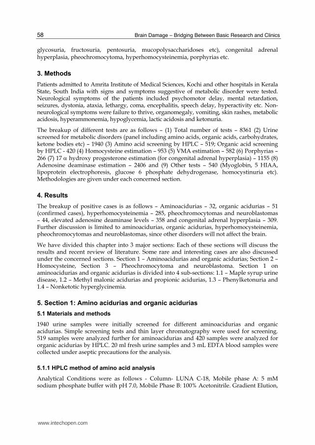
abnormal urine odor and clinical suspicion was MSUD, isovaleric aciduria or PKU. Laboratory analysis revealed ketonuria and metabolic acidosis. HPLC analysis of amino acid confirmed MSUD (Figure 1). Child died immediately afterwards.
Patient 2 presented at Day 12 with metabolic acidosis, abnormal urine odor, ketonuria and hepatosplenomegaly. Blood and urine studies revealed the diagnosis of MSUD. Aggressive treatment was started including branched chain amino acid restricted diet and supplementation. Patient has survived until 3 years of age, without any episode of exacerbation afterwards. Patient is on follow up. Levels of leucine, isoleucine and valine came down to normal level (Table 1).
In the case of the first patient (Patient 1), diagnosis was delayed and hence treatment could not be instituted and the baby died. But in the second case (Patient 2) diagnosis and treatment was started early in life and outcome was better. These two case studies indicate the importance of early diagnosis and treatment in MSUD.
5.2.2 Biochemical abnormalities
The name originates from the characteristic smell of urine (similar to burnt sugar or maple sugar) due to excretion of branched chain keto acids. Maple syrup urine disease (MSUD) or branched chain ketoaciduria is caused by deficiency of branched chain keto acid dehydrogenase complex (BCKAD). The basic biochemical defect is deficient decarboxylation of branched chain keto acids (BCKA). It leads to accumulation of branched chain amino acids (BCAA) Leucine, Isoleucine and Valine and corresponding branched chain keto acids (BCKA). Five distinct phenotypes are present: Classic, Intermediate, Intermittent, Thiamine-responsive and Dihydrolipoyl dehydrogenase (E3) deficient.
Fig. 1. Chromatogram of patient 1 (Leucine 2240 mol/L, Valine 411 mol/L, Isoleucine
180 mol/L; Normal Leu <150, Val <255 and Ile <80mol/L)
www.intechopen.com

Inborn Errors of Metabolism and Brain Involvement – 5 Years Experience from a Tertiary Care Center in South India
61
Valine (µmol/l)
Leucine (µmol/l)
Isoleucine (µmol/l)
11.04.2009 518 3020 437 25.07.2009 194 1708 350 15.03.2010 98 424 166 06.08.2010 135 479 69
Table 1. Serial levels of Branched chain amino acids in patient 2 diagnosed with MSUD –
Classic MSUD is the most common and is the most severe type. It has a neonatal-onset type of encephalopathy. Other types generally have onset by 2 years. BCAA, generally Leucine, is elevated in blood and urine. Presence of alloisoleucine is diagnostic. It has autosomal recessive inheritance. Worldwide frequency is 1 in 1,85,000.
BCAA comprise 35% of indispensable amino acids in muscles. Majority of untreated classic patients die within the early months of life from recurrent metabolic crises and neurologic deterioration. Treatment involves long-term dietary management and aggressive intervention during acute metabolic decompensation. Age at diagnosis and subsequent metabolic control are the most important determinants of long-term control. Patients in whom treatment is initiated after 10 days of age rarely achieve normal intellect.
Disease starts in the first week of life. It is characterized by convulsions, severe mental retardation, vomiting, acidosis, coma and death within the first year of life. Urine contains branched chain keto acids, valine, leucine and isoleucine. Rothera's test is positive, but unlike in cases of ketoacidosis, even boiled and cooled urine will give the test. Diagnosis depends on enzyme analysis in cells. Diagnosis should be done prior to 1 week after birth. Giving a diet low in branched chain amino acids. Mild variant is called intermittent branched chain ketonuria. This will respond to high doses of thiamine. This is because the decarboxylation of the BCKA requires thiamine. Liver transplantation has been successfully tried in some cases of MSUD.
5.2.3 Review of literature
Chen et al (2010) has reviewed 15 cases of MSUD from China and they suggest that early diagnosis and treatment can help prevent neurologic signs. Pangkanon et al (2008) report 13 cases of MSUD in Thai infants. All patients had neurological manifestations and psychomotor retardation. Lee et al (2008) report 47 Filipino patients with MSUD which is the commonest IEM in Philippines. They report that clinical outcome is poor in their series of patients.
Barschak et al (2009) report MSUD is associated with lipid peroxidation. They also report reduced amino acids methionine and tryptophan, which are amino acids with antioxidant activity. Mescka et al (2011) report protective effect of carnitine against oxidative stress induced by MSUD.
Ribeiro et al (2008) report that the major metabolites accumulating in MSUD disturb brain aerobic metabolism by compromising the citric acid cycle and the electron flow through the respiratory chain. They hypothesize that this might explain the neurological features in MSUD.
Brunetti-Pierri et al (2011) report successful use of phenylbutyrate in bringing down branched chain amino acid levels in MSUD. Shellmer et al (2011) studied 14 patients who received liver transplantation for MSUD and found that liver transplantation reduced further CNS damage in these patients. Strauss et al ((2010) has suggested novel therapeutic modalities in the management of MSUD based on their experience in treating 79 patients over 20 years.
www.intechopen.com

Brain Damage – Bridging Between Basic Research and Clinics
62
Zinnanti et al (2009) report that rapid brain leucine accumulation displaces other essential amino acids resulting in neurotransmitter depletion and disruption of normal brain growth and development in mouse model. They also report that administration of norleucine reduces branched chain amino acid accumulation in brain, blood and milk. Norleucine also substantially delayed encephalopathy in intermediate type MSUD. They conclude that brain damage in MSUD might be due to two factors – (1) Neurotransmitter deficiencies and growth restriction associated with BCAA accumulation, and (2) Energy deprivation through Krebs’ cycle disruption associated with BCAA accumulation.
Ibarra-Gonzalez et al (2007) report increased mortality and disabilities, especially neurological, in a cohort of 36 patients with MSUD from Mexico. Kowalik et al (2007) report deficiency of iron, zinc, copper, Vitamin B1, B2, niacin and Vitamin C in treated MSUD patients. Wajner et al (2007) report that BCAA inhibited Na-K ATPase pump in the brain and alanine prevented this inhibition. They conclude that this mechanism might contribute to neurological damage in MSUD, as Na-K ATPase pump is a critical enzyme for normal brain development and functioning. Bridi et al (2005) also report that alpha keto acids accumulating in MSUD stimulate lipid peroxidation and reduce antioxidant defense in cerebral cortex.
There are a large number of studies which report on CT and MR scans of the brain in MSUD [Cakmakci et al (2010), Tu et al (2005), Sener (2004), Schonberger et al (2004) and others]. Bindu et al (2007) have described neuroradiological findings in 3 patients with intermediate MSUD from South India.
5.3 Section 1.2: Methylmalonic aciduria and propionic aciduria
5.3.1 Results
We have detected 16 cases of propionic acidurias and 15 cases of methyl malonic acidurias in our study. Most of these patients had neurological manifestations and presented with metabolic acidosis and/or hyperammonemia. 60% of patients had neurological abnormalities including psychomotor delay, mental retardation, seizures, dystonia, ataxia, lethargy, extrapyramidal symptoms, encephalopathy, coma, visual deficiency, speech delay etc.
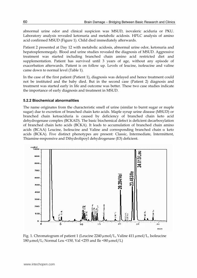
Fig. 2. Screening test for methyl malonic aciduria (Emerald green is positive test; other test tube is control urine sample)
www.intechopen.com
Inborn Errors of Metabolism and Brain Involvement – 5 Years Experience from a Tertiary Care Center in South India
63
Abnormal MRI findings were found in 11 patients including macrocephaly, cerebral atrophy and cerebral edema. Further details of this work can be seen in our paper (Narayanan et al, 2011; Vaidyanathan et al, 2011). Figure 2 gives a screening test for methyl malonic acid.
5.3.2 Biochemical abnormalities
Propionyl CoA is primarily converted to methyl malonyl CoA, which is subsequently converted to Succinyl CoA. Enzymes involved are propionyl CoA carboxylase, methyl malonyl CoA racemase and methyl malonyl CoA mutase. Biotin is needed for first and cobalamin for the third enzyme. Propionyl CoA carboxylase has two non-identical sub-units and biotin binds to sub-units, located on chromosomes 13 and 3 respectively. Methyl malonyl CoA mutase has 2 identical sub-units, located on chromosome 6. Holocarboxylase deficiency and biotinidase deficiency are known. Other known disorder is multiple carboxylase deficiency. PCC deficiency leads to propionic acidemia, and elevated 3 hydroxy propionate, methyl citrate, tiglyl Glycine, and unusual ketone bodies in urine. Severe metabolic ketoacidosis in neonatal period is seen. Alkali therapy and protein restriction are needed.
Inherited deficiency of the mutase enzyme or abnormalities in cobalamin can result in methyl malonic aciduria. Neonatal or infantile metabolic ketoacidosis are the hallmarks. Patients with abnormal binding ability of enzyme to cobalamin, cannot be treated by cobalamin therapy. These cases may be treated with dietary protein restriction and antibiotic therapy. Other patients respond to cobalamin or hydroxycobalamin therapy. This can be used in combination with dietary protein restriction. Therapy has been found to reduce methyl malonate levels. Mutations leading to impaired adenosyl cobalamin and methyl cobalamin and deficient activity of methyl malonyl CoA mutase and N5 methyl tetrahydro folate reductase. Homocysteine methyl transferase have methyl malonic aciduria combined with homocystinuria. Features include failure to thrive, developmental retardation, megaloblastic anemia and macrocytosis. Therapy includes protein restriction, pharmacological doses of hydroxocobalamin and betaine supplementation.
Both disorders are inherited as autosomal recessive disorders. Prenatal diagnosis is possible by enzyme assays on chorionic villus biopsy or cultured amniotic cells and chemical determinations on amniotic fluid or maternal urine.
5.3.3 Review of literature
Liu et al (2010) reports 24 mutations in the MMACHC gene to be responsible for cblC type of combined methyl malonic aciduria and homocystinuria in 79 unrelated Chinese patients. 5 mutations are responsible for 80% of cases and suggest a role for mutation detection in early diagnosis. Cosson et al (2009) reports on long term outcome of 30 French patients with methyl malonic acidurias. 15 patients had neonatal onset, 13 had severe neurological involvement, 14 had chronic renal failure and 5 died during a metabolic crisis. Patients with a mut(0) phenotype had a severe phenotype and early and more severe CRF than patients with mut-/cblA phenotype.
Chandler et al (2007) reports successful use of adenoviral mediated gene therapy for methylmalonic aciduria in murine models and human patients with MUT gene mutation. Yang et al (2006) and other authors report that in China methylmalonic acidurias is more commonly associated with homocysteinemia. Filippi et al (2010) describes the use of N –
www.intechopen.com
Brain Damage – Bridging Between Basic Research and Clinics
64
carbamyl glutamate in the emergency management of hyperammonemia in neonatal acute onset propionic aciduria and methylmalonic aciduria. Perez et al (2009) describes pseudoexon exclusion by antisense therapy in MMA. Haas et al (2009) report that Coenzyme Q (10) is significantly decreased in MMA patients.
Wajner et al (2009) identified 34 patients with MMA and 18 patients with PA from Brazil in 15 years. Zwickler et al (2008) reviews MMA patients in 14 centers in Germany and outlines the management principles. Most centers used hydroxocobalamin or cyanocobalamin for cobalamin-responsive patients while cobalamin – nonresponsive patients are supplemented with carnitine. Intestinal decontamination by antibiotic therapy, D-A-CH or Dewey recommendations for protein therapy and precursor-free amino acid supplements were used by most centers. Zhang et al (2007) studied the clinical picture of 96 patients with MMA over a 10 year period. Most of the patients had neurological abnormalities including developmental delay, seizures, psychomotor degeneration and motor disorders. A significant proportion of patients had MMA along with homocysteinemia.
Longo et al (2005) describes neurological abnormalities in MMA patients detected by MRI and 1H-MRS. Burlina et al (2003) and Rossi et al (2001) also describe neurological damage in MMA. Bodamer et al (2005) studied creatine metabolism in MMA patients and found that guanidinoacetate is elevated. They suggest that guanidinoacetate may be responsible for neurotoxicity in MMA. Vara et al (2011) discusses the importance of liver transplantation in propionic aciduria (PA) patients. Liver transplantation reduces the risk of metabolic decompensation and improves the quality of life. Romano et al (2010) report that cardiomyopathy in PA is reversible with liver transplantation.
Ah Mew et al (2010) also reports that NCG reduces ammonia and glutamine and induces
ureagenesis in PA. Schwahn et al (2010) also reports of NCG in PA. Chandler et al (2011)
describes the use of gene therapy using adeno associated virus (AAV8) for the treatment of
PA. Ribas et al (2010) report that PA and MMA induce DNA damage and L – carnitine
prevents this damage. Haberlandt et al (2009) report seizures in 9 patients with PA and
hypothesize that some intermediates might be involved in the pathogenesis of epilepsy.
Rigo et al (2006) suggests the involvement of NMDA receptors in the pathogenesis of
neurological manifestations.
de Keyzer et al (2009) report multiple OXPHOS deficiency in liver, kidney, heart and
skeletal muscle in patients with MMA and PA. Desviat et al (2009) report high frequency of
large gene deletions in PA patients. This finding underscores the need for gene dosage
analysis in additional to mutation testing in PA patients. Perez Cerda et al (2004) report the
utility of prenatal diagnosis by molecular methods in 19 unrelated families with PA.
5.4 Section 1.3: Phenylketonuria (PKU)
5.4.1 Case report
We hereby report two patients with PKU. Patient 3 is a 21 year old woman with sub-normal intelligence and suspected to have phenylketonuria, though not confirmed previously
(Phenylalanine level – 1427 mol/L, Normal <65 mol/L). Patient 4 is 19 years old and is
the sibling of Patient 2 (Phenylalanine level 1177 mol/L). She also had sub-normal intelligence. Both patients had pleasant social manners. At the time of presentation, Patient 3
www.intechopen.com
Inborn Errors of Metabolism and Brain Involvement – 5 Years Experience from a Tertiary Care Center in South India
65
was pregnant and had hence sought advice. Three months after presentation, Patient 4 also became pregnant. Both patients were confirmed to have phenylketonuria by urine and blood tests for phenylalanine. Phenylalanine restricted diet was advised, but compliance
was not satisfactory and phenylalanine levels remained above 1000 mol/L. Patient 3 delivered and child suffered from clinical and laboratory signs of maternal hyperphenylalaninemia. Child died in the immediate post natal period. The child of the other patient died in utero.
5.4.2 Biochemical abnormalities
Hyperphenylalaninemias are due to disorders of phenylalanine hydroxylation reaction. The minimum requirements for phenylalanine metabolism to occur are the enzyme phenyl alanine hydroxylase (PAH), molecular oxygen (O2), L-Phenyl Alanine and tetrahydrobiopterin (BH4). Other components include dihydrobiopterin (DHPR), reduced pyridine nucleotide, 4carbinolamine dehydratase (for BH4 recycling), GTP cyclohydrolase (GTP – CH) and 6 pyruvoyl tetrahydropterin synthase (6 PTS). Hyperphenylalaninemia is defined as Phenylalanine levels above 120 µM (2 mg/dl). Normal plasma level of phenyl alanine is 58±15 µM.
Phenylalanine cannot be converted to tyrosine. So phenylalanine accumulates. Phenylalanine level in blood is elevated. So alternate minor pathways are opened. Phenyl ketone (phenyl pyruvate), phenyl lactate and phenyl acetate are excreted in urine. Phenyl pyruvate inhibits pyruvate decarboxylase enzyme in brain, but not in liver. Hence myelin formation defects and mental retardation are seen. Brain effects are due to phenylalanine and its metabolites (phenyl ketones, namely Pyruvate, lactate, acetate, acetyl glutamine and ethyl amine) that accumulate via alternate pathway. Myelination and protein synthesis are affected and there is deficient neurotransmitter supply. Peculiarities of gait, stance and sitting posture are additional features. Brain calcification may be seen in DHPR deficient type.
Phenylketonuria is well known for the neurological manifestations. The classical PKU child is mentally retarded with an IQ of 50. About 20% inmates of psychiatric hospitals may have PKU. Agitation, hyperactivity, tremors and convulsions are often manifested. This may be because phenylalanine interferes with neurotransmitter synthesis. The child often has hypopigmentation, explained by the decreased level of tyrosine. Phenyl lactic acid in sweat may lead to mousy body odor.
5.4.3 Maternal hyperphenylalaninemia
Female child, on growing to adulthood may become pregnant (maternal hyper phenylalaninemia). Then again special diet is to be given, because the increased phenylalanine level will affect the brain development of the fetus. Maternal hyperphenylalaninemia (PKU embryo-fetopathy) cause embryopathy/ fetopathy comprising impaired growth, congenital cardiac malformations, microcephaly and mental retardation in the embryo/fetus. Fetal phenylalanine level is 1.5 – 2 fold higher than maternal blood level. Further fetal blood brain barrier concentrates phenylalanine to another 2-4 fold. Intraneuronal phenylalanine level of 600mol interferes with brain development. Phenylalanine restricted diet should be started at least 3 months prior to planned pregnancy. Phe level in mother to be maintained at 60-180mol/L. Linoleic and linolenic acid supplements should be maintained at high level.
www.intechopen.com

Brain Damage – Bridging Between Basic Research and Clinics
66
5.4.4 Review of literature
Oddason et al (2011) report 27 patients diagnosed with PKU in Iceland since 1947. Classical PKU is the commonest type. Macdonald et al (2011) in a study from UK report that PKU patients, especially older, are not fully compliant with treatment and hence have higher than acceptable phenylalanine levels. At the same time, van Rijn et al (2011) report that well-controlled adult PKU patients can tolerate larger dietary variations in phenylalanine levels. ten Hoedt et al (2011) report that high phenylalanine levels can directly affect mood and sustained attention in adult PKU patients (Randomized, double-blind, placebo-controlled, crossover trial).
Ribas et al (2011) suggest that oxidative stress may play a role in pathogenesis of PKU. Sanayama et al (2011) provide experimental evidence for the same and report that oxidative stress status is closely linked with phenylalanine levels. Sitta et al (2011) report that administration of L-carnitine and selenium can reduce oxidative stress in PKU patients.
Hanley (2011) in a review states that “non-PKU mild hyperphenylalaninemia” (MHP) also might have neuropsychological function deficits and therefore may need treatment with tetrahydrobiopterin and/or phenyl alanine restricted diet. Campistol et al (2011) discusses on the extent of neuro-congitive dysfunction in mild PKU (mPKU) and conclude that further studies are needed on mPKU to clearly answer this question. van Spronsen (2011) addresses the question on treatment of mPKU and reaches similar conclusion.
Vernon et al (2010), Trefz et al (2010), Harding (2010), Burton et al (2010), Somaraju & Merrin (2010) and others report that saptopterin hydrochloride, a synthetic analog of tetrahydrobiopterin, is a promising new drug in the treatment of PKU. This modality of treatment is especially important considering the report by Enns et al (2010) who state that PKU treated with diet alone has sub-optimal outcome with relation to neurocognitive, psychological and physical parameters. Lee et al (2009) and others however, report better outcome with dietary treatment alone. Blau et al (2010) reports on the management practices in PKU in 165 PKU centers in 23 European countries. They emphasize that treatment recommendations vary tremendously in different countries and hence there is an urgent need to pool long-term data in registries to generate evidence based international guidelines. Ahring et al (2009) and van Spronsen (2009) give similar reports.
Rocha and Martel (2009) report the use of large neutral amino acids in the treatment of PKU. The same carrier transports phenylalanine as well as large neutral amino acids into the brain; hence their use diminishes toxicity due to phenylalanine. Weigel et al (2008) report low free carnitine levels in PKU patients given low phenylalanine diet. They suggest that carnitine level should be monitored in PKU patients. Sitta et al (2009) reach similar conclusions. A study by Maillot et al (2008) reports on the importance of maintaining blood phenylalanine during pregnancy. They conclude that maintenance of maternal blood phenylalanine level within the target range predicts good offspring outcomes; and further suggests that variations even within that range should be avoided.
5.5 Section 1.4: Nonketotic hyperglycinemia
5.5.1 Case report
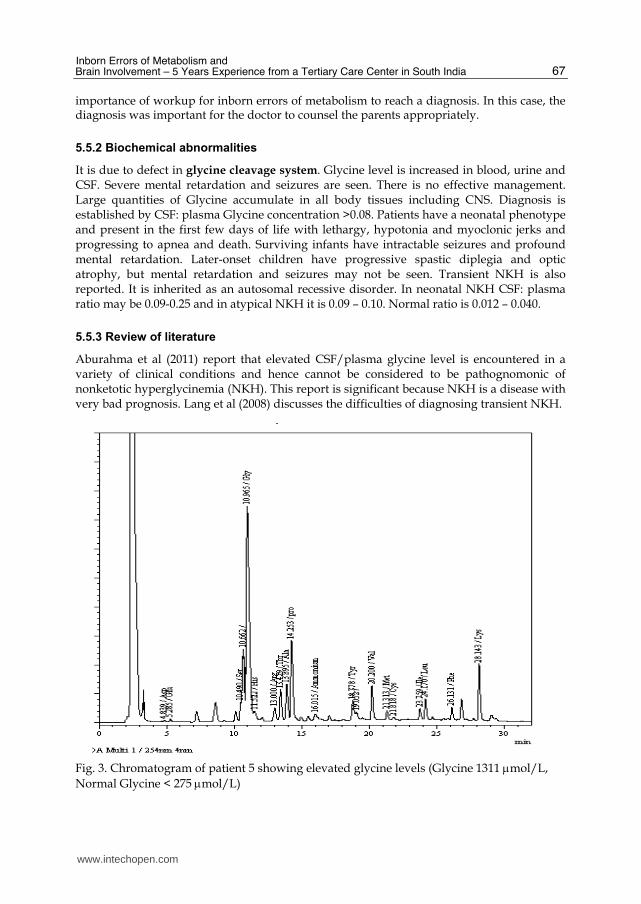
We report here one male baby (Patient 5) with intractable seizures who was 7 days old. All antiepileptic drugs were tried without any response. The parents were complaining about medical negligence. HPLC analysis of amino acid revealed nonketotic hyperglycinemia (NKH) (Figure 3). Even though the child could not be revived; this case shows the
www.intechopen.com
Inborn Errors of Metabolism and Brain Involvement – 5 Years Experience from a Tertiary Care Center in South India
67
importance of workup for inborn errors of metabolism to reach a diagnosis. In this case, the diagnosis was important for the doctor to counsel the parents appropriately.
5.5.2 Biochemical abnormalities
It is due to defect in glycine cleavage system. Glycine level is increased in blood, urine and CSF. Severe mental retardation and seizures are seen. There is no effective management. Large quantities of Glycine accumulate in all body tissues including CNS. Diagnosis is established by CSF: plasma Glycine concentration >0.08. Patients have a neonatal phenotype and present in the first few days of life with lethargy, hypotonia and myoclonic jerks and progressing to apnea and death. Surviving infants have intractable seizures and profound mental retardation. Later-onset children have progressive spastic diplegia and optic atrophy, but mental retardation and seizures may not be seen. Transient NKH is also reported. It is inherited as an autosomal recessive disorder. In neonatal NKH CSF: plasma ratio may be 0.09-0.25 and in atypical NKH it is 0.09 – 0.10. Normal ratio is 0.012 – 0.040.
5.5.3 Review of literature
Aburahma et al (2011) report that elevated CSF/plasma glycine level is encountered in a variety of clinical conditions and hence cannot be considered to be pathognomonic of nonketotic hyperglycinemia (NKH). This report is significant because NKH is a disease with very bad prognosis. Lang et al (2008) discusses the difficulties of diagnosing transient NKH.
Fig. 3. Chromatogram of patient 5 showing elevated glycine levels (Glycine 1311 mol/L,
Normal Glycine < 275 mol/L)
www.intechopen.com
Brain Damage – Bridging Between Basic Research and Clinics
68
Leipnitz et al (2009) report lipid peroxidation and reduced antioxidant levels in NKH.
Kanno et al (2007) reviews the genetic causes of NKH. NKH can be caused by genes in the
glycine cleavage system, including GLDC, AMT and GCSH. They report significant number
of GLDC mutations by MLPA (multiple ligation-dependent probe amplification) analysis.
Conter et al (2006) and Kure et al (2006) also report significant number of mutations in these
genes.
Tan et al (2007) report that currently tandem mass spectrometry employed for newborn
screening does not identify NKH without significant error rate. Raghavendra et al (2007)
and others report significant neurological abnormalities in patients with NKH. Generally
NKH is a disease refractory to treatment. A number of authors report the use of sodium
benzoate and dextromethrophan in the treatment of NKH, some of them with and others
without any beneficial effect.
6. Section 2: Hyperhomocysteinemia
6.1 Materials and methods
Homocysteine estimation was done by ELISA method (BioRad Laboratories Inc.). 5 ml
blood was drawn from the patients. 953 patients were analyzed during this 5 year period,
110 patients were from the Department of Cardiology, 656 from the Department of
Neurology and the remaining from other departments.
6.2 Results
285 patients had elevated homocysteine levels. 226 cases had hyperhomocysteinemia from
the Department of Neurology (226/656, 34.5%), 31 had hyperhomocysteinemia from the
Department of Cardiology (31/110, 28.2%), and the remaining were other cases like
peripheral artery disease, deep vein thrombosis etc. Neurological disorders included
different types of stroke including medullary stroke, ischemic stroke, young stroke, transient
ischemic attack, sagittal sinus thrombosis, lacunar thalamic stroke and recurrent stroke. All
patients with hyperhomocysteinemia from the Department of Cardiology were suffering
from Coronary artery disease (CAD).
6.3 Biochemical abnormalities
Normal homocysteine level in blood is 5-15 mol/L. In diseases, it may be increased to 50 to
100 times. Moderate increase is seen in aged persons, vitamin B12 or B6 deficiency, tobacco
smokers, alcoholics and in hypothyroidism. Substantial increase is noticed in congenital
enzyme deficiencies. Large amounts of homocysteine are excreted in urine. In plasma,
homocysteine (with -SH group) and homocysteine (disulfide, -S-S- group) exist. Both of
them are absent in normal urine; but if present, it will be the homocysteine (disulfide) form.
If homocysteine level in blood is increased, there is increased risk for coronary artery
diseases. Homocystinuria/ hyperhomocysteinemia may be due to many causes. These
include impaired activity of CBS (genetic CBS deficiency, INH therapy), methionine
synthase defect, MTHFR defect, impaired metabolism of vitamin B12, renal insufficiency,
pyridoxine and folate deficiency etc.
www.intechopen.com
Inborn Errors of Metabolism and Brain Involvement – 5 Years Experience from a Tertiary Care Center in South India
69
6.4 Review of literature
Hyperhomocysteinemia is associated with a number of diseases including coronary artery diseases, stroke, retinal vein thrombosis, diabetic peripheral neuropathy, schizophrenia, preeclampsia, chronic pancreatitis etc. Herrmann and Obeid (2011) have reviewed the role of hyperhomocysteinemia in neurodegenerative diseases like Alzheimer’s disease, vascular dementia, cognitive impairment and stroke. Damelan et al (2010) report hyperhomocysteinemia is ischemic stroke patients from France. Valentino et al ((2010) report elevated blood and CSF levels of homocysteine in amyotropic lateral sclerosis (ALS).
Sniezawska et al (2011) report that antiepileptic drug (AED) treatment in epileptics leads to increase in homocysteine and asymmetric dimethyl arginine (ADMA) levels. Greater increase in homocysteine is observed in patients with MTHFR CT (C677T) and MTHFD1 GG (G1958A) polymorphisms. Linnebank et al (2011) report reduced serum Vitamin B12 and folate levels on treatment with AED. Zhuo et al (2010) report that normalization of homocysteine values in rat models with hyperhomocysteinemia resulted in improvement of cognitive defects and brain amyloidosis.
Paoli et al (2010) report that protein N homocysteinylation induces the formation of toxic amyloid like amyloid protofibrils. The authors hypothesize that this could be responsible for pathophysiology of hyperhomocysteinemia. da Cunha et al (2010) report increase in inflammatory markers in brain and blood of mice after acute homocysteine administration. This study suggests that inflammation may be at least partially responsible for neurological and cardiovascular response of homocysteine. Green et al (2010) report that homocysteine lowering vitamins do not lower S adenosyl homocysteine levels in older people. They suggest that S adenosyl homocysteine might be a better indicator for vascular events. They hypothesize that this might explain the lack of clinical benefit of B vitamins in some patients with hyperhomocysteinemia. Almawi et al (2009) and others have investigated the role of MTHFR C677T polymorphism and high homocysteine levels in patients with stroke. Dutta et al (2009) report mild increase in serum homocysteine levels in Indian patients with idiopathic mental retardation.
7. Section 3: Pheochromocytoma, neuroblastoma
7.1 Materials and methods
VMA estimation was done using column method (BioRad VMA by column test). 24 hr urine samples were collected following dietary restrictions for 3 days. Samples were collected in 6N HCl and stored in the refrigerator during collection time. If the pH was more than 3.5, the samples were rejected. The analysis was done following manufacturer’s guidelines. 582 samples were collected during this time period.
7.2 Results
44 patients had elevated VMA levels. This included 24 cases of pheochromocytoma, 15 cases of neuroblastoma and 5 cases of paraganglioma. The analysis correlated positively with histopathology studies. All patients with pheochromocytoma and paraganglioma were above 10 years of age; whereas 60% cases of neuroblastoma cases were below 10 years of age. The sensitivity and specificity of our results were 88% and 86% respectively. All neuroblastoma cases showed elevated VMA level.
www.intechopen.com
Brain Damage – Bridging Between Basic Research and Clinics
70
7.3 Biochemical abnormalities
Catecholamines are important biological compounds, essential to maintain the proper functioning of the body. Secretion of excessive catecholamines can produce stress, palpitation, paroxysmal hypertension, congestive heart failure, thyroid hormone deficiency and arrhythmias. Measurement of catecholamines and its metabolites is primarily used in the diagnosis of Neuroendocrine tumors; Neuroblastomas, Pheochromocytomas and paragangliomas. Most common metabolites of catecholamines are Vanillylmandelic acid (VMA), urinary free catecholamines, metanephrine, normetanephrine, homovanillic acid (HVA), plasma catecholamines, plasma metanephrines and chromogranin A. Most of the metabolites are measured by spectrophotometric assays, nowadays replaced by HPLC and mass spectrometry. Vanillylmandelic acid (VMA) is a major catecholamine metabolite formed by the actions of catechol-O-methyl transferase and MAO. VMA is excreted by kidney and represents 40%-50% urinary excretory product of norepinephrine and epinephrine. Norepinephrine is the major source of VMA.
Neuroblastomas are the most common solid extra-cranial tumors in children, and account for 7-10 % of all tumors. In about 90% of cases of neuroblastoma, elevated levels of catecholamines or its metabolites are found in the urine or blood. Pheochromocytomas are chromaffin-cell tumors; 80-85 % arises from the adrenal medulla and 15-20 % arises from extra-adrenal chromaffin tissues (paragangliomas). They are characterized by excessive production of catecholamines. If not diagnosed or if left untreated, the excessive secretion of catecholamines by these tumors can have devastating consequences. Paraganglioma is a rare neuroendocrine tumor that arises from extraadrenal sympathochromaffin tissue, usually in the abdomen. 10% of the catecholamine producing tumors are paragangliomas that develop in head, neck, thorax and abdomen. About 97% are benign and 3% metastatic.
7.4 Review of literature
Elevated urinary catecholamines have been described in 90 – 95 % of patients with
neuroblastomas [Smith et al (2010)]. Studies suggest that dopamine nervous systems are
involved in the pathogenesis of autistic disorder. Quantification of urine homovanillic acid
(HVA) and vanillylmandelic acid (VMA) by GC/MS are very important in the study of
dopamine metabolism in autistic children [Kaluzna-Czaplinska et al (2010)]. Allenbrand and
Garg (2010) also report quantification of urine HVA and VMA by GC/MS. Li et al (2010)
describe that the ratio of HVA/VMA ratio is useful as a disease marker in neuroblastomas
and pheochromocytomas. Aydin et al (2010) report that VMA is a poor prognostic factor in
patients with neuroblastoma.
Hickman et al (2009) report that in patients with pheochromocytoma, plasma free metanephrines displayed superior diagnostic sensitivity and specificity compared with other biochemical markers of catecholamine output and metabolism. Boyle et al (2007) came to a similar conclusion. Lionetto et al (2008) describe an HPLC-tandem mass spectrometric method for the simultaneous quantification of VMA, HVA and 5-HIAA in human urine.
8. Conclusions
We have detected a high incidence of metabolic disorders in our population. The commonest disorders detected were organic acidurias, congenital adrenal hyperplasia and
www.intechopen.com
Inborn Errors of Metabolism and Brain Involvement – 5 Years Experience from a Tertiary Care Center in South India
71
hyperhomocysteinemia. Aminoacidurias like PKU are rare; whereas organic acidurias (PAA, MMA and MSUD) are more common. Prompt identification and treatment is important and early diagnosis helps to institute treatment measures which prevents further morbidity and reduces mortality rates.
Acknowledgements: We thank Kerala State Council for Science, Technology and Environment (KSCSTE), Indian Council of Medical Research (ICMR) for the financial support; Sumithra K, Rajesh PC and Anoop PA for the technical assistance.
9. References
Aburahma S, Khassawneh M, Griebel M, Sharp G, Gibson J. Pitfalls in measuring cerebrospinal fluid glycine levels in infants with encephalopathy. J Child Neurol. 2011 Jun;26(6):703-6. Epub 2011 Feb 18.
Ah Mew N, McCarter R, Daikhin Y, Nissim I, Yudkoff M, Tuchman M. N-carbamylglutamate augments ureagenesis and reduces ammonia and glutamine in propionic acidemia. Pediatrics. 2010 Jul;126(1):e208-14. Epub 2010 Jun 21.
Allenbrand R, Garg U. Quantitation of homovanillic acid (HVA) and vanillylmandelic acid (VMA) in urine using gas chromatography-mass spectrometry (GC/MS). Methods Mol Biol. 2010;603:261-9.
Almawi WY, Khan A, Al-Othman SS, Bakhiet M. Case-control Study of methylenetetrahydrofolate reductase mutations and hyperhomocysteinemia and risk of stroke. J Stroke Cerebrovasc Dis. 2009 Sep-Oct;18(5):407-8.
Aydin GB, Kutluk MT, Yalcin B, Varan A, Akyuz C, Buyukpamukcu M. The prognostic significance of vanillylmandellic acid in neuroblastoma. Pediatr Hematol Oncol. 2010 Sep;27(6):435-48.
Barschak AG, Sitta A, Deon M, Busanello EN, Coelho DM, Cipriani F, Dutra-Filho CS, Giugliani R, Wajner M, Vargas CR. Amino acids levels and lipid peroxidation in maple syrup urine disease patients. Clin Biochem. 2009 Apr;42(6):462-6. Epub 2008 Dec 24.
Bindu PS, Shehanaz KE, Christopher R, Pal PK, Ravishankar S. Intermediate maple syrup urine disease: neuroimaging observations in 3 patients from South India. J Child Neurol. 2007 Jul;22(7):911-3.
Blau N, Belanger-Quintana A, Demirkol M, Feillet F, Giovannini M, MacDonald A, Trefz FK, van Spronsen F; European PKU centers. Management of phenylketonuria in Europe: survey results from 19 countries. Mol Genet Metab. 2010 Feb;99(2):109-15. Epub 2009 Sep 13.
Bodamer OA, Sahoo T, Beaudet AL, O'Brien WE, Bottiglieri T, Stockler-Ipsiroglu S, Wagner C, Scaglia F. Creatine metabolism in combined methylmalonic aciduria and homocystinuria. Ann Neurol. 2005 Apr;57(4):557-60.
Boyle JG, Davidson DF, Perry CG, Connell JM. Comparison of diagnostic accuracy of urinary free metanephrines, vanillyl mandelic Acid, and catecholamines and plasma catecholamines for diagnosis of pheochromocytoma. J Clin Endocrinol Metab. 2007 Dec;92(12):4602-8. Epub 2007 Jul 17.
Bridi R, Braun CA, Zorzi GK, Wannmacher CM, Wajner M, Lissi EG, Dutra-Filho CS. alpha- keto acids accumulating in maple syrup urine disease stimulate lipid peroxidation
www.intechopen.com

Brain Damage – Bridging Between Basic Research and Clinics
72
and reduce antioxidant defences in cerebral cortex from young rats. Metab Brain Dis. 2005 Jun;20(2):155-67.
Brunetti-Pierri N, Lanpher B, Erez A, Ananieva EA, Islam M, Marini JC, Sun Q, Yu C, Hegde M, Li J, Wynn RM, Chuang DT, Hutson S, Lee B. Phenylbutyrate therapy for maple syrup urine disease. Hum Mol Genet. 2011 Feb 15;20(4):631-40. Epub 2010 Nov 23.
Burlina AP, Manara R, Calderone M, Catuogno S, Burlina AB. Diffusion-weighted imaging in the assessment of neurological damage in patients with methylmalonic aciduria. J Inherit Metab Dis. 2003;26(5):417-22.
Burton BK, Bausell H, Katz R, Laduca H, Sullivan C. Sapropterin therapy increases stability of blood phenylalanine levels in patients with BH4-responsive phenylketonuria (PKU). Mol Genet Metab. 2010 Oct-Nov;101(2-3):110-4. Epub 2010 Jun 27.
Cakmakci H, Pekcevik Y, Yis U, Unalp A, Kurul S. Diagnostic value of proton MR spectroscopy and diffusion-weighted MR imaging in childhood inherited neurometabolic brain diseases and review of the literature. Eur J Radiol. 2010 Jun;74(3):e161-71. Epub 2009 Jun 21.
Campistol J, Gassio R, Artuch R, Vilaseca MA; PKU Follow-up Unit. Neurocognitive function in mild hyperphenylalaninemia. Dev Med Child Neurol. 2011 May;53(5):405-8. Epub 2011 Mar 21.
Chandler RJ, Chandrasekaran S, Carrillo-Carrasco N, Senac JS, Hofherr SE, Barry MA, Venditti CP. Adeno-associated virus serotype 8 gene transfer rescues a neonatal lethal murine model of propionic acidemia. Hum Gene Ther. 2011 Apr;22(4):477-81. Epub 2011 Feb 16.
Chandler RJ, Tsai MS, Dorko K, Sloan J, Korson M, Freeman R, Strom S, Venditti CP. Adenoviral-mediated correction of methylmalonyl-CoA mutase deficiency in murine fibroblasts and human hepatocytes. BMC Med Genet. 2007 Apr 30;8:24.
Chen Z, Luo F, Wu XJ, Shi LP. Maple syrup urine disease of neonates: report of two cases and review of literature. Zhonghua Er Ke Za Zhi. 2010 Sep;48(9):680-4.
Conter C, Rolland MO, Cheillan D, Bonnet V, Maire I, Froissart R. Genetic heterogeneity of the GLDC gene in 28 unrelated patients with glycine encephalopathy. J Inherit Metab Dis. 2006 Feb;29(1):135-42.
Cosson MA, Benoist JF, Touati G, Dechaux M, Royer N, Grandin L, Jais JP, Boddaert N, Barbier V, Desguerre I, Campeau PM, Rabier D, Valayannopoulos V, Niaudet P, de Lonlay P. Long-term outcome in methylmalonic aciduria: a series of 30 French patients. Mol Genet Metab. 2009 Jul;97(3):172-8. Epub 2009 Mar 24.
da Cunha AA, Ferreira AG, Wyse AT. Increased inflammatory markers in brain and blood of rats subjected to acute homocysteine administration. Metab Brain Dis. 2010 Jun;25(2):199-206. Epub 2010 Apr 28.
Damelan K, Kom A, Kossivi A, Koffi BA, Emile A, Kodjo GE. Hyperhomocysteinemia among ischaemic stroke victims in the teaching hospital of Lome. Ann Biol Clin (Paris). 2010 Nov-Dec;68(6):669-73.
de Keyzer Y, Valayannopoulos V, Benoist JF, Batteux F, Lacaille F, Hubert L, Chretien D, Chadefeaux-Vekemans B, Niaudet P, Touati G, Munnich A, de Lonlay P. Multiple OXPHOS deficiency in the liver, kidney, heart, and skeletal muscle of patients with methylmalonic aciduria and propionic aciduria. Pediatr Res. 2009 Jul;66(1):91-5.
www.intechopen.com
Inborn Errors of Metabolism and Brain Involvement – 5 Years Experience from a Tertiary Care Center in South India
73
Desviat LR, Sanchez-Alcudia R, Perez B, Perez-Cerda C, Navarrete R, Vijzelaar R, Ugarte M. High frequency of large genomic deletions in the PCCA gene causing propionic acidemia. Mol Genet Metab. 2009 Apr;96(4):171-6. Epub 2009 Jan 20.
Dutta S, Chatterjee A, Sinha S, Chattopadhyay A, Mukhopadhyay K. Correlation between cystathionine beta synthase gene polymorphisms, plasma homocysteine and idiopathic mental retardation in Indian individuals from Kolkata. Neurosci Lett. 2009 Apr 10;453(3):214-8. Epub 2009 Feb 21.
Enns GM, Koch R, Brumm V, Blakely E, Suter R, Jurecki E. Suboptimal outcomes in patients with PKU treated early with diet alone: revisiting the evidence. Mol Genet Metab. 2010 Oct-Nov;101(2-3):99-109. Epub 2010 Jun 22.
Filippi L, Gozzini E, Fiorini P, Malvagia S, la Marca G, Donati MA. N-carbamylglutamate in emergency management of hyperammonemia in neonatal acute onset propionic and methylmalonic aciduria. Neonatology. 2010;97(3):286-90. Epub 2009 Nov 4.
Green TJ, Skeaff CM, McMahon JA, Venn BJ, Williams SM, Devlin AM, Innis SM. Homocysteine-lowering vitamins do not lower plasma S-adenosylhomocysteine in older people with elevated homocysteine concentrations. Br J Nutr. 2010 Jun;103(11):1629-34. Epub 2010 Jan 21.
Haas D, Niklowitz P, Horster F, Baumgartner ER, Prasad C, Rodenburg RJ, Hoffmann GF, Menke T, Okun JG. Coenzyme Q(10) is decreased in fibroblasts of patients with methylmalonic aciduria but not in mevalonic aciduria. J Inherit Metab Dis. 2009 Aug;32(4):570-5. Epub 2009 Jun 7.
Haberlandt E, Canestrini C, Brunner-Krainz M, Moslinger D, Mussner K, Plecko B, Scholl- Burgi S, Sperl W, Rostasy K, Karall D. Epilepsy in patients with propionic acidemia. Neuropediatrics. 2009 Jun;40(3):120-5. Epub 2009 Dec 17.
Hanley WB. Non-PKU mild hyperphenylalaninemia (MHP) - The dilemma. Mol Genet Metab. 2011 May 14. [Epub ahead of print]
Harding CO. New era in treatment for phenylketonuria: Pharmacologic therapy with sapropterin dihydrochloride. Biologics. 2010 Aug 9;4:231-6.
Herrmann W, Obeid R. Homocysteine: a biomarker in neurodegenerative diseases. Clin Chem Lab Med. 2011 Mar;49(3):435-41.
Hickman PE, Leong M, Chang J, Wilson SR, McWhinney B. Plasma free metanephrines are superior to urine and plasma catecholamines and urine catecholamine metabolites for the investigation of phaeochromocytoma. Pathology. 2009 Feb;41(2):173-7.
Ibarra-Gonzalez I, Fernandez-Lainez C, Belmont-Martinez L, Vela-Amieva M. Increased mortality and disability in a cohort of Mexican children with maple syrup urine disease. Gac Med Mex. 2007 May-Jun;143(3):197-201.
Kaluzna-Czaplinska J, Socha E, Rynkowski J. Determination of homovanillic acid and vanillylmandelic acid in urine of autistic children by gas chromatography/mass spectrometry. Med Sci Monit. 2010 Sep;16(9):CR445-50.
Kanno J, Hutchin T, Kamada F, Narisawa A, Aoki Y, Matsubara Y, Kure S. Genomic deletion within GLDC is a major cause of non-ketotic hyperglycinaemia. J Med Genet. 2007 Mar;44(3):e69.
Keskinen P, Siitonen A, Salo M. Hereditary urea cycle diseases in Finland. Acta Paediatr. 2008 Oct;97(10):1412-9. Epub 2008 Jul 9.
www.intechopen.com

Brain Damage – Bridging Between Basic Research and Clinics
74
Kowalik A, Narojek L, Sykut-Cegielska J. Compliance of the diet restricted with leucine, isoleucine and valine in maple syrup urine disease (MSUD) children. Rocz Panstw Zakl Hig. 2007;58(1):95-101.
Kure S, Kato K, Dinopoulos A, Gail C, DeGrauw TJ, Christodoulou J, Bzduch V, Kalmanchey R, Fekete G, Trojovsky A, Plecko B, Breningstall G, Tohyama J, Aoki Y, Matsubara Y. Comprehensive mutation analysis of GLDC, AMT, and GCSH in nonketotic hyperglycinemia. Hum Mutat. 2006 Apr;27(4):343-52.
Lang TF, Parr JR, Matthews EE, Gray RG, Bonham JR, Kay JD. Practical difficulties in the diagnosis of transient non-ketotic hyperglycinaemia. Dev Med Child Neurol. 2008 Feb;50(2):157-9.
Lee JY, Chiong MA, Estrada SC, Cutiongco-De la Paz EM, Silao CL, Padilla CD. Maple syrup urine disease (MSUD)-Clinical profile of 47 Filipino patients. J Inherit Metab Dis. 2008 Nov 10. [Epub ahead of print]
Lee PJ, Amos A, Robertson L, Fitzgerald B, Hoskin R, Lilburn M, Weetch E, Murphy G. Adults with late diagnosed PKU and severe challenging behaviour: a randomised placebo-controlled trial of a phenylalanine-restricted diet. J Neurol Neurosurg Psychiatry. 2009 Jun;80(6):631-5. Epub 2009 Feb 9.
Leipnitz G, Solano AF, Seminotti B, Amaral AU, Fernandes CG, Beskow AP, Dutra Filho CS, Wajner M. Glycine provokes lipid oxidative damage and reduces the antioxidant defenses in brain cortex of young rats. Cell Mol Neurobiol. 2009 Mar;29(2):253-61. Epub 2008 Oct 2.
Li Q, Batchelor-McAuley C, Compton RG. Electrooxidative decarboxylation of vanillylmandelic acid: voltammetric differentiation between the structurally related compounds homovanillic acid and vanillylmandelic acid. J Phys Chem B. 2010 Jul 29;114(29):9713-9.
Linnebank M, Moskau S, Semmler A, Widman G, Stoffel-Wagner B, Weller M, Elger CE. Antiepileptic drugs interact with folate and vitamin B12 serum levels. Ann Neurol. 2011 Feb;69(2):352-9. doi: 10.1002/ana.22229. Epub 2011 Jan 19.
Lionetto L, Lostia AM, Stigliano A, Cardelli P, Simmaco M. HPLC-mass spectrometry method for quantitative detection of neuroendocrine tumor markers: vanillylmandelic acid, homovanillic acid and 5-hydroxyindoleacetic acid. Clin Chim Acta. 2008 Dec;398 (1- 2):53-6. Epub 2008 Aug 8.
Liu MY, Yang YL, Chang YC, Chiang SH, Lin SP, Han LS, Qi Y, Hsiao KJ, Liu TT. Mutation spectrum of MMACHC in Chinese patients with combined methylmalonic aciduria and homocystinuria. J Hum Genet. 2010 Sep;55(9):621-6. Epub 2010 Jul 15.
Longo D, Fariello G, Dionisi-Vici C, Cannata V, Boenzi S, Genovese E, Deodato F. MRI and 1H-MRS findings in early-onset cobalamin C/D defect. Neuropediatrics. 2005 Dec;36(6):366-72.
Lou Y, Yin N, Chen FQ, Cheng YY, Xu LJ, Dai F, Song XT. Selective screening of inborn errors of metabolism by using the tandem mass spectrometry: pilot study of 552 children at high risk. Zhongguo Dang Dai Er Ke Za Zhi. 2011 Apr; 13 (4): 296 – 9.
Macdonald A, Nanuwa K, Parkes L, Nathan M, Chauhan D. Retrospective, observational data collection of the treatment of phenylketonuria in the UK, and associated clinical and health outcomes. Curr Med Res Opin. 2011 Jun;27(6):1211-22. Epub 2011 Apr 19.
www.intechopen.com
Inborn Errors of Metabolism and Brain Involvement – 5 Years Experience from a Tertiary Care Center in South India
75
Maillot F, Lilburn M, Baudin J, Morley DW, Lee PJ. Factors influencing outcomes in the offspring of mothers with phenylketonuria during pregnancy: the importance of variation in maternal blood phenylalanine. Am J Clin Nutr. 2008 Sep;88(3):700-5.
Mescka C, Moraes T, Rosa A, Mazzola P, Piccoli B, Jacques C, Dalazen G, Coelho J, Cortes M, Terra M, Regla Vargas C, Dutra-Filho CS. In vivo neuroprotective effect of L- carnitine against oxidative stress in maple syrup urine disease. Metab Brain Dis. 2011 Mar;26(1):21-8. Epub 2011 Mar 5.
Narayanan MP, Vaidyanathan K, Vinayan KP, Vasudevan DM. Diagnosis of major organic acidurias in children: Two years experience at a tertiary care centre. Ind J Clin Biochem (Oct – Dec 2011) 26(4): 347 – 353.
Niu DM, Chien YH, Chiang CC, Ho HC, Hwu WL, Kao SM, Chiang SH, Kao CH, Liu TT, Chiang H, Hsiao KJ. Nationwide survey of extended newborn screening by tandem mass spectrometry in Taiwan. J Inherit Metab Dis. 2010 Oct;33(Suppl 2):S295-305. Epub 2010 Jun 22.
Oddason KE, Eiriksdottir L, Franzson L, Dagbjartsson A. Phenylketonuria (PKU) in Iceland. Laeknabladid. 2011 Jun;97(6):349-352.
Pangkanon S, Charoensiriwatana W, Sangtawesin V. Maple syrup urine disease in Thai infants. J Med Assoc Thai. 2008 Oct;91 Suppl 3:S41-4.
Paoli P, Sbrana F, Tiribilli B, Caselli A, Pantera B, Cirri P, De Donatis A, Formigli L, Nosi D, Manao G, Camici G, Ramponi G. Protein N-homocysteinylation induces the formation of toxic amyloid-like protofibrils. J Mol Biol. 2010 Jul 23;400(4):889-907. Epub 2010 May 25.
Perez B, Rincon A, Jorge-Finnigan A, Richard E, Merinero B, Ugarte M, Desviat LR. Pseudoexon exclusion by antisense therapy in methylmalonic aciduria (MMAuria). Hum Mutat. 2009 Dec;30(12):1676-82.
Perez-Cerda C, Perez B, Merinero B, Desviat LR, Rodriguez-Pombo P, Ugarte M. Prenatal diagnosis of propionic acidemia. Prenat Diagn. 2004 Dec 15;24(12):962-4.
Raghavendra S, Ashalatha R, Thomas SV, Kesavadas C. Focal neuronal loss, reversible subcortical focal T2 hypointensity in seizures with a nonketotic hyperglycemic hyperosmolar state. Neuroradiology. 2007 Apr;49(4):299-305. Epub 2007 Jan 3.
Ribas GS, Manfredini V, de Marco MG, Vieira RB, Wayhs CY, Vanzin CS, Biancini GB, Wajner M, Vargas CR. Prevention by L-carnitine of DNA damage induced by propionic and L-methylmalonic acids in human peripheral leukocytes in vitro. Mutat Res. 2010 Sep 30;702(1):123-8. Epub 2010 Jul 24.
Ribas GS, Sitta A, Wajner M, Vargas CR. Oxidative Stress in Phenylketonuria: What is the Evidence? Cell Mol Neurobiol. 2011 Jul;31(5):653-62. Epub 2011 Apr 23.
Ribeiro CA, Sgaravatti AM, Rosa RB, Schuck PF, Grando V, Schmidt AL, Ferreira GC, Perry ML, Dutra-Filho CS, Wajner M. Inhibition of brain energy metabolism by the branched-chain amino acids accumulating in maple syrup urine disease. Neurochem Res. 2008 Jan;33(1):114-24. Epub 2007 Aug 8.
Rigo FK, Pasquetti L, Malfatti CR, Fighera MR, Coelho RC, Petri CZ, Mello CF. Propionic acid induces convulsions and protein carbonylation in rats. Neurosci Lett. 2006 Nov 13;408(2):151-4. Epub 2006 Sep 25.
Rocha JC, Martel F. Large neutral amino acids supplementation in phenylketonuric patients. J Inherit Metab Dis. 2009 Aug;32(4):472-80. Epub 2009 May 13.
www.intechopen.com
Brain Damage – Bridging Between Basic Research and Clinics
76
Romano S, Valayannopoulos V, Touati G, Jais JP, Rabier D, de Keyzer Y, Bonnet D, de Lonlay P. Cardiomyopathies in propionic aciduria are reversible after liver transplantation. J Pediatr. 2010 Jan;156(1):128-34.
Rossi A, Cerone R, Biancheri R, Gatti R, Schiaffino MC, Fonda C, Zammarchi E, Tortori- Donati P. Early-onset combined methylmalonic aciduria and homocystinuria: neuroradiologic findings. AJNR Am J Neuroradiol. 2001 Mar;22(3):554-63.
Sanayama Y, Nagasaka H, Takayanagi M, Ohura T, Sakamoto O, Ito T, Ishige-Wada M, Usui H, Yoshino M, Ohtake A, Yorifuji T, Tsukahara H, Hirayama S, Miida T, Fukui M, Okano Y. Experimental evidence that phenylalanine is strongly associated to oxidative stress in adolescents and adults with phenylketonuria. Mol Genet Metab. 2011 Jul;103(3):220-5. Epub 2011 Mar 29.
Schonberger S, Schweiger B, Schwahn B, Schwarz M, Wendel U. Dysmyelination in the brain of adolescents and young adults with maple syrup urine disease. Mol Genet Metab. 2004 May;82(1):69-75.
Schwahn BC, Pieterse L, Bisset WM, Galloway PG, Robinson PH. Biochemical efficacy of N- carbamylglutamate in neonatal severe hyperammonaemia due to propionic acidaemia. Eur J Pediatr. 2010 Jan;169(1):133-4. Epub 2009 Aug 14.
Sener RN. Diffusion magnetic resonance imaging patterns in metabolic and toxic brain disorders. Acta Radiol. 2004 Aug;45(5):561-70.
Shellmer DA, DeVito Dabbs A, Dew MA, Noll RB, Feldman H, Strauss KA, Morton DH, Vockley J, Mazariegos GV. Cognitive and adaptive functioning after liver transplantation for maple syrup urine disease: a case series. Pediatr Transplant. 2011 Feb;15(1):58-64. Epub 2010 Oct 8.
Shigematsu Y, Hata I, Tajima G. Useful second-tier tests in expanded newborn screening of isovaleric acidemia and methylmalonic aciduria. J Inherit Metab Dis. 2010 Oct;33(Suppl 2):S283-8. Epub 2010 May 4.
Sitta A, Vanzin CS, Biancini GB, Manfredini V, de Oliveira AB, Wayhs CA, Ribas GO, Giugliani L, Schwartz IV, Bohrer D, Garcia SC, Wajner M, Vargas CR. Evidence that L-carnitine and selenium supplementation reduces oxidative stress in phenylketonuric patients. Cell Mol Neurobiol. 2011 Apr;31(3):429-36. Epub 2010 Dec 30.
Smith SJ, Diehl NN, Smith BD, Mohney BG. Urine catecholamine levels as diagnostic markers for neuroblastoma in a defined population: implications for ophthalmic practice. Eye (Lond). 2010 Dec;24(12):1792-6. Epub 2010 Sep 24.
Sniezawska A, Dorszewska J, Rozycka A, Przedpelska-Ober E, Lianeri M, Jagodzinski PP, Kozubski W. MTHFR, MTR, and MTHFD1 gene polymorphisms compared to homocysteine and asymmetric dimethylarginine concentrations and their metabolites in epileptic patients treated with antiepileptic drugs. Seizure. 2011 May 2. [Epub ahead of print]
Somaraju UR, Merrin M. Sapropterin dihydrochloride for phenylketonuria. Cochrane Database Syst Rev. 2010 Jun 16;(6):CD008005.
Strauss KA, Wardley B, Robinson D, Hendrickson C, Rider NL, Puffenberger EG, Shelmer D, Moser AB, Morton DH. Classical maple syrup urine disease and brain development: principles of management and formula design. Mol Genet Metab. 2010 Apr;99(4):333-45. Epub 2010 Jan 12.
www.intechopen.com
Inborn Errors of Metabolism and Brain Involvement – 5 Years Experience from a Tertiary Care Center in South India
77
Tan ES, Wiley V, Carpenter K, Wilcken B. Non-ketotic hyperglycinemia is usually not detectable by tandem mass spectrometry newborn screening. Mol Genet Metab. 2007 Apr;90(4):446-8. Epub 2007 Jan 4.
ten Hoedt AE, de Sonneville LM, Francois B, ter Horst NM, Janssen MC, Rubio-Gozalbo ME, Wijburg FA, Hollak CE, Bosch AM. High phenylalanine levels directly affect mood and sustained attention in adults with phenylketonuria: a randomised, double-blind, placebo-controlled, crossover trial. J Inherit Metab Dis. 2011 Feb;34(1):165-71. Epub 2010 Dec 10.
Trefz FK, Scheible D, Frauendienst-Egger G. Long-term follow-up of patients with phenylketonuria receiving tetrahydrobiopterin treatment. J Inherit Metab Dis. 2010 Mar 9. [Epub ahead of print]
Tu YF, Chen CY, Lin YJ, Chang YC, Huang CC. Neonatal neurological disorders involving the brainstem: neurosonographic approaches through the squamous suture and the foramen magnum. Eur Radiol. 2005 Sep;15(9):1927-33. Epub 2005 Apr 5.
Tuchman M, Lee B, Lichter-Konecki U, Summar ML, Yudkoff M, Cederbaum SD, Kerr DS, Diaz GA, Seashore MR, Lee HS, McCarter RJ, Krischer JP, Batshaw ML; Urea Cycle Disorders Consortium of the Rare Diseases Clinical Research Network. Cross- sectional multicenter study of patients with urea cycle disorders in the United States. Mol Genet Metab. 2008 Aug;94(4):397-402. Epub 2008 Jun 17.
Vaidyanathan K, Narayanan MP, Vasudevan DM. Organic acidurias: An updated review. Ind J Clin Biochem (Oct-Dec 2011) 26(4): 319 – 325.
Valentino F, Bivona G, Butera D, Paladino P, Fazzari M, Piccoli T, Ciaccio M, La Bella V. Elevated cerebrospinal fluid and plasma homocysteine levels in ALS. Eur J Neurol 2010 Jan, 17 (1): 84 – 9. Epub 2009 Jul 29.
van Spronsen FJ. Mild hyperphenylalaninemia: to treat or not to treat. J Inherit Metab Dis. 2011 Jun;34(3):651-6. Epub 2011 Feb 24.
Vara R, Turner C, Mundy H, Heaton ND, Rela M, Mieli-Vergani G, Champion M, Hadzic N. Liver transplantation for propionic acidemia in children. Liver Transpl. 2011 Jun;17(6):661-7.
Vernon HJ, Koerner CB, Johnson MR, Bergner A, Hamosh A. Introduction of sapropterin dihydrochloride as standard of care in patients with phenylketonuria. Mol Genet Metab. 2010 Jul;100(3):229-33. Epub 2010 Apr 3.
Wajner A, Burger C, Dutra-Filho CS, Wajner M, de Souza Wyse AT, Wannmacher CM. Synaptic plasma membrane Na(+), K (+)-ATPase activity is significantly reduced by the alpha-keto acids accumulating in maple syrup urine disease in rat cerebral cortex. Metab Brain Dis. 2007 Mar;22(1):77-88.
Wajner M, Coelho Dde M, Ingrassia R, de Oliveira AB, Busanello EN, Raymond K, Flores Pires R, de Souza CF, Giugliani R, Vargas CR. Selective screening for organic acidemias by urine organic acid GC-MS analysis in Brazil: fifteen-year experience. Clin Chim Acta. 2009 Feb;400(1-2):77-81. Epub 2008 Nov 1.
Walter JH, Patterson A, Till J, Besley GT, Fleming G, Henderson MJ. Bloodspot acylcarnitine and amino acid analysis in cord blood samples: efficacy and reference data from a large cohort study. J Inherit Metab Dis. 2009 Feb;32(1):95-101. Epub 2009 Jan 13.
Wasant P, Liammongkolkul S, Kuptanon C, Vatanavicharn N, Sathienkijakanchai A, Shinka T. Organic acid disorders detected by urine organic acid analysis: twelve cases in
www.intechopen.com
Brain Damage – Bridging Between Basic Research and Clinics
78
Thailand over three-year experience. Clin Chim Acta. 2008 Jun;392(1-2):63-8. Epub 2008 Feb 23.
Weigel C, Kiener C, Meier N, Schmid P, Rauh M, Rascher W, Knerr I. Carnitine status in early-treated children, adolescents and young adults with phenylketonuria on low phenylalanine diets. Ann Nutr Metab. 2008;53(2):91-5. Epub 2008 Oct 22.
Yang Y, Sun F, Song J, Hasegawa Y, Yamaguchi S, Zhang Y, Jiang Y, Qin J, Wu X. Clinical and biochemical studies on Chinese patients with methylmalonic aciduria. J Child Neurol. 2006 Dec;21(12):1020-4.
Zhang Y, Song JQ, Liu P, Yan R, Dong JH, Yang YL, Wang LF, Jiang YW, Zhang YH, Qin J, Wu XR. Clinical studies on fifty-seven Chinese patients with combined methylmalonic aciduria and homocysteinemia. Zhonghua Er Ke Za Zhi. 2007 Jul;45(7):513-7.
Zhuo JM, Pratico D. Normalization of hyperhomocysteinemia improves cognitive deficits and ameliorates brain amyloidosis of a transgenic mouse model of Alzheimer's disease. FASEB J. 2010 Oct;24(10):3895-902. Epub 2010 Jun 2.
Zinnanti WJ, Lazovic J, Griffin K, Skvorak KJ, Paul HS, Homanics GE, Bewley MC, Cheng KC, Lanoue KF, Flanagan JM. Dual mechanism of brain injury and novel treatment strategy in maple syrup urine disease. Brain. 2009 Apr;132(Pt 4):903-18. Epub 2009 Mar 17.
Zwickler T, Lindner M, Aydin HI, Baumgartner MR, Bodamer OA, Burlina AB, Das AM, DeKlerk JB, Gokcay G, Grunewald S, Guffon N, Maier EM, Morava E, Geb S, Schwahn B, Walter JH, Wendel U, Wijburg FA, Muller E, Kolker S, Horster F. Diagnostic work-up and management of patients with isolated methylmalonic acidurias in European metabolic centres. J Inherit Metab Dis. 2008 Jun;31(3):361-7. Epub 2008 May 27.
www.intechopen.com
Brain Damage - Bridging Between Basic Research and ClinicsEdited by Dr. Alina Gonzalez-Quevedo
ISBN 978-953-51-0375-2Hard cover, 282 pagesPublisher InTechPublished online 16, March, 2012Published in print edition March, 2012
InTech EuropeUniversity Campus STeP Ri Slavka Krautzeka 83/A 51000 Rijeka, Croatia Phone: +385 (51) 770 447 Fax: +385 (51) 686 166www.intechopen.com
InTech ChinaUnit 405, Office Block, Hotel Equatorial Shanghai No.65, Yan An Road (West), Shanghai, 200040, China
Phone: +86-21-62489820 Fax: +86-21-62489821
"Brain Damage - Bridging Between Basic Research and Clinics" represents a collection of papers in an attemptto provide an up-to-date approach to the fascinating topic of brain damage in different pathological situations,combining the authors' personal experiences with current knowledge in this field. In general, the necessary linkbetween basic and clinical neurosciences is highlighted, as it is through this interaction that the theoreticalunderstanding of the pathophysiological mechanisms can be successfully translated into better ways todiagnose, treat and prevent the catastrophic events that occur when the brain suffers from external or internalnoxious events. The book spans different aspects of brain injury, starting from damage occurring in the fetaland child brain, followed by different neurodegenerative processes. Attention is also focused on the negativeeffects of drug addictions and sleep deprivation on the brain, as well as on the early assessment of brain injuryfor preventive strategies employing sensitive biomarkers.
How to referenceIn order to correctly reference this scholarly work, feel free to copy and paste the following:
Kannan Vaidyanathan, M. P. Narayanan and D. M. Vasudevan (2012). Inborn Errors of Metabolism and BrainInvolvement - 5 Years Experience from a Tertiary Care Center in South India, Brain Damage - BridgingBetween Basic Research and Clinics, Dr. Alina Gonzalez-Quevedo (Ed.), ISBN: 978-953-51-0375-2, InTech,Available from: http://www.intechopen.com/books/brain-damage-bridging-between-basic-research-and-clinics/inborn-errors-of-metabolism-and-brain-involvement-5-years-experience-from-a-tertiary-care-center-in-

© 2012 The Author(s). Licensee IntechOpen. This is an open access articledistributed under the terms of the Creative Commons Attribution 3.0License, which permits unrestricted use, distribution, and reproduction inany medium, provided the original work is properly cited.