inactivation of parietal and prefrontal cortex reveals
TRANSCRIPT

Inactivation of Parietal and Prefrontal Cortex RevealsInterdependence of Neural Activity During Memory-Guided Saccades
MATTHEW V. CHAFEE AND PATRICIA S. GOLDMAN-RAKICSection of Neurobiology, Yale University School of Medicine, New Haven, Connecticut 06520
Chafee, Matthew V. and Patricia S. Goldman-Rakic.Inactivationof parietal and prefrontal cortex reveals interdependence of neuralactivity during memory-guided saccades.J. Neurophysiol. 83:1550–1566, 2000. Dorsolateral prefrontal and posterior parietal cor-tex share reciprocal projections. They also share nearly identicalpatterns of neuronal activation during performance of memory-guidedsaccades. To test the hypothesis that the reciprocal projections be-tween parietal and prefrontal neurons may entrain their parallel acti-vation, the present experiments have combined cortical cooling in onecortical area with single-unit recording in the other to more preciselydetermine the physiological interactions between the two duringworking memory performance. The activity of 105 cortical neuronsduring the performance of an oculomotor delayed response (ODR)task (43 parietal neurons during prefrontal cooling, 62 prefrontalneurons during parietal cooling) was compared across two blocks oftrials collected while the distant cortical area either was maintained atnormal body temperature or cooled. The mean firing rates of 71% ofthe prefrontal neurons during ODR performance changed significantlywhen parietal cortex was cooled. Prefrontal neurons the activity ofwhich was modulated during the cue, delay, or saccade periods of thetask were equally vulnerable to parietal inactivation. Further, bothlower and higher firing rates relative to the precool period were seenwith comparable frequency. Similar results were obtained from theconverse experiment, in which the mean firing rates of 76% of theparietal neurons were significantly different while prefrontal cortexwas cooled, specifically in those task epochs when the activity of eachneuron was modulated during ODR performance. These effects againwere seen equally in all epochs of the ODR task in the form ofaugmented or suppressed activity. Significant effects on the latency ofneuronal activation during cue and saccade periods of the task wereabsent irrespective of the area cooled. Cooling was associated in somecases with a shift in the best direction of Gaussian tuning functions fitto neuronal activity, and these shifts were on average larger duringparietal than prefrontal cooling. In view of the parallel between thesimilarity in activity patterns previously reported and the largelysymmetrical cooling effects presently obtained, the data suggest thatprefrontal and parietal neurons achieve matched activation duringODR performance through a symmetrical exchange of neuronal sig-nals between them; in both cortical areas, neurons activated during thecue, delay, and also saccade epochs of the ODR task participate inreciprocal neurotransmission; and the output of each cortical areaproduces a mixture of excitatory and inhibitory drives within itstarget.
I N T R O D U C T I O N
Our recent interest (Chafee and Goldman-Rakic 1998) hasbeen a comparison of patterns of activity evoked in parietal
area 7ip and prefrontal area 8a neurons during an oculomotordelayed-response (ODR) task. The motivating hypothesis hasbeen that neurons in parietal and prefrontal cortex interactduring the ODR task, and as such this task may serve as amodel for parietal-prefrontal interaction whenever a spatialdatum derived from visual input is loaded into working mem-ory. The interaction between prefrontal and posterior parietalneurons is predictable on several grounds. First, numerousinvestigators have described the large and reciprocal cortico-cortical projection extending between prefrontal and posteriorparietal cortex (Andersen et al. 1985a, 1990a; Barbas 1988;Barbas and Mesulam 1981; Cavada and Goldman-Rakic 1989;Petrides and Pandya 1984; Schall et al. 1995; Schwartz andGoldman-Rakic 1984; Stanton et al. 1995). Second, their out-put is tightly linked, efferent projections from the two corticalareas travel in parallel to target the same$15 cortical andsubcortical targets, where they terminate either in interdigitat-ing columns or alternating cortical lamina (Selemon and Gold-man-Rakic 1988). Finally, distinct but related functions havebeen ascribed to both regions. Posterior parietal cortex isbelieved to combine both retinal and extraretinal signals tobuild a three-dimensional representation of visual space(Andersen and Mountcastle 1983; Andersen et al. 1985b, 1987,1990b; Brotchie et al. 1995). In addition, parietal cortex isbelieved to contribute to motor command signals moving theeyes (Andersen et al. 1987; Barash et al. 1991a,b; Gnadt andMays 1995; Mazzoni et al. 1996; Mountcastle et al. 1975) andhands (Johnson et al. 1996; Mountcastle et al. 1975; Snyder etal. 1997, 1998) to points defined in this space. The dorsolateralprefrontal cortex also has been associated with the spatialguidance of both eye and arm (Butters and Pandya 1969; Niki1974a,b; Niki and Watanabe 1976) movements, but has beenassociated particularly with spatial working memory (Gold-man-Rakic 1987, 1988, 1995); in this context, the internalrepresentation of a spatial coordinate to direct these move-ments when none is specified by a currently available stimulus(Funahashi et al. 1989, 1993; Sawaguchi and Goldman-Rakic1991 particularly). It would be advantageous to know whatprinciples might govern the physiological interaction betweenparietal and prefrontal neurons suggested by these facts, in thatthese subsequently may indicate how distributed representa-tions in the visuospatial domain emerge and are stored by theconcerted action of groups of interacting cortical areas.
Interestingly, it does not appear to be the case that parietaland prefrontal neurons exhibit categorically different patternsof activity during such a process (Chafee and Goldman-Rakic1998; Quintana and Fuster 1992) in spite of the prediction tothe contrary that lesion studies would seem to support. For
The costs of publication of this article were defrayed in part by the paymentof page charges. The article must therefore be hereby marked “advertisement”in accordance with 18 U.S.C. Section 1734 solely to indicate this fact.
1550 0022-3077/00 $5.00 Copyright © 2000 The American Physiological Society

example, the effects of lesions of parietal and prefrontal cortexhave been considered to exemplify a “double dissociation”because damage to parietal cortex rarely has been associatedwith memory problems (Butters and Pandya 1969; Jacobsen1936; Pu et al. 1993; although see Quintana and Fuster 1993),and perceptual difficulties are uncommon after prefrontal le-sions (Goldman et al. 1971; Jacobsen 1936; Pohl 1973; Un-gerleider and Brody 1977). However, a direct comparison ofactivity within neuronal populations in parietal area 7ip andprefrontal area 8a has indicated that the two cortical areascontain the same heterogeneity of defined neuronal types whilemonkeys performed the ODR task (Chafee and Goldman-Rakic 1998). The patterns of activation characteristic of eachof these subpopulations were matched to a greater extent(Chafee and Goldman-Rakic 1998) than could be gleaned fromindependent studies of the two populations using similar, butnot identical, tasks (Andersen et al. 1990b; Bruce and Goldberg1985; Funahashi et al. 1989–1991; Gnadt and Andersen 1988).This would suggest that whatever physiological principlesdrive the interaction between neurons in these two corticalareas, the net result appears to be that changes in neuronalactivity within them are virtually coincident during at leastsome behaviors. Contributions made by either prefrontal orparietal neurons to their aggregate activity might only becomeevident when the normally integrated system is perturbed.Thus a more direct examination of physiological interactionbetween prefrontal and parietal cortices at a neuronal levelseems warranted to address the contribution made to the acti-vation of neurons in each cortical area by the operation of thenetwork in which both are embedded.
Toward this end, single-unit recording and reversible cryo-genic inactivation are combined in the present experiments todetermine whether changes in the activity of parietal neuronsduring ODR performance depend on the normal function ofprefrontal cortex and vice versa. This approach has beenadopted previously in two studies using cryoinactivation toaddress interactions between prefrontal and parietal (Quintanaet al. 1989) and inferotemporal (Fuster et al. 1985) cortexduring the performance of a task in which the color rather thanthe spatial location of a cue stimulus was stored in workingmemory. The present experiments sought to extend these databy addressing the operation of the parietal-prefrontal systemwhen the visuospatial dimensions of a stimulus were critical totask performance. Parietal and prefrontal neurons are nearlyidentical in their activation during ODR performance (Chafeeand Goldman-Rakic 1998). It remains a possibility that theactivities of these distant neuronal populations are brought intoregister by the operation of corticocortical projections betweenthem as a result of the exchange of neuronal signals throughoutdistributed systems in the cortex (Mountcastle 1978, 1995,1998). The present experiments are intended to reveal whethersuch an exchange between parietal and prefrontal cortex mighttake place and what patterns of activity it might include duringODR performance.
M E T H O D S
The neurons presently described represent a subset of those theactivity of which during the ODR task was the subject of aprevious report: the effects of cortical cooling on the activation ofthese neurons are addressed here. Additional detail regarding the
surgical and single-unit recording methods can be found in thatearlier study (Chafee and Goldman-Rakic 1998). All proceduresconformed to the Guiding Principles for Research Involving Ani-mals and Human Beings of the American Physiological Society.Briefly, using aseptic surgical procedures, two male rhesus ma-caque monkeys (7 and 9 kg) were implanted (in stages) with ahead-restraint device, a scleral search coil (Judge et al. 1980), andrecording chambers positioned over parietal and prefrontal cortexin the right cerebral hemisphere. A Peltier cryothermode (seefollowing text) or a microelectrode (glass-coated Elgiloy or var-nish-coated tungsten: FHC, part 120-110-1, Brunswick, ME) couldbe introduced into either recording chamber to cool the underlyingcortex or to record single-unit activity from within it. The micro-electrode signal was amplified (BAK MDA-4, BAK Electronics,Germantown, MD) and filtered (Khrone-Hite 3700, Khrone-Hite,Avon, MA) before being input to a PC-based waveform discrim-ination system (8701 waveform discriminator, Signal ProcessingSystems, Prospect, South Australia). A PDP 11/73 computer ran aprogram (generously made available by C. J. Bruce) controlling theexperiment. This program generated visual stimuli via a Graph-11graphics card (Pacific Binary Systems) that were presented on avideo monitor (NEC DM3000P, NEC Technologies, Itasca, IL) 57cm in front of the monkey. The program also collected digitizedsamples (ADAC, Woburn, MA: 0.1° resolution) of the horizontaland vertical eye position outputs of the eye coil system at afrequency of 500 Hz. Saccades were recognized on-line, and thetime as well as horizontal and vertical positions of each saccadestart and end point were saved to the data file. The occurrence ofthe discriminated action potentials of up to two simultaneouslyrecorded units also were sampled at 500 Hz.
ODR task
Two visual stimuli were presented on each trial: a small (0.1°)stimulus always appearing at the center of the monitor (the fixationtarget) and a larger (0.5°) stimulus (the cue) presented in the visualperiphery. Both stimuli were square and solid white in color. The trialbegan with the presentation of the fixation target (Fig. 1A1). Themonkey was required to fixate this target for an initial period of 500ms (Fig. 1A2), after which the peripheral cue was presented for anadditional 500 ms (Fig. 1A3) at one of eight possible locations,equally spaced on a circle 13° in radius centered on the fixation target(Fig. 1B). The location of the cue among these eight possible locationswas chosen pseudorandomly each trial and was therefore unpredict-able. After cue offset, the fixation target remained visible for a fixed3-s delay period (Fig. 1A4). Continual fixation of the fixation target(within a central 4–6° eye position window) was required during bothcue presentation and the subsequent delay period, a break in fixationterminated the trial. After the end of the delay period, signaled by theoffset of the fixation target, the monkey was allowed 500 ms tocomplete a memory-guided saccade in the dark (Fig. 1A5). If thatsaccade brought the eyes to within 4–6° of thex-y location where theperipheral stimulus appeared before the delay (Fig. 1A3), the responsewas rewarded with a drop of juice. Relatively large eye positionwindows were made necessary by systematic errors in memory-guided saccades; nonetheless the average saccade ofmonkeys JKandARbegan within 2° of the fixation target and ended within#3° of theactual cue location (Chafee and Goldman-Rakic 1998).
Unit recording and cortical cooling
A cryoprobe (described below) that could be mounted tempo-rarily within either the prefrontal or parietal recording chamberswas used to cool the brain. The cryoprobe was mounted firmlyagainst the tissue at the bottom of each recording chamber andfixed in place with set screws in the walls of the chamber. This wasnecessary for cold to penetrate the volume of thickened dura and
1551PARIETAL, PREFRONTAL COOLING EFFECTS ON UNIT ACTIVITY

granulation tissue present in both monkeys at the bottom of therecording chamber. As one cortical area was cooled, single-unitactivity was collected from the other. Locations of recording andcooling were switched between parietal and prefrontal cortex onsubsequent days. A search for neuronal activity was conducted bylowering the electrode into the chosen brain area while the cryo-probe mounted in the other chamber was maintained at normalbody temperature (37°C). Once the activity of a unit was isolated,its activity was recorded for;8 –10 ODR trials per cue location. Ingeneral, cooling was reserved for units showing clear changes inactivity during one or more epochs of the ODR task. If isolationswere stable and the activity clearly task-related, the temperature ofthe cryoprobe was then lowered from 37°C to between 2 and 5°Cin ;1 min and held at this temperature (60.5°C). During thecooling procedure, care was taken to assure the stability of the unitisolations by comparison of incoming wave forms against samplesstored at the beginning of the run. Once the cold temperature hadbeen achieved, data collection resumed after a 5-min waitingperiod, allowing some time for brain temperature to stabilize andthe physiological response to cold to develop. The monkeys per-formed the ODR task throughout the transition from warm to coldtemperatures without interruption and did not show any overt signsof discomfort during this interval. The activity of the neuron thenwas recorded for a second set of between 8 and 10 ODR trialsper cue location. Some isolations lasted long enough to enablecollection of additional trials after the temperature of the brain hadbeen returned to normal, and a few allowed for multiple coolingcycles.
Cryoprobe and brain temperature
The cryoprobe (Fig. 2A) consisted of a lower cylindrical piece ofgold-plated copper made to fit within the recording chamber (16 or 20mm ID) to which a vertical rectangular piece of copper was solderedto provide a mounting surface for two Peltier thermoelectric coolingdevices (CP Series, Melcor, Trenton, NJ) connected in series and
mounted on either side of the vertical copper piece. A64 A DCpower supply used in conjunction with a control circuit adjusted boththe amount and polarity of the current delivered to the Peltier devicesso that the cryoprobe could be either cooled or warmed (by reversingthe direction of current) and small adjustments in current couldmaintain any desired set temperature. This circuit employed the dif-ference between a desired set temperature and the temperature of thecryoprobe measured by a small (0.010 in) copper-constantan thermo-couple (Omega 5TC-TT-T-30-36, Omega Engineering, Stamford, CT)cemented to its under surface, at the interface between the cryoprobeand underlying tissue within the chamber. Excess heat accumulatingat the outer face of each Peltier device during the cooling of thecryoprobe was removed by water circulating within copper heat sinks(Fig. 2A). To serve as a secondary temperature measurement device,a bead thermistor (YSI 44033, YSI, Yellow Springs, OH) was con-nected to a telethermometer (YSI) and cemented close to the dura atthe bottom of a hole drilled through the long axis of the cryoprobe.
At the end of these experiments, the temperature within the brainunderneath the cryoprobe was measured in one animal. To make thesemeasurements, the bead thermistor in the central hole within thecryoprobe was removed. While the cryoprobe was mounted within thechamber and maintained at 2°C, a needle temperature probe (Physi-temp, Clifton, NJ) attached to the telethermometer was loweredthrough the hole along the central axis of the cryoprobe into the brainand the temperature within the tissue directly measured at 1-mmintervals. This relationship was essentially linear at depths between 3and 10 mm, temperature increasing on average an additional2.8°C/mm beneath the cryoprobe (Fig. 3A). Below a depth of 10 mm,the temperature gradient was less steep, warming 1.3°C/mm. Mea-surements within prefrontal cortex were on average within 0.8°C ofparietal measurements at each depth (the largest discrepancy be-tween the 2 being 1.7°C, data not shown). Temperature measure-ments obtained with this technique were likely to underestimateactual brain temperature because the temperature of the tip of theprobe would reflect both the ambient temperature of the brain andalso the temperature of the shaft of the needle probe cooled directlyalong its course through the cryoprobe. To establish an approxi-
FIG. 1. Oculomotor delayed response (ODR)task. A: sequential events of the ODR trial. Mon-key’s gazing location is represented by the crosssymbol in all panels. Monkey initiated the trial byacquiring fixation of a central target (1). After main-taining fixation for 500 ms (2) and while continuingto fixate the central target, a cue stimulus was pre-sented at a peripheral location for an additional 500ms (3). If the monkey broke central fixation to fo-veate the peripheral cue the trial was terminated.After extinction of the peripheral cue, the centralfixation target remained, and the monkey continuedfixation for a 3-s delay period (4). At the end of thedelay, extinction of the central target provided a gosignal for the memory-guided saccade. If the mon-key executed a saccade in the dark (5) the end pointof which fell within an eye-position window (notvisible to the monkey) centered on the peripheral cuelocation (dotted circle), the trial was rewarded.B:cue array. ODR cue appeared in 1 of the 8 locations(shaded squares) indicated, selected pseudorandomlyeach trial.C: polar plots represent the mean distancein degrees of visual angle (saccade error) betweenthe endpoint of each saccade and its correspondingtarget (indicated by the directional coordinate oneach axis). Saccade errors observed when prefrontalcortex was cooled (dark gray) exceed those when theprefrontal cortex was at normal temperature (lightgray). D: saccade errors when parietal cortex wascooled (dark gray) did not differ significantly fromthose observed when parietal cortex was at normaltemperature (light gray).
1552 M. V. CHAFEE AND P. S. GOLDMAN-RAKIC
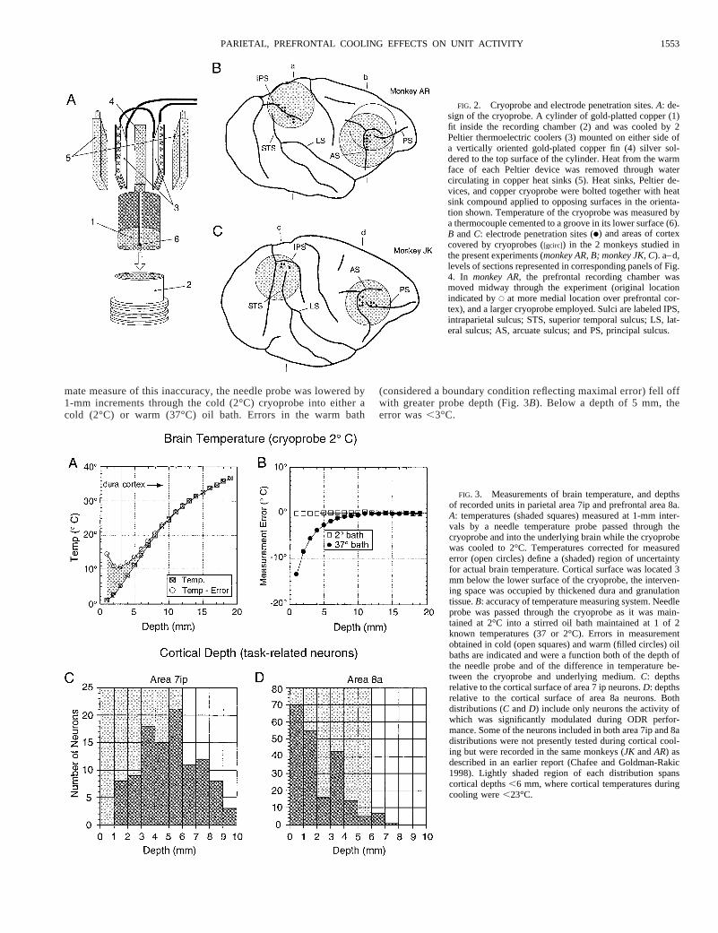
mate measure of this inaccuracy, the needle probe was lowered by1-mm increments through the cold (2°C) cryoprobe into either acold (2°C) or warm (37°C) oil bath. Errors in the warm bath
(considered a boundary condition reflecting maximal error) fell offwith greater probe depth (Fig. 3B). Below a depth of 5 mm, theerror was,3°C.
FIG. 2. Cryoprobe and electrode penetration sites.A: de-sign of the cryoprobe. A cylinder of gold-platted copper (1)fit inside the recording chamber (2) and was cooled by 2Peltier thermoelectric coolers (3) mounted on either side ofa vertically oriented gold-plated copper fin (4) silver sol-dered to the top surface of the cylinder. Heat from the warmface of each Peltier device was removed through watercirculating in copper heat sinks (5). Heat sinks, Peltier de-vices, and copper cryoprobe were bolted together with heatsink compound applied to opposing surfaces in the orienta-tion shown. Temperature of the cryoprobe was measured bya thermocouple cemented to a groove in its lower surface (6).B andC: electrode penetration sites (●) and areas of cortexcovered by cryoprobes ([gcirc]) in the 2 monkeys studied inthe present experiments (monkey AR, B; monkey JK, C). a–d,levels of sections represented in corresponding panels of Fig.4. In monkey AR,the prefrontal recording chamber wasmoved midway through the experiment (original locationindicated byE at more medial location over prefrontal cor-tex), and a larger cryoprobe employed. Sulci are labeled IPS,intraparietal sulcus; STS, superior temporal sulcus; LS, lat-eral sulcus; AS, arcuate sulcus; and PS, principal sulcus.
FIG. 3. Measurements of brain temperature, and depthsof recorded units in parietal area 7ip and prefrontal area 8a.A: temperatures (shaded squares) measured at 1-mm inter-vals by a needle temperature probe passed through thecryoprobe and into the underlying brain while the cryoprobewas cooled to 2°C. Temperatures corrected for measurederror (open circles) define a (shaded) region of uncertaintyfor actual brain temperature. Cortical surface was located 3mm below the lower surface of the cryoprobe, the interven-ing space was occupied by thickened dura and granulationtissue.B: accuracy of temperature measuring system. Needleprobe was passed through the cryoprobe as it was main-tained at 2°C into a stirred oil bath maintained at 1 of 2known temperatures (37 or 2°C). Errors in measurementobtained in cold (open squares) and warm (filled circles) oilbaths are indicated and were a function both of the depth ofthe needle probe and of the difference in temperature be-tween the cryoprobe and underlying medium.C: depthsrelative to the cortical surface of area 7 ip neurons.D: depthsrelative to the cortical surface of area 8a neurons. Bothdistributions (C andD) include only neurons the activity ofwhich was significantly modulated during ODR perfor-mance. Some of the neurons included in both area 7ip and 8adistributions were not presently tested during cortical cool-ing but were recorded in the same monkeys (JK andAR) asdescribed in an earlier report (Chafee and Goldman-Rakic1998). Lightly shaded region of each distribution spanscortical depths,6 mm, where cortical temperatures duringcooling were,23°C.
1553PARIETAL, PREFRONTAL COOLING EFFECTS ON UNIT ACTIVITY

Statistical analysis of cooling effects on neuronal activity
Trials were segregated according to cue direction and cryoprobetemperature. Then within each group of trials, neuronal firing rateswere measured in five time windows, within the cue, delay, early-saccade, late-saccade, and intertrial periods. The cue and delay win-dows were coextensive with the corresponding 500 and 3,000 msperiods in the ODR trial. The boundaries of early (100–400 ms afterfixation target offset)- and late (400–700 ms after fixation targetoffset)-saccade period windows were adjusted to coincide with theaverage timing and duration of pre- and postsaccadic neuronal activity(Chafee and Goldman-Rakic 1998). These windows in the saccadeperiod were not defined relative to saccade initiation because vari-ability in the on-line recognition of saccades could vary the number oftrials recognized in a minority number of cases, and the ANOVA weemployed required a constant number of trials across each of therepeated measures (trial periods). The window in the intertrial periodwas the last 2,000 ms of the 2,500-ms intertrial interval (in a minorityof the data, a shorter 2,000-ms intertrial interval occasionally wasincluded, and the window spanned the entire period). The monkeywas free to make eye movements during the intertrial period, and thisoccasionally may have included the centering saccade the monkeymade after reward delivery (typically this saccade was completedwithin 500 ms of reward and so would not be included in the bulk ofthe data with the longer intertrial interval).
Thus three factors described each neuronal firing rate measurement;these were task period (5 levels), cue direction (8 levels), and cryo-probe temperature (2 levels, this analysis was limited to the firing ratesobserved while the brain was at normal temperature before coolingwas initiated, and firing rates observed during the subsequent set oftrials administered while cooling was in effect). A three-way repeatedmeasure ANOVA (as implemented in the SYSTAT computer statis-tics package) was used to analyze the data. Task period was treated asa repeated measure. If theF statistic for the main effect of trial periodin the overall analysis was significant, the neuron was defined astask-related. In these cases, additional tests isolated which trial peri-ods contained significantly modulated activity. Four planned compar-isons contrasted the mean firing rates in cue, delay, and early- andlate-saccade periods each to the intertrial interval. Depending onwhich of these yielded significance, neurons were assigned a combi-nation of C (cue), D (delay), or S (either early or late saccade)designations. If theF statistic for the main effect of temperature in theoverall analysis was significant, the significance of this effect for eachof the repeated measures was examined to determine whether cryo-probe temperature impacted firing rates in each trial period. An alphalevel of P #0.05 was employed throughout. The analysis was limitedfurther only to those trial periods in which neurons exhibited signif-icantly modulated activity. Thus cooling effects were identified ascases where two conditions were met; neuronal firing rates within agiven trial period were significantly different from the intertrial inter-val and the main effect of temperature was significant on firing rateswithin the same trial period. In this way, the analysis was focused oncooling induced changes in task-related activity. The significance ofthe main effect of temperature on activity within the intertrial periodassessed whether cooling had an overall effect on background activity.This was determined for each neuron. The magnitude of coolingeffects was defined as the ratio of the mean firing rate during a giventask epoch when the brain was cold, to the mean firing rate during thesame epoch when the brain was warm (after Sandell and Schiller1982). This ratio was,1 when cooling lowered firing rates and.1when cooling elevated firing rates. Recovery of cooling effects wasassessed in a separate three-way repeated measures ANOVA directlyanalogous to the analysis described in the preceding text, with theexception that neuronal firing rates during cooling were comparedwith firing rates after the brain had been returned to normal temper-ature.
Latencies of neuronal activation were defined with the method of
MacPherson and Aldridge (1979) based on confidence intervals es-tablished around a spike density function of each neuron’s activity.We employed Gaussian curves to measure the spatial tuning ofneuronal activity. Within each trial period containing significantlymodulated activity, the mean neuronal firing rate was measured acrossthe eight cue/saccade directions tested. Then a reiterative curve-fittingalgorithm determined the parameters defining the Gaussian curve thatbest fit these eight mean firing rates (separate fits were made to datacollected during warm and cold conditions). Only cases in which thecurve fitting procedure provided anF-statistic significant at theP#0.05 level were included in subsequent analyses. Further details ofboth latency and spatial tuning analyses were described previously(Chafee and Goldman-Rakic 1998). Changes in the spatial tuning ofneuronal activity were defined as the difference in the best direction orwidth parameters of the Gaussian tuning functions fit to warm andcold data sets for a given neuron. Two separate two-way analyses ofvariance then were conducted on the populations of these differencemeasures each using trial period and cortical area as factors. Thisaddressed whether the mean shift in either spatial tuning parametervaried depending on which cortical area was cooled (main effect ofarea), and which task period was considered (main effect of trialperiod).
R E S U L T S
Database
A total of 105 neurons were isolated and their activityrecorded within one cortical area as the monkeys completedtwo full sets of ODR trials, one conducted while the othercortical region remained at normal body temperature and asecond set while the temperature of that region was lowered.Forty three of these neurons were located in parietal cortex(Fig. 2, B andC), and their activity collected while prefrontalcortex was subjected to cold. The majority of these (31 neu-rons) were located in parietal area 7ip (LIP) in the lateral bankof the intraparietal sulcus (Fig. 4,A andC), but a few also werelocated in area 7a (7 neurons) in the inferior parietal gyrus andalso in area DP in the dorsal prelunate gyrus (5 neurons).Conversely, 62 of the neurons in the database were located inprefrontal cortex (Fig. 2,B andC), the majority of which (52neurons) was located in area 8a in the anterior bank of thearcuate sulcus (Fig. 4,B andD), in the approximate location ofthe FEF, whereas the remainder (10 neurons) was located inthe principal sulcus, in area 46.
The results of the repeated-measure ANOVA indicated that90% of neurons within the database were task responsive anddirectionally selective. (i.e., the main effects of trial epoch andcue direction, or their interaction, were significant in the anal-ysis, see Table 1). Using a set of planned comparisons (METH-ODS), the present analysis recognized seven types of neuron onthe basis of whether significant activity modulation occurredduring the cue, delay, and/or saccade periods or in combina-tions of these periods. Neurons tested during cooling in bothparietal and prefrontal cortex included examples from most ofthese types (Fig. 5,A andB).
Extent of cryoinactivation
Cortical cooling involved a group of areas in parietal andprefrontal cortex (Fig. 2,B andC), with the coldest tempera-tures existing within the more superficial cortex immediatelybeneath the cryoprobes (Figs. 3A and 4,A–D). Progressivelywarmer brain temperatures occurred at greater depths. At a
1554 M. V. CHAFEE AND P. S. GOLDMAN-RAKIC

cryoprobe temperature of 2°C, cortical temperatures at depthsof 3, 6, and 9 mm beneath the surface of the brain were 14, 23,and 29°C, respectively (Figs. 3A and 4, A–D). The lateralspread of cooling was not measured in the present experimentsbut was likely to extend beyond the boundaries of the cryo-probe (Fuster and Bauer 1974). Thus cooling established agradient of subnormal temperature and functional inactivationacross considerable portions of parietal and prefrontal cortex.Changes in the activity of area 7ip neurons resulted thereforenot only from the cooling of area 8a, but of an expanse ofprefrontal cortex which included it. Similarly, changes in theactivity of area 8a neurons resulted from the cooling of aportion of parietal cortex that included area 7ip but was notlimited to this cortical area. The degree to which the volume ofcooled cortex involved areas 7ip and 8a can be estimated fromthe depths of neurons in these areas the activity of which wassignificantly modulated during ODR performance (Chafee andGoldman-Rakic 1998) and from the temperature measurements
made at these depths (Fig. 3A). Most ODR task-related neuronsin monkeys JKandARsampled in area 7ip (Fig. 3C) and nearlyall of those sampled in area 8a (Fig. 3D) were located atcortical depths,6 mm (lightly shaded region, Fig. 3,C andD),where the temperature of the cortex was,23°C (Fig. 3A). Ithas been shown that whereas colder temperatures are requiredto block neuronal responsiveness entirely, the response of V1neurons to an optimal visual stimulus is reduced by;80% ata temperature of 20°C (Girard and Bullier 1989). Thus coolingin the present experiments would be expected to substantiallyreduce the activity and output of a large portion of the neuronsin parietal area 7ip and prefrontal area 8a. Because neurons inarea 8a that were driven during ODR performance were moresuperficially located, the degree of functional inactivationachieved in area 8a was likely to exceed that in area 7ip.
In parietal cortex, cooling involved a group of cortical areas,which included, in addition to area 7ip in the lateral bank of theintraparietal sulcus, portions of area 5 in monkey AR (Fig. 2B),
FIG. 4. Reconstruction of levels of coolingand recording sites within the brain.●, locationsof a subset of the neurons in the present data-base.A–D: sections through the brain beneaththe cryoprobes in parietal (A andC) and prefron-tal (B andD) cortex, inmonkeys AR(A andB),andJK (C andD). Units were recorded primarilyfrom the cortex of the lateral bank of the intrapa-rietal sulcus (IPS) in parietal cortex (A andC),and from the anterior bank of the arcuate sulcus(AS) in prefrontal cortex (B andD). Brain tem-peratures measured inmonkey AR(A andB) areindicated at 3-mm intervals. Same temperaturelevels are projected onto the reconstruction ofmonkey JK(C andD, brain temperature was notmeasured in this animal). Axes underneath eachcryoprobe are presented for scale (1-mm divi-sions).
TABLE 1. Neurons with significant main effects and their interactions
Direction Temperature EpochDirection 3Temperature
Direction 3Epoch
Temperature3Epoch
Direction 3Temperature3
Epoch
ParietalSignificant 41 33 40 8 42 27 18Not Significant 2 10 3 35 1 16 25Total 43 43 43 43 43 43 43
PrefrontalSignificant 56 44 61 13 58 26 16Not Significant 6 18 1 49 4 36 46Total 62 62 62 62 62 62 62
Numbers of neurons for which cue direction, cortical temperature, and oculomotor delayed response (ODR) trial epoch, or their interaction, were significantfactors (P # 0.05) effecting firing rate as determined by a three-way repeated-measures ANOVA of neuronal activity obtained in parietal and prefrontal cortex.
1555PARIETAL, PREFRONTAL COOLING EFFECTS ON UNIT ACTIVITY

as well as the dorsal prelunate gyrus (including area DP) andparts of striate cortex inmonkey JK(Fig. 2C). Parts of area 7awere cooled in both animals. In prefrontal cortex, coolingincluded not only area 8a in the anterior bank of the arcuatesulcus but also portions of area 46 in the posterior principalsulcus, posterior portions of the dorsolateral and inferior pre-frontal convexities, and area 6 (Fig. 2,B andC).
Effect of cold on ODR performance
Cooling prefrontal cortex produced an impairment in mem-ory-guided saccade performance (Fig. 1C). In a two-wayANOVA on the mean error distance between the endpoint ofmemory-guided saccades and their respective visual cues (withcue direction and cortical temperature as factors), error dis-tance increased during cryogenic depression of prefrontal cor-tex–both the main effect of temperature (Ftemp5149.47, df51, P , 0.001) and the interaction between temperature anddirection (Ftemp*dir 5 15.01, df5 10, P , 0.001) were signif-icant in this analysis. This behavioral deficit was confined
largely to saccades toward targets appearing contralateral to thecooled prefrontal hemisphere (Fig. 1C). During cooling, mon-keys would maintain fixation of the central target until itsdisappearance but then frequently make inaccurate saccades(sometimes into the visual hemifield opposite the target). Thesetrials were interspersed with others in which comparativelyaccurate saccades were made. Cooling parietal cortex produceda much smaller impact on the accuracy of memory-guidedsaccades (Fig. 1D), failing to significantly effect the mean errorbetween memory-guided saccade endpoints and their targets(Ftemp 5 3.41, df5 1, P 5 0.065;Ftemp*dir 5 1.50, df5 10,P 5 0.131).
Effect of cold on neuronal firing rate
Cooling affected the intensity of activation of neurons invarious ODR trial epochs in both parietal and prefrontal cortex.The parietal area 7ip neuron illustrated in Fig. 6A exhibitedsustained delay period activation when the cue appeared in theupper (90°) and upper left (135°) locations before the prefron-tal cortex was cooled. When the activity of the same neuronwas recorded with the prefrontal cortex cooled, delay periodactivity was attenuated sharply in the 135° direction (Fig. 6B;Ftemp 5 45.99, df5 1, P , 0.001), and early-saccade periodactivity also was suppressed significantly (Ftemp5 25.69, df51, P , 0.001). The activity in the cue period also was affectedbut less strongly and not significantly (Ftemp 5 0.357, df5 1,P 5 0.551). The delay period activity of nine additional pari-etal area 7ip neurons significantly changed during prefrontalcooling (4 neurons suppressed, 5 augmented; Fig. 9C).
Comparable effects were observed among prefrontal neu-rons while cooling parietal cortex. Figure 7A illustrates aneuron in the principal sulcus (area 46) recorded during cryo-genic inactivation of the parietal cortex. Like the parietalneuron described in the preceding text, the activity of thisprefrontal neuron was reduced significantly by cooling (Fig.7B), in this instance during the cue period (Ftemp5 99.29, df51, P , 0.001). There was also a general suppression of thelevel of activity during the intertrial interval in this case(Ftemp5 108.60, df5 1, P , 0.001). Six additional prefrontalneurons (1 in area 46 and 5 in area 8a) similarly exhibitedreduced cue period activation when parietal cortex was cooled,whereas augmented cue period activation was observed in 11prefrontal area 8a neurons (Fig. 9B). Thus whereas the illus-trated cooling effects fit well those that prefrontal mnemonicand parietal visuospatial functions would predict (namely thereduction of cue period activity in prefrontal neurons and delayperiod activity in parietal neurons), counter examples wereequally numerous. For example, delay period activity of sev-eral prefrontal area 8a neurons was altered by parietal cooling(9 neurons, Figs. 9D and 11,E–H), and prefrontal coolingaltered the activation of parietal 7ip neurons in response to thevisual stimulus (9 neurons, Fig. 9A). Neurons whose primaryactivation during ODR performance occurred during the sac-cade period also were impacted by cooling either area (17parietal 7ip neurons, 25 prefrontal 8a neurons, Fig. 9,E andF).Thus prefrontal cooling could reduce the presaccadic activationof neurons in area 7ip (Fig. 8,A andB; Ftemp5 34.54, df5 1,P , 0.001), and cooling parietal cortex could augment thepresaccadic activation of neurons in prefrontal area 8a (Fig. 8,C andD; Ftemp 5 34.89, df5 1, P , 0.001). In general, both
FIG. 5. Distribution of neurons recorded during cortical cooling in thepresent database by response class.A: C, D, and S designations indicateneurons the activity of which was elevated significantly relative to the baselinelevel during cue, delay, and saccade ODR task periods, respectively, asassessed by a 3-way repeated-measures ANOVA (SeeMETHODS. Combinationsof letters indicate neurons the activity of which was elevated during combi-nations of corresponding task periods).B: total numbers of neurons the activityof which was elevated significantly in each task period collapsed across thecombinations indicated inA.
1556 M. V. CHAFEE AND P. S. GOLDMAN-RAKIC

enhanced and suppressed levels of activation were observed inall task periods after the transient inactivation of either corticalarea (Fig. 9,A–H).
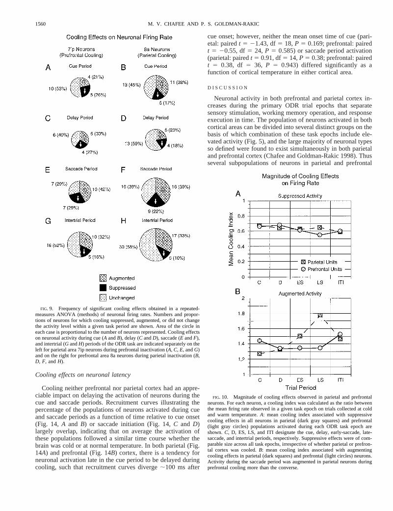
MAGNITUDE AND FREQUENCY OF EFFECTS. In the case ofsignificant suppressive cooling effects, the mean coolingindex was quite constant both across trial period and corticalarea (Fig. 10A). In this population of neurons, firing rateswere reduced by;40% of their level of activation seen atnormal brain temperature. It was not the case that coolingone area produced stronger suppressive effects than coolingthe other or that stronger suppressive effects were seen insome trial periods. In a two-way ANOVA of the cooling
indices of suppressed neurons (cortical area by trial period),neither the main effects of area or trial period nor the interactionbetween them were significant (Farea5 0.10, df5 1, P 5 0.75;Fperiod5 0.37, df5 4, P 5 0.83;Farea*period5 0.19, df5 4, P 50.94). Similarly, in the case of enhancing effects, neither the maineffects of area (Farea5 0.89, df5 1, P 5 0.35) or trial period(Fperiod5 1.47, df5 4, P 5 0.22) were significant, although theinteraction term was (Farea3period5 2.72, df5 4,P 5 0.03). Bothearly- and late-saccade period activities were more strongly aug-mented in parietal units under prefrontal cooling than the converse(Fig. 10B).
Most often, cooling impacted firing rates in a given trial
FIG. 6. Delay period activity recordedfrom a neuron in parietal area 7ip duringcooling of prefrontal cortex. Point of en-trance of the electrode penetration fromwhich the neuron was recorded is indicated inthe central drawings of the right cerebralhemisphere. Circle over prefrontal cortex de-notes the area of brain underneath the pre-frontal cryoprobe. Eight rasters in each group(A and B) show activity on ODR trials inwhich the cue appeared in a single location,corresponding to 1 of the 8 cue locationsillustrated in Fig. 1B, each indicated by thedirectional coordinate at thetop left. In eachraster, the duration of the cue (dark gray bar)corresponds to the 500-ms interval betweenthe 1st and 2nd vertical lines. Duration of thedelay period (light gray bar) corresponds tothe 3-s interval between the 2nd and 3rdvertical lines. Memory-guided saccade wasmade within the 500-ms immediately afterthe end of the delay period (3rd vertical line).A spike density function formed by convolu-tion of the corresponding histogram with aGaussian function accompanies each raster.A: activity of this parietal area 7ip neuronwhile prefrontal cortex was maintained atnormal temperature. Activity increased afterthe onset of the cue in the upper (90°) andupper left (135°) locations, and persisted af-ter cue offset throughout the delay period,ending with an additional burst in activityimmediately before initiation of the memory-guided saccade. Later activation on 270 and315° trials is most likely due to the centeringsaccade made in the intertrial interval afterthese trials in anticipation of the reappear-ance of the fixation target.B: activity of thesame parietal area 7 ip neuron recorded whilethe cryoprobe over prefrontal cortex wascooled to 2°C. Delay period activity, partic-ularly in the 135° location, is sharply atten-uated.
1557PARIETAL, PREFRONTAL COOLING EFFECTS ON UNIT ACTIVITY

epoch equally across trials of different cue and saccade direc-tion. This is indicated by the fact that the interaction betweentemperature and direction in the ANOVA applied to neuronalfiring rates (METHODS) was significant in a minority of neurons(19% of parietal neurons, 21% of prefrontal neurons, Table 1).On the other hand, a significant interaction was found betweencortical temperature and trial epoch in a larger proportion ofthe sample, in 62% of parietal neurons, and 42% of prefrontalneurons (Table 1). This finding indicates that firing rates indifferent task epochs changed by different amounts. Coolingeffects such as these, as well as others that were observedacross multiple cooling cycles (Fig. 11), would be difficult toascribe to spurious sources such as changes in the overall level
of activity of a neuron or changes in the quality of the isolationof its activity. Comparable fractions of the neurons activatedduring the cue, delay, or saccade periods of the ODR task hadtheir activity augmented or suppressed irrespective of whichcortical area was cooled (Fig. 9,A–H). Thus transient inacti-vation of parietal and prefrontal cortex produced largely sym-metrical effects on the patterns of neuronal activity distributedbetween them.
REVERSIBILITY OF EFFECTS. The activity of 52 neurons wasrecorded after the brain had been returned to normal temper-ature. A repeated-measures ANOVA similar to the originalanalysis (METHODS) was performed on these neurons to deter-
FIG. 7. Cue period activity recorded froma neuron in prefrontal area 8a during coolingof parietal cortex (conventions as in Fig. 6).A: firing rate increases during the cue period,and trails into the delay period, when the cueappears in upper (90°) and upper left (135°)locations.B: activity of the same neuron afterparietal cortex had been cooled. Increase inactivity during the cue period is reduced.
1558 M. V. CHAFEE AND P. S. GOLDMAN-RAKIC

mine whether changes seen under cooling reversed on warmingthe brain. Nine of the 33 neurons (27%) whose activity wasaugmented by cooling, and 9 of the 19 neurons (47%) whoseactivity was suppressed by cooling, exhibited significantchanges in activity in the opposite direction when the brain wasreturned to normal temperature. Thus significant recovery wasmore common among those neurons suppressed by cooling. Inother neurons, changes in activity seen on cooling did notimmediately reverse on warming the brain within the period oftime over which that activity was sampled. In some neurons,changes in activity were consistently observed across multiplecooling cycles. For example, cooling prefrontal cortex had areversible and repeatable impact on the activity of the neuronlocated in parietal area 7ip (Fig. 11,A–D, same neuron as inFig. 6). Before cooling, the firing rate of this neuron waselevated during the delay period (Fig. 11A). During the firstcooling of prefrontal cortex, the delay period activation wasreduced (Fig. 11B) and then after the brain was warmed backto normal temperature, rebounded to a level greater than thatoriginally seen before cooling was initiated (compare Fig. 11,C and A). Delay period firing rates were again suppressed asecond time when prefrontal cortex was again cooled (Fig.11D). The neuron in prefrontal area 8a exhibited a tonicexcitation during the delay period (Fig. 11E) that was attenu-ated when parietal cortex was cooled (Fig. 11F), regained itsoriginal strength when parietal cortex was warmed (Fig. 11G),and was attenuated again when parietal cortex was cooled asecond time (Fig. 11H).
Cooling effects on spatial tuning
The technique employed to quantify the spatial tuning ofneuronal activity (METHODS) provided for each neuron the pa-rameters of the Gaussian curve representing the best fit to themean firing rates observed in each of the eight cue/saccade
directions tested. Significant Gaussian fits were obtained to theactivity of 19 parietal neurons and 32 prefrontal neurons in atleast one ODR task period in both warm and cold conditions.Comparison of tuning curves fit with the activity of these 51neurons within a given trial period across temperature indi-cated whether cooling affected the breadth of tuning (Td pa-rameter of the Gaussian equation) or the best direction (Dparameter) of that activity. The best directions of fits to cue,delay, and saccade period activities shifted,10° in the largemajority of neurons (Fig. 12A, neurons contributed multipleshift values to this distribution if they were activated in.1ODR task epoch). However, larger shifts occasionally wereseen. For example, after cooling prefrontal cortex, the bestdirection of the saccade period activity of an area 7ip parietalneuron shifted counterclockwise (leftward) by 28° due largelyto opposite changes in mean firing rate observed at off-peak 45and 135° directions (Fig. 13A). In a neuron in prefrontal area8a, a 26° clockwise (rightward) shift in the preferred directionof saccade period activity was seen (Fig. 13C). Comparing thesize of the shifts in best direction across parietal and prefrontalcooling in a two-way ANOVA with cortical area and taskepoch as factors, the main effect of cortical area was significant(Farea5 5.08, df5 1, P 5 0.027), indicating that larger shiftsin best directions were associated with cooling parietal thanprefrontal cortex (Fig. 12A). The size of the shift in bestdirection did not vary with task period (Fepoch5 1.89, df5 2,P 5 0.157). Cooling also could affect the width of tuning (Tdparameter; Fig. 12B), and a minority of neurons exhibitedconsiderable shifts. The best direction of the saccade periodactivity of the illustrated area 8a neuron remained constant asthe width of tuning changed markedly (Fig. 13B). Coolingparietal and prefrontal cortex did not differentially impact thismeasure (Farea 5 0.64, df 5 1, P 5 0.424), which wascomparable across task epoch (Fepoch 5 0.32, df 5 1, P 50.724).
FIG. 8. Saccade period activity recordedfrom neurons in parietal area 7ip (A andB) andprefrontal area 8a (C andD). Trials in each ofthe 8 different cue directions are displayedtogether in each raster and spike density func-tion. Other conventions as in Fig. 6.A andC:neuronal activity recorded in area 7ip (A) andarea 8a (C) when the alternate cortical area wasat normal temperature. In both cases, bursts inactivity that are evident after the end of thedelay period precede the initiation of the mem-ory-guided saccade.B and D: activity of thesame 2 neurons after the remote cortical areahad been cooled. In the case of the parietalneuron, the saccade related burst is markedlyreduced (B). In the case of the prefrontal neu-ron, saccade period activity is augmented (D).
1559PARIETAL, PREFRONTAL COOLING EFFECTS ON UNIT ACTIVITY
Cooling effects on neuronal latency
Cooling neither prefrontal nor parietal cortex had an appre-ciable impact on delaying the activation of neurons during thecue and saccade periods. Recruitment curves illustrating thepercentage of the populations of neurons activated during cueand saccade periods as a function of time relative to cue onset(Fig. 14, A and B) or saccade initiation (Fig. 14,C and D)largely overlap, indicating that on average the activation ofthese populations followed a similar time course whether thebrain was cold or at normal temperature. In both parietal (Fig.14A) and prefrontal (Fig. 14B) cortex, there is a tendency forneuronal activation late in the cue period to be delayed duringcooling, such that recruitment curves diverge;100 ms after
cue onset; however, neither the mean onset time of cue (pari-etal: pairedt 5 21.43, df5 18, P 5 0.169; prefrontal: pairedt 5 20.55, df5 24, P 5 0.585) or saccade period activation(parietal: pairedt 5 0.91, df5 14,P 5 0.38; prefrontal: pairedt 5 0.38, df 5 36, P 5 0.943) differed significantly as afunction of cortical temperature in either cortical area.
D I S C U S S I O N
Neuronal activity in both prefrontal and parietal cortex in-creases during the primary ODR trial epochs that separatesensory stimulation, working memory operation, and responseexecution in time. The population of neurons activated in bothcortical areas can be divided into several distinct groups on thebasis of which combination of these task epochs include ele-vated activity (Fig. 5), and the large majority of neuronal typesso defined were found to exist simultaneously in both parietaland prefrontal cortex (Chafee and Goldman-Rakic 1998). Thusseveral subpopulations of neurons in parietal and prefrontal
FIG. 10. Magnitude of cooling effects observed in parietal and prefrontalneurons. For each neuron, a cooling index was calculated as the ratio betweenthe mean firing rate observed in a given task epoch on trials collected at coldand warm temperature.A: mean cooling index associated with suppressivecooling effects in all neurons in parietal (dark gray squares) and prefrontal(light gray circles) populations activated during each ODR task epoch areshown.C, D, ES, LS, and ITI designate the cue, delay, early-saccade, late-saccade, and intertrial periods, respectively. Suppressive effects were of com-parable size across all task epochs, irrespective of whether parietal or prefron-tal cortex was cooled.B: mean cooling index associated with augmentingcooling effects in parietal (dark squares) and prefrontal (light circles) neurons.Activity during the saccade period was augmented in parietal neurons duringprefrontal cooling more than the converse.
FIG. 9. Frequency of significant cooling effects obtained in a repeated-measures ANOVA (methods) of neuronal firing rates. Numbers and propor-tions of neurons for which cooling suppressed, augmented, or did not changethe activity level within a given task period are shown. Area of the circle ineach case is proportional to the number of neurons represented. Cooling effectson neuronal activity during cue (A andB), delay (C andD), saccade (E andF),and intertrial (G andH) periods of the ODR task are indicated separately on theleft for parietal area 7ip neurons during prefrontal inactivation (A, C, E,andG)and on the right for prefrontal area 8a neurons during parietal inactivation (B,D, F, andH).
1560 M. V. CHAFEE AND P. S. GOLDMAN-RAKIC

cortex appear to be physiologically synchronized in the sensethat the levels of their activity rise and fall together throughoutthe ODR trial. The present experiments were intended in partto address the mechanisms through which this parallel activa-tion might come about, specifically whether reciprocal neuro-transmission between these cortical areas may be involved.The known projection between parietal neurons in area 7ip andprefrontal neurons in area 8a (Andersen et al. 1990a; Cavadaand Goldman-Rakic 1989; Schall et al. 1995; Stanton et al.1995) implies that activity is exchanged between these neuro-nal populations during ODR performance but not whether thisexchange is symmetrical or asymmetrical nor whether itequally includes neuronal activation during the cue, delay, andsaccade epochs of the task. As some of these signals (specif-ically neuronal activity sustained throughout the delay period)appear to effect the retention of a spatial datum defined by thelocation of a visual stimulus in working memory, the questionis of interest as to how prefrontal cortex may engage, and inturn be engaged by, other cortical areas as working memory
operates. Our results included the following observations:1)neurons the activation of which occurred during periods ofsensory input, working memory operation, or saccade execu-tion were equally vulnerable to the effects of cooling.2) Theeffects of cooling parietal versus prefrontal cortex were largelyequivalent (with a few exceptions discussed in the followingtext). 3) The average change in neuronal activity associatedwith cooling was on the order of 40% of the firing rateobserved in each trial epoch at normal temperature and tendedto be equally strong across all cue/saccade directions tested.And 4) both the suppression and enhancement of activationwere observed. How these findings might bear on the nature ofthe interaction between prefrontal and posterior parietal neu-rons during the ODR task will be considered in turn. It shouldbe noted that using the current technique it is not possible toconclude whether cooling effects were mediated by directmonosynaptic projections between parietal and prefrontal neu-rons or via multisynaptic pathways involving other cortical orsubcortical areas.
Neuronal activity in all ODR task epochs was affected
The similarity that parietal and prefrontal neuronal activitiesachieve during ODR performance might arise through any oneof several different mechanisms. One possibility is that thissimilarity is the consequence of a redundancy of oculomotor
FIG. 12. Effects of cold on spatial tuning. Gaussian curves fit to the meanfiring rate observed across cue direction in a given task epoch provided ameasure of the width (Td parameter) and best direction (D parameter) ofspatial tuning. For each neuron, separate Gaussian functions were fit to datafrom trials collected at normal brain temperature and during cooling, and theabsolute difference between the best directions of these functions (D param-eters) was determined along with the absolute difference between their widths(Td parameters). Data obtained from 17 parietal and 32 prefrontal neurons areillustrated.A: distribution of the changes seen in best directions of parietalneurons (n) and prefrontal neurons (u) associated with cooling. Best directionsof the large majority of neurons changed,10°. B: distribution of the changesseen in the width of tuning of parietal neurons (n) and prefrontal neurons (u)during cooling.
FIG. 11. Repeatability of cooling effects. Activity each of the 2 illustratedneurons was elevated during the delay period on preferred direction trials (onlypreferred direction trials are shown). Trials in each raster are segregated into4 groups, collected during a particular temperature condition. Four groupscorrespond a warm (A andE), cold (B andF), warm (C andG), and cold (Dand H) temperature sequence. Pairs of spike density functions (SDF) beloweach raster compare activation on the 1st (top) and 2nd (bottom) coolingcycles. In each panel, the SDF at normal temperature is shaded, whereas theSDF during cooling is open. Other conventions as in Fig. 6.A–D: delay periodactivity of an area 7ip neuron observed after the cue appeared in the 135°location was repeatedly suppressed through 2 cycles of cooling prefrontalcortex (the activity of this neuron across all target locations is depicted in Fig.6). E–H: similar observations in a prefrontal area 8a neuron.
1561PARIETAL, PREFRONTAL COOLING EFFECTS ON UNIT ACTIVITY

function between parietal and prefrontal cortex, which generatesimilar patterns of neuronal activity in physiological isolationwithout requiring or involving the exchange of neuronal sig-nals between them. As this would predict that cooling onecortical area ought to have negligible effect on the other, thepresent data indicate that a physiological interaction betweenparietal and prefrontal neurons takes place during ODR per-formance and that this interaction contributes to the modulationof activity that neurons in both areas exhibit. In this manner,the physiological operations performed by the two corticalareas appear to be linked. It was observed further that thereduction of output that could be assumed to accompany cool-ing of one of the two cortical areas produced equal effects onneuronal activation during cue, delay, and saccade epochsconcurrently recorded in the remaining cortical area (Figs. 9and 10). This was evidence that the cortical output suppressedby cooling was normally active during each of these ODR taskepochs to contribute to the modulation of neuronal activitytaking place in the other cortical area during those same timesin the ODR trial. Thus the present data favor the hypothesisthat neural signals associated with sensory input, working
memory operation, and saccade execution in both parietal andprefrontal cortex all participate in cortical output. The hetero-geneity of output signals suggested by these data are consistentwith the results of Pare´ and Wurtz (1997), who have combinedantidromic activation and unit recording to demonstrate thattwo physiologically distinct classes of 7ip neuron, one activeduring the cue period, and another the activity of which ex-tended into the delay and saccade periods of a memory-guidedsaccade task, both give rise to axons projecting to the superiorcolliculus.
The present results are in agreement with the data presentedby Quintana and colleagues (1989) in their study of the effectsof parietal cooling on prefrontal unit activity during delayedmatch to sample and conditional position discrimination tasks,both of which employed the color of a cue stimulus to direct adelayed arm movement. These authors found that coolingparietal cortex significantly altered the level of activity of someprefrontal neurons in each of the cue, delay, choice, andresponse epochs comprising these tasks. Further, cooling pro-duced a mixture of increased and decreased levels of activa-tion. Our results extend these by examining the impact ofprefrontal inactivation on parietal unit activity and the use of atask employing a visuospatial cue and an oculomotor responseto investigate this system. In their study of interactions be-tween prefrontal and inferotemporal cortex, Fuster and col-leagues (1985) observed that in either cortical area, remotecortical inactivation had a significant impact on the neuronalactivity observed during cue, delay, and choice epochs of thesame delayed color match to sample task. The agreementbetween these data suggests a general pattern of intracorticalcommunication existing between prefrontal cortex and areasproviding its input during a variety of working memory-de-pendent behaviors.
Parietal and prefrontal cooling were equivalent
Parietal and prefrontal cooling were largely equivalent intheir impact on neuronal activity. Cooling had a significantimpact on cue, delay, and saccade class neurons with compa-rable frequency irrespective of which cortical area was cooled(Fig. 9). Furthermore suppressive effects were of comparablemagnitude (Fig. 10A), although saccade responses were en-hanced to a larger degree after prefrontal cooling (Fig. 10B)than the converse. Thus it did not appear to be the case that thesymmetrical patterning of neuronal activity patterns previouslydescribed (Chafee and Goldman-Rakic 1998) was achievedthrough an asymmetrical exchange of signals between parietaland prefrontal neurons. For example, it is possible that activa-tion elicited by the visual stimulus during the cue periodoriginates in parietal cortex and then is transmitted to prefron-tal cortex in a strictly feedforward direction. Along similarlines, prefrontal neurons might employ this input to generatelocally a neural signal that is sustained throughout the delayperiod that once initiated drives parietal neurons in a strictlyfeedback manner. This exchange of different neural signals infeedforward and feedback components of the reciprocal pro-jection between parietal and prefrontal neurons would have theeffect of mixing activity patterns between the two populationsthat were initially unique to one or the other. However, thepresent data do not support the possibility that activation dur-ing any individual period of the ODR task is transmitted in
FIG. 13. Examples of changes in the spatial tuning of individual neurons.Gaussian tuning functions fit to mean firing rates observed while the brain wasat normal temperature (‚) and during cortical cooling ([gcirc]). A: saccade periodactivity of a neuron in parietal area 7ip. Cooling is associated with a clockwise(leftward) shift in the best direction.B: saccade period activity of a neuron inprefrontal area 8a. In this instance, the best direction remained constant whilethe width of tuning changed (broadened).C: saccade period activity of aneuron in prefrontal area 8a. A counterclockwise (rightward) shift in the bestdirection of the neuron occurred during cooling of parietal cortex.
1562 M. V. CHAFEE AND P. S. GOLDMAN-RAKIC

only one direction between parietal and prefrontal neurons.Instead, they argue that all neuronal signals in this system areconcurrently feedforward and feedback signals and conse-quently that the input prefrontal cortex provides to parietalneurons conveys the same signals as the input from parietalcortex to prefrontal neurons. This is supported by the symme-try of the effects of cooling parietal and prefrontal cortex ontheir shared activity. That the suppression of function of pre-frontal cortex was able to modify the sustained modulation ofneuronal activity during a working memory interval in a distantcortical area (Figs. 6, 9C, and 10,A andB) indicates that therole of prefrontal cortex in working memory may be partlycarried out through its output projections to other corticalareas. It is of further interest that prefrontal cooling was effec-tive in altering the response of posterior parietal neurons to thepresentation of a visual stimulus in the present experiment.This argues for a prefrontal modulation of even early sensoryprocessing taking place in posterior sensory association cortex,where neurons appear to be driven from two sides in effect,from earlier extrastriate areas on the one hand and prefrontalcortex on the other. Similar feedback influences on neuronalactivation evoked by a visual stimulus have been described forV1 neurons, the responses of which can be suppressed bycryoinactivation of V2 (Sandell and Schiller 1982), and alsofor IT neurons, whose differential activation to the color of acue stimulus diminishes under conditions of prefrontal inacti-vation (Fuster et al. 1985). Furthermore, in split-brain mon-
keys, prefrontal feedback can drive pattern-selective visualactivity in inferotemporal neurons that have been otherwisedeprived of feedforward visual input from ipsilateral extrastri-ate cortex (Tomita et al. 1999). Such a dynamic opens apossibility that dysfunction of prefrontal cortex also mightimpact sensory processing in disease states, contributing topathology observed in schizophrenia, for example, that is as-sociated with abnormalities in both anatomic and functionalcharacteristics of prefrontal cortex (Goldman-Rakic andSelemon 1997) and also is characterized by sensory hallucina-tions.
Both enhancement and suppression were observed
In the present experiments, cooling was associated both withenhancement and suppression of the response magnitude ob-served before cooling, depending on the neuron under study. Asimilar mixture of enhanced and suppressed unit responsesafter cryoinactivation has been reported in several studies ofcorticocortical interaction between striate and extrastriate cor-tex (Girard et al. 1992; Rodman et al. 1989; Sandell andSchiller 1982), between prefrontal and inferotemporal cortex(Fuster et al. 1985), and between prefrontal and posteriorparietal cortex (Quintana et al. 1989). Corticocortical projec-tions target both pyramidal neurons and also GABAergic in-terneurons within the target area (Somogyi et al. 1998). Assuch, cooling the neurons that give rise to a corticocortical
FIG. 14. Effects of cold on neuronal latency. Latency ofneuronal activation relative to the onset of the cue (A andB) andthe initiation of the memory-guided saccade (C and D) weredetermined for each neuron. Recruitment curves show the cu-mulative percentage of the population of neurons that havebecome active at successive time points during cue and saccadeperiods in parietal (A andC) and prefrontal (B andD) cortex.AandB: activation of parietal and prefrontal neurons during thecue period. Recruitment curves overlap early in the cue periodand diverge after;120 ms with activation on warm trialspreceding that on cold trials. Trend was not significant, how-ever. C and D: activation of parietal and prefrontal neuronsduring the saccade period. Recruitment curves largely overlapin both parietal (C) and prefrontal (D) cortex.
1563PARIETAL, PREFRONTAL COOLING EFFECTS ON UNIT ACTIVITY

projection would be expected to produce a mixture of effectson target pyramidal neurons, a direct suppression of theiractivity as the level of excitatory input they received wasreduced and an indirect augmentation of activity, as excitatorydrive to interneurons was reduced and pyramidal neurons intheir vicinity were released from inhibition. AlthoughGABAergic interneurons are less numerous than pyramidalcells by;5 to 1, in the case of basket and chandelier cells, theoutput of each targets;300 surrounding pyramidal neurons(Salin and Bullier 1995). Reduced drive to a small number ofinterneurons could impact a much larger number of pyramidalcells. There is also supporting evidence that activation ofcorticocortical inputs can produce an exclusively inhibitoryinfluence in some target neurons. Intracellular recording ofmotor cortical pyramidal neurons demonstrated that in a mi-nority of cases, stimulation of corticocortical inputs from pre-motor and somatosensory cortex produced inhibitory postsyn-aptic potentials without a preceding excitatory postsynapticpotential (Ghosh and Porter 1988). In the present data, perhapsthe clearest difference between prefrontal and parietal coolingrelated to the augmentation of saccade period activities duringcooling. The degree of augmentation was stronger in this taskperiod when prefrontal cortex was cooled and parietal neuronsrecorded from than the converse (Fig. 10B).
It is also possible that cooling directly enhanced the activityof some neurons within the cooled cortical region and soincreased their output. Cooling depolarizes the resting mem-brane potential and if cooling is slight (5° below normaltemperature), higher firing rates result (Adey 1974). Neuronshave been observed to discharge repetitively at somewhatcolder temperatures (9° below normal temperature) beforeentering a state of electrical silence (Moseley et al. 1972). Thusneurons cooled to intermediate temperatures may have exhib-ited an increased level of activity, contributing to the augmen-tation of neuronal activity we observed in the target corticalregion.
Both the enhancing and depressing effects of cooling ob-served in the present experiments support the view that corti-cocortical projections within the same hemisphere, perhaps inaddition to influencing the temporal structure of spike trains,can and do directly influence the total number of spikes emittedby target neurons during a given behavioral epoch, contribut-ing therefore to changes in average firing rate over time, or ratecoding, as this feature of neuronal physiology has been termed(Shadlen and Newsome 1994).
COOLING COMMONLY MODULATED BUT DID NOT BLOCK
NEURONAL ACTIVATION. Cooling effects were common in thepresent results; firing rates of the majority of the current sampleof task-related neurons were significantly different at coldtemperatures. Some of these significant effects were nonethe-less subtle, and the general pattern of activity across the ODRtrial typically persisted through cryoinactivation. The effects ofcooling were on average640% of the activity level in a giventask epoch measured before cooling began (Fig. 10). In themonkey visual system, cryoinactivation of area V1 completelysilences neurons in areas V2–V4 the receptive fields of whichoverlap those represented in the inactivated portion of V1(Salin and Bullier 1995). However, in cases where projectionsexist to circumnavigate the blocked projection (such as the catwhere LGN projections target V2), the effects of cooling V1
are commonly less complete (Salin and Bullier 1995). In thepresent experiments, two structures in association cortex wereexamined that share reciprocal projections with 15 corticalstructures (Selemon and Goldman-Rakic 1988). Thus the net-work of input projections that could convey activity potentiallyrelevant to ODR performance into parietal and prefrontal cor-tex is broadly distributed. Among the cortical projectionsalone, this network includes inputs from area STP in the upperbank of the superior temporal sulcus (Seltzer and Pandya1984,1989) and from other extrastriate cortical areas (Barbasand Mesulam 1981; Cavada and Goldman-Rakic 1989; Huertaet al. 1987) as well as from area 7m located on the medial wallof the cerebral hemisphere (Cavada and Goldman-Rakic 1989),all of which are potential conduits for parallel visual input toprefrontal and parietal cortices initiating the chain of eventsproducing delay and ultimately saccade-related neuronal re-sponses within them.
Direct interaction between areas 7ip and 8a
The experimental technique presently employed did notlimit inactivation to parietal area 7ip and prefrontal area 8a norwas inactivation of these areas (particularly area 7ip) complete.However, the volumes of cooled cortex included substantialportions of both areas 7ip and 8a (Figs. 3 and 4). Projectionsbetween parietal area 7ip and prefrontal area 8a are strong andselective (Andersen et al. 1985a, 1990a; Barbas 1988; Cavadaand Goldman-Rakic 1989; Schall et al. 1995). Granting thetechnological limitations of the cooling method employed, thepreferential interconnection of areas 8a and 7ip suggest thatchanges in neuronal activation we observed in each corticalarea were mediated at least in part by changes that coolingimposed on the direct interaction between the two. Thus thecooling effects observed in prefrontal area 8a, for example,were much more likely to be due to partial cooling of area 7ipwith which it is directly and reciprocally connected (Cavadaand Goldman-Rakic 1989) than to partial cooling of areassurrounding 7ip (such as area 5) that are indirectly or not at allconnected to area 8a. However, cooling of some parietal areas,such as areas 7a and DP, were more likely to contribute to theobserved effects as these areas both contain neurons that aremodulated during ODR performance (Chafee and Goldman-Rakic 1998) and project to prefrontal area 8a (Andersen et al.1990a). Similarly area 46 in the principal sulcus containsneurons that are driven during ODR performance (Chafee andGoldman-Rakic 1998; Funahashi et al. 1989), and the posteriorprincipal sulcus projects to area 7ip (Cavada and Goldman-Rakic 1989). The present results can be characterized thereforeas the effect on the neuronal activity specifically within parietalarea 7ip and prefrontal 8a of cooling groups of neighboringareas in each cortical region including but not limited to thesetwo areas.
We estimate, on the basis of the depth of the recordedneurons, the measured temperature-depth relationship (Fig. 3),and a study of the direct effects of cooling in the striate cortex(Girard and Bullier 1989), that cooling suppressed neuronalactivation by$80% in the majority of neurons in parietal area7ip and prefrontal area 8a. This represents a substantial ifincomplete reduction in output. There is further evidence in anumber of systems that the physiological response to cold atthis level and beyond is graded and progressive (Gahwiler et al.
1564 M. V. CHAFEE AND P. S. GOLDMAN-RAKIC

1972; Girard and Bullier 1989; Jasper et al. 1970; Moseley etal. 1972). Deeper inactivation of parietal area 7ip or prefrontalarea 8a therefore might be expected to produce stronger effectsin more neurons but may not be expected to yield substantivelydifferent results from those presently obtained. The possibilitycannot be excluded at present, however, that an asymmetry inthe physiological effects of inactivating areas 7ip and 8a mightnot emerge if total inactivation of each area was achieved.Finally, changes in neuronal activity we observed may havebeen secondary to changes in oculomotor behavior that coolingproduced. Arguing against this interpretation of the results wasthe finding that cooling parietal and prefrontal cortex producedlargely equivalent effects on neuronal physiology but haddifferential effects on ODR performance.
The results of the present experiment agree with work inother distributed cortical networks in the visual (Bressler1995,1996; Payne et al. 1996; Salin and Bullier 1995) andmotor systems (Alexander and Crutcher 1990a,b; Ashe andGeorgopoulos 1994; Caminiti et al. 1996; Crutcher and Alex-ander 1990; Johnson et al. 1996) to indicate that patterns ofneuronal activity recorded within a single cortical area arelikely to reflect concurrent processing in many others. Taken inconjunction with anatomic data indicating the prevalence ofprojections between cortical areas (Felleman and Van Essen1991), the present data support a dynamic view in which thepatterns of neuronal activity modulation associated with be-havioral events are generated by the concerted action of groupsof interacting cortical areas. This interaction appears to involvethe flux across cortical areas of a group of shared signalsexchanged between neurons with similar patterns of activitymodulation. The present data favor the inclusion of workingmemory in such a process through which prefrontal cortex andother cortical areas interact to drive the sustained neuronaldischarge associated with the formation and maintenance ofinternal representations.
The authors thank S. Funahashi for important assistance during early phasesof this research. We thank J. Fuster for generously providing a prototypecryoprobe on which the current design was based, S. O´ Scalaidhe for insightfuldiscussions regarding this work and for assistance regarding the method ofanalysis of the data, and C. Bruce for the computer program that controlled theexperiments. We also thank G. Leydon for the development of data analysisprograms, T. Beattie, P. Pivirotto, and M. Papero for assistance with animalcare, and J. Coburn and M. Pappy for assistance in histological processing.
This work was supported by National Institute of Mental Health GrantsMH-38546 and MH-44866.
Present address of M. V. Chafee: Brain Sciences Center, Dept. of VeteransAffairs Medical Center, Minneapolis, MN 55417.
Address reprint requests to P. S. Goldman-Rakic.
Received 18 May 1999; accepted in final form 29 November 1999.
REFERENCES
ADEY, W. R. Biophysical and metabolic bases of cooling effects on corticalmembrane potentials in the cat.Exp. Neurol.42: 113–140, 1974.
ALEXANDER, G. E.AND CRUTCHER, M. D. Neural representations of the target(goal) of visually guided arm movements in three motor areas of themonkey.J. Neurophysiol.64: 164–178, 1990a.
ALEXANDER, G. E. AND CRUTCHER, M. D. Preparation for movement: neuralrepresentations of intended direction in three motor areas of the monkey.J. Neurophysiol.64: 133–150, 1990b.
ANDERSEN, R. A., ASANUMA, C., AND COWAN, W. M. Callosal and prefrontalassociational projecting cell populations in area 7A of the macaque monkey:a study using retrogradely transported fluorescent dyes.J. Comp. Neurol.232: 443–455, 1985a.
ANDERSEN, R. A., ASANUMA, C., ESSICK, G.,AND SIEGEL, R. M. Corticocorticalconnections of anatomically and physiologically defined subdivisions withinthe inferior parietal lobule.J. Comp. Neurol.296: 65–113, 1990a.
ANDERSEN, R. A., BRACEWELL, R. M., BARASH, S., GNADT, J. W.,AND FOGASSI,L. Eye position effects on visual, memory, and saccade-related activity inareas LIP and 7a of macaque.J. Neurosci.10: 1176–1196, 1990b.
ANDERSEN, R. A., ESSICK, G. K., AND SIEGEL, R. M. Encoding of spatiallocation by posterior parietal neurons.Science230: 456–458, 1985b.
ANDERSEN, R. A., ESSICK, G. K.,AND SIEGEL, R. M. Neurons of area 7 activatedby both visual stimuli and oculomotor behavior.Exp. Brain Res.67: 316–322, 1987.
ANDERSEN, R. A. AND MOUNTCASTLE, V. B. The influence of the angle of gazeupon the excitability of the light-sensitive neurons of the posterior parietalcortex.J. Neurosci.3: 532–548, 1983.
ASHE, J. AND GEORGOPOULOS, A. P. Movement parameters and neural activityin motor cortex and area 5.Cereb. Cortex4: 590–600, 1994.
BARASH, S., BRACEWELL, R. M., FOGASSI, L., GNADT, J. W., AND ANDERSEN,R. A. Saccade-related activity in the lateral intraparietal area. I. Temporalproperties; comparison with area 7a.J. Neurophysiol.66: 1095–1108,1991a.
BARASH, S., BRACEWELL, R. M., FOGASSI, L., GNADT, J. W., AND ANDERSEN,R. A. Saccade-related activity in the lateral intraparietal area. II. Spatialproperties.J. Neurophysiol.66: 1109–1124, 1991b.
BARBAS, H. Anatomic organization of basoventral and mediodorsal visualrecipient prefrontal regions in the rhesus monkey.J. Comp. Neurol.276:313–342, 1988.
BARBAS, H. AND MESULAM, M. M. Organization of afferent input to subdivi-sions of area 8 in the rhesus monkey.J. Comp. Neurol.200: 407–431, 1981.
BRESSLER, S. L. Large-scale cortical networks and cognition.Brain Res. BrainRes. Rev.20: 288–304, 1995.
BRESSLER, S. L. Interareal synchronization in the visual cortex.Behav BrainRes.76: 37–49, 1996.
BROTCHIE, P. R., ANDERSEN, R. A., SNYDER, L. H., AND GOODMAN, S. J. Headposition signals used by parietal neurons to encode locations of visualstimuli. Nature375: 232–375, 1995.
BRUCE, C. J.AND GOLDBERG, M. E. Primate frontal eye fields. I. Single neuronsdischarging before saccades.J. Neurophysiol.53: 603–635, 1985.
BUTTERS, N. AND PANDYA , D. Retention of delayed-alternation: effect ofselective lesions of sulcus principalis.Science165: 1271–1273, 1969.
CAMINITI , R., FERRAINA, S., AND JOHNSON, P. B. The sources of visual infor-mation to the primate frontal lobe: a novel role for the superior parietallobule.Cereb. Cortex6: 319–328, 1996.
CAVADA , C. AND GOLDMAN-RAKIC, P. S. Posterior parietal cortex in rhesusmonkey. II. Evidence for segregated corticocortical networks linking sen-sory and limbic areas with the frontal lobe.J. Comp. Neurol.287: 422–445,1989.
CHAFEE, M. V. AND GOLDMAN-RAKIC, P. S. Matching patterns of activity inprimate prefrontal area 8a and parietal area 7ip neurons during a spatialworking memory task.J. Neurophysiol.79: 2919–2940, 1998.
CRUTCHER, M. D. AND ALEXANDER, G. E. Movement-related neuronal activityselectively coding either direction or muscle pattern in three motor areas ofthe monkey.J. Neurophysiol.64: 151–163, 1990.
FELLEMAN, D. J.AND VAN ESSEN, D. C. Distributed hierarchical processing inthe primate cerebral cortex.Cereb. Cortex1: 1–47, 1991.
FUNAHASHI, S., BRUCE, C. J.,AND GOLDMAN-RAKIC, P. S. Mnemonic coding ofvisual space in the monkey’s dorsolateral prefrontal cortex.J. Neurophysiol.61: 331–349, 1989.
FUNAHASHI, S., BRUCE, C. J.,AND GOLDMAN-RAKIC, P. S. Visuospatial codingin primate prefrontal neurons revealed by oculomotor paradigms.J. Neuro-physiol.63: 814–831, 1990.
FUNAHASHI, S., BRUCE, C. J.,AND GOLDMAN-RAKIC, P. S. Neuronal activityrelated to saccadic eye movements in the monkey’s dorsolateral prefrontalcortex.J. Neurophysiol.65: 1464–1483, 1991.
FUNAHASHI, S., CHAFEE, M. V., AND GOLDMAN-RAKIC, P. S. Prefrontal neuronalactivity in rhesus monkeys performing a delayed anti-saccade task.Nature365: 753–756, 1993.
FUSTER, J. M. AND BAUER, R. H. Visual short-term memory deficit fromhypothermia of frontal cortex.Brain Res.81: 393–400, 1974.
FUSTER, J. M., BAUER, R. H.,AND JERVEY, J. P. Functional interactions betweeninferotemporal and prefrontal cortex in a cognitive task.Brain Res.330:299–307, 1985.
GAHWILER, B. H., MAMOON, A. M., SCHLAPFER, W. T., AND TOBIAS, C. A.Effects of temperature on spontaneous bioelectric activity of cultured nervecells.Brain Res.40: 527–533, 1972.
1565PARIETAL, PREFRONTAL COOLING EFFECTS ON UNIT ACTIVITY

GHOSH, S. AND PORTER, R. Corticocortical synaptic influences on morpholog-ically identified pyramidal neurons in the motor cortex of the monkey.J. Physiol. (Lond.)400: 617–629, 1988.
GIRARD, P. AND BULLIER, J. Visual activity in area V2 during reversibleinactivation of area 17 in the macaque monkey.J. Neurophysiol.62:1287–1302, 1989.
GIRARD, P., SALIN , P. A., AND BULLIER, J. Response selectivity of neurons inarea MT of the macaque monkey during reversible inactivation of area V1.J. Neurophysiol.67: 1437–1446, 1992.
GNADT, J. W. AND ANDERSEN, R. A. Memory related motor planning activityin posterior parietal cortex of macaque.Exp. Brain Res.70: 216–220, 1988.
GNADT, J. W.AND MAYS, L. E. Neurons in monkey parietal area LIP are tunedfor eye-movement parameters in three-dimensional space.J. Neurophysiol.73: 280–297, 1995.
GOLDMAN, P. S., ROSVOLD, H. E., VEST, B., AND GALKIN , T. W. Analysis of thedelayed-alternation deficit produced by dorsolateral prefrontal lesions in therhesus monkey.J. Comp. Physiol. Psychol.77: 212–220, 1971.
GOLDMAN-RAKIC, P. S. Circuitry of primate prefrontal cortex and regulation ofbehavior by representational memory. In:Handbook of Physiology. TheNervous System. Higher Functions of the Brain.Bethesda, MD: Am.Physiol. Soc., 1987, sect. 1, vol. V, chapt. 9, p. 373–417.
GOLDMAN-RAKIC, P. S. Topography of cognition: parallel distributed networksin primate association cortex.Annu. Rev. Neurosci.11: 137–156, 1988.
GOLDMAN-RAKIC, P. S. Cellular basis of working memory.Neuron14: 477–485, 1995.
GOLDMAN-RAKIC, P. S.AND SELEMON, L. D. Functional and anatomical aspectsof prefrontal pathology in schizophrenia (see comments).Schizophr. Bull.23: 437–458, 1997.
HUERTA, M. F., KRUBITZER, L. A., AND KAAS, J. H. Frontal eye field as definedby intracortical microstimulation in squirrel monkeys, owl monkeys, andmacaque monkeys. II. Cortical connections.J. Comp. Neurol.265: 332–361,1987.
JACOBSEN, C. F. Studies of cerebral function in primates.Comp. Psychol.Monogr.13: 1–68, 1936.
JASPER, H. H., SHACTER, D. G.,AND MONTPLAISIR, J. The effect of local coolingupon spontaneous and evoked electrical activity of cerebral cortex.Can.J. Physiol. Pharmacol.48: 640–652, 1970.
JOHNSON, P. B., FERRAINA, S., BIANCHI, L., AND CAMINITI , R. Cortical networksfor visual reaching: physiological and anatomical organization of frontal andparietal lobe arm regions.Cereb. Cortex6: 102–119, 1996.
JUDGE, S. J., RICHMOND, B. J.,AND CHU, F. C. Implantation of magnetic searchcoils for measurement of eye position: an improved method.Vision Res.20:535–538, 1980.
MACPHERSON, J. M. AND ALDRIDGE, J. W. A quantitative method of computeranalysis of spike train data collected from behaving animals.Brain Res.175:183–187, 1979.
MAZZONI, P., BRACEWELL, R. M., BARASH, S., AND ANDERSEN, R. A. Motorintention activity in the macaque’s lateral intraparietal area. I. Dissociationof motor plan from sensory memory.J. Neurophysiol.76: 1439–1456, 1996.
MOSELEY, J. I., OJEMANN, G. A., AND WARD, A. A., JR. Unit activity duringfocal cortical hypothermia in the normal cortex.Exp. Neurol.37: 152–163,1972.
MOUNTCASTLE, V. B. An organizing principle for cerebral function: the unitmodule and the distributed system. In:The Mindful Brain,edited by G. E.Edelman and V. B. Mountcastle. Cambridge, MA: MIT Press, 1978, p.7–50.
MOUNTCASTLE, V. B. The evolution of ideas concerning the function of theneocortex.Cereb. Cortex5: 289–295, 1995.
MOUNTCASTLE, V. B.Perceptual Neuroscience. The Cerebral Cortex.Cam-bridge, MA: Harvard Univ. Press, 1998, p. 224–279.
MOUNTCASTLE, V. B., LYNCH, J. C., GEORGOPOULOS, A., SAKATA , H., AND
ACUNA, C. Posterior parietal association cortex of the monkey: commandfunctions for operations within extrapersonal space.J. Neurophysiol.38:871–908, 1975.
NIKI , H. Prefrontal unit activity during delayed alternation in the monkey. I.Relation to direction of response.Brain Res.68: 185–196, 1974a.
NIKI , H. Prefrontal unit activity during delayed alternation in the monkey. II.Relation to absolute versus relative direction of response.Brain Res.68:197–204, 1974b.
NIKI , H. AND WATANABE, M. Prefrontal unit activity and delayed response:relation to cue location versus direction of response.Brain Res.105: 79–88,1976.
PARE, M. AND WURTZ, R. H. Monkey posterior parietal cortex neurons anti-dromically activated from superior colliculus.J. Neurophysiol.78: 3493–3497, 1997.
PAYNE, B. R., LOMBER, S. G., VILLA , A. E., AND BULLIER, J. Reversibledeactivation of cerebral network components (see comments).Trends Neu-rosci. 19: 535–542, 1996.
PETRIDES, M. AND PANDYA , D. N. Projections to the frontal cortex from theposterior parietal region in the rhesus monkey.J. Comp. Neurol.228:105–116, 1984.
POHL, W. Dissociation of spatial discrimination deficits following frontal andparietal lesions in monkeys.J. Comp. Physiol. Psychol.82: 227–239, 1973.
PU, X., MA, Y. AND CAI, J. A study on the effect of lesions of area 7 of theparietal cortex on the short-term visual spatial memory of rhesus monkeys(Macaca mulatta).Brain Res.600: 187–192, 1993.
QUINTANA , J.AND FUSTER, J. M. Mnemonic and predictive functions of corticalneurons in a memory task.Neuroreport3: 721–724, 1992.
QUINTANA , J. AND FUSTER, J. M. Spatial and temporal factors in the role ofprefrontal and parietal cortex in visuomotor integration.Cereb. Cortex3:122–132, 1993.
QUINTANA , J., FUSTER, J. M.,AND YAJEYA, J. Effects of cooling parietal cortexon prefrontal units in delay tasks.Brain Res.503: 100–110, 1989.
RODMAN, H. R., GROSS, C. G.,AND ALBRIGHT, T. D. Afferent basis of visualresponse properties in area MT of the macaque. I. Effects of striate cortexremoval.J. Neurosci.9: 2033–2050, 1989.
SALIN , P. A. AND BULLIER, J. Corticocortical connections in the visual system:structure and function.Physiol. Rev.75: 107–154, 1995.
SANDELL, J. H.AND SCHILLER, P. H. Effect of cooling area 18 on striate cortexcells in the squirrel monkey.J. Neurophysiol.48: 38–48, 1982.
SAWAGUCHI, T. AND GOLDMAN-RAKIC, P. S. D1 dopamine receptors in pre-frontal cortex: involvement in working memory.Science251: 947–950,1991.
SCHALL, J. D., MOREL, A., KING, D. J.,AND BULLIER, J. Topography of visualcortex connections with frontal eye field in macaque: convergence andsegregation of processing streams.J. Neurosci.15: 4464–4487, 1995.
SCHWARTZ, M. L. AND GOLDMAN-RAKIC, P. S. Callosal and intrahemisphericconnectivity of the prefrontal association cortex in rhesus monkey: relationbetween intraparietal and principal sulcal cortex.J. Comp. Neurol.226:403–420, 1984.
SELEMON, L. D. AND GOLDMAN-RAKIC, P. S. Common cortical and subcorticaltargets of the dorsolateral prefrontal and posterior parietal cortices in therhesus monkey: evidence for a distributed neural network subserving spa-tially guided behavior.J. Neurosci.8: 4049–4068, 1988.
SELTZER, B. AND PANDYA , D. N. Further observations on parieto-temporalconnections in the rhesus monkey.Exp. Brain Res.55: 301–312, 1984.
SELTZER, B. AND PANDYA , D. N. Frontal lobe connections of the superiortemporal sulcus in the rhesus monkey.J. Comp. Neurol.281: 97–113,1989.
SHADLEN, M. N. AND NEWSOME, W. T. Noise, neural codes and corticalorganization.Curr. Opin. Neurobiol.4: 569–579, 1994.
SNYDER, L. H., BATISTA, A. P.,AND ANDERSEN, R. A. Coding of intention in theposterior parietal cortex (see comments).Nature386: 167–170, 1997.
SNYDER, L. H., BATISTA, A. P., AND ANDERSEN, R. A. Change in motor plan,without a change in the spatial locus of attention, modulates activity inposterior parietal cortex.J. Neurophysiol.79: 2814–2819, 1998.
SOMOGYI, P., TAMAS, G., LUJAN, R., AND BUHL, E. H. Salient features ofsynaptic organisation in the cerebral cortex.Brain Res. Brain Res. Rev.26:113–135, 1998.
STANTON, G. B., BRUCE, C. J.,AND GOLDBERG, M. E. Topography of projec-tions to posterior cortical areas from the macaque frontal eye fields.J. Comp.Neurol.353: 291–305, 1995.
TOMITA, H., OHBAYASHI, M., NAKAHARA , K., HASEGAWA, I., AND MIYASHITA ,Y. Top-down signal from prefrontal cortex in executive control of memoryretrieval.Nature401: 699–703, 1999.
UNGERLEIDER, L. G. AND BRODY, B. A. Extrapersonal spatial orientation: therole of posterior parietal, anterior frontal, and inferotemporal cortex.Exp.Neurol.56: 265–280, 1977.
1566 M. V. CHAFEE AND P. S. GOLDMAN-RAKIC