in vitro induced mutation for screening of red rot (colletotrichum
TRANSCRIPT

Pak. J. Bot., 39(6): 1979-1994, 2007.
IN VITRO INDUCED MUTATION FOR SCREENING OF RED ROT (COLLETOTRICHUM FALCATUM) RESISTANCE IN
SUGARCANE (SACCHARUM OFFICINARUM)
AAMIR ALI1*, SHAGUFTA NAZ2, S. SARWAR ALAM3 AND JAVED IQBAL4
1Department of Biological Sciences, University of Sargodha, Sargodha, Pakistan,
2Lahore College for Women University, Lahore, Pakistan 3Nuclear Institute of Agriculture and Biology Faisalabad, Pakistan
4School of Biological Sciences, University of the Punjab, Lahore, Pakistan [email protected]
Abstract
Explants from leaf, shoot apical meristem and parenchymatous pith were cultured on MS
medium supplemented with different concentrations of 2,4-D either alone or in combination with BAP. MS medium containing 3.0 mg/l 2,4-D exhibited maximum callus induction. Seven week old well developed calli were either treated with different concentrations of sodium azide ranging from 1.0 to 5.0 mg/l or irradiated with 10 Gy, 20 Gy, 30 Gy, 40 Gy and 50 Gy doses of γ- rays for induction of mutation. Partially purified toxin of Colletotrichum falcatum ranging from 0.05% to 0.5% was added in callus regenerating medium. Minimum plants were regenerated at 0.5% toxin with maximum callus death. These plants regenerated from callus, which was insensitive to red rot toxin were supposed to be disease resistant. These In vitro screened plants after rooting were hardened and acclimatized in the glass house and were shifted into the field. Field screening was carried out against two different isolates of Colletotrichum falcatum by using syringe method of inoculation (Hussnain & Afghan, 2001). Out of 164 In vitro selected resistant lines, after two years field trials, only 8 were found to be resistant against red rot disease according to red rot disease rating scales as proposed by Srinivasan & Bhat (1961). Introduction
Red rot disease of sugarcane caused by fungus Colletotrichum falcatum is the most dreaded disease, which has whiped several important sugarcane varieties out of cultivation. It occurs in many cane-growing countries of the world but a continuous threat for subtropical countries. The most satisfactory, dependable, long lasting and economical means of fighting red rot is the use of resistant varieties. It has been observed that the resistance of the particular variety breaks down and become susceptible with time due to the emergence of highly virulent pathotypes of Colletotrichum falcatum. For maintenance of resistance against red rot a continuous flow of resistant varieties through breeding is essential. Sugarcane cultivars, grown today are highly heterogenous and complex polyploids produced through interspecific hybridization involving three or four species of Saccharum. Consequently, a considerable time is required to improve sugarcane varieties through conventional breeding program. Keeping this limitation in view, sugarcane breeders have explored other possibilities to rectify specific defects or to improve a specific desirable trait of highly adapted and genetically balanced sugarcane cultivars without involving a sexual cycle. In this context, In vitro induced mutagenesis thus appears to be an important source of genetic variability. This technique has opened up possibilities for its application to overcome various biotic and abiotic stresses, which are serious threat to sustained sugarcane production to fulfill the sugar demand of ever increasing population. This methodology is considered particularly useful in improving

AAMIR ALI ET AL.,
1980
vegetatively propagated crops, as the mutated character can be maintained through asexual propagation. Ahloowalia (1995) reported that the development of desired genotypes is only possible through somaclonal variation or through In vitro mutagenesis in case of vegetatively propagated plants. After treatment with mutagenic agent the plant can be screened In vitro against selective agent to target the specific character. Selection for increased tolerance to specific disease has been achieved using known toxins (Duncan, 1995; Jones, 1990; Hidalgo, 1999; Jarausch et al., 1999). Regenerates, which are not affected by the toxin are consequently also disease resistant.
Keeping this all background information in view the present research work was under taken to develop mutant lines resistant to red rot through In vitro techniques in sugarcane cv CP 77,400. Material and Methods Surface sterilization: Explants of sugarcane variety CP 77,400 were first treated for sterilization with household detergent for 5 minutes. This was followed by washing with tap water to remove all the traces of detergent. The explant was then further sterilized with 1% Sodium hypochlorite for 15 minutes. The explants were washed three times with autoclaved distilled water to remove all the traces of sodium hypochlorite. Murashige & Skoog (1962) medium were prepared containing 3% sucrose and 0.7% agar and pH of the medium was adjusted to 5.74 before autoclaving at 121°C for 15 minutes at 15 Ibs/in2. Callus induction and organogenesis: MS medium was supplemented with 12 different combinations of auxins and cytokinins used for callus induction. Shoot apical meristem, spindle leaves and pith parenchyma in different sizes were placed in the medium for the induction of callus. Data was recorded on the frequency of callus formation, its colour, type of callus i.e., embryogenic and non- embryogenic, compactness, smoothness and fresh weight. Different explants were first sub-cultured first time after 28 days and rest of all sub culturing were done after 14 days on same hormonal combination and concentration for callus induction. To standardize the medium for regeneration potential of callus, different concentrations of growth hormones were used. The cultures were maintained at 16 hour photoperiod and 8 hours dark period in each 24 hours cycle. The light intensity was 3000 lux. Induction of mutation Induction of mutation through chemical mutagens (Sodium azide): In the present investigation, sugarcane In vitro cultures (calluses) of same age (42 days) were treated with different concentration of Sodium azide (1.0–5.0 mg/l) for 5 days. After treatment, calli were washed with liquid MS medium and transferred to maintenance medium without Sodium azide. Further cultivation of calluses was carried out by normal procedure as for regeneration. Induction of mutation through (Gamma rays): Forty two days old calluses and embryogenic calluses with somatic embryoes were irradiated with gamma rays (60Co source) in a gamma cell, a fiscal at Nuclear Institute for Agriculture and Biology at Faisalabad. Dose rate at the time of irradiation was 30.86 Gy/min. Callus weighing 1.50 gm was exposed to 10 Gy, 20 Gy, 30 Gy, 40 Gy, 50 Gy. Immediately after irradiation calluses were transferred to fresh medium for further growth and proliferation.

INDUCED MUTATION FOR SCREENING OF RED ROT RESISTANCE IN SUGARCANE
1981
Growth of fungus, toxin production and In vitro screening of mutants: A fully-grown colony of fungus was transferred into suspension by adding autoclaved distilled water. Number of spores per milliliter of this suspension was calculated by using Haemacytometer. Purity of the fungus was confirmed by Koch’s postulate. This spore suspension was used to grow fungus in liquid media for toxin production. For partial purification of toxin pH of culture filtrate was brought to 3.0 by the addition of H2SO4 (1.0 M). The acidified solution was partitioned three times against equal volumes of ethyl acetate (EtoAc). The (EtoAc) fraction were combined and dried over anhydrous Na2SO4 before evaporating to dryness on a film evaporator at <40°C. The aqueous phase was also film evaporated to dryness. The residues from both phases were dissolved in 100 ml holding buffer (same volume as was of culture filtrate) and were used in cell bioassay. This partially purified toxin was used for In vitro screening of red rot resistant plants. The well proliferated calluses were shifted on to the regeneration medium containing different concentrations of partially purified toxin ranging from 0.05% to 0.5%. Rooting of regenerated shoots and field trials: Plants regenerated from irradiated callus after toxin treatment when obtained considerable size (3.5 to 4.0 cm) were shifted on to the MS media containing different concentrations of different Auxins for rooting. Well-rooted plants were shifted in the green house for hardening. After hardening the plants were shifted in the field for R generation. Twenty eight plants were selected from R generation and were further planted conventionally for R1 generation. In R1 generation total 8 plants maintained their resistance against red rot disease. The syring method of inoculation (Husnain & Afghan, 2001) was used for studying red rot disease rating scales as proposed by Srinivasan & Bhat, (1961). Results Callus induction and organogenesis: In the present study three explants i.e., leaf, shoot apical meristem and pith segments were used for callogenesis. Data presented in Table 1 shows that explant from leaf had highest potential for callus formation and proliferation. Among various types of leaves, young and newly formed leaves resulted in better callus formation. On the other hand, explant from pith proved less efficient for callus formation and growth. Among the various concentrations of auxins used for callus induction, the concentration of 2,4-D at 3.0 mg/l (Medium C3) proved best for maximum callus induction and proliferation in all kinds of explants of both the varieties (Table 1). At this concentration the rate of callus formation was 100% in leaf, 90% in meristem and 60 % in pith. Increase or decrease in the concentration of 2,4–D adversely affected the rate of callus formation and growth (Table 1). All other combinations failed to give good results for callus induction. In dark regimes, callus induction was 2 days earlier than in light. The callus formed in dark was white to yellowish white in colour. On the other hand callus induced in light was golden brown in colour. In case of dark conditions mostly dry nodular and compact callus was formed which was morphogenic in nature. In light conditions two type of calluses were formed: Dry nodular and compact which was morphogenic and smooth compact which was non morphogenic in nature. The proliferation response of callus varied with respect to sub-culturing. Proliferation response increased with sub-culturing and highest proliferation response was obtained in 3rd subculturing. From the data, it is concluded that the best selected medium regarding callus induction and proliferation was C3 i.e., 2,4- D at the concentration of 3.0 mg/l. Maximum morphogenic response of callus was noticed in 3rd sub-culture.

AAMIR ALI ET AL.,
1982
Table 1. Effect of different hormones on callus growth and development in sugarcane. % Age of callus
induction Proliferation* Treatment No.
Media Concentration (mg/l) Leaf SAM Pith Leaf SAM Pith
C1 C2 C3 C4
MS+2,4-D1.0 MS+2,4-D 2.0 MS+2,4-D3.0 MS+2,4-D4.0
60 80 100 80
50 70 90 60
30 40 60 40
++ +++
++++ +++
++ +++
++++ +++
+ ++
++++ ++
C5 C6 C7 C8
MS+2,4D 2.0+BAP 0.5 MS+2,4-D 3.0+BAP 0.5 MS+2,4-D 3.0+BAP 0.25 MS+2,4-D 4.0+BAP 0.25
40 60 90 80
30 50 80 60
20 30 60 30
++ ++
+++ +++
+ ++
+++ +++
+ +
+++ +
C9 C10 C11 C12
MS+2,4-D 2.0+Kin0.25 MS+2,4-D 3.0+Kin0.25 MS+2,4-D 3.0+Kin0.5 MS+2,4-D 4.0+Kin0.5
30 60 70 60
20 40 60 50
20 20 40 30
+ ++
+++ ++
+ +
+++ ++
+ +
++ +
SAM = Shoot Apical Meristem; + = Poor; ++ = Fair; +++ = Good; ++++ = Excellent
Table 2. Organogenesis from callus in sugarcane. % Age of Shoot Induction Treatment
No. Media Conc. (mg/l) Leaf SAM Pith
M1 MS Basal 60 40 20 M2 M3 M4 M5
MS + BAP
1.0 1.5 2.0 2.5
70 50 40 20
70 40 40 20
40 30 20 10
M6 M7 M8 M9
MS+BAP+Kinetin
0.25+0.25 0.5+0.25 1.0+ 0.50 2.0+0.50
60 50 30 20
50 40 30 10
30 30 20 10
Photoperiod = Sixteen hours light and eight hours dark. Number of cultures examined = 10 each and Age of culture = 11 week
Two types of calluses i.e., morphogenic and non morphogenic, obtained from third
sub-culture after eight week of inoculation were further sub-cultured either on growth regulators free or supplemented MS medium for efficient organ induction. When the morpghogenic callus was transferred to growth regulators free MS medium (M1), the rate of regeneration was 60, 40 and 20% in leaf, shoot apical meristem and pith, respectively (Table 2). To further accelerate the rate of regeneration, MS medium was supplemented with different concentrations and combinations of BAP and kinetin. Maximum shoot differentiation was obtained in fifth sub-culture in M2 Medium (MS medium supplemented with 1.0 mg/l BAP) (Fig. 2). At this combination 70% shoot induction was observed in callus obtained both from leaf and shoot apical meristem and 40% in callus from pith explant. Among the callus obtained from different explants, the callus derived from leaf showed better results for organ (shoot and root) induction, followed by shoot apical meristem and pith explants respectively (Table 2). Maximum organogenic response from morphogenic callus was noticed after ten week of inoculation in fifth sub-culture. The shoots produced were subjected to half (½) as well as full strength of MS medium both in basal form as well as in combination with different auxins for rooting.

INDUCED MUTATION FOR SCREENING OF RED ROT RESISTANCE IN SUGARCANE
1983
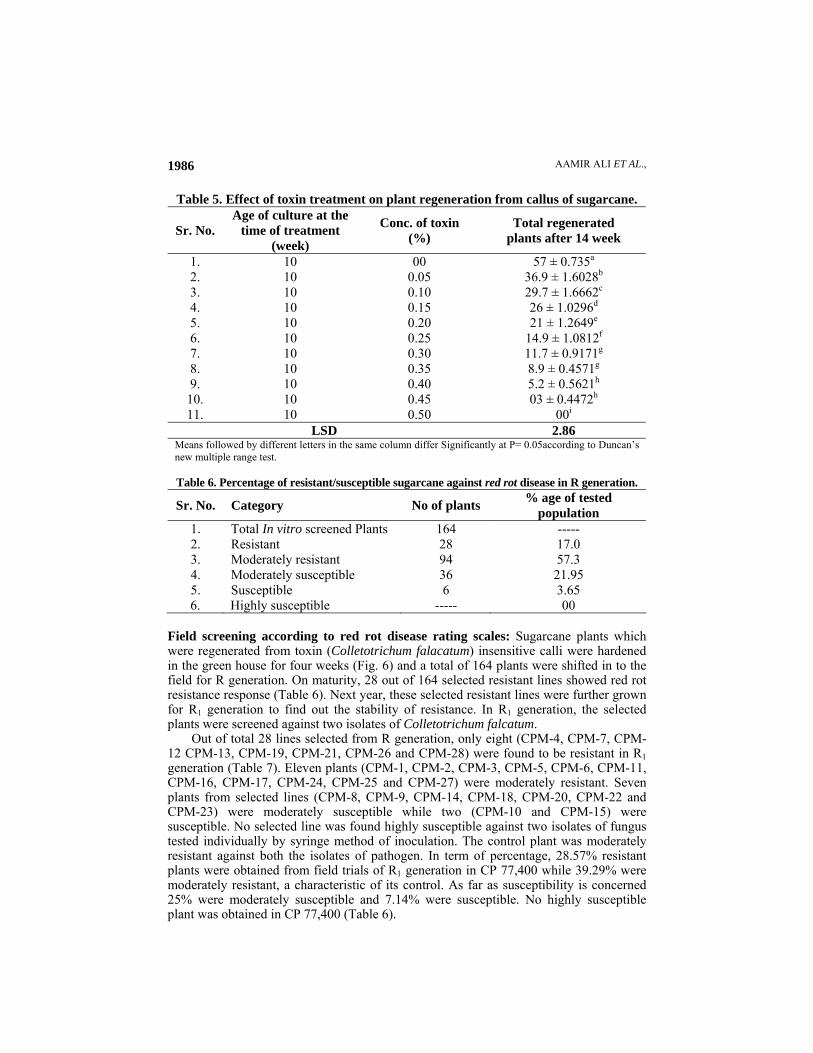
Induction of mutation and screening of mutants Effect of different treatment of Sodium azide on callus proliferation and plant regeneration: The effect of sodium azide treatment on callus proliferation and plant regeneration are depicted in Table 3, where it is obvious that in the control cultures, 1.81 gm of callus was produced after 12 week of incubation which on regeneration produced 57 plants. However, incorporation of Sodium azide into the medium adversely affected the rate of callus proliferation and plant regeneration, which was found to be decreased with the concentration of Sodium azide. When the concentration of Sodium azide was increased from 0, 1.0 mg/l to 5.0 mg/l the rate of callus proliferation was decreased by 8.8, 10.49, 26.52, 37.01 and 52.49% at 1.0, 2.0, 3.0, 4.0 and 5.0 mg/l of sodium azide and number of plants regenerated were 48, 44, 38, 34 and 22 respectively (Table 3; Fig. 3 a, b, c, d, e and f). Maximum number of morphological variants were obtained at the concentration of 4.0 mg/l of Sodium azide (Table 3). At this concentration 19 plants showed growth abnormalities (either albino, viridis or with very thin and small leaves). It was also noticed that all the morphologically abnormal plants produced (viridis or albino) due to mutagenic treatment and did not survive for further growth. Effect of gamma rays on callus proliferation and plant regeneration: In control cultures, 1.86 gm of callus was proliferated after 12 week of incubation which on regeneration produced 59 plants (Table 4). After irradiation a stimulatory effect at low doses (i.e 10 Gy and 20 Gy) of gamma rays on In vitro growing cultures was noticed (Fig. 4a, b and c). The weight of proliferated callus at 10 and 20 Gy exposures was 5.91% and 6.99% more than the control (Table 4). Total 62 plants were produced after 10Gy exposure with 4 morphological variants while at 20Gy exposure 63 plants were produced. So the rate of plant regeneration was 5.8% and 6.8% more over control at these exposures. At exposures of 30, 40 and 50 Gy the lethal effects were observed. The rate of callus proliferation and plant regeneration was decreased and most of callus became necrotic. At 30 Gy exposures proliferation of callus was 1.64 gm, which produced 40 plants with 12 morphological variants, a decrease by 11.82% in callus proliferation and 32.2% in regenerated plants than the control. High exposures of gamma rays became lethal and showed very poor rate of plant regeneration where 24 and 16 plants were produced after 40 and 50 Gy exposures, which were 59.32% and 72.88% less than their control. Most of the morphological variants were albino (Fig. 4 a, b, c, d, e, and f). In vitro selection of red rot resistant plants: Well proliferated and mutagen treated (Sodium azide and γ- rays) 10 week old calli were inoculated in MS media supplemented with different concentrations of partially purified toxin ranging from 0.05% to 0.50% to select the mutant resistant to red rot disease of sugarcane. The control cultures produced 57 plants. Toxin concentration greater than 0.1% retarded callus growth and proliferation that resulted in decrease in the number of regenerated plants (Table 5). Toxin concentration of 0.5% completely inhibited the callus proliferation and plant regeneration. At 0.05% concentration of toxin, the surviving callus produced 37 plants per culture tube after 14 week of incubation in regeneration medium. At 0.3% toxin concentration callus regeneration decreased almost half and numbers of regenerated plants were 12 at this stage (Table 5). Highest damage of callus proliferation and plant regeneration was observed at 0.45% toxin and only three plants were regenerated (Fig. 5).

AAMIR ALI ET AL.,
1984
(a) Control (b) 1.0 mg/l (c) 2.0 mg/l (d) 3.0 mg/l (e) 4.0 mg/l (f) 5.0 mg/l
Fig. 3. (a) Showing shoot regeneration from control b and c showing decreased shoot regeneration potential after treatment with 1.0 and 2.0 mg/l of sodium azide while at 3.0 mg/l treatmetn (fig. 3 d) number of variants (particularly Albino) plants was increased znd at 4.0 and 5.0 mg/l conc. number of regenerated plants was decreased and rate of necrosis was increased (Fig. 3 e & f).
(a) Control (b) 10 Gy (c) 20 Gy (d) 30 Gy (e) 40 Gy (f) 50 Gy Fig. 4. a showing shoot regeneration from control culture, b and c showing efficient shoot regeneration after irradiation with (10 and 20 Gy) gamma rays while at 30 and 40 Gy exposures (fig. 4 d and e) necrosis was observed with less number of regenerated shoots while at 50 Gy exposure (Fig. 4 f) more than 70% callus became necrotic.
Fig. 1. Well developed dry nodular compact andhighly morphogenic callus formed from leaf explantafter 56 days of inoculation in 3rd subculture in MSmedium containing 3.0 mg/l 2,4-D (2.5X).
Fig. 2. Differentiation of callus obtained from leaf explant into small bunches of plantlets after 10 week of inoculation on MS medium containing 1.0 mg/l BAP (4X)

INDUCED MUTATION FOR SCREENING OF RED ROT RESISTANCE IN SUGARCANE
1985
AAMIR ALI ET AL.,
1986
Table 5. Effect of toxin treatment on plant regeneration from callus of sugarcane.
Sr. No. Age of culture at the
time of treatment (week)
Conc. of toxin (%)
Total regenerated plants after 14 week
1. 10 00 57 ± 0.735a 2. 10 0.05 36.9 ± 1.6028b 3. 10 0.10 29.7 ± 1.6662c 4. 10 0.15 26 ± 1.0296d
5. 10 0.20 21 ± 1.2649e 6. 10 0.25 14.9 ± 1.0812f 7. 10 0.30 11.7 ± 0.9171g 8. 10 0.35 8.9 ± 0.4571g 9. 10 0.40 5.2 ± 0.5621h
10. 10 0.45 03 ± 0.4472h 11. 10 0.50 00i
LSD 2.86 Means followed by different letters in the same column differ Significantly at P= 0.05according to Duncan’s new multiple range test.
Table 6. Percentage of resistant/susceptible sugarcane against red rot disease in R generation.
Sr. No. Category No of plants % age of tested population
1. Total In vitro screened Plants 164 ----- 2. Resistant 28 17.0 3. Moderately resistant 94 57.3 4. Moderately susceptible 36 21.95 5. Susceptible 6 3.65 6. Highly susceptible ----- 00
Field screening according to red rot disease rating scales: Sugarcane plants which were regenerated from toxin (Colletotrichum falacatum) insensitive calli were hardened in the green house for four weeks (Fig. 6) and a total of 164 plants were shifted in to the field for R generation. On maturity, 28 out of 164 selected resistant lines showed red rot resistance response (Table 6). Next year, these selected resistant lines were further grown for R1 generation to find out the stability of resistance. In R1 generation, the selected plants were screened against two isolates of Colletotrichum falcatum.
Out of total 28 lines selected from R generation, only eight (CPM-4, CPM-7, CPM-12 CPM-13, CPM-19, CPM-21, CPM-26 and CPM-28) were found to be resistant in R1 generation (Table 7). Eleven plants (CPM-1, CPM-2, CPM-3, CPM-5, CPM-6, CPM-11, CPM-16, CPM-17, CPM-24, CPM-25 and CPM-27) were moderately resistant. Seven plants from selected lines (CPM-8, CPM-9, CPM-14, CPM-18, CPM-20, CPM-22 and CPM-23) were moderately susceptible while two (CPM-10 and CPM-15) were susceptible. No selected line was found highly susceptible against two isolates of fungus tested individually by syringe method of inoculation. The control plant was moderately resistant against both the isolates of pathogen. In term of percentage, 28.57% resistant plants were obtained from field trials of R1 generation in CP 77,400 while 39.29% were moderately resistant, a characteristic of its control. As far as susceptibility is concerned 25% were moderately susceptible and 7.14% were susceptible. No highly susceptible plant was obtained in CP 77,400 (Table 6).

INDUCED MUTATION FOR SCREENING OF RED ROT RESISTANCE IN SUGARCANE
1987

AAMIR ALI ET AL.,
1988
Fig. 5. Callus death and plant regeneration aftertreatment with 0.45% toxin in CP77, 400. At hightoxin treatment most of the callus died and only a smallpart of it differentiated and gave rise to resistant plantsP 77,400) (4X) (four week after toxin treatment).
Fig. 6. 0.45%, 0.4%, 0.35% well developed In vitroscreened and rooted plants 8 week after toxin treatment (1X).
Fig. 7. Hardening of plants survived after toxintreatment (10 week after toxin treatment (1.0X).
Fig. 8. Longitudinal section of control cane (moderately resistant) (1.0 X).
Fig. 9. Longitudinal section of moderatelysusceptible cane.
Fig. 10. Longitudinal section of red rot resistant cane control plant (1.0X).

INDUCED MUTATION FOR SCREENING OF RED ROT RESISTANCE IN SUGARCANE
1989
Discussion Callogenesis and organogenesis: A great deal of work has been conducted for induction and proliferation of callus in cereals. Different combinations of phytohormones supplemented in MS media were tried. The hormone mediated callus induction and subsequent growth on a priori is dependent on certain factors which may trigger the complete chain of events that influence the ability of cultured cells to grow in an organized fashion. Plant tissues, therefore, must have receptors for hormones. These hormones interact with specific receptors that reside either on cell membrane or within the cytoplasm (Mockeviciute & Anisimoviene, 1999). Affinity and concentration of receptors on the surface of the target tissues determine the type of response. A class of proteins called expansins mediates the proton ability to cause cell wall loosening (McQueen et al., 1995). These expansins break the hydrogen bonds between the polysaccharide components of the wall (Cosgrove, 2001). Proton (H+) pumping and lowering of cytosolic pH result in an elevation of intracellular calcium level (Shishova et al., 1999). Both cytosolic pH and calcium ions act as secondary messengers in early auxin action (Zhang, 2003). Calcium ions, either themselves and or along with calcium binding proteins e.g., calmodulin, activate the protein kinase cascade which in turn activates other proteins, including the transcription factors (Wagner, 2001). These factors presumably interact with the auxin-response elements and regulate the expression of auxin-inducible or auxin-responsive genes and exert its effect on cell cycle and stimulate cell division (Johri & Mitra, 2001).
The cultured explant in this study comprised of meristem, newly formed leaves around meristem and pith parenchyma. As these different explants consisting of callus are triggered by some hormone, the higher the meristematic region within the callus mass, the greater will be the probability of differentiation (Fitch & Moore, 1993). The results of our findings also reaffirm same results. So the callus formed from meristem and newly formed whorl of leaves around meristem exhibited maximum morphogenic potential due to their greater meristematic nature.
Organogenesis In vitro consists of many aspects such as phytohormone perception, dedifferentiation of differentiated cells to acquire organogenic competence (Lo et al., 1997), re-entry of quiescent cells into cell cycle and organization of cell division to form specific organ primordia and meristems (Burritt & Leung, 1996). The present work revealed that morphogenic callus (dry nodular and compact) exhibited more potential for shoot regeneration even on hormone free medium. Initiation of differentiation begins with differentiation of group of parenchyma cells to produce centre of meristematic activity called meristemoids which leads to organ formation. The fate and shape of differentiated plant cells can be determined by the size of their undifferentiated precursors.
Cytokinin plays a role in organogenesis by stimulating cell division both In vivo and In vitro. Immunocytochemistry and direct measurement of cytokinin both revealed high cytokinin levels in mitotically active areas, such as the root and shoot meristems, and very low levels are found in tissues where the cell cycle is arrested (Corbesier et al., 2003, Rashotte, 2005). Application of exogenous cytokinin to some organs that normally lack this hormone has been shown to induce cell division (Agostino & Joseph, 1999). The ability of cytokinins to initiate shoots from callus in tissue culture and the initiation of ectopic meristems in cytokinin overproducing plants suggest a role for cytokinins in SAM (Shoot apical meristem) development. One possible mechanism by which cytokinins influence SAM development is by regulating gene expression. The knotted1

AAMIR ALI ET AL.,
1990
(kn1) homeobox family of genes is expressed exclusively in the SAM and is involved in its development and maintenance (Kerstetter, 1997). Rupp et al., (1999) examined the expression of KNAT1 and STM (Shoot meristemless gene) (Arabidopsis homologs of kn1) in transgenic Arabidopsis expressing it under the control of a heat shock promoter. The steady-state mRNA levels of both KNAT1 and STM were elevated following heat shock, and were correlated to elevated cytokinin levels. Elevated KNAT1 and STM transcript levels were also observed in untreated amp1 plants, implying that endogenous cytokinin can also induce expression of these homeobox genes. These results suggest that cytokinins may act upstream of KNAT1 and STM in regulating SAM development. Induction of mutation and In vitro screening of mutants: Mutation breeding as a methodology for crop improvement is based on the possibility of altering genes by exposing their vegetative parts, cells, tissues, gametes or seeds to physical and chemical mutagens. Mutagenesis of In vitro cultures avoids the need for large-scale facilities and allows better control of treatment, as vitrified tissues may be more permeable to mutagens (Ziv, 1991).
In this study Sodium azide was used as chemical mutagen. Hibberd et al., (1982) reported that Sodium azide has potential in tissue culture mutagenesis for inducing biochemical mutants. They evaluated azide mutagenicity in friable embryogenic cultures using selection for lysine plus threonine resistance. Bhagwat (1998) used sodium azide to induce mutation against Fusarium oxysporium in Banana. Maniu et al., (1998) used sodium azide for In vitro mutagenesis in barley. The effect of different concentration of Sodium azide treatment on callus proliferation and its regeneration is shown in Tables 3. Sodium azide is an S stage mutagen (Sander & Nilan, 1974, Nilan et al., 1975). It was determined that azide acts through a promutagen or organic metabolite. This metabolite has been isolated and identified as azidoalanine (Owais et al., 1988). Sodium azide also produce DNA single strand breaks (Veleminsky et al., 1985), it readily induces base substitutions, but not "frameshifts" or deletions (Niknejad et al., 1972). It preferentially induced G: C → A: T transitions at the second codon position (Koch et al., 1994). Although sodium azide causes mutation at gene level but few chromosme aberrations have also been detected (El-Den, 1993). Del Campo (1999) also reported the induction of chromosomal aberrations during replication phase (S) by Sodium azide. Mendhulkar (2002) reported mitotic chromosomal aberrations (laggards, bridges, chromosome stickiness) by single treatment with Sodium azide. The immediate effect of Sodium azide on meristematic cells appears to be a blocking action at the beginning of the genome separation reducing its velocity of the progression in the cell cycle; he further concluded that it acts as the respiratory chain inhibitor. Johnsen et al., (2002) also mentioned Sodium azide as electron transport blocker. However exact mechanism of Sodium azide action in In vitro cultures at molecular level is still needed to be investigated.
The irradiation of callus cultures which are capable of embryogenesis and organogenesis can be used to obtain mutants quickly and in large number (Ahloowalia, 1995). Results of exposures of gamma rays on callus proliferation and plant regeneration is presented in Table 4.
Radioactive materials like 60Co emit high energy photons which are called as gamma radiation. These radiations can alter the structure of chromosome in two ways: • Directly by quanta of energy which hit the chromosomes like bullets hitting a target and • Indirectly by ionization which produces free radicals.

INDUCED MUTATION FOR SCREENING OF RED ROT RESISTANCE IN SUGARCANE
1991
Among different type of variations obtained after gamma rays treatment in the present study, mainly chlorophyll variants i.e., albino and viridis were predominant. Khan et al., (1998) also reported more chlorophyll variants in sugarcane after gamma ray treatment. Although other variants such as plants with thin and curled leaves, vitrified and plants with comparatively smaller and narrow leaves were also observed. Lethal and mutagenic effects of ionizing radiation result principally from incompletely or incorrectly repaired DNA lesions (Ward, 1994). Among these lesions, strand breaks are considered to be most important (Schulte, 1987) as they interrupt the continuity and integrity of the double helix. An unrepaired single stranded break (SSB) in ssDNA (Fiers & Sinsheimer, 1962); an unrepaired double strand break (DSBs) in dsDNA (Frankenberg et al., 1981) and crosslinking of DNA to itself or proteins (Cress et al., 1990) has been shown to be responsible for the lethal effects of ionising radiations. Ward (1995) reported that radiation induced mutagenesis is due to the involvement of multiple damaged sites (such as double-strand breaks). In vitro screening and field study of red rot resistanc: Carlson (1973) for the first time demonstrated that cells and plant protoplast could be selected in cultures for resistance to a pathogenic toxins and plants with an altered response to infection by pathogen could be regenerated from these cultured cells.
The preliminary step to obtain pathogen resistant plants is to subject mutagen treated calli to toxin and to select the tolerant or resistant one for further development of plantlets. Culture filtrate or partially purified toxin, obtained from fungal pathogen has been extensively used for in vitro selection of resistant host species (Buiatti & Ingram 1991). The selection of tissue for resistance to toxin is apparent from the observation that tissues finally surviving the toxin at different stages results in small proportion of viable shoots, which may have potential to develop into disease resistant genotypes. Toxin tolerance detected to the host cultures has been reported to be positively correlated with In vivo resistance (Gentile et al., 1992). Jin et al., (1996) reported similar response when he exposed susceptible soyabean calli to high levels of Fusarium solani culture filtrate showing reduced growth and regeneration.
Mohanraj (1995) have reported that in sugarcane, relatively high concentration of red rot toxin used resulted in limited survival of calli and shoot differentiation. The results of present study also clearly show that high concentration of toxin markedly inhibits development of In vitro cultures at all stages via callus proliferation, regeneration and shoot growth. The brownish discolouration of the calli in the present study associated with necrosis in toxin incorporation medium suggests the involvement of phenolic compounds and their oxidation products similar to those observed in sugarcane plant infected with red rot pathogen. Although there are several reports available on the use of pathogen toxins in tissue culture to evolve disease resistant genotype of host plant, but only a few reports are available on similar attempts with specie of Colletotrichum. Naik et al., (1997) used toxin of Colletotrichum falcatum in the selection of red rot resistant genotypes of sugarcane. Olufolaji (2000) and Mohanraj (2003) used the toxin of Colletotrichum falcatum for In vitro selection of red rot resistance in sugarcane. The results of present study, suggests the possibility of generating In vitro plants resistant to red rot using tissue culture technique. Small proportions of survived shoots at high toxin concentration were rooted and hardened in the green house. The hardened plants were then shifted in the field for final screening through field trials. As reported by Perez

AAMIR ALI ET AL.,
1992
(1987), the stability of induced mutation in agronomic characters needs to be confirmed in the next generation under field conditions. A total of 164 lines obtained after In vitro screening and hardening were shifted for field trials. On maturity of R generation crop, 28 lines were selected for R1 generation on the basis of their resistance response against red rot disease and morphological characters (number of tillers, Cane length, Cane diameter, internodal length and sugar recovery). Next year, these selected lines were planted for R1 generation by conventional method of cultivation and to confirm the stability of field characters and red rot resistance. From Table 7 it is obvious that out of 28 selected lines tested, 8 lines were resistant while 11 selected lines were moderately resistant. Recovery of plants at different level of susceptibility regenerated even after toxin resistant callus was due to the existence of high genetic complexity and chromosomal mosaicism in sugarcane (Jambhale et al., 1998). Mohanraj (1996) screened 700 somaclones against red rot and out of these 120 showed moderately resistant reaction in the field trials. Similarly Javed et al., (2003) obtained 35 and 205 variants after In vitro screening in two different varieties of sugarcane and out of these only 10 and 57 were found to be resistant in both the varieties respectively after field trials. In vitro selection and final screening of red rot resistant lines of sugarcane after field trials has also been reported by Naik et al., (1997); Javed et al., (2001) and Shahid et al., (2003). References Agostino, D, I.B. and J.K. Joseph. 1999. Molecular mechanisms of cytokinin action. Curr. Opinion
in Plant Biol., 2: 359-364. Ahloowalia, B.S. 1995. In vitro mutagenesis for improvement of vegetatively propagated plants. In:
Proceeding, Induced mutation and molecular techniques for Crop Improvement, pp. 531-541. International Symposium,, IAEA and Food Agriculture Organization of the United Nations. IAEA SM-340, Vienna.
Bhagwat, B. and E.J. Duncan. 1998. Mutation breeding of banana cv Highgate (Musa spp., AAA Group) for tolerance to Fusarium oxysporum f.sp. cubense using chemical mutagens. Sci. Hort., 73: 11 -22.
Buiatti, M. and D.S. Ingram. 1991. Phytotoxins as a tool in breeding and selection of disease resistant plants. Experientia, 47: 811-819.
Burritt, D.J. and D.W.M. Leung. 1996. Organogenesis in cultured petiole explants of Begonia Xerythrophylla: the timing and specificity of the inductive stimuli. J. Exp. Bot., 47: 557-567.
Carlson, P.S. 1973. Methionine sulfoximine-resistant mutants of tobacco. Sci., 180: 1366-1368. Corbesier, L., E. Prinsen, A. Jacqmard, P. Lejeune, H. Van Onckelen, C. Pe´rilleux and G. Bernier.
2003. Cytokinin levels in leaves, leaf exudate and shoot apical meristem of Arabidopsis thaliana during floral transition. J. Exp. Bot., 54: 2511-2517.
Cosgrove. 2001. Wall structure and wall loosening. A look backwards and forwards. Plant Physiol., 125: 131-134.
Cress, A.E., K.M. Kurath, B. Stea and G.T. Bowden. 1990. J. Cancer Res. Clin. Oncol., 116: 324-330.
Del Campo, G., R. Antonio and O. Coletto. 1999. Induction of chromosomal aberrations during replication phase (S) by Sodium azide in root meristems of Allium cepa L. Ciencia (Maracaibo), 7(2): 118-125.
Duncan, R.R., R.M. Wasko, and M.W. Nabors. 1995. In vitro screening and field evaluation of tissue culture regenerated sorghum (Sorghum bicolor) for soil stress tolerance. Euphytica, 85: 373-380.
El-Den, J.B. 1993. Development of a new barley line by induced mutation. Rachis, 12(½): 8-10. Fiers, W. and R.L. Sinsheimer. 1962. J. Mol. Biol., 5: 408-419.

INDUCED MUTATION FOR SCREENING OF RED ROT RESISTANCE IN SUGARCANE
1993
Fitch, M.M.M. and P.H. Moore. 1993. Long-term culture of embryogenic sugarcane cells. Plant Cell Tiss. and Org. Cul., 32(3): 335-343.
Frankenberg, D., M. Frankenberg, D. Blocher and R. Harbich. 1981. Radiat Res., 88: 524-532. Gentile, A., E. Tribulato, G. Continella and A. Vardi. 1992. Differential responses of citrus calli
and protoplasts to culture filtrate and toxin of Phoma tracheifila. Theoret. Appl. Genet., 83: 759-764.
Hibberd, K.A. and C.E. Green. 1982. Inheritance and expression of Lysine plus Threonine resistance selected in maize tissue cul., PNAS. 79: 559-563.
Hidalgo, Santos, Tussel, Pires de Matos, Cabral and Perez. 1999. Phytotoxicity of Fusarium subglutinans culture filtrate on In vitro plantlets and calli of resistant and susceptible pineapple (Ananas comosus). Plant Path., 48(6): 756.
Hussnain, Z. and S. Afghan. 2001. A comparative study on artificial inoculation methods for red rot (Colletotrichum falcatum (went) screening. Pak. Sugar J., 16(2): 3-5.
Jambhale, N.D. and S.C. Patil. 1998. "Yashwant" an elite clone of Eucalyptus camaldulensis. J. of Maharashtra Agri. Uni., 23(2): 179-180.
Javed, M.A., B.A. Chaudhry, M.K. Tanvir, M.T.H. Shahid and Makhdoom Hussain. 2001. Development and screening of sugarcane somaclones against diseases. Pak. Sugar J., 16(6): 36-39.
Javed, M.A., M.T.H. Shahid, S. Rehman, Z. Saleem, M. Hussain and A. Rauf. 2003. Induction of sugarcane somaclones and their evaluation against diseases. Pak. Sugar J., 18(6): 48-51.
Jarausch, Lansac, Bliot and Dosba. 1999. Phytoplasma transmission by In vitro graft inoculation as a basis for a preliminary screening method for resistance in fruit trees. Plant Path., 48(2): 283.
Jin, H., G.L. Hartman, C.D. Nickell and J.M. Widholm. 1996. Phytotoxicity of culture filtrate from Fusarium solani, the causal agent of soybean sudden death syndrome. Plant Dis., 80: 922-927.
Johnsen, A.R., K. Bendixen and U. Karlson. 2002. Detection of microbial growth on polycyclic aromatic hydrocarbons in microtiter plates by using the respiration indicator WST-1. Appl. and Env. Microbiol., 68(6): 2683-2689.
Johri, M.M. and D. Mitra. 2001. Action of plant hormones. Curr. Sci., 80(2): 199-205. Jones, P.W. 1990. In vitro selection for disease resistnace. In: Plant Cell Lines Selection, 113-149. Kerstetter, R.A. and S. Hake. 1997. Shoot meristem formation in vegetative development. Plant
Cell, 9: 1001-1010. Khan, I.A., A. Khatri, M. Ahmad, S.H. Siddiqui, G.S. Nizamani, M.H. Khanzada, N.A. Dahar and
R. Khan. 1998. In vitro mutagenesis in sugarcane. Pak. J. of Bot., 30(2): 253-261. Koch, W.H., E.N. Henrikson, E. Kupchella and A. Cebula Thomas. 1994. Salmonella typhimurium
strain TA 100 differentiates several classes of carcinogens and mutagens by base substitution specificity. Carcinogenesis, 15(1): 79-88.
Lo, K.H., K.L. Giles and V.K. Sawhney. 1997 Acquisition of competence for shoot regeneration in leaf discs of Saintpaulia ionantha X confuse hybrids (African violet) cultured In vitro. Plant Cell Rep., 16: 416-420.
Maniu, M. and A. Mihailescu. 1998. Sodium azide in vitro mutagenesis in barley. Analele Siintifice ale Universitatii "Al I Cuza" din Iasi. Sectiunea II a. Biologie Vegetala, 44: 177-182.
McQueen, M.S. and D.J. Cosgrove. 1995. Expansin mode of action on cell walls: analysis of wall hydrolysis, stress relaxation, and binding. Plant Physiol., 107: 87-100.
Mendhulkar, V.D. 2002. Synergistic effect of Sodium azide in combination with Maleic hydrazide in Triticum aestivum Linn. Adv. in Plant Sci., 15(1): 213-219.
Murashige, T. and F. Skoog. 1962. A revised medium for rapid growth and bioassay with tobacco tissue cultures. Physiol. Plant, 15: 473-487.
Mockeviciute, R. and N. Anisimoviene. 1999. Indole-3-acetic acid receptors in the cytosol. Biologija, 4: 90-93.
Mohanraj, D., P. Padmanaban, R. Jothi and K.C. Alexander. 1995. Possible role of phytotoxin(s) in red rot disease of sugarcane. In: Proceedings of the National Seminar on Strategies for Research and Management of Red Rot. (Eds.): V.P. Agnihotri, O.K. Sinha and R.P. Singh IISR. Lucknow, 263-270.

AAMIR ALI ET AL.,
1994
Mohanraj, D. 1996. Evaluation of red rot toxin resistant somaclones for disease reaction. Sugarcane Breeding Ins. Ann. Rep., 1995-96, 32.
Mohanraj, D., P. Padmanaban and M. Karunakaran. 2003. Effect of phytotoxin of Colletotrichum falcatum Went. (Physalospora tucumanensis) on sugarcane in tissue culture. Acta Phytopathologica et Entomologica Hungarica, 38(½): 21-28.
Naik, G.R. and A.B. Vedamurthy. 1997. In vitro evaluation of red rot toxin influence on sugarcane (Saccharum officinarum) Var. CoC 671. Curr. Sci., 73(4): 367-369.
Niknejad, M., C.F. Konzak, E. Donaldson, I. Wickham, R.A. Nilan and C. Sander. 1972. Azide, a potent new mutagen - near additivity of mutation yields from post-treatment combination of barley treated with N-methyl,N'-nitrosourea. Agronomy, 56-61.
Nilan, R.A. and O.W. Pearson. 1975. Lack of chromosome breakage by azide in embryonic shoots and microspores of barley. BGN, 5: 33.
Olufolaji, D.B. 2000. Rapid toxin test for red rot disease in sugarcane. Sugar Tach., 2(4): 6-11. Owais, W.M. and A. Kleinhofs. 1988. Metabolic activation of the mutagen azide in biological
systems. Mut. Res., 197: 313-323. Perez, P.J.N. 1987. Die Nutzung der in vitro kultur und die induction von Mutationen bei der
Zuchtung von Zuckerrohr (Saccharum spp). Thesis, Dr. habil., Universitat Leipzig. Rashotte, A.M., H.S. Chae, B.B. Maxwell and J.J. Kieber. 2005.Interaction of cytokinins with
othewr signals. Physiol. Plantarum, 123: 184-194. Rupp, H.M., M. Frank, T. Werner, M. Strnad and T. Schmülling. 1999. Increased steady state
mRNA levels of the STM and KNATI homeobox genes in cytokinin overproducing Arabidopsis thaliana indicate a role for cytokinins in the shoot apical meristem. Plant J., 18: 557-563.
Sander, C. and R.A. Nilan. 1974. Increasing the mutagenic efficiency of Sodium azide in barley. Barley Genetics Newsletter, 4: 63-65.
Schulte, F.D. 1987. Br J. Cancer, 55: suppl. VIII, 129-134. Shahid, M.T.H., S. Rehman, M.A. Javed, M. Hussain, Z. Saleem and M.K. Tanvir. 2003. Variation
for red rot resistance and other characters of economic importance in the calliclones of sugarcane (Saccharum officinarum L.) cultivar BF-162. Pak. Sugar J., 18(6): 41-44.
Shishova, M.F., S. Lindberg and V.V. Polevoi. 1999. Auxin activation of Ca2+ transport across the plasmalemma of plant cells. Russian J. of Plant Physiol., 46(5): 626-633.
Srinivasan, K.V. and N.R. Bhat. 1961. Red Rot of Sugarcane: Criteria for breeding resistance. J. Ind. Bot. Soc., 40: 566-577.
Veleminsky, J., J. Satava, A. Kleinhofs, R.A. Nilan and T. Gichner. 1985. Induction of proteinase K-sensitive sites and M. luteus endonuclease-sensitive sites in DNA of barley embryos by sodium azide. Mut. Res., 149(3): 431-436.
Wagner, T.A. and B.D. Kohorn. 2001. Wall-Associated Kinases Are Expressed throughout Plant Development and Are Required for Cell Expansion. Plant Cell. (13): 303-318.
Ward, J. F. 1994. Int. J. Rad. Biol., 66: 427-432. Ward, J.F. 1995. Rad Res. 142: 362-368. Zhang, L. and Y.T. Lu. 2003. Calmodulin-binding protein kinases in plants. Trends Plant Sci., 8:
123-127. Ziv, M. 1991. Vitrification: Morphological and physiological disorders of in vitro plants. In:
Mocropropagation: Technology and Application, pp. 45-69. (Eds.): P.C. Debergh and R.H. Zimmerman). Dordrecht: Kliwer.
(Received for publication 13 June 2007)