improvement of mechanical properties and biocompatibility of
TRANSCRIPT

J O U R N A L O F T H E M E C H A N I C A L B E H AV I O R O F B I O M E D I C A L M A T E R I A L S 3 ( 2 0 1 0 ) 5 3 0 – 5 3 7
available at www.sciencedirect.com
journal homepage: www.elsevier.com/locate/jmbbm
Research paper
Improvement of mechanical properties and biocompatibilityof forsterite bioceramic addressed to bone tissue engineeringmaterials
M. Kharaziha∗, M.H. Fathi
Biomaterials Group, Department of Materials Engineering, Isfahan University of Technology, Isfahan, 8415683111, Iran
A R T I C L E I N F O
Article history:
Received 13 March 2010
Received in revised form
16 June 2010
Accepted 18 June 2010
Published online 26 June 2010
Keywords:
Forsterite
Two step sintering
Fracture toughness
Biocompatibility
Bone tissue engineering
A B S T R A C T
This work deals with the fabrication and characterization of nanostructured forsterite
bulk. This material may have better biocompatibility and mechanical properties than
coarse grain forsterite for the development of bone tissue engineering materials.
Nanostructured forsterite bulks were prepared by two step sintering of sol–gel derived
forsterite nanopowder. Their sinterability and mechanical properties were then studied.
Biocompatibility of the nanostructured forsterite bulk was also evaluated by cell
attachment and proliferation experiments. In addition, the effects of ionic products from
forsterite nanopowder dissolution on osteoblasts were studied. Results show that dense
nanostructured forsterite bulk was prepared with hardness and fracture toughness of
about 1102 Hv and 4.3 MPa m1/2, respectively. Nanostructured forsterite was biocompatible
and the MTT test confirmed that the products from forsterite nanopowder dissolution
significantly promoted osteoblast proliferation within a certain concentration range. In
addition, cells attached to and spread on the surface of nanostructured forsterite bulks.
Mechanical properties of the nanostructured forsterite were much higher than that of
hydroxyapatite. It was concluded that nanostructured forsterite is a bioactive ceramic with
good biocompatibility that can be used as a bone tissue engineering material.c© 2010 Elsevier Ltd. All rights reserved.
d
1. Introduction
Bone tissue engineering proposes a suitable way to repairand regenerate lost bones. Different materials have beenconsidered for use in bone tissue engineering (Chim et al.,2006). The biocompatibility of bioceramics makes them themost important group of biomaterials. Hydroxyapatite (HA) isa significant success of bioceramics as a bone tissue repairingmaterial. HA is osteoconductive; a property that promotesbone tissue growth (Sprio et al., 2009). Weak mechanical
∗ Corresponding author. Tel.: +98 311 6676858; fax: +98 311 3912752.E-mail address: [email protected] (M. Kharaziha).
1751-6161/$ - see front matter c© 2010 Elsevier Ltd. All rights reservedoi:10.1016/j.jmbbm.2010.06.003
properties such as low fracture toughness limit the clinicalapplications of HA (Hench, 1998). Dense HA has lower fracturetoughness than cortical bone, and higher Young’s modulusthan cortical bone (Hench, 1998).
Forsterite (Mg2SiO4) is a new bioceramic. Previous researchsuggests that Mg is one of the most important elements inthe human body. It indirectly influences the body mineralmetabolism. Also, it is closely associated with mineralizationof calcined tissues (Althoff et al., 1982). In addition, silicon isan essential element in skeletal development and is uniquely
.
J O U R N A L O F T H E M E C H A N I C A L B E H AV I O R O F B I O M E D I C A L M A T E R I A L S 3 ( 2 0 1 0 ) 5 3 0 – 5 3 7 531
localized in the active areas of young bone. Silicon is involvedin the early stage of bone calcification (Carlisle, 1970; Schwarzand Milne, 1972).
Research shows that forsterite has better mechanicalproperties than calcium phosphate ceramics such as HA(Ni et al., 2007). However, it is not mechanically strong forload bearing applications. Thus, improvement of mechanicalfeatures of forsterite is fundamental to reach equilibriumand reduce the mismatch to favor the osteointegrationprocess. On the other hand, nanostructured ceramics havesuperior mechanical properties (Cottom and Mayo, 1996).In addition, the nanometer-sized grains and the highvolume fraction of grain boundaries in nanostructuredmaterials are reported to show improved biocompatibilityover normal materials (Kim et al., 2008), and increasedosteoblast adhesion and proliferation (Catledge et al., 2008;Du et al., 1999). Webster et al. (Webster et al., 1998)designed the first nanophase ceramics with the aim ofimproving osteointegrative properties of orthopedic anddental materials. In contrast to conventional materials,nanophase ceramics can be designed with surface properties,mechanical properties, and grain size distribution similar tonatural bone (Gutwein and Webster, 2002).
According to the above points, nanostructured forsteritebioceramic is expected to have better mechanical propertiesand biocompatibility than coarser crystals. It was reportedthat coarse grain forsterite had an extremely low degradationrate and was not bioactive (Ni and Chou, 2008). Ourrecent study indicated that forsterite nanopowder, unlikemicron-sized forsterite, possessed apatite-formation ability(Kharaziha and Fathi, 2009). The bioactivity of the nanophaseforsterite when compared to coarse grain forsterite showsa greater effect of the nanophase forsterite on its iondissolution in biological solution. As stated above, Si andMg containing ionic products from the dissolution offorsterite nanopowder might have inhibitory effects on cellproliferation. Thus, an in vitro cytocompatibility evaluationof the nanostructured forsterite ceramics must be done.
In our previous research, the fabrication of densenanostructured forsterite bulk with mechanical alloyingderived forsterite nanopowder was studied (Fathi andKharaziha, 2009). In this study, dense forsterite bulk wasprepared by forsterite nanopowder derived via the sol–gelmethod (which has a smaller particle size and a narrowparticle size distribution) to study the effects of these factorson the mechanical properties of the nanostructured forsteritedense ceramic. For the first time, an in vitro cytocompatibilityevaluation of the nanostructured forsterite was performed.The effects of ionic products from forsterite nanopowderdissolution on an osteoblast were studied.
2. Experimental procedures
2.1. Preparation of forsterite ceramic
Forsterite nanopowder was prepared according to themodified sol–gel process described in our previous report(Kharaziha and Fathi, 2009). In brief, water-based solutionsof the magnesium salts and colloidal silica were prepared.
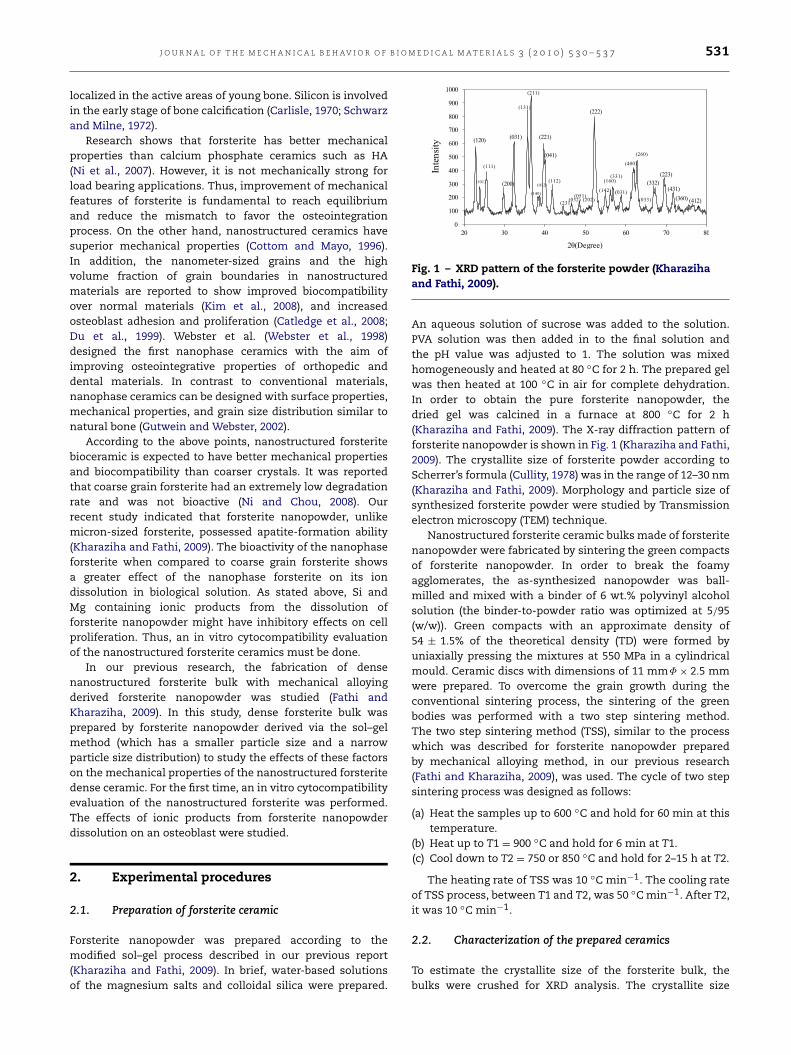
Fig. 1 – XRD pattern of the forsterite powder (Kharazihaand Fathi, 2009).
An aqueous solution of sucrose was added to the solution.PVA solution was then added in to the final solution andthe pH value was adjusted to 1. The solution was mixedhomogeneously and heated at 80 ◦C for 2 h. The prepared gelwas then heated at 100 ◦C in air for complete dehydration.In order to obtain the pure forsterite nanopowder, thedried gel was calcined in a furnace at 800 ◦C for 2 h(Kharaziha and Fathi, 2009). The X-ray diffraction pattern offorsterite nanopowder is shown in Fig. 1 (Kharaziha and Fathi,2009). The crystallite size of forsterite powder according toScherrer’s formula (Cullity, 1978) was in the range of 12–30 nm(Kharaziha and Fathi, 2009). Morphology and particle size ofsynthesized forsterite powder were studied by Transmissionelectron microscopy (TEM) technique.
Nanostructured forsterite ceramic bulks made of forsteritenanopowder were fabricated by sintering the green compactsof forsterite nanopowder. In order to break the foamyagglomerates, the as-synthesized nanopowder was ball-milled and mixed with a binder of 6 wt.% polyvinyl alcoholsolution (the binder-to-powder ratio was optimized at 5/95(w/w)). Green compacts with an approximate density of54 ± 1.5% of the theoretical density (TD) were formed byuniaxially pressing the mixtures at 550 MPa in a cylindricalmould. Ceramic discs with dimensions of 11 mmΦ × 2.5 mmwere prepared. To overcome the grain growth during theconventional sintering process, the sintering of the greenbodies was performed with a two step sintering method.The two step sintering method (TSS), similar to the processwhich was described for forsterite nanopowder preparedby mechanical alloying method, in our previous research(Fathi and Kharaziha, 2009), was used. The cycle of two stepsintering process was designed as follows:
(a) Heat the samples up to 600 ◦C and hold for 60 min at thistemperature.
(b) Heat up to T1 = 900 ◦C and hold for 6 min at T1.(c) Cool down to T2 = 750 or 850 ◦C and hold for 2–15 h at T2.
The heating rate of TSS was 10 ◦C min−1. The cooling rateof TSS process, between T1 and T2, was 50 ◦Cmin−1. After T2,it was 10 ◦C min−1.
2.2. Characterization of the prepared ceramics
To estimate the crystallite size of the forsterite bulk, thebulks were crushed for XRD analysis. The crystallite size
532 J O U R N A L O F T H E M E C H A N I C A L B E H AV I O R O F B I O M E D I C A L M A T E R I A L S 3 ( 2 0 1 0 ) 5 3 0 – 5 3 7
of the forsterite bulk was estimated from the XRD peakbroadening based on Scherrer’s formula (Cullity, 1978). Ascanning electron microscopy (SEM) was used to study thefracture surface of the forsterite ceramics. To accomplish this,the sintered specimens were mechanically ground and thenpolished. The grain size of the specimens was determined bymultiplying the average linear intercept by 1.56 (Mendelson,1969). For each specimen, 20 line segments were taken intoaccount.
Linear shrinkage of the sintered samples was determinedusing the following equation (Eq. (1)):
Shrinkage(%) =lg − lp
lg× 100 (1)
where lg is the length of the green samples and lp is the lengthof the sintered products.
Apparent density of the bulks was measured byArchimedes’ method. At least three sample sizes were usedto determine the average grain size, shrinkage, and densitycorresponding to each data point.
The microhardness of each sample was tested using theVickers indentation technique. This was done by applying aloading of 9.8 N for 15 s. Ten indentations were made onthe polished surface perpendicular to the height direction.The fracture toughness was determined using the direct crackmeasurement method according to Niihara’s formula (Eq. (2))(Niihara et al., 1982):
KIC = 0.203(c/a)−3/2Hv.a1/2 (2)
where KIC is fracture toughness in MPa m1/2; c is the lengthof the crack measured from the center of the indentation athalf of the average length of two indent diagonals; and Hv isthe hardness.
2.3. Cytotoxicity assay
The method was carried out with a dilution of powder extractin contact with osteoblasts according to the InternationalStandard Organization (ISO/EN 10993–5, 1999). A dilution offorsterite nanopowder extract in contact with osteoblast-likeG292 Cells was used in the RPMI-1640 medium supplementedwith 10% FCS (Fetal Calf serum). Prior to testing, the forsteritenanopowder were washed in 75% ethanol solution, sterilizedfor 20 min under ultraviolet light, and autoclaved for 30 minat 120 ◦C.
The dissolution extracts of forsterite nanopowder wereprepared by adding forsterite nanopowder to PBS. The ratioof the forsterite nanopowder weight and the medium volumewas 200 mg/ml. After 24 h incubation at 37 ◦C, the mixturewas centrifuged and the supernatant was collected. Forfurther cell culture experiments, the obtained extract liquidwas diluted with PBS. After the cell suspension was adjustedto a density of 1× 10⁴ cells/ml, 180 µl of the cell suspensionswas added to each well of a 96-well plate and incubated for24 h at 37 ◦C and 5% CO2.50 µl of diluted extracts (1, 1/2,1/4 1/8, 1/16, 1/32) of the forsterite nanopowder were addedto each well of the plate, respectively. The negative controlwas prepared with the RPMI-1640 medium supplementedwith 10% FCS without the addition of diluted extracts ineach well. The positive control was prepared with 50 µl of
RPMI-1640 medium supplemented with 10% FCS and 50 µlsolution of Taxol. After the cells incubated for 1–7 days,20 µl of 5 mg/ml 3-(4, 5-dimethylthiazol-2-yl)-2, 5-diphenyltetrazolium bromide (MTT) solution were added to eachwell. The plate was then incubated for 4 h. After, Dimethylsulfoxide (DMSO) was added to each well to stop the reactionbetween the MTT and the cells. The optical density (OD) wasmeasured at the wavelength of 540 nm by an enzyme-linkedimmunoadsorbent assay (ELISA) reader.
2.4. Cell adhesion and growth assay
The method was carried out with nanostructured discs incontact with osteoblast-like G292 cells in the RPMI-1640medium supplemented with 10% FCS. Prior to cell seeding,nanostructured forsterite ceramic discs were cleaned in 75%ethanol solution, sterilized for 20 min under ultraviolet light,and autoclaved for 30 min at 120 ◦C. After the discs wereplaced in a 24-well culture plate, 25 µL of culture mediumcontaining 3× 10⁴ cells were seeded onto the top of the discs.In order the cells to attach, the plate was incubated for 1 h.After, 1 mL of fresh culture medium was added to each welland the cells were incubated for 1–7 days. Culture media wererefreshed every 2 days.
After incubation, the disks were washed with PBS solutionand fixed for 30 min in 2.5% glutaraldehyde in phosphate-buffered solutions. The fixed cells were washed three timeswith PBS and dehydrated in varying concentrations of ethanolsolution (30%, 50%, 70%, 90%, 95%, 100% (v/v)) for 10 mineach. The discs were treated by immersion in the 50% alcohol-HMDS (hexamethyldisilazane) solution (v/v) for 10 min andthen in pure HMDS for 10 min. Finally, they were dried in adesiccator overnight.
The cell morphology on nanostructured forsterite ceram-ics was observed with a scanning electron microscopy (SEM).
2.5. Statistical analysis
All data were collected with N = 3 and expressed as means± standard deviation (SD) in each experiment. Statisticalanalysis was done by two-way ANOVA with Duncan test.Differences were considered statistically significant at p <
0.05.
3. Results and discussion
3.1. Characterization of forsterite ceramic
Fig. 2 shows the TEMmicrograph of the forsterite nanopowderproduced via sol–gel and mechanical alloying methods,respectively. As seen in Fig. 2(a, b), forsterite particlesproduced via the sol–gel method show a spherical shapewith narrow particle size distribution in the 25–45 nmrange (Kharaziha and Fathi, 2009). Fig. 2(c, d) show theTEM micrograph of forsterite nanopowder produced bymechanical alloying method (Fathi and Kharaziha, 2009).In contrast to the previous forsterite powder, the powderproduced by mechanical alloying shows particles with

J O U R N A L O F T H E M E C H A N I C A L B E H AV I O R O F B I O M E D I C A L M A T E R I A L S 3 ( 2 0 1 0 ) 5 3 0 – 5 3 7 533
Fig. 2 – TEM micrographs of forsterite nanopowdersprepared by (a), (b) sol–gel method and (c), (d) mechanicalalloying.
irregular shapes and with wide particle size distribution inthe 25–70 nm range (Fathi and Kharaziha, 2009).
Fig. 3 shows the relative densities (RD) of the sinteredsamples as a function of T1 (the first step sinteringtemperature) and grain size at two different second steptemperatures (750 and 850 ◦C). As shown with this figure, byincreasing the first step temperature up to 1000 ◦C, significantdensification (about 79%–85%TD) was obtained in the firststep of sintering at all temperatures (0 h-curve). Densityimproved from a lower value up to 98.6%TD in the secondstep of sintering. By increasing the holding time at the secondstep temperature, a fully dense ceramic was obtained (withT1 ≥ 1200 ◦C and a holding time of T2 ≥ 5 h) although graingrowth occurred when T2 = 850 ◦C.
Figs. 4 and 5 show the effect of the first step temperatureof the TSS process on the fracture toughness and hardnessvalues of forsterite ceramic. As can be observed, under thisTSS regime (T1 = 1200 ◦C and T2 = 750 ◦C) with a prolongedsoaking at 750 ◦C (up to 5 h), fracture toughness increasedfrom 1.10±0.5 MPa m1/2 (at T1 = 900 ◦C) to 4.3±0.3 MPa m1/2
(at T1 = 1200 ◦C) and hardness increased from 520 ± 45 Hv(at T1 = 900 ◦C) to 980 ± 20 Hv (at T1 = 1200 ◦C). Under theTSS regime (T1 = 1200 ◦C and T2 = 850 ◦C) with a prolongedsoaking at 850 ◦C up to 5 h, full density was observed.
The results also showed high hardness and fracturetoughness of about 955 ± 15 Hv and 3.2 ± 0.25 MPa m1/2,respectively. With prolonged soaking at 850 ◦C up to 15 h,hardness and fracture toughness declined.
Wang et al. (2006) suggests that for achieving densificationwithout a significant grain growth, grain boundary diffusionneeds to remain active, while the grain boundary migration issuppressed. A mechanism to slow down the grain boundarymovement is the triple-point drag. Grain growth entails acompetition between grain boundary mobility and junction
a
b
60
65
70
75
80
85
90
95
100
T (°C)R
elat
ive
Den
sity
(%)
Gra
in s
ize
(nm
) 850-2h
850-5h
850-15h
850-2h
850-5h
850-15h
Grain size Relative Density
60 70
120
170
220
270
320
370
420
65
70
75
80
85
90
95
100
Rel
ativ
e D
ensi
ty (
%)
900 1000 1100 1200 1300
T1(°C)
Gra
in s
ize
(nm
)
Grain sizeRelative Density
100
300
500
700
900
1100
1300
1500
1700
1900
0-h
750-5h
750-15h
0-h
750-5h
750-15h
Fig. 3 – Relative densities (RD) of the sintered samples as afunction of T1 and grain size in the two second steptemperatures ((a) 750 ◦C, (b) 850 ◦C).
Fig. 4 – Fracture toughness of forsterite bulk as a functionof sintering temperature.
Fig. 5 – Vickers microhardness of forsterite bulk as afunction of sintering temperature.
mobility. Once the latter decreases, particularly at lowtemperatures in which junctions are rather immobile, thedrag will occur. The grain growth prohibition is, therefore,achievable under the above circumstances. For the forsteriteceramic, T2 = 850 ◦C might be too high to immobilize the

534 J O U R N A L O F T H E M E C H A N I C A L B E H AV I O R O F B I O M E D I C A L M A T E R I A L S 3 ( 2 0 1 0 ) 5 3 0 – 5 3 7
Table 1 – Physical structure and mechanical properties of the nanostructured forsterite bulk obtained at T1 = 1200 ◦C andT2 = 750 ◦C in different second step holding times of the TSS process.
Sintering time(h)
Relative density(% TD)
Shrinkage (%) Crystallite size(nm)
Hardness (Hv) Fracturetough-ness
(MPam1/2)
2 83.1 ± 4.2 10.7 ± 3.2 20–22 800 ± 55 1.86 ± 0.215 98.6 ± 0.22 32.1 ± 1.8 30– 45 1102 ± 25 4.3 ± 0.19
15 98.6 ± 0.13 33.4 ± 0.3 48–57 1092 ± 18 3.98 ± 0.25
Fig. 6 – SEM micrograph of the fracture surface of forsteritewith T1 = 1200 ◦C, T2 = 750 ◦C (5 h).
grain boundary. The best condition for the TSS processing ofthe forsterite nanopowder is the TSS1 regime (T1 = 1200 ◦Cand T2 = 750 ◦C) with soaking at 750 ◦C up to 5 h. Also,the presence of a lot of pores between grains when the firststep temperature was lower than 1200 ◦C and the second steptemperature was 750 ◦C resulted in grain growth. At lowertemperatures the grain growth was significant.
Fig. 6 shows the SEM micrograph of the fracture surface offorsterite ceramic after the densification process under T1 =1200 ◦C and T2 = 750 ◦C with a 5 h holding time at TSS secondstep. The micrograph of fracture surface of forsterite showsintergranular fracture, uniform grain size distribution in the120–150 nm range without any observable cracks or poresand no abnormal grain growth. Also, well-defined grains arevisible with almost negligible porosity.
Table 1 shows the results of relative density, linearshrinkage, crystallite size, hardness, and fracture toughnessvalues obtained at T1 = 1200 ◦C and T2 = 750 ◦C at differentsecond step sintering times of the TSS process. By increasingthe second step sintering time of the TSS process from 2 to15 h, the linear shrinkage and relative density of sinteredforsterite ceramic bulks increased from 10.7% to 33.4% andfrom 83.1% to 98.6% TD, respectively. According to the resultsof Table 1, prolonged soaking of up to 5 h, gives the maximumvalues of hardness and fracture toughness which are about1102 Hv and 4.3 MPa m1/2, respectively.
In our previous work, nanostructured forsterite bulkwas prepared by two step sintering of the mechanicalalloying derived forsterite nanopowder (Kharaziha and Fathi,2009). The improved mechanical properties at a lowersintering temperature were obtained in the present study.According to the TEM micrograph of forsterite nanopowderprepared via sol–gel and mechanical alloying processes, the
smaller particles and narrow particle size distribution offorsterite prepared via the sol–gel method could induce bettercompatibility and sinterability. In addition, because of lowerresidual pores and smaller grain size in the sintered bulk,mechanical properties could be modified.
Ni et al. (2007) prepared fully dense forsterite ceramicfrom coarse grain forsterite powder with a relative densityof about 92.5% TD by uniaxially pressing at 10 MPa, thencold isostatic pressing at 200 MPa. Sintering was done at1450 ◦C with an 8 h holding time. The maximum KIC value forforsterite reported in this report was 2.4 MPa m1/2 (Ni et al.,2007). Thus, the higher value of fracture toughness obtainedin the present work was encouraging. This improvement infracture toughness could be attributed to both the improvedproperties of the synthesized forsterite powder, as well as theimproved sinterability of these powders with the limited graingrowth obtained through TSS method.
It is well known that nanostructured materials havea higher driving force for densification than coarse grainpowders which improves their sinterability (Edelstein andCammarata, 1996). A comparison of the density valuesattained here with the results of the studies involvingnormal sintering indicates that the two step sinterednanoceramics reached higher density values, thus affectingtheir mechanical properties. However, the accelerated graingrowth is reported in many studied cases (Han et al., 2007;Mazaheri et al., 2007; Ghosh et al., 2007; Yu et al., 2007),showing that the two step schedule could control the graingrowth in the nanostructured materials. Two step sintering isvery different from the conventional sintering methods.
Usually, in the normal sintering method, green compactsare heated in a first cycle at a predetermined rate, andheld at a desired temperature until the highest densificationlevel is reached. The grain size increases continuously asdensity increases which reduces the fracture toughnessvalue. Whereas in the two step sintering methods, greencompacts are heated up to a high temperature and held fora short time to reduce the pore sizes in the subcritical scale,then cooled to the lower temperature in order to complete thesintering process.
To the best of our knowledge, it is the first timethat fully dense nanostructured forsterite ceramic withsuch a small grain size (in about 120–150 nm) and goodmechanical properties (KIC = 4.3 MPa m1/2) is achieved. Ascan be seen, nanostructured forsterite ceramic has bettermechanical properties than calcium phosphate ceramics,such as hydroxyapatite ceramics (KIC = 0.75–1.2 MPa m1/2)(Ni et al., 2007). In addition, it is bioactive. Thus, it mightbe a good replacement for HA ceramic as a bone tissue
J O U R N A L O F T H E M E C H A N I C A L B E H AV I O R O F B I O M E D I C A L M A T E R I A L S 3 ( 2 0 1 0 ) 5 3 0 – 5 3 7 535
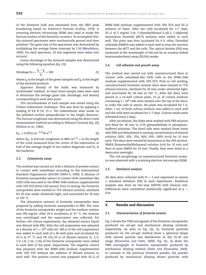
Fig. 7 – Results of MTT assay of the forsterite nanopowderproducts with different concentrations after different timesof cell culture using G292 cells. Data are presented as themean ± SD. (*: Significant difference (p < 0.05)).
engineering material. Compared to other bioactive ceramicscontaining SiO2, such as diopside (CaMgSi2O6) (Nonamiand Tsutsumi, 1999), akermanite (Ca2MgSi2O7) (Wu et al.,2006) and bredigite (Ca7MgSi4O16) ceramics (Wu et al., 2005),nanostructured forsterite possessed significantly improvedmechanical properties, which are some of themost importantproperties in the bone repairing ceramics.
Because of the higher surface energy in the nanostruc-tured materials than the coarse grain samples, nanostruc-tured forsterite might release inordinate ions in biologicalconditions. This could have diverse effects on the bone cells.Hence, we studied the in vitro biocompatibility of nanostruc-tured forsterite.
3.2. Cytotoxicity assay
Fig. 7 shows the results of the MTT assay of forsteritenanopowder dissolution after different times of cell cultur-ing. The results reveal that after 1 day of incubation, signifi-cant differences could not be detected among different con-centrations of the forsterite nanopowder. Cells proliferatedcontinually with increasing culture time in all samples. In thehigh concentrations of forsterite (50–200 mg/ml), cell num-bers increased slowly. In contrast, in the low concentrationsof forsterite (from 6.25 to 50 mg/ml), cells proliferated moreactively compared to the other samples (p < 0.05). By cultur-ing up to 7 days, the OD values of samples were significantlyhigher than the negative control (p < 0.05). The higher pro-liferation rate of samples having forsterite extracts than thenegative control confirmed that forsterite promoted the pro-liferation of cells without a cytotoxic effect. Recent studieshave demonstrated the positive stimulatory effects of extra-cellular Ca, Si, and Mg containing ionic products from the dis-solution of MgO–SiO2–CaO ceramic systems at a certain con-centration range on the osteoblast-like cells (Wu et al., 2005).In summary, in spite of the important role of Mg ion dissolu-tion in the bioactivity of inorganic biomaterials, at high con-centration of extracts, the inhibitory effect on cell prolifera-tion might be observed (Wu et al., 2005).
Based on the above results, although ionic productsfrom forsterite nanopowder dissolution could stimulate cellproliferation only at a certain concentration range, inhibitoryeffects on cell growth were not seen in other concentration
a
b
c
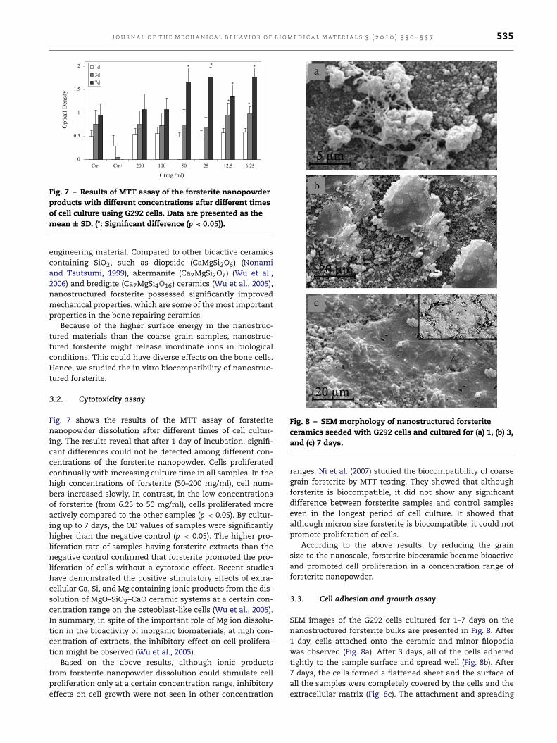
Fig. 8 – SEM morphology of nanostructured forsteriteceramics seeded with G292 cells and cultured for (a) 1, (b) 3,and (c) 7 days.
ranges. Ni et al. (2007) studied the biocompatibility of coarsegrain forsterite by MTT testing. They showed that althoughforsterite is biocompatible, it did not show any significantdifference between forsterite samples and control sampleseven in the longest period of cell culture. It showed thatalthough micron size forsterite is biocompatible, it could notpromote proliferation of cells.
According to the above results, by reducing the grainsize to the nanoscale, forsterite bioceramic became bioactiveand promoted cell proliferation in a concentration range offorsterite nanopowder.
3.3. Cell adhesion and growth assay
SEM images of the G292 cells cultured for 1–7 days on thenanostructured forsterite bulks are presented in Fig. 8. After1 day, cells attached onto the ceramic and minor filopodiawas observed (Fig. 8a). After 3 days, all of the cells adheredtightly to the sample surface and spread well (Fig. 8b). After7 days, the cells formed a flattened sheet and the surface ofall the samples were completely covered by the cells and theextracellular matrix (Fig. 8c). The attachment and spreading
536 J O U R N A L O F T H E M E C H A N I C A L B E H AV I O R O F B I O M E D I C A L M A T E R I A L S 3 ( 2 0 1 0 ) 5 3 0 – 5 3 7
of the cells on biomaterials are important processes of thecell/material interactions (Kirpatrick et al., 1997). Our resultsshowed that G292 cells completely attached to and spread onthe nanostructured forsterite ceramics.
Results indicate that nanostructured forsterite bioceramicpossesses good bioactivity, biocompatibility and mechanicalproperties that make it especially suitable as a bone tissuerepairing material. In our study, ionic concentrations ofdifferent samples showed obvious differences during cellculture. This indicates that it is possible to modulate therelease rates of active ions (Mg and Si) by making acomposite of forsterite nanopowder with other ceramics suchas bioactive glass, Hydroxyapatite, or polymers to obtainthe best desirable properties (i.e., mechanical properties andbiocompatibility). Based on the results, the forthcoming studyfor the present authors is the in vivo evaluation of preparedbioactive forsterite.
4. Conclusion
Nanostructured forsterite (Mg2SiO4) bulks, made of forsteritenanopowder (grainsize = 25–45 nm) produced via sol–gelmethod, with grain size of 120–150 nm and crystallitesize of 30–45 nm, was prepared with a two step sinteringmethod. Their sintering behavior and mechanical propertieswere studied. Hardness (1102 Hv) and fracture toughness(4.3 MPa m1/2) of nanostructured forsterite ceramics werehigher than those of the currently available hydroxyapatiteand coarse grain forsterite ceramics. The products fromforsterite nanopowder dissolution significantly promoted cellproliferations at a certain concentration range. In addition,the in vitro study showed significant G292 cell adhesion,spread, and growth on the surface of the nanostructuredforsterite ceramic. Results indicated that nanostructuredforsterite bioceramic possesses good in vitro bioactivity,biocompatibility, and mechanical properties and can be usedas a bioactive bone tissue engineering material. However,further in vivo studies need to be conducted to explore theapplicability of this ceramic as a bone tissue material.
Acknowledgement
The authors are grateful for the support of this research byIsfahan University of Technology.
R E F E R E N C E S
Althoff, J., Quint, P., Krefting, E.R., Hohling, H.J., 1982. Morphologi-cal studies on the epiphyseal growth plate combined with bio-chemical and X-ray microprobe analysis. Histochemistry 74,541–552.
Carlisle, E.M., 1970. Silicon: a possible factor in bone calcification.Science 167, 279–280.
Catledge, S.A., Fries, M.D., Vohra, Y.K., Lacefield, W.R., 2008.Nanostructured ceramics for biomedical implants. J. Nanosci.Nanotech. 2, 293–312.
Chim, H., Hutmacher, D.W., Chou, A.M., Oliveira, A.L.,Reis, R.L., Lim, T.C., 2006. Load-bearing applications in bonetissue engineering. Int. J. Oral Maxillofac. Surg. 35, 928–934.
Cottom, B.A., Mayo, M.J., 1996. Fracture toughness of nanocrys-talline ZrO2-3 mol% Y2O3 determined by vickers indentation.Scripta Mat. 34, 809–814.
Cullity, B.D, 1978. Elements of X-ray Diffraction. Prentice Hall,Addison-Wesley.
Du, C., Cui, F.Z., Zhu, X.D., Groot, K.de., 1999. Three-dimensionalnano-HAp/collagen matrix loading with osteogenic cells inorgan culture. J. Biomed. Mater. Res. 44, 407–415.
Edelstein, A.S., Cammarata, R.C., 1996. Nanomaterials: Synthesis,Properties and Applications. Institute of Physics (IOP), England.
Fathi, M.H., Kharaziha, M., 2009. Two-step sintering of dense,nanostructural forsterite. Mater. Lett. 63, 1455–1458.
Ghosh, A., Suri, A.K., Rao, B.T., Ramamohan, T.R., 2007. Low-temperature sintering and mechanical property evaluation ofnanocrystalline 8 mol% yttria fully stabilized zirconia. J. Am.Ceram. Soc. 90, 2015–2023.
Gutwein, L.G., Webster, T.J., 2002. Osteoblast and chrondrocyteproliferation in the presence of alumina and titania nanopar-ticles. J. Nanoparticle Res. 4, 231–238.
Han, M., Tang, X., Yin, H., Peng, S., 2007. Fabrication, microstruc-ture and properties of a YSZ electrolyte for SOFCs. J. PowerSources. 165, 757–763.
Hench, L.L., 1998. Bioceramics. J. Am. Ceram. Soc. 81 (7),1705–1728.
ISO/EN 10993–5, 1999. Biological Evaluation of Medical Devices,Part 5 Tests for Cytotoxicity, In Vitro Methods: 8.2 Tests onExtracts.
Kharaziha, M., Fathi, M.H., 2009. Synthesis and characterization ofbioactive forsterite nanopowder. Ceram. Int. 35, 2449–2454.
Kim, T.N., Balakrishnan, A., Lee, B.C., Kim, W.S., Dvorankova, B.,Smetana, K., Park, J.K., Panigrahi, B.B., 2008. In vitro fibroblastresponse to ultra fine grained titanium produced by a severeplastic deformation process. J. Mater. Sci., Mater. Med. 19,553–557.
Kirpatrick, C.J., Wagner, M., Kohler, H., Bittinger, F., Otto, M.,Klein, C.L., 1997. The cell and molecular biological approach tobiomaterial research: a perspective. J. Mater. Sci. Mater. Med.8, 131–141.
Mazaheri, M., Zahedi, A.M., Sadrnezhaad, S.K., 2007. Two-step sintering of nanocrystalline ZnO compacts: effect oftemperature on densification and grain growth. J. Am. Ceram.Soc. 91 (1), 56–63.
Mendelson, M.I., 1969. Average grain size in polycrystallineceramics. J. Am. Ceram. Soc. 52, 443–446.
Ni, S., Chou, L., Chang, J., 2007. Preparation and characterizationof forsterite (Mg2SiO4) bioceramics. Ceram. Int. 33, 83–88.
Ni, S., Chou, L., 2008. In vitro studies of novel CaO–SiO2–MgOsystem composite bioceramics. J. Chang. J. Mat. Sci., Mat. Med.19, 359–367.
Niihara, K., Morena, R., Hasselman, D.P., 1982. Evaluation of KIcof brittle solids by the indentation method with low crack-to-indent ratios. J. Mater. Sci. Lett. 1, 13–16.
Nonami, T., Tsutsumi, S., 1999. Study of diopside ceramics forbiomaterials. J. Mater. Sci., Mater. Med. 10, 475–479.
Schwarz, K., Milne, D.B., 1972. Growth-promoting effects of siliconin rats. Nature 239, 333–334.
Sprio, S., Tampieri, A., Celotti, G., Landi, E., 2009. Development ofhydroxyapatite/calcium silicate composites addressed to thedesign of load-bearing bone scaffolds. J. Mech. Behav. Biomed.Mater. 2, 147–155.
Wang, X.H., Chen, P.L., Chen, I.W., 2006. Two-step sintering ofceramics with constant grain-size. J. Am. Ceram. Soc. 89,431–437.
Webster, T.J., Siegel, R.W., Bizios, R., 1998. An in vitroevaluation of nanophase alumina for orthopaedic/dentalapplications. In: LeGeros, R.Z., LeGeros, J.P. (Eds.), Bioceramics

J O U R N A L O F T H E M E C H A N I C A L B E H AV I O R O F B I O M E D I C A L M A T E R I A L S 3 ( 2 0 1 0 ) 5 3 0 – 5 3 7 537
11: Proceedings of the 11th International Symposium onCeramics in Medicine. World Scientific, New York, pp. 273–276.
Wu, C., Chang, J., Wang, J., Ni, S., Zhai, W., 2005. Preparationand characteristics of a calciummagnesium silicate (bredigite)bioactive ceramic. Biomaterials 26, 2925–2931.
Wu, C., Chang, J., Ni, S., Wang, J., 2006. The in vitro bioactivity ofakermanite ceramics. J. Biomed. Mater. Res. A 76, 73–80.
Yu, P.C., Li, Q.F., Fuh, J.Y.H., Li, T., Lu, L., 2007. Two-stage sinteringof nanosized yttria stabilized zirconia process by powderinjection molding. J. Mater. Process. Technol. 192–193. 312–318.