impact of acute undernutrition on growth, ileal morphology and … · impact of acute...
TRANSCRIPT

Impact of acute undernutrition on growth, ilealmorphology and nutrient transport in a murine model
I.C. Sampaio1, P.H.Q.S. Medeiros1, F.A.P. Rodrigues1, P.A. Cavalcante1, S.A. Ribeiro1,J.S. Oliveira1, M.M.G. Prata1, D.V.S. Costa1, S.G.C. Fonseca2, M.M. Guedes1, A.M. Soares1,
G.A.C. Brito1, A. Havt1, S.R. Moore3* and A.A.M. Lima1
1Departamento de Fisiologia e Farmacologia, Faculdade de Medicina, Instituto de Biomedicina,Universidade Federal do Ceará, Fortaleza, CE, Brasil
2Departamento de Farmácia, Universidade Federal do Ceará, Fortaleza, CE, Brasil3Division of Gastroenterology, Hepatology, and Nutrition, Cincinnati Children’s Hospital Medical Center, Cincinnati, OH, USA
Abstract
Undernutrition represents a major public health challenge for middle- and low-income countries. This study aimed to evaluatewhether a multideficient Northeast Brazil regional basic diet (RBD) induces acute morphological and functional changes in theileum of mice. Swiss mice (B25 g) were allocated into two groups: i) control mice were fed a standard diet and II) under-nourished mice were fed the RBD. After 7 days, mice were killed and the ileum collected for evaluation of electrophysiologicalparameters (Ussing chambers), transcription (RT-qPCR) and protein expression (western blotting) of intestinal transporters andtight junctions. Body weight gain was significantly decreased in the undernourished group, which also showed decreased cryptdepth but no alterations in villus height. Electrophysiology measurements showed a reduced basal short circuit current (Isc) inthe undernourished group, with no differences in transepithelial resistance. Specific substrate-evoked Isc related to affinity andefficacy (glutamine and alanyl-glutamine) were not different between groups, except for the maximum Isc (efficacy) induced byglucose. Transcription of Sglt1 and Pept1was significantly higher in the undernourished group, while SN-2 transcription was decreased.No changes were found in transcription of CAT-1 and CFTR, while claudin-2 and occludin transcriptions were significantly increased inthe undernourished group. Despite mRNA changes, SGLT-1, PEPT-1, claudin-2 and occludin protein expression showed no differencebetween groups. These results demonstrate early effects of the RBD onmice, which include reduced body weight and crypt depth in theabsence of significant alterations to villus morphology, intestinal transporters and tight junction expression.
Key words: Undernutrition; Ion transport; Intestinal absorption
Introduction
Undernutrition is defined as a physiological outcome ofillness and/or hunger, which subclassifies as wasting (anacute state), stunting (a chronic state), underweight (mixedacute and/or chronic states) and micronutrient deficiencies.This condition has a great impact on global public health,especially in middle- and low-income countries (1,2), and isthe cause of 3.1 million child deaths annually. Further, it isestimated that 25.7% (about 165 million) of children youngerthan 5 years suffer from stunting, while 10.9% (B70 million)and 15.7% (B100 million) suffer from wasting and under-weight, respectively (3). Further evaluation of childhoodundernutrition-associated consequences has worrisomesocial implications, such as impaired school performance (4),increased economic costs (5), impaired immunity (6) and
a significantly higher number of deaths due to infectiousdiseases (7).
The regional basic diet (RBD) is an experimental rodentdiet based on the nutritional intake of the northeasternBrazilian population and is characterized by a deficit of pro-tein, fat and minerals, which trigger some clinical symptoms ofkwashiorkor and stunting, commonly reported in this popula-tion (8,9). Some studies have used the RBD in mouse modelsof undernutrition (10–12). Chronically, the RBD promotes del-eterious effects in the small intestine with altered villous heightand crypt depth, reduced transmucosal resistance, increasedpermeability and enhanced epithelial apoptosis (12).
Several studies seek to understand the pathophysiologyof undernutrition by characterizing alterations of transcellular
Correspondence: A.A.M. Lima: <[email protected]>
*The current address of S.R. Moore is: Division of Gastroenterology and Nutrition, Department of Pediatrics, University of Virginia,Charlottesville, VA, USA.
Received April 8, 2016 | Accepted June 24, 2016
Braz J Med Biol Res | doi: 10.1590/1414-431X20165340
Brazilian Journal of Medical and Biological Research (2016) 49(10): e5340, http://dx.doi.org/10.1590/1414-431X20165340ISSN 1414-431X 1/10
and paracellular transports (12–19). Transmembrane pro-teins, such as sodium-glucose linked transporter 1 (SGLT-1),peptide transporter 1 (PEPT-1) and cationic amino acidtransport-1 (CAT-1), are modulated by undernourishedstates (14–16,19). Conversely, the paracellular transportmediating tight junctions, such as claudins, occludins andzonula occludens, have been described as fundamental forabsorption of electrolytes and water in the small intestine,and are modulated by undernutrition as well (12,13,17–19).
Generally, the clinical management of undernutritionrequires early detection of pathophysiological changes.As the majority of studies have shown only the long-termeffects of RBD, we examined if the RBD induces acutemorphological and functional changes in the ileum in ashort period of time, evaluating modifications of electro-physiological parameters and transcription and expres-sion of intestinal transporters and tight junction proteins.
Material and Methods
Animals and experimental designMale Swiss mice (total of 28) weighing B25 g were
obtained from the Departamento de Fisiologia e Farma-cologia, Universidade Federal of Ceará, and maintainedunder controlled temperature (21±2°C) and humidity(60±5%), and a 12:12 h light:dark cycle. Mice had accessto a standard commercial diet and water ad libitum. Theanimal protocols were performed according to the normsof the National Council for Control of Animal Experimenta-tion (CONCEA) and were approved by the Ethics Com-mittee of Animal Research of the Universidade Federal doCeará, Fortaleza, CE, Brazil (protocol #62/11).
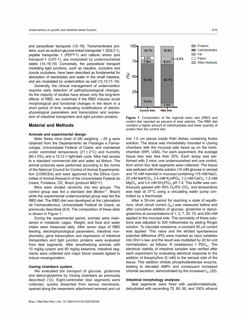
Mice were divided randomly into two groups. Thecontrol group was fed a standard diet (Biotecs, Brazil)while the experimental undernourished group was fed theRBD diet. The RBD diet was developed at the Laboratóriode Farmacotécnica, Universidade Federal do Ceará, aspreviously described (8,9). The composition of these dietsis shown in Figure 1.
During the experimental period, animals were main-tained in metabolic cages. Weight, and food and waterintake were measured daily. After seven days of RBDfeeding, electrophysiological parameters, intestinal mor-phometry, gene transcription and expression of intestinaltransporters and tight junction proteins were evaluatedfrom ileal segments. After anesthetizing animals with10 mg/kg xylazin and 90 mg/kg ketamine, intestinal seg-ments were collected and major blood vessels ligated toinduce exsanguination.
Ussing chambers systemWe evaluated the transport of glucose, glutamine
and alanyl-glutamine by Ussing chambers as previouslydescribed (12). Eight-centimeter ileal segments werecollected, quickly dissected from serous membrane,opened along the mesenteric attachment remnant and cut
into 1.5 cm pieces inside Petri dishes containing Krebssolution. The tissue was immediately mounted in Ussingchambers with the mucosal side faced up on the hemi-chamber (WPI, USA). For each experiment, the averagetissue loss was less than 20%. Each assay was per-formed with 2 mice; one undernourished and one control,from which four ileal segments were collected. The tissuewas perfused with Krebs solution (10 mM glucose in serosaland 10 mM mannitol in mucosa) containing 115 mM NaCl,25 mM NaHCO3, 2.4 mM K2HPO4, 1.2 mM CaCl2, 1.2 mMMgCl2, and 0.4 mM KH2PO4, pH 7.4. This buffer was con-tinuously gassed with 95% O2/5% CO2, and temperaturewas kept at 37°C using a circulating water pump con-trolled by a thermostat.
After a 30-min period for reaching a state of equilib-rium, short circuit current (Isc) was measured before andafter cumulative addition of glucose, glutamine or alanyl-glutamine at concentrations of 1, 2, 7, 20, 70, and 200 mMapplied to the mucosal side. The osmolarity of these solu-tions was adjusted to 300 milliosmoles by adding Ringersolution. To calculate resistance, a constant 50 mA currentwas applied. This value and the elicited spontaneouspotential difference (PD) were inserted as input variablesinto Ohm’s law, and the result was multiplied by 20 for unitnormalization, as follows: R (resistance) = PD/Isc. Theelectrical viability of intestinal samples was verified aftereach experiment by evaluating electrical response to theaddition of theophylline (5 mM) to the serosal side of thetissue. This addition inhibits phosphodiesterase enzyme,leading to elevated AMPc and consequent increasedchloride secretion, demonstrated by the increased Isc (20).
Intestinal morphology analysesIleal segments were fixed with paraformaldehyde,
dehydrated with ascending 70, 80, 90, and 100% ethanol
Figure 1. Composition of the regional basic diet (RBD) andcontrol diet reported as percent of total calories. The RBD dietcontains a higher amount of carbohydrates and lower quantity ofprotein than the control diet.
Braz J Med Biol Res | doi: 10.1590/1414-431X20165340
Undernutrition on growth and intestinal barrier function 2/10
and processed in paraffin. The resulting blocks weresliced into 5-mm-thick sections, stained with hematoxylinand eosin (H&E), and observed under a light microscope(� 400). The area of the villi and crypt depth were measuredas previously described (21) using Image J software ver-sion 1.6.0 (National Institutes of Health, USA).
Evaluation of gene transcription of intestinaltransporters and tight junctions
Gene transcription of sodium-glucose linked transporter(SGLT-1), system N-transporter (SN-2), cationic aminoacid transporter (CAT-1), peptide transporter 1 (PEPT-1),cystic fibrosis transmembrane conductance regulator(CFTR,) zonula occludens-1 (ZO-1), claudin-1, claudin-2,and occludin was determined by quantitative reversetranscription-polymerase chain reaction (qRT-PCR). Ini-tially, total RNA was isolated from mucosal membrane of
ileal segments using RNeasy mini Kit (Qiagen, Germany)according to the manufacturer’s instructions. The cDNAwas synthesized using iScriptt cDNA Synthesis Kit (Invitro-gen, USA). The reference gene peptidylprolyl isomerase Awas used for this experiment (22). The primers design wasbased on mRNA sequences obtained from the NationalCenter for Biotechnology Information (http://www.ncbi.nlm.nih.gov; accessed on February 4, 2014).
The qRT-PCR reactions were performed in a finalvolume of 25 mL containing 12.5 mL of iQ SYBR greensupermix (Bio-Rad, USA), 200 nM (each) primers, and 1 mLof cDNA from sample. All primers and conditions for qRT-PCR are shown in Table 1. To measure the specificity ofthe applied amplifications (i.e., to determine whether theformed products were specific for the tested genes), weperformed a melting curve analysis in which the reactiontemperature was increased 0.5°C every 15 s, beginning at
Table 1. Description of genes, primers sequences, accession numbers (NCBI) and qRT-PCR conditions.
Genes/Primerssequences (50-30)
GenBank (NCBI)accession number
Annealing temperature(40 cycles)*
ZO-1GACCATCGCCTACGGTTTGA NM_001163574.1 2000, 60°CAGGTCTCGGGGATGCTGATT
Claudin-1TCTACGAGGGACTGTGGATG NM_016674.4 2000, 58°CTCAGATTCAGCAAGGAGTCG
Claudin -2CCCACCACCACCAGCTTAAT NM_016675.4 2000, 60°CGAAATGGCTTCCAGGTCAGC
OccludinAAGAGCAGCCAAAGGCTTCC NM_008756.2 2000, 60°CCGTCGGGTTCACTCCCATTA
SGLT-1CGGAAGAAGCGATCTGAGAA NM_019810.4 2000, 58°CAATCAGCACGAGGATGAACA
SN-2ATATCCTCGTCATCTGTGTGC NM_172479.2 2000, 60°CCAGTAGGTACAATACGGAGGTAGA
CAT-1
CTTTGGATTCTCTGGTGTCCTGTC NM_007513.4 2000, 58°CGTTCTTGACTTCTTCCCCTGTGG
PEPT-1
AGGGGAGAACGGAATCAGGT NM_053079.2 2000, 60°CCTTTTCGCCAGAAGGGAAGA
CFTR
GGATGCTGAGGAAGCAACTC NM_021050.2 2000, 58°CCCAGCCTGGAACTCTCTTTG
Ppia
AATGCTGGACCAAACACAAA NM_008907.1 2000, 58°CTTCCACAATGTTCATGCCTT
*Every cycle began with a denaturing step (20 s at 95°C) and finished with an extension of 45 s to 1 minat 72°C.
Braz J Med Biol Res | doi: 10.1590/1414-431X20165340
Undernutrition on growth and intestinal barrier function 3/10
the annealing temperature of the tested set of primersand ending at 95°C. Throughout the curve constructionprocess, the changes in fluorescence were measured,and the data obtained, using CFX Manager software(version 3.0; Bio-Rad), were based on the values for thethreshold cycle, i.e., where the observed fluorescencewas 10-fold higher than the basal fluorescence for eachreaction. Gene transcription was obtained by applying themathematical 2-DDCT method (23).
Immunoblot analysisIn order to quantify SGLT-1, PEPT-1, claudin-2 and
occludin proteins, mucosal membranes from ileal seg-ments were homogenized in RIPA lysis buffer (25 mMTris-HCL, pH 7.6; 150 mM NaCl; 5 mM EDTA; 1% NP40;1% triton X-100; 1% sodium deoxycholate; 0.1% SDS)and protease inhibitor (1 mL inhibitor: 100 mL RIPA). Thehomogenates were centrifuged (17,949 g, 17 min, 4°C),and the supernatant was collected. Protein concentrationswere determined through the bicinchoninic acid assayusing PierceTM BCA protein assay kit (Thermo Scientific,USA). The protein (20 mg) was prepared adding Laemmlisample buffer with b-mercaptoethanol and denatured at95°C for 5 min, except for PEPT-1, SGLT-1 and occludin.Then, proteins were separated in a SDS-polyacrylamidegel (8% for occludin, SGLT-1 and PEPT-1 analysis and12.5% for claudin analysis) under a condition of 120 voltsand transferred to polyvinylidene difluoride (PVDF) mem-branes by electrophoresis for 2 h. PVDF membraneswere blocked with 5% bovine serum albumin for 1 h andincubated overnight with primary rabbit antibody [anti-bactin (1:1000), anti-SGLT-1 (1:500), anti-PEPT-1 (1:200),anti-claudin-2 (1:100), or anti-occludin (1:1000); SantaCruz Biotechnology, USA]. Chemiluminescent detectionusing Clarity Western ECL Substrate (Bio-Rad) wasperformed after incubation of the membrane with second-ary antibody (1:1000) for 1 h. Finally, bands were capturedusing the ChemiDoc system (Bio-Rad). Densitometricquantification of bands was made using Image J softwareversion 1.6.0 (National Institutes of Health).
Statistical analysisData are reported as means±SE. One- or two-way
analysis of variance (ANOVA) followed by the Bonferroni’spost hoc test were used for parametric data. Gene tran-scription data were evaluated by the Mann-Whitney test.Differences were considered to be statistically significantwhen Po0.05. Analysis was performed using GraphPadPrism 5.0 (GraphPad Software, USA).
Results
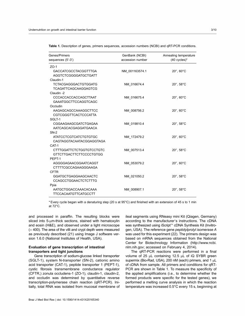
Impact of RBD diet for 7 days on body weightRBD feeding triggered a significant decrease in body
weight gain when compared to the nourished group duringthe short period of 7 days (Po0.001). This difference was
observable by the second day of intake, showing a reduc-tion of 13.3% from the previous time point (nourished30.0±0.8 vs 26.10±0.7 g undernourished; Figure 2A). Inaddition, food intake was higher in the nourished groupuntil day 2 (Po0.01), and this difference was not observedsubsequently (Figure 2B). Water intake did not differstatistically between the groups (Figure 2C).
Impact of RBD diet for 7 days on intestinalmorphology
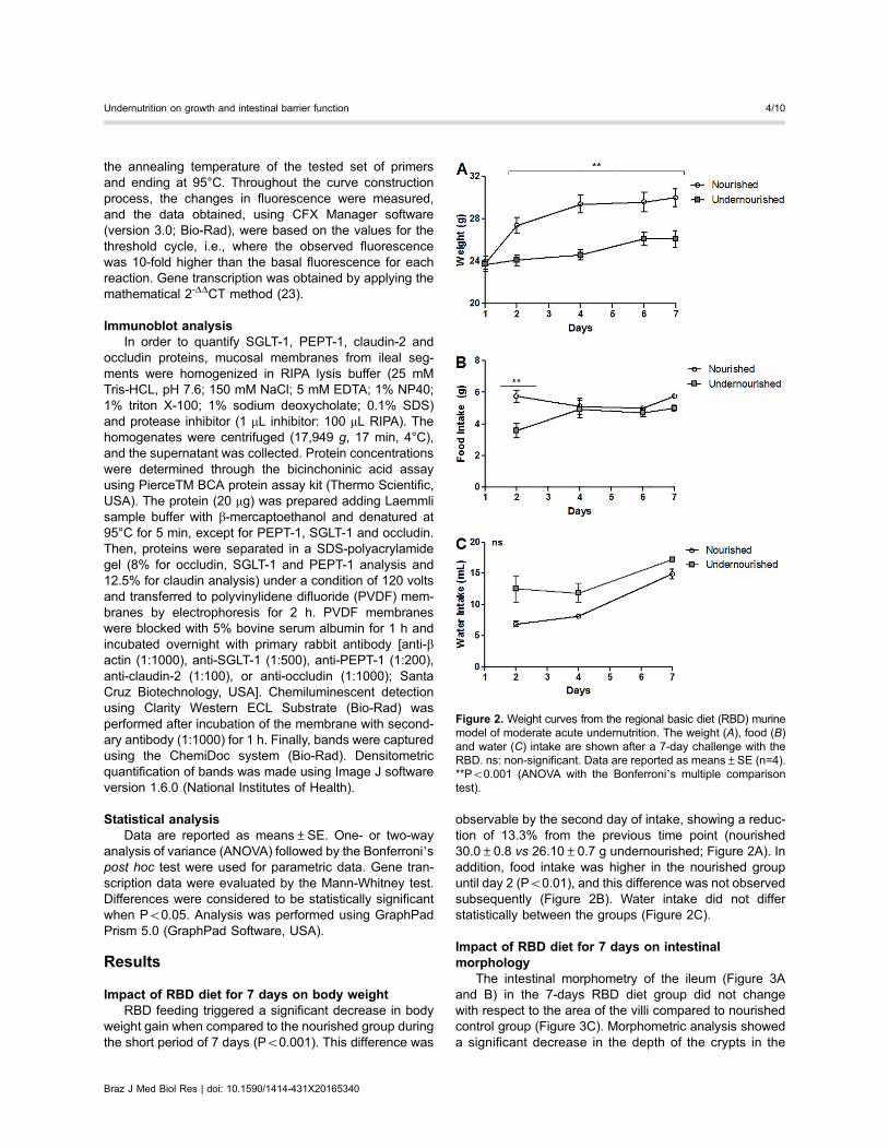
The intestinal morphometry of the ileum (Figure 3Aand B) in the 7-days RBD diet group did not changewith respect to the area of the villi compared to nourishedcontrol group (Figure 3C). Morphometric analysis showeda significant decrease in the depth of the crypts in the
Figure 2. Weight curves from the regional basic diet (RBD) murinemodel of moderate acute undernutrition. The weight (A), food (B)and water (C) intake are shown after a 7-day challenge with theRBD. ns: non-significant. Data are reported as means±SE (n=4).**Po0.001 (ANOVA with the Bonferroni’s multiple comparisontest).
Braz J Med Biol Res | doi: 10.1590/1414-431X20165340
Undernutrition on growth and intestinal barrier function 4/10
undernourished group compared to nourished control(133.6±4.12 vs 86.50±5.871 mm; Po0.001; Figure 3D).
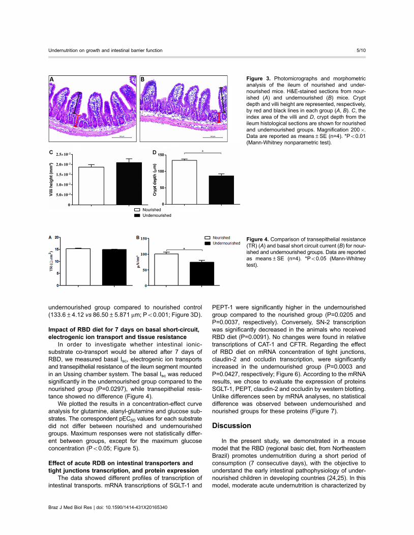
Impact of RBD diet for 7 days on basal short-circuit,electrogenic ion transport and tissue resistance
In order to investigate whether intestinal ionic-substrate co-transport would be altered after 7 days ofRBD, we measured basal Isc, electrogenic ion transportsand transepithelial resistance of the ileum segment mountedin an Ussing chamber system. The basal Isc was reducedsignificantly in the undernourished group compared to thenourished group (P=0.0297), while transepithelial resis-tance showed no difference (Figure 4).
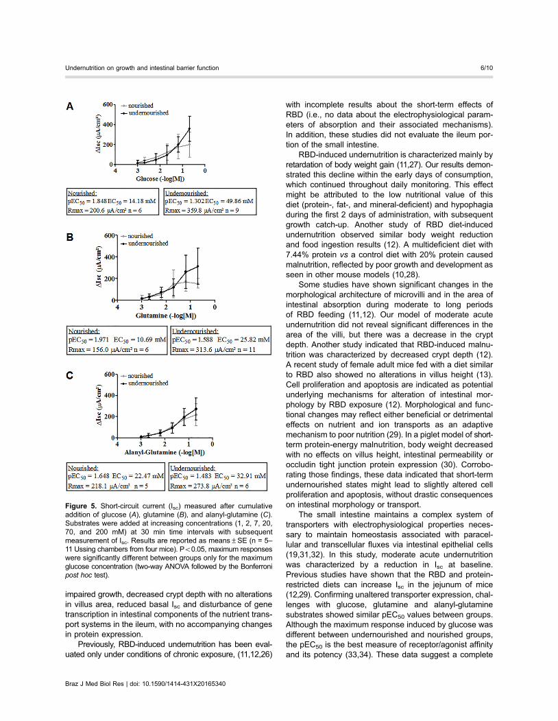
We plotted the results in a concentration-effect curveanalysis for glutamine, alanyl-glutamine and glucose sub-strates. The correspondent pEC50 values for each substratedid not differ between nourished and undernourishedgroups. Maximum responses were not statistically differ-ent between groups, except for the maximum glucoseconcentration (Po0.05; Figure 5).
Effect of acute RDB on intestinal transporters andtight junctions transcription, and protein expression
The data showed different profiles of transcription ofintestinal transports. mRNA transcriptions of SGLT-1 and
PEPT-1 were significantly higher in the undernourishedgroup compared to the nourished group (P=0.0205 andP=0.0037, respectively). Conversely, SN-2 transcriptionwas significantly decreased in the animals who receivedRBD diet (P=0.0091). No changes were found in relativetranscriptions of CAT-1 and CFTR. Regarding the effectof RBD diet on mRNA concentration of tight junctions,claudin-2 and occludin transcription, were significantlyincreased in the undernourished group (P=0.0003 andP=0.0427, respectively; Figure 6). According to the mRNAresults, we chose to evaluate the expression of proteinsSGLT-1, PEPT, claudin-2 and occludin by western blotting.Unlike differences seen by mRNA analyses, no statisticaldifference was observed between undernourished andnourished groups for these proteins (Figure 7).
Discussion
In the present study, we demonstrated in a mousemodel that the RBD (regional basic diet, from NortheasternBrazil) promotes undernutrition during a short period ofconsumption (7 consecutive days), with the objective tounderstand the early intestinal pathophysiology of under-nourished children in developing countries (24,25). In thismodel, moderate acute undernutrition is characterized by
Figure 3. Photomicrographs and morphometricanalysis of the ileum of nourished and under-nourished mice. H&E-stained sections from nour-ished (A) and undernourished (B) mice. Cryptdepth and villi height are represented, respectively,by red and black lines in each group (A, B). C, theindex area of the villi and D, crypt depth from theileum histological sections are shown for nourishedand undernourished groups. Magnification 200�.Data are reported as means±SE (n=4). *Po0.01(Mann-Whitney nonparametric test).
Figure 4. Comparison of transepithelial resistance(TR) (A) and basal short circuit current (B) for nour-ished and undernourished groups. Data are reportedas means±SE (n=4). *Po0.05 (Mann-Whitneytest).
Braz J Med Biol Res | doi: 10.1590/1414-431X20165340
Undernutrition on growth and intestinal barrier function 5/10
impaired growth, decreased crypt depth with no alterationsin villus area, reduced basal Isc and disturbance of genetranscription in intestinal components of the nutrient trans-port systems in the ileum, with no accompanying changesin protein expression.
Previously, RBD-induced undernutrition has been eval-uated only under conditions of chronic exposure, (11,12,26)
with incomplete results about the short-term effects ofRBD (i.e., no data about the electrophysiological param-eters of absorption and their associated mechanisms).In addition, these studies did not evaluate the ileum por-tion of the small intestine.
RBD-induced undernutrition is characterized mainly byretardation of body weight gain (11,27). Our results demon-strated this decline within the early days of consumption,which continued throughout daily monitoring. This effectmight be attributed to the low nutritional value of thisdiet (protein-, fat-, and mineral-deficient) and hypophagiaduring the first 2 days of administration, with subsequentgrowth catch-up. Another study of RBD diet-inducedundernutrition observed similar body weight reductionand food ingestion results (12). A multideficient diet with7.44% protein vs a control diet with 20% protein causedmalnutrition, reflected by poor growth and development asseen in other mouse models (10,28).
Some studies have shown significant changes in themorphological architecture of microvilli and in the area ofintestinal absorption during moderate to long periodsof RBD feeding (11,12). Our model of moderate acuteundernutrition did not reveal significant differences in thearea of the villi, but there was a decrease in the cryptdepth. Another study indicated that RBD-induced malnu-trition was characterized by decreased crypt depth (12).A recent study of female adult mice fed with a diet similarto RBD also showed no alterations in villus height (13).Cell proliferation and apoptosis are indicated as potentialunderlying mechanisms for alteration of intestinal mor-phology by RBD exposure (12). Morphological and func-tional changes may reflect either beneficial or detrimentaleffects on nutrient and ion transports as an adaptivemechanism to poor nutrition (29). In a piglet model of short-term protein-energy malnutrition, body weight decreasedwith no effects on villus height, intestinal permeability oroccludin tight junction protein expression (30). Corrobo-rating those findings, these data indicated that short-termundernourished states might lead to slightly altered cellproliferation and apoptosis, without drastic consequenceson intestinal morphology or transport.
The small intestine maintains a complex system oftransporters with electrophysiological properties neces-sary to maintain homeostasis associated with paracel-lular and transcellular fluxes via intestinal epithelial cells(19,31,32). In this study, moderate acute undernutritionwas characterized by a reduction in Isc at baseline.Previous studies have shown that the RBD and protein-restricted diets can increase Isc in the jejunum of mice(12,29). Confirming unaltered transporter expression, chal-lenges with glucose, glutamine and alanyl-glutaminesubstrates showed similar pEC50 values between groups.Although the maximum response induced by glucose wasdifferent between undernourished and nourished groups,the pEC50 is the best measure of receptor/agonist affinityand its potency (33,34). These data suggest a complete
Figure 5. Short-circuit current (Isc) measured after cumulativeaddition of glucose (A), glutamine (B), and alanyl-glutamine (C).Substrates were added at increasing concentrations (1, 2, 7, 20,70, and 200 mM) at 30 min time intervals with subsequentmeasurement of Isc. Results are reported as means±SE (n = 5–11 Ussing chambers from four mice). Po0.05, maximum responseswere significantly different between groups only for the maximumglucose concentration (two-way ANOVA followed by the Bonferronipost hoc test).
Braz J Med Biol Res | doi: 10.1590/1414-431X20165340
Undernutrition on growth and intestinal barrier function 6/10

responsiveness of the intestinal absorption ability of theundernourished ileum. Furthermore, transepithelial resis-tance was not altered, which corroborates the unimpairedparacellular transport (29).
In order to maintain membrane stability between epithelialcells in the small intestine, claudins play an important rolein the complex intestinal transporters system, contributingfor the proper functioning of SGLT-1 and PepT-1 carriersplus several transporters for amino acids (19,35,36). Inour study, undernutrition caused diverse regulation onmRNA levels of intestinal transporters and tight junctions:claudin-2, occludin, SGLT-1 and PepT-1 mRNAs wereincreased while SN-2 mRNA was decreased. Despite thevariation on mRNA levels, the protein expression of SGLT-1,PetT-1, claudin-2 and occludin did not vary. The lack ofcorrelation between mRNA and protein levels of intestinaltransporters and tight junctions has also been reported inother studies that addressed dietary restrictions in animals
(37,14). Furthermore, this study evaluated these param-eters in parallel, and it is well known that mRNA andprotein production rates of a gene may occur at differenttime-points (38,39). Moreover, post-transcriptional and trans-lational mechanisms of regulation might help to explainthese findings (40). Furthermore, the protein expressionresults are in agreement with the results of Isc-evokedsubstrates variation, measured in the concentration-effectapproach.
It was shown previously that dietary protein restrictionin pregnant rats promoted increased transcription andprotein expression of intestinal transporter genes (SGLT-1and PepT-1) in the duodenum of their offspring, but not inthe jejunum or ileum (14). Interestingly, removal of luminalnutrition in rats, by applying total parenteral nutrition forinduction of intestinal damage, also increased mRNA ofsome transporter genes, as PEPT-1, although it did notmodify CAT1 and SN-2 (15). In addition, a study of
Figure 6. Effects of the regional basic diet on relative expression of A, SGLT-1; B, PepT-1; C, CAT-1; D, CFTR; E, SN-2; F, ZO-1;G, claudin-1; H, claudin-2; I, occludin. *Po0.05, Mann-Whitney test (n=7).
Braz J Med Biol Res | doi: 10.1590/1414-431X20165340
Undernutrition on growth and intestinal barrier function 7/10
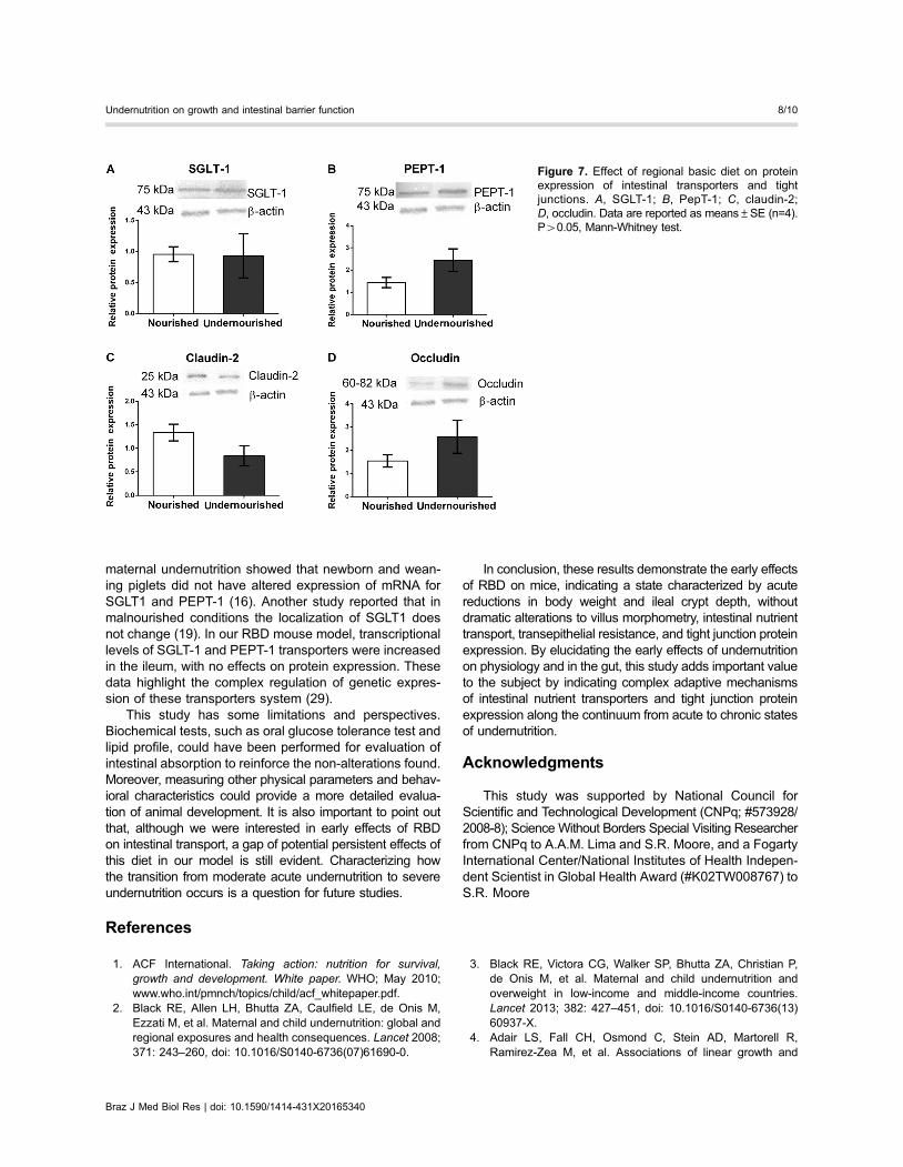
maternal undernutrition showed that newborn and wean-ing piglets did not have altered expression of mRNA forSGLT1 and PEPT-1 (16). Another study reported that inmalnourished conditions the localization of SGLT1 doesnot change (19). In our RBD mouse model, transcriptionallevels of SGLT-1 and PEPT-1 transporters were increasedin the ileum, with no effects on protein expression. Thesedata highlight the complex regulation of genetic expres-sion of these transporters system (29).
This study has some limitations and perspectives.Biochemical tests, such as oral glucose tolerance test andlipid profile, could have been performed for evaluation ofintestinal absorption to reinforce the non-alterations found.Moreover, measuring other physical parameters and behav-ioral characteristics could provide a more detailed evalua-tion of animal development. It is also important to point outthat, although we were interested in early effects of RBDon intestinal transport, a gap of potential persistent effects ofthis diet in our model is still evident. Characterizing howthe transition from moderate acute undernutrition to severeundernutrition occurs is a question for future studies.
In conclusion, these results demonstrate the early effectsof RBD on mice, indicating a state characterized by acutereductions in body weight and ileal crypt depth, withoutdramatic alterations to villus morphometry, intestinal nutrienttransport, transepithelial resistance, and tight junction proteinexpression. By elucidating the early effects of undernutritionon physiology and in the gut, this study adds important valueto the subject by indicating complex adaptive mechanismsof intestinal nutrient transporters and tight junction proteinexpression along the continuum from acute to chronic statesof undernutrition.
Acknowledgments
This study was supported by National Council forScientific and Technological Development (CNPq; #573928/2008-8); Science Without Borders Special Visiting Researcherfrom CNPq to A.A.M. Lima and S.R. Moore, and a FogartyInternational Center/National Institutes of Health Indepen-dent Scientist in Global Health Award (#K02TW008767) toS.R. Moore
References
1. ACF International. Taking action: nutrition for survival,growth and development. White paper. WHO; May 2010;www.who.int/pmnch/topics/child/acf_whitepaper.pdf.
2. Black RE, Allen LH, Bhutta ZA, Caulfield LE, de Onis M,Ezzati M, et al. Maternal and child undernutrition: global andregional exposures and health consequences. Lancet 2008;371: 243–260, doi: 10.1016/S0140-6736(07)61690-0.
3. Black RE, Victora CG, Walker SP, Bhutta ZA, Christian P,de Onis M, et al. Maternal and child undernutrition andoverweight in low-income and middle-income countries.Lancet 2013; 382: 427–451, doi: 10.1016/S0140-6736(13)60937-X.
4. Adair LS, Fall CH, Osmond C, Stein AD, Martorell R,Ramirez-Zea M, et al. Associations of linear growth and
Figure 7. Effect of regional basic diet on proteinexpression of intestinal transporters and tightjunctions. A, SGLT-1; B, PepT-1; C, claudin-2;D, occludin. Data are reported as means±SE (n=4).P40.05, Mann-Whitney test.
Braz J Med Biol Res | doi: 10.1590/1414-431X20165340
Undernutrition on growth and intestinal barrier function 8/10

relative weight gain during early life with adult healthand human capital in countries of low and middle income:findings from five birth cohort studies. Lancet 2013; 382:525–534, doi: 10.1016/S0140-6736(13)60103-8.
5. Horton S, Steckel RH. Malnutrition - Global economic lossesattributable to malnutrition 1900-2000 and projections to2050. Copenhagen Consensus on Human Challenges. Copen-hagen; 2011; http://www.copenhagenconsensus.com/sites/default/files/malnutrition.pdf.
6. Rytter MJ, Kolte L, Briend A, Friis H, Christensen VB. Theimmune system in children with malnutrition - a systematicreview. PLoS One 2014; 9: e105017, doi: 10.1371/journal.pone.0105017.
7. Caulfield LE, de Onis M, Blossner M, Black RE. Under-nutrition as an underlying cause of child deaths associatedwith diarrhea, pneumonia, malaria, and measles. Am J ClinNutr 2004; 80: 193–198.
8. Brigide P, Ataide TR, Baptista AS, Canniatti-Brazaca SG,Abdalla AL, Nascimento Filho V, et al. Bioavailability of ironin the regional basic diet (RBD) with dietary supplement inBrazil. Biol Trace Elem Res 2011; 140: 53–65, doi: 10.1007/s12011-010-8681-6.
9. Teodosio NR, Lago ES, Romani SA, Guedes RC. A regionalbasic diet from northeast Brazil as a dietary model ofexperimental malnutrition. Arch Latinoam Nutr 1990; 40:533–547.
10. Bolick DT, Roche JK, Hontecillas R, Bassaganya-Riera J,Nataro JP, Guerrant RL. Enteroaggregative Escherichia colistrain in a novel weaned mouse model: exacerbation bymalnutrition, biofilm as a virulence factor and treatment bynitazoxanide. J Med Microbiol 2013; 62: 896–905, doi:10.1099/jmm.0.046300-0.
11. de Queiroz CA, Fonseca SG, Frota PB, Figueiredo IL,Aragao KS, Magalhaes CE, et al. Zinc treatment amelioratesdiarrhea and intestinal inflammation in undernourished rats.BMC Gastroenterol 2014; 14: 136, doi: 10.1186/1471-230X-14-136.
12. Ueno PM, Oria RB, Maier EA, Guedes M, de Azevedo OG,Wu D, et al. Alanyl-glutamine promotes intestinal epithelialcell homeostasis in vitro and in a murine model of weanlingundernutrition. Am J Physiol Gastrointest Liver Physiol 2011;301: G612–G622, doi: 10.1152/ajpgi.00531.2010.
13. Brown EM, Wlodarska M, Willing BP, Vonaesch P, Han J,Reynolds LA, et al. Diet and specific microbial exposuretrigger features of environmental enteropathy in a novelmurine model. Nat Commun 2015; 6: 7806, doi: 10.1038/ncomms8806.
14. Pinheiro DF, Pinheiro PF, Buratini J, Jr., Castilho AC, LimaPF, Trinca LA, et al. Maternal protein restriction duringpregnancy affects gene expression and immunolocalizationof intestinal nutrient transporters in rats. Clin Sci 2013; 125:281–289, doi: 10.1042/CS20120400.
15. Howard A, Goodlad RA, Walters JR, Ford D, Hirst BH.Increased expression of specific intestinal amino acid andpeptide transporter mRNA in rats fed by TPN is reversed byGLP-2. J Nutr 2004; 134: 2957–2964.
16. Cao M, Che L, Wang J, Yang M, Su G, Fang Z, et al. Effectsof maternal over- and undernutrition on intestinal morphol-ogy, enzyme activity, and gene expression of nutrient trans-porters in newborn and weaned pigs. Nutrition 2014; 30:1442–1447, doi: 10.1016/j.nut.2014.04.016.
17. Furuse M, Hirase T, Itoh M, Nagafuchi A, Yonemura S,Tsukita S, et al. Occludin: a novel integral membrane proteinlocalizing at tight junctions. J Cell Biol 1993; 123: 1777–1788,doi: 10.1083/jcb.123.6.1777.
18. Itoh M, Nagafuchi A, Yonemura S, Kitani-Yasuda T,Tsukita S, Tsukita S. The 220-kD protein colocalizing withcadherins in non-epithelial cells is identical to ZO-1, a tightjunction-associated protein in epithelial cells: cDNA cloningand immunoelectron microscopy. J Cell Biol 1993; 121:491–502, doi: 10.1083/jcb.121.3.491.
19. Wada M, Tamura A, Takahashi N, Tsukita S. Loss ofclaudins 2 and 15 from mice causes defects in paracellularNa+ flow and nutrient transport in gut and leads to deathfrom malnutrition. Gastroenterol 2013; 144: 369–380, doi:10.1053/j.gastro.2012.10.035.
20. Field M. Intestinal secretion: effect of cyclic AMP and itsrole in cholera. N Engl J Med 1971; 284: 1137–1144,doi: 10.1056/NEJM197105202842008.
21. Coutinho BP, Oria RB, Vieira CM, Sevilleja JE, Warren CA,Maciel JG, et al. Cryptosporidium infection causes under-nutrition and, conversely, weanling undernutrition intensifiesinfection. J Parasitol 2008; 94: 1225–1232, doi: 10.1645/GE-1411.1.
22. Sirakov M, Borra M, Cambuli FM, Plateroti M. Definingsuitable reference genes for RT-qPCR analysis on intestinalepithelial cells. Mol Biotechnol 2013; 54: 930–938, doi:10.1007/s12033-012-9643-3.
23. Livak KJ, Schmittgen TD. Analysis of relative gene expres-sion data using real-time quantitative PCR and the 2(-DeltaDelta C(T)) Method. Methods 2001; 25: 402–408, doi: 10.1006/meth.2001.1262.
24. Lima AA, Anstead GM, Zhang Q, Figueiredo IL, Soares AM,Mota RM, et al. Effects of glutamine alone or in combinationwith zinc and vitamin A on growth, intestinal barrier function,stress and satiety-related hormones in Brazilian shantytownchildren. Clinics 2014; 69: 225–233, doi: 10.6061/clinics/2014(04)02.
25. Platts-Mills JA, Babji S, Bodhidatta L, Gratz J, Haque R,Havt A, et al. Pathogen-specific burdens of communitydiarrhoea in developing countries: a multisite birth cohortstudy (MAL-ED). Lancet Glob Health 2015; 3: e564–e575,doi: 10.1016/S2214-109X(15)00151-5.
26. Maier EA, Weage KJ, Guedes MM, Denson LA, McNealMM, Bernstein DI, et al. Protein-energy malnutrition altersIgA responses to rotavirus vaccination and infection butdoes not impair vaccine efficacy in mice. Vaccine 2014; 32:48–53, doi: 10.1016/j.vaccine.2013.10.072.
27. Barros KM, Manhaes-De-Castro R, Lopes-De-Souza S,Matos RJ, Deiro TC, Cabral-Filho JE, et al. A regionalmodel (Northeastern Brazil) of induced mal-nutritiondelays ontogeny of reflexes and locomotor activity in rats.Nutr Neurosci 2006; 9: 99–104, doi: 10.1080/10284150600772148.
28. Azevedo OG, Bolick DT, Roche JK, Pinkerton RF, LimaAA, Vitek MP, et al. Apolipoprotein E plays a key roleagainst cryptosporidial infection in transgenic undernour-ished mice. PLoS One 2014; 9: e89562, doi: 10.1371/journal.pone.0089562.
29. Ferraris RP, Carey HV. Intestinal transport during fastingand malnutrition. Annu Rev Nutr 2000; 20: 195–219, doi:10.1146/annurev.nutr.20.1.195.
Braz J Med Biol Res | doi: 10.1590/1414-431X20165340
Undernutrition on growth and intestinal barrier function 9/10

30. Jacobi SK, Moeser AJ, Blikslager AT, Rhoads JM, Corl BA,Harrell RJ, et al. Acute effects of rotavirus and malnutritionon intestinal barrier function in neonatal piglets. World JGastroenterol 2013; 19: 5094–5102, doi: 10.3748/wjg.v19.i31.5094.
31. Gawenis LR, Hut H, Bot AG, Shull GE, de Jonge HR, StienX, et al. Electroneutral sodium absorption and electrogenicanion secretion across murine small intestine are regulatedin parallel. Am J Physiol Gastrointest Liver Physiol 2004;287: G1140–G1149, doi: 10.1152/ajpgi.00177.2004.
32. Chen M, Singh A, Xiao F, Dringenberg U, Wang J,Engelhardt R, et al. Gene ablation for PEPT1 in miceabolishes the effects of dipeptides on small intestinal fluidabsorption, short-circuit current, and intracellular pH. Am JPhysiol Gastrointest Liver Physiol 2010; 299: G265–G274,doi: 10.1152/ajpgi.00055.2010.
33. Neubig RR, Spedding M, Kenakin T, Christopoulos A.International Union of Pharmacology Committee on Recep-tor Nomenclature and Drug Classification. XXXVIII. Updateon terms and symbols in quantitative pharmacology. Phar-macol Rev 2003; 55: 597–606, doi: 10.1124/pr.55.4.4.
34. Fincan GSÖ, Gündüz MG, Vural ÝM, Simsek R, Sarioglu Y,Safak C. Investigation of myorelaxant activity of 9-aryl-3,4,6,7-tetrahydroacridine-1,8-(2H,5H,9H,10H)-diones in isolatedrabbit gastric fundus. Med Chem Res 2012; 21: 1817–1824,doi: 10.1007/s00044-011-9698-x.
35. Suzuki H, Tani K, Tamura A, Tsukita S, Fujiyoshi Y. Modelfor the architecture of claudin-based paracellular ion chan-nels through tight junctions. J Mol Biol 2015; 427: 291–297,doi: 10.1016/j.jmb.2014.10.020.
36. Tamura A, Hayashi H, Imasato M, Yamazaki Y, Hagiwara A,Wada M, et al. Loss of claudin-15, but not claudin-2, causesNa+ deficiency and glucose malabsorption in mouse smallintestine. Gastroenterol 2011; 140: 913–923, doi: 10.1053/j.gastro.2010.08.006.
37. Elfers K, Marr I, Wilkens MR, Breves G, Langeheine M,Brehm R, et al. Expression of tight junction proteins andcadherin 17 in the Small intestine of young goats offered areduced N and/or Ca diet. PLoS One 2016; 11: e0154311,doi: 10.1371/journal.pone.0154311.
38. Fournier ML, Paulson A, Pavelka N, Mosley AL, Gaudenz K,Bradford WD, et al. Delayed correlation of mRNA andprotein expression in rapamycin-treated cells and a rolefor Ggc1 in cellular sensitivity to rapamycin. Mol Cell Pro-teomics 2010; 9: 271–284, doi: 10.1074/mcp.M900415-MCP200.
39. Vogel C, Silva GM, Marcotte EM. Protein expression regu-lation under oxidative stress. Mol Cell Proteomics 2011; 10:M111, doi: 10.1074/mcp.M111.009217.
40. Vogel C, Marcotte EM. Insights into the regulation of proteinabundance from proteomic and transcriptomic analyses.Nat Rev Genet 2012; 13: 227–232.
Braz J Med Biol Res | doi: 10.1590/1414-431X20165340
Undernutrition on growth and intestinal barrier function 10/10