immunity in drosophila melanogaster — from microbial ...buchonlab.com/publications/nri3763.pdf ·...
TRANSCRIPT

The fruit fly Drosophila melanogaster is a whole-animal model system that has been used to study how physio-logical responsiveness is integrated with immunity and how dysregulation of this integration leads to pathol-ogy. The study of D. melanogaster is highly relevant to mammalian biology given the evolutionary conserva-tion of many of the key signal transduction pathways and transcriptional regulators that control develop-ment, metabolism and immunity1–3. Indeed, many of the organ systems of flies are homologous to those in vertebrates (FIG. 1). Flies can be used to study infec-tious diseases as they are infected naturally by bacte-ria, fungi and viruses, and can also be experimentally infected with human pathogens4–6. D. melanogaster is also a useful model in which to study the response of arthropods and insect vectors to human pathogens. Most important to experimental immunologists, there is a wide array of genetic tools for studying flies that allow for the fine manipulation of cells and tissues both spatially and temporally7,8. In the past few years, these genomic and molecular genetic technologies have provided many insights into immunity in this model system.
In this Review, we discuss key observations that dem-onstrate that D. melanogaster has much to offer to our understanding of innate immunity and how immune regulation fits into the broader context of organism biology. We begin by highlighting some of the key mechanisms and pathways involved in innate immune recognition of infection in D. melanogaster. We then dis-cuss intestinal immunity, as the gut is at the intersection between immune defence and metabolic control. Next, we discuss the interplay between metabolism and immu-nity, in which D. melanogaster research has also begun to provide important mechanistic insights. Finally, we highlight recent discoveries in D. melanogaster that have advanced our understanding of the modulation of immunity — both locally and systemically — by hor-mones and endocrine signalling molecules produced by cells in the brain.
Microbial recognitionIn 1972, Hans Boman and colleagues9 discovered an inducible antibacterial immune response in D. mela-nogaster, thereby introducing a new model system in which to study innate immunity . This led to the
1Department of Entomology, Cornell University, Ithaca, New York 14853, USA.2Division of Infectious Diseases and Immunology, University of Massachusetts School of Medicine, Worcester, Massachusetts 01605, USA.3Department of Microbiology, Penn Genome Frontiers Institute, University of Pennsylvania, Philadelphia, Pennsylvania 19104, USA.Correspondence to S.C. e‑mail: [email protected]:10.1038/nri3763
Immunity in Drosophila melanogaster — from microbial recognition to whole-organism physiologyNicolas Buchon1, Neal Silverman2 and Sara Cherry3
Abstract | Since the discovery of antimicrobial peptide responses 40 years ago, the fruit fly Drosophila melanogaster has proven to be a powerful model for the study of innate immunity. Early work focused on innate immune mechanisms of microbial recognition and subsequent nuclear factor‑κB signal transduction. More recently, D. melanogaster has been used to understand how the immune response is regulated and coordinated at the level of the whole organism. For example, researchers have used this model in studies investigating interactions between the microbiota and the immune system at barrier epithelial surfaces that ensure proper nutritional and immune homeostasis both locally and systemically. In addition, studies in D. melanogaster have been pivotal in uncovering how the immune response is regulated by both endocrine and metabolic signalling systems, and how the immune response modifies these systems as part of a homeostatic circuit. In this Review, we briefly summarize microbial recognition and antiviral immunity in D. melanogaster, and we highlight recent studies that have explored the effects of organism‑wide regulation of the immune response and, conversely, the effects of the immune response on organism physiology.
R E V I E W S
796 | DECEMBER 2014 | VOLUME 14 www.nature.com/reviews/immunol
© 2014 Macmillan Publishers Limited. All rights reserved

discovery of antimicrobial peptides (AMPs), which not only have conserved sequences from insects to mammals10, but are also regulated by conserved nuclear factor-κB (NF-κB) signalling cascades11. The discovery of D. melanogaster Toll (also known as Toll-1) as a key component of the antifungal immune response was a defining moment in the study of innate immunity, as this signalling pathway is similar to the myeloid differentiation primary response protein 88
(MYD88)-dependent Toll-like receptor (TLR) pathway in mammals12,13 (FIG. 2). D. melanogaster encodes nine Toll proteins, and although Toll is indirectly involved in pathogen recognition and functions as a recep-tor for the secreted cytokine Spätzle, Toll-7 directly recog nizes viral glycoproteins, which is similar to TLRs in mammals14–16. Toll-8, which is expressed by the respiratory epithelium, negatively regulates NF-κB signalling7.
Nature Reviews | Immunology
HumanDrosophila melanogaster
Macrophage Natural killer cell
Granulocyte
Plasmatocyte(phagocytosis)
Lamellocyte(encapsulation)
Crystal cell (melanization and clotting)
Cellular response(Haemolymph in flies, and blood and lymph in humans)• Phagocytosis• Clotting and coagulation• Cytokine secretion
Systemic response(Fat body in flies and liver in humans)• Production of AMPs• Acute phase response
Digestive system(Gut in flies and humans)• Production of AMPs• Local ROS production
via Duox and Nox
Central nervous system• Production of AMPs
and/or cytokines• Inflammation• Neuronal death and
degeneration
Respiratory system(Trachea in flies and lungs in humans)• Production of AMPs
Excretory system(Malpighian tubules in flies and kidneys in humans) • Production of AMPs• Hormonal regulation
Figure 1 | Innate immunity in Drosophila melanogaster. The organ systems of Drosophila melanogaster are analogous to those in vertebrates; the gut absorbs nutrients, whereas the fat body stores nutrients and functions as a nutrient sensor, similar to the mammalian liver and adipose tissue. The Malpighian tubules in flies carry out the same basic functions as the kidneys in vertebrates. The D. melanogaster heart is essential for the circulation of nutrients and immune cells; however, flies have an open circulatory system, rather than a vasculature, and oxygen is delivered by an independent tracheal system. Several D. melanogaster organ systems contribute to innate immune defence. Similar to mammals, who have various blood cell types, flies have several types of circulating cells, collectively known as haemocytes. Immediately upon infection, macrophage‑like plasmatocytes begin to phagocytose microbial invaders. Other circulating cells, such as the crystal cells in larvae, activate melanization, which generates bactericidal reactive oxygen species (ROS) at infection sites and promotes coagulation. Large pathogens are encapsulated by large haemocytes known as lamellocytes. The hallmark of the D. melanogaster humoral response is the inducible synthesis and secretion of antimicrobial peptides (AMPs), which are released into the haemolymph as a systemic response. The fat body is a primary systemic source of AMPs. Barrier epithelial cells in D. melanogaster are also capable of generating AMPs, similar to mammals. The trachea, Malpighian tubules and gut produce tissue‑specific AMPs in response to local microbial infection. In the gut, the inducible generation of ROS by NADPH oxidases — such as dual oxidase (Duox) and NADPH oxidase (Nox) — has a role in both pathogen infection and regulation of the gut microbiota. The D. melanogaster central nervous system coordinates both organism physiology and immunity through the secretion of hormones.
R E V I E W S
NATURE REVIEWS | IMMUNOLOGY VOLUME 14 | DECEMBER 2014 | 797
© 2014 Macmillan Publishers Limited. All rights reserved

Nature Reviews | Immunology
Gram-negative bacteria
Pathogen-derivedmetabolite
DAP-typepeptidoglycan
Virulence(proteases)
Polymericpeptidoglycan
Uracil
PGRP-LC
PGRP-SAand GNBP1
Lysine-type peptidoglycan β-glucans
GNBP3
PGRP-LE Autophagosome
Duox GPCR?
Monomericpeptidoglycan
Persephone
SPE
Spätzle
Myd88
Cytosol
Nucleus
Toll
Gram-positive bacteria Yeast and fungi
Imd
Gαq
PLCβCa2+
Mekk1 Tak1 Fadd
Tube Pelle
modSP
DreddIKK IKK
Duox Diptericin
Enterocyte
Haemocyte or fat body cell
Drosomycin
MKK3
p38
Atf2
ER
Relish
Relish
P PDif
Dif Cactus
P
Necrosis
Figure 2 | Immune recognition of microbial agents in Drosophila melanogaster. Two classical signalling pathways control inducible immune responses to bacteria and fungi in D. melanogaster: the Toll pathway and the immune deficiency (Imd) pathway. The Toll pathway is active in the fat body and, together with the Imd pathway, controls the systemic production of antimicrobial peptides (AMPs). The Imd pathway is also active in barrier epithelial surfaces including the gut, and functions in antimicrobial responses together with reactive oxygen species (ROS)‑generating enzymes, such as dual oxidase (Duox). These pathways are activated in response to the detection of microbial cell wall components. Peptidoglycan recognition protein LC (PGRP‑LC) and PGRP‑LE recognize the diaminopimelic acid (DAP)‑type peptidoglycan from Gram‑negative bacteria and certain Gram‑positive bacteria, and activate the Imd pathway. PGRP‑SA and Gram‑negative bacteria‑binding protein 1 (GNBP1) recognize the lysine‑type peptidoglycan of Gram‑positive bacteria, and GNBP3 recognizes the β‑glucans of yeasts and fungi to activate Toll signalling. In addition, the Toll pathway can be activated through the sensing of danger signals — including microbial proteases — or abnormal cell death, triggering the maturation of the protease Persephone. In all variations of the Toll pathway, immune recognition activates a proteolytic cascade that
culminates in the maturation of the cytokine Spätzle, which is mediated by the protease Spätzle‑processing enzyme (SPE). Toll activation ultimately leads to the nuclear translocation of the nuclear factor‑κB (NF‑κB) transcription factor Dif, to induce the expression of AMP genes such as Drosomycin, as well as other target genes. Activation of the Imd pathway leads to the nuclear translocation of the NF‑κB transcription factor Relish to activate the expression of AMP genes such as Diptericin. ROS also have important roles in antimicrobial defence and Duox activity is triggered by the recognition of uracil, which is a pathogen‑derived small molecule that activates an unidentified G protein‑coupled receptor (GPCR) and promotes the release of calcium from the endoplasmic reticulum (ER). In addition, both the Imd pathway and GPCR signalling, through a phospholipase Cβ (PLCβ)‑dependent pathway, lead to the activation of a Mekk1–p38 mitogen‑activated protein kinase (p38 MAPK) axis that promotes the sustained expression of Duox upon infection. Atf2, activating transcription factor 2; Dredd, death‑related ced‑3/Nedd2‑ like caspase; Fadd, FAS‑associated death domain orthologue; Gαq, G protein αq‑subunit; IKK, inhibitor of NF‑κB kinase (also known as Ird5); MKK3, MAPK kinase 3 (also known as Licorne); modSP, modular serine protease; Tak1, TGFβ‑activated kinase 1.
R E V I E W S
798 | DECEMBER 2014 | VOLUME 14 www.nature.com/reviews/immunol
© 2014 Macmillan Publishers Limited. All rights reserved

Selfish genetic elementsDNA sequences that enhance their own transmission relative to other elements in the genome, and that are thought to be either neutral or detrimental to the fitness of the organism. These elements, which include transposons, constitute a large proportion of eukaryotic genomes.
RNA interference(RNAi). RNA-directed inhibition of gene expression that is typically achieved by causing the degradation of specific mRNA molecules.
Spätzle is a cystine-knot cytokine that is structur-ally similar to mammalian interleukin-17 (IL-17)17, and is produced as a pro-form that is activated by proteo-lytic cleavage in response to both infection and cellular damage through serine protease cascades that converge on the activation of Spätzle-processing enzyme (SPE). For example, some virulent fungi and bacteria release proteases during infection, and the circulating host serine protease Persephone senses these virulence-associated proteases and triggers SPE activation; this process is referred to as effector-triggered immunity18,19. Endogenous signals released by necrotic cells also trigger a Persephone-dependent response20.
In addition, Spätzle cleavage is triggered by microbial cell wall components. For example, fungal cell walls are recognized by the circulating β-glucan receptor Gram-negative bacteria-binding protein 3 (GNBP3)19, whereas lysine-type peptidoglycan — which is common to many Gram-positive bacteria — is recognized by a complex of GNBP1 and peptidoglycan recognition protein SA (PGRP-SA)21. Both ligand–receptor complexes trigger activation of the same protease, modular serine protease (modSP). In turn, modSP initiates a protease cascade that culminates in SPE activation and Spätzle cleavage19,22.
The immune deficiency (Imd) pathway is a second important microbial-sensing system in D. melanogaster and is similar to the tumour necrosis factor (TNF) path-way and TIR domain-containing adaptor protein induc-ing IFNβ (TRIF)-dependent TLR pathways (FIG. 2). The Imd pathway is triggered by the recognition of diami-nopimelic acid (DAP)-type peptidoglycan (which is pro-duced by Gram-negative bacteria and Bacillus species) by surface-bound PGRP-LC and cytosolic PGRP-LE. This initiates an NF-κB signalling pathway that func-tions in parallel with the Toll-triggered NF-κB path-way and induces the expression of an overlapping but distinct set of effector proteins, including AMPs23–25. Both PGRP-LC and PGRP-LE activate strong NF-κB signalling, whereas the cytosolic receptor PGRP-LE also triggers an antibacterial autophagic response that is essential for the control of intracellular pathogens such as Listeria species23,26. Although the Toll and Imd signalling pathways have been well studied, there is still much to be learnt about the molecular mechanisms underlying these NF-κB-activating systems, and future studies will continue to contribute to our understand-ing of these ancient and highly conserved pathways. Integrating these pathways into whole-organism biology will provide a better understanding of the homeostatic mechanisms that result in pathology when dysregulated.
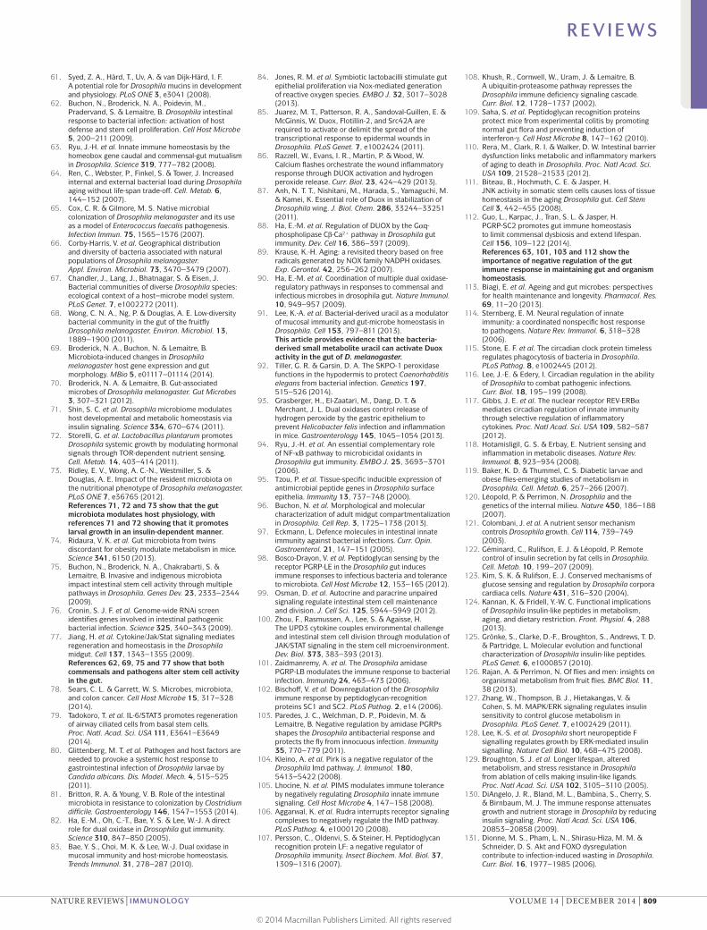
Nucleic acid and virus recognitionNucleic acid recognition is central to host detection of viruses and to antiviral immunity in diverse organ-isms, from plants to mammals27,28. Although in some cases NF-κB-dependent pathways are triggered in response to viruses, how viruses activate this pathway and the effector proteins induced by this sensing are less clear29,30. The antiviral Janus kinase (JAK)–signal transducer and activator of transcription (STAT) sig-nalling pathway that is activated during viral infection
in mammals is also involved in the response to some viruses in flies. However, evidence suggests that in flies, this pathway is induced by damage-associated mole-cules that are released during cytolytic infection, rather than by direct sensing of viral ligands31. Furthermore, studies in flies have revealed that small noncoding RNAs have fundamental roles in the defence against diverse viruses in the soma and selfish genetic elements in the germ line32.
RNA interference and exogenous viruses. Studies that were first carried out in plants and later in D. mela-nogaster demonstrated that RNA interference (RNAi) has a major role in defence against viral pathogens28,33 (FIG. 3). During viral replication, viral RNAs are processed into small interfering RNAs (siRNAs), which are then used to direct antiviral RNAi34–36. Thus, loss-of-function muta-tions in the host genes required for this RNAi pathway lead to an increased susceptibility to viral infection35,37–39. DNA viruses are also subject to RNAi-mediated restric-tion, as their genomes encode foreign double-stranded RNA (dsRNA) structures (such as repetitive hairpins and convergent transcripts)40,41. However, the mecha-nisms by which particular RNA structures or interme-diates are targeted during infection remain unclear42–44. Intriguingly, RNA fragments from various different RNA viruses can be reverse-transcribed early during infection to generate DNA of viral origin. These are potentially transcribed to generate additional substrates for RNAi to amplify the signal and provide a form of long-term inhibition of viral replication45. Many viral pathogens of insects encode suppressors of RNAi, high-lighting the central role of RNAi in antiviral responses. Moreover, viral suppressors that bind dsRNAs also attenuate interferon (IFN) signalling in vertebrates by blocking the recognition of these potent pathogen-associated molecular patterns by other classes of pattern recognition receptors46–50.
Insects have two somatic small RNA pathways — the antiviral siRNA pathway (which is Dicer-2 depend-ent) and the modulatory microRNA (miRNA) pathway (which is Dicer-1 dependent). Mammals only have one DICER gene (also known as DICER1) that carries out the functions of D. melanogaster Dicer-1 and Dicer-2. As the miRNA pathway components are essential for life in all organisms, the antiviral activity of DICER in mammals has been more difficult to study and its importance is debated (reviewed in REF. 51). Indeed, it has been sug-gested that in mammals, the RNA-based antiviral RNAi pathway has been replaced by the protein-based antiviral IFN response. However, two recent studies found that RNAi could have an antiviral role in some contexts; for example, in undifferentiated cells, which do not have a robust IFN pathway52,53.
Other factors involved in nucleic acid recogni-tion and antiviral defence. In addition to the small RNA pathways, other RNA-binding proteins act as pattern recognition receptors to induce antiviral responses. In mammals, the DEAD-box helicases retinoic acid-inducible gene I (RIG-I) and melanoma
R E V I E W S
NATURE REVIEWS | IMMUNOLOGY VOLUME 14 | DECEMBER 2014 | 799
© 2014 Macmillan Publishers Limited. All rights reserved

differentiation-associated protein 5 (MDA5; also known as IFIH1) — which are collectively known as RIG-I-like receptors (RLRs) — are canonical cytosolic receptors that bind to viral RNAs, activate NF-κB and induce IFN production54,55. Recent studies in flies have linked the small RNA pathway to signalling, suggest-ing greater similarities. The closest D. melanogaster homologue of mammalian RLRs is Dicer-2, which not only cleaves viral RNAs into siRNAs but also induces the transcription of antiviral effector proteins56. Recent genetic screens in D. melanogaster have found antiviral roles for additional DEAD-box helicases57,58, such as DDX17 (known as Rm62 in D. melanogaster), which has antiviral activity against arthropod-borne bunya-viruses in a range of species from insects to humans57. Many open questions remain about how diverse RNAs are recognized and how these activities are integrated to restrict infection. Even less is known about DNA virus recognition and future studies are required to unveil the mechanisms involved.
D. melanogaster as a model for gut immunityThe intestine is a major entry point for pathogens and is therefore central to the defence against infec-tion. The insect intestine is structurally similar to that in vertebrates, and also contains a complex microbiota (FIG. 4). In both insects and vertebrates, the intestine is a highly compartmentalized tubular organ59 with an epi-thelial monolayer that is physically separated from the lumen by barriers. One such barrier is the peritrophic membrane that lines the epithelium — analogous to mammalian mucus — and acts as a first line of defence against microbial invaders60. In addition, D. melanogaster encodes multiple mucin-like proteins that produce layers of mucus that are maintained between the peritrophic membrane and the gut epithelium; many of these mucin-like proteins are regulated by infection, which suggests an important but understudied role in immunity61,62. D. melanogaster has emerged as a powerful model in which to investigate interactions between pathogens and gut-associated microorganisms in the intestinal tract59.
Figure 3 | Nucleic acid recognition and antiviral defences in Drosophila melanogaster. RNA viruses often encode structured RNAs or produce double‑stranded RNA (dsRNA) intermediates. Some DNA viruses produce convergent transcripts that form dsRNAs and also transcribe structured RNAs. These RNAs are recognized and cleaved by Dicer‑2 to form virus‑derived small interfering RNAs (siRNAs), which are loaded into an Argonaute 2‑containing RNA‑induced silencing complex (RISC) to silence viral expression. Dicer‑2 also initiates the transcription of antiviral genes through an as‑yet‑unidentified pathway. Endogenous retrotransposon‑encoded reverse transcriptase can generate viral‑derived cDNA, which can be further transcribed and amplify the RNA interference (RNAi) response. Another RNAi pathway, the PIWI‑interacting RNA (piRNA) pathway, protects the cell from endogenous mobile genetic elements, especially those in the germ line. In addition to these nucleic‑acid‑triggered responses, some viruses can be directly sensed by Toll‑7 to induce antiviral autophagy dependent on the conserved AKT pathway involving phosphoinositide 3‑kinase (PI3K) and target of rapamycin (Tor).
Nature Reviews | Immunology
DNA virus RNA virus
Toll-7
PI3K
AKT
Tor
Autophagy
Viral suppressorsof RNAi
Viral DNA
dsRNA
Stem–loop RNA
Retrotransposonreverse transcriptase
Dicer-2
Antiviralresponse
Transposon
?
?
?
?
siRNApiRNA
piRNA locus
Nucleus
Cytosol
RISC
Amplification
RNAi
R E V I E W S
800 | DECEMBER 2014 | VOLUME 14 www.nature.com/reviews/immunol
© 2014 Macmillan Publishers Limited. All rights reserved
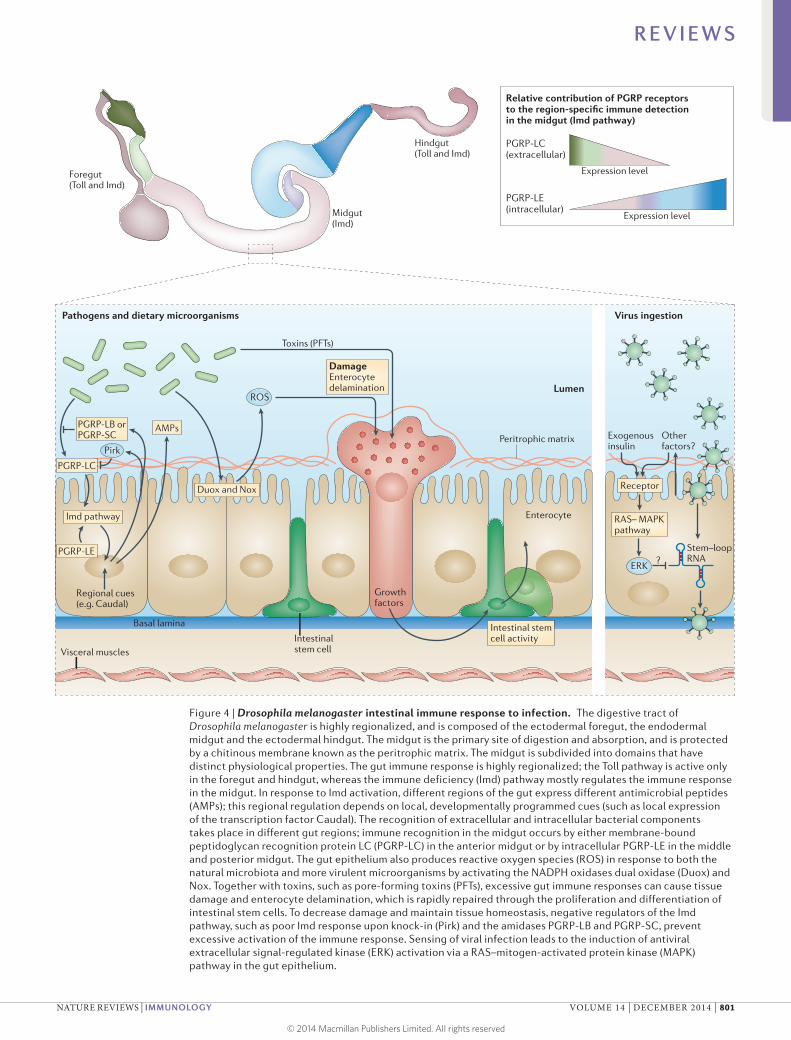
Figure 4 | Drosophila melanogaster intestinal immune response to infection. The digestive tract of Drosophila melanogaster is highly regionalized, and is composed of the ectodermal foregut, the endodermal midgut and the ectodermal hindgut. The midgut is the primary site of digestion and absorption, and is protected by a chitinous membrane known as the peritrophic matrix. The midgut is subdivided into domains that have distinct physiological properties. The gut immune response is highly regionalized; the Toll pathway is active only in the foregut and hindgut, whereas the immune deficiency (Imd) pathway mostly regulates the immune response in the midgut. In response to Imd activation, different regions of the gut express different antimicrobial peptides (AMPs); this regional regulation depends on local, developmentally programmed cues (such as local expression of the transcription factor Caudal). The recognition of extracellular and intracellular bacterial components takes place in different gut regions; immune recognition in the midgut occurs by either membrane‑bound peptidoglycan recognition protein LC (PGRP‑LC) in the anterior midgut or by intracellular PGRP‑LE in the middle and posterior midgut. The gut epithelium also produces reactive oxygen species (ROS) in response to both the natural microbiota and more virulent microorganisms by activating the NADPH oxidases dual oxidase (Duox) and Nox. Together with toxins, such as pore‑forming toxins (PFTs), excessive gut immune responses can cause tissue damage and enterocyte delamination, which is rapidly repaired through the proliferation and differentiation of intestinal stem cells. To decrease damage and maintain tissue homeostasis, negative regulators of the Imd pathway, such as poor Imd response upon knock‑in (Pirk) and the amidases PGRP‑LB and PGRP‑SC, prevent excessive activation of the immune response. Sensing of viral infection leads to the induction of antiviral extracellular signal‑regulated kinase (ERK) activation via a RAS–mitogen‑activated protein kinase (MAPK) pathway in the gut epithelium.
Nature Reviews | Immunology
Pathogens and dietary microorganisms Virus ingestion
Lumen
Regional cues(e.g. Caudal)
DamageEnterocytedelamination
RAS– MAPKpathway
Imd pathway
Duox and Nox Receptor
Toxins (PFTs)
Foregut(Toll and Imd)
Midgut(Imd)
Hindgut(Toll and Imd)
Expression level
Expression level
AMPsPeritrophic matrix Exogenous
insulinOtherfactors?
PGRP-LB orPGRP-SC
PGRP-LE
ROS
Intestinalstem cellVisceral muscles
Intestinal stem cell activity
Basal lamina
Growthfactors
Enterocyte
?Stem–loop RNA
PGRP-LC
PGRP-LC (extracellular)
PGRP-LE (intracellular)
Relative contribution of PGRP receptors to the region-specific immune detection in the midgut (Imd pathway)
Pirk
ERK
R E V I E W S
NATURE REVIEWS | IMMUNOLOGY VOLUME 14 | DECEMBER 2014 | 801
© 2014 Macmillan Publishers Limited. All rights reserved

Gut microorganisms of D. melanogaster. Recent experi-ments with wild and laboratory-reared flies indicate that the gut microbiome of D. melanogaster consists of 1–30 bacterial species, which are mostly of the Lactobacillus and Acetobacter genera63–69 (Lactobacillus species are also part of the normal human microbiome). This simple micro-biome composition and the fact that these bacterial spe-cies are all culturable allow for efficient functional analyses through the use of germ-free and gnotobiotic flies70.
As in mammals, the intestinal microbiota influences the metabolic and immune status of flies. Although flies that are reared under germ-free conditions are viable when provided with a rich food source, the presence of the gut microbiota is essential under limiting conditions, and promotes key aspects of development and intesti-nal homeostasis. First, gut microorganisms promote D. melanogaster growth through the induction of the insulin pathway, which in turn accelerates larval devel-opment71–73. This process is controlled by Acetobacter pomorum (through the generation of acetic acid) and Lactobacillus plantarum (potentially through alterations in circulating amino acid levels). Given these relation-ships, it is clear that dysbiosis of the microbiota can affect systemic growth, and that there may be a link between dysbiosis and metabolic disorders in flies, as there is in humans74. Second, germ-free flies have broad alterations in intestinal physiology. The microbiota positively regu-lates the renewal of the midgut epithelium through stem cell proliferation69,71,75. Intestinal stem cells (ISCs) differ-entiate into two essential lineages — absorptive epithe-lial cells and secretory enteroendocrine cells. Moreover, microorganisms skew cell lineage commitment, as germ-free flies have an increased ratio of enteroendocrine cells to absorptive epithelial cells69. Indeed, a common feature of both pathogenic and commensal microorganisms is their ability to promote the production of unpaired 3 (Upd3) — a cytokine analogue of mammalian IL-6 — by the gut epithelium62,69,75. In turn, Upd3 promotes homeo-static responses by altering the ISC niche function, and by increasing ISC proliferation and differentiation69,71,75. The microbiota triggers this homeostatic programme during steady-state conditions, whereas intestinal path-ogens induce a stronger homeostatic response to repair infectious damage to the epithelium71,75–77. Flies deficient in this compensatory proliferation succumb rapidly to infection, which provides strong evidence for the cen-tral role of active tissue repair in survival. However, it is unclear whether repair mechanisms alter bacterial elimination (resistance), the ability to withstand infec-tion (tolerance) or both. The relationship between the microbiota and epithelial cell proliferation has direct para llels with colorectal cancer, in which the microbiota has been suggested to be an aetiological factor78. A simi-lar relationship occurs in mammalian lungs, where IL-6 is produced in response to chemical injury, and coordinates both immune and homeostatic responses79.
In a range of organisms from flies to humans, the microbiota also has protective roles. Germ-free larvae are more susceptible to pathogenic infection by Candida albicans than larvae reared in normal conditions80. This protective effect is probably mediated by direct
competition for a survival niche, as the conventional microbiota remained protective in immune-deficient animals. This type of niche competition is important in humans and is best exemplified by Clostridium difficile, which can take hold when the normal niche is depopu-lated of healthy commensal microorganisms during anti-biotic treatment81. Germ-free mice also show increased susceptibility to pathogens, reduced cytokine production and altered stem cell activity. Together, these findings suggest that D. melanogaster is a useful model in which to identify both host pathways and microbial metabolites that regulate immunity and intestinal physiology.
Reactive oxygen species in gut immunity. In response to bacterial ingestion, organisms from flies to humans induce two key responses to fight infection — the produc-tion of reactive oxygen species (ROS) and the expression of AMPs59. ROS are induced by two conserved NADPH enzymes; dual oxidase (Duox) generates microbicidal hydrogen peroxide (H2O2) and hypochlorous acid82–84, whereas NADPH oxidase (Nox) generates H2O2. In addition to its microbicidal activity, ROS have instructive roles in tissue repair, wound healing and haematopoiesis in both flies and mammals, during which they act as a second messengers or signalling modulators85–87. How particular bacteria are sensed to induce these responses is unclear. In D. melanogaster, commensal and pathogenic microorganisms stimulate ROS production through Duox and Nox; commensals, such as L. plantarum, stim-ulate Nox, whereas pathogenic microorganisms, such as Erwinia carotovora subsp. carotovora 15 (also known as Pectobacterium carotovorum) or Pseudomonas entomoph-ila, activate Duox82–84. Flies that lack Duox activity are more susceptible to infection with enteric pathogens and have increased mortality in the presence of dietary bacte-ria and yeasts82. This suggests that ROS are central to both resistance to infection and regulation of commensals. In addition, flies that are deficient in either Duox or Nox, or that lack the ROS-detoxifying enzyme immune-regulated catalase, have a shortened lifespan, which demonstrates that healthy ageing can only be achieved when there is tight regulation of the activity of NADPH oxidases84,88,89.
As excessive ROS production is ultimately toxic to the host, a sophisticated regulatory network controls Duox expression and activity in D. melanogaster90. The tran-scriptional induction of Duox depends on the conserved p38 mitogen-activated protein kinase (p38 MAPK)−activating transcription factor 2 (Atf2) pathway90. This pathway can be induced by the metabolite uracil and the bacterial cell wall component peptidoglycan90,91. Uracil is selectively secreted by two distinct pathogenic bacteria and sensed by an as-yet-unidentified G protein-coupled receptor that activates phospholipase Cβ (PLCβ; also known as Plc21C in D. melanogaster) in a pathway that depends on the G protein αq-subunit (Gαq), which in turn drives p38 MAPK signalling88,90. Peptidoglycan also activates p38 MAPK through the activation of the Imd pathway and TGFβ-activated kinase 1 (Tak1)88,90. Uracil stimulates Duox activity through the PLCβ pathway, which promotes the release of Ca2+ from endoplasmic reticulum stores to bind and activate Duox directly88,90. However,
R E V I E W S
802 | DECEMBER 2014 | VOLUME 14 www.nature.com/reviews/immunol
© 2014 Macmillan Publishers Limited. All rights reserved

it remains unclear whether uracil is the sole agonist of Duox activity. Nevertheless, when levels of uracil are low, concentrations of cytosolic Ca2+ are reduced and Duox activity is attenuated88. This attenuation is further enforced because basal levels of PLCβ induce MAPK phosphatase 3 (Mkp3), which reduces the expression of Duox in a calci-neurin B-dependent manner90. Loss of Mkp3 in the fly gut increases the levels of ROS, triggering increased epithelial cell death and early ageing90. This suggests that multiple layers of control are required to ensure that Duox activity is kept proportional to the microbial threat and is maintained at levels that are beneficial to the host. The recent iden-tification of Duox as a key regulator of innate immunity in species ranging from worms to humans suggests that Duox function and, in turn, the role of ROS in infection, are an ancient and central immune defence mechanism92,93. Whether microbiota-derived metabolites such as uracil are involved in this regulation in mammals remains unclear.
Local regulation of gut immunity and links with ageing. Throughout the animal kingdom, AMPs are produced by the gut to control infection. Indeed, ROS and AMPs are complementary, as AMPs are crucial to control ROS-resistant bacteria94. In both flies and mammals, the induction of AMP production is compartmentalized, and distinct AMPs are expressed regionally throughout the gut62,95–97. This suggests that, as is found in mammals, local cues modulate the immune response throughout the gut in flies. Spatial control of AMP expression is not well understood, but probably involves regulation by locally expressed transcription factors. For example, expression of the homeobox gene Caudal is restricted to the D. melanogaster posterior midgut, where it represses the expression of AMPs63. Mutations in Caudal are asso-ciated with increased AMP expression in this gut region, resulting in dysbiosis, cell death and early lethality63.
It is known that systemic control of AMPs is NF-κB dependent, whereas the regulation of AMPs in the gut is more complex. The Toll pathway, which is a key systemic activator of NF-κB, is non-functional in the midgut. In addition, the expression of microbial-sensing receptors that activate the Imd pathway is also regionalized in the gut; PGRP-LC is mainly expressed in the anterior midgut, whereas PGRP-LE is expressed in the middle and poste-rior midgut96,98 (FIG. 4). This suggests that bacterial sensing and the production of specific AMPs are regionally con-trolled within the gut to fine-tune the immune response. For example, in the posterior midgut, the transit of food and bacteria slows down, and the enrichment for cytosol-expressed PGRP-LE might prioritize immune responses to invasive species that gain access to the cytosol in this region over other types of responses98. Furthermore, the JAK–STAT pathway regulates three AMPs of the Drosomycin class in the midgut but not elsewhere in the fly gut62,99. Importantly, the JAK–STAT pathway is induced not by microbial products but by the cytokine Upd3, which is activated locally by stress or damage75,77,100. So, the intestinal immune response in D. melanogaster is poised to recognize not only microorganisms but also damage caused by infection or toxins, similar to how virus-induced JAK–STAT activation is thought to occur.
Similar to ROS production, excessive or prolonged activation of NF-κB signalling is deleterious to the host and causes developmental defects and advanced ageing. To prevent these effects in the intestine, the gut secretes enzymatic PGRPs, such as PGRP-SC or PGRP-LB, that digest peptidoglycan and thus decrease the local levels of this immunostimulant101–103. In addition, several nega-tive regulators of the Imd pathway have been identified: poor Imd response upon knock-in (Pirk) modulates PGRP-LC and PGRP-LE signalling104–106; PGRP-LF is a PGRP receptor that dimerizes with PGRP-LC to decrease its ability to activate Imd signalling107; and SkpA is proposed to ubiquitylate and degrade the active NF-κB homologue Relish108. Finally, transcription factors such as Caudal function as locally expressed negative regulators of target genes activated by Relish, enabling fine-tuning of their expression63. Flies deficient for PGRP-LB or Caudal have shortened lifespans that can be rescued by the elimination of commensal bac-teria63,103, which highlights the important role of these negative regulators in maintaining immune homeo stasis in the complex milieu of the gut. Accordingly, mam-mals encode four PGRPs (PGLYRP1 to PGLYRP4) that are required for healthy management of the microbiota, and deficiencies in these PGRPs are associated with dys-biosis and increased susceptibility to colitis109. However, the sequence of events that underlies the loss of intestinal homeostasis remains unclear in both flies and mammals.
These studies demonstrate the complexity of the regulation and compartmentalization of the D. mela-nogaster gut immune response. The constant exposure to microorganisms throughout life makes it challeng-ing to maintain this delicate balance, and recent studies have shown that gut immunity affects healthy ageing in D. melanogaster75,110–112. The microbial load and diversity increases with age64,75,112, despite an increased production of ROS and AMPs in the intestine69,75,90,112. This hyper-activation is likely to have multiple effects on the gut. First, ROS cause damage to the epithelium. Second, the increased levels of ROS and AMPs will affect the micro-biota species differently and cause dysbiosis. Third, this dysbiosis induces chronic epithelial stress that stimulates high levels of ISC proliferation, which alters both the differentiation and the renewal of the gut tissue75,111,112. These age-related changes in gut homeostasis are cor-related with barrier failure and the induction of systemic immune responses, which are potentially caused by the translocation of bacteria or bacterial products across the failed epithelium of the gut110. Recent investigations into the mechanisms underlying this age-related dysbiosis have shown that the transcription factor forkhead box subgroup O (Foxo) is chronically activated in the gut of ageing flies, potentially due to oxidative stress, and that Foxo dampens the expression of PGRP-SC2 — a nega-tive regulator of the Imd pathway — resulting in hyper-activation of the immune response112. These results shed light on the links between intestinal immune homeo-stasis, dysbiosis and barrier dysfunction. Given the para-llel findings of age-related dysbiosis in humans113, these studies point to mechanisms that are likely to occur in ageing humans.
R E V I E W S
NATURE REVIEWS | IMMUNOLOGY VOLUME 14 | DECEMBER 2014 | 803
© 2014 Macmillan Publishers Limited. All rights reserved

AutophagyAn evolutionarily conserved process in which an acidic double-membrane-bound vesicle, known as an autophagosome, sequesters intracellular contents (such as damaged organelles and macromolecules, or pathogens) and targets them for degradation through fusion to lysosomes. This process is essential for the response to starvation because it facilitates the recycling of cellular components, and it can also be targeted to intracellular bacteria or viruses to restrict their growth.
HaemocytesCells found within the haemolymph of an insect that are equivalent to the blood cells in vertebrates. Different types of haemocyte are plasmatocytes, crystal cells and lamellocytes. These cells have important roles in immunity through the secretion of cytokines and phagocytic clearance of invaders.
Systemic control of the immune systemHormones have central roles in regulating whole- organism physiology, including direct effects on immu-nity114. In addition, circadian circuits, which are known as homeostatic oscillators of physiology, also affect immunity in flies and humans115–117. Indeed, an increas-ing number of connections between immunity and metabolic homeostatic control have been made in recent years. Acute and chronic infections can trigger meta-bolic dysregulation, and many metabolic syndromes are associated with inflammation, which has led to the hypothesis that inflammation drives metabolic dysfunc-tion118. Dissecting this relationship has provided insights into both immunity and the aetiology of metabolic syn-dromes. In this section, we aim to integrate recent stud-ies that have explored the interplay between physiology, metabolism and immune defence in flies.
Nutrient sensing and immune defence. Insulin signal-ling is central to metabolic regulation, and studies have implicated inflammatory signalling in the regulation of insulin responsiveness118 (FIG. 5). Similar to the mamma-lian liver and white adipose tissue, the D. melanogaster fat body stores excess fat as triglycerides and responds to metabolic shifts119,120. The fat body responds to dietary signals by releasing factors that affect insulin secretion, organism growth and metabolism121,122. A delicate balance between fat storage and breakdown is crucial to main-tain energy homeostasis. Peptide hormones released by specialized cells are key regulators of metabolism in both flies and mammals; in flies, these include glucagon-like peptide123 and insulin-like peptides124,125, which are mostly produced by neurosecretory cells in the brain. D. mela-nogaster insulin-like peptides bind a single insulin recep-tor that is orthologous to insulin receptors in vertebrates. Insulin receptor activation in both insects and mammals induces the same two conserved downstream path-ways — those mediated by AKT (also known as Akt1 in D. melanogaster) and extracellular signal-regulated kinase (ERK; known as Rolled in D. melanogaster). On binding of insulin to its receptor, AKT negatively regulates the transcription factor Foxo, which otherwise suppresses the storage of energy as fat and glycogen, thereby pro-moting long-term energy storage. AKT also positively regulates target of rapamycin (Tor; known as mTOR in mammals), which is central to growth factor signalling, as it cell-autonomously responds to amino acids, ATP and oxygen; induces anabolic processes including translation; and inhibits catabolic pathways including autophagy126. In turn, ERK activation affects organism growth, and modulates insulin sensitivity and metabolic flux127,128. Importantly, adult flies fed a high-fat or high-sugar diet develop excess adiposity and insulin resistance, suggesting that the molecular mechanisms of insulin resistance are conserved126. In adult flies, insulin functions in metabolic homeostasis, resistance to stress, fecundity, lifespan and, as shown more recently, has direct links to immunity125,129.
In flies, as in humans, infections result in metabolic disruption. The molecular mechanisms that drive shifts in metabolic homeostasis, including cachexia caused by infection, are incompletely understood and probably
have diverse aetiologies, including inflammatory sig-nalling pathways130,131. Redistribution of resources from metabolism to immunity during infection is thought to be central to these shifts. Indeed, prolonged or exces-sive immune activation can drive metabolic dysregu-lation and cause wasting in mammals and flies132. In flies, systemic infection with Mycobacteria species, Listeria species or fungi leads to metabolic shifts such as decreases in total triglyceride levels130,131,133. Infection of fly haemocytes leads to a systemic reduction in AKT activation, which increases Foxo activity and triggers the loss of metabolic stores131. Foxo can also directly affect inflammatory signalling by binding to and acti-vating the promoters of a subset of NF-κB-dependent target genes, which suggests direct coordination of NF-κB and Foxo pathways134. In this way, genes encod-ing AMPs can be activated under normal physiological conditions in response to the oscillating energy status of cells and tissues; whether this is beneficial or can lead to pathologies under some conditions remains unclear. Furthermore, in the fed state, the fat body secretes the leptin family hormone Upd2, which is a ligand for the JAK–STAT pathway135. This affects brain insulin secretion and directly communicates metabolic information between the fat body and the brain, influ-encing organism growth and energy metabolism. As the JAK–STAT pathway also has fundamental roles in immunity, this is another clear example of integrating metabolic activities with immune defence.
Mechanistic links between metabolism and immu-nity are probably integrated in the D. melanogaster fat body. Not only does the fat body have roles that are simi-lar to the mammalian liver and adipose tissue, but it is also immune responsive and can produce large quan-tities of AMPs during infections. Although mammals have separated these functions into distinct adipose tissue, liver and haematopoietic systems, it is likely that over lapping pathways regulate metabolic and immune functions through conserved signalling systems. Indeed, pathogen-sensing and nutrient-sensing pathways directly regulate each another in flies130,136.
However, immune defence is energetically costly137, and this robust response must draw resources from other physiological processes130,138. Activation of Toll signalling in the fat body of larvae either genetically or by systemic infection leads to decreased AKT signalling and increased Foxo-dependent transcription, leading to the depletion of nutrient stores and reduced organism growth130. Shifting the metabolic state of the host affects fecundity, which suggests that there are trade-offs between immunity and host fitness. Furthermore, tissue necrosis at distal sites activates the Toll–AKT crosstalk in the fat body, promoting an energy-wasting pheno-type and decreased levels of circulating S-adenosyl-methionine (SAM), which is a major building block for organism growth139. Tissue-derived DNA damage, which is another classic danger signal, also leads to the systemic induction of pro-inflammatory signalling and alters insulin signalling in the fat body140. Together, these data suggest that crosstalk between the micro-bial recognition system and AKT signalling evolved to
R E V I E W S
804 | DECEMBER 2014 | VOLUME 14 www.nature.com/reviews/immunol
© 2014 Macmillan Publishers Limited. All rights reserved

Figure 5 | Systemic regulation of Drosophila melanogaster immune responses. The Drosophila melanogaster immune response is integrated with multiple inter‑organ regulatory circuits. a | The fat body is a central regulator of metabolic control. Here, Toll signalling through the nuclear factor‑κB (NF‑κB) transcription factor Dif inhibits insulin signalling and retards growth, whereas forkhead box subgroup O (Foxo) — the downstream target of the insulin pathway — regulates the production of antimicrobial peptides (AMPs) and anabolic genes. b | In larvae, the steroid hormone Ecdysone is produced in a secretory organ associated with the brain (known as the ring gland), whereas the source of Ecdysone is less well characterized in adult flies. Ecdysone is a major regulator of the D. melanogaster lifecycle, as well as the immune response, through controlling the expression of peptidoglycan recognition protein LC (PGRP‑LC) in the fat body and the phagocytic activity of pupal haemocytes. c | The gut microbiota further influences the insulin signalling pathway by regulating the production of Drosophila insulin‑like peptides (Dilps) in the brain. Dilp production and robust growth are triggered by acetic acid that is produced by Acetobacter species in the gut, and potentially triggered by optimization of amino acid levels provided by other components of microbiota. d | The central nervous system further regulates immune responses in the Malpighian tubules through the production of neuropeptides that are thought to trigger a nitric oxide (NO)‑dependent NF‑κB response in this organ. The faded part of this panel indicates a hypothetical role for the Imd pathway. cGMP, cyclic guanosine monophosphate; EcR, Ecdysone receptor; GC, guanylyl cyclase; Imd, immune deficiency; IPC, insulin‑producing cell; Nos, nitric oxide synthase; Tor, target of rapamycin.
Nature Reviews | Immunology
a
Gut bacteria
Ecdysone Infection
Infection?dc
Ca2+
NOS GC
NO cGMP ?
Neuropeptide Neuropeptidereceptor
EcR AMPsRelish
Relish
P
Relish
Relish
PRelish
Relish
PAMPs andmetabolic genes
AMPs andstress genes
Infection bDilps
Toll
Cytosol
Nucleus
Spätzle
Insulin-likereceptor
Dif
Dif
Foxo
FoxoAMPs andmetabolism
Tor
AKT
Cactus
P
P
PGRP-LC
Haemocyteactivation
Haemocyte
Activephagocytichaemocyte
Imd
PGRPPGRP-LC
PGRP-LE
ImdImd
• Amino acids?• Acetic acid?• Others?
• Haemolymph• Dilp levels
Neuropeptides
Dilps
IPCs
Ecdysone
Ring gland
Brain
Gut
Bacteria
Amino acids and acetic acid Malpighian
tubules
Fat body (storage)
R E V I E W S
NATURE REVIEWS | IMMUNOLOGY VOLUME 14 | DECEMBER 2014 | 805
© 2014 Macmillan Publishers Limited. All rights reserved

MitophagyA specialized form of autophagy that selectively targets and degrades mitochondria.
balance energy expenditure between organism growth and the acute demand of combating infection. This has an impact on the induction of protective mechanisms locally (such as autophagy) and systemically (such as AMPs and SAM). Thus, as in humans, chronic inflam-mation can directly affect metabolic state and insulin resistance.
Metabolic pathways and inflammatory responses are integrated at the level of transcription by the transcrip-tion factor myocyte enhancer factor 2 (Mef2), which activates distinct classes of genes depending on the state of the organism136. Under normal conditions, Mef2 is phosphorylated, potentially by kinases downstream of AKT signalling, and promotes the expression of ana-bolic enzymes. However, immune stimulation reduces its phosphorylation levels and alters its target genes to those encoding immune effectors, which results in decreased anabolism and increased immunity136. Mef2 has also been linked to the regulation of Eiger, which is the D. melanogaster homologue of TNF141. TNF signal-ling is involved in cell death pathways and immunity in a range of organisms from flies to humans, which suggests another link between metabolism and innate responses. Furthermore, many infections attenuate AKT signalling and thus Mef2 may emerge as a central control point.
Attenuation of AKT signalling upon microbial recog-nition can also activate autophagy. Sensing of cytosolic Listeria species by PGRP-LE and of viruses by Toll-7 induces autophagy in infected cells to restrict infec-tion15,26. In addition, the ubiquitin ligase Parkin promotes a specialized form of autophagy known as mitophagy to target intracellular pathogens such as mycobacteria in both flies and humans142.
In addition to the effects of inflammatory signalling on growth that are discussed above, growth signals can affect inflammatory signalling. Insulin and growth fac-tor signalling both induce the MAPK–ERK pathway, which is involved in growth and development, and also lead to the activation of Pirk, which is a negative regu-lator of inflammatory NF-κB signalling104–106,143. This negative feedback is probably protective, as hyperac-tive inflammatory signalling is detrimental to all species and is a hallmark of metabolic disorders144. ERK signal-ling also promotes antiviral defence in the intestine145. Oral challenge of vector insects by blood-acquired viral patho gens occurs during a nutrient-rich blood meal, and this triggers antiviral ERK signalling in the absorp-tive epithelial cells. This couples antiviral defences with nutrient acquisition and might be an ancient prophy-lactic defence mechanism in the gut. Furthermore, nutrient signals can influence systemic immunity, as diet restriction leads to decreased resistance to infec-tion with Listeria species but increased tolerance to infection with Salmonella species146.
Hormonal control of immunity. In addition to insulin, the steroid hormone Ecdysone modulates immune responses. Ecdysone is the major regulator of the insect life cycle147 and signals through a classical nuclear hor-mone receptor heterodimer consisting of the Ecdysone receptor (EcR) and Ultraspiracle (USP), which are
orthologous to mammalian farnesoid X receptor and liver X receptor, and retinoid X receptor, respectively148. Early fly embryos, which are devoid of Ecdysone, cannot mount an antibacterial response unless exog-enous Ecdysone is provided149. Later during embryo-genesis, Ecdysone pulses released from extra-embryonic tissues provide the priming needed for immune respon-siveness149. During the third and final larval stage, a large Ecdysone pulse initiates pupariation, which regu-lates many aspects of physiology, including the death of many larval tissues, haemocyte-mediated phagocyto-sis of dead cells and full induction of genes encoding AMPs upon infection150–153. Hormone-triggered haemocyte activation involves several actin-dependent processes — including haemocyte motility and phago-cytic activity — that are also required for resistance to infection and for wound healing at the pupal stage154. During development, the embryo is covered in a shell that protects it from external challenge, and thus the embryo may not require AMP production until hatch-ing. By contrast, larvae are constantly exposed to patho-gens, possibly even more so during moulting, when the cuticle must be rebuilt. During pupariation, when the entire organism is remodelled, it has been suggested that microorganisms are released from the gut into the haemolymph154. Thus, the coupling of immune prim-ing with these major developmental transitions could confer a survival advantage, as it may tailor the immune response to deal with relevant threats.
In adult flies, the induction of AMP gene expres-sion and host survival following bacterial infection requires Ecdysone signalling, as it is crucial for acti-vation of the Imd pathway and directs the expres-sion of the upstream receptor PGRP-LC152. It remains unclear which Ecdysone-triggered transcription factors directly regulate PGRP-LC expression but its upregu-lation during infection suggests that Ecdysone itself may be induced. Ecdysone is known to be regulated by multiple stressors, including heat shock and sleep deprivation155. Nutrient deprivation increases Ecdysone levels in the ovaries to block oogenesis, thereby serv-ing a checkpoint function156. So, Ecdysone-dependent induction of PGRP-LC expression may integrate stress signals with immune function. Like stress, age-ing is associated with a dysregulated immune system, elevated AMP expression, defects in phagocytosis, elevated levels of cytokines (such as IL-6 in verte-brates) and immunosenescence. Reduced Ecdysone signalling increases lifespan157, which suggests that it has a fundamental role in age-dependent changes in immunity. Given that infection is inherently stressful, stress hormone priming of both the humoral Imd path-way and the cellular phagocytic immune response may protect the animal from infection, but also lead to the pathology that is found in aged animals.
Other hormones, including neuropeptides, respond to physiological stressors and modulate the immune response in D. melanogaster (FIG. 5). Excretory functions are partially achieved by tubular epithelial structures known as the Malpighian tubules, which are analo-gous to vertebrate kidneys. The function of Malpighian
R E V I E W S
806 | DECEMBER 2014 | VOLUME 14 www.nature.com/reviews/immunol
© 2014 Macmillan Publishers Limited. All rights reserved

tubules is regulated by neuropeptides that respond to stressors; Malpighian tubules also sense systemic infec-tion through tissue-autonomous sensing of peptido-glycan23,158. Neuropeptides — such as D. melanogaster Capability (also known as Capa-1)159, which enhances tolerance to desiccation, and neuropeptide receptors, such as guanylyl cyclase at 76C (Gyc76C)160 — elicit an immune response in the Malpighian tubules and parallels were observed in human embryonic kidney cells, sug-gesting that physiological stressors, such as desiccation, interact with immune pathways through neurohormonal signalling across hosts.
Neuronal–inflammatory connectionsThe brain coordinates many systemic responses, includ-ing the production of hormones. The brain is also rela-tively immune privileged, so as to protect non-dividing yet essential neuronal cells. Recently, neuronal inflam-mation has been linked to age-related pathological processes, including neurodegeneration161. Indeed, neurodegenerative triggers — such as amyloid-β pep-tide — can activate classical microbial-sensing recep-tors such as TLRs and the NOD-, LRR- and pyrin domain-containing 3 (NRLP3; also known as NALP3) inflammasome161,162. Mechanistically, it is unclear how these pathways intersect. Interestingly, recent studies in D. melanogaster models of neurodegeneration have linked activation of the insect innate immune response to disease progression. In a D. melanogaster model for Alzheimer’s disease that is based on the expression of the amyloid-β42 peptide in the developing eye163, loss of any intracellular component of the Toll pathway, but not of the Imd pathway or the canonical ligand Spätzle, ameliorated the amyloid-β42-induced eye phenotype163. These findings suggest a conserved connection between innate immune signalling and amyloid-β-dependent pathologies in the central nervous system.
The Imd pathway is linked to neurodegeneration in other models. In one study, spontaneous vacuolar lesions in the D. melanogaster brain neuropile — a hall-mark of neurodegeneration — were found to occur at a markedly increased rate in flies that lack defence repres-sor 1 (Dnr1)164, which is a negative regulator of the Imd pathway165. Increased neurodegeneration in the Dnr1 mutant was found to be NF-κB dependent, and enforced expression of AMPs in either the neurons or glia was sufficient to phenocopy neurodegeneration164. In another system, mutation of ATM (which encodes ataxia telangiectasia mutated kinase; also known as Tefu) in glia induced NF-κB-dependent neuro degeneration, suggesting a previously unknown connection between the ATM-dependent DNA damage response pathway and Imd immune signalling. Moreover, NF-κB activa-tion in the glia alone was sufficient to drive neuronal cell death and vacuolar lesions in the fly central nerv-ous system166,167. However, it remains unclear how the loss of ATM affects Relish and NF-κB signalling, and whether this involves any of the known components of the ATM-dependent DNA damage response path-way. In a third study, a D. melanogaster model of retinal degeneration also revealed a dependence on NF-κB
signalling168. Light-dependent NorpA-deficient retinal degeneration was strongly suppressed by mutations in the Imd signalling components Relish, Kenny (also known as IKKγ) or death-related ced-3/Nedd2-like cas-pase (Dredd; also known as caspase 8). However, this phenotype was not affected by the loss of either Imd or Fadd (which encodes FAS-associated death domain orthologue), which are upstream components in the classical Imd pathway. Together, these four studies support a previously unknown role for components of the Imd pathway in neuronal cell death, with their effects being partially mediated by NF-κB-dependent AMP expression. Clearly, inappropriate activation can lead to serious pathology169. Although these papers describe sterile inflammatory conditions contribut-ing to neuro degeneration, the theme here is similar to what is observed in the gut — restoring homeostasis after inflammatory responses is key to maintaining healthy organs.
Future perspectivesIn this Review, we have described the diverse areas of immunity that have been studied in D. melanogaster in recent years. Early studies focused on molecules involved in sensing and signalling, whereas more recent studies have started to investigate immunity using a more holistic approach. Studies in flies can reveal how immunity and its dysregulation can affect whole-body pathophysiology. Such studies have also shown that innate immune genes are under strong positive selec-tion170,171, which suggests that parasites impose strong evolutionary pressures on their hosts. Animals from fruit flies to humans are colonized by a range of micro-organisms that must be tolerated by the ever-evolving and adapting immune response, and how these bacte-ria have co-evolved with other organisms remains to be elucidated. Dissecting the complex relationships between hosts and their microbiota is challenging but essential; advances in this area might help us to under-stand complex diseases such as inflammatory bowel disease, and to develop treatments for some intestinal infectious and autoinflammatory diseases. Indeed, flies harbouring symbiotic intracellular bacteria of the Wolbachia genus have been found to be less suscepti-ble to viral infections172,173,179. This discovery has led to field trials involving the release of Wolbachia-colonized mosquitoes to break the transmission cycle of dengue virus in Australia180. Recent studies have also high-lighted that successful immunity to infection requires two facets: first, targeting the microbe for elimina-tion, termed resistance; and second, inducing tissue-protective responses, termed disease tolerance174–176. Mechanistically, they are defined by their effects on both organism survival and pathogen load. Resistance mechanisms promote survival by decreasing bacte-rial numbers, whereas tolerance mechanisms improve survival without necessarily affecting bacterial load. Recent studies have provided evidence that tolerance is a feature that is conserved across species from insects to humans177, and work in D. melanogaster has found that active repair mechanisms and metabolic adaptations are
R E V I E W S
NATURE REVIEWS | IMMUNOLOGY VOLUME 14 | DECEMBER 2014 | 807
© 2014 Macmillan Publishers Limited. All rights reserved

1. St Johnston, D. & Nusslein-Volhard, C. The origin of pattern and polarity in the Drosophila embryo. Cell 68, 201–219 (1992).
2. Padmanabha, D. & Baker, K. D. Drosophila gains traction as a repurposed tool to investigate metabolism. Trends Endocrinol. Metab. 25, 518–527 (2014).
3. Lemaitre, B. & Hoffmann, J. A. The host defense of Drosophila melanogaster. Annu. Rev. Immunol. 25, 697–743 (2007).
4. Limmer, S. et al. Pseudomonas aeruginosa RhlR is required to neutralize the cellular immune response in a Drosophila melanogaster oral infection model. Proc. Natl Acad. Sci. USA 108, 17378–17383 (2011).
5. Vallet-Gely, I., Lemaitre, B. & Boccard, F. Bacterial strategies to overcome insect defences. Nature Rev. Microbiol. 6, 302–313 (2008).
6. Xu, J. & Cherry, S. Viruses and antiviral immunity in Drosophila. Dev. Comparative Immunol. 42, 67–84 (2014).
7. Venken, K. J. T. & Bellen, H. J. Chemical mutagens, transposons, and transgenes to interrogate gene function in Drosophila melanogaster. Methods 68, 15–28 (2014).
8. Mohr, S. E., Hu, Y., Kim, K., Housden, B. E. & Perrimon, N. Resources for functional genomics studies in Drosophila melanogaster. Genetics 197, 1–18 (2014).
9. Boman, H. G., Nilsson, I. & Rasmuson, B. Inducible antibacterial defence system in Drosophila. Nature 237, 232–235 (1972).
10. Strominger, J. L. Animal antimicrobial peptides: ancient players in innate immunity. J. Immunol. 182, 6633–6634 (2009).
11. Hoffmann, J. A. & Reichhart, J. M. Drosophila innate immunity: an evolutionary perspective. Nature Immunol. 3, 121–126 (2002).
12. Lemaitre, B., Nicolas, E., Michaut, L., Reichhart, J. M. & Hoffmann, J. A. The dorsoventral regulatory gene cassette spätzle/Toll/cactus controls the potent antifungal response in Drosophila adults. Cell 86, 973–983 (1996).
13. Lemaitre, B., Reichhart, J. M. & Hoffmann, J. A. Drosophila host defense: differential induction of antimicrobial peptide genes after infection by various classes of microorganisms. Proc. Natl Acad. Sci. USA 94, 14614–14619 (1997).
14. Shelly, S., Lukinova, N., Bambina, S., Berman, A. & Cherry, S. Autophagy is an essential component of Drosophila immunity against vesicular stomatitis virus. Immunity 30, 588–598 (2009).
15. Nakamoto, M. et al. Virus recognition by Toll-7 activates antiviral autophagy in Drosophila. Immunity 36, 658–667 (2012).This study establishes that D. melanogaster Toll‑7 directly senses RNA viruses, similar to the function of mammalian TLRs.
16. Moy, R. H., Gold, B. & Molleston, J. Antiviral autophagy restricts Rift Valley fever virus infection and is conserved from flies to mammals. Immunity 40, 51–65 (2014).
17. Hymowitz, S. G. et al. IL-17s adopt a cystine knot fold: structure and activity of a novel cytokine, IL-17F, and implications for receptor binding. EMBO J. 20, 5332–5341 (2001).
18. Chamy, L. E., Leclerc, V., Caldelari, I. & Reichhart, J. M. Sensing of ‘danger signals’ and pathogen-associated molecular patterns defines binary signaling pathways ‘upstream’ of Toll. Nature Immunol. 9, 1165–1170 (2008).
19. Gottar, M. et al. Dual detection of fungal infections in Drosophila via recognition of glucans and sensing of virulence factors. Cell 127, 1425–1437 (2006).
20. Ming, M., Obata, F., Kuranaga, E. & Miura, M. Persephone/Spätzle pathogen sensors mediate the activation of Toll receptor signaling in response to
endogenous danger signals in apoptosis-deficient Drosophila. J. Biol. Chem. 289, 7558–7568 (2014).
21. Gobert, V. et al. Dual activation of the Drosophila Toll pathway by two pattern recognition receptors. Science 302, 2126–2130 (2003).
22. Buchon, N. et al. A single modular serine protease integrates signals from pattern-recognition receptors upstream of the Drosophila Toll pathway. Proc. Natl Acad. Sci. USA 106, 12442–12447 (2009).
23. Kaneko, T. et al. PGRP-LC and PGRP-LE have essential yet distinct functions in the drosophila immune response to monomeric DAP-type peptidoglycan. Nature Immunol. 7, 715–723 (2006).
24. Lim, J.-H. et al. Structural basis for preferential recognition of diaminopimelic acid-type peptidoglycan by a subset of peptidoglycan recognition proteins. J. Biol. Chem. 281, 8286–8295 (2006).
25. Chang, C., Chelliah, Y., Borek, D., Mengin-Lecreulx, D. & Deisenhofer, J. Structure of tracheal cytotoxin in complex with a heterodimeric pattern-recognition receptor. Science 311, 1761–1764 (2006).References 24 and 25 provide atomic detail on the specific recognition of DAP‑type peptidoglycan by the D. melanogaster Imd pathway.
26. Yano, T. et al. Autophagic control of listeria through intracellular innate immune recognition in drosophila. Nature Immunol. 9, 908–916 (2008).
27. Gürtler, C. & Bowie, A. G. Innate immune detection of microbial nucleic acids. Trends Microbiol. 21, 413–420 (2013).
28. Ding, S.-W. & Voinnet, O. Antiviral immunity directed by small RNAs. Cell 130, 413–426 (2007).
29. Costa, A., Jan, E., Sarnow, P. & Schneider, D. S. The Imd pathway is involved in antiviral immune responses in Drosophila. PLoS ONE 4, e7436 (2009).
30. Zambon, R. A., Nandakumar, M., Vakharia, V. N. & Wu, L. P. The Toll pathway is important for an antiviral response in Drosophila. Proc. Natl Acad. Sci. USA 102, 7257–7262 (2005).
31. Dostert, C. et al. The Jak-STAT signaling pathway is required but not sufficient for the antiviral response of drosophila. Nature Immunol. 6, 946–953 (2005).
32. van Rij, R. P. & Berezikov, E. Small RNAs and the control of transposons and viruses in Drosophila. Trends Microbiol. 17, 163–171 (2009).
33. Kemp, C. & Imler, J.-L. Antiviral immunity in drosophila. Curr. Opin. Immunol. 21, 3–9 (2009).
34. Galiana-Arnoux, D., Dostert, C., Schneemann, A., Hoffmann, J. A. & Imler, J.-L. Essential function in vivo for Dicer-2 in host defense against RNA viruses in drosophila. Nature Immunol. 7, 590–597 (2006).
35. Wang, X. et al. RNA interference directs innate immunity against viruses in adult Drosophila. Science 312, 452–454 (2006).
36. Sabin, L. R. et al. Ars2 regulates both miRNA- and siRNA-dependent silencing and suppresses RNA virus infection in Drosophila. Cell 138, 340–351 (2009).
37. Zambon, R., Vakharia, V. & Wu, L. P. RNAi is an antiviral immune response against a dsRNA virus in Drosophila melanogaster. Cell. Microbiol. 8, 880–889 (2006).
38. Aliyari, R. et al. Mechanism of induction and suppression of antiviral immunity directed by virus-derived small RNAs in Drosophila. Cell Host Microbe 4, 387–397 (2008).
39. Mueller, S. et al. RNAi-mediated immunity provides strong protection against the negative-strand RNA vesicular stomatitis virus in Drosophila. Proc. Natl Acad. Sci. USA 107, 19390–19395 (2010).
40. Kemp, C. et al. Broad RNA interference-mediated antiviral immunity and virus-specific inducible responses in Drosophila. J. Immunol. 190, 650–658 (2012).
41. Bronkhorst, A. et al. The DNA virus Invertebrate iridescent virus 6 is a target of the Drosophila RNAi machinery. Proc. Natl Acad. Sci. USA 109, E3604–E3613 (2012).
42. Sabin, L. R. et al. Dicer-2 processes diverse viral RNA species. PLoS ONE 8, e55458 (2013).
43. Brackney, D. E., Beane, J. E. & Ebel, G. D. RNAi targeting of west nile virus in mosquito midguts promotes virus diversification. PLoS Pathog. 5, e1000502 (2009).
44. Flynt, A., Liu, N., Martin, R. & Lai, E. C. Dicing of viral replication intermediates during silencing of latent Drosophila viruses. Proc. Natl Acad. Sci. USA 106, 5270–5275 (2009).
45. Goic, B. et al. RNA-mediated interference and reverse transcription control the persistence of RNA viruses in the insect model Drosophila. Nature Immunol. 14, 396–403 (2013).The data in this study suggest a mechanism for the amplification and persistence of the RNAi pathway in insects.
46. Nayak, A. et al. Cricket paralysis virus antagonizes Argonaute 2 to modulate antiviral defense in Drosophila. Nature Struct. Mol. Biol. 17, 547–554 (2010).
47. Chao, J. A. et al. Dual modes of RNA-silencing suppression by Flock House virus protein B2. Nature Struct. Mol. Biol. 12, 952–957 (2005).
48. Fagegaltier, D. et al. The endogenous siRNA pathway is involved in heterochromatin formation in Drosophila. Proc. Natl Acad. Sci. 106, 21258–21263 (2009).
49. Berry, B., Deddouche, S., Kirschner, D., Imler, J.-L. & Antoniewski, C. Viral suppressors of RNA silencing hinder exogenous and endogenous small RNA pathways in Drosophila. PLoS ONE 4, e5866 (2009).
50. van Mierlo, J. T. et al. Convergent evolution of argonaute-2 slicer antagonism in two distinct insect RNA viruses. PLoS Pathog. 8, e1002872 (2012).
51. Cullen, B. R., Cherry, S. & tenOever, B. R. Is RNA interference a physiologically relevant innate antiviral immune response in mammals? Cell Host Microbe 14, 374–378 (2013).
52. Maillard, P. V. et al. Antiviral RNA interference in mammalian cells. Science 342, 235–238 (2013).
53. Li, Y., Lu, J., Han, Y., Fan, X. & Ding, S.-W. RNA interference functions as an antiviral immunity mechanism in mammals. Science 342, 231–234 (2013).
54. Mukherjee, K., Korithoski, B. & Kolaczkowski, B. Ancient origins of vertebrate-specific innate antiviral immunity. Mol. Biol. Evol. 31, 140–153 (2014).
55. Loo, Y.-M. & Gale, M. Immune signaling by RIG-I-like receptors. Immunity 34, 680–692 (2011).
56. Deddouche, S. et al. The DExD/H-box helicase Dicer-2 mediates the induction of antiviral activity in drosophila. Nature Immunol. 9, 1425–1432 (2008).
57. Moy, R. H. et al. Stem-loop recognition by DDX17 facilitates miRNA processing and antiviral defense. 158, 764–777 (2014).This paper shows that additional RNA sensors detect RNA viruses and that these are conserved across species.
58. Hopkins, K. C. et al. A genome-wide RNAi screen reveals that mRNA decapping restricts bunyaviral replication by limiting the pools of Dcp2-accessible targets for cap-snatching. Genes Dev. 27, 1511–1525 (2013).
59. Buchon, N., Broderick, N. A. & Lemaitre, B. Gut homeostasis in a microbial world: insights from Drosophila melanogaster. Nature Rev. Microbiol. 11, 615–626 (2013).
60. Hegedus, D., Erlandson, M., Gillott, C. & Toprak, U. New insights into peritrophic matrix synthesis, architecture, and function. Annu. Rev. Entomol. 54, 285–302 (2009).
key to surviving infection62,136. Undoubtedly, the power of D. melanogaster genetics will further our under-standing of the genetic basis for tolerance mechanisms and the means by which a host adapts to infection-associated damage. How the D. melanogaster holobiont interacts with different environmental insults — such as altered diets, toxins and more virulent pathogens — will be the focus of future work. Such studies will provide
new insights into the physiological consequences of microbial colonization and how immunity interacts with behaviour, metabolism, physiology and hormo-nal regulation. Further study of infections, immune defences and their systemic regulators will continue to open up new routes for translational research and new treatment strategies, and result in a more complete understanding of health and disease.
R E V I E W S
808 | DECEMBER 2014 | VOLUME 14 www.nature.com/reviews/immunol
© 2014 Macmillan Publishers Limited. All rights reserved
61. Syed, Z. A., Härd, T., Uv, A. & van Dijk-Härd, I. F. A potential role for Drosophila mucins in development and physiology. PLoS ONE 3, e3041 (2008).
62. Buchon, N., Broderick, N. A., Poidevin, M., Pradervand, S. & Lemaitre, B. Drosophila intestinal response to bacterial infection: activation of host defense and stem cell proliferation. Cell Host Microbe 5, 200–211 (2009).
63. Ryu, J.-H. et al. Innate immune homeostasis by the homeobox gene caudal and commensal-gut mutualism in Drosophila. Science 319, 777–782 (2008).
64. Ren, C., Webster, P., Finkel, S. & Tower, J. Increased internal and external bacterial load during Drosophila aging without life-span trade-off. Cell. Metab. 6, 144–152 (2007).
65. Cox, C. R. & Gilmore, M. S. Native microbial colonization of Drosophila melanogaster and its use as a model of Enterococcus faecalis pathogenesis. Infection Immun. 75, 1565–1576 (2007).
66. Corby-Harris, V. et al. Geographical distribution and diversity of bacteria associated with natural populations of Drosophila melanogaster. Appl. Environ. Microbiol. 73, 3470–3479 (2007).
67. Chandler, J., Lang, J., Bhatnagar, S. & Eisen, J. Bacterial communities of diverse Drosophila species: ecological context of a host–microbe model system. PLoS Genet. 7, e1002272 (2011).
68. Wong, C. N. A., Ng, P. & Douglas, A. E. Low-diversity bacterial community in the gut of the fruitfly Drosophila melanogaster. Environ. Microbiol. 13, 1889–1900 (2011).
69. Broderick, N. A., Buchon, N. & Lemaitre, B. Microbiota-induced changes in Drosophila melanogaster host gene expression and gut morphology. MBio 5, e01117–01114 (2014).
70. Broderick, N. A. & Lemaitre, B. Gut-associated microbes of Drosophila melanogaster. Gut Microbes 3, 307–321 (2012).
71. Shin, S. C. et al. Drosophila microbiome modulates host developmental and metabolic homeostasis via insulin signaling. Science 334, 670–674 (2011).
72. Storelli, G. et al. Lactobacillus plantarum promotes Drosophila systemic growth by modulating hormonal signals through TOR-dependent nutrient sensing. Cell. Metab. 14, 403–414 (2011).
73. Ridley, E. V., Wong, A. C.-N., Westmiller, S. & Douglas, A. E. Impact of the resident microbiota on the nutritional phenotype of Drosophila melanogaster. PLoS ONE 7, e36765 (2012).References 71, 72 and 73 show that the gut microbiota modulates host physiology, with references 71 and 72 showing that it promotes larval growth in an insulin‑dependent manner.
74. Ridaura, V. K. et al. Gut microbiota from twins discordant for obesity modulate metabolism in mice. Science 341, 6150 (2013).
75. Buchon, N., Broderick, N. A., Chakrabarti, S. & Lemaitre, B. Invasive and indigenous microbiota impact intestinal stem cell activity through multiple pathways in Drosophila. Genes Dev. 23, 2333–2344 (2009).
76. Cronin, S. J. F. et al. Genome-wide RNAi screen identifies genes involved in intestinal pathogenic bacterial infection. Science 325, 340–343 (2009).
77. Jiang, H. et al. Cytokine/Jak/Stat signaling mediates regeneration and homeostasis in the Drosophila midgut. Cell 137, 1343–1355 (2009).References 62, 69, 75 and 77 show that both commensals and pathogens alter stem cell activity in the gut.
78. Sears, C. L. & Garrett, W. S. Microbes, microbiota, and colon cancer. Cell Host Microbe 15, 317–328 (2014).
79. Tadokoro, T. et al. IL-6/STAT3 promotes regeneration of airway ciliated cells from basal stem cells. Proc. Natl. Acad. Sci. USA 111, E3641–E3649 (2014).
80. Glittenberg, M. T. et al. Pathogen and host factors are needed to provoke a systemic host response to gastrointestinal infection of Drosophila larvae by Candida albicans. Dis. Model. Mech. 4, 515–525 (2011).
81. Britton, R. A. & Young, V. B. Role of the intestinal microbiota in resistance to colonization by Clostridium difficile. Gastroenterology 146, 1547–1553 (2014).
82. Ha, E.-M., Oh, C.-T., Bae, Y. S. & Lee, W.-J. A direct role for dual oxidase in Drosophila gut immunity. Science 310, 847–850 (2005).
83. Bae, Y. S., Choi, M. K. & Lee, W.-J. Dual oxidase in mucosal immunity and host-microbe homeostasis. Trends Immunol. 31, 278–287 (2010).
84. Jones, R. M. et al. Symbiotic lactobacilli stimulate gut epithelial proliferation via Nox-mediated generation of reactive oxygen species. EMBO J. 32, 3017–3028 (2013).
85. Juarez, M. T., Patterson, R. A., Sandoval-Guillen, E. & McGinnis, W. Duox, Flotillin-2, and Src42A are required to activate or delimit the spread of the transcriptional response to epidermal wounds in Drosophila. PLoS Genet. 7, e1002424 (2011).
86. Razzell, W., Evans, I. R., Martin, P. & Wood, W. Calcium flashes orchestrate the wound inflammatory response through DUOX activation and hydrogen peroxide release. Curr. Biol. 23, 424–429 (2013).
87. Anh, N. T. T., Nishitani, M., Harada, S., Yamaguchi, M. & Kamei, K. Essential role of Duox in stabilization of Drosophila wing. J. Biol. Chem. 286, 33244–33251 (2011).
88. Ha, E.-M. et al. Regulation of DUOX by the Gαq-phospholipase Cβ-Ca2+ pathway in Drosophila gut immunity. Dev. Cell 16, 386–397 (2009).
89. Krause, K.-H. Aging: a revisited theory based on free radicals generated by NOX family NADPH oxidases. Exp. Gerontol. 42, 256–262 (2007).
90. Ha, E.-M. et al. Coordination of multiple dual oxidase-regulatory pathways in responses to commensal and infectious microbes in drosophila gut. Nature Immunol. 10, 949–957 (2009).
91. Lee, K.-A. et al. Bacterial-derived uracil as a modulator of mucosal immunity and gut-microbe homeostasis in Drosophila. Cell 153, 797–811 (2013).This article provides evidence that the bacteria‑ derived small metabolite uracil can activate Duox activity in the gut of D. melanogaster.
92. Tiller, G. R. & Garsin, D. A. The SKPO-1 peroxidase functions in the hypodermis to protect Caenorhabditis elegans from bacterial infection. Genetics 197, 515–526 (2014).
93. Grasberger, H., El-Zaatari, M., Dang, D. T. & Merchant, J. L. Dual oxidases control release of hydrogen peroxide by the gastric epithelium to prevent Helicobacter felis infection and inflammation in mice. Gastroenterology 145, 1045–1054 (2013).
94. Ryu, J.-H. et al. An essential complementary role of NF-κB pathway to microbicidal oxidants in Drosophila gut immunity. EMBO J. 25, 3693–3701 (2006).
95. Tzou, P. et al. Tissue-specific inducible expression of antimicrobial peptide genes in Drosophila surface epithelia. Immunity 13, 737–748 (2000).
96. Buchon, N. et al. Morphological and molecular characterization of adult midgut compartmentalization in Drosophila. Cell Rep. 3, 1725–1738 (2013).
97. Eckmann, L. Defence molecules in intestinal innate immunity against bacterial infections. Curr. Opin. Gastroenterol. 21, 147–151 (2005).
98. Bosco-Drayon, V. et al. Peptidoglycan sensing by the receptor PGRP-LE in the Drosophila gut induces immune responses to infectious bacteria and tolerance to microbiota. Cell Host Microbe 12, 153–165 (2012).
99. Osman, D. et al. Autocrine and paracrine unpaired signaling regulate intestinal stem cell maintenance and division. J. Cell Sci. 125, 5944–5949 (2012).
100. Zhou, F., Rasmussen, A., Lee, S. & Agaisse, H. The UPD3 cytokine couples environmental challenge and intestinal stem cell division through modulation of JAK/STAT signaling in the stem cell microenvironment. Dev. Biol. 373, 383–393 (2013).
101. Zaidmanremy, A. et al. The Drosophila amidase PGRP-LB modulates the immune response to bacterial infection. Immunity 24, 463–473 (2006).
102. Bischoff, V. et al. Downregulation of the Drosophila immune response by peptidoglycan-recognition proteins SC1 and SC2. PLoS Pathog. 2, e14 (2006).
103. Paredes, J. C., Welchman, D. P., Poidevin, M. & Lemaitre, B. Negative regulation by amidase PGRPs shapes the Drosophila antibacterial response and protects the fly from innocuous infection. Immunity 35, 770–779 (2011).
104. Kleino, A. et al. Pirk is a negative regulator of the Drosophila Imd pathway. J. Immunol. 180, 5413–5422 (2008).
105. Lhocine, N. et al. PIMS modulates immune tolerance by negatively regulating Drosophila innate immune signaling. Cell Host Microbe 4, 147–158 (2008).
106. Aggarwal, K. et al. Rudra interrupts receptor signaling complexes to negatively regulate the IMD pathway. PLoS Pathog. 4, e1000120 (2008).
107. Persson, C., Oldenvi, S. & Steiner, H. Peptidoglycan recognition protein LF: a negative regulator of Drosophila immunity. Insect Biochem. Mol. Biol. 37, 1309–1316 (2007).
108. Khush, R., Cornwell, W., Uram, J. & Lemaitre, B. A ubiquitin-proteasome pathway represses the Drosophila immune deficiency signaling cascade. Curr. Biol. 12, 1728–1737 (2002).
109. Saha, S. et al. Peptidoglycan recognition proteins protect mice from experimental colitis by promoting normal gut flora and preventing induction of interferon-γ. Cell Host Microbe 8, 147–162 (2010).
110. Rera, M., Clark, R. I. & Walker, D. W. Intestinal barrier dysfunction links metabolic and inflammatory markers of aging to death in Drosophila. Proc. Natl Acad. Sci. USA 109, 21528–21533 (2012).
111. Biteau, B., Hochmuth, C. E. & Jasper, H. JNK activity in somatic stem cells causes loss of tissue homeostasis in the aging Drosophila gut. Cell Stem Cell 3, 442–455 (2008).
112. Guo, L., Karpac, J., Tran, S. L. & Jasper, H. PGRP-SC2 promotes gut immune homeostasis to limit commensal dysbiosis and extend lifespan. Cell 156, 109–122 (2014).References 63, 101, 103 and 112 show the importance of negative regulation of the gut immune response in maintaining gut and organism homeostasis.
113. Biagi, E. et al. Ageing and gut microbes: perspectives for health maintenance and longevity. Pharmacol. Res. 69, 11–20 (2013).
114. Sternberg, E. M. Neural regulation of innate immunity: a coordinated nonspecific host response to pathogens. Nature Rev. Immunol. 6, 318–328 (2006).
115. Stone, E. F. et al. The circadian clock protein timeless regulates phagocytosis of bacteria in Drosophila. PLoS Pathog. 8, e1002445 (2012).
116. Lee, J.-E. & Edery, I. Circadian regulation in the ability of Drosophila to combat pathogenic infections. Curr. Biol. 18, 195–199 (2008).
117. Gibbs, J. E. et al. The nuclear receptor REV-ERBα mediates circadian regulation of innate immunity through selective regulation of inflammatory cytokines. Proc. Natl Acad. Sci. USA 109, 582–587 (2012).
118. Hotamisligil, G. S. & Erbay, E. Nutrient sensing and inflammation in metabolic diseases. Nature Rev. Immunol. 8, 923–934 (2008).
119. Baker, K. D. & Thummel, C. S. Diabetic larvae and obese flies-emerging studies of metabolism in Drosophila. Cell. Metab. 6, 257–266 (2007).
120. Léopold, P. & Perrimon, N. Drosophila and the genetics of the internal milieu. Nature 450, 186–188 (2007).
121. Colombani, J. et al. A nutrient sensor mechanism controls Drosophila growth. Cell 114, 739–749 (2003).
122. Géminard, C., Rulifson, E. J. & Léopold, P. Remote control of insulin secretion by fat cells in Drosophila. Cell. Metab. 10, 199–207 (2009).
123. Kim, S. K. & Rulifson, E. J. Conserved mechanisms of glucose sensing and regulation by Drosophila corpora cardiaca cells. Nature 431, 316–320 (2004).
124. Kannan, K. & Fridell, Y.-W. C. Functional implications of Drosophila insulin-like peptides in metabolism, aging, and dietary restriction. Front. Physiol. 4, 288 (2013).
125. Grönke, S., Clarke, D.-F., Broughton, S., Andrews, T. D. & Partridge, L. Molecular evolution and functional characterization of Drosophila insulin-like peptides. PLoS Genet. 6, e1000857 (2010).
126. Rajan, A. & Perrimon, N. Of flies and men: insights on organismal metabolism from fruit flies. BMC Biol. 11, 38 (2013).
127. Zhang, W., Thompson, B. J., Hietakangas, V. & Cohen, S. M. MAPK/ERK signaling regulates insulin sensitivity to control glucose metabolism in Drosophila. PLoS Genet. 7, e1002429 (2011).
128. Lee, K.-S. et al. Drosophila short neuropeptide F signalling regulates growth by ERK-mediated insulin signalling. Nature Cell Biol. 10, 468–475 (2008).
129. Broughton, S. J. et al. Longer lifespan, altered metabolism, and stress resistance in Drosophila from ablation of cells making insulin-like ligands. Proc. Natl Acad. Sci. USA 102, 3105–3110 (2005).
130. DiAngelo, J. R., Bland, M. L., Bambina, S., Cherry, S. & Birnbaum, M. J. The immune response attenuates growth and nutrient storage in Drosophila by reducing insulin signaling. Proc. Natl Acad. Sci. USA 106, 20853–20858 (2009).
131. Dionne, M. S., Pham, L. N., Shirasu-Hiza, M. M. & Schneider, D. S. Akt and FOXO dysregulation contribute to infection-induced wasting in Drosophila. Curr. Biol. 16, 1977–1985 (2006).
R E V I E W S
NATURE REVIEWS | IMMUNOLOGY VOLUME 14 | DECEMBER 2014 | 809
© 2014 Macmillan Publishers Limited. All rights reserved

132. Fitzpatrick, M. & Young, S. P. Metabolomics — a novel window into inflammatory disease. Swiss Med. Wkly 143, w13743 (2013).
133. Chambers, M. C., Song, K. H. & Schneider, D. S. Listeria monocytogenes infection causes metabolic shifts in Drosophila melanogaster. PLoS ONE 7, e50679 (2012).
134. Becker, T. et al. FOXO-dependent regulation of innate immune homeostasis. Nature 463, 369–373 (2010).The data in this study show an evolutionarily conserved mechanism of cross‑regulation of metabolism and innate immunity that allows an organism to adapt to changing environmental conditions.
135. Rajan, A. & Perrimon, N. Drosophila cytokine unpaired 2 regulates physiological homeostasis by remotely controlling insulin secretion. Cell 151, 123–137 (2012).
136. Clark, R. I. et al. MEF2 is an in vivo immune-metabolic switch. Cell 155, 435–447 (2013).This study shows that Mef2 is a crucial transcriptional switch in the adult fat body that mediates switching between promoting anabolic or immune genes in response to metabolic changes.
137. Lazzaro, B. P. & Galac, M. R. Disease pathology: wasting energy fighting infection. Curr. Biol. 16, R964–R965 (2006).
138. Zerofsky, M., Harel, E., Silverman, N. & Tatar, M. Aging of the innate immune response in Drosophila melanogaster. Aging Cell 4, 103–108 (2005).
139. Obata, F. et al. Necrosis-driven systemic immune response alters SAM metabolism through the FOXO-GNMT axis. Cell Rep. 7, 821–833 (2014).
140. Karpac, J., Younger, A. & Jasper, H. Dynamic coordination of innate immune signaling and insulin signaling regulates systemic responses to localized DNA damage. Dev. Cell 20, 841–854 (2011).
141. Pallavi, S. K., Ho, D. M., Hicks, C., Miele, L. & Artavanis-Tsakonas, S. Notch and Mef2 synergize to promote proliferation and metastasis through JNK signal activation in Drosophila. EMBO J. 31, 2895–2907 (2012).
142. Manzanillo, P. S. et al. The ubiquitin ligase parkin mediates resistance to intracellular pathogens. Nature 501, 512–516 (2013).
143. Ragab, A. et al. Drosophila Ras/MAPK signalling regulates innate immune responses in immune and intestinal stem cells. EMBO J. 30, 1123–1136 (2011).
144. Gregor, M. F. & Hotamisligil, G. S. Inflammatory mechanisms in obesity. Annu. Rev. Immunol. 29, 415–445 (2011).
145. Xu, J. et al. ERK signaling couples nutrient status to antiviral defense in the insect gut. Proc. Natl Acad. Sci. USA 110, 15025–15030 (2013).
146. Ayres, J. S. & Schneider, D. S. The role of anorexia in resistance and tolerance to infections in Drosophila. PLoS Biol. 7, e1000150 (2009).
147. Di Cara, F. & King-Jones, K. How clocks and hormones act in concert to control the timing of insect development. Curr. Top. Dev. Biol. 105, 1–36 (2013).
148. King-Jones, K. & Thummel, C. S. Nuclear receptors — a perspective from Drosophila. Nature Rev. Genet. 6, 311–323 (2005).
149. Tan, K. L., Vlisidou, I. & Wood, W. Ecdysone mediates the development of immunity in the Drosophila embryo. Curr. Biol. 24, 1145–1152 (2014).This paper shows that immune responsiveness of the D. melanogaster embryo requires developmentally programmed Ecdysone production.
150. Meister, M. & Richards, G. Ecdysone and insect immunity: the maturation of the inducibility of the diptericin gene in Drosophila larvae. Insect Biochem. Mol. Biol. 26, 155–160 (1996).
151. Reichhart, J. M. et al. Insect immunity: developmental and inducible activity of the Drosophila diptericin promoter. 11, 1469–1477 (1992).
152. Rus, F. et al. Ecdysone triggered PGRP-LC expression controls Drosophila innate immunity. EMBO J. 32, 1626–1638 (2013).
153. Franc, N. C., Dimarcq, J. L., Lagueux, M., Hoffmann, J. A. & Ezekowitz, R. A. Croquemort, a novel Drosophila hemocyte/macrophage receptor that recognizes apoptotic cells. Immunity 4, 431–443 (1996).
154. Regan, J. C. et al. Steroid hormone signaling is essential to regulate innate immune cells and fight bacterial infection in Drosophila. PLoS Pathog. 9, e1003720 (2013).References 152 and 154 establish that the steroid hormone Ecdysone controls cellular and humoral immune defences in larval and adult stages.
155. Hirashima, A., Rauschenbach, I. Y. & Sukhanova, M. J. Ecdysteroids in stress responsive and nonresponsive Drosophila virilis lines under stress conditions. Biosci. Biotechnol. Biochem. 64, 2657–2662 (2000).
156. Terashima, J., Takaki, K., Sakurai, S. & Bownes, M. Nutritional status affects 20-hydroxyecdysone concentration and progression of oogenesis in Drosophila melanogaster. J. Endocrinol. 187, 69–79 (2005).
157. Simon, A. F., Shih, C., Mack, A. & Benzer, S. Steroid control of longevity in Drosophila melanogaster. Science 299, 1407–1410 (2003).
158. Beyenbach, K. W., Skaer, H. & Dow, J. A. T. The developmental, molecular, and transport biology of Malpighian tubules. Annu. Rev. Entomol. 55, 351–374 (2010).
159. Terhzaz, S., Overend, G., Sebastian, S., Dow, J. A. T. & Davies, S. A. The D. melanogaster capa-1 neuropeptide activates renal NF-kB signaling. Peptides 53, 218–224 (2014).
160. Overend, G. et al. The receptor guanylate cyclase Gyc76C and a peptide ligand, NPLP1-VQQ, modulate the innate immune IMD pathway in response to salt stress. Peptides 34, 209–218 (2012).
161. Heneka, M. T., Kummer, M. P. & Latz, E. Innate immune activation in neurodegenerative disease. Nature Rev. Immunol. 14, 463–477 (2014).
162. Stewart, C. R. et al. CD36 ligands promote sterile inflammation through assembly of a Toll-like receptor 4 and 6 heterodimer. Nature Immunol. 11, 155–161 (2010).
163. Tan, L., Schedl, P., Song, H.-J., Garza, D. & Konsolaki, M. The Toll→NFκB signaling pathway mediates the neuropathological effects of the human Alzheimer’s Aβ42 polypeptide in Drosophila. PLoS ONE 3, e3966 (2008).
164. Cao, Y., Chtarbanova, S., Petersen, A. J. & Ganetzky, B. Dnr1 mutations cause neurodegeneration in Drosophila by activating the innate immune response in the brain. Proc. Natl Acad. Sci. USA 110, E1752–E1760 (2013).This study establishes that the classic Imd pathway and individual AMPs can trigger neurodegeneration when activated in the brain.
165. Guntermann, S., Primrose, D. A. & Foley, E. Dnr1-dependent regulation of the Drosophila immune deficiency signaling pathway. Dev. Comp. Immunol. 33, 127–134 (2009).
166. Petersen, A. J., Rimkus, S. A. & Wassarman, D. A. ATM kinase inhibition in glial cells activates the innate immune response and causes neurodegeneration in Drosophila. Proc. Natl Acad. Sci. USA 109, E656–E664 (2012).
167. Petersen, A. J., Katzenberger, R. J. & Wassarman, D. A. The innate immune response transcription factor relish is necessary for neurodegeneration in a Drosophila model of ataxia-telangiectasia. Genetics 194, 133–142 (2013).
168. Chinchore, Y., Gerber, G. F. & Dolph, P. J. Alternative pathway of cell death in Drosophila mediated by NF-κB transcription factor Relish. Proc. Natl Acad. Sci. USA 109, E605–612 (2012).
169. Hanke, M. L. & Kielian, T. Toll-like receptors in health and disease in the brain: mechanisms and therapeutic potential. Clin. Sci. 121, 367–387 (2011).
170. Sackton, T. B. et al. Dynamic evolution of the innate immune system in Drosophila. Nature Genet. 39, 1461–1468 (2007).
171. Obbard, D. J., Jiggins, F. M., Halligan, D. L. & Little, T. J. Natural selection drives extremely rapid evolution in antiviral RNAi genes. Curr. Biol. 16, 580–585 (2006).
172. Teixeira, L., Ferreira, A. & Ashburner, M. The bacterial symbiont Wolbachia induces resistance to RNA viral infections in Drosophila melanogaster. PLoS Biol. 6, e2 (2008).
173. Chrostek, E. et al. Wolbachia variants induce differential protection to viruses in Drosophila melanogaster: a phenotypic and phylogenomic analysis. PLoS Genet. 9, e1003896 (2013).
174. Frentiu, F. D. et al. Limited dengue virus replication in field-collected Aedes aegypti mosquitoes infected with Wolbachia. PLoS Negl. Trop. Dis. 8, e2688 (2014).
175. Schneider, D. S. & Ayres, J. S. Two ways to survive infection: what resistance and tolerance can teach us about treating infectious diseases. Nature Rev. Immunol. 8, 889–895 (2008).
176. Medzhitov, R., Schneider, D. S. & Soares, M. P. Disease tolerance as a defense strategy. Science 335, 936–941 (2012).
177. Ferrandon, D. The complementary facets of epithelial host defenses in the genetic model organism Drosophila melanogaster: from resistance to resilience. Curr. Opin. Immunol. 25, 59–70 (2013).
178. Howick, V. M. & Lazzaro, B. P. Genotype and diet shape resistance and tolerance across distinct phases of bacterial infection. BMC Evol. Biol. 14, 56 (2014).
179. Hedges, L. M., Brownlie, J. C., O’Neill, S. L. & Johnson, K. N. Wolbachia and virus protection in insects. Science 322, 702 (2008).References 173, 174 and 179 have established Wolbachia as a tenable strategy for blocking the transmission of arboviruses in vector insects.
180. Hoffmann, A. A. et al. Successful establishment of Wolbachia in Aedes populations to suppress dengue transmission. Nature 476, 454–457 (2011).
AcknowledgementsN.B. is supported by New York State Stem Cell Science (grant C029542) and the US National Science Foundation (grant 1354421). S.C is supported by Burroughs Wellcome Fund, and both S.C. and N.S. are supported by the US Department of Health and Human Services and National Institutes of Health, National Institute of Allergy and Infectious Diseases grants: R01 AI095500-01A1 and R01 AI074951-06A1 (to S.C.), and R01 AI060025, R01 AI099708 and P01 AG033561 (to N.S.).
Competing interests statementThe authors declare no competing interests.
R E V I E W S
810 | DECEMBER 2014 | VOLUME 14 www.nature.com/reviews/immunol
© 2014 Macmillan Publishers Limited. All rights reserved