imi – myopia control reports overview and introduction
TRANSCRIPT
OPEN ACCESS
Special Issue | February 2019
IMI – Myopia Control ReportsOverview and IntroductionJames S. Wolffsohn; Daniel Ian Flitcroft; Kate L. Gifford; Monica Jong; Lyndon Jones; CarolineC. W. Klaver; Nicola S. Logan; Kovin Naidoo; Serge Resnikoff; Padmaja Sankaridurg; Earl L.Smith, III; David Troilo; Christine F. Wildsoet
! Author Affiliations & Notes
Investigative Ophthalmology & Visual Science February 2019, Vol.60, M1-M19.doi:10.1167/iovs.18-25980
AbstractWith the growing prevalence of myopia, already at epidemic levels in
some countries, there is an urgent need for new management
approaches. However, with the increasing number of research
publications on the topic of myopia control, there is also a clear necessity
for agreement and guidance on key issues, including on how myopia
should be defined and how interventions, validated by well-conducted
clinical trials, should be appropriately and ethically applied. The
International Myopia Institute (IMI) reports the critical review and
synthesis of the research evidence to date, from animal models, genetics,
clinical studies, and randomized controlled trials, by more than 85
multidisciplinary experts in the field, as the basis for the
recommendations contained therein. As background to the need for
myopia control, the risk factors for myopia onset and progression are
reviewed. The seven generated reports are summarized: (1) Defining and
Classifying Myopia, (2) Experimental Models of Emmetropization and
Myopia, (3) Myopia Genetics, (4) Interventions for Myopia Onset and
https://iovs.arvojournals.org/article.aspx?articleid=2727310 01/03/19, 12@41Pagina 1 di 51

Progression, (5) Clinical Myopia Control Trials and Instrumentation, (6)
Industry Guidelines and Ethical Considerations for Myopia Control, and
(7) Clinical Myopia Management Guidelines.
1. Previous Guidance on Myopia ControlWhile eye care professionals have put forward views on how to slow myopia
progression for centuries, the first evidence-based review to make clinical
recommendations appears to have been in 2002, based on the only 10
randomized controlled trials to have been conducted at that time. This report
concluded that bifocal spectacle lenses and soft contact lenses are not
recommended for slowing the progression of myopia in children, nor is the
routine use of atropine eye drops. Since that time, more than 170 peer-
reviewed articles on myopia control have been published, making it difficult for
clinicians to keep abreast of the latest findings and how they should affect the
optimum management of their patients. Few, if any, professional bodies have
issued documented guidance on the treatment of myopia (in contrast to the
correction of the refractive error). While eye care practitioners from across the
globe seem concerned about the increasing levels of myopia in their practices,
especially in Asia, and report relatively high levels of activity in controlling
myopia, most still prescribe single-vision spectacles and contact lenses to their
progressing myopes. Hence, there is a need for evidence-based intervention
strategies, informed by animal model and genetic studies, with agreement on
how myopia should be defined, validated by well-designed and ethically applied
clinical trials. The International Myopia Institute (IMI) reports represent the work
of more than 85 multidisciplinary experts in the field, who set out to critically
review, synthesize, and summarize the research evidence to date (Table 1), and
serve to inform both clinical practice and future research.
Table 1
1
2
https://iovs.arvojournals.org/article.aspx?articleid=2727310 01/03/19, 12@41Pagina 2 di 51
" View Table
International Myopia Institute (IMI) Report Subcommittee Members
2. The IMI Report Generation ProcessAs highlighted in the accompanying editorial, the foundation of the IMI was an
outcome of the World Health Organization–associated global scientific meeting
on Myopia, held at the Brien Holden Vision Institute in Sydney, Australia, in 2015.
As part of the IMI's mission to address identified key issues related to myopia,
they approached a group of experts to produce two white papers in November
2015, one focused on Myopia Interventions (optical, pharmaceutical, and
behavioral/environmental) and the other on Definitions and Classification of
Myopia (high myopia, pathologic myopia, and myopic macular degeneration). An
IMI steering and an advisory board were established also in November 2015 at
the American Academy of Ophthalmology meeting in Las Vegas to oversee the
process. A separate initiative at a similar time, led by James Wolffsohn and Nicola
Logan of Aston University (Birmingham, UK), approached leading experts in the
field to establish a steering committee to put together an evidence-based global
consensus on myopia control, in particular to inform clinicians, based on the
well-established approach taken by the Tear Film and Ocular Surface Society. The
two groups agreed to bring the initiatives together at a meeting at The
Association for Research in Vision and Ophthalmology (ARVO) in May 2016 in
Seattle. It was agreed that Earl Smith and James Wolffsohn would chair the
initiative supported by the IMI. Monica Jong from the Brien Holden Vision
Institute facilitated the entire process. In March 2017, the new white papers to
accompany the original two had been agreed on and potential chairs
approached.
https://iovs.arvojournals.org/article.aspx?articleid=2727310 01/03/19, 12@41Pagina 3 di 51
In developing this set of reports, the IMI has collaborated closely with the past
and present organizers of The International Myopia Conference (IMC), an
international event that has been in existence since 1964 and is now a biennial
event (Table 2). The IMC is devoted to promoting all aspects of myopia research
at the basic level through to translational research and clinical myopia research,
thereby bringing together a wide range of disciplines. The attendance at the
congress reflects the diversity of persons involved in myopia-related activities,
including researchers, academics, practitioners, policy makers, industry
representatives, and students. The IMC started more than 50 years ago;
however, it was Sek Jin Chew in collaboration with Josh Wallman who was
instrumental in reviving the conference in 1990. The site-hosting organization
and organizing committee change for each meeting, thus ensuring diversity at
many levels. Chew and Wallman re-established the IMC meetings, using local
organizing committees beginning in 1990, adopting the numbering based on the
original Myopia International Research Foundation sponsored meetings.
Table 2
" View Table
Past International Myopia Conferences
Experts in the field (as identified by the IMI and IMC) were approached for
expressions of interest to contribute to one of the reports of their choice. An
inclusive approach was adopted, while limiting the number of participants from
any one research group to ensure a balanced representation. Discussion
between the chairs resulted in report selection for each individual, based on
their expertise. The then IMI steering board (David Friedman, Mingguang He,
https://iovs.arvojournals.org/article.aspx?articleid=2727310 01/03/19, 12@41Pagina 4 di 51
Jonas Jost, Ohno-Matsui Kyoko, Kovin Naidoo [chair], Jason Nichols, Serge
Resnikoff, Earl Smith, Hugh Taylor, Christine Wildsoet, James Wolffsohn, Tien
Wong) and the chairs met at ARVO in May 2017 in Baltimore. The steering
committee was responsible for developing the specific aims and mission, along
with the strategy for these reports, and agreed on the topics, conflict of interest
policy, chairs, and committee members. The chairs (Table 3) presented to a
special session at the IMC in Birmingham, United Kingdom, in September 2017
and the report committee membership was expanded based on further interest
and feedback. The report committees also met to finalize their paper's outline
and to allocate the workload immediately after the meeting. Shortly after this
meeting an agreement was put in place to publish all the reports in a special
issue of Investigative Ophthalmology & Visual Science (IOVS).
Table 3
" View Table
Report Committees, Chairs, and Harmonizers
By early 2018, the draft report was put together from the contributions of each
committee, and authorship was determined on the basis of contribution. The
draft reports were circulated to that committee to review as a whole, to ensure
all issues were adequately addressed. In March 2018, the report drafts were
circulated to all 88 members of the IMI committees (who came from 17 counties)
for review by July. At ARVO in May 2018 the IMI steering committee received
reports from each of the committee chairs. Reviewer comments were received
by the report chairs and addressed one by one, as occurs in a traditional peer
review of academic manuscripts, to ensure all views were considered. Experts in
https://iovs.arvojournals.org/article.aspx?articleid=2727310 01/03/19, 12@41Pagina 5 di 51
the field who work for industry were not excluded from the report committees
owing to their valuable experience, but the review process outlined ensured no
undue influence. The sponsors contributed to publication costs of the
International Myopia Reports. The appointed harmonizer to each report (see
Table 3) was then responsible for ensuring the reviewers' comments had been
adequately addressed, that overlap between the reports was minimized (with
appropriate cross-referencing), and that the report styles were unified as much
as possible.
The harmonizers had a meeting in August 2018 and subsequent email
communication to resolve any issues arising. It was acknowledged that some
areas of overlap would remain where aspects were approached from a different
angle (such as crafting a clinical trial protocol as compared to clinical guidance).
The imperative of promoting myopia control as an ethical imperative, due to the
evidence-based risk of complications from higher levels of myopia and the
availability of treatments with proven effectiveness (compared to the risk of
complications from the treatment modality), was of particular note. Hence, the
reports promote open communication with patients and their parents/guardians
regarding the risk versus benefits, such that a fully informed, joint decision on
treatment adoption can be made. Finalized harmonized reports were submitted
for publication in IOVS in October 2018.
3. Background to the Need for MyopiaControl3.1 Refractive DevelopmentFrom birth, eye growth continues and refractive state normally undergoes a
gradual shift toward emmetropia. In the first 6 months of life, human newborns
typically have a variable, but low hyperopic, cycloplegic refractive error with
mean of approximately +2.00 diopters (D) (±SD 2.75 D), which shows a normal
distribution in the population. Emmetropization over the subsequent 6 to 123–6
https://iovs.arvojournals.org/article.aspx?articleid=2727310 01/03/19, 12@41Pagina 6 di 51

months of age leads to a reduction in hyperopia, and the normal distribution of
refractive errors seen in neonates becomes more leptokurtic as the eye
matures. For the next several years, hyperopic refractive error will reduce slowly
such that, by 5 to 7 years of age, most children will have a refractive error in the
low hyperopic range (plano to +2.00 D). In populations with relatively low to
modest education levels, refractive error is likely to endure at this level
throughout the teenage and adult years. In some individuals, for reasons not
well understood, the refractive error will become myopic and is likely to progress
for a period of time.
3.2 Myopia OnsetIn children younger than 6 years the prevalence of myopia is low. Even in
East Asia and Singapore, where the prevalence of myopia is considered to be
alarmingly high in young adults, most studies show a prevalence
rate of myopia in the pre–6-year-old age group to be less than 5%. In certain
populations, myopia has been found in more than 5% of children younger than 6
years, although the prevalence rarely exceeds 10%. Recent studies have
reported that the incidence of myopia in this age group may be increasing. Fan
et al. report that the prevalence of myopia in Hong Kong preschoolers (mean
age, 4.6 ± 0.9 years; range, 3–6 years) has increased significantly from 2.3% to
6.3% over 10 years.
The incidence of myopia increases dramatically in at-risk populations from
approximately 6 years of age. Previous studies have linked this change with the
beginning of primary school education, and a link between the intensity of the
education system and myopia onset has been determined. The annual
incidence of myopia onset is reasonably constant between the ages of
approximately 7 and 15 years in Chinese populations and, by the age of 18 years,
some 80% of the urban-based Han population in China is myopic, regardless of
geographic locality. Singapore, Hong Kong, Taiwan, South Korea, and
Japan show similar patterns, although incidence may be higher in Singapore,
7
3,6,8,9
10
11–19
12,14,15,17,20–22
11,14,23
23
24
10,24,25
17,26–28
29–37
https://iovs.arvojournals.org/article.aspx?articleid=2727310 01/03/19, 12@41Pagina 7 di 51
Taiwan, and Hong Kong at younger ages. A systematic review and meta-
analysis by Rudnicka et al. has reported an increase of 23% in the prevalence
of myopia over the last decade among East Asians.
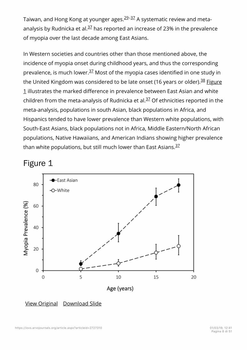
In Western societies and countries other than those mentioned above, the
incidence of myopia onset during childhood years, and thus the corresponding
prevalence, is much lower. Most of the myopia cases identified in one study in
the United Kingdom was considered to be late onset (16 years or older). Figure
1 illustrates the marked difference in prevalence between East Asian and white
children from the meta-analysis of Rudnicka et al. Of ethnicities reported in the
meta-analysis, populations in south Asian, black populations in Africa, and
Hispanics tended to have lower prevalence than Western white populations, with
South-East Asians, black populations not in Africa, Middle Eastern/North African
populations, Native Hawaiians, and American Indians showing higher prevalence
than white populations, but still much lower than East Asians.
Figure 1
View Original Download Slide
29–37
37
37
38
37
37
https://iovs.arvojournals.org/article.aspx?articleid=2727310 01/03/19, 12@41Pagina 8 di 51

Modeled prevalence of myopia by age for East Asian and white children and
teenagers from a systematic review and quantitative meta-analysis fitted to the
year 2005. Graph created from data in Table 3 of Rudnicka et al.
Models, such as those reviewed in the accompanying IMI – Defining and
Classifying Myopia Report, are likely to be efficient in predicting myopia onset,
due in part at least, to identification of a process of myopic shift already under
way. Since the predominant refractive error of young children is usually a low
degree of hyperopia, and the consensus diagnostic criterion for myopia is −0.50
D, there is clearly a transition stage of refractive development for those destined
to become myopic. The onset of the myopic trajectory is relatively sudden
compared to a subtle loss of hyperopia seen in those who remain
emmetropic. The myopic shift and acceleration of axial elongation that
precedes the onset of myopia may be evident up to 4 years earlier and does not
seem to vary between different ethnicities. The high predictive value of the
models of Zadnik et al. and Zhang et al. is therefore likely based on detection
of values of refractive error and ocular biometry during the transition phase,
which depart from those found in emmetropes of the same age.
3.3 Myopia ProgressionProgression of myopic refractive error tends to be studied less frequently than
onset and prevalence in population-based studies. However, understanding the
mechanisms and risk factors for both onset and progression, and the degree to
which they vary, are important, so the phenomena are considered separately
here. Longitudinal studies are optimal, but are resource intensive and
consequently uncommon. Cross-sectional studies are useful when the mean
refractive errors of myopes are segregated by age.
Donovan and colleagues have conducted a meta-analysis of studies reporting
myopia progression rates in children of Asian or European descent living in
urban areas and corrected with single-vision spectacles. The analysis uses data
37
39
40
41–43
42
44 45
46
https://iovs.arvojournals.org/article.aspx?articleid=2727310 01/03/19, 12@41Pagina 9 di 51
from 20 studies, 14 intervention trials, and 6 longitudinal observation studies, to
predict the progression of myopia and shows that among existing myopes,
progression rate declines with increasing age. For example, according to the
equation provided in the study of Donovan et al., progression declines from
−1.12 D/y at age 7 years to −0.50 D/y at age 12 years among Asian children.
The progression rates presented by Donovan et al. arise principally from
control groups of intervention trials, which may not be representative of the
general population. For example, parents of participants in such trials may have
enrolled them because of concern that their children's myopia was progressing
at a rapid rate when compared with their peers. Population-based and school-
based studies tend to report somewhat slower progression. In a rural district in
China with baseline data collected in 1998, a total of 4662 myopic (≤−0.5 D)
children with a mean age of 9.8 years showed −0.84 D progression during 28.5
months, an average annual progression rate of −0.35 D. The timing of the
study and rural habitation of this population may explain some of the difference
in myopic progression rate compared with the meta-data reported by Donovan
et al. The average annual progression rate for a sample including more than
7500 myopic children aged 5 to 16 (mean, 9.3) years in Hong Kong was reported
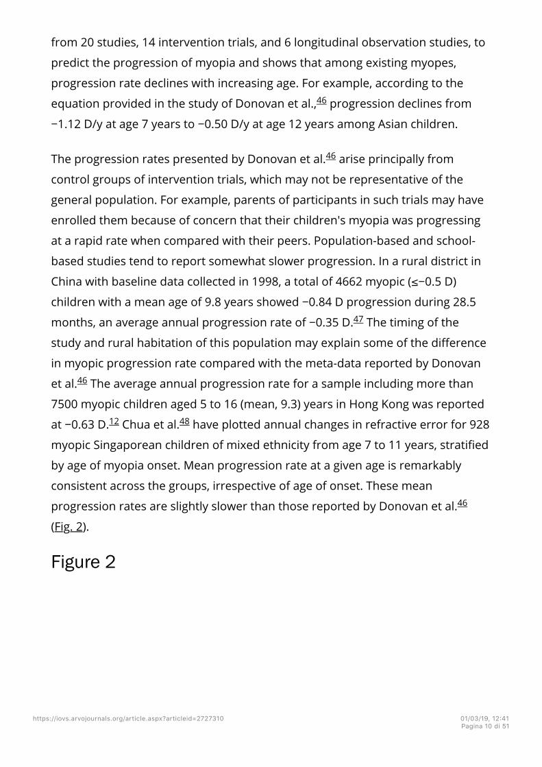
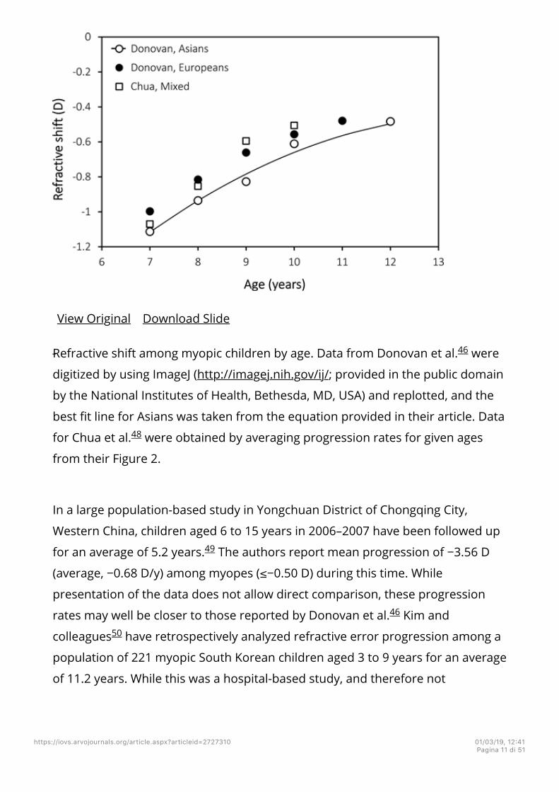
at −0.63 D. Chua et al. have plotted annual changes in refractive error for 928
myopic Singaporean children of mixed ethnicity from age 7 to 11 years, stratified
by age of myopia onset. Mean progression rate at a given age is remarkably
consistent across the groups, irrespective of age of onset. These mean
progression rates are slightly slower than those reported by Donovan et al.
(Fig. 2).
Figure 2
46
46
47
46
12 48
46
https://iovs.arvojournals.org/article.aspx?articleid=2727310 01/03/19, 12@41Pagina 10 di 51
View Original Download Slide
Refractive shift among myopic children by age. Data from Donovan et al. were
digitized by using ImageJ (http://imagej.nih.gov/ij/; provided in the public domain
by the National Institutes of Health, Bethesda, MD, USA) and replotted, and the
best fit line for Asians was taken from the equation provided in their article. Data
for Chua et al. were obtained by averaging progression rates for given ages
from their Figure 2.
In a large population-based study in Yongchuan District of Chongqing City,
Western China, children aged 6 to 15 years in 2006–2007 have been followed up
for an average of 5.2 years. The authors report mean progression of −3.56 D
(average, −0.68 D/y) among myopes (≤−0.50 D) during this time. While
presentation of the data does not allow direct comparison, these progression
rates may well be closer to those reported by Donovan et al. Kim and
colleagues have retrospectively analyzed refractive error progression among a
population of 221 myopic South Korean children aged 3 to 9 years for an average
of 11.2 years. While this was a hospital-based study, and therefore not
46
48
49
46
50
https://iovs.arvojournals.org/article.aspx?articleid=2727310 01/03/19, 12@41Pagina 11 di 51
necessarily representative of the population at large, the progression rate of
approximately −0.50 D/y between the ages of 7 and 13 years was surprisingly
modest. Hsu and coworkers have reviewed a population-based cohort in
Taiwan of 3256 myopic children, of average age 7.5 years, after 1 year and noted
average progression in the group of only −0.42 D, well below that predicted by
Donovan et al. Some of these children were being treated with cycloplegics to
slow myopia progression and all had been exposed to a large-scale eye care
education program, which may explain the lower progression rate. Most
recently, Wu et al. have found annual progression of −0.79 D among a school-
based control population of 89 myopes aged 6 and 7 years in Taiwan. This is also
less than predicted by Donovan et al., but it should be noted that those in the
sample population receiving myopia treatment were excluded from the analysis.
Further details of likely progression can be obtained from centile progression
curves. Chen et al. have constructed reference age-specific centile curves of
refraction from cross-sectional population-based data from the Guangzhou
Refractive Error Study in Children. However, apparent progression among the
myopes between ages of 7 and 12 years is observed to be only approximately
−0.5 to −0.6 D/y, comparatively constant across ages, and less again than that of
Donovan et al., particularly at a younger age. The implications of these
differences are not clear. Tideman et al. have also produced age-specific centile
curves for axial length. The relative functionality of these curves compared to
those for refractive error is yet to be determined.
Based on the above literature review, greater myopia progression rates are
expected at younger ages (i.e., −0.50 to −1.00 D/y for 6- to 9-year-olds) than at
older ages (i.e., −0.35 to −0.75 D for those older than 10 years).
3.4 High MyopiaOne of the major ethical challenges for practitioners is accurate identification of
those at risk of becoming highly myopic or, at the very least, of those whose
myopia is progressing at an unacceptably fast rate. Few analyses are available on
51
46
52
46
53
46
54
48
https://iovs.arvojournals.org/article.aspx?articleid=2727310 01/03/19, 12@41Pagina 12 di 51
this topic, but the breakdown by Chua et al. probably represents the most
comprehensive data available. They have found age of onset of myopia to be the
strongest predictor of high myopia among Singaporean children. As expected,
duration of myopia progression was also important in predicting high myopia.
For children with high myopia at age 11 years, there was an 87% chance that the
child became myopic at 7 years of age or younger or had a duration of myopia
progression of 4 years or more. Reports from other countries (Denmark,
Argentina, United Kingdom) reliably reproduce this observation. However, in
contrast to the report by Chua and colleagues, Williams et al. have found that
age of onset only accounts for a modest proportion (approximately 15%) of the
variance in severity of myopia.
3.5 Adult-Onset Myopia and ProgressionMost of the myopia cases in one study in Britain were considered to be late
onset (16 years or older). Although myopia onset past the adolescent stage of
life is of clinical interest and has shown an association with environmental
factors, eye care practitioners are generally more concerned from an ethical
standpoint with identifying patients at risk for development of higher degrees of
myopia, which typically involves juvenile-onset myopia and its associated
potential to progress to sight-threatening pathology.
The prevailing perception is that myopia stabilizes in the late teenage years.
Certainly, annual progression in most myopic patients slows with time and for
many myopes whose condition has progressed through the teenage years,
myopia will stabilize before they reach 20 years of age. However, there are
patients whose myopia will continue to progress through adult years. These
patients include those doing intense near work, especially students, and those
who have higher degrees of myopia. Continued assessment of refraction and
initiation of treatment in patients showing continued progression are warranted.
Higher levels of myopia will result from continued progression through
adulthood, placing these individuals at higher risk for development of myopia-
48
48
55–57
48 57
38
58
41
58–60
https://iovs.arvojournals.org/article.aspx?articleid=2727310 01/03/19, 12@41Pagina 13 di 51

associated pathologies.
3.6 Genetic and Environmental RiskRisk factors for myopia onset have been identified and included in a number of
multivariate models, although to our knowledge there is currently no
comprehensive clinical model that provides good predictive value, aside from
those using refractive or biometric information. McMonnies has provided a
review of risk factors for onset and progression of myopia and produced a
comprehensive table of those factors and how they may influence the prognosis
and treatment decisions for individual patients. However, he also notes that the
lack of clinical data on the topic of risk “undermines the confidence with which
individual prognoses and clinical decisions about interventions can be made.”
3.6.1 Myopia OnsetGenetics and Personal CharacteristicsHeritability statistics can be used to estimate the proportion of variation in a
phenotypic trait of a population that is due to genetics, and further details can be
found in the accompanying IMI – Myopia Genetics Report. Heritability
estimates for myopia vary from 0.11 to 0.98, the latter higher value being found
among a highly specific group of Finnish female twins aged 28 to 29 years. A
meta-analysis places heritability at 0.71 for refractive error, which would
suggest that the majority of influence is from genetics rather than environment.
Genome-wide association studies (GWAS) have demonstrated complex
inheritance of refractive error traits, with identification of more than 150 gene
loci associated with myopia and good correlation between studies. However, the
identified loci explain a meagre percentage of the variance in refractive error.
For example, a genetic risk score (GRS) has estimated that these loci explain only
0.6% and 2.3% of the variance in refractive error at ages 7 and 15 years,
respectively. The difference between heritability from twin studies and GWAS is
known as “missing heritability” or the “heritability gap” and is a well-known
characteristic of other phenotypes and diseases.
61
62
63,64
65
66
67
68
66
https://iovs.arvojournals.org/article.aspx?articleid=2727310 01/03/19, 12@41Pagina 14 di 51
While the nature versus nurture debate continues in relation to myopia
development, recognition of the importance of gene-environment interactions in
phenotypic expression has been a significant step forward. Fan et al. have
tested for evidence of interactions between near work or time spent outdoors
and 39 previously identified loci from GWAS in refractive development in a
pediatric cohort. Five variants have shown apparent interaction with near work,
while neither variant nor GRS effects were altered with time outdoors.
The most useful clinical indicator for genetic risk short of genetic testing is
parental history of myopia. Older studies demonstrating this association have
been reviewed by Goss and Jackson. Studies since that time show significant
association between number of myopic parents and incident myopia, as
summarized in a recent meta-analysis. Odds ratios (ORs) ranging from 1.44 to
2.96 for having a myopic child compared to not having a myopic child were
calculated, depending on the number of myopic parents and adjustment for bias
and missing studies. More recent studies confirm the connection.
Parental myopia has also been found to interact with other risk factors. In one
study of 1770 grade-7 Chinese students, those with close reading distances and
two myopic parents have a 26-fold higher odds for prevalent myopia than
children with reading distances of greater than 20 cm and no myopic
parents. Also, unsurprisingly, parental myopia correlates with certain ocular
components, particularly axial length.
There are some further considerations around parental myopia as a risk factor.
The additive genetic portion of phenotypic variance is smaller in younger
families, reflecting the trend for increasing environmental influences. The odds
of a child with two myopic parents becoming myopic is thus different to the odds
of a myopic child having two myopic parents. In part, this stems from increased
myopia prevalence, meaning that there will likely be more children with myopia
than there are parents with myopia. Number of myopic parents is a relatively
gross instrument and a knowledge of degree of myopia in family members may
be a more useful factor for predicting progression. Because of these factors,
68
68
69
70
70 27,71–76
77,78
79,80
81
43
61,82
https://iovs.arvojournals.org/article.aspx?articleid=2727310 01/03/19, 12@41Pagina 15 di 51
the sensitivity of number of myopic parents in predicting childhood myopia is
correspondingly low.
Rudnicka et al. also have found that sex differences emerge in myopia
prevalence at approximately 9 years of age in whites and East Asians. By 18
years of age, white females have 2.0 (95% confidence interval [CI], 1.4–2.9) times
the odds of myopia as white males, and East Asian females have 2.3 (95% CI, 2.0–
2.6) times the odds of myopia as East Asian males. Others since have
confirmed the propensity for greater myopia prevalence among females. The
extent to which this influence is environmental as opposed to genetic has yet to
be determined.
EnvironmentRamamurthy and colleagues have reviewed the large number of
environmental risk factors for myopia. Two key environmental influences upon
myopia development are time spent outdoors and amount of near work. The
reason time spent outdoors is protective against myopia development remains
unexplained. Although there is some evidence from animal studies showing that
high light levels or chromaticity might be the critical factor, Flitcroft and Ngo
et al. present a counter-argument as to why the dioptric field, perhaps in an
interaction with the high light level, is central to protection from time spent
outdoors. Xiong et al. have reviewed multiple studies and suggest a clear
connection between time spent outdoors and myopia onset. However,
differentiation between consequence and causality can only be shown in
prospective randomized studies. As spending time outdoors is an intervention to
prevent or delay myopia development, detailed description of this risk factor for
myopia onset is presented in the accompanying IMI – Interventions for
Controlling Myopia Onset and Progression Report.
Despite some indications that near work may not be directly related to myopia,
more recent evidence suggests a clear link. “Near work” has been defined and
measured in a multitude of ways across different studies (e.g., education level,
82,83
37
27,71,73
84
84 85
86
87
88
89
https://iovs.arvojournals.org/article.aspx?articleid=2727310 01/03/19, 12@41Pagina 16 di 51
duration of continuous study time, time spent reading books for pleasure,
number of books read per week, time spent on reading and close work, time
spent indoors studying, closer working distance, short reading distance, distance
from near work, font size, and screen-viewing activities) and is, by its nature,
difficult to quantify. Nonetheless, in a systematic review and meta-analysis,
Huang et al. have found more time spent on near-work activities is associated
with higher odds of myopia, increasing by 2% for every additional diopter-hour
of near work per week. Multiple subsequent articles not included in this meta-
analysis also confirm the association of some index of near work with
development and progression of myopia, often independently from time spent
outdoors in multivariate analyses. French and coworkers
have presented data that illustrate a strong interaction between the effect of
time spent outdoors and near work. In children with baseline mean age of 6
years, those who spend low amounts of time outdoors and perform high levels
of near work have dramatically increased odds of incident myopia by age 12
years (OR, 15.9; 95% CI, 3.5–73.4) as compared with those who spend high
amounts of time outdoors and low amount of time involved in near work.
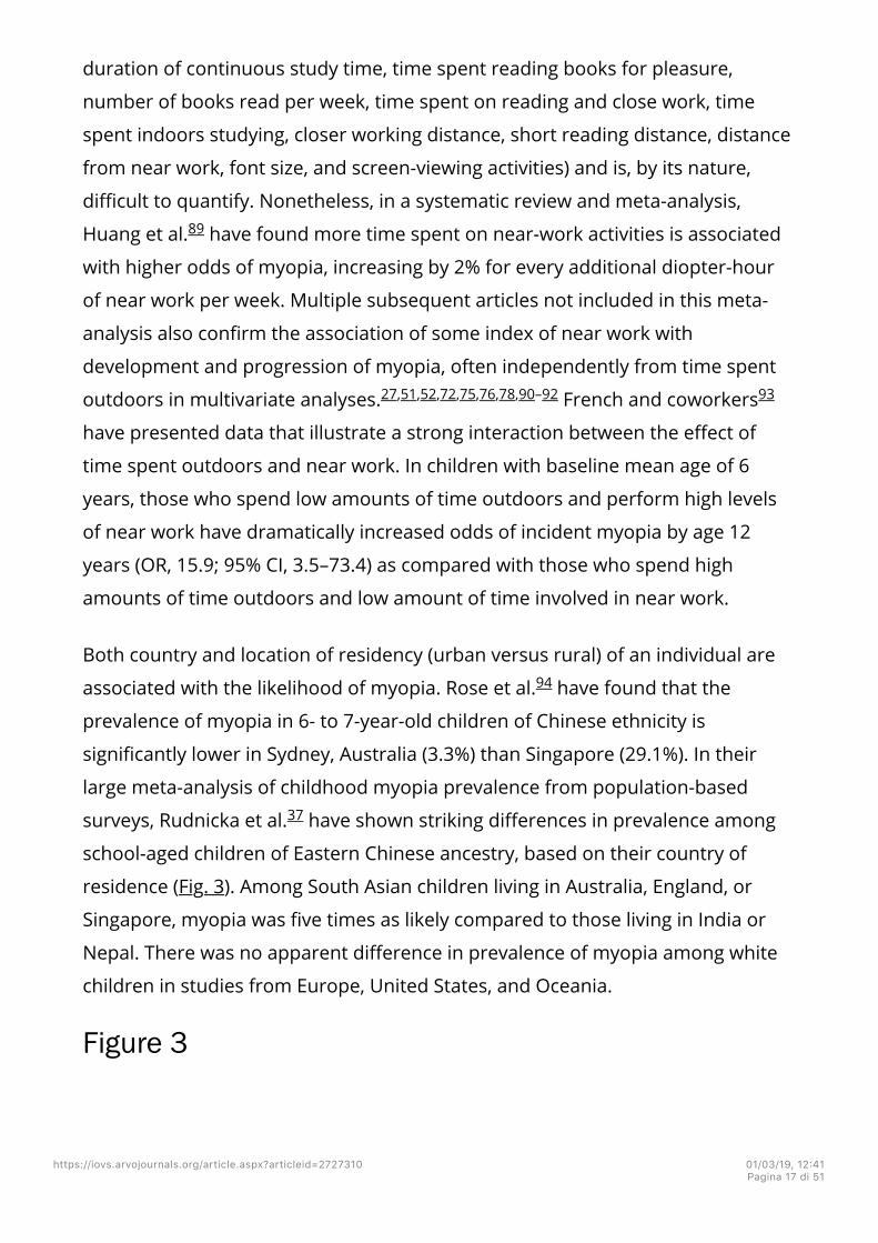
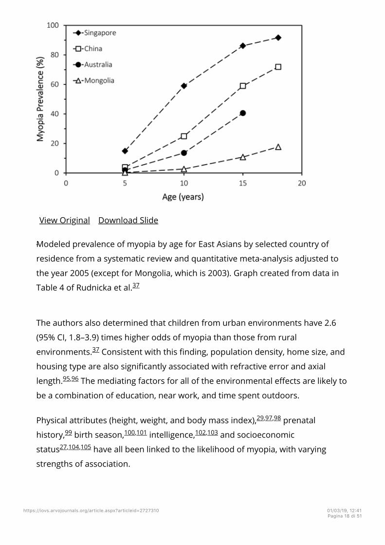
Both country and location of residency (urban versus rural) of an individual are
associated with the likelihood of myopia. Rose et al. have found that the
prevalence of myopia in 6- to 7-year-old children of Chinese ethnicity is
significantly lower in Sydney, Australia (3.3%) than Singapore (29.1%). In their
large meta-analysis of childhood myopia prevalence from population-based
surveys, Rudnicka et al. have shown striking differences in prevalence among
school-aged children of Eastern Chinese ancestry, based on their country of
residence (Fig. 3). Among South Asian children living in Australia, England, or
Singapore, myopia was five times as likely compared to those living in India or
Nepal. There was no apparent difference in prevalence of myopia among white
children in studies from Europe, United States, and Oceania.
Figure 3
89
27,51,52,72,75,76,78,90–92 93
94
37
https://iovs.arvojournals.org/article.aspx?articleid=2727310 01/03/19, 12@41Pagina 17 di 51
View Original Download Slide
Modeled prevalence of myopia by age for East Asians by selected country of
residence from a systematic review and quantitative meta-analysis adjusted to
the year 2005 (except for Mongolia, which is 2003). Graph created from data in
Table 4 of Rudnicka et al.
The authors also determined that children from urban environments have 2.6
(95% CI, 1.8–3.9) times higher odds of myopia than those from rural
environments. Consistent with this finding, population density, home size, and
housing type are also significantly associated with refractive error and axial
length. The mediating factors for all of the environmental effects are likely to
be a combination of education, near work, and time spent outdoors.
Physical attributes (height, weight, and body mass index), prenatal
history, birth season, intelligence, and socioeconomic
status have all been linked to the likelihood of myopia, with varying
strengths of association.
37
37
95,96
29,97,98
99 100,101 102,103
27,104,105
https://iovs.arvojournals.org/article.aspx?articleid=2727310 01/03/19, 12@41Pagina 18 di 51
Binocular VisionIt has long been postulated that myopia onset and progression may be related to
dysfunctional accommodation and convergence. An elevated
accommodation-convergence/accommodation (AC/A) ratio has been observed
before the onset of myopia. In a large, ethnically diverse group of children
followed up for an extensive period of time, Mutti and colleagues have found
the AC/A ratios of those who become myopic begin to increase approximately 4
years before myopia diagnosis, continue increasing until diagnosis, and then
plateau at a level higher than those who remain emmetropic.
Another feature of accommodation that has been observed is that measured lag
of accommodation is larger among myopes than nonmyopes. It was
thought that the presence of lag before onset may produce hyperopic retinal
defocus, stimulating myopia onset. However, this effect only appears at the time
of onset, not before, and does not seem to impact progression. An aspect
of accommodative lag worthy of mention is that spurious measurement of
accommodative error is well documented. So-called lag may be
substantially a function of the measurement technique, where depth of focus
and increasing negative spherical aberration with accommodation and
developing myopia are not taken into account.
The shift in refraction (in terms of a reduction in hyperopia) observed in those
who will become myopic compared to those who remain emmetropic begins
several years before diagnosis. Changes to the AC/A ratio merely seem to
parallel such changes. Accommodative lag does not seem to appear until myopia
onset. Thus, while binocular vision attributes are an interesting research
adjacency in the onset and development of myopia, from our current knowledge
they do not seem to add any additional benefit in risk assessment over
refraction and biometric parameters, genetics, or environmental effects.
3.6.2 Myopia Progression
Compared with onset, there is a lower volume of literature describing risk of
106
44,107
108
109,110
108,111
112–114
115
https://iovs.arvojournals.org/article.aspx?articleid=2727310 01/03/19, 12@41Pagina 19 di 51
progression for existing myopes other than age and initial refractive error. Some
studies have looked at group progression, including emmetropes and hyperopes
as well as myopes in their analyses, which does not allow specific interpretation
regarding progression among myopes.
Genetics and Personal CharacteristicsDonovan et al. report that myopia in European children progresses more
slowly on average than in Asians (−0.55 D/y and −0.82 D/y, respectively, at mean
age of 9.3 years), although age-specific progression data by baseline age for
Europeans in their analysis are derived from a single article. For studies
conducted in somewhat homogeneous Western societies, the analysis of Mutti et
al. supports the ethnic differences in progression rates found in the study by
Donovan and colleagues, although French et al. did not establish
significance of an ethnicity effect. Gwiazda and colleagues have looked at risk
factors for high myopia, which can be considered a corollary of fast progression.
Reporting on an ethnically diverse population of children aged 6 to 11 years with
initial myopia between −4.50 and −1.25 D at four sites within the United States,
they did not find an effect of ethnicity on progression rates. Environment is also
likely to play a role in myopic progression rates, which may be inferred from
higher degrees of myopia among Asian children living in Asia compared to those
living in Western societies; however, a thorough review of differences in
progression rates between ethnically similar populations in different
environments does not seem to have been undertaken.
In their study, Gwiazda et al. found that the number of myopic parents is a
risk factor for high myopia. Some studies support the proposition that parental
myopia is associated with faster progression rates, where others do not.
Females show faster progression than males according to Donovan et al.
(−0.80 D/y and −0.71 D/y, respectively, at mean age 8.8 years) and Zhou et al.
(OR, 1.45; 95% CI, 1.12–1.84). However, such a difference is not evident in the
study of Gwiazda et al.
46
42
46 116
117
117
91,118,119
46
49
117
https://iovs.arvojournals.org/article.aspx?articleid=2727310 01/03/19, 12@41Pagina 20 di 51
EnvironmentIn their meta-analysis, Xiong et al. report that outdoor time is not effective in
slowing progression in eyes that are already myopic. However, a more recent
prospective study suggests that outdoor time does have a protective effect on
rate of progression. Subsequent cohort studies yield mixed results.
Support for the protective effect of time spent outdoors on myopia progression
may be inferred from numerous studies that have found a seasonal variation in
myopia progression; see the accompanying Interventions for Controlling
Myopia Onset and Progression Report.
Many of the same environmental factors that are linked to the incidence or
prevalence of myopia are also related to progression. Multiple articles link near
work, with various descriptors of activity, to myopia
progression. Other associations include urbanization and
increasing family income.
Binocular VisionTwo studies that have considered binocular vision effects as part of the
treatment protocol (esophoria or low lag of accommodation) have had good
success, suggesting that some aspect of binocular function may be a risk factor
for progression.
3.7 Summary of Findings on Risk FactorsThe observations reported above present an unambiguous message. The
younger the age of onset of myopia, the greater the likelihood that a child will
experience progression to vision-threatening levels of myopia. Practitioners and
parents should be active in addressing both myopia onset and progression at as
young an age as possible. No formal procedures have been identified that
recognize those at risk of myopia onset before the triggering of the steady
progression in refractive error that ultimately leads to myopia diagnosis.
However, it is clear, for example, that Chinese children living in urban regions of
Asia, who are immersed in an intensive education environment and have two
87
52
51,74,118,120
121–123
88
51,52,72,74,91,119,120
90,91
124,125
https://iovs.arvojournals.org/article.aspx?articleid=2727310 01/03/19, 12@41Pagina 21 di 51
myopic parents, have a much greater risk for onset and development of
significant myopia than a Caucasian living in a rural environment in Australia
with no myopic parents. Not all children who are young at myopia onset will
experience progression to high myopia, but age of onset is the current best
determinant for identifying children at risk of progression. While noting the risk
of high myopia is greatest in those with early onset, practitioners should also be
cognizant that the condition of some individuals with later onset (say 11 years or
older) may also progress to higher degrees of myopia, where the rate of
progression is high. Practitioners should be vigilant in identifying and treating
those at risk of rapid progression, regardless of age of onset.
4. Subcommittees and Their Report Focusand Advancements4.1 IMI – Defining and Classifying Myopia ReportMyopia has been the topic of scientific study for more than 400 years, but it is
only more recently that it has been recognized as a serious public health issue,
owing to its being a significant cause of visual loss and a risk factor for a range of
pathologic ocular conditions. Its prevalence is increasing on a global basis and
has reached epidemic levels in much of Asia. Myopia has been defined in a wide
variety of ways in the past, such as based on its assumed etiology, age of onset,
progression rate, degree of myopia (in diopters), and structural complications.
This has led to a confusing accumulation of terms. Hence this subcommittee's
aim was to provide a standardized set of terminology, definitions, and thresholds
of myopia and its main ocular complications. A critical review of current
terminology and choice of myopia thresholds was undertaken to ensure that the
proposed standards are appropriate for clinical research purposes, relevant to
the underlying biology of myopia, acceptable to researchers in the field, and
useful for developing health policy. It is recommended that the many descriptive
terms of myopia be consolidated into the following descriptive categories:
39
https://iovs.arvojournals.org/article.aspx?articleid=2727310 01/03/19, 12@41Pagina 22 di 51

Myopia: A refractive error in which rays of light entering the eye parallel
to the optic axis are brought to a focus in front of the retina when
ocular accommodation is relaxed. This usually results from the eyeball
being too long from front to back, but can be caused by an overly
curved cornea, a lens with increased optical power, or both. It is also
called nearsightedness.
With qualifying terms:
Axial Myopia: A myopic refractive state that can be attributed to
excessive axial elongation.
Refractive Myopia: A myopic refractive state that can be attributed to
changes in the structure or location of the image-forming structures of
the eye (i.e., the cornea and lens).
Secondary Myopia: A myopic refractive state for which a single, specific
cause (e.g., drug, corneal disease, or systemic clinical syndrome) can be
identified that is not a recognized population risk factor for myopia
development.
https://iovs.arvojournals.org/article.aspx?articleid=2727310 01/03/19, 12@41Pagina 23 di 51

It was also recommended that in quantitative contexts, myopia should always be
treated as a negative value and that mathematical comparison symbols be used
in their strict mathematical sense.
To provide a framework for research into myopia prevention, the condition of
“premyopia” is defined.
Premyopia: A refractive state of an eye of ≤+0.75 D and >−0.50 D in
children where a combination of baseline refraction, age, and other
quantifiable risk factors provides a sufficient likelihood of the future
development of myopia to merit preventative interventions.
As a quantitative trait it is recommended that myopia be divided into myopia
(i.e., all myopia), low myopia, and high myopia as based on the current
consensus of publications:
Myopia: A condition in which the spherical equivalent refractive error of
an eye is ≤−0.5 D when ocular accommodation is relaxed.
Low Myopia: A condition in which the spherical equivalent refractive
error of an eye is ≤−0.5 D and >−6.00 D when ocular accommodation is
relaxed.
https://iovs.arvojournals.org/article.aspx?articleid=2727310 01/03/19, 12@41Pagina 24 di 51
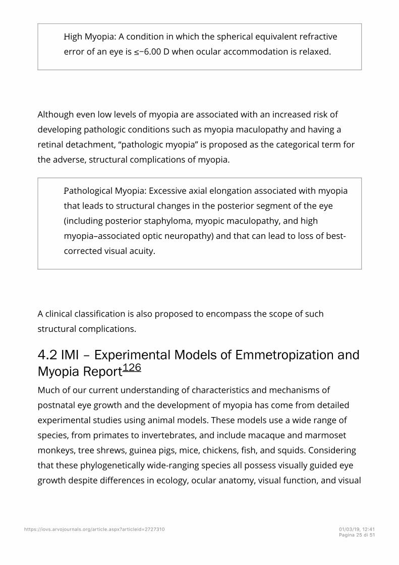
High Myopia: A condition in which the spherical equivalent refractive
error of an eye is ≤−6.00 D when ocular accommodation is relaxed.
Although even low levels of myopia are associated with an increased risk of
developing pathologic conditions such as myopia maculopathy and having a
retinal detachment, “pathologic myopia” is proposed as the categorical term for
the adverse, structural complications of myopia.
Pathological Myopia: Excessive axial elongation associated with myopia
that leads to structural changes in the posterior segment of the eye
(including posterior staphyloma, myopic maculopathy, and high
myopia–associated optic neuropathy) and that can lead to loss of best-
corrected visual acuity.
A clinical classification is also proposed to encompass the scope of such
structural complications.
4.2 IMI – Experimental Models of Emmetropization andMyopia ReportMuch of our current understanding of characteristics and mechanisms of
postnatal eye growth and the development of myopia has come from detailed
experimental studies using animal models. These models use a wide range of
species, from primates to invertebrates, and include macaque and marmoset
monkeys, tree shrews, guinea pigs, mice, chickens, fish, and squids. Considering
that these phylogenetically wide-ranging species all possess visually guided eye
growth despite differences in ecology, ocular anatomy, visual function, and visual
126
https://iovs.arvojournals.org/article.aspx?articleid=2727310 01/03/19, 12@41Pagina 25 di 51
acuity, this supports the hypothesis that visually guided eye growth is an
evolutionarily conserved process found in camera-type eyes. Each species
provides unique experimental advantages to study the mechanisms of visually
guided eye growth and the key signalling pathways that regulate refractive eye
development across species; however, anatomic and physiological differences
must be taken into account when interpreting and translating results to
humans.
The report summarizes the anatomic similarities and differences between the
eyes of the principal experimental species used for studies of emmetropization
and myopia. Surveying more than 800 published reports on the changes in eye
growth and refractive state in response to experimental manipulations of visual
conditions, the report offers a summary of the evidence supporting the role of
vision in eye development and the mechanisms that underlie the visual
regulation of eye growth and emmetropization development. Also discussed are
the key operating characteristics of experimental emmetropization to
experimentally imposed retinal defocus including local retinal mechanisms
controlling regional eye growth, the spatial and temporal integration of visual
signals, the impact of simultaneous competing defocus signals, the relationships
of various ocular circadian rhythms to induced changes in eye growth, and the
critical periods for visual experience–invoked myopia. Studies of the
characteristics of the visual signals affecting eye growth are also reviewed and
discussed, including the intensity of ambient illumination, the spectral
composition of light, longitudinal chromatic aberration, higher-order
monochromatic aberrations, and astigmatism. The report reviews the
biochemistry of refractive error development, including the roles of various
retinal neurotransmitters, neuromodulators, and growth promotors such as
dopamine, vasoactive intestinal peptide, melanopsin, glucagon, and insulin, and
nitric oxide. Pharmacologic studies of the mechanisms of emmetropization and
myopia are discussed including the effects of cholinergic, GABAergic, and
adenosine antagonistic drugs and drugs affecting nitric oxide and
https://iovs.arvojournals.org/article.aspx?articleid=2727310 01/03/19, 12@41Pagina 26 di 51
neuropeptides. Finally, the article reviews the molecular biology of gene
expression in the eye and retina and possible gene-environment interactions.
The report reviews and summarizes several confirmed findings from animal
models that have provided important proofs of concept that helped to transform
treatment strategies for myopia control. These findings include the eye's ability
to detect the sign of retinal defocus and undergo compensatory growth, the local
retinal control of eye growth, regulatory changes in choroidal thickness, and the
identification of biochemical signal cascades regulating postnatal eye growth and
refractive state. Experimental animal models continue to provide new insights
into the cellular and molecular mechanisms of eye growth control, including the
identification of potential new targets for drug development and future
treatments needed to stem the increasing prevalence of myopia and the vision-
threatening conditions associated with this disease.
4.3 IMI – Myopia Genetics ReportLike other complex traits, myopia has benefitted enormously from the dramatic
improvements in DNA technologies and significant reduction in costs for
genotyping during the last decade. The IMI – Myopia Genetics Report
summarizes the developments in gene identification for refractive error and
myopia, and addresses their implications for molecular pathways. An extensive
literature search identified almost 200 genetic loci that have been reported for
refractive error, myopia, or axial length, and many overlap between these
endophenotypes. Risk variants have mostly been identified outside the protein
coding regions, and by themselves carry a low risk. Nevertheless, totalling all
genetic risk variants in a polygenic risk score shows that those with a high
genetic load are >40 times more likely to become myopic, and high myopes and
high hyperopes can be separated by their genetic score. The most significant
contribution of the current gene dissection is the insights into the molecular
machinery underlying eye growth. Functions of the annotated genes include
retinal cell physiology, light processing, glutamate receptor signalling,
62
https://iovs.arvojournals.org/article.aspx?articleid=2727310 01/03/19, 12@41Pagina 27 di 51
extracellular matrix modulation, anterior segment morphology, but also
posttranscriptional regulation indicating control of gene expression at the RNA
level. In silico and in vitro experiments have shown that all cell types in the
retina, but also RPE, vascular bed, and connective tissue are sites of gene
expression. This implies that the retinal signalling cascade responding to a visual
trigger and leading to eye growth involves a complex network of molecules from
many different cells and tissues. Another lesson learned from the genetic studies
is that most genes are not eye specific and have a plethora of effects outside of
the eye. A fair number of genes for common myopia are involved in a wide range
of syndromes, including neurodegenerative and connective tissue disorders.
How this broad spectrum of gene functions leads to scleral remodelling and an
increase of axial length remains intriguing. Addressing this “black box” requires
taking myopia molecular genetics to the next level: to explore new high-
throughput, wide coverage genotyping assays; determine the protein function
and the elements that regulate gene expression; investigate how DNA, proteins,
and the environment interact to determine eye size; and create possibilities for
storage and reuse of massive genomic data. The forecast of understanding and
solving myopia makes these challenges worth taking.
4.4 IMI – Interventions for Controlling Myopia Onsetand Progression ReportThis report examines the evidence basis for various interventions in current use
for controlling myopia progression in children, organized under the categories of
optical, pharmacologic, environmental (behavioral), and surgical interventions
(aimed at stabilizing highly myopic eyes). There is equivocal evidence concerning
whether single-vision spectacles cause faster myopic progression than soft
contact lenses, but any difference is likely to be clinically irrelevant.
Undercorrection is still adopted as a myopia control strategy by some
practitioners, yet some but not all clinical trials indicate this strategy has no
clinically significant benefit in slowing myopia. Single-vision spectacle lenses
designed to alter peripheral defocus had only a small treatment effect, of less
88
https://iovs.arvojournals.org/article.aspx?articleid=2727310 01/03/19, 12@41Pagina 28 di 51
than 14% reduction in myopia progression. The treatment effects on myopia
progression of bifocal and progressive addition spectacles tend to be larger,
although variable and questioned in terms of clinical significance in some cases
(6%–51%). Overall, single-vision contact lenses, whether soft or rigid, seem to
have little effect on myopia progression, in contrast to significant treatment
effects with contact lenses that impose multifocality. Center-distance multifocal
lenses have been used off-label successfully, demonstrating a sample size–
weighted average of 38%, slowing both myopia progression and axial elongation,
although these two assessment elements did not always correspond tightly.
Orthokeratology has also proven to be effective in slowing axial length
elongation, by between 30% to 55%.
Pharmacologic myopia control trials have principally used atropine, although
other muscarinic antagonists such as M1 selective pirenzepine, ocular
hypotensive agents including topical timolol (a nonselective β-adrenergic
antagonist), and oral 7-methylxanthine, an adenosine antagonist, have also
undergone trial. Although the reduction in myopia progression seems to be
higher with 1% atropine (around 60%–80%), more recent atropine studies use
much lower doses (e.g., 0.01%), with a reduced effect on refractive error
retardation (around 45%) and no apparent effect of axial length compared to
historical controls, but with fewer side effects and apparent rebound after
discontinuation.
Time outdoors appears to be more effective in preventing incident myopia than
slowing progression of existing myopia. However, the evidence for vitamin D
levels being related to myopic control is weak. Seasonal trends in myopia
progression have also been interpreted as indirect evidence of outdoor effects
on myopia progression, based on observed faster myopia progression during
the darker winter than the brighter summer months. In one study, every
additional hour of outdoor time per week has been found to reduce the risk of
developing myopia by 2%. In another study, the time children spend engaged in
near work outside of school and time spent outdoor were not found to be
https://iovs.arvojournals.org/article.aspx?articleid=2727310 01/03/19, 12@41Pagina 29 di 51
related, as might be expected. Deployment of wearable technologies in place of
questionnaires as study tools may help to resolve apparent inconsistencies and
unresolved questions, including whether the quality of indoor lighting is
important.
4.5 IMI – Clinical Myopia Control Trials andInstrumentation ReportClinical trials on myopia control conducted to date were reviewed to inform a
consensus on best practice in the design of clinical trials to assess the
effectiveness of treatments and the impact on patients. As myopia control
interventions will be applied for multiple years throughout the time during which
myopia is progressing, and treatment effects have been shown to often reduce
after an initial period, it is important that clinical trials evaluate efficacy over a
long period (3 years being the recommendation) to ensure continued efficacy
beyond any initial treatment effect. Assessment of rebound should also be
considered, with a minimum recommended period of 1 year due to seasonal
effects. Typical inclusion criteria are cycloplegic spherical or spherical equivalent
myopia of at least −0.75 D; astigmatism ≤ 1.00 D; anisometropia ≤ 1.50 D; ages 6
to 12 years; and 20/20 (0.0 logMAR) minimum visual acuity. Exclusion criteria
typically are previous rigid contact lens wear; history of previous myopia control
treatment; ocular pathology; binocular vision anomaly; medications that may
affect pupil size, accommodation, or have an impact on ocular surface; and
systemic disease that may affect vision, vision development, or the treatment
modality. Appropriate control group selection depends on the intervention being
studied, but often myopia control studies cannot be fully masked. Studies with
no control group are unable to demonstrate treatment efficacy; as the rate of
myopia progression decreases naturally with age and has seasonal variation, it is
not possible to distinguish between naturally declining progression and reduced
progression attributable to the treatment, without a simultaneously conducted
control group. Randomization should be applied to treatment allocation, and
stratification by key factors known to influence myopia progression (such as age
127
https://iovs.arvojournals.org/article.aspx?articleid=2727310 01/03/19, 12@41Pagina 30 di 51

and race/ethnicity) should be considered. Ocular health, including a slit lamp
examination and baseline/periodic dilated fundus examination, along with
standardized adverse event reporting, should also be embedded in the trial
protocol. Binocular vision associations in myopia control treatments have also
been found, so should be investigated at baseline and periodic intervals during
the study. Other safety-related assessments include visual acuity and
dysphotopsia. Finally, there is not a specific minimum percentage reduction in
myopia progression that has been published for a treatment effect to be
considered clinically meaningful; any such percentage reduction threshold could
theoretically vary with multiple other factors, including duration of treatment,
sample population, and study design considerations. Sample size estimations
based on currently available measurement variability data are provided.
Outcome measures were classified as primary, secondary, and exploratory.
Primary outcome measures are refractive error (ideally assessed objectively with
autorefraction of the eye cyclopleged in optical intervention studies with 1%
tropicamide) or axial length (ideally measured with noncontact interferometry)
or both. Secondary outcome measures focus on patient-reported outcomes
(usually assessed by questionnaire and can include the parent's/guardian's
experience as well as the patient's) and treatment compliance (ideally in real
time, such as with text messaging responses or wearable sensors connected to
data loggers). Exploratory outcome measures are particularly useful in trying to
understand the mechanism of action and associated factors. These include
peripheral refraction (such as measured with autorefractors or wavefront
aberrometers), accommodative changes (including accommodative lag and
dynamics), ocular alignment, pupil size, outdoor activity/lighting levels, anterior
and posterior segment structural changes (typically imaged with Scheimpflug
imaging, optical coherence tomography, and retinal photography with a
particular interest in choroidal thickness changes), and tissue biomechanics (of
the sclera and cornea).
4.6 IMI – Industry Guidelines and Ethical128
https://iovs.arvojournals.org/article.aspx?articleid=2727310 01/03/19, 12@41Pagina 31 di 51
Considerations for Myopia Control ReportThe aim of this subcommittee was to discuss guidelines and ethical
considerations associated with the development and prescription of treatments
intended for myopia control. A critical review of published articles and guidance
documents was undertaken, with a view to carefully consider the ethical
standards associated with the investigation, development, registration,
marketing, prescription, and use of myopia control treatments.
From an ethical standpoint, deciding whether to implement a myopia control
strategy represents a classical medical risk versus benefit ratio. A principal
motivation for slowing myopia progression is based on the premise that limiting
myopia progression reduces risk of the development of vision-threatening
disease in later life. However, conclusive evidence that this is the case is unlikely
to be available for decades. Nonetheless, if this assumption is correct, then the
benefits could be substantial, given the clear relationship between myopia-
related ocular pathology and the degree of myopia. Thus, the risk-benefit
analysis must take account of the outcomes arising from nonintervention in
deciding if implementation of a myopia control strategy with an individual
patient is warranted. Other factors to consider include the known improvements
in quality-of-life issues arising from the use of corrective devices; adults with
pathologic myopia and associated visual impairment report significant social and
emotional impacts and reduced life satisfaction. Additional factors that must be
accounted for in the decision to undertake myopia control include the regulatory
status of the treatment being considered, availability of the treatment, access to
appropriate eye care services, and pricing and convenience of the treatment,
which are all potential barriers to accessing the myopia control treatment being
considered.
These considerations place a burden of responsibility on the practitioner to be
fully cognizant of the risks for the patient of developing different levels of
myopia, the implications that progression to higher levels of myopia may have,
128
https://iovs.arvojournals.org/article.aspx?articleid=2727310 01/03/19, 12@41Pagina 32 di 51
the likely benefits of treatment, the side effects of treatment and other
associated factors, so as to provide appropriate advice and care.
Researchers and clinicians often partner with companies to conduct myopia
control studies. However, there is a risk for these partnerships to introduce bias,
and practitioners should be aware of the importance of evaluating any real (or
perceived) conflict of interest when recommending a management plan for
myopia control. The interactions between researchers, practitioners, and
manufacturers of myopia control treatments should meet the highest possible
standards of integrity and transparency and must be declared in the reporting of
the results obtained. Relationships between clinicians and patients should not be
compromised by commercial or other interests that could subvert the principle
that the interests of patients are of primary concern.
Most myopia control treatments are currently off-label in many countries. Most
regulatory bodies do not restrict practitioners from discussing off-label
treatment uses with their patients. However, given that patients and their
families generally assume that a treatment prescribed by their clinician has been
proven safe and effective and is supported by scientific evidence, it is
recommended that practitioners ensure that a formal informed consent process
is adopted, to ensure that the patient (and parents/guardians in some cases) is
aware of the risks, benefits, and alternatives for any myopia control treatment
discussed.
Regulatory bodies, manufacturers, academics, practitioners, and patients are all
stakeholders and play an important role in ensuring the appropriate prescribing
and success of myopia control treatments. Approval of a treatment by a
regulatory body relies on the risk-benefit assessment and is informed by science,
medicine, policy, and judgment, in accordance with applicable legal and
regulatory standards. Manufacturers have a large part to play in the ethical
decisions around the practitioner's prescribing of myopia control treatments by
ensuring that the discussion of the efficacy of a treatment is appropriately
https://iovs.arvojournals.org/article.aspx?articleid=2727310 01/03/19, 12@41Pagina 33 di 51

reported and that the treatments are manufactured by using rigorous methods
to ensure their quality. Academics have an important role in disseminating
scientific information related to myopia control treatments, which is typically
undertaken in the form of peer-reviewed journal articles, in addition to abstracts
and presentations at major scientific conferences. Practitioners have a
responsibility to care for their patients by recommending myopia control
treatments using evidence-based practice. With a condition as multifactorial and
individual as myopia, this means using published evidence along with clinical
judgment to determine the best course of action for the young myopic patient.
Finally, patients should be well informed about the nature of the product's
marketing authorization status for the intended use and, in case of off-
label/unlicensed treatments, that the risks associated with the treatment might
be unknown. Such information should be provided in a neutral, balanced, and
nonbiased way by the practitioner and be accompanied by easily accessible
online and printed information.
Undertaking myopia control treatment in minors creates an ethical challenge for
a wide variety of stakeholders. Regulatory bodies, manufacturers, academics,
and clinicians all share an ethical responsibility to ensure that the products used
for myopia control are safe and efficacious and that patients understand the
benefits and potential risks of such products.
4.7 IMI – Clinical Myopia Management GuidelinesReportThis report draws on the evidence basis outlined principally in the IMI –
Interventions for Controlling Myopia Onset and Progression Report for
establishing clinical guidelines to inform the management of the progressing
myopic patient. This includes risk factor identification from the assessment of
refractive error, binocular visual function, parental refraction, and visual
environment (such as educational intensity and time spent outdoors) at around
ages 6 to 11 years; discussion of the prospect of developing myopia and the
129
88
https://iovs.arvojournals.org/article.aspx?articleid=2727310 01/03/19, 12@41Pagina 34 di 51

associated risks, along with treatment option efficacy, risks, and additional
correction benefits with the parents/guardians and the patient in lay terms;
setting realistic expectations; gaining informed consent; agreement of
compliance and a follow-up schedule; and off-label considerations. Key baseline
examination procedures include a detailed ocular and general health history
(including parental refractive error, myopia onset, any previous
correction/treatment, and time spent outdoors/doing detailed near work),
subjective refraction (objective refraction following cycloplegia when indicated),
visual acuity, binocular vision (principally vergence) and accommodation
(particularly lag and amplitude) assessment, corneal topography (if considering
orthokeratology), slit lamp biomicroscopy of the anterior eye (including signs of
dry eye disease), intraocular pressure measurement (if considering
pharmaceutical treatment), dilated fundus examination, and ideally noncontact
axial length measurement. Exploratory tests that may be used clinically in future
include uncorrected relative peripheral refraction, ocular aberrations, pupil size,
subfoveal choroidal thickness, and wearable devices to track visual habits and
the environment. Treatment strategies need to be agreed upon in conjunction
with the patient and parents/guardians with aspects such as their risks/benefits,
the patient's lifestyle, and ease of compliance taken into account. Myopia
“calculators” can be useful to visualize the average potential outcome based on
research studies, but it must be noted that projections are based on carefully
selected subjects examined for between 2 and 5 years only. Owing to the
inherent risks of any treatment (contact lens, pharmaceuticals), treatment is not
generally advisable until the myopia is visually significant (−0.50 D to −0.75 D),
and baseline refractive error will determine the availability and potential
effectivity of treatment. Although undercorrecting myopia is still practiced in
some countries, most robust studies show it to either have no effect or increase
the rate of myopia progression, hence children should be encouraged to wear
their myopic correction full time. Children should not be prevented from
participating in near-work activity, but regular breaks and fixation changes from
intense near work should be encouraged, along with sufficient time (8–15
https://iovs.arvojournals.org/article.aspx?articleid=2727310 01/03/19, 12@41Pagina 35 di 51

hours/week) outdoors. Treatments are likely to be most effective at younger
ages, when rapid progression is underway; the efficacy of some treatments may
wane after the first 6 months to 2 years of treatment and the effects could
rebound after cessation (particularly with higher-dose pharmaceuticals). The
guidelines recommend 6 monthly follow-ups to monitor safety and efficacy of
the myopia control treatment, performing the same tests as at baseline, but with
cycloplegic refraction and dilated fundus examination conducted annually or on
indication. The future research directions of myopia interventions and
treatments are discussed, along with the provision of clinical references,
resources, and recommendations for continuing professional education in this
growing area of clinical practice.1552
AcknowledgmentsThe authors thank David Sullivan, the chief executive of the Tear Film and Ocular
Surface Society, for sharing his expertise in putting together global consensuses.
An individual who contributed significantly to this paper wishes to remain
anonymous and declined authorship or acknowledgment.
Supported by the International Myopia Institute. The publication costs of the
International Myopia Institute reports were supported by donations from the
Brien Holden Vision Institute, Carl Zeiss Vision, CooperVision, Essilor, Alcon, and
Vision Impact Institute.
Disclosure: J.S. Wolffsohn, Alcon (F), Allergan (F), Aston EyeTech (F, I), Atiya Vision
(C), Bausch & Lomb (F), BetterVision Ltd. (F), British Contact Lens Association (C),
CooperVision (F, C), Eaglet Eye (F), European Union (F), Eyebag (F), EMPharma (F),
EyeDocs (F), Gelflex (F), Innovate UK (F), Johnson & Johnson Vision Care (F, C, R),
Lenstec (F), Medmont (F), Optimec (F), Rayner (F), RB (C), Santen (C, R), Shire (C),
Tearlab (F), Théa (F), University of Houston (C), Visioncare Research (F, C), P; D.I.
Flitcroft, None; K.L. Gifford, Myopia Profile Pty Ltd. (I), CooperVision (C), Menicon
(C), Alcon (R), CooperVision (R), Menicon (R), Visioneering Technologies (R); M.
https://iovs.arvojournals.org/article.aspx?articleid=2727310 01/03/19, 12@41Pagina 36 di 51
Jong, None; L. Jones, Alcon (F, C, R), Allergan (F), Contamac (F), CooperVision (F, C,
R), Essilor (F), GL Chemtec (F), Inflamax Research (F), J&J Vision (F, C, R), Menicon
(F), Nature's Way (F), Novartis (F, C, R), Ophtecs (C, R), Safilens (F), Santen (F), Shire
(F), SightGlass (F), TearLab (F), TearScience and Visioneering (F); C.C.W. Klaver,
Topcon (F), Thea Pharma (C); N.S. Logan, CooperVision, Visioncare Research (F);
K. Naidoo, None; S. Resnikoff, Brien Holden Vision Institute (C); P. Sankaridurg,
Brien Holden Vision Institute (E), P; E.L. Smith III, Brien Holden Vision Institute (F),
Tree House Eyes, SightGlass Vision (C), P; D. Troilo, Johnson & Johnson Vision (C),
P; C.F. Wildsoet, P
IMI – Defining and Classifying Myopia: A Proposed Set of Standards for Clinical
and Epidemiologic Studies
Disclosure: D.I. Flitcroft, None; M. He, None; J.B. Jonas, P; M. Jong, None; K.
Naidoo, None; K. Ohno-Matsui, Bayer (F), Japanese Society for Promotion of
Science (F), Novartis (F), Santen (F), Senju (F); J. Rahi, None; S. Resnikoff, Brien
Holden Vision Institute (C); S. Vitale, None; L. Yannuzzi, None
IMI – Report on Experimental Models of Emmetropization and Myopia
Disclosure: D. Troilo, Alcon (C), Essilor (C), Johnson & Johnson (C), P; E.L. Smith III,
Brien Holden Vision Institute (F), SightGlass Vision (C), Tree House Eyes (C), P; D.
Nickla, None; R. Ashby, P; A. Tkatchenko, P; L.A. Ostrin, None; T.J. Gawne, None;
M.T. Pardue, None; J.A. Summers, P; C. Kee, None; F. Schroedl, None; S. Wahl,
Carl Zeiss Vision International (E); L. Jones, Alcon (F, C, R), Allergan (F), Contamac
(F), CooperVision (F, C, R), Essilor (F), GL Chemtec (F), Inflamax Research (F),
Johnson & Johnson Vision (F, C, R), Menicon (F), Nature's Way (F), Novartis (F, C,
R), Ophtecs (C, R), Safilens (F), Santen (F), Shire (F), SightGlass (F), TearLab (F),
TearScience (F), Visioneering (F)
IMI – Myopia Genetics Report
M.S. Tedja, None; A.E.G. Haarman, None; M.A. Meester-Smoor, None; J. Kaprio,
https://iovs.arvojournals.org/article.aspx?articleid=2727310 01/03/19, 12@41Pagina 37 di 51

None; D.A. Mackey, None; J.A. Guggenheim, None; C.J. Hammond, None; V.J.M.
Verhoeven, None; C.C.W. Klaver, Bayer (C), Novartis (C), Optos (C), Topcon (F),
Thea Pharma (C)
IMI – Interventions for Controlling Myopia Onset and Progression Report
Disclosure: C.F. Wildsoet, P; A. Chia, None; P. Cho, None; J.A. Guggenheim,
None; J.R. Polling, None; S. Read, Cylite Pty Ltd. (F), P; P. Sankaridurg, Brien
Holden Vision Institute (E), P; S.-M. Saw, P; K. Trier, Theialife (I), P; J.J. Walline,
Bausch & Lomb (F), SightGlass (C); P.-C. Wu, None; J.S. Wolffsohn, Alcon (F),
Allergan (F), Aston EyeTech (F, I), Atiya Vision (C), Bausch & Lomb (F), BetterVision
Ltd. (F), British Contact Lens Association (C), CooperVision (F, C), Eaglet Eye (F),
European Union (F), Eyebag (F), EMPharma (F), EyeDocs (F), Gelflex (F), Innovate
UK (F), Johnson & Johnson Vision Care (F, C, R), Lenstec (F), Medmont (F), Rayner
(F), Santen (C, R), Shire (C), Tearlab (F), Théa (F), Optimec (F), University of
Houston (C), Visioncare Research (F, C), P
IMI – Clinical Myopia Control Trials and Instrumentation Report
Disclosure: J.S. Wolffsohn, Alcon (F), Allergan (F), Aston EyeTech (F, I), Atiya Vision
(C), Bausch & Lomb (F), BetterVision Ltd. (F), British Contact Lens Association (C),
CooperVision (F, C), Eaglet Eye (F), European Union (F), Eyebag (F), EMPharma (F),
EyeDocs (F), Gelflex (F), Innovate UK (F), Johnson & Johnson Vision Care (F, C, R),
Lenstec (F), Medmont (F), Rayner (F), Santen (C, R), Shire (C), Tearlab (F), Théa (F),
Optimec (F), University of Houston (C), Visioncare Research (F, C), P; P.S.
Kollbaum, Alcon (F), Allergan (F), CooperVision (F, C, R), Johnson & Johnson Vision
Care (F, R), Luminopia (F), P; D.A. Bernsten, Bausch & Lomb (F), Visioneering
Technologies (C); D.A. Atchison, Carl Zeiss Vision (F), Cylite Optics (F), ForgetSpecs
(F), P; A. Benavente, None; A. Bradley, CooperVision (F, C), Alcon (F, C), Vistakon
(F, C), Allergan (F, C); H. Buckhurst, None; M. Collins, Johnson & Johnson Vision
Care (F, C, R), Cylite (F), Johnson & Johnson Vision (F), P; T. Fujikado, Nidek (F),
Topcon (F), Menicon (F), Hoya (F), P; T. Hiraoka, Johnson & Johnson Vision Care
https://iovs.arvojournals.org/article.aspx?articleid=2727310 01/03/19, 12@41Pagina 38 di 51
(F), Seed (F), Alpha Corporation (F), Menicon (R), Santen (R); M. Hirota, None; D.
Jones, CooperVision (C), Shire (C), Alcon (C); N.S. Logan, CooperVision (F),
Visioncare Research (F); L. Lundström, Abbott (F), Alcon (C), P; H. Torii, P, S.A.
Read, Cylite Pty Ltd. (F), P; K. Naidoo, None
IMI – Industry Guidelines and Ethical Considerations for Myopia Control
Report
Disclosure: L. Jones, Alcon (F, C, R), Allergan (F), Contamac (F), CooperVision (F, C,
R), Essilor (F), GL Chemtec (F), Inflamax Research (F), Johnson & Johnson Vision (F,
C, R), Menicon (F), Nature's Way (F), Novartis (F, C, R), Ophtecs (C, R), Safilens (F),
Santen (F), Shire (F), SightGlass (F), TearLab (F), TearScience (F), Visioneering (F); B.
Drobe, Essilor (E), P; J.M. González-Méijome, Alcon (F, C, R), Bausch & Lomb (C,
R), CooperVision (F, C, R), Essilor (F), Johnson & Johnson Vision (F), Menicon (F),
Paragon Vision Science (F), Procornea (F, C, R); L. Gray, None; T. Kratzer, Carl
Zeiss Vision GmbH (E), P; S. Newman, Menicon Co. Pty Ltd. (E), P; J.J. Nichols,
Alcon, (C, R), Bruder Healthcare (F, C), Allergan (F, C), Kala Pharmaceuticals (F, C),
Olympic Ophthalmics (C), Shire (C), Johnson & Johnson Vision Care (F), Sun
Pharmaceuticals (C), ScienceBased Health (C), Oyster Point (C), Sight Sciences (C),
Silk Technologies (C), Topivert (C), Tear-Solutions (F), Tearfilm Innovations (I); A.
Ohlendorf, Carl Zeiss Vision International GmbH (F, E); S. Ramdass, Bausch &
Lomb (F, C), Contamac (F), Euclid (F), Paragon Vision Sciences (F, R), Specialeyes
(F), SynergEyes (F), Wink Production, Inc. (R); J. Santodomingo-Rubido, Menicon
Company Ltd. (E); K.L. Schmid, Carl Zeiss Vision (F); D. Tan, Santen (F, C, R), Eye-
Lens (C), P; K.-O. Tan, Brian Holden Vision Institute (E); F.A. Vera-Diaz, None; Y.-L.
Wong, Essilor International R and D (E); K.L. Gifford, Alcon (R), CooperVision (C,
R), Menicon (C, R), Visioneering Technologies (R), Myopia Profile Pty Ltd. (I); S.
Resnikoff, Brien Holden Vision Institute (C)
IMI – Clinical Management Guidelines Report
Disclosure: K.L. Gifford, Alcon (R), CooperVision (C, R), Essilor (C), Menicon (C, R),
https://iovs.arvojournals.org/article.aspx?articleid=2727310 01/03/19, 12@41Pagina 39 di 51

1.
2.
3.
4.
5.
6.
7.
8.
Myopia Profile Pty Ltd. (I), Visioneering Technologies (R); K. Richdale, Alcon Euclid
(F), Novartis (C); P. Kang, Bausch + Lomb Australia (F), CooperVision Australia (F),
CooperVision USA (F), Paragon Vision Sciences USA (F); T.A. Aller, Essilor (C, R),
Johnson & Johnson (I), Nevakar (R), Pentavision (R), Reopia (C), Specialeyes, LLC
(F), Treehouse Eyes, LLC (F), Vision CRC, P; Visioneering Technologies, Inc. (F, C, R),
P; C.S. Lam, Hoya Corporation (F), Johnson & Johnson (F), P; Y.M. Liu, Paragon (F,
C); L. Michaud, Allergan (R), Bausch & Lomb (R), Blanchard Labs (F, R),
CooperVision (F, R), Johnson & Johnson (F), Shire (R), P; J. Mulder, None; J.B. Orr,
None; K.A. Rose, None; K.J. Saunders, None; D. Seidel, None; J.W. Tideman,
None; P. Sankaridurg, Brian Holden Vision Institute (E), P
ReferencesSaw SM, Shih-Yen EC, Koh A, Tan D. Interventions to retard myopia progression in
children: an evidence-based update. Ophthalmology. 2002; 109: 415–421.
Wolffsohn JS, Calossi A, Cho P, et al. Global trends in myopia management attitudes
and strategies in clinical practice. Cont Lens Anterior Eye. 2016; 39: 106–116.
Duckman RH. Refractive error. In: Visual Development, Diagnosis, and Treatments of the
Pediatric Patient. Philadelphia, PA: Lippincott Williams & Wilkins; 2006: 69–88.
Mutti DO, Mitchell GL, Jones LA, et al. Accommodation, acuity, and their relationship to
emmetropization in infants. Optom Vis Sci. 2009; 86: 666–676.
Wood IC, Hodi S, Morgan L. Longitudinal change of refractive error in infants during the
first year of life. Eye (Lond). 1995; 9: 551–557.
Mayer DL, Hansen RM, Moore BD, Kim S, Fulton AB. Cycloplegic refractions in healthy
children aged 1 through 48 months. Arch Ophthalmol. 2001; 119: 1625–1628.
Mutti DO, Mitchell GL, Jones LA, et al. Axial growth and changes in lenticular and
corneal power during emmetropization in infants. Invest Ophthalmol Vis Sci. 2005; 46:
3074–3080.
Mohindra I, Held R. Refraction in humans from birth to five years. In: Fledelius HC,
Alsbirk PH, Goldschmidt E, eds. Third International Conference on Myopia Copenhagen,
August 24–27, 1980. Dordrecht: Springer Netherlands; 1981: 19–27.
https://iovs.arvojournals.org/article.aspx?articleid=2727310 01/03/19, 12@41Pagina 40 di 51
9.
10.
11.
12.
13.
14.
15.
16.
17.
18.
19.
20.
21.
Gwiazda J, Thorn F, Bauer J, Held R. Emmetropization and the progression of manifest
refraction in children followed from infancy to puberty. Clin Vis Sci. 1993; 8: 337–344.
Morgan I, Rose K. How genetic is school myopia? Prog Retin Eye Res. 2005; 24: 1–38.
He M, Zeng J, Liu Y, Xu J, Pokharel GP, Ellwein LB. Refractive error and visual impairment
in urban children in Southern China. Invest Ophthalmol Vis Sci. 2004; 45: 793–799.
Fan DS, Cheung EY, Lai RY, Kwok AK, Lam DS. Myopia progression among preschool
Chinese children in Hong Kong. Ann Acad Med Singapore. 2004; 33: 39–43.
Giordano L, Friedman DS, Repka MX, et al. Prevalence of refractive error among
preschool children in an urban population: the Baltimore Pediatric Eye Disease Study.
Ophthalmology. 2009; 116: 739–746.
Dirani M, Zhou B, Hornbeak D, et al. Prevalence and causes of decreased visual acuity
in Singaporean Chinese preschoolers. Br J Ophthalmol. 2010; 94: 1561–1565.
Lan W, Zhao F, Lin L, et al. Refractive errors in 3-6 year-old Chinese children: a very low
prevalence of myopia? PLoS One. 2013; 8: e78003.
Wen G, Tarczy-Hornoch K, McKean-Cowdin R, et al. Prevalence of myopia, hyperopia,
and astigmatism in non-Hispanic white and Asian children: Multi-Ethnic Pediatric Eye
Disease Study. Ophthalmology. 2013; 120: 2109–2116.
Wu JF, Bi HS, Wang SM, et al. Refractive error, visual acuity and causes of vision loss in
children in Shandong, China: The Shandong Children Eye Study. PLoS One. 2013; 8:
e82763.
Li Z, Xu K, Wu S, et al. Population-based survey of refractive error among school-aged
children in rural northern China: the Heilongjiang Eye Study. Clin Exp Ophthalmol. 2014;
42: 379–384.
Guo X, Fu M, Ding X, Morgan IG, Zeng Y, He M. Significant axial elongation with minimal
change in refraction in 3- to 6-year-old Chinese preschoolers: the Shenzhen
Kindergarten Eye Study. Ophthalmology. 2017; 124: 1826–1838.
Gong Q, Janowski M, Luo M, et al. Efficacy and adverse effects of atropine in childhood
myopia: a meta-analysis. JAMA Ophthalmol. 2017; 135: 624–630.
Atkins D, Eccles M, Flottorp S, et al. The GRADE Working Group. Systems for grading the
https://iovs.arvojournals.org/article.aspx?articleid=2727310 01/03/19, 12@41Pagina 41 di 51
22.
23.
24.
25.
26.
27.
28.
29.
30.
31.
32.
33.
34.
quality of evidence and the strength of recommendations I: critical appraisal of existing
approaches. BMC Health Serv Res. 2004; 4: 38.
Aller TA. Clinical management of progressive myopia. Eye (Lond). 2014; 28: 147–153.
Fan DS, Lai C, Lau HH, Cheung EY, Lam DS. Change in vision disorders among Hong
Kong preschoolers in 10 years. Clin Exp Ophthalmol. 2011; 39: 398–403.
Ma Y, Qu X, Zhu X, et al. Age-specific prevalence of visual impairment and refractive
error in children aged 3-10 years in Shanghai, China. Invest Ophthalmol Vis Sci. 2016; 57:
6188–6196.
Morgan IG, Rose KA. Myopia and international educational performance. Ophthalmic
Physiol Opt. 2013; 33: 329–338.
Guo K, Yang DY, Wang Y, et al. Prevalence of myopia in schoolchildren in Ejina: the Gobi
Desert Children Eye Study. Invest Ophthalmol Vis Sci. 2015; 56: 1769–1774.
Wu LJ, You QS, Duan JL, et al. Prevalence and associated factors of myopia in high-
school students in Beijing. PLoS One. 2015; 10: e0120764.
You QS, Wu LJ, Duan JL, et al. Prevalence of myopia in school children in greater Beijing:
the Beijing Childhood Eye Study. Acta Ophthalmol. 2014; 92: e398–e406.
Jung SK, Lee JH, Kakizaki H, Jee D. Prevalence of myopia and its association with body
stature and educational level in 19-year-old male conscripts in Seoul, South Korea.
Invest Ophthalmol Vis Sci. 2012; 53: 5579–5583.
Koh V, Yang A, Saw SM, et al. Differences in prevalence of refractive errors in young
Asian males in Singapore between 1996-1997 and 2009-2010. Ophthalmic Epidemiol.
2014; 21: 247–255.
Lam CS, Goldschmidt E, Edwards MH. Prevalence of myopia in local and international
schools in Hong Kong. Optom Vis Sci. 2004; 81: 317–322.
Lee JH, Jee D, Kwon JW, Lee WK. Prevalence and risk factors for myopia in a rural
Korean population. Invest Ophthalmol Vis Sci. 2013; 54: 5466–5471.
Matsumura H, Hirai H. Prevalence of myopia and refractive changes in students from 3
to 17 years of age. Surv Ophthalmol. 1999; 44 (suppl 1): S109–S115.
Lin LL, Shih YF, Hsiao CK, Chen CJ. Prevalence of myopia in Taiwanese schoolchildren:
https://iovs.arvojournals.org/article.aspx?articleid=2727310 01/03/19, 12@41Pagina 42 di 51
35.
36.
37.
38.
39.
40.
41.
42.
43.
44.
45.
46.
1983 to 2000. Ann Acad Med Singapore. 2004; 33: 27–33.
Fan DS, Lam DS, Lam RF, et al. Prevalence, incidence, and progression of myopia of
school children in Hong Kong. Invest Ophthalmol Vis Sci. 2004; 45: 1071–1075.
Saw SM, Tong L, Chua WH, et al. Incidence and progression of myopia in Singaporean
school children. Invest Ophthalmol Vis Sci. 2005; 46: 51–57.
Rudnicka AR, Kapetanakis VV, Wathern AK, et al. Global variations and time trends in
the prevalence of childhood myopia, a systematic review and quantitative meta-
analysis: implications for aetiology and early prevention. Br J Ophthalmol. 2016; 100:
882–890.
Rahi JS, Cumberland PM, Peckham CS. Myopia over the lifecourse: prevalence and early
life influences in the 1958 British birth cohort. Ophthalmology. 2011; 118: 797–804.
Flitcroft DI, He M, Jonas JB, et al. IMI – Defining and classifying myopia: a proposed set
of standards for clinical and epidemiologic studies. Invest Ophthalmol Vis Sci. 2019; 60:
M20–M30.
Chew SJ, Ritch R. Parental history and myopia: taking the long view [author reply in
JAMA. 1994; 272:1256]. JAMA. 1994; 272: 1255.
Thorn F, Gwiazda J, Held R. Myopia progression is specified by a double exponential
growth function. Optom Vis Sci. 2005; 82: 286–297.
Mutti DO, Hayes JR, Mitchell GL, et al. Refractive error, axial length, and relative
peripheral refractive error before and after the onset of myopia. Invest Ophthalmol Vis
Sci. 2007; 48: 2510–2519.
Xiang F, He M, Morgan IG. Annual changes in refractive errors and ocular components
before and after the onset of myopia in Chinese children. Ophthalmology. 2012; 119:
1478–1484.
Zadnik K, Sinnott LT, Cotter SA, et al. Prediction of juvenile-onset myopia. JAMA
Ophthalmol. 2015; 133: 683–689.
Zhang M, Gazzard G, Fu Z, et al. Validating the accuracy of a model to predict the onset
of myopia in children. Invest Ophthalmol Vis Sci. 2011; 52: 5836–5841.
Donovan L, Sankaridurg P, Ho A, Naduvilath T, Smith ELIII, Holden BA. Myopia
https://iovs.arvojournals.org/article.aspx?articleid=2727310 01/03/19, 12@41Pagina 43 di 51

47.
48.
49.
50.
51.
52.
53.
54.
55.
56.
57.
58.
progression rates in urban children wearing single-vision spectacles. Optom Vis Sci.
2012; 89: 27–32.
Zhao J, Mao J, Luo R, Li F, Munoz SR, Ellwein LB. The progression of refractive error in
school-age children: Shunyi district, China. Am J Ophthalmol. 2002; 134: 735–743.
Chua SY, Sabanayagam C, Cheung YB, et al. Age of onset of myopia predicts risk of high
myopia in later childhood in myopic Singapore children. Ophthalmic Physiol Opt. 2016;
36: 388–394.
Zhou WJ, Zhang YY, Li H, et al. Five-year progression of refractive errors and incidence
of myopia in school-aged children in Western China. J Epidemiol. 2016; 26: 386–395.
Kim YS, Lee SY, Park SH. Longitudinal changes in refractive error in a pediatric referral
population in Korea. J Pediatr Ophthalmol Strabismus. 2017; 54: 43–51.
Hsu CC, Huang N, Lin PY, et al. Risk factors for myopia progression in second-grade
primary school children in Taipei: a population-based cohort study. Br J Ophthalmol.
2017; 101: 1611–1617.
Wu PC, Chen CT, Lin KK, et al. Myopia prevention and outdoor light intensity in a
school-based cluster randomized trial. Ophthalmology. 2018; 125: 1239–1250.
Chen Y, Zhang J, Morgan IG, He M. Identifying children at risk of high myopia using
population centile curves of refraction. PLoS One. 2016; 11: e0167642.
Tideman JWL, Polling JR, Vingerling JR, et al. Axial length growth and the risk of
developing myopia in European children. Acta Ophthalmol. 2018; 96: 301–309.
Jensen H. Myopia in teenagers: an eight-year follow-up study on myopia progression
and risk factors. Acta Ophthalmol Scand. 1995; 73: 389–393.
Iribarren R, Cortinez MF, Chiappe JP. Age of first distance prescription and final myopic
refractive error. Ophthalmic Epidemiol. 2009; 16: 84–89.
Williams KM, Hysi PG, Nag A, Yonova-Doing E, Venturini C, Hammond CJ. Age of myopia
onset in a British population-based twin cohort. Ophthalmic Physiol Opt. 2013; 33: 339–
345.
Bullimore MA, Reuter KS, Jones LA, Mitchell GL, Zoz J, Rah MJ. The Study of Progression
of Adult Nearsightedness (SPAN): design and baseline characteristics. Optom Vis Sci.
https://iovs.arvojournals.org/article.aspx?articleid=2727310 01/03/19, 12@41Pagina 44 di 51

59.
60.
61.
62.
63.
64.
65.
66.
67.
68.
69.
70.
2006; 83: 594–604.
Bullimore MA, Jones LA, Moeschberger ML, Zadnik K, Payor RE. A retrospective study of
myopia progression in adult contact lens wearers. Invest Ophthalmol Vis Sci. 2002; 43:
2110–2113.
Kinge B, Midelfart A, Jacobsen G, Rystad J. The influence of near-work on development
of myopia among university students: a three-year longitudinal study among
engineering students in Norway. Acta Ophthalmol Scand. 2000; 78: 26–29.
McMonnies CW. Clinical prediction of the need for interventions for the control of
myopia. Clin Exp Optom. 2015; 98: 518–526.
Tedja MS, Haarman AEG, Meester-Smoor MA, et al.; for the CREAM Consortium. IMI –
Myopia Genetics Report. Invest Ophthalmol Vis Sci. 2019; 60: M89–M105.
Angi MR, Clementi M, Sardei C, Piattelli E, Bisantis C. Heritability of myopic refractive
errors in identical and fraternal twins. Graefes Arch Clin Exp Ophthalmol. 1993; 231: 580–
585.
Teikari JM, Kaprio J, Koskenvuo MK, Vannas A. Heritability estimate for refractive errors
—a population-based sample of adult twins. Genet Epidemiol. 1988; 5: 171–181.
Sanfilippo PG, Hewitt AW, Hammond CJ, Mackey DA. The heritability of ocular traits.
Surv Ophthalmol. 2010; 55: 561–583.
Williams KM, Hysi P, Hammond CJ. Twin studies, genome-wide association studies and
myopia genetics. Ann Eye Sci. 2017; 2: 69.
Verhoeven VJ, Hysi PG, Wojciechowski R, et al. Genome-wide meta-analyses of
multiancestry cohorts identify multiple new susceptibility loci for refractive error and
myopia. Nat Genet. 2013; 45: 314–318.
Fan Q, Guo X, Tideman JW, et al. Childhood gene-environment interactions and age-
dependent effects of genetic variants associated with refractive error and myopia: The
CREAM Consortium. Sci Rep. 2016; 6: 25853.
Goss DA, Jackson TW. Clinical findings before the onset of myopia in youth: 4—parental
history of myopia. Optom Vis Sci. 1996; 73: 279–282.
Zhang X, Qu X, Zhou X. Association between parental myopia and the risk of myopia in
https://iovs.arvojournals.org/article.aspx?articleid=2727310 01/03/19, 12@41Pagina 45 di 51

71.
72.
73.
74.
75.
76.
77.
78.
79.
80.
81.
82.
a child. Exp Ther Med. 2015; 9: 2420–2428.
Lyu Y, Zhang H, Gong Y, et al. Prevalence of and factors associated with myopia in
primary school students in the Chaoyang District of Beijing, China. Jpn J Ophthalmol.
2015; 59: 421–429.
Wu LJ, Wang YX, You QS, et al. Risk factors of myopic shift among primary school
children in Beijing, China: a prospective study. Int J Med Sci. 2015; 12: 633–638.
Guo L, Yang J, Mai J, et al. Prevalence and associated factors of myopia among primary
and middle school-aged students: a school-based study in Guangzhou. Eye (Lond). 2016;
30: 796–804.
Guo Y, Liu LJ, Tang P, et al. Outdoor activity and myopia progression in 4-year follow-up
of Chinese primary school children: the Beijing Children Eye Study. PLoS One. 2017; 12:
e0175921.
Sun JT, An M, Yan XB, Li GH, Wang DB. Prevalence and related factors for myopia in
school-aged children in Qingdao. J Ophthalmol. 2018; 2018: 9781987.
Shah RL, Huang Y, Guggenheim JA, Williams C. Time outdoors at specific ages during
early childhood and the risk of incident myopia. Invest Ophthalmol Vis Sci. 2017; 58:
1158–1166.
Si JK, Tang K, Bi HS, Guo DD, Guo JG, Wang XR. Orthokeratology for myopia control: a
meta-analysis. Optom Vis Sci. 2015; 92: 252–257.
Li SM, Li SY, Kang MT, et al. Near work related parameters and myopia in Chinese
children: the Anyang Childhood Eye Study. PLoS One. 2015; 10: e0134514.
Zadnik K, Satariano WA, Mutti DO, Sholtz RI, Adams AJ. The effect of parental history of
myopia on children's eye size. JAMA. 1994; 271: 1323–1327.
Parssinen O, Kauppinen M. What is the influence of parents' myopia on their children's
myopic progression: a 22-year follow-up study. Acta Ophthalmol. 2016; 94: 579–585.
Ahn H, Lyu IS, Rim TH. The influence of parental myopia on children's myopia in
different generations of parent-offspring pairs in South Korea. Semin Ophthalmol. 2017;
33: 419–428.
Wenbo L, Congxia B, Hui L. Genetic and environmental-genetic interaction rules for the
https://iovs.arvojournals.org/article.aspx?articleid=2727310 01/03/19, 12@41Pagina 46 di 51
83.
84.
85.
86.
87.
88.
89.
90.
91.
92.
93.
94.
myopia based on a family exposed to risk from a myopic environment. Gene. 2017; 626:
305–308.
Jones-Jordan LA, Sinnott LT, Manny RE, et al. Early childhood refractive error and
parental history of myopia as predictors of myopia. Invest Ophthalmol Vis Sci. 2010; 51:
115–121.
Ramamurthy D, Lin Chua SY, Saw SM. A review of environmental risk factors for myopia
during early life, childhood and adolescence. Clin Exp Optom. 2015; 98: 497–506.
Flitcroft DI. The complex interactions of retinal, optical and environmental factors in
myopia aetiology. Prog Retin Eye Res. 2012; 31: 622–660.
Ngo C, Saw SM, Dharani R, Flitcroft I. Does sunlight (bright lights) explain the protective
effects of outdoor activity against myopia? Ophthalmic Physiol Opt. 2013; 33: 368–372.
Xiong S, Sankaridurg P, Naduvilath T, et al. Time spent in outdoor activities in relation
to myopia prevention and control: a meta-analysis and systematic review. Acta
Ophthalmol. 2017; 95: 551–566.
Wildsoet C, Chia A, Cho P, et al. IMI – Interventions for Controlling Myopia Onset and
Progression Report. Invest Ophthalmol Vis Sci. 2019; 60: M106–M131.
Huang HM, Chang DS, Wu PC. The association between near work activities and myopia
in children-a systematic review and meta-analysis. PLoS One. 2015; 10: e0140419.
Hua WJ, Jin JX, Wu XY, et al. Elevated light levels in schools have a protective effect on
myopia. Ophthalmic Physiol Opt. 2015; 35: 252–262.
Lee YY, Lo CT, Sheu SJ, Yin LT. Risk factors for and progression of myopia in young
Taiwanese men. Ophthalmic Epidemiol. 2015; 22: 66–73.
You X, Wang L, Tan H, et al. Near work related behaviors associated with myopic shifts
among primary school students in the Jiading District of Shanghai: a school-based one-
year cohort study. PLoS One. 2016; 11: e0154671.
French AN, Morgan IG, Mitchell P, Rose KA. Risk factors for incident myopia in
Australian schoolchildren: the Sydney Adolescent Vascular and Eye Study.
Ophthalmology. 2013; 120: 2100–2108.
Rose KA, Morgan IG, Smith W, Burlutsky G, Mitchell P, Saw SM. Myopia, lifestyle, and
https://iovs.arvojournals.org/article.aspx?articleid=2727310 01/03/19, 12@41Pagina 47 di 51
95.
96.
97.
98.
99.
100.
101.
102.
103.
104.
105.
schooling in students of Chinese ethnicity in Singapore and Sydney. Arch Ophthalmol.
2008; 126: 527–530.
Choi KY, Yu WY, Lam CHI, et al. Childhood exposure to constricted living space: a
possible environmental threat for myopia development. Ophthalmic Physiol Opt. 2017;
37: 568–575.
Wu X, Gao G, Jin J, et al. Housing type and myopia: the mediating role of parental
myopia. BMC Ophthalmol. 2016; 16: 151.
Terasaki H, Yamashita T, Yoshihara N, Kii Y, Sakamoto T. Association of lifestyle and
body structure to ocular axial length in Japanese elementary school children. BMC
Ophthalmol. 2017; 17: 123.
Tideman JWL, Polling JR, Hofman A, Jaddoe VW, Mackenbach JP, Klaver CC.
Environmental factors explain socioeconomic prevalence differences in myopia in 6-
year-old children. Br J Ophthalmol. 2018; 102: 243–247.
O'Connor AR, Stephenson TJ, Johnson A, Tobin MJ, Ratib S, Fielder AR. Change of
refractive state and eye size in children of birth weight less than 1701 g. Br J
Ophthalmol. 2006; 90: 456–460.
Mandel Y, Grotto I, El-Yaniv R, et al. Season of birth, natural light, and myopia.
Ophthalmology. 2008; 115: 686–692.
McMahon G, Zayats T, Chen YP, Prashar A, Williams C, Guggenheim JA. Season of birth,
daylight hours at birth, and high myopia. Ophthalmology. 2009; 116: 468–473.
Teasdale TW, Fuchs J, Goldschmidt E. Degree of myopia in relation to intelligence and
educational level. Lancet. 1988; 2: 1351–1354.
Rosner M, Belkin M. Intelligence, education, and myopia in males. Arch Ophthalmol.
1987; 105: 1508–1511.
Saw SM, Gazzard G, Koh D, et al. Prevalence rates of refractive errors in Sumatra,
Indonesia. Invest Ophthalmol Vis Sci. 2002; 43: 3174–3180.
Lim HT, Yoon JS, Hwang SS, Lee SY. Prevalence and associated sociodemographic
factors of myopia in Korean children: the 2005 Third Korea National Health and
Nutrition Examination Survey (KNHANES III). Jpn J Ophthalmol. 2012; 56: 76–81.
https://iovs.arvojournals.org/article.aspx?articleid=2727310 01/03/19, 12@41Pagina 48 di 51
106.
107.
108.
109.
110.
111.
112.
113.
114.
115.
116.
117.
118.
de Jong P. Myopia: its historical contexts. Br J Ophthalmol. 2018; 102: 1021–1027.
Gwiazda J, Thorn F, Held R. Accommodation, accommodative convergence, and
response AC/A ratios before and at the onset of myopia in children. Optom Vis Sci.
2005; 82: 273–278.
Mutti DO, Mitchell GL, Jones-Jordan LA, et al. The response AC/A ratio before and after
the onset of myopia. Invest Ophthalmol Vis Sci. 2017; 58: 1594–1602.
Gwiazda J, Thorn F, Bauer J, Held R. Myopic children show insufficient accommodative
response to blur. Invest Ophthalmol Vis Sci. 1993; 34: 690–694.
Mutti DO, Mitchell GL, Hayes JR, et al. Accommodative lag before and after the onset of
myopia. Invest Ophthalmol Vis Sci. 2006; 47: 837–846.
Weizhong L, Zhikuan Y, Wen L, Xiang C, Jian G. A longitudinal study on the relationship
between myopia development and near accommodation lag in myopic children.
Ophthalmic Physiol Opt. 2008; 28: 57–61.
Buehren T, Collins MJ. Accommodation stimulus-response function and retinal image
quality. Vision Res. 2006; 46: 1633–1645.
Lopez-Gil N, Martin J, Liu T, Bradley A, Diaz-Munoz D, Thibos LN. Retinal image quality
during accommodation. Ophthalmic Physiol Opt. 2013; 33: 497–507.
Thibos LN, Bradley A, Lopez-Gil N. Modelling the impact of spherical aberration on
accommodation. Ophthalmic Physiol Opt. 2013; 33: 482–496.
Gwiazda JE, Hyman L, Norton TT, et al. Accommodation and related risk factors
associated with myopia progression and their interaction with treatment in COMET
children. Invest Ophthalmol Vis Sci. 2004; 45: 2143–2151.
French AN, Morgan IG, Burlutsky G, Mitchell P, Rose KA. Prevalence and 5- to 6-year
incidence and progression of myopia and hyperopia in Australian schoolchildren.
Ophthalmology. 2013; 120: 1482–1491.
Gwiazda J, Hyman L, Dong LM, et al. Factors associated with high myopia after 7 years
of follow-up in the Correction of Myopia Evaluation Trial (COMET) cohort. Ophthalmic
Epidemiol. 2007; 14: 230–237.
Li SM, Li H, Li SY, et al. Time outdoors and myopia progression over 2 years in Chinese
https://iovs.arvojournals.org/article.aspx?articleid=2727310 01/03/19, 12@41Pagina 49 di 51
119.
120.
121.
122.
123.
124.
125.
126.
127.
128.
129.
children: the Anyang Childhood Eye Study. Invest Ophthalmol Vis Sci. 2015; 56: 4734–
4740.
Oner V, Bulut A, Oruc Y, Ozgur G. Influence of indoor and outdoor activities on
progression of myopia during puberty. Int Ophthalmol. 2016; 36: 121–125.
Saxena R, Vashist P, Tandon R, et al. Incidence and progression of myopia and
associated factors in urban school children in Delhi: The North India Myopia Study (NIM
Study). PLoS One. 2017; 12: e0189774.
Donovan L, Sankaridurg P, Ho A, et al. Myopia progression in Chinese children is slower
in summer than in winter. Optom Vis Sci. 2012; 89: 1196–1202.
Fujiwara M, Hasebe S, Nakanishi R, Tanigawa K, Ohtsuki H. Seasonal variation in
myopia progression and axial elongation: an evaluation of Japanese children
participating in a myopia control trial. Jpn J Ophthalmol. 2012; 56: 401–406.
Gwiazda J, Deng L, Manny R, Norton TT, Group CS. Seasonal variations in the
progression of myopia in children enrolled in the correction of myopia evaluation trial.
Invest Ophthalmol Vis Sci. 2014; 55: 752–758.
Cheng D, Woo GC, Drobe B, Schmid KL. Effect of bifocal and prismatic bifocal spectacles
on myopia progression in children: three-year results of a randomized clinical trial.
JAMA Ophthalmol. 2014; 132: 258–264.
Aller TA, Liu M, Wildsoet CF. Myopia control with bifocal contact lenses: a randomized
clinical trial. Optom Vis Sci. 2016; 93: 344–352.
Troilo D, Smith EL III, Nickla DL, et al. IMI – Report on Experimental Models of
Emmetropization and Myopia. Invest Ophthalmol Vis Sci. 2019; 60: M31–M88.
Wolffsohn JS, Kollbaum PS, Berntsen DA, et al. IMI – Clinical Myopia Control Trials and
Instrumentation Report. Invest Ophthalmol Vis Sci. 2019; 60: M132–M160.
Jones L, Drobe B, González-Méijome JM, et al. IMI – Industry Guidelines and Ethical
Considerations for Myopia Control Report. Invest Ophthalmol Vis Sci. 2019; 60: M161–
M183.
Gifford KL, Richdale K, Kang P, et al. IMI – Clinical Myopia Management Guidelines
Report. Invest Ophthalmol Vis Sci. 2019; 60: M184–M203.
https://iovs.arvojournals.org/article.aspx?articleid=2727310 01/03/19, 12@41Pagina 50 di 51
AppendixTable A1
" View Table
Consolidated Acronymn/Abbreviation List for IMI – Reports
Copyright 2019 The AuthorsThis work is licensed under a Creative Commons Attribution-NonCommercial-
NoDerivatives 4.0 International License.
https://iovs.arvojournals.org/article.aspx?articleid=2727310 01/03/19, 12@41Pagina 51 di 51