imbalance of the reciprocally inhibitory loop between the ... · lin he 1, xinhua liu1,2,3, ... yue...
TRANSCRIPT

ARTICLE
Imbalance of the reciprocally inhibitory loop between theubiquitin-specific protease USP43 and EGFR/PI3K/AKTdrives breast carcinogenesisLin He1, Xinhua Liu1,2,3, Jianguo Yang1, Wanjin Li1, Shumeng Liu2, Xujun Liu1, Ziran Yang1, Jie Ren1, Yue Wang2, Lin Shan2,Chengjian Guan4, Fei Pei5, Liandi Lei6, Yu Zhang1, Xia Yi1, Xiaohan Yang1, Jing Liang1, Rong Liu4, Luyang Sun1 and Yongfeng Shang1,2,3
Hyperactivation of EGFR/PI3K/AKT is a prominent feature of various human cancers. Thus, understanding how this molecularcascade is balanced is of great importance. We report here that the ubiquitin-specific protease USP43 is physically associated withthe chromatin remodeling NuRD complex and catalyzes H2BK120 deubiquitination. Functionally this coordinates the NuRDcomplex to repress a cohort of genes, including EGFR, which are critically involved in cell proliferation and carcinogenesis. We showthat USP43 strongly suppresses the growth and metastasis of breast cancer in vivo. Interestingly, USP43 also exists in the cytoplasm,where it is phosphorylated by AKT, enabling its binding to the 14-3-3β/ε heterodimer and sequestration in the cytoplasm.Significantly, hyperactivation of EGFR/PI3K/AKT in breast cancer is associated with the cytoplasmic retention of USP43 and thus, theinhibition of its transcriptional regulatory function. Moreover, cancer-associated mutations of USP43 affect its subcellularlocalization and/or epigenetic regulatory functions. Nuclear USP43 is significantly reduced in breast carcinomas and is associatedwith EGFR accumulation and AKT hyperactivation. A low level of nuclear USP43 correlates with higher histologic grades and poorprognosis. Our study identifies USP43 to be an H2BK120 deubiquitinase and a potential tumor suppressor and reveals a reciprocallyinhibitory loop between USP43 and EGFR/PI3K/AKT, whose imbalance drives breast carcinogenesis.
Cell Research (2018) 0:1–18; https://doi.org/10.1038/s41422-018-0079-6
INTRODUCTIONIt has been well established that epidermal growth factor recep-tor (EGFR) is required for cell proliferation and animal develop-ment and that dysregulation of EGFR is critically involved inmalignant transformation and progression of a broad variety ofcancers.1–3 Binding of EGFR to its cognate ligands leads tothe autophosphorylation of receptor tyrosine kinase and subse-quent activation of downstream intracellular signaling cascadesespecially the phosphatidylinositol 3-kinase-AKT serine/threo-nine kinase 1 (PI3K-AKT) pathway, a molecular axis that is vitalfor cell proliferation, growth, survival, motility and metabolism.4–6
AKT kinase activity is regulated positively by PI3K7 and negativelyby phosphatase and tensin homolog (PTEN).8 Remarkably, gain-of-function mutation/amplification of EGFR9 and/or PI3K10,11 and loss-of-function or loss of heterozygosity of PTEN,12 which all leadto hyperactivation of AKT, also frequently occur in human cancers.Nevertheless, genetic alterations cannot fully account for thehyperactivation of the EGFR/PI3K/AKT cascade.13 It is reportedthat AKT kinase activity is elevated in approximately 40% ofbreast and ovarian cancers and in >50% of prostate cancer.Moreover, nearly 80% of tumors with activated AKT are higher-grade carcinomas.14 Clearly, understanding the molecular basis,
including epigenetic mechanisms, underlying the dysregulationof the EGFR/PI3K/AKT pathway is of great significance.Ubiquitination is an important cellular process implicated in a
multitude of events from endocytosis to transcription and fromcell cycle progression to DNA repair. In effect, the covalentattachment of ubiquitin molecules to substrates influencesmany cellular signaling pathways either through proteasomaldegradation or by modulating the activity and/or localizationof constituent proteins. Ubiquitination levels are balanced byubiquitinating enzymes (E1, E2 and E3) and deubiquitinatingenzymes (DUBs).15 The human genome encodes approximately 98putative DUBs, which belong to the protease superfamily and aregrouped into several subclasses including ubiquitin-specificproteases (USPs), the largest subclass in DUB family with >50members. A hallmark of USPs is the presence of a catalytic corewith so-called histidine and cysteine boxes, along with ubiquitin-binding, ubiquitin-like or zinc-finger domains lying N-terminallyand/or C-terminally to the catalytic domain.16
It has been shown that USPs are involved in a broad spectrumof cellular processes17 and implicated in the pathogenesis ofvarious human diseases including cancer.18 Several prominentcellular signaling pathways, such as the p53, NFκB, Wnt and TGF-β
Received: 22 March 2018 Revised: 18 June 2018 Accepted: 17 July 2018
1Key Laboratory of Carcinogenesis and Translational Research (Ministry of Education), Department of Biochemistry and Molecular Biology, School of Basic Medical Sciences,Peking University Health Science Center, Beijing 100191, China; 2Department of Biochemistry and Molecular Biology, School of Basic Medical Sciences, Capital Medical University,Beijing 100069, China; 3Department of Biochemistry and Molecular Biology, School of Basic Medical Sciences, Tianjin Medical University, Tianjin 300070, China; 4Department ofHepatobiliary and Pancreatic Surgical Oncology, Chinese People’s Liberation Army General Hospital, Beijing 100853, China; 5Department of Pathology, School of Basic MedicalSciences, Peking University Health Science Center, Beijing 100191, China and 6Laboratory of Molecular Imaging, Peking University Health Science Center, Beijing 100191, ChinaCorrespondence: Luyang Sun ([email protected]) or Yongfeng Shang ([email protected])
www.nature.com/crwww.cell-research.com
© IBCB, SIBS, CAS 2018

pathways, which are frequently altered in many types of cancer,are profoundly influenced by the activity of various USPs includingUSP2, USP4, USP5, USP10, USP11, USP15, USP29 and USP34.19 Inaddition, we reported recently that USP7, by influencing severalkey cell cycle regulators,20 and USP9X, by impacting oncentrosome duplication,21 are involved in breast carcinogenesis.USP9X also regulates mitotic cell death and chemoresistance inaggressive B-cell lymphoma.22 Moreover, USP20 and USP33 areimplicated in the pathogenesis of Von Hippel–Lindau’s (VHL)syndrome.23,24 Nevertheless, the enzymatic activity and biologicalfunction of the majority of proteins in the USP family includingUSP43 remain to be determined.Ubiquitination also occurs on histones, particularly H2A and
H2B.25 Mono-ubiquitination of H2A and H2B represents a type ofhistone modification that impacts on a range of chromatin-associated events including transcription, DNA replication andchromatin segregation.26 Dysregulation of H2A/H2B ubiquitina-tion underlies several pathological states, particularly cancer.27–29
Analogously, histone ubiquitination is balanced by the activities ofspecific ubiquitin ligases and deubiquitinases. Although additionof H2B120 ubiquitination (H2BK120ub) is mainly catalyzed by theRNF20/40 complex,30 removal of this mark has been documentedfor multiple USP species including USP3,31 USP22,32 USP12 andUSP46,33 USP44,18 and USP49.34 Thus, it becomes important tounderstand how the enzymatic activities of these USPs areregulated/coordinated and what kind of cellular environmentfosters the action of a particular USP in the context of H2BK120ubdeubiquitination. Moreover, although it is becoming increasinglyclear that different types of histone modification interplay or“crosstalk” to control the epigenetic output,35 how histoneubiquitination acts in coordination with other types of histonemodification such as histone acetylation remains to be elucidated.In the current study, we determined the biochemical activity
and cellular function of USP43 and explored its mechanisticinvolvement in breast carcinogenesis. We investigated themolecular basis and clinical/pathological significance of thecellular compartmentalization of USP43 in relation to the EGFR/PI3K/AKT axis in the development and progression of breastcancer. This enabled us to identify a reciprocally inhibitory loopbetween USP43 and EGFR/PI3K/AKT, whose imbalance drivesbreast carcinogenesis.
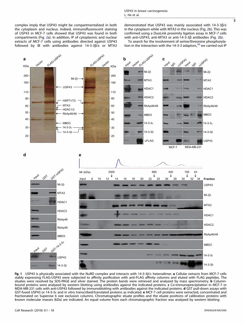
RESULTSUSP43 is physically associated with the NuRD complex andinteracts with the 14-3-3β/ε heterodimerAs stated above, recent studies implicate DUBs in the develop-ment and progression of various malignancies by influencingmultiple key cellular signaling pathways.36,37 To further under-stand the scope and the variety of the mechanistic involvementof DUBs in human cancers, we cloned the gene encoding forUSP43 from a human mammary complementary DNA (cDNA)library, a gene that has not been functionally characterized. ThecDNA for USP43 (GeneBank NM_153210.4) is 3369 bp in lengthand contains an open reading frame encoding for a protein of1123 amino acids. Bioinformatics analysis indicates that USP43harbors a putative USP domain (Supplementary information,Figure S1a). An amino-acid sequence alignment revealed thathuman USP43 shares 100% identity with its homolog in Pantroglodytes, and the similarity of the amino-acid sequence ofUSP43 with orthologues in other organisms is 82% in Bos taurus,79% in Mus musculus, 79% in Rattus norvegicus and 50% inXenopus tropicalis. Phylogenetic analysis also indicates that USP43is an evolutionarily well-conserved gene (Supplementary informa-tion, Figure S1b).To explore the cellular function of USP43, we first employed
affinity purification and mass spectrometry to interrogate theUSP43 interactome in vivo. In these experiments, FLAG-tagged
USP43 (FLAG-USP43) was stably expressed in mammary adeno-carcinoma MCF-7 cells. Cellular extracts were subjected to affinitypurification using anti-FLAG affinity columns, and the boundproteins were analyzed by mass spectrometry. The results showedthat USP43 co-purified with Mi-2β, MTA3, HDAC1/2, RbAp46/48and MBD3, all components of the nucleosome remodeling anddeacetylase (NuRD) complex, as well as with several other proteinsincluding 14-3-3β and 14-3-3ε (Fig. 1a), members of the 14-3-3family of adaptors that are primarily localized in the cytoplasmand bind to client proteins as homo- or hetero-dimers in aphosphoserine/threonine motif-dependent manner.38 The pre-sence of the NuRD subunits and 14-3-3 species in the USP43interactome was confirmed by western blotting of the column-bound proteins with antibodies against the correspondingputative partner proteins (Fig. 1b). The detailed results of themass spectrometric analysis are provided in the Supplementaryinformation, Table S1.To verify the in vivo interaction of USP43 with the NuRD
complex and 14-3-3 proteins, total proteins from MCF-7 or MDA-MB-231 cells were extracted for immunoprecipitation (IP) experi-ments using antibodies detecting the endogenous proteins. IPwith commercial polyclonal antibodies against USP43 followed byimmunoblotting (IB) with antibodies against Mi-2β, MTA3, HDAC1,HDAC2, RbAp46/48, MBD3, 14-3-3β or 14-3-3ε demonstrated thateach of these tested NuRD components and also 14-3-3β and 14-3-3ε efficiently co-IP with USP43 from extracts of breast cancer celllines with low or high metastatic potential (Fig. 1c).To understand the molecular interaction between USP43 and
the NuRD complex, glutathione S-transferase (GST) pull-downassays were performed with GST-fused USP43 (GST-USP43) andindividual components of the NuRD complex synthesized bycoupled transcription/translation in vitro. These experimentsrevealed that USP43 was capable of interacting with MTA3 andHDAC1, but not with the other components of the NuRD complexthat we tested (Fig. 1d), thus suggesting that the association ofUSP43 with the NuRD complex is through interactions with MTA3and HDAC1. Analogously, GST pull-down assays with GST-14-3-3εand USP43 or 14-3-3β synthesized in vitro by coupled transcrip-tion/translation showed that USP43 was able to interact with the14-3-3β/ε heterodimer (Fig. 1d).To further support the physical interaction of USP43 with
the NuRD complex and 14-3-3β/ε in vivo, cellular proteinsextracted in high salt from MCF-7 cells were fractionated by sizeexclusion using fast protein liquid chromatography (FPLC)with Superose 6 columns. We found that native USP43 fromMCF-7 cell extracts was eluted with an apparent molecularmass much greater than that of the monomeric protein; USP43immunoreactivity was detected in chromatographic fractionswith an elution pattern that largely overlapped with the NuRDproteins Mi-2β, MTA3, HDAC1/2, RbAp46/48 and MBD3 andat least partially overlapped with that of 14-3-3β/ε (Fig. 1e).Importantly, analysis of the FLAG-USP43 affinity eluate fromFPLC after Superose 6 gel filtration revealed that USP43 existedin multiprotein complexes; one complex peaked in fraction 14containing Mi-2β, MTA3, HDAC1/2, RbAp46/48 and MBD3, andanother peaked in fraction 30 containing 14-3-3β/ε and HSP71/72(Supplementary information, Figure S2). Collectively, these experi-ments support the observation that USP43 is physically associatedwith the NuRD complex and interacts with the 14-3-3β/εheterodimer in vivo in a transient yet likely functionally relevantmanner.
Phosphorylation of USP43 by AKT leads it to bind to the 14-3-3β/εheterodimer and be sequestered in the cytoplasmAs stated above, 14-3-3s are adaptor proteins that are primarilylocalized in the cytoplasm,38 whereas NuRD is a chromatinremodeling complex existing in the nucleus.39–41 Thus, thephysical interactions of USP43 with 14-3-3β/ε and the NuRD
USP43 in breast carcinogenesisL. He et al.
2
Cell Research (2018) 0:1 – 18
1234567890();,:
complex imply that USP43 might be compartmentalized in boththe cytoplasm and nucleus. Indeed, immunofluorescent stainingof USP43 in MCF-7 cells showed that USP43 was found in bothcompartments (Fig. 2a). In addition, IP of cytoplasmic and nuclearextracts of MCF-7 cells using antibodies directed against USP43followed by IB with antibodies against 14-3-3β/ε or MTA3
demonstrated that USP43 was mainly associated with 14-3-3β/εin the cytoplasm while with MTA3 in the nucleus (Fig. 2b). This wasconfirmed using a DuoLink proximity ligation assay in MCF-7 cellswith anti-USP43, anti-MTA3 or anti-14-3-3β antibodies (Fig. 2b).To search for the involvement of serine/threonine phosphoryla-
tion in the interaction with the 14-3-3 adaptors,38 we carried out IP
Mi-2β
MTA3
HDAC1
HDAC2
RbAp46
RbAp48
MBD3
d
USP43
14-3-3β
Fraction34
e
USP43
Mi-2β
MTA3
HDAC1
HDAC2
RbAp46/48
MBD3
14-3-3ε
14-3-3β
2000 669 440 158
10 12 14 16 18 20 22 24 26 30
Mr (kDa)
28 32Input
43
MCF-7 MDA-MB-231
cb
Mi-2β
MTA3
HDAC1
HDAC2
RbAp46/48
MBD3
14-3-3ε
14-3-3β
αFLAG
Mi-2β
MTA3
HDAC1
HDAC2
RbAp46/48
MBD3
14-3-3ε
14-3-3β
USP43
8
KDaKDa
a
260
160
110
80
60
50
40
30
20
260
160
110
80
60
50
40
30
20
InputVecto
rFLAG-U
SP43
Vector
FLAG-USP43
InputIgG αU
SP43
InputIgG αU
SP43
Vector
FLAG-USP43
InputGST
GST-USP43
InputGST
GST-14-3-3ε
Mi-2β
USP43
MBD314-3-3ε
14-3-3β
HSP71/72
MTA3HDAC1/2RbAp46/48
Fig. 1 USP43 is physically associated with the NuRD complex and interacts with 14-3-3β/ε heterodimer. a Cellular extracts from MCF-7 cellsstably expressing FLAG-USP43 were subjected to affinity purification with anti-FLAG affinity columns and eluted with FLAG peptides. Theeluates were resolved by SDS-PAGE and silver stained. The protein bands were retrieved and analyzed by mass spectrometry. b Column-bound proteins were analyzed by western blotting using antibodies against the indicated proteins. c Co-immunoprecipitation in MCF-7 orMDA-MB-231 cells with anti-USP43 followed by immunoblotting with antibodies against the indicated proteins. d GST pull-down assays withGST-fused USP43 or 14-3-3ε and in vitro transcribed/translated proteins as indicated. e MCF-7 cell proteins were extracted, concentrated andfractionated on Superose 6 size exclusion columns. Chromatographic eluate profiles and the eluate positions of calibration proteins withknown molecular masses (kDa) are indicated. An equal volume from each chromatographic fraction was analyzed by western blotting
USP43 in breast carcinogenesisL. He et al.
3
Cell Research (2018) 0:1 – 18

of MCF-7 cell lysates with antibodies against USP43 followed by IBwith antibodies against phospho-(Ser/Thr). This clearly indicatedthat USP43 is a phosphoprotein (Fig. 2c). In this regard, it isinteresting to note that a recent stable isotope labeling by aminoacids in cell culture (SILAC)-based quantitative phosphoproteomicstudy upon a human protein microarray-based AKT kinase assayplatform showed that aberrant activation of PI3K pathway leads toan increased phosphorylation of a number of proteins includingCTTN, TRIP10, PRKCD and USP43 in PIK3CA-mutated cancer cells.11
Thus, it is possible that phosphorylation of USP43 by AKT regulates
the interaction of USP43 with the 14-3-3β/ε heterodimer. To testthis, we overexpressed constitutively active AKT42 or knockeddown endogenous AKT in MCF-7 cells and examined thephosphorylation status and the subcellular localization of USP43by IP/IB or immunofluorescent staining. We found that over-expression of constitutively active AKT (Myr-AKT) was associatedwith increased phosphorylation of USP43, accompanied by itsincreased cytoplasmic localization and decreased nuclear abun-dance. In contrast, knockdown of AKT was associated withdecreased phosphorylation of USP43, its decreased cytoplasmic
USP43 in breast carcinogenesisL. He et al.
4
Cell Research (2018) 0:1 – 18

localization and increased nuclear abundance (Fig. 2d). In addition,IP of USP43 from MCF-7 cells followed by IB with antibodiesagainst 14-3-3β, 14-3-3ε or MTA3 showed that overexpression ofconstitutively active AKT was associated with enhanced interac-tion of USP43 with the 14-3-3β/ε heterodimer and a weakenedinteraction with MTA3. In contrast, AKT depletion resulted in aweakened interaction of USP43 with the 14-3-3β/ε heterodimerand enhanced interaction with MTA3 (Fig. 2e). Together, theseexperiments strongly suggest that AKT-mediated USP43 phos-phorylation triggers 14-3-3 binding and thus, the cytoplasmicretention of USP43.To gain further support for this notion, MCF-7 cells were treated
with MK-2206, an allosteric AKT inhibitor.43 Western blotanalysis of the cytoplasmic and nuclear extracts of these cellsshowed that inhibition of AKT activity resulted in a decreasedphosphorylation and cytoplasmic localization of USP43, accom-panied by an increased level of USP43 in the nucleus (Fig. 2f).In agreement, immunofluorescent staining of USP43 in thesecells showed that inhibition of AKT activity led to decreasedcytoplasmic localization and increased nuclear abundance ofthis protein (Fig. 2f). Furthermore, the in vitro phosphorylationof bacterially expressed GST-USP43 by AKT expressed in Sf9cells followed by liquid chromatography/tandem mass spectro-metry (LC-MS/MS) demonstrated that AKT could phosphorylateUSP43 upon Ser29 (Fig. 2g). To validate a requirement forphosphorylation of USP43 at Ser29 to its cytoplasmic retentionby the 14-3-3β/ε heterodimer, we transfected MCF-7 cellswith Myr-AKT and/or USP43/S29A plasmids or treated the cellswith 14-3-3β small interfering RNA (siRNA) before separatingnucleus and cytoplasm fractions for western blot analysis of USP43expression. The results showed that either depletion of 14-3-3βor overexpression of USP43/S29A abolished the AKT-mediatedcytoplasmic retention of USP43 (Supplementary information,Figure S3). Collectively, the above experiments support thenotion that USP43 is phosphorylated by AKT at Ser29, leadingto its binding to the 14-3-3β/ε heterodimer and sequestration inthe cytoplasm.
USP43 is a deubiquitinase acting on H2BK120 ubiquitinationTo understand the functional significance of the physicalinteraction of USP43 with the NuRD complex and to explore thepotential deubiquitinase activity of USP43, we first investigatedwhether or not USP43 could promote deubiquitination to enhancethe stability of MTA3 and HDAC1, subunits of the NuRD complexthat were identified to interact with USP43 directly. Western blotanalysis showed that neither overexpression nor knockdown ofUSP43 affected the protein levels of MTA3 and HDAC1 (Fig. 3a)arguing against these proteins being targeted for deubiquitina-tion/stabilization by USP43.
The NuRD complex is empowered with both ATP-dependentchromatin remodeling and histone deacetylase activities andfunctions in epigenetic regulation.39,40 The interaction ofUSP43 with this complex suggests that USP43 might befunctionally linked to epigenetic regulation, particularly histoneubiquitination. To test this hypothesis, we utilized the SILAC-LC-MS/MS approach to quantify dynamic changes in the level ofhistone ubiquitination in vivo. In these experiments, MCF-7 cellswere infected with empty lentiviral vectors or vectors carryinga USP43 expression construct and maintained in “light” [12C6]-L-lysine- or “heavy” [13C6]-L-lysine-containing medium, respectively.Histones were acid extracted from chromatin, trypsin-digestedand enriched using anti-di-Gly-conjugated agarose beads for LC-MS/MS analysis (Fig. 3b). The signal intensity from light and heavypeptides provides a quantitative comparison of their relativeabundances. We were able to detect multiple ubiquitinated lysinesites on histones, including H2BK120, H3K56, H4K31, H2BK108,H2AK118 and H2AK119 (Table 1). Among these histone ubiqui-tinations, the levels of H2BK120ub, H3K56ub and H4K31ub weresignificantly reduced upon USP43 overexpression, whereas thelevels of H2BK108ub, H2AK118ub and H2AK119ub were essen-tially unaffected (Fig. 3c). Specifically, LC-MS/MS detected a 2.17-fold decrease in H2BK120ub in cells overexpressing USP43(Fig. 3d), suggesting that H2BK120ub is at least one of the targetsfor deubiquitination by USP43.As stated above, several DUBs have already been identified
as deubiquitinases for H2BK120ub.18,31–34 To gain furthersupport for the deubiquitination of H2BK120ub by USP43 andto have a global view of H2BK120ub deubiquitination by DUBs,we carried out a systematic screen to knockdown each of81 DUBs using an siRNA library containing a pool of fourdifferent siRNAs against each target and assaying for H2BK120ubdeubiquitination. High-content screening (HCS) verified theknown deubiquitination of H2BK120ub by DUBs USP12 andUSP22 (Fig. 3e). These experiments also showed that theaverage immunofluorescent intensity of H2BK120ub significantlyincreased upon USP43 depletion (Fig. 3e), supporting its rolein H2BK120ub deubiquitination. In addition, treatment ofHeLa cells with siRNA pools specific for USP12, USP22, USP43 orUSP30 and western blot analysis of acid extracted histoneswith anti-H2BK120ub showed that the level of H2BK120ubincreased upon knockdown of either USP12, USP22 or USP43,whereas depletion of USP30 had limited effect (Fig. 3f). Moreo-ver, western blotting showed that overexpression of USP43in U2OS and HeLa cells resulted in a decrease in H2BK120ublevel whereas knockdown of USP43 in HEK293T and MCF-7cells led to increased H2BK120ub levels (Fig. 3g). Together,these results support the role of USP43 in H2BK120ubdeubiquitination.
Fig. 2 Phosphorylation of USP43 by AKT leads to its binding to 14-3-3β/ε heterodimer and sequestration in cytoplasm. a The endogenousUSP43 in MCF-7 cells was immunofluorescently stained with polyclonal antibodies against USP43. DAPI staining was included to visualize thenucleus (blue). b Co-immunoprecipitations of nuclear or cytoplasmic proteins in MCF-7 cells with anti-USP43 followed by immunoblottingwith antibodies against the indicated proteins (upper). Duolink proximity ligation assay in MCF-7 cells was performed with anti-USP43, anti-MTA3 or anti-14-3-3β antibodies (lower). c Co-immunoprecipitations in MCF-7 cells with anti-USP43 followed by immunoblotting with anti-phospho-(Ser/Thr) or anti-USP43. d MCF-7 cells were transfected with constitutively active AKT plasmids (Myr-AKT) or AKT siRNAs prior tonucleus-cytoplasm separation for western blot analysis of USP43 expression and the level of AKT phosphorylation (left). The subcellularlocalization of USP43 in MCF-7 cells transfected with Myr-AKT plasmids or treated with AKT siRNAs was analyzed by immunofluorescentstaining (right). e Co-immunoprecipitation assays in MCF-7 cells upon overexpression or knockdown of AKT with anti-USP43 followed byimmunoblotting with the indicated antibodies. The bands were quantified with ImageJ software. The numbers indicate the relative levels ofthe indicated proteins or modifications. f MCF-7 cells were treated with DMSO or AKT inhibitor prior to nucleus-cytoplasm separation forwestern blot analysis of USP43 expression and the level of AKT phosphorylation (left). The subcellular localization of USP43 in MCF-7 cellstreated with DMSO or AKT inhibitor was analyzed by immunofluorescent staining and the quantification is shown (right). The data presentedare mean ± SD for three independent experiments (***p < 0.001). g In vitro phosphorylation assays with bacterially expressed GST-USP43 andSf9 cells-expressed AKT followed by LC-MS/MS. The relevant ion fragments are labeled, and the corresponding peptide positions areillustrated. b and y represent the cleavage points of the peptide backbone when the charge is retained at the N- or C-terminal fragments ofthe peptide, respectively
USP43 in breast carcinogenesisL. He et al.
5
Cell Research (2018) 0:1 – 18
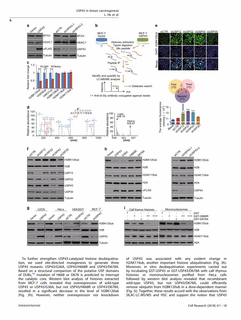
To further strengthen USP43-catalyzed histone deubiquitina-tion, we used site-directed mutagenesis to generate threeUSP43 mutants: USP43/G36A, USP43/H668R and USP43/D678A.Based on a structural comparison of the putative USP domainsof DUBs,16 mutation of H668 or D678 is predicted to interruptthe catalytic core. Western blot analysis of histones extractedfrom MCF-7 cells revealed that overexpression of wild-typeUSP43 or USP43/G36A, but not USP43/H668R or USP43/D678A,resulted in a significant decrease in the level of H2BK120ub(Fig. 3h). However, neither overexpression nor knockdown
of USP43 was associated with any evident change inH2AK119ub, another important histone ubiquitination (Fig. 3h).Moreover, in vitro deubiquitination experiments carried outby incubating GST-USP43 or GST-USP43/D678A with calf thymushistones or mononucleosomes purified from HeLa cellsfollowed by western blot analysis revealed that recombinantwild-type USP43, but not USP43/D678A, could efficientlyremove ubiquitin from H2BK120ub in a dose-dependent manner(Fig. 3i). Together, these results accord with the observations fromSILAC-LC-MS/MS and HSC and support the notion that USP43
0
20
40
60
80
100
USP43 in breast carcinogenesisL. He et al.
6
Cell Research (2018) 0:1 – 18
negatively regulates H2BK120ub through its deubiquitinaseactivity.
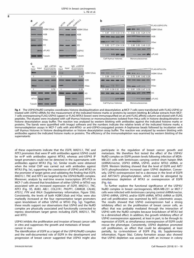
The USP43/NuRD complex coordinates histone deubiquitinationand deacetylationTo provide further support for USP43-catalyzed H2BK120ubdeuibiquitination and to explore the functional significance ofthe physical interaction between USP43 and the NuRD complex,we studied the effects of gain- or loss-of-USP43 function in MCF-7cells. Western blot analysis of histones extracted from these cellsrevealed that overexpression of USP43 resulted in a decrease inthe level of not only H2BK120ub but also H3 acetylation (H3ac).In contrast, knockdown of USP43 led to an increase in the levelof not only H2BK120ub but also H3ac (Fig. 4a), suggestingUSP43 is associated with cooperative deubiquitinase and deace-tylase activities.To substantiate these observations, USP43-containing protein
complex and the NuRD complex were IP from MCF-7 cellsexpressing FLAG-USP43 or FLAG-MTA3, respectively, with anti-FLAG. The IPs were incubated with calf thymus histones ormononucleosomes derived from HeLa cells, and the enzymaticactivities of the immunocomplexes were analyzed by westernblotting for H2BK120ub and H3ac. As expected, the USP43-containing complex possessed an enzymatic activity that ledto a significant decrease in the level of H2BK120ub. Remarkably,the IPs also exhibited dose-dependent deacetylase activityfor H3ac (Fig. 4b). Analogously, the MTA3-containing NuRDcomplex possessed not only deacetylase activity for H3acbut also deubiquitinase activity for H2BK120ub (Fig. 4b).Moreover, an anti-FLAG antibody was used to precipitate MTA3-containing NuRD complex from WT or USP43-KO MCF-7 cellsexpressing FLAG-MTA3, or to precipitate USP43-containingprotein complex from WT or MTA3-KO MCF-7 cells expressingFLAG-USP43. Analysis of the IPs after incubation with calfthymus histones by western blotting with anti-H2BK120 andanti-H3ac showed severely compromised interdependence ofsuch activities (Supplementary information, Figure S4). Theseexperiments demonstrate that the USP43/NuRD complex is
essential for crosstalk between histone deubiquitination anddeacetylation.We further performed immunodepletion assays in which the IPs
were incubated with anti-USP43- or anti-MTA3-conjugated proteinA-Sepharose beads. After two rounds of incubation and theremoval of the resins by centrifugation, the supernatants werethen used to perform deubiquitinase and deacetylase with histonesubstrates. The results showed that immunodepletion of USP43resulted in a drastic decrease in the deacetylation activity of theUSP43-containing complex and immunodepletion of MTA3 led toa significant loss in the deubiquitination activity of the MTA3-containing protein complex (Fig. 4c). These experiments stronglysupport the notion that USP43 and the NuRD complex are bothphysically and functionally associated.
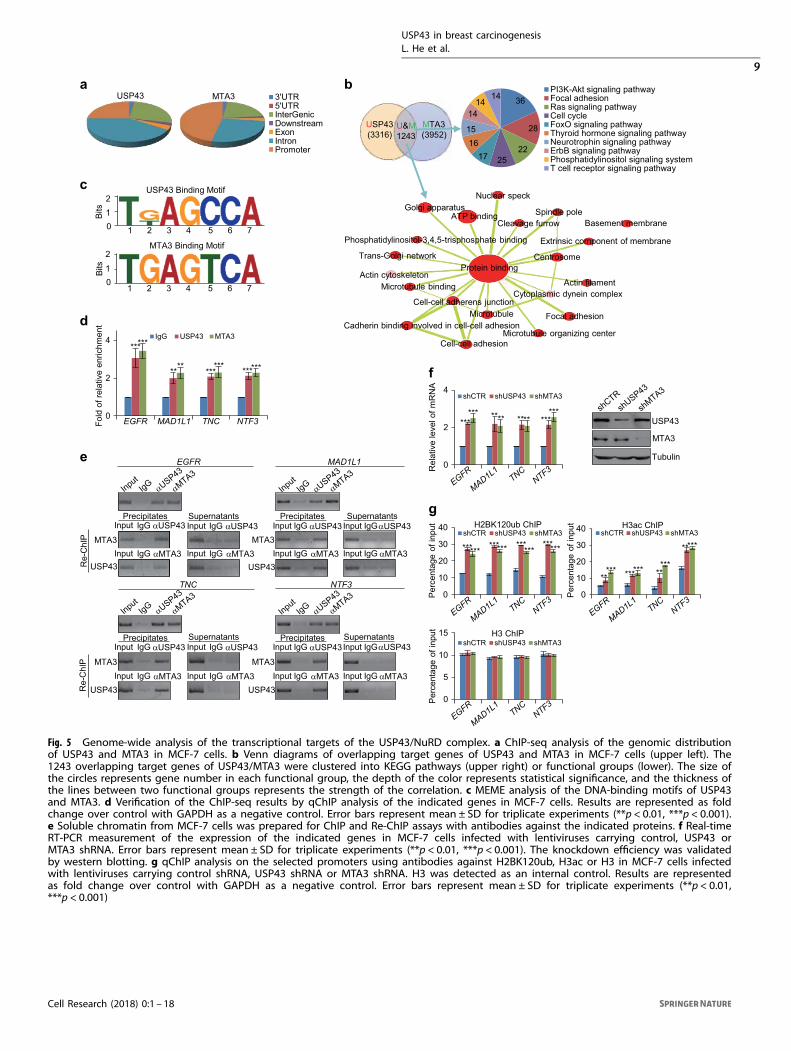
Genome-wide analysis of the transcriptional targets of the USP43/NuRD complexTo further explore the functional significance of the physicalassociation between USP43 and the NuRD complex, we nextanalyzed the genome-wide transcriptional targets of USP43. Tothis end, we first performed chromatin IP (ChIP)-based deepsequencing (ChIP-seq) in MCF-7 cells using antibodies againstUSP43. Following ChIP, USP43-associated DNAs were amplifiedusing non-biased conditions, labeled, and then sequenced usingHiSeq2500. Using MACS Version 2 and with a p-value cutoff of10−3, we identified 31,556 USP43-specific binding peaks (Fig. 5a).The data for USP43 were then cross-analyzed with our previouslypublished ChIP-seq data for MTA3 in MCF-7 cells (GSE67206)44 foroverlapping DNA sequences/gene promoters. These promoterswere considered to be targets of the USP43/NuRD complex. Theseexperiments identified a total of 1243 target genes, which wereclassified by gene ontology with DAVID (https://david.ncifcrf.gov/)into different KEGG pathways (Fig. 5b). These KEGG pathwaysinclude PI3K-AKT, focal adhesion and cell cycle pathways that arecritically involved in cell proliferation, survival, migration andinvasion (Fig. 5b). Significantly, analysis of the genomic signaturesof USP43 and MTA3 revealed a similar binding motif for thesetwo proteins (Fig. 5c), strongly supporting the physical interactionand functional connection between USP43 and MTA3. Quantita-tive ChIP (qChIP) analysis in MCF-7 cells using specific antibodiesagainst USP43 or MTA3 on selected genes including EGFR,MAD1L1, TNC, NTF3 (Fig. 5d), BUB3, ABL1, COL27A1, PHLPP1,CAMK2B, CALM2, ATG12, FYN and RALA (Supplementary informa-tion, Figure S5a) showed strong enrichment of USP43 and MTA3on the promoters of these genes, validating the ChIP-seq results.To verify that USP43 and MTA3 exist in the same protein
complex on target gene promoters, we performed sequentialChIP or ChIP/Re-ChIP on the four representative target genes,EGFR, MAD1L1, TNC and NTF3. In these experiments, solublechromatin was first IP with antibodies against USP43 and the IPswere subsequently re-IP with antibodies against MTA3. The results
Fig. 3 USP43 is a deubiquitinase acting on H2BK120ub. a The expression of the indicated proteins was measured by western blotting in MCF-7 cells following overexpression or knockdown of USP43. b The experimental workflow of SILAC-LC-MS/MS for quantification of histone lysineubiquitination. c The ratio of the light and heavy peptides identified in SILAC-LC-MS/MS. d Full MS and MS/MS spectra of H2BK120ub in SILAC-LC-MS/MS. b and y represent the cleavage points of the peptide backbone when the charge is retained at the N- or C-terminal fragments ofthe peptide, respectively. e HeLa cells were transfected with siRNA pools against DUBs and immunofluorescently stained with antibodiesagainst H2BK120ub (green). DAPI staining was included to visualize the nucleus (blue). The images represent one field under microscopy ineach group (left). Bar, 100 μm. High-content screening (HCS) with automatic image processing was applied to determine the meanimmunofluorescence intensity per cell (right). Error bars represent mean ± SD for triplicate experiments (***p < 0.001). f The level of theindicated histone marks or proteins was measured by western blotting in HeLa cells transfected with siRNAs against USP12, USP22, USP43 orUSP30. g The levels of the indicated histone marks or specific proteins was measured by western blotting in U2OS or HeLa cells transfectedwith FLAG-USP43 expression construct or in HEK293T or MCF-7 cells treated with USP43 siRNA. h The levels of the indicated histone marks orproteins was measured by western blotting in MCF-7 cells transfected with the indicated expression constructs or specific siRNAs. i In vitrodeubiquitination assays with bacterially expressed GST-fused proteins as indicated and calf thymus histones or mononucleosomes. Thereaction was analyzed by western blotting with antibodies against the indicated histone marks or proteins
Table 1 Ubiquitinated lysine sites identified in SILAC-LC-MS/MS
Protein Position Modified sequence
H2B 120 _AVTK(gl)YTSSK_
H3 56 _YQK(gl)STELLIR_
H4 31 _DNIQGITK(gl)PAIR_
H2B 108 _PGELAK(gl)HAVSEGT_
H2A 118 _GVLPNIQAVLLPK(gl)K_
H2A 119 _K(gl)TESHHK_
USP43 in breast carcinogenesisL. He et al.
7
Cell Research (2018) 0:1 – 18
of these experiments indicate that the EGFR, MAD1L1, TNC andNTF3 promoters that were IP with antibodies against USP43 couldbe re-IP with antibodies against MTA3, whereas anti-USP43 IPtarget promoters could not be detected in the supernatants withantibodies against MTA3 (Fig. 5e). Similar results were obtainedwhen the initial ChIP was carried out with antibodies againstMTA3 (Fig. 5e), supporting the coexistence of USP43 and MTA3 onthe promoter of target genes and validating the finding that EGFR,MAD1L1, TNC and NTF3 are targeted by the USP43/NuRD complex.Moreover, analysis by real-time reverse transcription (RT)-PCR inMCF-7 cells showed that knockdown of either USP43 or MTA3 wasassociated with an increased expression of EGFR, MAD1L1, TNC,NTF3 (Fig. 5f), BUB3, ABL1, COL27A1, PHLPP1, CAMK2B, CALM2,ATG12, FYN and RALA (Supplementary information, Figure S5b).Consistently, the levels of both H2BK120ub and H3ac were alsomarkedly increased at the four representative target promotersupon knockdown of either USP43 or MTA3 (Fig. 5g). Together,these results support our observations that USP43 and the NuRDcomplex are physically associated and functionally connected torepress downstream target genes including EGFR, MAD1L1, TNCand NTF3.
USP43 inhibits the proliferation and invasion of breast cancer cellsin vitro and suppresses the growth and metastasis of breastcancer in vivoThe identification of EGFR as a target of the USP43/NuRD complexand the well-documented role of EGFR in the development andprogression of breast cancer suggested that USP43 might also
participate in the regulation of breast cancer growth andmetastasis. We therefore first tested the effect of the USP43/NuRD complex on EGFR protein levels following infection of MDA-MB-231 cells with lentiviruses carrying control short hairpin RNA(shRNA)/vector, USP43 shRNA, USP43, and/or MTA3 shRNA, orEGFR. Western blotting showed that the level of EGFR and AKT/S473 phosphorylation increased upon USP43 depletion. Conver-sely, USP43 overexpression led to a decrease in the level of EGFRand AKT/S473 phosphorylation, which could be abrogated bysimultaneous depletion of MTA3 or overexpression of EGFR(Fig. 6a).To further explore the functional significance of the USP43/
NuRD complex in breast carcinogenesis, MDA-MB-231 or MCF-7cells were infected with lentiviruses carrying control shRNA/vector,USP43 shRNA, USP43, and/or EGFR shRNA, MTA3 shRNA or EGFR,and cell proliferation was examined by MTS colorimetric assays.The results showed that USP43 overexpression had a stronginhibitory effect on the proliferation of breast cancer cells, aneffect that was probably mediated through the interaction ofUSP43 with the NuRD complex because knockdown of MTA3 ledto a diminished effect. In addition, the growth inhibitory effect ofUSP43 overexpression appeared, at least in part, to be through itsrepression of EGFR, as simultaneous overexpression of EGFR couldrescue the phenotype. Consistently, USP43 depletion promotedcell proliferation, an effect that could be abrogated, at leastpartially, by co-knockdown of EGFR (Fig. 6b; Supplementaryinformation, Figure S6a). Colony formation assays also showedthat USP43 depletion was associated with an increase in colony
b
Calf thymus histones Mononucleosomes
FLAG-USP43 IPs
H2BK120ub1 0.87 0.53 0.33 0.18 1 0.62 0.33 0.24 0.14
1 0.80 0.77 0.54 0.36 1 0.96 0.85 0.49 0.25
H2B
H3ac
H3
αFLAG
Calf thymus histones Mononucleosomes
1 0.91 0.39 0.32 0.29 1 0.91 0.59 0.47 0.18
1 0.89 0.66 0.57 0.42 1 0.72 0.49 0.42 0.28
FLAG-MTA3 IPs
H2BK120ub
H2B
H3ac
H3
αFLAG
H3ac
H3
H2BK120ub
H2B
a
H2BK120ub
H2B
H3ac
H3
αFLAG
Tubulin
H2BK120ub
H2B
H3ac
H3
USP43
Tubulin
cIgGIgG
InputNo
Input InputNo
InputSupernatantsαUSP43αMTA3
Supernatants
MTA3 USP43
Tubulin Tubulin
1 0.31 1 0.45
1 0.79 1 0.32
siCTR
siUSP43-1
siUSP43-2
Vector
USP43
Fig. 4 The USP43/NuRD complex coordinates histone deubiquitination and deacetylation. aMCF-7 cells were transfected with FLAG-USP43 ortreated with USP43 siRNAs for the measurement of the indicated histone marks or proteins by western blotting. b Cellular extracts from MCF-7 cells overexpressing FLAG-USP43 (upper) or FLAG-MTA3 (lower) were immunopurified on an anti-FLAG affinity column and eluted with FLAGpeptides. The eluates were incubated with calf thymus histones or mononucleosomes isolated from HeLa cells in histone deubiquitination orhistone deacetylation assay buffer. The reaction was analyzed by western blotting with antibodies against the indicated histone marks orproteins. The bands were quantified with ImageJ software and the numbers indicate the relative levels of the indicated histone marks. cImmunodepletion assays in MCF-7 cells with anti-MTA3- or anti-USP43-conjugated protein A-Sepharose beads followed by incubation withcalf thymus histones in histone deubiquitination or histone deacetylation assay buffer. The reaction was analyzed by western blotting withantibodies against the indicated histone marks or proteins. The efficiency of the immunodepletion was examined by western blotting of thesupernatants
USP43 in breast carcinogenesisL. He et al.
8
Cell Research (2018) 0:1 – 18
0
5
10
15shCTR shUSP43 shMTA3
3'UTR5'UTRInterGenicDownstream ExonIntronPromoter
0
2
4 IgG USP43 MTA3***
*****
***
** *********
aUSP43 MTA3
b PI3K-Akt signaling pathwayFocal adhesionRas signaling pathwayCell cycleFoxO signaling pathwayThyroid hormone signaling pathwayNeurotrophin signaling pathwayErbB signaling pathwayPhosphatidylinositol signaling systemT cell receptor signaling pathway
14 36
28
22
14
1716
15U&M1243
MTA3(3952)
USP43(3316)
Cell-cell adhesion
Cleavage furrowATP binding
Microtubule binding
Extrinsic component of membrane
Cell-cell adherens junction
Cadherin binding involved in cell-cell adhesionMicrotubule organizing center
Trans-Golgi networkProtein binding
Microtubule Focal adhesion
Cytoplasmic dynein complex
Phosphatidylinositol-3,4,5-trisphosphate binding
Centrosome
Spindle pole
Nuclear speck
Actin cytoskeletonActin filament
Golgi apparatus
Basement membrane
c
d
Bits
Bits
USP43 Binding Motif
EGFR MAD1L1 TNC NTF3
MTA3 Binding Motif
Fold
of r
elat
ive
enric
hmen
t
0
21
0
2
1
1 2 3 4 5 6 7
1 2 3 4 5 6 7
Supernatants
14
e
Re-
ChI
PR
e-C
hIP
MTA3
USP43
MTA3
USP43
Input IgG αUSP43
Input IgG αMTA3
EGFR
Input IgG αUSP43MTA3
Input IgG αMTA3USP43
Input IgG αUSP43
Input IgG αMTA3
TNC
Input IgG
MTA3Input IgG αMTA3
USP43
NTF3
Input IgG
Input IgG
Input IgG
Input IgGαMTA3
Input IgG αUSP43
Input IgG αMTA3
Input IgG
Input IgG
αUSP43
αMTA3
MAD1L1
Precipitates
Precipitates Supernatants Precipitates
Precipitates
Supernatants
SupernatantsαUSP43
αMTA3
αUSP43
αUSP43
0
2
4shCTR shUSP43 shMTA3
Rel
ativ
e le
vel o
f mR
NA
*** ************ USP43
MTA3
Tubulin
f
Per
cent
age
of in
put
0
10
20
30
40shCTR shUSP43 shMTA3
*** *********
********* ***
H2BK120ub ChIPg
0
10
20
30
40 shCTR shUSP43 shMTA3H3ac ChIP
Per
cent
age
of in
put
*** ***
***
***** *****
Per
cent
age
of in
put H3 ChIP
**
*****
shCTR
shUSP43
shMTA3
EGFR
MAD1L1TNC
NTF3
EGFR
MAD1L1TNC
NTF3EGFR
MAD1L1TNC
NTF3
EGFR
MAD1L1TNC
NTF3
InputIgG αU
SP43
αMTA3
InputIgG αU
SP43
αMTA3
InputIgG αU
SP43
αMTA3
InputIgG αU
SP43
αMTA3
25
Fig. 5 Genome-wide analysis of the transcriptional targets of the USP43/NuRD complex. a ChIP-seq analysis of the genomic distributionof USP43 and MTA3 in MCF-7 cells. b Venn diagrams of overlapping target genes of USP43 and MTA3 in MCF-7 cells (upper left). The1243 overlapping target genes of USP43/MTA3 were clustered into KEGG pathways (upper right) or functional groups (lower). The size ofthe circles represents gene number in each functional group, the depth of the color represents statistical significance, and the thickness ofthe lines between two functional groups represents the strength of the correlation. c MEME analysis of the DNA-binding motifs of USP43and MTA3. d Verification of the ChIP-seq results by qChIP analysis of the indicated genes in MCF-7 cells. Results are represented as foldchange over control with GAPDH as a negative control. Error bars represent mean ± SD for triplicate experiments (**p < 0.01, ***p < 0.001).e Soluble chromatin from MCF-7 cells was prepared for ChIP and Re-ChIP assays with antibodies against the indicated proteins. f Real-timeRT-PCR measurement of the expression of the indicated genes in MCF-7 cells infected with lentiviruses carrying control, USP43 orMTA3 shRNA. Error bars represent mean ± SD for triplicate experiments (**p < 0.01, ***p < 0.001). The knockdown efficiency was validatedby western blotting. g qChIP analysis on the selected promoters using antibodies against H2BK120ub, H3ac or H3 in MCF-7 cells infectedwith lentiviruses carrying control shRNA, USP43 shRNA or MTA3 shRNA. H3 was detected as an internal control. Results are representedas fold change over control with GAPDH as a negative control. Error bars represent mean ± SD for triplicate experiments (**p < 0.01,***p < 0.001)
USP43 in breast carcinogenesisL. He et al.
9
Cell Research (2018) 0:1 – 18

Fig. 6 USP43 inhibits the proliferation and invasion of breast cancer cells in vitro and suppresses the growth and metastasis of breast cancerin vivo. a MDA-MB-231 cells were infected with lentiviruses carrying control shRNA/vector, USP43 shRNA, USP43, and/or EGFR or MTA3 shRNAfor western blotting with antibodies as indicated. b MTS assays for the growth of MDA-MB-231 cells infected with lentiviruses carrying theindicated expression constructs and/or specific shRNAs. Error bars represent the mean ± SD for three independent experiments (*p < 0.05, **p< 0.01, ***p < 0.001). c MDA-MB-231 cells infected with lentiviruses carrying the indicated expression constructs and/or specific shRNAs werecultured for 14 days before staining with crystal violet and counting for colony numbers. Error bars represent the mean ± SD for threeindependent experiments (***p < 0.001). dMDA-MB-231 cells were infected with lentiviruses carrying the indicated expression constructs and/or specific shRNAs for the measurement of the expression of the indicated epithelial/mesenchymal markers by western blotting. The bandswere quantified with ImageJ software. The numbers indicate the relative levels of the indicated proteins. e MDA-MB-231 cells were infectedwith lentiviruses carrying the indicated expression constructs and/or specific shRNA for transwell invasion assays. The invaded cells werestained and counted. The images represent one microscope field in each group. Error bars represent mean ± SD for triplicate experiments(***p < 0.001). Bar, 100 μm. f MDA-MB-231-Luc-D3H2LN cells infected with lentiviruses carrying the indicated expression constructs and/orspecific shRNA were inoculated orthotopically onto the abdominal mammary fat pad of 6-week-old female SCID mice. Primary tumor size wasmeasured and metastases were quantified using bioluminescence imaging after 6 weeks of initial implantation. Representative primarytumors and bioluminescent images are shown. Error bars represent mean ± SD for three independent measurements (***p < 0.001).g Representative lung metastasis specimens were sectioned and stained with H&E. Bar, 100 μm. Error bars represent mean ± SD (***p < 0.001)
USP43 in breast carcinogenesisL. He et al.
10
Cell Research (2018) 0:1 – 18
number, which was partially attenuated upon co-knockdown ofEGFR. In contrast, USP43 overexpression was associated with adecrease in colony number, an effect that could be abrogated bysimultaneous depletion of MTA3 or, at least partially, by over-expression of EGFR (Fig. 6c; Supplementary information, Fig-ure S6b). Together, these results support a role for USP43 in theinhibition of cell proliferation and suggest that USP43 does sothrough its interaction with the NuRD complex and down-regulation of target genes including EGFR.The epithelial–mesenchymal transition (EMT) is a hallmark
of cancer and an early event in cancer invasion and metastasis.45
To investigate a possible role for USP43 in invasion and metastasisof breast cancer, the expression of epithelial/mesenchymalmarkers was analyzed by western blotting in MDA-MB-231 orMCF-7 cells. We found that depletion of USP43 resulted in areduction of epithelial markers including E-cadherin and γ-cateninand an induction of mesenchymal markers including fibronectinand N-cadherin, which were partially attenuated by co-knockdown of EGFR (Fig. 6d; Supplementary information, Fig-ure S6c). Conversely, overexpression of USP43 was associated withan induction of the epithelial markers and reduction of themesenchymal markers. However, simultaneous depletion of MTA3or overexpression of EGFR counteracted the effect of USP43overexpression on the expression patterns of the epithelial/mesenchymal markers (Fig. 6d; Supplementary information,Figure S6c). These results support a role for USP43 in theinhibition of the EMT, indicating that USP43 does so, through itsinteraction with the NuRD complex and downregulation of targetgenes including EGFR.We then investigated the role of USP43 in the cellular behavior
of breast cancer cells in vitro using transwell invasion assays. Wefound that USP43 knockdown was associated with an increase inthe invasive potential of MDA-MB-231 cells, whereas USP43overexpression was accompanied by a decrease in the invasivepotential of such cells (Fig. 6e). Moreover, in agreement with theexistence of a functional link between USP43 and EGFR, theincrease in invasive potential associated with USP43 knockdowncould be rescued, at least partially, by co-knockdown of EGFR andthe inhibitory effect of USP43 overexpression on the invasivepotential of MDA-MB-231 cells was partially offset by EGFRoverexpression. The inhibitory effect of USP43 overexpression oninvasiveness appeared to be achieved through concerted actionwith the NuRD complex because the effect diminished whenMTA3 was concomitantly knocked down in MDA-MB-231 cells(Fig. 6e). Taken together, these results support a role for USP43 inregulating the invasive potential of breast cancer cells and suggestthat it does so through its association with the NuRD complex anddownregulation of target genes including EGFR.To investigate the role of USP43 in breast cancer metastasis
in vivo, MDA-MB-231 cells that had been engineered to stablyexpress firefly luciferase (MDA-MB-231-Luc-D3H2LN, XenogenCorporation) were infected with a control lentivirus shRNA vectorand lentiviruses carrying USP43 shRNA, USP43, and/orMTA3 shRNA or EGFR. These cells were then implanted onto theleft abdominal mammary fat pad of 6-week-old female SCID mice(n= 6). The growth/dissemination of tumors was monitoredweekly by bioluminescence imaging with the IVIS imaging system(Xenogen). Tumor metastasis was defined as any detectableluciferase signal above background and away from the primarytumor site and was measured by quantitative bioluminescenceimaging after 6 weeks. We found that USP43 depletion promotedthe growth of the primary tumor (Fig. 6f) and lung metastasis(Fig. 6g) of the MDA-MB-231-Luc-D3H2LN tumors. Conversely,USP43 overexpression resulted in growth suppression of theprimary tumor (Fig. 6f), as well as lung metastasis (Fig. 6g) of MDA-MB-231-Luc-D3H2LN tumors. Remarkably, simultaneous depletionof MTA3 or overexpression of EGFR counteracted the USP43overexpression-associated suppression of the growth of primary
tumors (Fig. 6f) and lung metastases (Fig. 6g). Collectively, theseexperiments indicate that USP43 suppresses the growth andmetastasis of breast cancer and that it does so through itsinteraction with the NuRD complex and repression of target genesincluding EGFR.
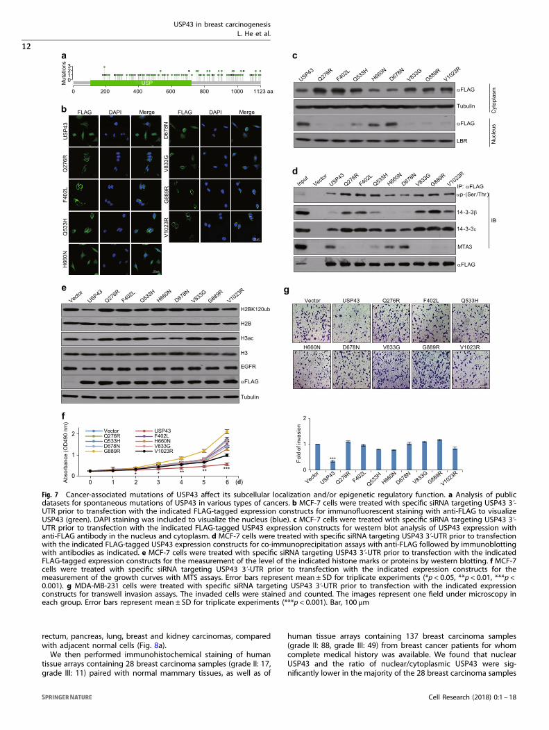
Cancer-associated mutations of USP43 affect its subcellularlocalization and/or epigenetic regulatory functionTo extend our observations to pathologically relevant contexts, wenext investigated the genetic alterations of USP43 in varioushuman carcinoma samples from the clinic. Analysis of thecBioPortal for Cancer Genomics (http://www.cbioportal.org/) forUSP43 mutations revealed that mutation in the N-terminal ofUSP43 is relatively rare in various human carcinoma samples, andthat mutations were observed at high frequency in amino-acidresidues located more C-terminally including Q276R, F402L,Q533H, H660N, D678N, V833G, G889R and V1023R (Fig. 7a). Thus,we generated USP43 mutant for each of these eight mutationsand first examined their subcellular localizations. Transfection ofFLAG-tagged wild-type USP43 and USP43 mutants into MCF-7cells and analysis by immunofluorescent staining with anti-FLAGshowed that wild-type USP43, USP43/H660N and USP43/D678Nwere present in both the cytoplasm and nucleus, the remainingUSP43 mutants were mainly localized in the cytoplasm (Fig. 7b).Western blot analysis of the cytoplasmic and nuclear extracts ofthese cells showed consistent results (Fig. 7c). We then tested theinteraction between the eight USP43 mutants and the 14-3-3β/εheterodimer or MTA3. IPs in MCF-7 cells expressing the FLAG-tagged USP43 mutants with anti-FLAG followed by IB withantibodies against phospho-(Ser/Thr), 14-3-3β, 14-3-3ε or MTA3demonstrated that each these mutants, except for USP43/H660Nand USP43/D678N, exhibited a higher phosphorylation level,stronger interaction with 14-3-3β/ε heterodimer and weakerassociation with MTA3, compared with wild-type USP43 (Fig. 7d).Consistently, transfections of FLAG-tagged USP43 mutants intoMCF-7 cells and subsequent western blot analysis revealed thatalmost all of the eight USP43 mutants showed compromisedability to deubiquitinate H2BK120ub and repress EGFR expression,compared with wild-type USP43, even though USP43/H660N andUSP43/D678N, which were found to be enriched in the nucleus,were associated with deacetylase activity for H3ac (Fig. 7e).Notably, both H660 and D678 reside in the conserved USP domainof USP43 and are important for the enzymatic activity of DUBs.16
Together, these results indicate that cancer-associated USP43mutants display either aberrant cytoplasmic retention or animpaired deubiquitinase activity toward H2BK120ub. This leads toimpaired transcription repression of downstream target genesincluding EGFR, and eventually, contributing to breast carcinogen-esis. In agreement, MTS assays showed that all the USP43 mutantslargely lost inhibitory effect on MCF-7 cell proliferation (Fig. 7f)and transwell invasion assays revealed that these mutants showedsignificantly compromised inhibition of the invasive potential ofMDA-MB-231 cells (Fig. 7g).
Nuclear USP43 is reduced in breast carcinomas, corresponding tothe high expression of EGFR and hyperactivation of AKTTo further extend our observations to pathologically relevantsettings, we examined the protein level of USP43 by immunohis-tochemical staining of human tissue arrays containing a series ofcarcinoma samples from thyroid, esophagus, stomach, colon,rectum, liver, pancreas, lung, breast and kidney cancer patients.These arrays held at least six samples of each type of carcinomapaired with the adjacent normal tissues. Strikingly, we found thatUSP43 protein was predominantly present in the cytoplasm ofcancer cells, especially in esophagus, colon, rectum, lung, andbreast carcinomas, whereas in adjacent cells, abundant USP43 wasdetected in the nucleus (Fig. 8a). Evidently, the ratio of nuclear/cytoplasmic USP43 was significantly reduced in esophagus, colon,
USP43 in breast carcinogenesisL. He et al.
11
Cell Research (2018) 0:1 – 18
rectum, pancreas, lung, breast and kidney carcinomas, comparedwith adjacent normal cells (Fig. 8a).We then performed immunohistochemical staining of human
tissue arrays containing 28 breast carcinoma samples (grade II: 17,grade III: 11) paired with normal mammary tissues, as well as of
human tissue arrays containing 137 breast carcinoma samples(grade II: 88, grade III: 49) from breast cancer patients for whomcomplete medical history was available. We found that nuclearUSP43 and the ratio of nuclear/cytoplasmic USP43 were sig-nificantly lower in the majority of the 28 breast carcinoma samples
Q533H
Fig. 7 Cancer-associated mutations of USP43 affect its subcellular localization and/or epigenetic regulatory function. a Analysis of publicdatasets for spontaneous mutations of USP43 in various types of cancers. b MCF-7 cells were treated with specific siRNA targeting USP43 3ʹ-UTR prior to transfection with the indicated FLAG-tagged expression constructs for immunofluorescent staining with anti-FLAG to visualizeUSP43 (green). DAPI staining was included to visualize the nucleus (blue). c MCF-7 cells were treated with specific siRNA targeting USP43 3ʹ-UTR prior to transfection with the indicated FLAG-tagged USP43 expression constructs for western blot analysis of USP43 expression withanti-FLAG antibody in the nucleus and cytoplasm. d MCF-7 cells were treated with specific siRNA targeting USP43 3ʹ-UTR prior to transfectionwith the indicated FLAG-tagged USP43 expression constructs for co-immunoprecipitation assays with anti-FLAG followed by immunoblottingwith antibodies as indicated. e MCF-7 cells were treated with specific siRNA targeting USP43 3ʹ-UTR prior to transfection with the indicatedFLAG-tagged expression constructs for the measurement of the level of the indicated histone marks or proteins by western blotting. f MCF-7cells were treated with specific siRNA targeting USP43 3ʹ-UTR prior to transfection with the indicated expression constructs for themeasurement of the growth curves with MTS assays. Error bars represent mean ± SD for triplicate experiments (*p < 0.05, **p < 0.01, ***p <0.001). g MDA-MB-231 cells were treated with specific siRNA targeting USP43 3ʹ-UTR prior to transfection with the indicated expressionconstructs for transwell invasion assays. The invaded cells were stained and counted. The images represent one field under microscopy ineach group. Error bars represent mean ± SD for triplicate experiments (***p < 0.001). Bar, 100 μm
USP43 in breast carcinogenesisL. He et al.
12
Cell Research (2018) 0:1 – 18

(22/28) compared with adjacent normal tissues. Remarkably, 3 outof the 137 breast carcinomas showed a lack of USP43 staining(Fig. 8b). Significantly, the level of nuclear USP43 negativelycorrelated with the histological grade of the 165 breast carcinomasamples (28+ 137) (Fig. 8b). Moreover, the level of nuclear USP43in these 165 breast carcinoma samples also negatively correlatedwith tumor size, depth of invasion, lymph node metastasis andadvanced TNM stage (Table 2). Consistent with our observationsthat EGFR is a downstream target of the USP43/NuRD complexand that AKT activation is associated with retention of USP43 incytoplasm, the ratio of nuclear/cytoplasmic USP43 also negatively
correlated with the levels of EGFR expression and AKT phosphor-ylation in the 165 breast carcinoma samples (Fig. 8c). Furthermore,the overall 5-year survival rate of patients with high nuclearexpression of USP43 was significantly higher than patients withlow nuclear expression of USP43 (Fig. 8d). Consistently, interroga-tion of public dataset (GSE36774) found that the level ofUSP43 shows a negative correlation and EGFR, a positivecorrelation with the histological grades of breast cancer (Fig. 8e).Strikingly, we found that low USP43 and high EGFR in breastcarcinomas strongly correlates with lymph node positivity of thepatients (Fig. 8e).
USP43 in breast carcinogenesisL. He et al.
13
Cell Research (2018) 0:1 – 18

To address the clinicopathological relevance of USP43 and EGFR,we next analyzed public datasets for mRNA levels of USP43 andEGFR in breast carcinomas. Analysis of two published clinicaldatasets (GSE36774 and GSE48390) revealed a statistically significantnegative correlation of expression between USP43 and EGFR inbreast carcinomas (Fig. 8f). Strikingly, analysis of public dataset(GSE48390) for USP43 or EGFR expression revealed that expressionof USP43 is higher in Luminal A and Luminal B subtypes and lowerin HER2-enriched and basal-like subtypes, where the expression ofEGFR is high (Supplementary information, Figure S7a). Kaplan–Meiersurvival analysis (http://kmplot.com/analysis/) demonstrated thateither higher USP43 expression (hazard ratio, HR= 0.66, p= 1.9e-07)or lower EGFR expression (HR= 1.2, p= 0.02) was associated with abetter overall survival for patients with breast cancer (Fig. 8g).Moreover, higher USP43 expression is associated with betterrelapse-free survival of breast cancer patients of Luminal A, LuminalB and even basal-like subtypes (Supplementary information,Figure S7b). Further stratification of patient groups (GSE42568)based on the inverse expression of USP43 and EGFR improved thepredictive capability of USP43 (HR= 0.53, p= 0.0372) (Fig. 8h).Collectively, these data are consistent with a role for USP43 insuppressing breast cancer development and progression bytargeting the downstream genes including EGFR.
DISCUSSIONGenetic mutation/deletion of chromosome 17p13.1 is consideredas a prototype of genetic abnormalities associated with malignanttransformation owing to its recurrent or invariable inclusionof TP53, an extensively studied tumor-suppressor gene that isbelieved to be the major driver of 17p13.1 loss-of-function-associated pathologies.46–48 Interestingly, the gene encodingthe USP family protein USP43 also maps to this cytologicalregion, suggesting that 17p13.1 mutations, especially deletions,could entail collateral damage to USP43. Indeed, analysis ofpublic datasets showed that USP43 suffers frequent mutationand/or deletion in various types of tumor, especially in invasivebreast carcinoma. Notably, the chromosome region 17p13.1encompasses ~4.3 million base pairs and harbors at least 144genes including, in addition to TP53 and USP43, important genessuch as MED31 and CHD3 (encoding for Mi-2α). It will be adaunting task to determine the scope and variety of the othergenes whose mutations/deletions accompany the geneticabnormalities of TP53 in specific tumor types. Nevertheless, it isa safe assumption that, in addition to mutation/deletion of TP53,
loss-of-function of one or more of these 144 genes might, at least,also contribute to tumorigenesis. Our finding that USP43 isfrequently mutated in a variety of human cancers suggests thatUSP43 might be another underappreciated candidate tumor-suppressor gene. Moreover, although USP43 is a predictedmember of the USP deubiquitinase family, its biological functionhas remained uncharacterized.We have found that USP43 is physically associated with the
NuRD complex and biochemically catalyzes H2BK120 deubiquiti-nation. We have shown that USP43 and the NuRD complex act incoordination to remove histone ubiquitination and acetylation.The USP43/NuRD complex represses a panel of genes includingEGFR that are critically involved in cell proliferation and tumorgrowth and we have demonstrated that the USP43/NuRD complexinhibits the proliferation and invasion of breast cancer cells in vitroand suppresses the growth and metastasis of breast cancerin vivo. These results support the notion that USP43 is a potentsuppressor of breast carcinogenesis and thus USP43 representsanother potential tumor suppressor.Extensive studies by us and others have implicated the NuRD
complex in the development and progression of breast can-cer.44,49–54 The NuRD complex contains several subunits whosepattern of expression is heterogeneous in various cell and tissuetypes, leading to the proposal that subunit heterogeneity confersthese complexes with additional regulatory capacity and withunique functional properties.41,49,55 Consistent with this notion,we reported previously that histone demethylases LSD149 andJARID1B50 are associated with the NuRD complex, thus expandingits enzymatic repertoire to include, in addition to ATPase anddeacetylase, the demethylase activity. Our current inclusion ofUSP43 in the NuRD complex extends the enzymatic arsenal of theNuRD complex further to include a deubiquitinase activity. It isbecoming increasingly clear that the NuRD complex acts as anepigenetic regulatory hub that coordinates different histonemodifications and directs distinct “histone crosstalk”. However,our ChIP-seq results clearly show an incomplete overlap betweenUSP43 and MTA3 targets, suggesting that NuRD complex couldrecruit some other transcriptional cofactors, whereas USP43also could deubiquitinate non-histone proteins independentof each other in some specific physiological states. Furtherinvestigations will determine whether the NuRD complex containsfurther enzymatic activities and additional chromatin modifyingcapabilities.Although H2BK120 ubiquitination is catalyzed mainly by the
RNF20/40 complex,30 at least six USPs, including USP3, USP12,
Fig. 8 Nuclear USP43 is reduced in breast carcinomas and the ratio of nuclear/cytoplasmic USP43 is negatively correlated with highexpression of EGFR and hyperactivation of AKT. a Immunohistochemical staining of USP43 in tissue arrays containing various types ofcarcinomas samples paired with adjacent normal tissues. Each type of carcinoma included at least six paired samples and the scores weredetermined by evaluating the extent and intensity of immunopositivity and were analyzed by paired samples t-test (*p < 0.05, **p < 0.01).Samples of tumors and normal tissues from breast and lung were sectioned and stained with H&E or immunostained with anti-USP43, andrepresentative images are shown. Bar, 100 μm (left), 50 μm (right). b Immunohistochemical staining of USP43 in tissue arrays containing 28breast carcinoma samples (grade II: 17, grade III: 11) paired with normal mammary tissues as well as tissue arrays containing 137 breastcarcinoma samples (grade II: 88, grade III: 49). Representative images from H&E staining and immunostaining with anti-USP43 are presented.Bar, 100 μm (left); 50 μm (right). The scores were determined by evaluating the extent and intensity of immunopositivity and were analyzed bytwo-tailed paired t-test (lower left) or two-tailed unpaired t-test (lower right) (*p < 0.05, ***p < 0.001). c Samples from adjacent normal tissuesand grade II/III breast cancers were H&E stained or immunostained with antibodies against USP43, EGFR or p-AKT. Representative images areshown. Bar, 100 μm (left); 50 μm (right). The ratio of nuclear and cytoplasmic USP43 was plotted against the scores of EGFR or p-AKT. Thecorrelation coefficients were calculated by R programming. d Time-to-event data were plotted using Kaplan–Meier curves, and the 5-yearsurvival rate of different groups was compared using the Mantel–Cox log-rank test (***p < 0.001). The y axis represents the percentage ofpatients, and the x axis represents the survival in months. e Analysis of public dataset (GSE36774) for the correlation between the level ofUSP43 or EGFR and the histological grades of breast carcinomas or lymph node metastasis of breast cancer patients (*p < 0.05, **p < 0.01). fAnalysis of public datasets (GSE36774 and GSE48390) for the expression of EGFR and USP43. The relative mRNA level of EGFR was plottedagainst that of USP43. g Kaplan–Meier survival analysis (http://kmplot.com/analysis/) for the relationship between survival time of breastcancer patients and the expression levels of USP43 or EGFR. h Kaplan–Meier survival analysis of public dataset (GSE42568) for the relationshipbetween survival time and the expression levels of USP43/EGFR in breast cancer. i The proposed model of the reciprocally inhibitory loopbetween EGFR/PI3K/AKT and USP43 in breast cancer carcinogenesis
USP43 in breast carcinogenesisL. He et al.
14
Cell Research (2018) 0:1 – 18

USP22, USP44, USP46 and USP49, have been reported to be ableto deubiquitinate H2BK120. Our identification of USP43 as anothereraser for H2BK120ub adds to the complexity of the regulation ofthis histone mark. The biological significance of the multifariousrequirement for the regulation of H2BK120ub deubiquitinationis currently unknown. It is possible that different USPs act onH2BK120ub in a tissue/gene-specific manner and/or in respondingto a different extracellular stimulus/cellular environment. Ourstudy indicates that the function of USP43 is tightly linked to theEGFR/PI3K/AKT signaling pathway in which USP43 is phosphory-lated by AKT, resulting in its binding to 14-3-3β/ε heterodimer andsequestration in cytoplasm. Logically, dephosphorylation of USP43leads to its nuclear translocation and interaction with the NuRDcomplex. Due to the scope of our current investigation, we havenot identified the phosphatase involved in the regulation ofUSP43’s nuclear translocation. Future investigations of this issuewill surely provide a more detailed account of the regulation ofthe cellular function of this deubiquitinase. Moreover, ourexperiments showed that elevated USP43 could also affect theubiquitination level of H3K56ub and H4K31ub. Although, due tothe lack of the appropriate antibodies, we were unable to studythe regulation of these histone marks by USP43 and further studyof this is warranted.Mutation/amplification of EGFR is also a frequent event in
malignancies from a broad spectrum of tissue origins and thisaberration is often correlated with advanced stages of cancerprogression and poor prognosis.1–3,9,56 Constitutive activation ofEGFR leads to hyperactivation of downstream signaling pathwaysincluding the PI3K-AKT pathway that are critically involved in cellproliferation and malignant transformation.4–6 Remarkably, gain-of-function mutation/amplification of PI3K,10,11,57 the positive regulator
of AKT7 and loss-of-function mutation of PTEN,12 a negativeregulator of AKT,8 also frequently occur in human cancers, all ofwhich could lead to hyperactivation of AKT. However, these geneticalterations cannot fully account for the hyperactivation of the EGFR/PI3K/AKT cascade.13 According to our model, hyperactivation of AKTwould result in hyperphosphorylation and cytoplasmic retention ofUSP43, leading to lost transcriptional regulation by USP43. On theother hand, loss-of-function mutation/deletion of USP43 will renderlost transcriptional repression of genes including EGFR anddiminished negative regulation of the EGFR/PI3K/AKT signaling byUSP43. If our interpretation is correct, there appears to exist areciprocally inhibitory loop inside the cell in which the activation ofEGFR/PI3K/AKT signaling sequestrates USP43 in the cytoplasm thusinhibiting the transcription regulatory function of USP43, leading tocell proliferation and invasion. On the other hand, USP43translocates into the nucleus and transcriptionally represses EGFR,leading to the inhibition of cell proliferation and invasion. Accordingto this model, the imbalance of this loop, either due tohyperactivation of EGFR/PI3K/AKT signaling resulting from gain-of-function mutations or amplification of these components, or todiminished transcriptional regulatory function of USP43 resultingfrom loss-of-function mutations or deletion, strongly favors breastcarcinogenesis. These scenarios place USP43 into a critical node inthe hierarchical regulatory network of the EGFR/PI3K/AKT signaling,indicating that USP43 is a powerful suppressor of breast carcinogen-esis. Future investigations will determine whether and how theEGFR/PI3K/AKT-USP43 reciprocally inhibitory loop functions innormal development, as well as in the development and progres-sion of tumors originated from other tissues.In summary, we report in the current study that USP43 is
physically associated with the NuRD complex and functionallycoordinates H2BK120 deubiquitination and histone deacetylation torepress downstream target genes including EGFR and to suppressbreast carcinogenesis. USP43 also exists in the cytoplasm where itis phosphorylated by AKT, leading to its binding to 14-3-3β/εheterodimer and cytoplasmic sequestration. Our study identifiedUSP43 as a histone deubiquitinase and a potent suppressor of breastcarcinogenesis. We propose the existence of a reciprocally inhibitoryloop between the EGFR/PI3K/AKT signaling and USP43, whoseimbalance drives breast carcinogenesis.
MATERIALS AND METHODSAntibodies and reagentsAntibodies used: anti-FLAG, anti-tubulin, anti-fibronectin (Sigma);anti-USP43 (Abgent); anti-Mi-2β, anti-MTA3, anti-HDAC1, anti-HDAC2, anti-RbAp46/48, anti-14-3-3ε, anti-14-3-3β (Santa CruzBiotechnology); anti-MBD3, anti-H2BK120ub, anti-H2AK119ub,anti-Phospho-(Ser/Thr), anti-E-cadherin, anti-γ-catenin, anti-N-cadherin (Cell Signaling Technology); anti-p-AKT (pSer473) (Servi-cebio); anti-H2B, anti-H2A (Merck Millipore); anti-H3, anti-EGFR(Abcam); anti-H3ac (Active Motif). Protein A/G Sepharose CL-4Bbeads were from Amersham Biosciences, and protease inhibitorcocktail was from Roche Applied Science.
Immunopurification and mass spectrometryMCF-7 or HEK293T cells stably expressing FLAG-USP43 werewashed twice with cold phosphate-buffered saline (PBS), scraped,and collected by centrifugation at 800 × g for 5 min. Cellularextracts were prepared by lysing the cells in lysis buffer containingprotease inhibitor cocktail (Roche) and phosphatase inhibitor(Applygen). Anti-FLAG immunoaffinity resin (Sigma) was preparedaccording to the manufacturer’s protocol. Cell lysates wereapplied to the immunoaffinity resin to enable adsorption of theprotein complex. After binding, the resin was washed with coldPBS plus 0.3% Nonidet P-40. FLAG peptide (Sigma) was applied tothe resin to elute the FLAG-tagged protein-associated complex asdescribed by the vendor. The eluates were collected and resolved
Table 2 Correlation between nuclear USP43 level and theclinicopathologic characteristics of breast carcinomas
Variables n Nuclear USP43 level p-Value
Positive (n, %) Negative (n, %)
Histological grade
II 101 52 (74.29%) 49 (57.65%) p=0.023
III 54 18 (25.71%) 36 (42.35%)
Age (years)
≥55 81 34 (46.58%) 47 (57.32%) p=0.181
<55 74 39 (53.42%) 35 (42.68%)
Location
Left 87 41 (54.67%) 46 (58.23%) p=0.745
Right 67 34 (45.33%) 33 (41.77%)
Tumor size (cm)
D ≤ 3.3 94 57 (79.17%) 37 (44.58%) p<0.001
D > 3.3 61 15 (20.83%) 46 (55.42%)
Depth of invasion
T0 and T1 40 28 (39.44%) 12 (14.63%) p<0.001
T2 103 42 (59.15%) 61 (74.39%)
T3 10 1 (1.41%) 9 (10.98%)
Lymph node
N0 78 45 (60.81%) 33 (40.74%) p=0.044
N1 43 16 (21.62%) 27 (33.33%)
N2 and N3 34 13 (17.57%) 21 (25.93%)
TNM stage
I 26 21 (29.58%) 5 (6.17%) p<0.001
II 87 37 (52.11%) 50 (61.73%)
III 39 13 (18.31%) 26 (32.10%)
USP43 in breast carcinogenesisL. He et al.
15
Cell Research (2018) 0:1 – 18
on NuPAGE 4–12% Bis-Tris gel (Invitrogen), silver-stained (Pierce)and subjected to LC-MS/MS sequencing and data analysis.
Fast protein liquid chromatographyCellular extracts were prepared by lysing the cells in lysis buffercontaining protease inhibitor and phosphatase inhibitor. Approxi-mately 5mg protein was concentrated to 0.5mL using a MilliporeUltra free centrifugal filter apparatus (3 kDa nominal molecular masslimit), and then applied to an 850 × 20mm Superose 6 sizeexclusion column (Amersham Biosciences) that had been equili-brated with buffer D containing 1mM dithiothreitol and calibratedwith protein standards (blue dextran, 2000 kDa; thyroglobulin, 669kDa; ferritin, 440 kDa; catalase, 232 kDa; bovine serum albumin, 67kDa; and RNase A, 13.7 kDa, all from Amersham Biosciences). Elutionwas carried out at a flow rate of 0.5mL/min and fractions collected.
SILAC labeling and quantitative proteomics analysisMCF-7 cells were grown in Dulbecco’s modified Eagle’s mediumsupplemented with 10% fetal bovine serum and either the “light”form of [12C6]-L-lysine or “heavy” [13C6]-L-lysine for more than sixgenerations to achieve >97% labeling efficiency. After that, the cellswere further expanded in SILAC media to the desired cell number(~5 × 10⁸) in 15 × 150mm2
flasks. Cells infected with lentivirusvector or vectors carrying USP43 expression constructs were thencollected and the histones were acid extracted. The histones fromthe “heavy” and “light” form cells were mixed 1:1 by protein content.Proteins were digested sequentially with trypsin followed by peptidedesalting. Peptides were IP with anti-di-Gly antibody-conjugatedagarose beads in the PTMScan® ubiquitin remnant motif (K-ε-GG) kit(Cell Signaling Technology) for 2 h at 4 °C. The eluted peptides werecleaned with C18 ZipTips (Millipore) according to the manufacturer’sinstructions, followed by analysis with LC-MS/MS. The resulting MS/MS data were processed using MaxQuant with integrated Andro-meda search engine (version 1.4.1.2).
High-content screeningHeLa cells were transfected with siRNA pools against DUBs andimmunofluorescently stained with antibodies against H2BK120ub.The level of H2BK120ub was analyzed by immunofluorescentstaining followed by HCS microscopy. The experiments werebiologically repeated three times and each time the experimentswere performed in triplicate for each DUB.
Preparation of mononucleosomesHeLa cells were collected by cold PBS, resuspended in lysis buffer(10 mM NaCl, 3 mM MgCl2, 0.4% Nonidet P-40 and 10mM Tris-HCl,pH 7.5) in the presence of protease inhibitor and the nuclei werepelleted. Glycerol buffer (0.1 mM EDTA, 5 mM MgAc2, 25% (vol/vol) glycerol and 10mM Tris-HCl, pH 7.4) was added to give a finalconcentration of 1–2mg/mL nuclei. To generate nucleosomalmaterial, digestions were conducted by adding 1 volume of 2 ×micrococcal nuclease (MNase) buffer (50 mM KCl, 8 mM MgCl2,2 mM CaCl2 and 100mM Tris-HCl, pH 7.4) and 3000 U/mL MNase.The reaction was incubated for 15 min at 37 °C and stopped byadding EDTA to a final concentration of 10mM. The mononucleo-somes were then purified on sucrose gradients.50,58
Histone deubiquitination and histone deacetylation assaysCalf thymus bulk histones (Sigma) or mononucleosomes wereincubated with FLAG-USP43 or FLAG-MTA3 IPs at 37 °C for 90 minin deubiquitination assay buffer (50 mM NaCl, 1 mM DTT and 50mM Tirs-HCl, pH 7.5) or histone deacetylation assay buffer (150mM NaCl, 1 mM DTT and 10mM Tirs-HCl, pH 8.0). Each reactionwas stopped by mixing with sodium dodecyl sulfate (SDS) sampleloading buffer followed by western blot analysis with antibodiesagainst H2BK120ub, H3ac, H2B or H3.
qChIP, ChIP and Re-ChIPMCF-7 cells were crosslinked using 1% formaldehyde for 10 min atroom temperature and quenched by the addition of glycine to afinal concentration of 125 mM for 5 min. The fixed cells wereresuspended in lysis buffer (1% SDS, 5 mM EDTA and 50mM Tris-HCl, pH 8.1) in the presence of protease inhibitors, then subjectedto 30 cycles (30-s on and off) of sonication (Bioruptor, Diagenode)to generate chromatin fragments of ~300 bp in length. Lysateswere diluted in buffer containing 1% Triton X-100, 2 mM EDTA,150mM NaCl, 20 mM Tris-HCl (pH 8.1) and protease inhibitors. ForIP, the diluted chromatin was incubated with normal IgG (control)or specific antibodies for 12 h at 4 °C with constant rotation, 50 μLof 50% (vol/vol) protein A/G Sepharose beads were then added,and the incubation was continued for an additional 2 h. Beadswere washed with the following buffers: TSE I (0.1% SDS, 1% TritonX-100, 2 mM EDTA, 150mM NaCl and 20mM Tris-HCl, pH 8.0); TSEII (0.1% SDS, 1% Triton X-100, 2 mM EDTA, 500mM NaCl and 20mM Tris-HCl, pH 8.0); TSE III (0.25 M LiCl, 1% Nonidet P-40, 1%sodium deoxycholate, 1 mM EDTA and 10mM Tris-HCl, pH 8.0)and TE (1 mM EDTA and 10mM Tris-HCl, pH 8.0). The pulled-downchromatin complex and input were de-crosslinked at 55 °C for 12 hin elution buffer (1% SDS and 0.1 M NaHCO3).
59 The eluted DNAwas purified with the QIAquick PCR Purification Kit. qChIPs wereperformed using Power SYBR Green PCR Master Mix and ABIPRISM 7500 sequence detection system (Applied Biosystems,Foster City, CA). Re-ChIPs were carried out in essentially the sameway as primary IPs. Bead eluates from the first IP were incubatedwith 10mM DTT at 37 °C for 30 min and diluted 1:50 in dilutionbuffer (1% Triton X-100, 2 mM EDTA, 150mM NaCl and 20mMTris-HCl, pH 8.0) followed by re-IP with the second antibodies. Thefinal elution step was performed using 1% SDS solution in Tris-EDTA buffer, pH 8.0. The sequences of the primers used are listedas following:
EGFR F 5ʹ-CACAAGTGGAAGCCCTCTCA-3ʹ
EGFR R 5ʹ-CGTGGCAGCCTCAATGATGT-3ʹ
MAD1L1 F 5ʹ-CTTGGAAGTGGCCGGGTTAT-3ʹ
MAD1L1 R 5ʹ-AGGGATGAGTCCTAACGGCT-3ʹ
TNC F 5ʹ-TGGGTCCGTTTTTGGGGAAA-3ʹ
TNC R 5ʹ-AGCCCGATCCACATCACATC-3ʹ
NTF3 F 5ʹ-CAGGCTCTCCAGCCAGTAAG-3ʹ
NTF3 R 5ʹ-TTCTGAATTTGCTGGGGGCA-3ʹ
BUB3 F 5ʹ-ACACATCTGCCGACACTAGC-3ʹ
BUB3 R 5ʹ-GCAGTTCTTTGGACGCAGTT-3ʹ
ABL1 F 5ʹ-CCTTGCTGTCAAGGGGTCTC-3ʹ
ABL1 R 5ʹ-GCCCTCGACTCTGACGAAAA-3ʹ
COL27A1 F 5ʹ-GGATGCGTTTCGTGTGAACC-3ʹ
COL27A1 R 5ʹ-CTACCACCTCTTGCCACTCG-3ʹ
PHLPP1 F 5ʹ-GCCGATCCCCTTACGAGAAC-3ʹ
PHLPP1 R 5ʹ-GCGGAGCTTCAGGGTAAACA-3ʹ
CAMK2B F 5ʹ-GCCCAGTGGTCTAAAGTCCC-3ʹ
CAMK2B R 5ʹ-TCTCCTTTTCCAGCACGGTC-3ʹ
CALM2 F 5ʹ-CCACGATAACAAGGGGCCAT-3ʹ
CALM2 R 5ʹ-ATCTGGACACGGGCAAATGT-3ʹ
ATG12 F 5ʹ-GCTGGATACTCACGTAGGGC-3ʹ
ATG12 R 5ʹ-GTCGCGTGCGGGAGG-3ʹ
FYN F 5ʹ-CCGATGCACTCCCAACTGTA-3ʹ
FYN R 5ʹ-CGGCGACTAAGGAACCTGAA-3ʹ
RALA F 5ʹ-AAAGAACTCGCAGACCGTCC-3ʹ
RALA R 5ʹ-GGGGTATTCAGAGCGGAGC-3ʹ
USP43 in breast carcinogenesisL. He et al.
16
Cell Research (2018) 0:1 – 18
Statistical analysisResults were reported as mean ± SD for triplicate experimentsunless otherwise indicated. SPSS V.17.0 and two-tailed unpaired t-test were used for statistical analysis. The correlation coefficientswere calculated by R programming. Breast tumor datasets weredownloaded from http://www.ncbi.nlm.nih.gov/geo. Data forKaplan–Meier survival analysis were from http://kmplot.com/analysis/index.php?p=service&cancer=breast.
ACKNOWLEDGEMENTSThis work was supported by grants (91219201 and 81530073 to Y.S., and 81372223,81422034 and 31571340 to L.S.) from the National Natural Science Foundation ofChina, and grants (2016YFC1302304 to Y.S. and 2014CB542004 to L.S.) from theMinistry of Science and Technology of China.
AUTHOR CONTRIBUTIONSL.H., Luyang S. and Y.S. conceived the project and designed the experiments; L.H.,Xinhua L. and J.Y. performed experiments and analyzed data; F.P. performedpathologic analysis; J.Y., Z.Y. and L.L. performed animal experiments; W.L., S.L., XujunL., J.R., Y.W., Lin S., C.G., Y.Z., Xiaohan Y., Xia Y., J.L. and R.L. provided technicalassistance; L.H., Luyang S. and Y.S. wrote the manuscript.
ADDITIONAL INFORMATIONSupplementary information accompanies this paper at https://doi.org/10.1038/s41422-018-0079-6.
Competing interests: The authors declare no competing interests.
REFERENCES1. Appert-Collin, A., Hubert, P., Cremel, G. & Bennasroune, A. Role of ErbB receptors
in cancer cell migration and invasion. Front. Pharmacol. 6, 283 (2015).2. Chan, S. K., Hill, M. E. & Gullick, W. J. The role of the epidermal growth
factor receptor in breast cancer. J. Mammary Gland Biol. Neoplasia 11, 3–11(2006).
3. Holbro, T., Civenni, G. & Hynes, N. E. The ErbB receptors and their role in cancerprogression. Exp. Cell Res. 284, 99–110 (2003).
4. Alanazi, I. O. & Khan, Z. Understanding EGFR signaling in breast cancer and breastcancer stem cells: overexpression and therapeutic implications. Asian Pac. J.Cancer Prev. 17, 445–453 (2016).
5. Engelman, J. A., Luo, J. & Cantley, L. C. The evolution of phosphatidylinositol 3-kinases as regulators of growth and metabolism. Nat. Rev. Genet. 7, 606–619(2006).
6. Engelman, J. A. Targeting PI3K signalling in cancer: opportunities, challenges andlimitations. Nat. Rev. Cancer 9, 550–562 (2009).
7. Banfic, H. et al. A novel integrin-activated pathway forms PKB/Akt-stimulatoryphosphatidylinositol 3,4-bisphosphate via phosphatidylinositol 3-phosphate inplatelets. J. Biol. Chem. 273, 13–16 (1998).
8. Davies, M. A. et al. Regulation of Akt/PKB activity, cellular growth, and apoptosisin prostate carcinoma cells by MMAC/PTEN. Cancer Res. 59, 2551–2556(1999).
9. Dheeraj, A. et al. Silibinin treatment inhibits the growth of hedgehog inhibitor-resistant basal cell carcinoma cells via targeting EGFR-MAPK-Akt and hedgehogsignaling. Photochem. Photobiol. 93, 999–1007 (2017).
10. Chakravarti, A. et al. The prognostic significance of phosphatidylinositol 3-kinasepathway activation in human gliomas. J. Clin. Oncol. 22, 1926–1933 (2004).
11. Wu, X. et al. Activation of diverse signalling pathways by oncogenic PIK3CAmutations. Nat. Commun. 5, 4961 (2014).
12. Stambolic, V. et al. Negative regulation of PKB/Akt-dependent cell survival bythe tumor suppressor PTEN. Cell 95, 29–39 (1998).
13. Davis, N. M. et al. Deregulation of the EGFR/PI3K/PTEN/Akt/mTORC1 pathway inbreast cancer: possibilities for therapeutic intervention. Oncotarget 5, 4603–4650(2014).
14. Sun, M. et al. AKT1/PKBalpha kinase is frequently elevated in human cancers andits constitutive activation is required for oncogenic transformation in NIH3T3cells. Am. J. Pathol. 159, 431–437 (2001).
15. Wilkinson, K. D. Regulation of ubiquitin-dependent processes by deubiquitinatingenzymes. FASEB J. 11, 1245–1256 (1997).
16. Nijman, S. M. et al. A genomic and functional inventory of deubiquitinatingenzymes. Cell 123, 773–786 (2005).
17. Sowa, M. E., Bennett, E. J., Gygi, S. P. & Harper, J. W. Defining the human deu-biquitinating enzyme interaction landscape. Cell 138, 389–403 (2009).
18. Fuchs, G. et al. RNF20 and USP44 regulate stem cell differentiation by modulatingH2B monoubiquitylation. Mol. Cell 46, 662–673 (2012).
19. Singh, N. & Singh, A. B. Deubiquitinases and cancer: a snapshot. Crit. Rev. Oncol.Hematol. 103, 22–26 (2016).
20. Wang, Q. et al. Stabilization of histone demethylase PHF8 by USP7 promotesbreast carcinogenesis. J. Clin. Invest. 126, 2205–2220 (2016).
21. Li, X. et al. USP9X regulates centrosome duplication and promotes breast carci-nogenesis. Nat. Commun. 8, 14866 (2017).
22. Engel, K. et al. USP9X stabilizes XIAP to regulate mitotic cell death and che-moresistance in aggressive B-cell lymphoma. EMBO Mol. Med. 8, 851–862(2016).
23. Li, Z. et al. Ubiquitination of a novel deubiquitinating enzyme requires directbinding to von Hippel-Lindau tumor suppressor protein. J. Biol. Chem. 277,4656–4662 (2002).
24. Li, Z. et al. Identification of a deubiquitinating enzyme subfamily as substrates ofthe von Hippel-Lindau tumor suppressor. Biochem. Biophys. Res. Commun. 294,700–709 (2002).
25. Weake, V. M. & Workman, J. L. Histone ubiquitination: triggering gene activity.Mol. Cell 29, 653–663 (2008).
26. Wright, D. E., Wang, C. Y. & Kao, C. F. Histone ubiquitylation and chromatindynamics. Front Biosci. (Landmark Ed.) 17, 1051–1078 (2012).
27. Jaaskelainen, T. et al. Histone H2B ubiquitin ligases RNF20 and RNF40 in andro-gen signaling and prostate cancer cell growth. Mol. Cell. Endocrinol. 350, 87–98(2012).
28. Wang, E. et al. Histone H2B ubiquitin ligase RNF20 is required for MLL-rearrangedleukemia. Proc. Natl. Acad. Sci. USA 110, 3901–3906 (2013).
29. Duan, Y. et al. Ubiquitin ligase RNF20/40 facilitates spindle assembly and pro-motes breast carcinogenesis through stabilizing motor protein Eg5. Nat. Com-mun. 7, 12648 (2016).
30. Zhu, B. et al. Monoubiquitination of human histone H2B: the factors involved andtheir roles in HOX gene regulation. Mol. Cell 20, 601–611 (2005).
31. Nicassio, F. et al. Human USP3 is a chromatin modifier required for S phaseprogression and genome stability. Curr. Biol. 17, 1972–1977 (2007).
32. Zhao, Y. et al. A TFTC/STAGA module mediates histone H2A and H2B deubiqui-tination, coactivates nuclear receptors, and counteracts heterochromatin silen-cing. Mol. Cell 29, 92–101 (2008).
33. Joo, H. Y. et al. Regulation of histone H2A and H2B deubiquitination and Xenopusdevelopment by USP12 and USP46. J. Biol. Chem. 286, 7190–7201 (2011).
34. Zhang, Z. et al. USP49 deubiquitinates histone H2B and regulates cotranscrip-tional pre-mRNA splicing. Genes & Dev. 27, 1581–1595 (2013).
35. Fuchs, G. & Oren, M. Writing and reading H2B monoubiquitylation. Biochim.Biophys. Acta 1839, 694–701 (2014).
36. Yuan, L. et al. Deubiquitylase OTUD3 regulates PTEN stability and suppressestumorigenesis. Nat. Cell Biol. 17, 1169–1181 (2015).
37. Pan, J. et al. USP37 directly deubiquitinates and stabilizes c-Myc in lung cancer.Oncogene 34, 3957–3967 (2015).
38. Liao, J. & Omary, M. B. 14-3-3 Proteins associate with phosphorylated simpleepithelial keratins during cell cycle progression and act as a solubility cofactor.J. Cell. Biol. 133, 345–357 (1996).
39. Zhang, Y., LeRoy, G., Seelig, H. P., Lane, W. S. & Reinberg, D. The dermatomyositis-specific autoantigen Mi2 is a component of a complex containing histonedeacetylase and nucleosome remodeling activities. Cell 95, 279–289(1998).
40. Xue, Y. et al. NURD, a novel complex with both ATP-dependent chromatin-remodeling and histone deacetylase activities. Mol. Cell 2, 851–861 (1998).
41. Denslow, S. A. & Wade, P. A. The human Mi-2/NuRD complex and gene regula-tion. Oncogene 26, 5433–5438 (2007).
42. Lee-Kwon, W. et al. Constitutively active phosphatidylinositol 3-kinase and AKTare sufficient to stimulate the epithelial Na+/H+exchanger 3. J. Biol. Chem. 276,31296–31304 (2001).
43. Lindsley, C. W. The Akt/PKB family of protein kinases: a review of small moleculeinhibitors and progress towards target validation: a 2009 update. Curr. Top. Med.Chem. 10, 458–477 (2010).
44. Si, W. et al. Dysfunction of the reciprocal feedback loop between GATA3- andZEB2-nucleated repression programs contributes to breast cancer metastasis.Cancer Cell. 27, 822–836 (2015).
45. Hanahan, D. & Weinberg, R. A. Hallmarks of cancer: the next generation. Cell 144,646–674 (2011).
46. Lee, J. H., Kavanagh, J. J., Wildrick, D. M., Wharton, J. T. & Blick, M. Frequent loss ofheterozygosity on chromosomes 6q, 11, and 17 in human ovarian carcinomas.Cancer Res. 50, 2724–2728 (1990).
47. Russell, S. E., Hickey, G. I., Lowry, W. S., White, P. & Atkinson, R. J. Allele loss fromchromosome 17 in ovarian cancer. Oncogene 5, 1581–1583 (1990).
USP43 in breast carcinogenesisL. He et al.
17
Cell Research (2018) 0:1 – 18
48. Liu, Y. et al. Deletions linked to TP53 loss drive cancer through p53-independentmechanisms. Nature 531, 471–475 (2016).
49. Wang, Y. et al. LSD1 is a subunit of the NuRD complex and targets the metastasisprograms in breast cancer. Cell 138, 660–672 (2009).
50. Li, Q. et al. Binding of the JmjC demethylase JARID1B to LSD1/NuRD suppressesangiogenesis and metastasis in breast cancer cells by repressing chemokineCCL14. Cancer Res. 71, 6899–6908 (2011).
51. Fujita, N. et al. MTA3, a Mi-2/NuRD complex subunit, regulates an invasive growthpathway in breast cancer. Cell 113, 207–219 (2003).
52. Li, D. Q., Pakala, S. B., Nair, S. S., Eswaran, J. & Kumar, R. Metastasis-associatedprotein 1/nucleosome remodeling and histone deacetylase complex in cancer.Cancer Res. 72, 387–394 (2012).
53. Lai, A. Y. & Wade, P. A. Cancer biology and NuRD: a multifaceted chromatinremodelling complex. Nat. Rev. Cancer 11, 588–596 (2011).
54. Shan, L. et al. FOXK2 elicits massive transcription repression and suppresses thehypoxic response and breast cancer carcinogenesis. Cancer Cell. 30, 708–722 (2016).
55. Bowen, N. J., Fujita, N., Kajita, M. & Wade, P. A. Mi-2/NuRD: multiple complexes formany purposes. Biochim. Biophys. Acta 1677, 52–57 (2004).
56. Vergara, D. et al. Resveratrol inhibits the epidermal growth factor-induced epi-thelial mesenchymal transition in MCF-7 cells. Cancer Lett. 310, 1–8 (2011).
57. Guerrero-Zotano, A., Mayer, I. A. & Arteaga, C. L. PI3K/AKT/mTOR: role in breastcancer progression, drug resistance, and treatment. Cancer Metastas. Rev. 35,515–524 (2016).
58. Shi, L. et al. Histone demethylase JMJD2B coordinates H3K4/H3K9 methylationand promotes hormonally responsive breast carcinogenesis. Proc. Natl. Acad. Sci.USA 108, 7541–7546 (2011).
59. Li, L. et al. SIRT7 is a histone desuccinylase that functionally links to chromatincompaction and genome stability. Nat. Commun. 7, 12235 (2016).
USP43 in breast carcinogenesisL. He et al.
18
Cell Research (2018) 0:1 – 18