imaging considerations for the diagnosis and management of
TRANSCRIPT

Imaging Considerations for the Diagnosis and Management of Bronchioloalveolar Carcinoma
Sara Alcorn, HMS-4Dr. Gillian Lieberman
March 2010

Overview• Presentation of index patient
– History of present illness– Radiologic findings
• Review of differential diagnoses
• Discussion of bronchioloalveolar carcinoma– General information– Roles of radiology in diagnosis and management
• Follow-up of index patient
• Summary

Overview• Presentation of index patient
– History of present illness– Radiologic findings
• Review of differential diagnoses
• Discussion of bronchioloalveolar carcinoma– General information– Roles of radiology in diagnosis and management
• Follow-up of index patient
• Summary

Our Patient: History of Present Illness
• 81 year old female with a history of CAD, admitted to BIDMC 12/09/09 for anterior STEMI
– Underwent cardiac catheterization for 3-vessel disease
– Was found to be hypoxic following this procedure, prompting imaging

Our Patient: Review of Systems• ROS positive for:
– 2 months of chronic cough productive of white sputum, refractory to treatment with courses of Ciprofloxacin and Azithromycin
– 20 pound unintentional weight loss over 3 months
– Otherwise negative, including no fevers, night sweats, hemoptysis, changes to bowel habits, sick contacts, or recent travel

Our Patient: Additional Background• PMH:
– CAD s/p MI and CABG in 1978– Small CVA in 12/2008
• Meds: clopidogrel, simvastatin
• Family history: Mother with h/o colorectal cancer and “bone cancer”
• Social history:– Married, retired. Independent in ADLs– Habits: ~60 pack-years of tobacco. Quit in 2008.

Our Patient: Physical Exam
• Vital signs: HR: 77, BP: 101/46, RR: 16, T: 98.8, O2 Sat: 91% on 4L at rest
• General: Looks younger than stated age. In NAD.
• Chest: Decreased breath sounds at right lung base, with crackles extending to mid-right lung field
• Cardiac, abdominal, extremities, and neurological exams within normal limits
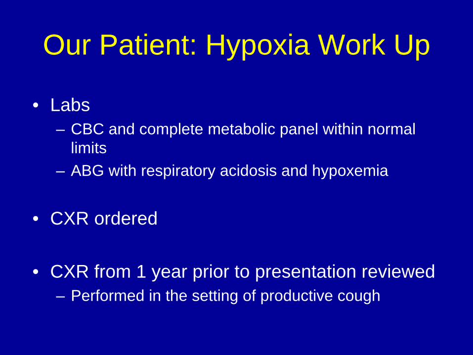
Our Patient: Hypoxia Work Up
• Labs– CBC and complete metabolic panel within normal
limits– ABG with respiratory acidosis and hypoxemia
• CXR ordered
• CXR from 1 year prior to presentation reviewed– Performed in the setting of productive cough

Overview• Presentation of index patient
– History of present illness– Radiologic findings
• Review of differential diagnoses
• Discussion of bronchioloalveolar carcinoma– General information– Roles of radiology in diagnosis and management
• Follow-up of index patient
• Summary

Our Patient: Chest CXR from One Year Prior to Presentation
From PACS, BIDMC
Upright PA and lateral chest X-ray

Please evaluate our patient’s CXR from one year prior to presentation for
abnormalities.

Our Patient: CXR with RLL Opacity
From PACS, BIDMC
Upright PA and lateral chest X-ray read as RLL opacity consistent with pneumonia.

Our Patient: CXR at Presentation
From PACS, BIDMC
Supine AP chest X-ray

Please evaluate our patient’s CXR at presentation for abnormalities.

Our Patient: CXR with Basilar Opacity
• Supine AP CXR read as:– New right basilar
opacity as compared with 1 year prior
– Consistent with PNA
– No other focal opacities suggestive of infection identified at the time
From PACS, BIDMC

Given the findings of persistent RLL on CXR from (a) one year prior and (b) at
the time of presentation, a chest CT was performed.

Our Patient: Anatomic Landmarks on Chest CT
• Right major fissure
• Left major fissure
From PACS, BIDMC
Axial C- Chest CT (lung window)

Our Patient: Multiple Nodules and Ground Glass Opacities
• Multiple nodules in LUL, LLL, RML, and RLL
– Varying sizes from 1- 4 mm
– Associated ground glass opacities (GGO):
•Hazy increased lung opacity with preservation of underlying bronchial and vascular margins
From PACS, BIDMC
Axial C- Chest CT (lung window)

Our Patient: Multiple Nodules, GGO, and Consolidation
• Moving inferiorly on CT:
– Multiple nodules and GGO, especially in RML
– Consolidation in RLL
From PACS, BIDMC
Axial C- Chest CT (lung window)

Our Patient: Dense RLL Consolidation
• Continuing to move inferiorly on CT:
– Dense consolidation of RLL with absence of aerated lung
– Air bronchograms:
Low attenuation, air-filled bronchi in the setting of a higher attenuation backgroundSuggest evacuation or
replacement of alveolar airFrom PACS, BIDMC
Axial C- Chest CT (lung window)

Our Patient: Aerated Portion of RLL
• What is this aerated space on our patient’s chest CT?
From PACS, BIDMC
Axial C- Chest CT, Lung Window

To clarify what comprises the aerated lung space on axial CT, let’s
compare this with the corresponding coronal CT view and with CXR…

Our Patient: Comparing Aerated Lung Space on Axial and Coronal CT
Axial C- Chest CT (lung window) Coronal C- Chest CT (lung window)
From PACS, BIDMC

Our Patient: Comparing Aerated Lung Space on CT and CXR
Supine AP CXR, 4 days after presentation
From PACS, BIDMC
Coronal C- Chest CT (lung window) at presentation

• Sharp borders between the consolidated and aerated spaces suggest that the consolidation follows an anatomic border.
• To further understand the involved and uninvolved spaces of the RLL, let’s review the segmental anatomy of the RLL…
Unknown Aerated Space in RLL: Conclusions from CT and CXR
Comparisons

RLL Segmental Anatomy
• The 5 segments of the RLL follow branching of the RLL bronchus
– Superior (S)– Anterior basal (AB)– Lateral basal (LB)– Posterior basal (PB)– Medial basal (MB)
Adapted from: http://koreacritcare.com/www/lung_segment.jpg

Our Patient: Aerated Space in RLL Identified
• RUL bronchus
• Bronchus intermedius
• RML
• Anterior basal segmental bronchus in the anterior basal lung segment
• MEDIAL BASAL LUNG SEGMENT
From PACS, BIDMC
Coronal C- Chest CT (lung window)

Overview• Presentation of index patient
– History of present illness– Radiologic findings
• Review of differential diagnoses
• Discussion of bronchioloalveolar carcinoma– General information– Roles of radiology in diagnosis and management
• Follow-up of index patient
• Summary

Differential Diagnostic Dilemma
• Are the radiologic findings of both consolidation and multiple nodules consistent with:
– One unifying diagnosis?
– Two or more concurrent processes leading to the mixed radiologic picture?

Differential Diagnosis: Multiple Pulmonary Nodules
• Neoplasm– Primary lung
– Metastases (breast, colon)
• Disseminated mycoses
• Septic emboli
• Sarcoidosis
• Mycobacterial disease

Narrowed Ddx for Multiple Pulmonary Nodules In Our Patient• Large number of nodules in close proximity, with sparing
of intervening parenchyma
– Makes inflammatory etiologies (mycoses, sarcoid) that would cause destruction and/or infiltration of surrounding parenchyma less likely
– Makes neoplasm more likely
• Lack of systemic signs of infection
– Makes extensive septic embolic disease and infection less likely

Differential Diagnosis: Lobar Consolidation
• Infection
• Edema
• Pulmonary hemorrhage
• Atelectasis
• Tumor

Narrowed Ddx for Consolidations in Our Patient
• Probable long-standing consolidation
– Lipoid PNA
– Chronic aspiration
– Pseudolymphoma (lymphoid hyperplasia)
– Bronchioloalveolar carcinoma

Our Patient: Presumed Diagnosis• Sputum cytology positive for adenocarcinoma
– Specific tumor markers initially pending
• Tissue sample not possible to obtain due to:– Ongoing anticoagulation therapy– Low respiratory performance status
• Pending specific identification of etiology, chemotherapy with premetrexed started – Based on radiologic appearance of presumed
cause: bronchioloalveolar carcinoma
Overview• Presentation of index patient
– History of present illness– Radiologic findings
• Review of differential diagnoses
• Discussion of bronchioloalveolar carcinoma– General information– Roles of radiology in diagnosis and management
• Follow-up of index patient
• Summary

Bronchioloalveolar Carcinoma (BAC): General Information
• Histology– Subtype of adenocarcinoma– Mucinous and non-mucinous histologies– Spreads mainly though lepidic growth (using lung architecture as
stroma) without destruction of underlying structure
• Epidemiology– Up to 7% of all primary lung neoplasms– 25-50% of patients with a history tobacco use
• Clinical features– Often incidentally diagnosed and asymptomatic – Symptoms: cough, sputum, SOB, weight loss, fever, bronchorrhea

Overview• Presentation of index patient
– History of present illness– Radiologic findings
• Review of differential diagnoses
• Discussion of bronchioloalveolar carcinoma– General information– Roles of radiology in diagnosis and management
• Follow-up of index patient
• Summary

Roles of Radiology in Diagnosis and Management of BAC
• To characterize the disease– Unique radiologic findings of BAC – Exclusion of other etiologies
• To identify patterns that correlate with specific BAC histologies (and thus prognosis)
• To further help establish prognosis and treatment by determining extent of disease
• To monitor treatment response

First, let’s discuss the radiologic characteristics that help identify BAC…
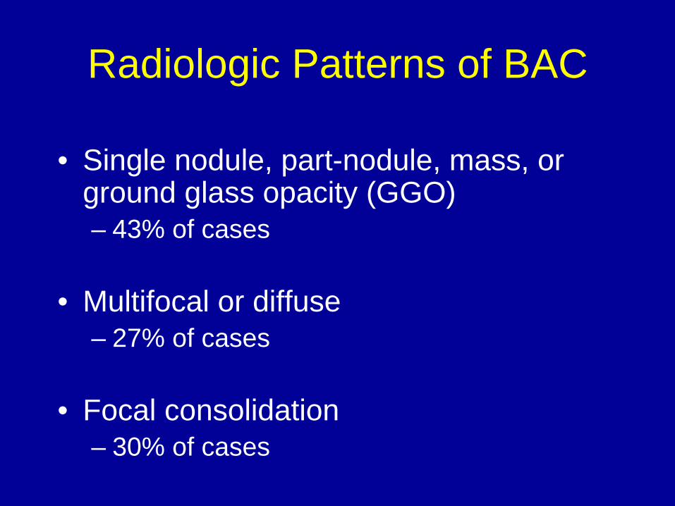
Radiologic Patterns of BAC
• Single nodule, part-nodule, mass, or ground glass opacity (GGO)– 43% of cases
• Multifocal or diffuse – 27% of cases
• Focal consolidation– 30% of cases

Companion Patient 1: BAC as a Solitary Nodule or Mass
• Solid nodule or mass
– Usually peripheral
– May be lobulated or ill- marginated
– May have heterogeneous CT attenuation, CT pseudocavitation, and/or air bronchogramsFrom PACS, BIDMC
Axial C+ Chest CT (lung window)

Companion Patient 2: BAC as Focal GGO
• Focal isolated GGO– Especially with
heterogeneous attenuation
• Can also be a part- nodule: – Partly solid opacity
with an associated area of GGO
From PACS, BIDMC
Axial C+ Chest CT (lung window)

Pathophysiology of CT Features in Nodular BAC
• Heterogeneous attenuation of nodules, masses and GGO – Due to non-destructive lepidic growth of tumors along alveolar
walls without disruption of the underlying architecture• Small patent airways or alveolar spaces left• Pseudocavitations and “bubble”-like areas of low attenuation
– Favors BAC versus other causes of nodules and/or GGO
• Air bronchograms: – Due to filling of alveoli adjacent to patent bronchi with tumor and mucin
• GGO:– Due to lepidic growth pattern of malignant cells and/or mucin production

Our Patient: BAC as Multiple Nodules
• Multiple nodules of varying size
– Seen bilaterally
– With and without GGO
• Associated with lymphatic spread
From PACS, BIDMC
Axial C- Chest CT (lung window)

Our Patient: Consolidative (Pneumonic) BAC
• Filling of the airspace with mucus
• Low-attenuation consolidation on CT due to mucin content
• Air bronchograms
• CT angiogram sign on C+ images:– Clearer visibility of vessels due to low
attenuation of surrounding tumor
• Delayed diagnosis common due to radiologic similarity to pneumoniaFrom PACS, BIDMC
Axial C+ Chest CT (bone window)

Our Patient: Consolidative BAC versus PNA
• CT changes of the air- filled bronchus in a consolidation that favor BAC to PNA:– Stretching– Squeezing– Sweeping– Widening of the branching
angle
• BAC is more chronic and with fewer systemic symptoms of infection
From PACS, BIDMC
Axial C- Chest CT (lung window)

Next, let’s discuss how radiologic findings correlate with histology and prognosis in BAC…

Radiologic Findings, Histology, and Prognosis in BAC
Radiologic pattern Typical histology 5-year survival s/p resection
<1 cm pure GGO or part nodule
Non-mucinous 100%
<1 cm solid nodule Non-mucinous 94%
Multifocal Mucinous 40%
Consolidative Mucinous 0%

Last, let’s discuss radiologic considerations for staging BAC…

TNM Staging in Lung Cancer: Imaging considerations
• Assessing tumor size (T)– Look for involvement of the main bronchus, pleura,
hilum, chest wall, diaphragm, pericardium vertebral bodies, mediastinum
• Assessing lymph nodes (N)– Look for peribronchial, ipsilateral hilar, mediastinal,
subarcinal nodes– Consider ipsilateral versus contralateral involvement
• Assessing distant metastases (M)– Consider whole body CT and/or PET

Overview• Presentation of index patient
– History of present illness– Radiologic findings
• Review of differential diagnoses
• Discussion of bronchioloalveolar carcinoma– General information– Roles of radiology in diagnosis and management
• Follow-up of index patient
• Summary

Our Patient: Diagnosis and Prognosis
• BAC confirmed with specific tumor markers
• General prognosis for BAC with multifocal and consolidative features:– Almost 0% survival at 5 years
• However, she is responding well after 4 courses of chemotherapy.

Our Patient: CT after 4 Cycles of Chemotherapy
Axial C- Chest CT (lung window), at presentation
• Fewer nodules and GGO in RML• Decreased extent of consolidation and more aerated space in RLL
Axial C+ Chest CT (lung window), after 4 cycles of chemotherapy
From PACS, BIDMC

Overview• Presentation of index patient
– History of present illness– Radiologic findings
• Review of differential diagnoses
• Discussion of bronchioloalveolar carcinoma– General information– Roles of radiology in diagnosis and management
• Follow-up of index patient
• Summary

Summary
• Radiologic findings of BAC– Patterns of growth
• Non-resolving singular nodule, mass or ground glass opacity• Multiple nodules or masses• Consolidations
– Heterogeneous attenuation on CT, with CT-angiogram sign, pseudocavitations, and air bronchograms
• Radiologic findings have implications for diagnosis, staging, prognosis, and treatment

Acknowledgements
• Dr. Paul Spirn• Dr. Ammar Sarwar• Dr. Prachi Dubey• Dr. Gillian Lieberman• Maria Levantakis

Select references• Akira M, Atagi S, Kawahara M, et al. 1999. High resolution CT findings of diffuse
bronchioloalveolar carcinoma in 38 patients. Am J Roentgenol. 172 (6): 1623-1629.
• Hansell DM, Bankier AA, MacMahon H, et al. 2008. Fleischner Society: Glossary of terms for thoracic imaging. Radiology. 246: 679-722.
• Jung, JI, Kim H, Park SH, et al. 2001.CT differentiation of pneumonic-type bronchioloalveolar cell carcinoma and infectious pneumonia. Br J Radiol. 74: 490-494.
• Lee KS, Kim Y, Ko EJ, et al. 1997. Bronchioloalveolar carcinoma: clinical, histopathologic, and radiologic findings. RadioGraphics. 17: 1345-1357.
• Lung Segments. Korea Critical Care. Accessed 3/15/10. Available online at: http://sites.google.com/a/koreacritcare.com/www/lung_segment.jpg
• Maldonado RL. 1992. The CT angiogram sign. Radiology. 210: 323-324.
• Mirtcheva RM, Vazquez M, Yankelevitz, DF, Henschke CI. 2002. Bronchioloalveolar carcinoma and adenocarcinoma with bronchioloalveolar features presenting as ground- glass opacities on CT. Clin Imgaing. 26 (2) 95-100.
• Patsios D, Roberts HC, Paul NS, et. al. 2007. Pictorial review of the many faces of bronchioloalveolar cell carcinoma. Br J Radiol. 80 (960): 1015-1023.
• Travis ED, Garg K, Franklin WA, et al. 2005. Evolving concepts in the pathology and CT imaging of lung adenocarcinoma and bronchioloalveolar carcinoma. J of Clin Onc. 23 (14): 3279-3287.