ichthyophthirius multifiliis (ciliophora) to serum from - inter research
TRANSCRIPT

DISEASES OF AQUATIC ORGANISMS Dis. aquat. Org. Published November 18
NOTE
Antibody binding following exposure of live Ichthyophthirius multifiliis (Ciliophora) to serum from
immune carp Cyprinus carpio
M. L. Cross*
Department ol Biological Sciences, University of Plymouth, Plymouth PL4 SAA, United Kingdom
ABSTRACT: Trophont stages of the c~l ia te fish parasite Ichthyophtl~jrius n~ult i f i l i is were Incubated alive in vitro in serum from immune carp Cyprinus carpi0 and subsequently examined immunoh~stoloyically for bound ant~body. Parasites became immobilized in immune serum and were enveloped by gelatinous mucus-llke material. Carp antibody could not be detected bound to cilia membranes (the major surface anti- gens), although it was detected at low levels in parasite food vacuoles and associated with the gelatinous material. This material is probably responsible for the immobilization of par- asites, and may form a barrier that prevents access of large amounts of ci l ia-membrane-spec~f~c antlbody to the parasite surface during in vitro incubat~on In f ~ s h serum.
KEY WORDS: Antibody . Carp . Ichthyophthlrius multifilijs L4ucocytes
The holotrich ciliate Ichthyophthirius n~ultifiliis is a major pathogen of freshwater fish worldwide (Nigrelli et al. 1976). Despite the marked pathogenicity of the disease (Hines & Spira 1974a. Ventura & Paperna 1985), fish which survive an initial infection can de- velop protective immunity and produce humoral anti- bodies specific to the parasite (Hines & Spira 197413, Wahli & Meier 1985, Clark et al. 1987, 1988). The major antigenic targets comprise integral proteins of the cilia membranes (Clark et al. 1988), and the contents of se- cretory mucocyst organellae (Cross & Matthews 1993). When parasites are incubated alive in vitro in immune fish serum they become immobilized, and gelatinous material is discharged from the mucocysts (Clark et al. 1987, Houghton 1987). However, the precise mode by which immobilization occurs is uncertain; in particular it is unclear whether this reflects direct agglutination of cilia via antibody-binding to the membrane anti-
Present address and address for correspondence: Depa.rt- ment of Veterinary Science, University of Arizona, Tucson, Arizona 85721, USA
gens, or whether immobilization occurs via a secon- dary effect. In the present study, the binding of anti- body to I. nlultifilijs is determined at the ultrastructural level, following incubation of parasites alive in im- mune carp Cyprinus carpio serum, to identify the fac- t o r ( ~ ) responsible for in vitro immobilization.
Methods. Ichthyophthirius multifiliis was isolated from freshwater fish obtained from local aquarists (Carassius auratus, held at 20 "C), and maintained in the laboratory by routine passage in naive hosts (Cyprinus ca~pio, held at 20 "C). To collect trophonts, fish were held temporarily in a glass vessel containing a small volun~e of buffered tap water (pH 7.2); the agi- tated movements of fish in such an enclosed environ- ment caused trophonts to be released from the skin. Within 2 to 3 min of release, groups of approximately 20 trophonts were collected with a glass pasteur pi- pette and transferred to 0.5 m1 perspex vials containing a minimum volume of fresh buffered tap water. 100 p1 of immune or control carp serum was then added to each vial to give a final dilution of 1/4 in water. The im- mune serum, which was derived from adult carp that had been immunized against I. multifiliis by controlled infections, was known to contain antibodies against cilia membrane antigens and the contents of secretory mucocyst organellae (Cross & Matthews 1993). The control serum, which was derived from similar-sized non-infected carp, was known to be non-reactive against parasite antigens (Cross & Matthews 1993). Following 1 h incubation at room temperature (approx- lmately 22 "C), trophonts were washed copiously with clean water, and subsequently fixed, embedded and sectioned for immunohistology as described previously (Cross & Matthews 1993).
Carp immunoglobulin was detected in tissue sec- tions by immunogold labelling techniques, performed
O Inter-Research 1993
160 Dis. aquat. Org. 17: 159-164, 1993
according to Cross & Matthews (1993). These proce- dures utilized a rabbit anti-carp 1gM serum that had been produced by immunizing male Dutch rabbits with chromatography-purified IgM fractions (Cross & Matthews 1993). Labelling was performed at both light and electron microscope levels. Briefly, for light level microscopy, semithin tissue sections on glass slides were labelled using 1/8 rabbit anti-carp IgM serum, which had been commercially conjugated with 15 nm colloidal gold (Biocell, Cardiff, UK), and the signal was amplified using an Intense-M silver enhancing kit (Janssen, UK). For electron microscopy, ultrathin tissue sections on nickel grids were labelled using 1/20 rabbit anti-carp IgM serum and 1/30 goat anti-rabbit IgG 20 nm colloidal gold conjugate (Bioclin, UK). The anti- serum controls for immunogold labelling procedures involved using pre-immune rabbit serum as the pri- mary antibody, or omitting the primary antibody step.
The efficacy of the above labelling procedures had been confirmed previously, by crnploying the same techniques and antisera to detect endogenous carp im- munoglobulin within tissue sections of carp epidermis (Cross 1990).
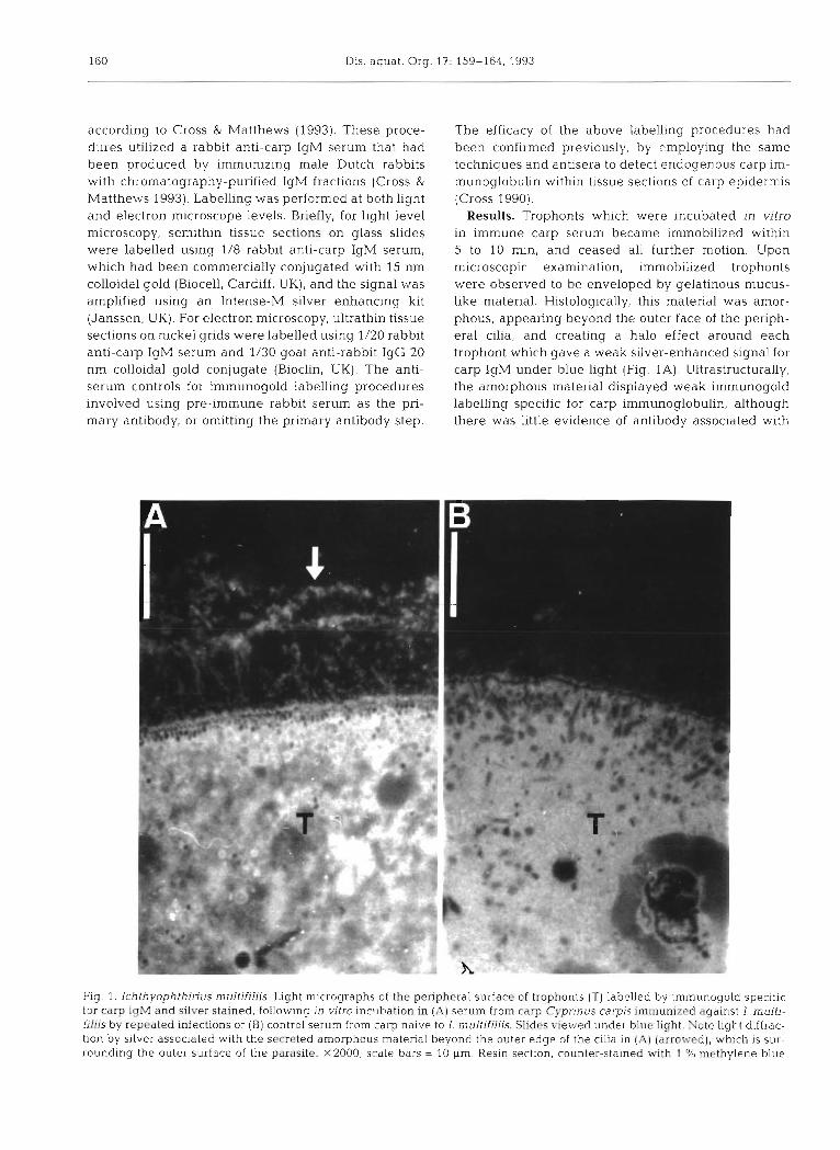
Results. Trophonts whlch were ~ncubated in vitro in immune carp serum became immobilized within 5 to 10 min, and ceased all further motion. Upon microscopic examination, immobilized trophonts were observed to be enveloped by gelatinous rnucus- like material. Histologically, this material was amor- phous, appearing beyond the outer face of the periph- eral cilia, and creating a halo effect around each trophont which gave a weak silver-enhanced signal for carp IgM under blue light (Fig. 1A). Ultrastructurally, the amorphous material displayed weak immunogold labelling specific for carp immunoglobulin, although there was little evidence of antibody associated with
Flg. 1. Ichthyophthinus multifillis. Light micrographs of the peripheral surface of trophonts (T) labelled by immunogold s p e c ~ f ~ c for carp IqM and silver stained, following in vitro incubation in (A) serum from carp Cyprinus carpis immunized against I. multi- filiis by repeated infections or (B) control serum from carp naive to I. multifiliis. Slides viewed under blue light. Note light diffrac- tion by silver associated w ~ t h the secreted amorphous material beyond the outer edge of the cilia in (AI (arrowed), which is sur- rounding the outer surface of the parasite. x2000; scale bars = 10 pm Resin, sectlon, counter-stained with 1 % methylene blue
Cross: Blnd~ny of carp antibody to a ciliate parasite 161
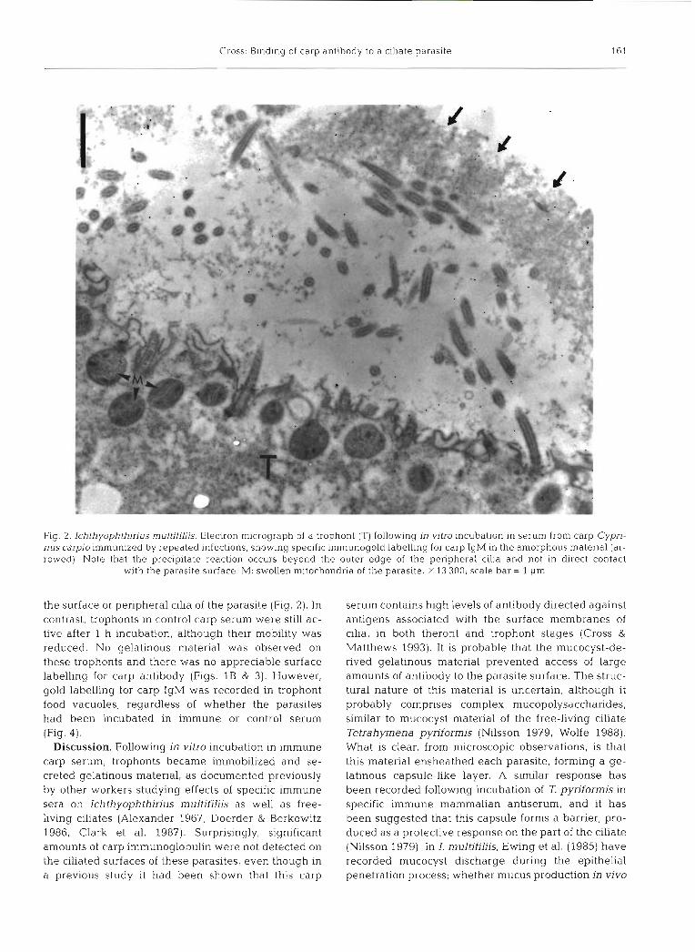
Fig. 2. Ichthyophthirius multifiliis. Electron micrograph of a trophont (T) following in vitro incubation in serum from carp Cypri- nus carpio immunized by repeated infections, showing specific immunogold labelling for carp IgM in the amorphous material (ar- rowed). Note that the precipitate reaction occurs beyond the outer edge of the peripheral cilia and not in direct contact
with the parasite surface. M: swollen mitochondria of the parasite. X 13 300; scale bar = 1 pm
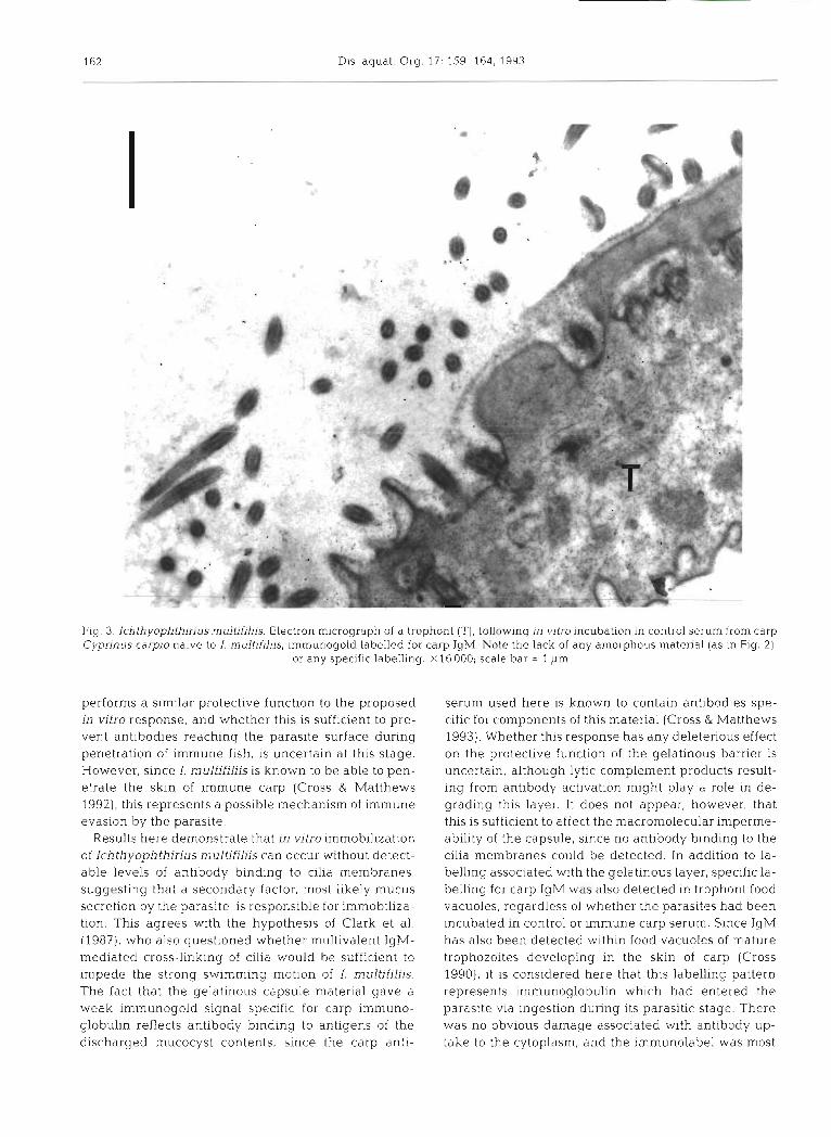
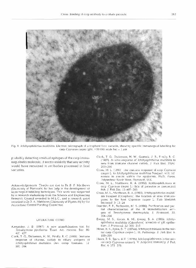
the surface or peripheral cilia of the parasite (Fig. 2 ) . In contrast, trophonts in control carp serum were still ac- tive after 1 h incubation, although their mobility was reduced. No gelatinous material was observed on these trophonts and there was no appreciable surface labelling for carp antibody (Figs. 1B & 3). However, gold labelling for carp IgM was recorded in trophont food vacuoles, regardless of whether the parasites had been incubated in immune or control serum (Fig. 4) .
Discussion. Following in vitro incubation in immune carp serum, trophonts became imnlobilized and se- creted gelatinous material, as documented previously by other workers studying effects of specific immune sera on Ichthyophthirius multifiliis as well as free- living ciliates (Alexander 1967, Doerder & Berkowitz 1986, Clark et al. 1987). Surprisingly, significant amounts of carp immunoglobulin were not detected on the ciliated surfaces of these parasites, even though in a previous study it had been shown that this carp
serum contains high levels of antibody directed against antigens associated with the surface membranes of cilia, in both theront and trophont stages (Cross & Matthews 1993). It is probable that the mucocyst-de- rived gelatinous material prevented access of large amounts of antibody to the parasite surface. The struc- tural nature of this material is uncertain, although it probably comprises complex mucopolysaccharides, similar to mucocyst material of the free-living ciliate Tetrahymena pyriformis (Nilsson 1979, Wolfe 1988). What is clear, from n~icroscopic observations, is that this material ensheathed each parasite, forming a ge- latinous capsule-like layer. A similar response has been recorded following incubation of T pyriformis in specific immune mammalian antiserum, and it has been suggested that this capsule forms a barrier, pro- duced as a protective response on the part of the ciliate (Nilsson 1979). In I. multifiliis, Ewing et al. (1985) have recorded mucocyst discharge during the epithelia1 penetration process; whether mucus production in vivo
162 Dis. aqual. Org. 17: 159-164, 1993
Fig. 3. lchthyophthirius multifiliis. Electron micrograph of a trophont (T), follaving in vitro incubation in control serum from carp Cyprinus carpio naive to I. mulfifiliis, imrnunogold labelled for carp IgM. Note the lack of any amorphous material (as in Fig. 2) ,
or any specific labelling. X16000; scale bar = 1 pm
performs a similar protective function to the proposed in vitro response, and whether this is sufficient to pre- vent antibodies reaching the parasite surface during penetration of immune fish, is uncertain at this stage. However, since I. multifiliis is known to be able to pen- etrate the skin of immune carp (Cross & Matthews 1992), this represents a possible mechanism of immune evasion by the parasite.
Results here demonstrate that in vitro immobilization of Ichthyophthirius multifiliis can occur without detect- able levels of antibody binding to cilia membranes, suggesting that a secondary factor, most likely mucus secretion by the parasite, is responsible for irnrnobiliza- tion. This agrees with the hypothesis of Clark et al. (1987), who also questioned whether multivalent IgM- mediated cross-linking of cilia would be sufficient to impede the strong swimmlng motion of I, multifiliis. The fact that the gelatinous capsule material gave a weak immunogold signal specific for carp immuno- globulin reflects antibody binding to antigens of the discharged mucocyst contents, since the carp anti-
serum used here is known to contain antibodies spe- cific for components of this material (Cross & Matthews 1993). Whether this response has any deleterious effect on the protective function of the gelatinous barrier is uncertain, although lytic complement products result- ing from antibody activation might play a role in de- grading this layer. It does not appear, however, that this is sufficient to affect the macromolecular imperme- ability of the capsule, since no antibody binding to the cilia membranes could be detected. In addition to la- belling associated with the gelatinous layer, specific la- belling for carp IgM was also detected in trophont food vacuoles, regardless of whether the parasites had been incubated in control or immune carp serum. Since IgM has also been detected within food vacuoles of m.ature trophozoites developing in the skin of carp (Cross 1990), it is considered here that this labelling pattern represents immunoglobulin which had entered the parasite via ingestion during its parasitic stage. There was no obvious damage associated with antibody up- take to the cytoplasm, and the immunolabel was most
Cross: Binding of carp antibody to a ciliate parasite 163
Fig. 4. Ichthyophlhirius multifiliis. Electron micrograph of a trophont food vacuole, showing specific immunogold labelling for carp Cyprinus carpio IgM. X 16 000; scale bar = 1 pm
probably detecting residual epitopes of the carp immu- Clark, T G., Dickerson, H. W.. Gratzek, J B., Findly, R. C. (1 987). In vitro response of Ichthyophlhirius multifiiis to
noglobulin molecule; it seems unllkely that any activity sera from immune channel catfish. J Fish Bi.01. 31(A). would have remained in antibodies processed in food. 203-208 vacuoles.
Acknowledgements. Thanks are due to Dr B. F. Matthews (University of Plymouth) for her help in the development of immunogold labelling techniques. This work was supported by a research studentship from the Science and Engineering Research Council awarded to M.L.C., and a research grant awarded to Dr R. A. Matthews (University of Plymouth) by the Polytechnic Central Funding Committee.
Cross, M. L. (1990). The immune response of carp Cyprinus carpio L. to Ichthyophthirius multifiliis Fouquet, with ref- erence to events within the epidermis. Ph.D. thesis, Polytechnic South West, Plymouth, U.K.
Cross, M. L., Matthews, R. A. (1992). Ichthyophthiriasis in carp Cyprinus carpio L.: fate of parasites in immunized fish. J. Fish Dis. 15: 497-505
Cross, M. L., Matthews, R. A. (1993). Ichthyophthirius multifi- 111s Fouquet (Ciliophora): the location of sites immuno- genic to the host Cypdnus carp10 L. Fish Shellfish Immunol. 3: 13-24
Doerder, F. P., Berkowitz, M. S. (1986). Purification and par- tial characterisation of the H immobilization anti- gen of Tetrahymena thermophila. J. Protozool. 33: 204-208
LITERATURE CITED Ekving, M. S., Kocan, K. M., Ewing, S. A. (1985). Ichthy- ophthirius multifiliis (Ciliophora): invasion of gill epithe-
Alexander. J. B. (1967). A new immobilization test for lium. J. Protozool. 32: 305-310 Tetrahymena pyj-ifoj-mjs. Trans. microsc, sot. 86. Hines, R. S., Spira, D. T (1974a). Ichthyophthiriasis in the mir- 421-427 ror carp Cyprinus carpio L. 111. Pathology. J. Fish Biol. 6:
Clark, T G , Dickerson, H. W., Findly, R C (1988). Immune 189-200 response of channel catfish to ciliary antigens of Hines, R. S., Spira, D.T (1974b). Ichthyophthiriasis in the mir- Ich thyophthirius multifiliis. Dev. comp. Immunol. 12: ror carp Cypnnus carpio L. V . Acquired immunity. J. Fish 581-594 Biol. 6: 373-378

164 Dis, aquat. Org. 17. 159-164, 1993
Houghton. G. (1987). The immune response in carp, Cyprinus carpio L. to Ichthyophthirius multifiliis, Fouquet 1876. Unpublished Ph. D. thesis, Plymouth Polytechnic, Plymouth, U.K.
Nigrelli, R. F., Pokorny, A. S., Ruggieri, G . D. (1976) Notes on Ichthyophthirius multifiljjs, a ciliate parasite on fresh- water fishes, with some remarks on possible physiolo- gical races and species. Trans. Am. microsc. Soc. 95: 607-613
Nilsson, J. R. (1979). Phagotrophy in Tetrahymena. In: Levandowsky, M.. Hunter, S. H. (eds.) Biochemistry and
Responsible Subject Editor: W. Korting, Hannover, Germany
physiology of protozoology, Vol 2. Academ~c Press. New York, p. 339-380
Ventura, M. T., Paperna, I. (1985). Histopathology of Ichthyophthirius multifiliis infections in fishes. J. Fish Brol. 27: 185-203
Wahli, T., Meier, W. (1985). Ichthyophthiriasis in trout: inve- stigations of natural defence mechanisms. In: Ellis, A. E. (ed.) Fish and shellfish pathology. Academic Press, London, p. 347-352
Wolfe, J. (1988). Analysis of Tetrahymena mucocyst material with lectins and alcian blue. J. Protozool. 351 46-51
Manuscript first received: March 9, I993 Revised version accepted: July 22, 1993