ice, ice baby - home page | phoenix children's hospital · ice, ice baby: treatment of infants...
TRANSCRIPT

Ice, Ice Baby:
Treatment of Infants with Hypoxic Ischemic Encephalopathy Using Hypothermic Therapy Within a
Successful Neuro NICU Program
•Cristina Carballo, M.D. •Neuro NICU Medical Director
•Kim Allred, NNP-BC
•Neuro NICU Program Coordinator
•Christine Bure RN, Clinical Supervisor, NICU Phoenix Children’s Hospital

NEURO NICU Concept
• 1st unit in the country opened in the UCSF NICU, Summer of 2007.
• 2nd unit in the country: PCH NICU, Summer of 2009.
Objectives
• Define HIE, identify HIE patients, and treatment modalities available
• Identify the necessity as well as the components of a successful Neuro NICU and the process for clinical education.

What is HIE?
(Hypoxic Ischemic Encephalopathy)

HIE (Hypoxic Ischemic Encephalopathy))
• Hypoxia = a decreased amount of oxygen
• Ischemia = a decreased amount of blood perfusing the brain
• Encephalopathy = any dysfunction of the brain
• HIE in general terms is a brain that has been deprived of oxygen just before and/or during delivery

Acute Brain Injury in Newborns
• Acute event
• Causes lack of oxygen to brain until oxygen is re-established
• Results in HIE (Hypoxic Ischemic Encephalopathy)

Causes of Acute Brain Injury in Newborns • Acute event
• Causes lack of oxygen to brain until oxygen is re-
established
• Results in HIE (Hypoxic Ischemic Encephalopathy)
• Antepartum: Placental insufficiencies, umbilical cord accidents,
viral infections, growth retardation, etc.
• Intrapartum: Placental abruption, cord prolapse, shoulder
dystocia, abnormal cord insertion, etc.
• Postpartum: Infection, aspiration causing asphyxial event,
brain blood vessel abnormality, etc.

HIE
Experimental studies have indicated that neuronal death occurs in 2 phases:
• Primary Neuronal death
• Delayed or Secondary Neuronal death

Primary neuronal death
• Cellular hypoxia with exhaustion of the cell’s high energy stores – also known as primary energy failure.
• It is the initial injury and is primarily necrotic cell death.
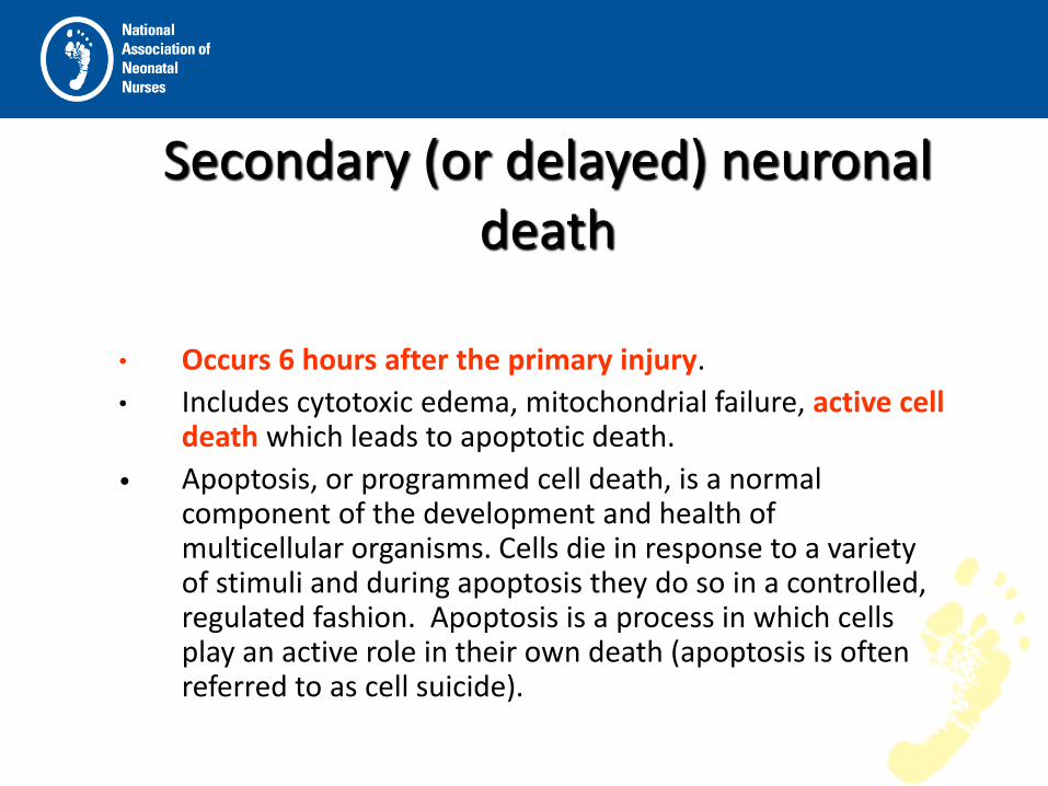
Secondary (or delayed) neuronal death
• Occurs 6 hours after the primary injury.
• Includes cytotoxic edema, mitochondrial failure, active cell death which leads to apoptotic death.
• Apoptosis, or programmed cell death, is a normal component of the development and health of multicellular organisms. Cells die in response to a variety of stimuli and during apoptosis they do so in a controlled, regulated fashion. Apoptosis is a process in which cells play an active role in their own death (apoptosis is often referred to as cell suicide).

Brott, NEJM 2000
Molecular Events Initiated in Brain Tissue by Acute Cerebral Ischemia

Mechanisms of Brain Injury in the Term Neonate • Oxidative stress and excitotoxicity, through downstream
intracellular signaling, produce both inflammation and repair
• Cell death begins immediately and continues during a period of days to weeks. The cell-death phenotype changes from an early necrotic morphology to a pathology resembling apoptosis. This evolution is called the necrosis– apoptosis continuum
• Donna Ferriero, NEJM, 2009
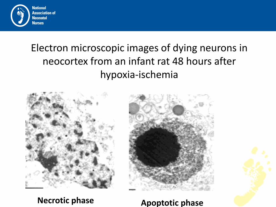
Electron microscopic images of dying neurons in neocortex from an infant rat 48 hours after
hypoxia-ischemia
Necrotic phase Apoptotic phase


Hypothermia therapy in neonates is now becoming more widely accepted and used in:
• Hypoxic Ischemic Encephalopathy (HIE)
• Hypoxic event post delivery, not related to delivery but within 6 hours of birth
• Prolonged hypoxic event prior to or during ECMO initiation

Impact of Brain Injury in Neonates
Life-long disabilities include: • Cerebral palsy: total cost in the US alone in 2007
was $4 Billion, with an individual cost $900 K/lifetime.
(Access Economic Study, CP Foundation) • Seizures • Hydrocephaly • Learning Disabilities and other cognitive
dysfunction • Speech and visual disturbances

HIE
(Hypoxic Ischemic Encephalopathy)
• 1 to 2/1000 term live births are at risk for HIE (Gluckman, Lancet 2005)
• 25-30% who survive will be left with life-long disabilities: CP (dyskinetic and spastic tetraplegia being the most common), seizures and/or cognitive dysfunction.
(Lin, J.Perin, 2006)

Impact of Early Identification and Treatment of Brain Injury/Pathology
• Decrease in CP severity
• Decrease in subclinical and clinical seizures resulting in improved brain function
• Decrease in cognitive deficits, visual and speech impairments

What can we do with this window of opportunity?
• Studies have found that HYPOTHERMIA provides a neuroprotective effect.
• Hypothermia may modify cells programmed for apoptosis – potentially leading to their survival.
• Hypothermia may also protect neurons by reducing cerebral metabolic rate.
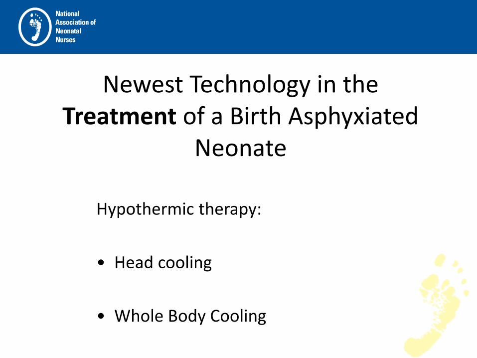
Newest Technology in the Treatment of a Birth Asphyxiated
Neonate
Hypothermic therapy:
• Head cooling
• Whole Body Cooling

Head Cooling VS Whole Body Cooling Both methods have similar outcomes
Head Cooling •PROS
• FDA Approved
CONS
• Limited MRI EEG access
• Scalp Edema
• Expense ($28,000)
Whole Body Cooling •PROS
•Easy MRI and EEG access
•Decreased scalp edema
•Decreased expense ($7,500)
•Increased access to infant
•CONS
•Pending FDA approval
•Risk for subcutaneous fat necrosis
•Possible exacerbation of PPHN

Medi-Therm® 7900 Olympic Cool-Cap® System

Inclusion Criteria
• Infant at ≥ to 36 weeks gestational age and at least ONE of the following:
• Apgar scores less than or equal to 5 at 10min after birth
• Continued need for resuscitation, including intubation or mask ventilation at 10min after birth
• Acidosis defined as either umbilical cord pH or any arterial pH within 60 minutes of birth less than 7.00
• Base deficit greater than or equal to -16 mmol/L in any blood sample within 60min of birth (arterial or venous)
• Prolonged resuscitation while being placed on ECMO

Inclusion Criteria (cont)
• Infant with moderate to severe encephalopathy consisting of altered state of consciousness (as shown by lethargy, stupor, or coma) and at least one of the following:
• Hypotonia • Abnormal reflexes, including oculomotor or pupillary
abnormalities • Absent or weak suck • Clinical seizures
Exclusion Criteria
• infants expected to be >6 hours of age before therapy initiated
• Major congenital abnormalities excluding Trisomy 21
• Evidence of head trauma or skull fracture
• Babies under 36wk of age
• Infants <1800 g birth weight

Sarnat Scoring
• A Neurologic evaluation including LOC, neuromuscular control, reflexes, autonomic function, evidence of seizures
• Three stages • Stage 1 – mild Good prognosis • Stage 2 – moderate Good prognosis if clinical and EEG recovery within 5 days Poor prognosis if periodic EEG with interburst intervals totally
isoelectric, bursting frequency less than every 6 seconds, bursting pattern (every 3-6 seconds) lasting more than 7 days.
• Stage 3 – severe Major neurologic sequelae, including microcephaly, mental
retardation, CP, seizures (all cases).


Tips for sending facilities and transport teams • Save and send placenta on ice for pathology studies • Early identification of eligible patients • Turn off all external heat sources
• Document time of heat source removal • Monitor rectal temperature (goal ~34.5 C) • Passively cool patients- not usually necessary to actively cool patients,
but maybe necessary to use ice packs in the armpit and/or groin area during hot summer months of transport
• Be prepared to slowly warm if the patient falls below 34 C • Obtain vascular access (becomes extremely difficult after cool-
vasoconstriction) • Recommend treating with morphine and ativan if needed for comfort • Treat only clinical seizures- phenobarbital load @ 20mg/kg • Support ventilation if necessary • Hemodynamic support:
• May need volume replacement • Expect a lower baseline HR with temperature decrease
Monitoring Neurological Status
• At present, the best technologies available include:
• the aEEG to monitor brain function
• Conventional EEGs are also done.
• INVOS to follow brain perfusion

• Single EEG Lead (3 wires)
What is aEEG?Special Filtering • Single EEG Lead (3 wires) • Bi-parietal • Monitor Global Electrocortical Activity • Special Filtering • Compression • Very Slow, Trend Display
PROS • RN initiation, ease of use, STAT clinical data, early identification of
seizure activities
CONS • Use of 29g needle electrode comes with minimal risk

aEEG tracings
Normal
Moderate suppression
Sleep/Wake Cycling Upper Margin > 10 µvolts Lower Margin > 5 µvolts Limited Variability
No Sleep/Wake Upper Margin > 10 µVolts Lower Margin < 5 µVolts Increased variability

aEEG tracings
Severe suppression
• No Sleep/Wake
• Upper Margin < 10 µVolts
• Greatly reduced variability

aEEG tracings - seizures
•Seizures cause sudden change in EEG amplitude – usually a rise during seizure followed by reduced amplitude at the end of seizure

Measure of quality of electrode contact Want it as low as possible Alarm if > 20 kOhm Can be used to detect lead motion artifact
Impedance


Use of NIRS (Near Infrared Spectroscopy). INVOS®
• Brain Spectroscopy: demonstrates brain perfusion and uptake of O2. A high number indicates decreased O2 uptake by tissue
• Renal/Body Spectroscopy: demonstrates organ perfusion, specifically renal and intestinal. Correlates with changes in hemodynamic stability

Use of NIRS • As new therapies are becoming available
with potential for neuroprotection, NIRS could potentially discriminate between infants at high and low risk for poor outcome in the first few hours of life, and could be used to monitor the safety and efficacy of treatment interventions.
• NINDS (National institute of Neurological Disorders and Stroke) April 2011


Reflecting the Color of Life®
• De-oxyhemoglobin and total hemoglobin are noninvasively assessed to generate a regional oxygen saturation (rSO2).
• This site-specific vital sign helps critical care teams detect and correct ischemia and oxygen imbalance to help prevent complications and poor outcomes
• “Provides a continuous, quantitative signal of the physiologic variable most related to injury and most amenable to intervention” Hoffman GM Ann Thorac Surg 2006

0
10
20
30
40
50
60
70
80
90
100
rSO
2
Series1Version 3.2
Case History Data 5 days
Day 1
0930
Day 2
Day 3
rewarming Day 4
Day 5
decannulated
Cerebral
Day 6

MRI with Spectroscopy
• MRI with Spectroscopy – done on Day 1 and Day 4 and repeated on either Day 14 or as outpatient.
• T1, T2
• Diffusion Weighted
• Cerebral Perfusion

• T1 imaging provides anatomic information as well as high contrast between gray and white matter

• T2 imaging provides contrast resolution between gray matter, unmylinated and mylinated white matter.

• Diffusion Weighted Imaging
– based on the microscopic movement of water molecules in brain tissue.

a | T2-weighted image depicting normal term brain. b | Diffusion-weighted MRI scan showing hypoxic–ischemic injury to the deep gray nuclei (arrows). c | Diffusion-weighted MRI scan with watershed pattern of hypoxic–ischemic injury (top and bottom arrows point to anterior and posterior watershed regions of the cortex and white matter, central arrows show injury to white matter). d | T1-weighted imaged depicting diffuse brain injury secondary to global hypoxic–ischemic insult. e | Diffusion-weighted image showing injury to the white matter and cortex (arrows) in an 11-day-old term infant with congenital heart disease. f | Diffusion-weighted MRI scan showing focal stroke (arrow) in a term infant who presented with focal seizures on the second day of life.

Spectroscopy (MRS)
• Acute injury (such as HIE) can be detected by MRS when both diffusion imaging and conventional imaging is negative.
• MRS can detect elevated lactate in the cerebral cortex or basal ganglia depending on the pattern of injury within the first 24 hours of the injury
• Reduced NAA and elevated glutamate and glutamine is usually detected after 24 hours.

Spectroscopy (MRS)
• It is a measure of brain chemistry – most commonly proton, sodium, phosphorus.
• Lipids – products of brain destruction
• Lactate – product of anaerobic glycolysis
• NAA – neuronal marker
• Glutamine – neurotransmitter
• Choline – cell membrane marker

Spectroscopy (MRS)
• When there is ischemia in the brain it switches to anaerobic glycolysis and lactate accumulates.
• A markedly elevated lactate in spectroscopy is seen in cerebral hypoxia and ischemia.
• Choline is elevated, NAA is reduced.
• If there is cerebral infarction, lipids increase.


Protocol while utilizing hypothermia • Minimize interventions • Monitor 72 hours of therapy– including q12hr scalp checks (head
cooling) and OFC • Hourly vital signs • Keep rectal temperature 34.0-35.0°C (head cooling). • Keep esophageal temperature 33.5°C (whole body cooling) • Morphine gtt • Watch closely for seizures (aEEG) • If seizures present – Load with Phenobarbital and follow guidelines • The heart rate will immediately fall, no intervention is necessary if BP
and O2 sats are stable • The infant may become edematous, reposition every four hours and
massage skin • closely watch urine output (indwelling uri-cath) • Labs: ABG’s, Electrolytes, CBC, DIC panel, Liver function • Infuse as necessary for clotting insufficiencies • HUS, MRI and EEG: goal of day one and after rewarm • Rewarm after 72 hours- SLOWLY, between 4-6 hour time frame

Case Presentations

Implementation at PCH
Our first Cool-Cap® baby:
• Was born 4/21/08 at an out-lying hospital via primary C-section secondary to placental abruption (otherwise uncomplicated pregnancy)
• Apgars: 4 at 1min, 5 at 5min, and 6 at 10min
• Cord pH 6.8
• Initial ABG pH 6.8 with a -19 base deficit
• Transported to PCH for further care – arrived and was placed on Cool-Cap at approx 5 hr of age.

Post Cool-Cap • MRI on 4/28 showed no acute intracranial
abnormality and some focal abnormality in the frontal lobe.
• EEG 4/28 was abnormal, showing some activity over the midline left central region, which appeared depressed.
• EEG 5/19 continued to show some abnormality, but a normal background with continued depression over the left and central regions.
Overall outcome is significantly improved with the use of the Cool-Cap®

Hypothermia Therapy post delivery
One patient: - Normal pregnancy and delivery - Mom given Ambien early in labor, sent home - Returned short time later while under the effects of Ambien and
delivered vaginally - Baby given to mom to breastfeed within one hour after delivery,
nurse left the room - Returned 5 minutes later to find mom having fallen asleep and baby
blue and unresponsive at the breast with apparent suffocation - Code ensued and baby transferred to us for possible cooling - Baby demonstrated moderate encephalopathy on the aEEG and by
SARNAT scoring - Baby cooled 72 hours, outcome was normal MRI, normal EEG and
normal developmental outcome.

Hypothermia Experience with ECMO
• 5 patients placed on hypothermia therapy (one Cool Cap® and four with whole body cooling)
• And
• within 24 hours then placed on ECMO
• (1 V-A 1 VV changed to VA, and 3 V-V ECMO).

Patients at Risk for HIE and PPHN
• MAS: Intrapartum hypoxic event increases meconium release prior to delivery.
• Chorio/Sepsis: increases risk for development of PPHN
• Malformations: CHD and CDH increase immediate postpartum hypoxia which increase risk for PPHN

Current Research • First published trials were in the UK:
• 1. 26 babies were enrolled in a pilot study of mild hypothermia during ECMO.
• All of these babies did well, no complications noted using both therapies
• 2. Second study in 2007, also in the UK, studied aEEG’s in infants on ECMO and receiving mild hypothermia. They showed that the aEEG reading was not affected by the mild hypothermia while on ECMO

Hypothermia Therapy experience in ECMO Centers in the US
Children’s National Medical Center (Billie Short): Protocol in place
• series of 10 patients, 6 MAS, 3 CDH, 1
tracheal stenosis. Results: • 3 CDH patients died, tracheal stenosis patient
died. Remaining 6 survivors includes 3 normal neurodevelopmental outcomes, 3 with abnormal tone

PCH Experience: Joaquin 1st Patient: DOB: 8/26/09 Gestational Age: 38 4/7 Maternal Hx: Unremarkable Birth Hx: • Shoulder Dystocia 30 seconds • APGARS: 1/3/4 at 1,5,10 minutes • Resuscitated, intubated, NaHCO3 given X 2 • Initial base deficit -21 • pH 6.9 • Pulmonary Hemorrhage
Transported for hypothermia therapy

PCH Experience (cont)
Neurological Evaluation upon arrival to PCH: • Level of Consciousness: Lethargic • Spontaneous Activity: Decreased • Posture: Complete Extension • Tone: Hypotonic • Primitive Reflexes: Absent suck and Moro • Autonomic System: Pupils poorly reactive, HR stable,
Respiratory: Apnea (Pulmonary hemorrhage, requiring HFJOV)
The above SARNAT SCORE is a 2 (Moderate)

PCH Experience (cont) INITIAL CLINICAL COURSE: • Upon arrival, baby placed on Cool Cap®
• Within less than 24 hrs, baby qualified for VA ECMO secondary to pulmonary hemorrhage and cardiovascular collapse despite Dopamine, Milrinone, and extensive support .
• ECHO that first day showed severely depressed bi-ventricular function, suprasystemic pressures, large PDA.

PCH Experience (cont) CONTINUED CLINICAL COURSE: • Cool Cap® continued for 72 hrs • ECMO continued for total of 6 days, without any
complications. • Extubated on DOL #10 to CPAP, HHLF on DOL
#12, RA on DOL #18 • All pressor support d/c’d on DOL #5 • Feeds started on DOL # 12, full feeds DOL # 19
and all PO DOL #28. • Home: DOL # 29

PCH Experience (cont)
STUDIES:
• On cooling and ECMO:
• INVOS was continuously running. Showed good cerebral perfusion.
• aEEG was also running, showed no seizure activity. Tracing showed moderate to severe encephalopathy.
• HUS’s remained normal

PCH Experience (cont)
POST COOLING and ECMO STUDIES:
EEG’s: improved from the first one on DOL #14 showing abnormal, recurrent slow delta activity over L temporal lobe and overall mild to moderate diffuse disturbance of cerebral function to the second on DOL #20 showing mild diffuse abnormality with otherwise normal background
PCH Experience (cont)
POST COOLING and ECMO STUDIES: MRI: done DOL #6 showed tiny foci of injury in the left posterior, parietal lobe, with otherwise normal parenchyma and spectroscopy, suggestive of NO hypoxic ischemic injury to the brain. MBS: DOL # 20, showed silent aspiration to all consistencies except honey consistency BAER passed bilaterally

PCH Experience (cont)
POST-DISCHARGE NEURO NICU F/U
• 4 month Bayley Scales of Infant and Toddler Development III: appropriate for gestation age in all areas, cognitive, motor and language development
• INFINIB and Gesell done show him to be age appropriate in all areas, and slightly delayed in gross motor by about one month.
• Ongoing visits for the continued above assessments.
PCH Experience: Patient #2 2nd Patient: DOB 8/6/10
• Gestational age: 41 6/7
• Maternal history: Lay Midwife care, +GBS, treated with chlorhexidine wipes and vaginal washes
• Birth history: Home delivery, Apgars 3 and 7. initially breathing then apneic and bradycardic at 15mins, required resuscitation.
• Initial baby gas: 6.8/89/242 -19 base deficit
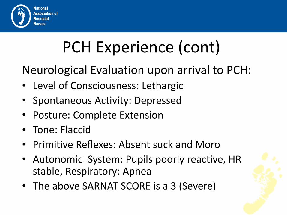
PCH Experience (cont) Neurological Evaluation upon arrival to PCH: • Level of Consciousness: Lethargic
• Spontaneous Activity: Depressed
• Posture: Complete Extension
• Tone: Flaccid
• Primitive Reflexes: Absent suck and Moro
• Autonomic System: Pupils poorly reactive, HR stable, Respiratory: Apnea
• The above SARNAT SCORE is a 3 (Severe)

PCH Experience (cont)
• Upon arrival, baby placed on whole body hypothermia blanket
• Within 6 hours, baby qualified for ECMO and was placed on V-V ECMO secondary to significant cardiovascular compromise (DOP, DOB and EPI drips with iNO)

PCH Experience (cont) CONTINUED CLINICAL COURSE: - Hypothermia therapy continued for 72 hrs
- ECMO continued for total of 8 days, without any complications.
- Extubated on DOL #11 to HHLF, RA on DOL #18
- All pressor support d/c’d on DOL #5
- Feeds started on DOL # 13, full feeds DOL # 24
- Back transported to continue learning po feeds: DOL # 25

PCH Experience (cont)
STUDIES:
• On cooling and ECMO:
• INVOS was continuously running. Showed good cerebral perfusion.
• aEEG was also running, showed no seizure activity. Tracing showed moderate to severe encephalopathy.
• HUS’s remained normal

PCH Experience (cont)
Neurologic Studies: • EEG: 1st on DOL 1 is very abnormal, no sz • 2nd on DOL 10, improved, no sz • 3rd on DOL 14, vastly improved, almost
normal
• MRI: 1st on DOL 10 (2nd to being on ECMO) was normal
• HUS: 5 studies during ECMO, all normal

PCH Experience (cont)
• MBS: Moderate to deep penetration 50% of time with ultrathin (breast milk) and thin consistency.
• BAER: passed bilaterally
• Follow-up post discharge: 1st 4 month neurodevelopmental eval in December, parents report all milestones being met

PCH Experience: Patient #3
3rd patient: DOB 10/19/10
• -Gestational Age: 36 5/7
• -Maternal HX: Previous two pregnancies with Gest DM, this pregnancy reportedly negative; +GBS received 3 doses of PenG prior to delivery
• -Birth HX: Attempted vaginal delivery, shoulder dystocia. Baby pushed back up the canal, cord prolapse and c-section. Apgars 0/4/5 at 1/5/10 mins. Cord pH 6.9

PCH Experience (cont)
Neurological Evaluation upon arrival to PCH: • Level of Consciousness: Lethargic
• Spontaneous Activity: None
• Posture: Complete Extension
• Tone: Flaccid
• Primitive Reflexes: Absent suck and Moro
• Autonomic System: Pupils reactive, HR stable, Respiratory: Apnea
• The above SARNAT SCORE is a 3 (Severe)

PCH Experience (cont)
• Upon arrival, baby placed on whole body hypothermia blanket
• Within 46 hours, baby qualified for ECMO and was placed on V-V ECMO. Prior to ECMO, baby on Dop, Dob and iNO. Increasing pCO2 despite HFJV.

PCH Experience (cont)
CONTINUED CLINICAL COURSE:
- Hypothermia therapy continued for 72 hrs
- ECMO continued for total of 3 days (60 hrs), with prolonged PT despite FFP and development of IVH.
- Extubated on DOL #6 to HHLF, RA on DOL #16
- All pressor support d/c’d on DOL #5
- Feeds started on DOL # 7

PCH Experience (cont)
STUDIES:
On cooling and ECMO:
• INVOS was continuously running. Showed initial poor cerebral perfusion.
• aEEG was also running, continued to severe encephalopathy. Some seizure activity recognized post-ECMO
• HUS’s showed R IVH with extension into septum pallucidum, but no intraparenchymal bleeds.

PCH Experience (cont) Neurologic Studies: • EEG: 1st on DOL 1 is very abnormal, no seizures • 2nd on DOL 6: worsening with seizure activity. • 3rd on DOL 8: improved but still abnormal, no seizures • 4th on DOL 14: continues to improve, still not completely
normal
• HUS: first showed edema, repeat subsequently showed R IVH and septum pallucidum bleed.
• MRI: 1st on DOL 1 showed mild diffuse white matter injury and R IVH with septum pallucidum bleed/cyst
• 2nd: on DOL 6: shows stable ventricular size, no parenchymal bleeds, stable cyst size
• 3rd: DOL 14: stable, continues to demonstrate white matter injury, normal gray matter

PCH Experience (cont)
• Brady continued to demonstrate significant emesis. UGI and pyloric U/S were normal. It was felt that the retching originated from pressure of the cyst sitting on the top of the brainstem.
• Neurosurgery became involved. A tapping reservoir was placed and the cyst drained.
• MBS soon after showed aspiration at all levels except honey so feeds were thickened.

PCH Experience – Cooling and ECMO: CONCLUSION
• Limited evidenced-based results presently available • Our five cases suggests that hypothermia therapy used with ECMO
can be done safely • University of Michigan has a protocol using whole body
hypothermia and the ECMO circuit to cool, rather than the Cool Cap (which was cumbersome to use while on ECMO).
• We feel, that at PCH, consideration for the use of both therapies concomitantly should be done on a case-by-case basis
• Use of either therapy should not preclude consideration of using the other
• Need for ECMO secondary to PPHN, may suggest an etiology for CNS injury that may benefit from the use of hypothermia therapy
• We may want to consider using hypothermia therapy in those situations were there is cardiac arrest going on to ECMO, as this hypoxic state may benefit from hypothermia therapy.
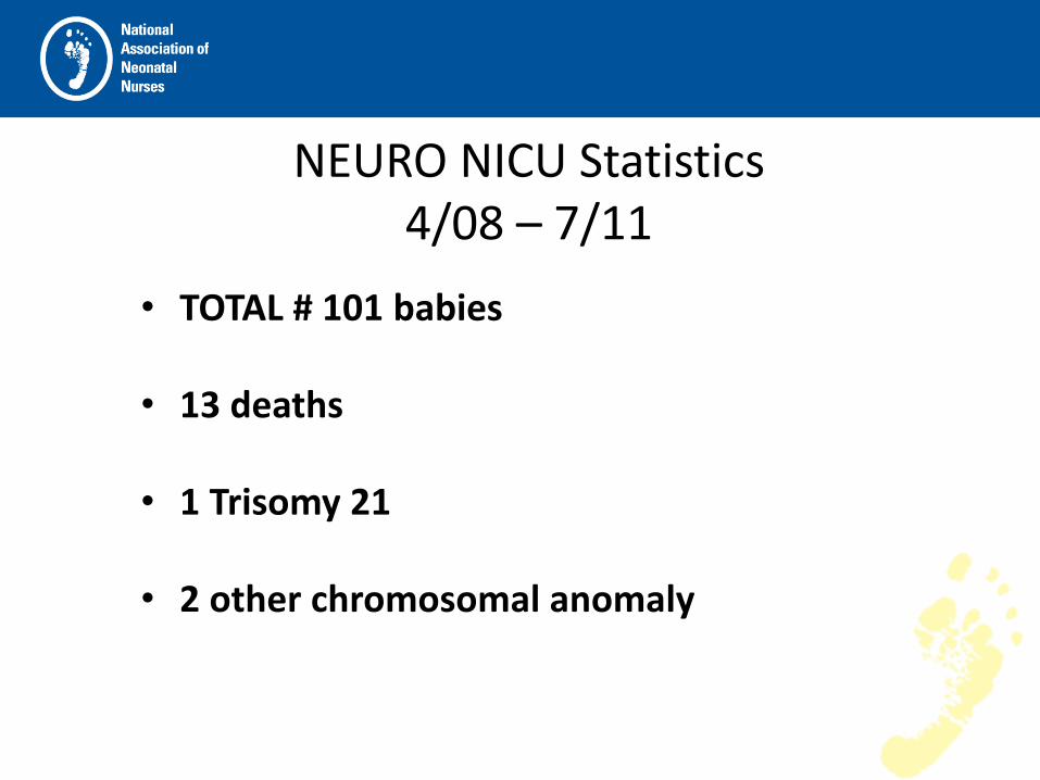
NEURO NICU Statistics 4/08 – 7/11
• TOTAL # 101 babies
• 13 deaths
• 1 Trisomy 21
• 2 other chromosomal anomaly

Statistics (cont) Maternal risk factor
• 15 Dec FHT
• 13 NR FHT
• 11 MAS
• 7 Abruption
• 4 Cord prolapse
• 4 Nuchal cord
• 3 True knot
• 3 Difficult delivery/Shoulder/Vac/Forceps

Statistics (cont) Maternal risk factor
• 2 Maternal death
• 2 No FHT
• 1 Post delivery code
• 1 Pulmonary hemorrhage
• 1 H1N1
• 1 Uterine rupture
• 1 Failure to progress
• 1 Repeat

Statistics (cont)
• pH range 6.51-7.36 avg=6.94
• Base deficit -8 to -30 avg= -18.7
• Lactic Acid 1-24 avg=8.4
• Sarnat mild 1 • moderate 44 • severe 21

Research
• Neuro NICU Data Registry:
• IRB approved.
• Allows entry of patient information into data base, tracks neurodevelopmental progress through 18 years of age and allows for ongoing studies concerning treatment modalities, early identification and interventions in relation to outcome measures.

Post Discharge Follow-up
• Neurodevelopmental Follow-up:
• to document neurodevelopmental assessments for research and recommend therapies for better patient care
• Rehab services: to document and implement therapy needs (OT/PT and Speech) to optimize neurodevelopmental outcomes

Neuro NICU Statistics (cont) • 66 currently consented infants:
• 24 babies have normal neurodevelopmental follow-ups
so far. The oldest is 3 1/2 yrs old
• 12 are delayed in motor
• 1 delayed cognitive
• 2 are delayed in speech
• 4 have documented CP
• 23 have not followed up
Neuro NICU Statistics (cont)
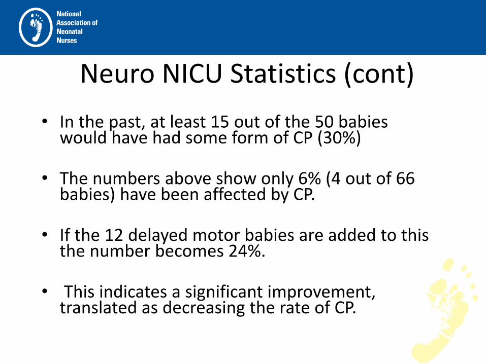
• In the past, at least 15 out of the 50 babies would have had some form of CP (30%)
• The numbers above show only 6% (4 out of 66 babies) have been affected by CP.
• If the 12 delayed motor babies are added to this the number becomes 24%.
• This indicates a significant improvement, translated as decreasing the rate of CP.

Components of a Neuro NICU
• Hypothermic Therapy
• Neonatal/Neurological Team Support/Rounds
• Neuroradiology Support
• NIDCAP
• Neurodevelopment Follow-up
• Research

Neuro NICU adjunct staff: • Specially trained NICU nurses
• Pediatric Neurologists and Neonatologists
• Neurosurgery
• All NNP’s
• Neuroradiology
• Genetics
• NIDCAP trained staff, Developmental Specialists
• Neurodevelopmental staff including OT/PT, Speech therapy
• Pathology

Guidelines for PT/OT evaluation for patients with HIE • All patients who met criteria for hypothermia therapy
– Evaluation is completed within day one of rewarming to within 72 hours of rewarming
– Parent education is initiated at or soon after initial evaluation and is ongoing throughout inpatient stay
– Currently using the AIMS (Alberta Infant Motor Scale)
– Discharge recommendations are made including follow-up and referrals

Occupational therapists • Assesses and supports neonate’s:
– Sensory processing: visual, auditory, olfactory
– Self regulation
– Fine motor skill and development
– Midline / bilateral skill development
– Positioning/ developmental alignment
– Support family bonding in relation to development and sensory processing
– Fabricate hand splints as needed
– Educate family and staff

Physical Therapists • Assesses and treats neonate’s:
– Gross motor skill development
– Lower body anomalies
– Spinal and lower extremity alignment
– Head shape
– Developmental positioning
– Fabricate lower body splints as needed
– Support and educate family and staff
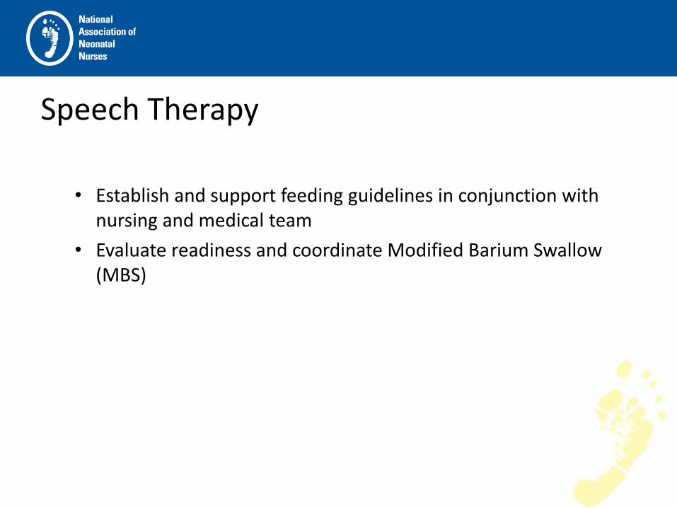
Speech Therapy
• Establish and support feeding guidelines in conjunction with
nursing and medical team
• Evaluate readiness and coordinate Modified Barium Swallow (MBS)

Referral resources
• PCH NICU follow-up clinic
• DDD: division of Developmental Disabilities
• AZEIP: Arizona Early Intervention Program
• CRS: Children’s Rehabilitation Services
• FBC: Foundation for Blind Children

Common Neuro NICU diagnoses • HIE
• Seizures
• Neuromuscular diseases
• Metabolic/Genetic Disorders
• Meningomyelocoele
• Intraventricular hemmhorages (IVH)/Intracranial bleeds (ICH)
• CNS Malformations


Current Research • MRI with Spectroscopy
• NIRS evaluation
• MBS (swallow study evaluation)
• Placental pathology
• Autologous cord blood stem cell therapy

MRI with Spectrocopy
• Can we determine outcomes based on spectroscopy evaluation?
• Can we correlate abnormal MBS findings in the spectroscopy?

NIRS Evaluation
• Trending the NIRS with clinical presentation, MRI, and outcome

Dysphagia in Neonates Treated with Hypothermic Therapy For HIE
Cristina Carballo, MD, P. David Adelson, MD, Kimberlee Allred, RN NNP-BC, Pamela Clarke-Levens, MS, CCC-SLP Paula Kinnard, RN
Neuro NICU, Phoenix Children’s Hospital (PCH), Children’s Neuroscience Institute, PCH, Phoenix, Arizona
Neonatology Associates Ltd

Abstract Introduction:. Hypoxic Ischemic Encephalopathy (HIE) is a significant cause of brain injury in neonates. The impact of HIE has varying degrees of neurodevelopmental sequelae including dysphagia. Objectives: To document that infants with moderate to severe HIE, who have received hypothermia treatment as standard of care, are at risk for dysphagia. Methods: A retrospective chart review over a one year period of patients with HIE whom received hypothermia therapy was done. Modified Barium Swallow (MBS) studies were performed on this patient population. A comparison between abnormal and normal MBS study results were evaluated.

Abstract Continued • Results: There were a total of 32 patients in this chart review.
Of the 32 patients, 23 had MBS studies done. Eighteen of 23 MBS were abnormal. This demonstrates a 78% abnormal rate in MBS in infants post hypothermia therapy for treatment of HIE.
• Conclusions: At this point, in the HIE population, using a specific feeding protocol as well as the inclusion of a MBS with Speech Therapy involvement, would be beneficial for optimum management of these high-risk infants.

Introduction Hypoxic Ischemic Encephalopathy (HIE)
Significant cause of brain injury in neonates ( 1-2/1000 term live births) Results in varying degrees of neurodevelopmental disabilities, including dysphagia
Treatment with hypothermia Proven to be neuroprotective in moderate to severe HIE incidence of dysphagia in post-cooling therapy for HIE has not been studied

Objective To evaluate the MBS studies done on infants who had the diagnosis of HIE and also receive hypothermia therapy.
To document that infants with moderate to severe HIE, who have been treated with hypothermia treatment, are at high risk for dysphagia's

Subjects and Methods
Study Design - Retrospective chart review of neonatal cases of HIE evaluated at a pediatric stand-alone facility between Jan 2009 - Jan 2010. - Information gathered included MBS study results.

Subjects and Methods continued Inclusion Criteria Infants received hypothermia therapy who met the following criteria:
> 36 weeks old and under 6 hrs of age -Plus one of the following:
-Cord and or baby gas under 60 minutes with pH >7.0 or base deficit > -16 -Prolonged resuscitation, greater than ten minutes -Sarnat score 2-3 and/or seizures -Abnormal aEEG, indicating moderate to severe encephalopathy and/or seizures
-infants cooled for the full 72hr
Modified Barium Swallow study with speech therapist present

Subjects and Methods continued Exclusion Criteria -All congenital and or chromosomal anomalies including Trisomy 21 -Infants with HIE who were not given hypothermia therapy Statistical Analysis -Descriptive statistics to characterize the findings. Data was analyzed to evaluate the incidence of dysphagia -in infants with a diagnosis of HIE treated with hypothermia therapy.

Summary -Dysphagia was seen in the majority of HIE affected infants treated with hypothermia therapy. -The definition of dysphagia was an abnormal MBS study that showed laryngeal penetration and/or aspiration. -HIE patients, who were treated with hypothermia therapy, are at high risk for dysphagia. -Dysphagia was treated by thickening the feeds with rice cereal to the consistency deemed safe during the MBS study. All of the 18 infants with abnormal MBS were able to be nipple feed despite the severity of the dysphagia encountered on the MBS study.

Summary continued
-Conclusions from these analyses must be interpreted cautiously since this was a one year review of charts of infants post-hypothermia therapy for HIE.
-Trending repeat MBS studies of these infants would provide further insight as to the evolution of the dysphagia.

Discussion
-We started doing MBS studies on all our infants treated with hypothermia therapy for HIE after we began to notice soft clinical signs of feeding difficulties in some of these infants.
-The implications of post-hypothermia treated infants for HIE, with apparently normal neurological exams, are that they are at an unexpected high risk for dysphagia. The dysphagia would not have not been identified except through MBS studies. Since this potential for dysphagia exists, not identifying it could lead to other morbidities, including long-term or late onset respiratory problems.

Discussion continued
-Through this retrospective look of infants who had hypothermia therapy for HIE, we have realized that there exists an unexpectedly high risk for dysphagia in an otherwise neurologically normal appearing infant.
-Based on these findings, we feel that in the post-hypothermia population diagnosed with HIE, using a specific feeding protocol as well as the inclusion of a MBS with Speech Therapy involvement, would be beneficial for optimum management of these high-risk infants.

• Table 1. MBS Study Analysis
MBS n %
Normal 5/23 22%
Abnormal 18/23 78%

Figure 1. Prevalence of Dysphagia

Table 2. Severity of Dysphagia

Figure 2. Severity of Dysphagia

Placental pathology
• Evaluation of the placenta can identify
– An acute injury VS chronic injury
– Sepsis, funicitis
– Hematological issues
Cord tissue vs Cord blood • Cord tissue - a rich source of mesenchymal stem
cells. These create structural and connective tissue,
• Cord blood - a rich source of hematopoietic stem cells. These create the blood and immune system.
• Since cord blood and cord tissue are rich sources of different types of stem cells, they may help repair the body in different ways.


• Cord tissue is a rich source of mesenchymal stem cells, which create structural and connective tissue.
– These stem cells have unique properties that make them promising for cellular therapies.
– Stem cells from cord tissue have demonstrated the power to heal spinal cord, brain, and cartilage injuries in laboratory studies.
– This research is at an early stage and medical treatments are not available today.

Autologous cord blood: Baby’s own cord blood
• Cord blood is collected because it contains stem cells, including hematopoietic cells, which can be used to treat hematopoietic and genetic disorders.
• Stem cells are the body’s “master cells” because they are the building blocks of organ tissue, blood, and the immune system.

Cord Blood • Cord blood, like bone marrow, is an invaluable source
of a type of stem cell that can be used in a variety of medical treatments.
• To date, cord blood stem cells have been used to treat many life-threatening diseases, such as leukemia and other cancers.
• Cord blood is showing potential in research to treat conditions like brain injury.

• Cord blood stem cells are biologically younger and have unique qualities and advantages compared to other stem cell sources like bone marrow: – There is less risk of complications when used in transplants.
– They are immediately available, and early treatment can minimize disease progression.
– Freezing them "stops the clock" and protects them from environmental damage, age, and common viruses that will impact the stem cells in our bodies over time.
– Collection of cord blood is simple, safe, and painless.
• Cord blood stem cells are not embryonic stem cells and are not controversial.

In 2005, the National Academy of Sciences published an Institute of Medicine (IoM) report which recommended that expectant parents be given a balanced perspective on their options for cord blood banking. State legislatures around the country are introducing legislation intended to help inform physicians and expectant parents on the options for donating, discarding or banking lifesaving newborn stem cells. Currently 17 states, have enacted legislation recommended by the IoM guidelines.

• There are 2 basic ways to store cord blood:
– Public – less expensive, used in research
– Private - more expensive
The company provides the kit to obtain the sample and send to them. It is frozen in a cryogenic storage tank until it needs to be used.

Clinical Trials

Duke University “Autologous Cord Blood Cells for Hypoxic Ischemic Encephalopathy Study 1. Phase 1 Study of Feasibility and Safety.”
– Initiated January 2008
– Patient population <14 days meeting criteria for HIE
– Infusions: 4 infusions of own volume reduced cord blood cells.
– Infants will be have neurodevelopmental follow up at 4-6 and 9-12mos
– MRI’s will be obtained between postnatal weeks 1 and 4, and, for study purposes at 4-6 postnatal months
Results to date have shown the therapy to be safe and “encouraging.”

Medical College of Georgia
• Medical College of Georgia researchers are conducting the first FDA-approved clinical trial to determine whether an infusion of stem cells from umbilical cord blood can improve the quality of life for children with cerebral palsy. The trial
• Initiated February 2010.
• Evaluating 40 children ages 2-12 with the diagnosis of CP who have autogolous cord blood stored.

Phoenix Children’s Implementation:
(Awaiting IRB approval)
• 2 randomized groups. Treatment will be after second abnormal MRI (close to DOL 4) vs No treatment.
• A third MRI will be done at 6 months of age as well as a neurodevelopmental evaluation.
If MRI remains abnormal a second injection will be given.
• Each infant will receive no more than 2 injections.

IN ANIMAL STUDIES, newborn stem cells from cord blood demonstrate an ability to cross the blood-brain barrier and migrate to damaged tissue to induce healing. The left-hand photo represents normal, healthy tissue in a rat’s brain and shows no evidence of cord blood stem cell migration even after being infused with human cord blood stem cells. The right-hand photo represents diseased rat brain tissue with the green dots showing where the newborn stem cells from cord blood migrated in an effort to induce healing.
Seizure guidelines • Currently there is a lack of evidence based guidelines for the
treatment of neonatal seizures. • Majority of NICU’s use Phenobarbital as the first line (A 2009
retrospective analysis of 6099 neonates in 31 US hospitals were treated with Phenobarbital). There was more variability of further treatment.
• There are only 2 published RCT using the Phenobarbital as a first line agent.
• One study results showed that when used alone up to drug levels of 25mcg/ml, controlled seizures in less than half of the babies. The addition of Fosphenytoin increased the efficacy to 62%.
• The other study showed even less success with Phenobarbital, only 4/14 babies responding to Phenobarbital to doses of 40/kg.

• In fact, there is a landmark study published in 2002 that demonstrated the potential harm that high dose anticonvulsants can do in a neonatal brain.
– The rat brain is more susceptible to the neuro-apoptotic affect of the Phenobarbital during the neonatal period than at any other time.

Suggested guidelines Seizures (clinical and/or electrographic)
– Load with 0.1mg/kg Bumex (blocks the NaK2Cl channel which has been proposed to be the etiology of the neonatal chloride gradient reversal phenomena; suppressing epileptiform activity).
– Load with 20mg/kg Phenobarbital and 30mg/kg Keppra (Phenobarbital at lower serum levels, not toxic. Keppra is a broad spectrum anticonvulsant with several case reports describing the efficacy in neonatal seizures.

Other adjunctive seizure treatment considerations:
• Magnesium at high end of normal
• Glucose and oxygen optimization
• Lidocaine drip

Goals for Seizure Treatment
• Minimize the time from seizure onset to treatment
• Minimize exposure to Phenobarbital
• Decrease duration of seizures
• Increase the efficacy of acute treatment
• Prophylaxis with non-apoptotic medication
Summary
• Hypothermic Therapy (head or whole body cooling) has shown an improvement in Neurodevelopmental outcomes.
• A Neuro NICU program implementation encourages a collaborative effort for patient follow up.
• Continued therapies and research could provide predictability of outcomes.

Conclusion
• Development of a Neuro NICU allows us to more effectively identify and treat brain injury/pathology.
• Earlier and more aggressive treatment of neonatal brain injury/pathology, whatever the etiology, will result in improved long term neurodevelopmental outcomes.
• We are a Center of Excellence in the treatment of neonatal brain pathology/injury in the Southwest and the USA

References Cochrane Database of Systematic Reviews, Issue 4, 2003 (Jacobs 2003)
Hau S, Reich DM, Scholz M, et al. “Evidence for neuroprotective properties of human umbilical cord blood cells after neuronal hypoxia in vitro.” BMC Neurosci. 2008;9:30.
Jacobs, Hunt, Tarnow-Mordi and Inder. “Cooling for newborns with hypoxic ischaemic encephalopathy”. 08/20/2007
Naqeeb, et al. Pediatrics 1999:103:1263
Natus Medical Cerebral Function Monitor 6000.
Neuhoff S, Moers J, Rieks M, et al. “Proliferation, differentiation, and cytokine secretion of human umbilical cord blood-derived mononuclear cells in vitro.” Exp Hematol. 2007;35(7):1119-1131
Olympic Medical Cool-Cap System
Phoenix Children’s Hospital protocol on Cool-Cap
Phoenix Children’s Hospital protocol on Cooling Blanket
Rocha V, Wagner JE, Jr., Sobocinski KA, et al. “Graft-versus-host disease in children who have received a cord-blood or bone marrow transplant from an HLA-identical sibling.” Eurocord and International Bone Marrow Transplant Registry Working Committee on Alternative Donor and Stem Cell Sources. N Engl J Med. 2000;342(25):1846-1854.
Thorenson, Simmonds, Satas, Tooley and Silver. “Effective head cooling during posthypoxic hypothermia in newborn piglets.” Pedatri. Res. 49, 594-599 (2001).

References Gunn AJ. Cerebral hypothermia for prevention of brain injury following perinatal asphyxia. Curr Opin. Pediatric 2000; 12: 111-115 Thornberg E., Thirimyer K, Odebuck A, Milsom I. Birth asphyxia: incidence, clinical course and outcome in a Swedish population. Acta Paediatr 1995; 84: 927-932 Dixon G, Badawi N, Kurinezvk JJ, Keogh JM, Stilborn Sr, Zubrick Sr, etal. Early developmental outcomes after newborn encephalopathy. Pediatrics 2002; 109: 26-33 Wilkinson D, Trevor D, etal. Hypothermia: A Neuroprotective Therapy for Neonatal Hypoxic-Ischemic Encephalopathy. Pediatrics 2007; 119: 422-423 Zanelli SA, Naylor M, Dobbins N, Yuigg M, Goodkin HP, Matsnoto SA, Fairchild KD. Implementation of a “Hypothermia for HIE program: 2 year experience in a single NICU”, Journal of Perinatology 2008; 28: 171-175 Shankaran S, etal. Whole Body Hypothermia for Neonates with Hypoxic Ischemic Encephalopathy. NEJM 2005; 353: 1574-1584 Gluckman P, Wyatt J, etal. Selective head cooling with mild systemic hypothermia after neonatal encephalopathy: multicentre randomized trial. The Lancet 2005; 365: 663-670 Higgins R, Tonse NK, etal. Hypothermia and Perinatal Asphyxia: Executive Summary of the National Institute of Child Health and Human Development Workshop. Journal of Pediatrics 2006; 148: 170-175 Kirplani H, Burks J, etal. Cooling for Neonatal Hypoxic Ischemic Encephalopathy: Do we have the answer? Pediatrics 2007; 120: 1126-1130 Morgan A., Ward E., Murdoch B., etal. 2003. Incidence, characteristics and predictive factors for dysphagia after pediatric traumatic brain injury. Journal of Head Trauma & Rehabilitation, 18 (3): 239-251 Neurology of the Newborn. Joseph J. Volpe. Fifth edition 2008; pages 430-431
www.emedicine.com
www.fda.gov
www.clinicaltrials.gov
www.natus.com

A special thank you to the families who gave permission for their stories to be shared and
who shared their families with us.

THANK YOU!!!!
QUESTIONS?????

Save the Date
Oct 18-20, 2012 - Palm Springs Convention Center
and Renaissance Hotel - Palm Springs, CA