(i) periplaneta americana. - betsy coul -...
TRANSCRIPT

Class XI Chapter 7– Structural Organisation in Animals Biology
Page 1 of 11 Website: www.vidhyarjan.com Email: [email protected] Mobile: 9999 249717
Head Office: 1/3-H-A-2, Street # 6, East Azad Nagar, Delhi-110051
(One Km from ‘Welcome Metro Station)
Question 1:
Answer in one word or one line.
(i) Give the common name of Periplaneta americana.
(ii) How many spermathecae are found in earthworm?
(iii) What is the position of ovaries in the cockroach?
(iv) How many segments are present in the abdomen of cockroach?
(v) Where do you find malphigian tubules?
Answer
(i) The common name of Periplaneta americana is the American cockroach.
(ii) Four pairs of spermathecae are present in earthworms. They are located
between sixth and the ninth segments. They help in receiving and storing the
spermatozoa during copulation.
(iii) In a cockroach, the pair of ovaries is located between 12th and 13th abdominal
segments.
(iv) In both sexes, the abdomen of a cockroach consists of ten segments.
(v) Malphigian tubules are main excretory organs of cockroaches. They form a part
of the alimentary canal.
Question 2:
Answer the following:
(i) What is the function of nephridia?
(ii) How many types of nephridia are found in earthworm based on their location?
Answer
(i) Nephridia are segmentally arranged excretory organs present in earthworms.
(ii) On the basis of their location, three types of nephridia are found in earthworms.
They are:
(a) Septal nephridia: These are present on both sides of the inter-segmental septa
behind the 15th segment. They open into the intestines.
(b) Integumentary nephridia: These lie attached to the body wall from the third
segment to the last segment, which opens on the body surface.

Class XI Chapter 7– Structural Organisation in Animals Biology
Page 2 of 11 Website: www.vidhyarjan.com Email: [email protected] Mobile: 9999 249717
Head Office: 1/3-H-A-2, Street # 6, East Azad Nagar, Delhi-110051
(One Km from ‘Welcome Metro Station)
(c) Pharyngeal nephridia: These are present as three paired tufts in fourth, fifth, and
sixth segments.
Question 3:
Draw a labelled diagram of the reproductive organs of an earthworm.
Answer
Question 4:
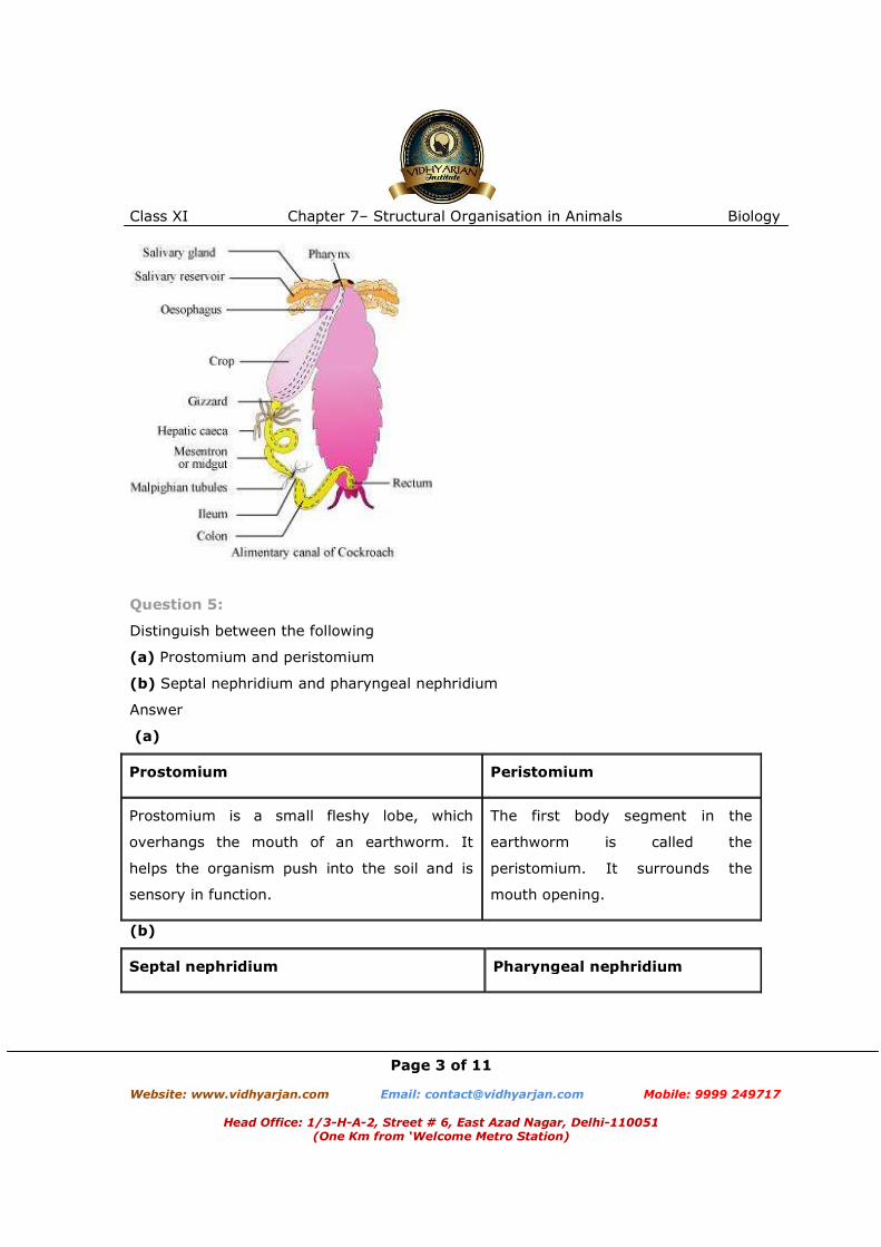
Draw a labelled diagram of alimentary canal of a cockroach.
Answer
Class XI Chapter 7– Structural Organisation in Animals Biology
Page 3 of 11 Website: www.vidhyarjan.com Email: [email protected] Mobile: 9999 249717
Head Office: 1/3-H-A-2, Street # 6, East Azad Nagar, Delhi-110051
(One Km from ‘Welcome Metro Station)
Question 5:
Distinguish between the following
(a) Prostomium and peristomium
(b) Septal nephridium and pharyngeal nephridium
Answer
(a)
Prostomium Peristomium
Prostomium is a small fleshy lobe, which
overhangs the mouth of an earthworm. It
helps the organism push into the soil and is
sensory in function.
The first body segment in the
earthworm is called the
peristomium. It surrounds the
mouth opening.
(b)
Septal nephridium Pharyngeal nephridium
Class XI Chapter 7– Structural Organisation in Animals Biology
Page 4 of 11 Website: www.vidhyarjan.com Email: [email protected] Mobile: 9999 249717
Head Office: 1/3-H-A-2, Street # 6, East Azad Nagar, Delhi-110051
(One Km from ‘Welcome Metro Station)
They are present on both sides of inter-
segmental septa behind the 15th segment.
They open into the intestines.
They are present as three paired
tufts in the fourth, fifth, and sixth
segments.
Question 6:
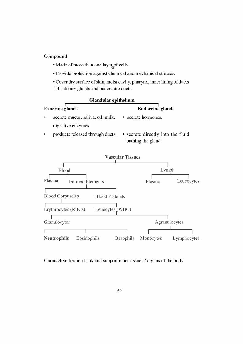
What are the cellular components of blood?
Answer
Components of blood include erythrocytes (RBCs), leucocytes (WBCs), and
thrombocytes (platelets). These components form 45% of blood. They are
suspended in the remaining fluid portion, called plasma.
Mammalian erythrocytes are biconcave, coloured cells devoid of a nucleus. They help
in transporting respiratory gases.
Leucocytes or white blood cells are nucleated cells. They can be divided into two
types, granulocytes (neutrophils, eosinophils, and basophils) and agranulocytes
(lymphocytes and monocytes). They help fight against various disease-causing
germs entering the body.
Thrombocytes are cell fragments produced from megarkaryocytes of the bone. They
play a major role during blood coagulation.
Question 7:
What are the following and where do you find them in animal body
(a) Chondriocytes
(b) Axons
(c) Ciliated epithelium
Answer
Chondriocytes:
They are cells of cartilages, and are present in small cavities within the matrix
secreted by them.
Axons:
Class XI Chapter 7– Structural Organisation in Animals Biology
Page 5 of 11 Website: www.vidhyarjan.com Email: [email protected] Mobile: 9999 249717
Head Office: 1/3-H-A-2, Street # 6, East Azad Nagar, Delhi-110051
(One Km from ‘Welcome Metro Station)
They are long, slender projections of neurons that help in carrying nerve impulses
from the neuron body. Axons aggregate in bundles which make up the nerves.
Ciliated epithelium:
It consists of simple columnar or cuboidal epithelium with cilia on their free surfaces.
It is present on the inner surface of the oviducts and bronchioles. It helps in the
movement of eggs or mucus in specific directions.
Question 8:
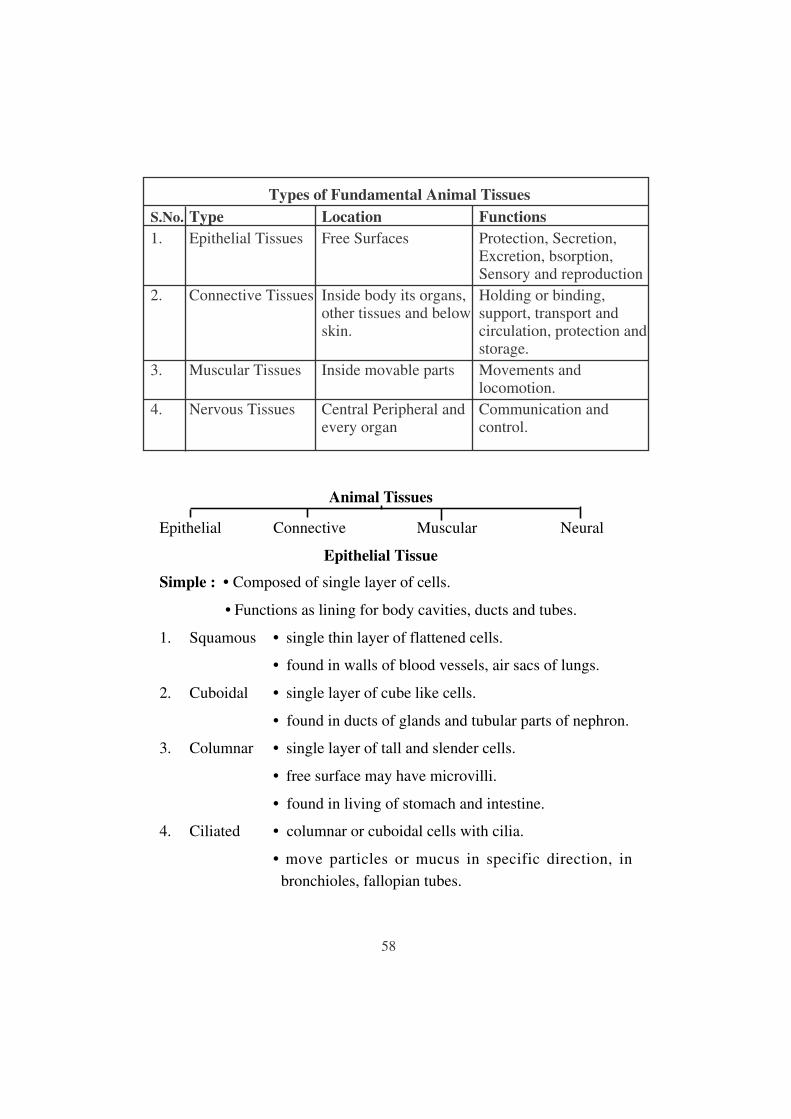
Describe various types of epithelial tissues with the help of labelled diagrams.
Answer
Epithelial tissue lines the surface of a body and forms a protective covering.
Epithelium cells are packed tightly together with little intercellular matrix. Epithelial
tissue in the body is of two types.
(a) Simple epithelium: It consists of a single layer of cells where cells are in direct
contact with the basement membrane. It is further sub-divided into the following
types:
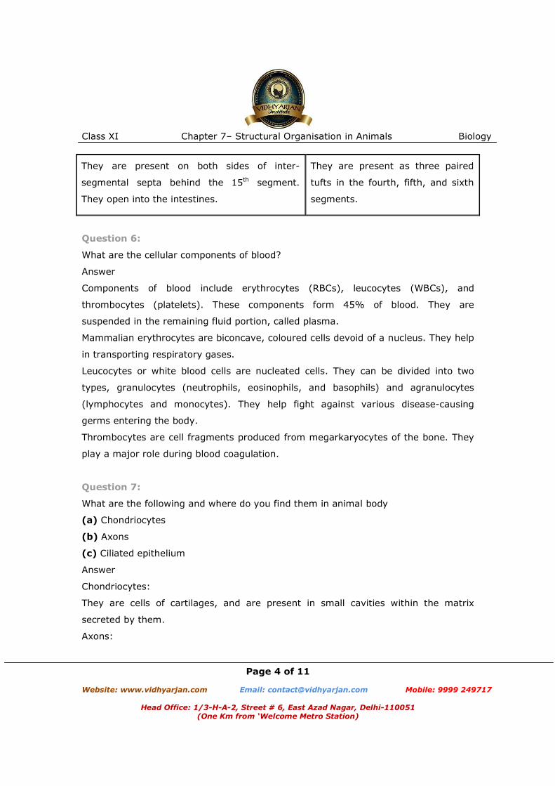
(i) Simple squamous epithelium: It consists of a single layer of flat cells with
irregular boundaries. It is found in the walls of the blood vessels and in the lining of
alveoli.
(ii) Simple cuboidal epithelium: It consists of a single layer of cube-like cells. It is
present in regions where secretion and absorption of substances takes place such as
the proximal convoluted tubule region of the nephron.
(iii) Simple columnar epithelium: It consists of a single layer of tall, slender cells
with their nuclei present at the base of the cells. They may bear micro-villi on the
free surfaces. Columnar epithelium forms the lining of the stomach and intestines,
and is involved in the function of secretion and absorption.
(iv) Ciliated epithelium: It consists of columnar or cuboidal cells with cilia on their
free surfaces. They are present in bronchioles and oviducts from where they direct
mucus and eggs in specific directions.
Class XI Chapter 7– Structural Organisation in Animals Biology
Page 6 of 11 Website: www.vidhyarjan.com Email: [email protected] Mobile: 9999 249717
Head Office: 1/3-H-A-2, Street # 6, East Azad Nagar, Delhi-110051
(One Km from ‘Welcome Metro Station)
(v) Glandular epithelium:It consists of columnar or cuboidal cells involved in the
secretion of substances. Glands are of two types, unicellular glands (goblet cells of
the alimentary canal) and multicellular glands (salivary glands). They can be
classified as exocrine (ductless glands) and endocrine glands (duct glands) by the
method through which they release enzymes.
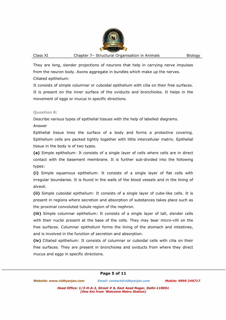
(b)
Compound epithelium: It consists of many layers of cells. It is involved mainly in the
function of providing protection and has a limited role in secretion and absorption.
Examples of compound epithelium include the dry surface of the skin or moist inner
lining of the buccal cavity, pharynx, pancreatic ducts, and the inner lining of ducts of
salivary glands.
Question 9:
Distinguish between
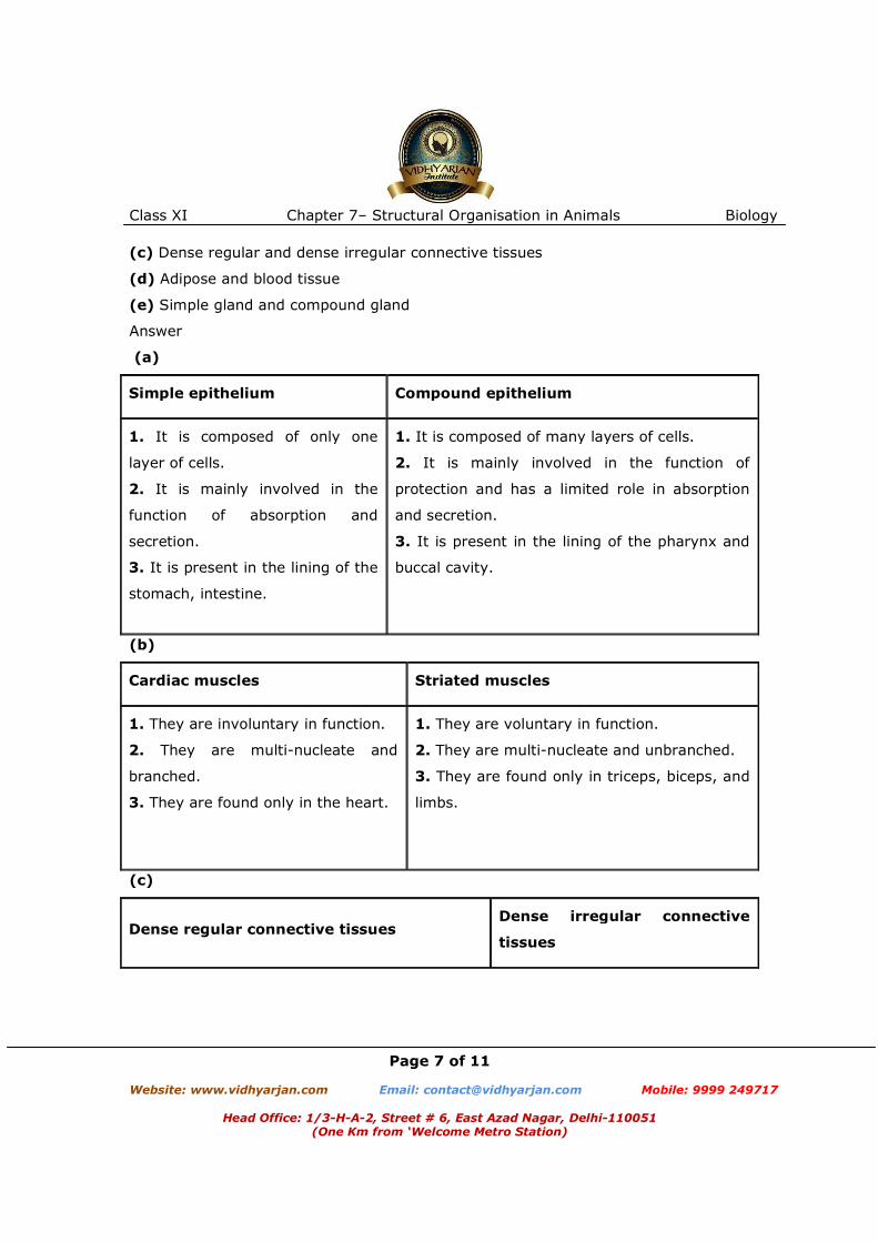
(a) Simple epithelium and compound epithelium.
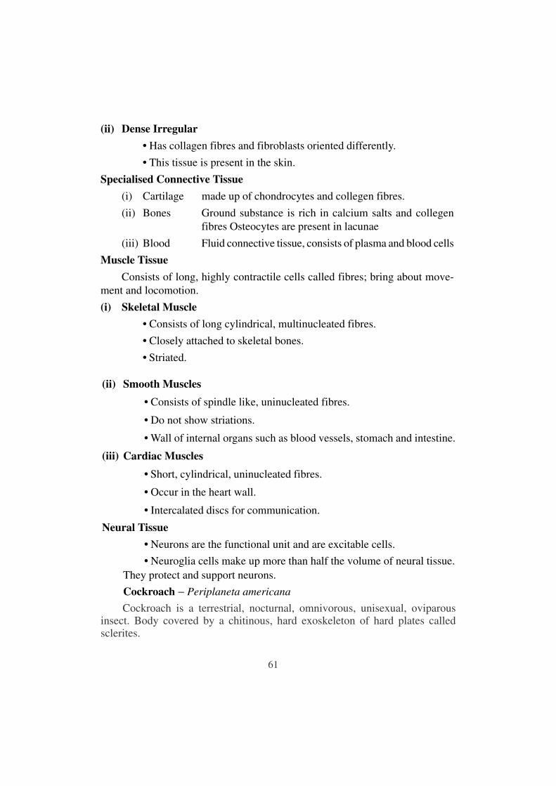
(b) Cardiac muscle and striated muscle
Class XI Chapter 7– Structural Organisation in Animals Biology
Page 7 of 11 Website: www.vidhyarjan.com Email: [email protected] Mobile: 9999 249717
Head Office: 1/3-H-A-2, Street # 6, East Azad Nagar, Delhi-110051
(One Km from ‘Welcome Metro Station)
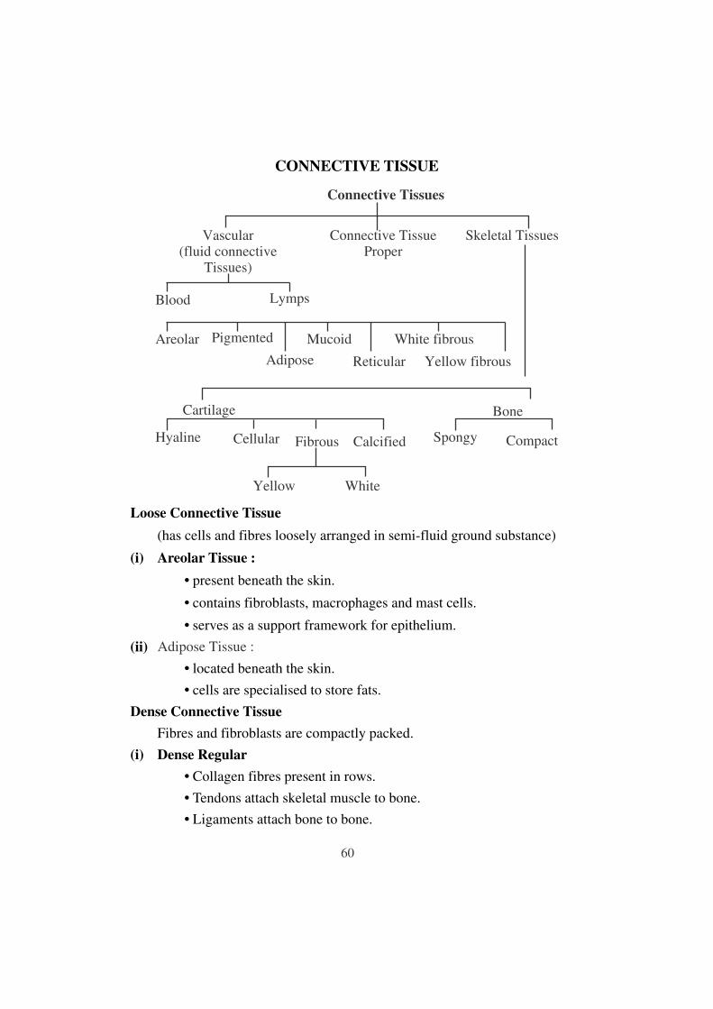
(c) Dense regular and dense irregular connective tissues
(d) Adipose and blood tissue
(e) Simple gland and compound gland
Answer
(a)
Simple epithelium Compound epithelium
1. It is composed of only one
layer of cells.
2. It is mainly involved in the
function of absorption and
secretion.
3. It is present in the lining of the
stomach, intestine.
1. It is composed of many layers of cells.
2. It is mainly involved in the function of
protection and has a limited role in absorption
and secretion.
3. It is present in the lining of the pharynx and
buccal cavity.
(b)
Cardiac muscles Striated muscles
1. They are involuntary in function.
2. They are multi-nucleate and
branched.
3. They are found only in the heart.
1. They are voluntary in function.
2. They are multi-nucleate and unbranched.
3. They are found only in triceps, biceps, and
limbs.
(c)
Dense regular connective tissues Dense irregular connective
tissues

Class XI Chapter 7– Structural Organisation in Animals Biology
Page 8 of 11 Website: www.vidhyarjan.com Email: [email protected] Mobile: 9999 249717
Head Office: 1/3-H-A-2, Street # 6, East Azad Nagar, Delhi-110051
(One Km from ‘Welcome Metro Station)
1. In dense regular connective tissues, collagen
fibres are present in rows between parallel
boundless fibres.
2. They are present in tendons and ligaments.
1. In dense irregular connective
tissues, fibres are arranged
irregularly.
2. They are present in the skin.
(d)
Adipose tissue Blood tissue
1. It is composed of collagen fibres, elastin
fibres, fibroblasts, macrophages, and
adipociytes.
2. It helps in the synthesis, storage, and
metabolism of fats.
3. It is present beneath the skin.
1. It is composed of RBCs, WBCs,
platelets, and plasma.
2. It helps in the transportation of
food, wastes, gases, and hormones.
3. It is present in the blood vessels.
(e)
Simple glands Compound glands
1. They contain isolated glandular cells.
2. They are unicellular.
3. Examples include goblet cells of the
alimentary canal.
1. They contain a cluster of
secretory cells.
2. They are multicellular.
3. Examples include salivary glands.
Question 10:
Mark the odd one in each series:
(a) Areolar tissue; blood; neuron; tendon
Class XI Chapter 7– Structural Organisation in Animals Biology
Page 9 of 11 Website: www.vidhyarjan.com Email: [email protected] Mobile: 9999 249717
Head Office: 1/3-H-A-2, Street # 6, East Azad Nagar, Delhi-110051
(One Km from ‘Welcome Metro Station)
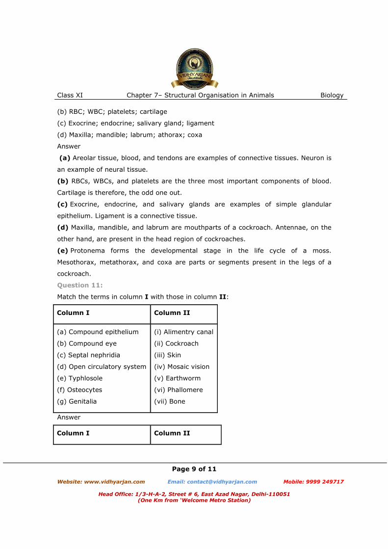
(b) RBC; WBC; platelets; cartilage
(c) Exocrine; endocrine; salivary gland; ligament
(d) Maxilla; mandible; labrum; athorax; coxa
Answer
(a) Areolar tissue, blood, and tendons are examples of connective tissues. Neuron is
an example of neural tissue.
(b) RBCs, WBCs, and platelets are the three most important components of blood.
Cartilage is therefore, the odd one out.
(c) Exocrine, endocrine, and salivary glands are examples of simple glandular
epithelium. Ligament is a connective tissue.
(d) Maxilla, mandible, and labrum are mouthparts of a cockroach. Antennae, on the
other hand, are present in the head region of cockroaches.
(e) Protonema forms the developmental stage in the life cycle of a moss.
Mesothorax, metathorax, and coxa are parts or segments present in the legs of a
cockroach.
Question 11:
Match the terms in column I with those in column II:
Column I Column II
(a) Compound epithelium
(b) Compound eye
(c) Septal nephridia
(d) Open circulatory system
(e) Typhlosole
(f) Osteocytes
(g) Genitalia
(i) Alimentry canal
(ii) Cockroach
(iii) Skin
(iv) Mosaic vision
(v) Earthworm
(vi) Phallomere
(vii) Bone
Answer
Column I Column II

Class XI Chapter 7– Structural Organisation in Animals Biology
Page 10 of 11 Website: www.vidhyarjan.com Email: [email protected] Mobile: 9999 249717
Head Office: 1/3-H-A-2, Street # 6, East Azad Nagar, Delhi-110051
(One Km from ‘Welcome Metro Station)
(a) Compound epithelium
(b) Compound eye
(c) Septal nephridia
(d) Open circulatory system
(e) Typhlosole
(f) Osteocytes
(g) Genitalia
(iii) Skin
(iv) Mosaic vision
(v) Earthworm
(ii) Cockroach
(i) Alimentary canal
(vii) Bone
(vi) Phallomere
Question 12:
Mention briefly about the circulatory system of earthworm
Answer
Earthworms (Pheretima) have closed blood vascular systems, which consists of the
heart, blood vessels, and capillaries. The heart pumps blood for circulating it in one
direction. Blood is supplied by smaller blood cells to the gut nerve cord and the body
wall. Blood glands are present in the 4th, 5th, and 6th segments, which produce blood
cells and haemoglobin dissolved in blood plasma. Blood cells in earthworms are
phagocytic in nature.
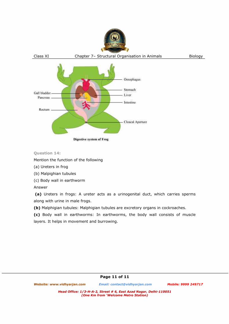
Question 13:
Draw a neat diagram of digestive system of frog.
Answer
Class XI Chapter 7– Structural Organisation in Animals Biology
Page 11 of 11 Website: www.vidhyarjan.com Email: [email protected] Mobile: 9999 249717
Head Office: 1/3-H-A-2, Street # 6, East Azad Nagar, Delhi-110051
(One Km from ‘Welcome Metro Station)
Question 14:
Mention the function of the following
(a) Ureters in frog
(b) Malpighian tubules
(c) Body wall in earthworm
Answer
(a) Ureters in frogs: A ureter acts as a urinogenital duct, which carries sperms
along with urine in male frogs.
(b) Malphigian tubules: Malphigian tubules are excretory organs in cockroaches.
(c) Body wall in earthworms: In earthworms, the body wall consists of muscle
layers. It helps in movement and burrowing.