hypothalamo-pituitary disorders · hypothalamo-pituitary disorders ... adrenal cortex, gonads and,...
TRANSCRIPT
INTRODUCTION
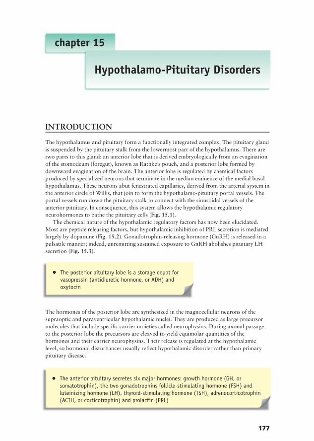
The hypothalamus and pituitary form a functionally integrated complex. The pituitary glandis suspended by the pituitary stalk from the lowermost part of the hypothalamus. There aretwo parts to this gland: an anterior lobe that is derived embryologically from an evaginationof the stomodeum (foregut), known as Rathke’s pouch, and a posterior lobe formed bydownward evagination of the brain. The anterior lobe is regulated by chemical factorsproduced by specialized neurons that terminate in the median eminence of the medial basalhypothalamus. These neurons abut fenestrated capillaries, derived from the arterial system inthe anterior circle of Willis, that join to form the hypothalamo-pituitary portal vessels. Theportal vessels run down the pituitary stalk to connect with the sinusoidal vessels of theanterior pituitary. In consequence, this system allows the hypothalamic regulatoryneurohormones to bathe the pituitary cells (Fig. 15.1).
The chemical nature of the hypothalamic regulatory factors has now been elucidated.Most are peptide releasing factors, but hypothalamic inhibition of PRL secretion is mediatedlargely by dopamine (Fig. 15.2). Gonadotrophin-releasing hormone (GnRH) is released in apulsatile manner; indeed, unremitting sustained exposure to GnRH abolishes pituitary LHsecretion (Fig. 15.3).
The hormones of the posterior lobe are synthesized in the magnocellular neurons of thesupraoptic and paraventricular hypothalamic nuclei. They are produced as large precursormolecules that include specific carrier moieties called neurophysins. During axonal passageto the posterior lobe the precursors are cleaved to yield equimolar quantities of thehormones and their carrier neurophysins. Their release is regulated at the hypothalamiclevel, so hormonal disturbances usually reflect hypothalamic disorder rather than primarypituitary disease.
177
chapter 15
Hypothalamo-Pituitary Disorders
● The posterior pituitary lobe is a storage depot forvasopressin (antidiuretic hormone, or ADH) andoxytocin
● The anterior pituitary secretes six major hormones: growth hormone (GH, orsomatotrophin), the two gonadotrophins follicle-stimulating hormone (FSH) andluteinizing hormone (LH), thyroid-stimulating hormone (TSH), adrenocorticotrophin(ACTH, or corticotrophin) and prolactin (PRL)
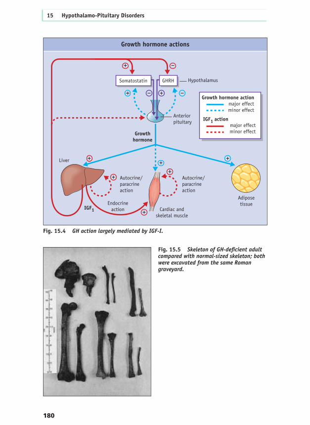
All the hormones of the anterior lobe except PRL act largely through target glands –including the thyroid, adrenal cortex, gonads and, in the case of GH, the liver, whichproduces IGF-I (Fig. 15.4). GH also stimulates other organs to produce IGF-I, which acts ina local paracrine fashion.
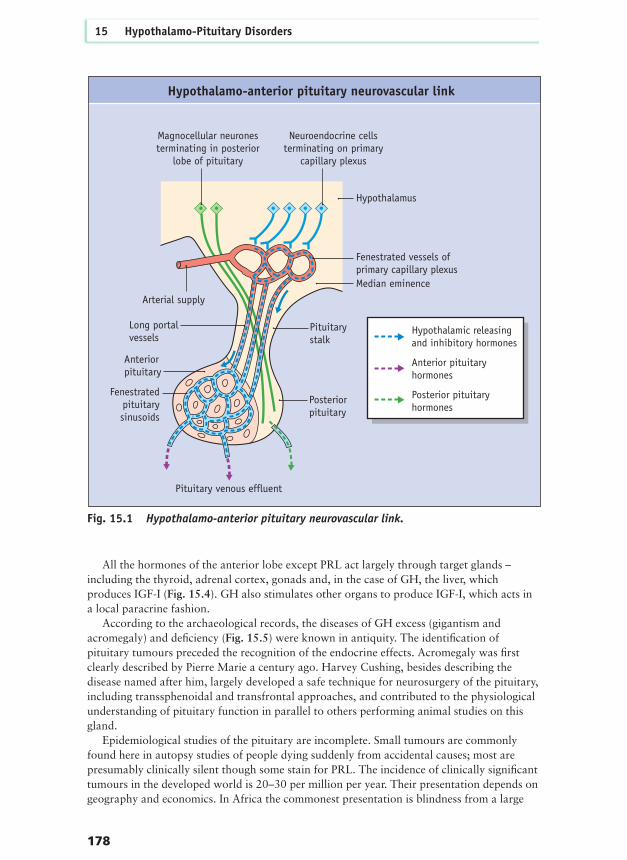
According to the archaeological records, the diseases of GH excess (gigantism andacromegaly) and deficiency (Fig. 15.5) were known in antiquity. The identification ofpituitary tumours preceded the recognition of the endocrine effects. Acromegaly was firstclearly described by Pierre Marie a century ago. Harvey Cushing, besides describing thedisease named after him, largely developed a safe technique for neurosurgery of the pituitary,including transsphenoidal and transfrontal approaches, and contributed to the physiologicalunderstanding of pituitary function in parallel to others performing animal studies on thisgland.
Epidemiological studies of the pituitary are incomplete. Small tumours are commonlyfound here in autopsy studies of people dying suddenly from accidental causes; most arepresumably clinically silent though some stain for PRL. The incidence of clinically significanttumours in the developed world is 20–30 per million per year. Their presentation depends ongeography and economics. In Africa the commonest presentation is blindness from a large
178
15 Hypothalamo-Pituitary Disorders
Magnocellular neuronesterminating in posterior
lobe of pituitary
Hypothalamus
Neuroendocrine cellsterminating on primary
capillary plexus
Fenestrated vessels ofprimary capillary plexusMedian eminence
Long portal vessels
Pituitarystalk
Pituitary venous effluent
Posteriorpituitary
Fenestratedpituitarysinusoids
Arterial supply
Hypothalamic releasing and inhibitory hormones
Anterior pituitaryhormones
Posterior pituitaryhormones
Anterior pituitary
Hypothalamo-anterior pituitary neurovascular link
Fig. 15.1 Hypothalamo-anterior pituitary neurovascular link.

Introduction
179
Prolactin TSH
Synergy
Hypothalamic releasing factorsMajor minor stimulation
Anterior pituitary hormones
Hypothalamic inhibitory factorsMajor minor stimulation
Ghrelin
Growthhormone
ACTH/POMC
Somato-statin
FSH LH
GnRHGHRH
Synergy
Dopamine TRH CRH ADH
Hypothalamic releasing/inhibitory factors andanterior pituitary hormone responses
Pulsatile Continuous Pulsatile
LH(n
g/m
l)
FSH
(ng/
ml)
0
5
10
15
20
10 5 0 5 10 15 20 25 30 350
Days
999200
150
100
50
Tonic secretion of pituitary gonadotrophin
Fig. 15.2 Hypothalamic releasing/inhibitory factors and anterior pituitary hormoneresponses.
Fig. 15.3 Differential influence of pulsatile versus sustained GnRH administration on LHrelease (modified from Belchetz et al. 1978 Science 202: 631–633, with permission).
180
15 Hypothalamo-Pituitary Disorders
Growth hormone actions
Hypothalamus
Anteriorpituitary
Growthhormone
IGF1Endocrine
action Cardiac andskeletal muscle
Autocrine/paracrine action
Autocrine/paracrine action
Adiposetissue
Growth hormone actionmajor effectminor effect
major effectminor effect
Liver
GHRHSomatostatin
IGF1 action
Fig. 15.4 GH action largely mediated by IGF-I.
Fig. 15.5 Skeleton of GH-deficient adultcompared with normal-sized skeleton; bothwere excavated from the same Romangraveyard.

tumour compressing the optic pathways. However, most tumours are functionless andpresent with features of a space-occupying lesion with various degrees of hypopituitarism.The recognition of hypersecreting lesions, especially GH and PRL and less frequently ACTH,gonadotrophins and TSH, follows from extra features that are detailed below. The impact ofthese disorders on mortality and morbidity is increasingly being recognized and defined.
PATHOGENESIS AND PATHOLOGY
PITUITARY TUMOURSPituitary tumours are rarely part of multiple endocrine neoplasia, type 1 (MEN-1) syndrome,which may be familial (see Ch. 23). The tumour suppressor gene for this syndrome locatedon chromosome 11 has been cloned and its common mutations resulting in MEN-1 havebeen identified. A constitutively active mutation of the stimulatory G protein (gsp) has beenpostulated as the cause of GH-secreting adenomas in up to 40% of cases. Rare familial casesof acromegaly have been recorded but so far the genetic abnormality has not been identifiedin non-MEN-1 cases. Aggressive tumours of the pituitary have been associated withabnormalities in a number of oncogenes and tumour suppressor genes (Fig. 15.6). It has longbeen noted that in many cases of Cushing’s disease either no discrete lesion can be found atsurgery or diffuse hyperplastic changes are noted. The blunted rather than fully resistantsuppression of cortisol production following dexamethasone administration, the exuberantACTH response to exogenous corticotrophin-releasing hormone (CRH, also called ACTH-releasing factor) and desmopressin together with the pathological features prompted earlyideas that hypothalamic secretion might be responsible. There are alternative explanations,however, such as constitutive activation in the pituitary receptors for releasing factors.Transgenic mice overexpressing the GHRH (GH-releasing hormone) gene display initialhyperplasia before developing tumours of somatroph cells. Also, most pituitary tumoursstudied are monoclonal, which suggests there is true neoplastic transformation.
The pathological characterization of pituitary tumours is based on classical haematoxylinand eosin staining and subsequent trichrome variants. There are three major categories:
Pathogenesis and pathology
181
● Acromegaly is estimated to have an incidence of 4–6new cases per million per year with a prevalence ofapproximately 40 per million. Overall mortality istwice normal but is reduced to near normal inpatients treated so that their mean GH averages lessthan 5 mU/L (2.5 ng/ml). The major causes of deathare cardiovascular, respiratory and cerebrovascular.There is an increased incidence and mortality fromcolonic cancer
Classification of pituitary tumours
● Chromophobe, often functionless tumours (Fig. 15.7a)● Eosinophilic, often producing GH or PRL (Fig. 15.7b)● Basophilic, particularly associated with Cushing’s disease (Fig. 15.76c)

Sensitive and specific immunostaining techniques reveal the existence of gonadotrophin-and TSH-secreting tumours, oncocytic tumours and functionless tumours staining forsynaptophysin and chromogranin. Immunostaining features do not invariably correlate withsecretory activity, however.
182
15 Hypothalamo-Pituitary Disorders
a b
c
Fig. 15.7 Histopathologicalclassification of pituitary tumoursaccording to haematoxylin and eosinstaining: (a) chromophobe, (b) eosinophilic and (c) basophilic.
Fig. 15.6 Oncogenes and tumour suppressor genes possibly related topituitary tumorigenesis
OncogenesGsP Mutations in Gs protein: 201 Arg → Cys/His or 227Gln → Arg/Leu ? 40% GH
secreting adenomas in Caucasian patients, fewer in JapaneseCREB ? facilitating role in somatotroph transformation and GH gene transcriptionRAS Found in metastases from pituitary carcinomas and aggressive prolactinomaPTTG Increased expression in many types, greater expression with more invasive
tumours
Tumour suppressor genes9p Early change in non-functional tumours, rare in somatotrophinomas9p21 Early change as above, via protein p16, interfering with cell cycle13q ? via RBI? close situated gene product. Somatotrophinomas, rare: non-
functional tumours10q23 Transition to invasive/metastatic phenotype, in all subtypes or pituitary tumour11q13 As above? via menin? closely situated gene product12p13 As above via p27? post-translational action on cell cycle regulation13q As above: all subtypes17p Via p53 as above, all subtypes17p21 Via m23 as above, all subtypes

The invasiveness of pituitary tumours varies greatly, but as with many endocrine tumoursthis characteristic correlates only poorly with observed features such as mitotic figures ornuclear pleomorphism. Dural invasion is often difficult to recognize at surgery but iscommonly the cause of recurrence. Pituitary carcinomas are extremely rare but examples aredocumented both for PRL-, GH-, ACTH- or TSH-secreting and for endocrinologicallyfunctionless tumours. They are defined by the presence of distant metastases within, or muchmore rarely outside, the CNS.
OTHER HYPOTHALAMO–PITUITARY DISORDERS For other pathologies in the hypothalamus-pituitary region a number of tests areappropriate:● Craniopharyngioma The presence of calcified cysts containing β-hCG
(beta human chorionic gonadotrophin)● Lymphocytic hypophysitis Lymphocytes for cd4 (cluster of differentiation 4)● Pituitary abscess Pus with evidence of bacterial infection● Cranial irradiation May be associated with cerebral vasculitis● Haemochromatosis Often marked iron deposition in the pituitary ● Infarction from apoplexy Yields pathological features which change with the
or Sheehan’s syndrome passage of time from the vascular catastrophe The application of electron microscopy can sometimes help define pathology while
cosecretion of more than one hormone by a single cell, suggested by immunostaining, can beconfirmed by demonstration of specific mRNA products.
INVESTIGATION AND DIAGNOSIS
Investigation of hypothalamo-pituitary disorders involves the assessment of endocrinefunction – whether deficient, excessive or mixed – and delineation of the anatomy, size andtopographical relations of any pituitary or parapituitary masses. Assessment of the effects ofa mass conventionally focuses on the visual pathways. Quality of life (QOL) evaluation andpsychometric testing are increasingly performed, especially in view of the long-termconsequences of surgery or other therapeutic interventions (Fig. 15.8). Recognition of adultGH deficiency syndrome prompts serial assessment of bone density and body composition aswell as cardiovascular risk factors such as lipid profile.
HORMONAL EVALUATION OF PITUITARY FUNCTIONBasal measurements adequately define most aspects of hormonal secretion, includingpituitary–thyroid and –gonadal axes, PRL secretion and water metabolism (Fig. 15.9). Thelarge variations in GH and ACTH/cortisol levels, which are determined by ultradian,circadian and stress-related mechanisms as well as classical negative feedback loops, oftenrequire dynamic testing (Fig. 15.10). Nevertheless a single 9 a.m. plasma cortisolmeasurement can provide adequate evidence of adrenal insufficiency if less than 180 nmol/L(6 µg/dl). Random GH measurements are useful only to exclude the diagnosis of acromegaly– this is unlikely if the value is less than 2 mU/L (1 ng/ml) – or for rough assessment of thedegree of GH hypersecretion in known acromegaly since there is a strong correlationbetween the fasting GH measurement and the mean of serial samples taken over periods upto 24 hours, notwithstanding the frequently observed pulsatility of GH release inacromegaly. Measurement of IGF-I provides an integrated perspective of overall GHsecretion. The level of IGF-I tends to be low in hypopituitarism even though it may lie withinthe age-related normal range in patients in whom stimulatory tests of GH reserve indicate
Investigation and diagnosis
183

184
15 Hypothalamo-Pituitary Disorders
Fig. 15.8 Psychometric consequences of pituitary disease and treatmentby transsphenoidal/transfrontal surgery and radiotherapy*
Treatment Transsphenoidal Transfrontal Medical Controlsurgery surgery treatment
Number 23 23 23 23Mean age (years) 42.7 41.6 38.7 38.9Years of education 10.9 11.3 11.5 11.6Estimated duration of 8.6 14.9 8.5 –illness (years)Estimated premorbid IQ 107.9 109.8 111.0 112.1
MeasuresPremorbid ability National Adult Reading TestAttention Digit subtest of Wechsler Adult Intelligence Scale (Revised)Memory Auditory – verbal learning test
Story recall – from Wechsler Memory ScaleRecognition – memory test for faces
Executive functions Controlled oral word association testBlock design subtest of Wechsler Adult Intelligence Scale (Revised)Trail-making test
Results Three or more tests below tenth percentileTranssphenoidal = 30.4%, Transfrontal = 43.5%, Medical = 21.7%, Control = 4.3%
Radiotherapy had NO adverse effect on cognitive function
*From Peace et al. Clinical Endocrinology 1998; 49(3): 391–396
Fig. 15.9 Basal blood tests for assessment ofpituitary function
Chemistry Hormones
Electrolytes (Na+/K+) Total/free T4 (and T3)Urea/creatinine TSHOsmolality ProlactinCalcium profile FSH/LHFasting glucose α-subunit (if glycoprotein-
producing tumour)Fasting triglycerides IGF-1Cholesterol Growth hormone (acromegaly)
HDL-C 9am cortisolLDL-C ACTH (Cushing’s disease)
Midnight cortisol (Cushing’s disease)Men: testosterone, SHBGWomen < 50 years: 17β oestradiol

Investigation and diagnosis
185
Fig. 15.10 Dynamic tests of hypothalamo-pituitary-adrenal function
Test Contraindication Precaution Protocol Interpretation
Insulin Frank Check 9 a.m.Fast patient Normal response:tolerance hypoadrenalism cortisol overnight and weigh. peak cortisoltest (ITT) ≥ 180 Insert venous cannula. ≥ 550 nmol/L
Epilepsy nmol/L Obtain blood for (18 µg/dl) if(6 µg/dl) plasma glucose and adequate
Cardiac ischaemia Check cortisol (and GH – hypoglycaemiaincluding patients history see below) at 0, 30, achieved > 70 years old Check 45, 60, 120 min. (≤ 2.2 mmol/L)– in whom IHD history and Inject 0.15 U soluble (40 mg/dl) +is likely 12 lead ECG insulin/kg appropriate
If patient insulin symptoms.resistant e.g. obese, NB Only testacromegalic, Cushings: validated clinicallydouble dose. – ability to If not hypoglycaemic withstand surgicalby 45 min stressreadminister initial dose and regard this as time zero.If severe response to hypoglycaemia viz coma, fits, chest pain: correct hypoglycaemia with i.v. glucose immediately but continue sampling for stress evoked responses
Glucagon Diabetes mellitus Prepare for Fast patient overnight Non-peak cortisoltest nausea and weigh. response
(30%) or Insert venous cannula. ≥ 500 nmol/Lvomiting Obtain blood for (17 µg/dl)(15%) plasma glucose, Post-glucagon
cortisol (and GH). subcutaneousInject glucagon injection peak time1 mg i.m. (if weight more variable,< 90 kg) hence need for1.5 mg (if weight multiple samples≥ 90 kg).Repeat blood samples at 150 and 180 min after glucagon i.m.injection.NB Classical subcutaneous test:(same doses butsamples at 0, 90, 120,150, 180, 210 and 240 min)
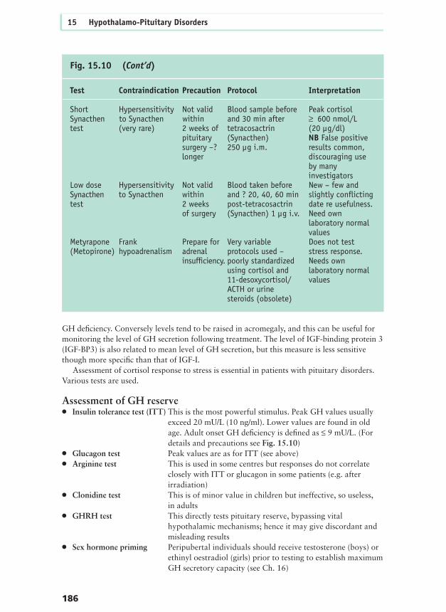
GH deficiency. Conversely levels tend to be raised in acromegaly, and this can be useful formonitoring the level of GH secretion following treatment. The level of IGF-binding protein 3(IGF-BP3) is also related to mean level of GH secretion, but this measure is less sensitivethough more specific than that of IGF-I.
Assessment of cortisol response to stress is essential in patients with pituitary disorders.Various tests are used.
Assessment of GH reserve● Insulin tolerance test (ITT) This is the most powerful stimulus. Peak GH values usually
exceed 20 mU/L (10 ng/ml). Lower values are found in old age. Adult onset GH deficiency is defined as ≤ 9 mU/L. (For details and precautions see Fig. 15.10)
● Glucagon test Peak values are as for ITT (see above)● Arginine test This is used in some centres but responses do not correlate
closely with ITT or glucagon in some patients (e.g. after irradiation)
● Clonidine test This is of minor value in children but ineffective, so useless, in adults
● GHRH test This directly tests pituitary reserve, bypassing vital hypothalamic mechanisms; hence it may give discordant and misleading results
● Sex hormone priming Peripubertal individuals should receive testosterone (boys) or ethinyl oestradiol (girls) prior to testing to establish maximum GH secretory capacity (see Ch. 16)
186
15 Hypothalamo-Pituitary Disorders
Fig. 15.10 (Cont’d)
Test Contraindication Precaution Protocol Interpretation
Short Hypersensitivity Not valid Blood sample before Peak cortisolSynacthen to Synacthen within and 30 min after ≥ 600 nmol/Ltest (very rare) 2 weeks of tetracosactrin (20 µg/dl)
pituitary (Synacthen) NB False positivesurgery –? 250 µg i.m. results common,longer discouraging use
by many investigators
Low dose Hypersensitivity Not valid Blood taken before New – few and Synacthen to Synacthen within and ? 20, 40, 60 min slightly conflictingtest 2 weeks post-tetracosactrin date re usefulness.
of surgery (Synacthen) 1 µg i.v. Need own laboratory normalvalues
Metyrapone Frank Prepare for Very variable Does not test (Metopirone) hypoadrenalism adrenal protocols used – stress response.
insufficiency. poorly standardized Needs own using cortisol and laboratory normal11-desoxycortisol/ valuesACTH or urine steroids (obsolete)

Assessment of vasopressin secretionFrank DI presents with thirst, polyuria including nocturia with the urinary volume exceeding3 litres over 24 hours, and with a serum sodium level of more than 145 mmol/L. Milderdegrees require evaluation with a water deprivation test including documenting response todesmopressin (DDAVP) (Fig. 15.11). On occasion, measuring plasma (arginine vasopressin,or AVP) responses to 5% saline infused at 0.04 ml/kg per minute for 2 hours will clarifyissues (Fig. 15.12).
IMAGING OF THE HYPOTHALAMO-PITUITARY REGIONMRI is the modality of choice in most cases. However, the presence of ferromagneticmaterial (e.g. some ocular prostheses) is an absolute contraindication. MRI is safe but mayprove claustrophobic, noisy and prolonged for some patients. Enhancement with gadoliniumvery rarely causes adverse reactions. Pituitary adenomas as small as 3 mm can be detected(Fig. 15.13). T1 and T2 signal characteristics inform about tumour qualities such as watercontent, haemorrhage and fibrosis. The optic chiasm is visualized well and its relationshipsto the pituitary well defined (Fig. 15.14). The posterior lobe normally appears as a
Investigation and diagnosis
187
Water only overnight, no caffeine or tobacco
9am Urine1: volume1, urine osmolality110am Urine2: volume2, urine osmolality211am Urine3: volume3, urine osmolality3
8am start water deprivation, empty bladder
Repeat hourly until 3 successive urineosmolalities differ by < 30 mosm/kg:
χ hour Urineχ: volumeχ, urine osmolalityχ
Inject desmopressin 2 µg im
χ+1 hour Urineχ+1: volumeχ+1, urine osmolalityχ+1
χ+2 hour Urineχ+2: volumeχ+2, urine osmolalityχ+2
Finish test: allow water and food
N.B. This modification minimizes test time till patient's maximum urinary concentration is achieved
weigh1
plasma osmolality1
weigh2
plasma osmolality2
• distinguishes partial from complete DI• distinguishes cranial from nephrogenic DI
Water deprivation testFig. 15.11 Waterdeprivation test forassessment of DI.

hyperintense signal (Fig. 15.15). The lateral relations of the pituitary show up well and giveevidence of abutment upon or invasion of cavernous sinuses (Fig. 15.16).
CT scanning is still useful – especially when access to MRI is limited or delayed. Pituitarytumours are almost as well imaged but chiasmal and cavernosal relations are less so. Bonyrelations including erosion are particularly well delineated (Fig. 15.17). The irradiation loadis high so efforts are made to avoid the eye lenses; this may involve the uncomfortable‘hanging head’ position to obtain coronal images. Dental fillings may cause streak artefacts.Contrast media give useful information but may be associated with severe reactions.
188
15 Hypothalamo-Pituitary Disorders
Plas
ma
Osm
alal
ity
Plas
ma
Osm
alal
ity
Normal osmolar threshold forthirst similar to AVP release
Note virtualinstantaneous
switch-off of thirst ondrinking thirst score
Note rapid AVP secretionswitch-off on drinking
preceding fall in osmolality
Thirst score (analogue scale 1–10)
Complete DI
Examples of neurosecretoryabnormalities of thirst
Examples of neurosecretoryabnormalities of AVP secretion
NormalAdipsic DI
Plasma AVP
Normal
Partial DIComplete DI
Ad libaccess to water
Hypertonic saline solution
Normal osmolarthreshold for AVP secretion
Partial DI
Plasma Osmality
Plasma Osmality
Hypertonic saline responses (a) thirst responses (b) plasma AVP levels
(a)
(b)
Fig. 15.12 AVP responses to hypertonic saline.

Calcified lesions such as craniopharyngiomas are well demonstrated, but MRI may also berequired to show the full extent of tumour (Fig. 15.18).
Angiography may be needed to display arterial anatomy – for instance if aneurysms aresuspected (Fig. 15.19). Ectatic loops of carotid arteries are often seen in acromegaly.Magnetic resonance angiography (MR-A) is of use, though has a lower exclusion value forsmall aneurysms than conventional angiography.
Investigation and diagnosis
189
a b
Fig. 15.13 MRI of pituitary microadenoma (a) before gadolinium – invisible, and(b) after – seen as hypointense area.
Fig. 15.14 MRI imaging of a pituitarytumour compressing the optic chiasm.
Fig. 15.15 MRI showing a high intensitysignal from the posterior lobe of thepituitary.

190
15 Hypothalamo-Pituitary Disorders
Fig. 15.16 MRI illustrating a pituitarytumour invading the cavernous sinus(enwrapping the intracavernosal carotidartery), (a) before and (b) after gadolinium.
Fig. 15.18 (a) and (b) MRI versus (c) and (d) CT scanning of craniopharyngioma.
Fig. 15.17 CT scan demonstrating bonyerosion by a pituitary tumour.
M31043-15-f17.tif
a b
c d
a b

INVESTIGATION OF ACROMEGALYA number of tests may be appropriate:● Random GH Values less than 2 mU/L (1ng/dl) exclude acromegaly● IGF-I Above the age-matched normal is suggestive – investigate
further● Oral GTT with GH The normal response is a fall to less than 2 mU/L (1ng/dl)
measurements (Fig. 15.20)● MRI Add, if necessary, MR-A or angiogram, and skull X-rays
(Fig. 15.21)● PRL● Thyroid function tests Plus a thyroid scan if goitre is present● Gonadotrophins and As clinically indicated
sex steroid measurements● ECG, chest X-ray Plus echocardiogram if there is hypertension● Serum calcium, phosphate, Plus if necessary X-ray of the renal tract
24 hour urinary calcium● Full blood count If there is anaemia, bowel symptoms, or a family history of
colon cancer add colonoscopy● Dorsolumbar spine X-ray, If there is backache, kyphosis, or loss of height
DEXA (dual-energy X-ray absorptiometry)
● Nerve conduction studies If there is neuropathy, especially carpaltunnel syndrome
● Sleep studies If there is snoring or daytime somnolence● Skin thickness measurement (Fig. 15.22)● Ring size measurement (Fig. 15.23)
INVESTIGATION OF CUSHING’S DISEASEIn patients with established Cushing’s syndrome (see Ch. 18) the following tests should beperformed:● Low dose dexamethasone test
Investigation and diagnosis
191
a b
Fig. 15.19 Cerebral aneurysms in proximity to the pituitary (containing coilsintroduced to thrombose them).

192
15 Hypothalamo-Pituitary Disorders
Minutes75 g
glucose p.o.
Acromegaly(paradoxical rise post-glucose)
Acromegaly(inadequate suppression post-glucose)
0
2
5
10
50
100
0 30 60 90 120
High basal < 2 mU/L
Normal
Plas
ma
GH
1h post-glucose
GH responses during GTT inacromegaly versus normal
Fig. 15.20 GTT showingGH responses in a patientwith acromegaly and in anormal individual.
Fig. 15.21 Skull X-ray of a patient withacromegaly showing an enlarged sella, `thickened calvarium, large frontal airsinuses, occipital bossing and prognathism.
Fig. 15.22 Skinfold measurements inacromegaly.

● CRH test ● Desmopressin test ● MRI scan● Inferior petrosal sinus sampling
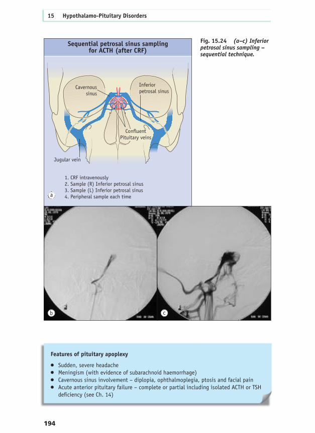
Petrosal sinus sampling is useful for differentiating pituitary from ectopic sources ofACTH secretion in Cushing’s syndrome. Catheters are introduced via the femoral veins andpassed under radiological screening up through the heart and jugular veins to the inferiorpetrosal sinus (IPS). Conventionally, a catheter is inserted each side and a further lineestablished for peripheral venous sampling. After central (right and left) and peripheral bloodsamples are taken, CRH 100 mg is injected intravenously. Repeat blood samples are takenfrom both IPSs and the peripheral vein three times over a 15 minute period following the CRHinjection for ACTH measurement (in EDTA tubes, sent to the laboratory on ice, rapidly spunand frozen; ensure accurate labelling). Rare but serious complications, including haematoma,thrombosis and brainstem lesions, have led to the development of sequential sampling of eachIPS after CRH injection (since the ACTH elevation from pituitary lesions is prolonged), as thishas lower morbidity (Fig. 15.24). If ACTH values from either IPS exceed twice thesimultaneous peripheral value a pituitary source is strongly suggested. Note though that the IPSside with peak ACTH values is not necessarily the side of the pituitary tumour.
CLINICAL FEATURES
PITUITARY TUMOURS
Headache, which is common with pituitary tumours even of modest size, is oftenattributed to pressure on the dura mater, especially the diaphragma sellae. However, rarely itis due to massive tumours causing raised intracranial pressure.
Pituitary infarction is commonly partial and may be asymptomatic but major episodescause pituitary apoplexy characterized by the following features:
Clinical features
193
Fig. 15.23 Ring size measurement inacromegaly.
Space-occupying features
● Headache● Visual disturbance● Hypopituitarism● Hydrocephalus, hypothalamic syndrome● Cerebrospinal fluid (CSF) leak● Epilepsy
194
15 Hypothalamo-Pituitary Disorders
Fig. 15.24 (a–c) Inferiorpetrosal sinus sampling –sequential technique.
Features of pituitary apoplexy
● Sudden, severe headache● Meningism (with evidence of subarachnoid haemorrhage)● Cavernous sinus involvement – diplopia, ophthalmoplegia, ptosis and facial pain● Acute anterior pituitary failure – complete or partial including isolated ACTH or TSH
deficiency (see Ch. 14)
Sequential petrosal sinus samplingfor ACTH (after CRF)
Jugular vein
Cavernoussinus
Inferiorpetrosal sinus
ConfluentPituitary veins
1. CRF intravenously2. Sample (R) Inferior petrosal sinus3. Sample (L) Inferior petrosal sinus4. Peripheral sample each time
Fig 15 24a
b c
a

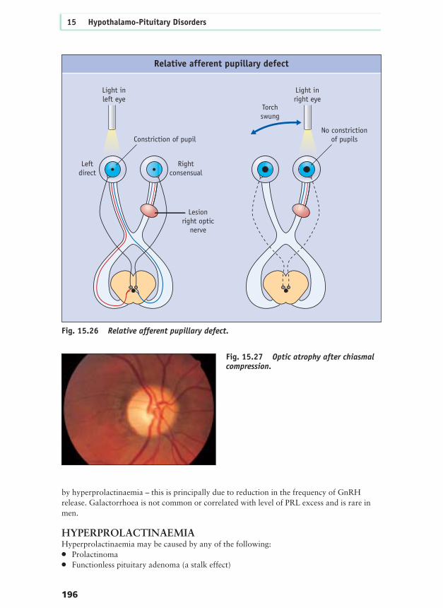
The most common visual disturbance is chiasmal compression. Classically this beginswith upper outer field loss progressing to bitemporal hemianopia or even complete blindness.Clinical testing by confrontation should begin with a red pin (Fig. 15.25) as loss of colourvision frequently long predates the inability to see finger movements. Other visual testsinclude the use of Ishihara colour charts and evidence of relative afferent pupillary defect(Fig. 15.26). Assessment of visual acuity should involve the reading of lines of print, as oftenletters become lost at the beginning and end of lines. Pituitary tumours may be asymmetricalwith striking differences in field defects between the right and left eyes. Fundoscopy oftenreveals optic atrophy with established chiasmal compression (Fig. 15.27); papilloedema israre. Ophthalmoplegia indicates cavernous sinus involvement.
Hypopituitarism due to pituitary tumour characteristically progresses sequentially:● GH deficiency. Is the first detectable feature● Gonadotrophin deficiency. Appears early on, with LH deficiency preceding that of FSH● PRL excess. Is often early but modest; it is due to stalk compression● ACTH deficiency. Is a late feature● TSH deficiency. Is usually last to develop● DI. If present at presentation this strongly suggests a parapituitary disorder
GROWTH HORMONE DEFICIENCYGH deficiency in childhood is dealt with in Chapter 16. Adult GH deficiency isdiagnostically valuable and clinically important (Fig. 15.28), since recombinant GH therapyis available. Gonadotrophin deficiency in premenopausal women is generally recognizedearly on account of primary or secondary amenorrhoea, infertility or loss of libido. Menoften delay presentation on developing impotence or loss of libido, though with widerrecognition of effective management of erectile dysfunction this pattern is reversing,provided other practitioners check for hormonal causes. Gonadal dysfunction is augmented
Clinical features
195
Visual field testing using red pinon confrontation
Fig. 15.25 Visual field testing byconfrontation using a red pin.
by hyperprolactinaemia – this is principally due to reduction in the frequency of GnRHrelease. Galactorrhoea is not common or correlated with level of PRL excess and is rare inmen.
HYPERPROLACTINAEMIAHyperprolactinaemia may be caused by any of the following:● Prolactinoma● Functionless pituitary adenoma (a stalk effect)
196
15 Hypothalamo-Pituitary Disorders
Relative afferent pupillary defect
Light inleft eye
Light inright eye
Constriction of pupil
Leftdirect
Rightconsensual
Lesionright optic
nerve
Torchswung
No constrictionof pupils
Fig. 15.26 Relative afferent pupillary defect.
Fig. 15.27 Optic atrophy after chiasmalcompression.

● Hypothalamic disease due to tumour, granuloma, or irradiation● Primary hypothyroidism● Dopamine antagonists such as phenothiazines, butyrophenones, metoclopramide and
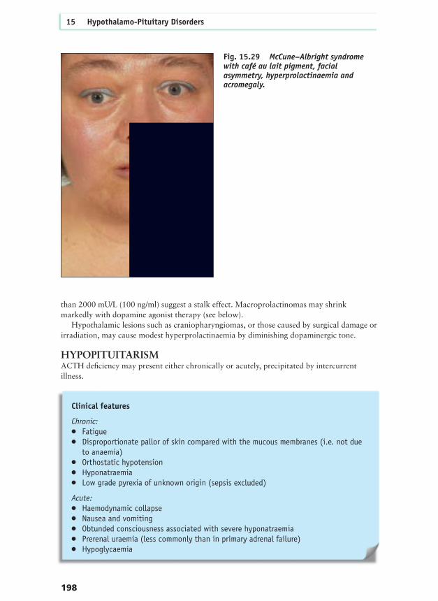
domperidone● Opioid peptides and alkaloids● Oestrogen – in high dosage, or pregnancy● H2 antagonists – such as intravenous (not oral) cimetidine● Polycystic ovary syndrome (PCOS)● Acromegaly● McCune–Albright syndrome (Fig. 15.29)● Chronic renal failure● Hepatic cirrhosis● Chest wall disease
Microprolactinomas (less than 10 mm maximum diameter) are common and do not causespace-occupying problems, with the occasional exception of headache; nor are they usuallyassociated with other hormone deficiencies, with the exception of the expectedhypogonadism and less often GH deficiency. Macroadenomas secreting PRL contrast withlarge functionless tumours causing hyperprolactinaemia by stalk compression that interfereswith delivery of hypothalamic dopamine, thus disinhibiting normal pituitary lactotrophs.Clinical distinction between the two tumours usually depends on the level of PRL secretion:values over 8000 mU/L (400 ng/ml) strongly suggest macroprolactinoma, whereas those less
Clinical features
197
Visceral obesityLean body massBone densityTotal body water
Stimulated peak GH < 9mU/LLow/low–normal age-adjusted/IGF1Hyperlipidaemia: Cholesterol
TriglycerideLDL cholesterolHDL cholesterol
EnergyPoor body imageDepressionPoor work recordMartial/social success
Mortality increased? cause? cardiovascular?
Body composition
Quality of life
Biochemistry
Adult onset GH deficiencyFig. 15.28 Features ofadult onset GH deficiency.
than 2000 mU/L (100 ng/ml) suggest a stalk effect. Macroprolactinomas may shrinkmarkedly with dopamine agonist therapy (see below).
Hypothalamic lesions such as craniopharyngiomas, or those caused by surgical damage orirradiation, may cause modest hyperprolactinaemia by diminishing dopaminergic tone.
HYPOPITUITARISMACTH deficiency may present either chronically or acutely, precipitated by intercurrentillness.
198
15 Hypothalamo-Pituitary Disorders
Fig. 15.29 McCune–Albright syndromewith café au lait pigment, facialasymmetry, hyperprolactinaemia andacromegaly.
Clinical features
Chronic:● Fatigue● Disproportionate pallor of skin compared with the mucous membranes (i.e. not due
to anaemia)● Orthostatic hypotension● Hyponatraemia● Low grade pyrexia of unknown origin (sepsis excluded)
Acute:● Haemodynamic collapse● Nausea and vomiting● Obtunded consciousness associated with severe hyponatraemia● Prerenal uraemia (less commonly than in primary adrenal failure)● Hypoglycaemia

TSH deficiency presents with the usual features of hypothyroidism but is often milderthan primary hypothyroidism. Myxoedema is not usually seen in secondary hypothyroidismalthough the skin is often dramatically cold and dry to the touch.
TUMOURS WITH EXTRASELLAR EXTENSIONLarge tumours can extend upward compressing the hypothalamus and third ventricle.Hydrocephalus may result in dementia, loss of balance and abnormalities of micturition. Invasion of the temporal lobes can cause epilepsy. Many neural problemsdevelop, often years later, after surgery or radiotherapy. Tumours eroding through the pituitary fossa and sphenoid bone can lead to the subarachnoid space communicating with the nasopharynx, resulting in CSF rhinorrhoea and the potential for meningitis.
HYPERSECRETING PITUITARY TUMOURSCushing’s disease – see Ch. 18.
ACROMEGALYGH excess causing gigantism (Fig. 15.30) prepubertally occurs rarely. Apart from theincreased growth of long bones leading to an ultimate height often in excess of 2 metres, thisis associated with kyphoscoliosis and distortions of the rib cage. The pituitary tumours arefrequently large.
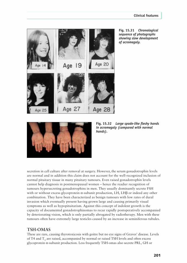
Adult onset acromegaly is most frequently diagnosed in patients in the middle years oflife, but retrospective analysis of photographs often discloses developing features becomingapparent 10 or more years before diagnosis (Fig. 15.31).
Clinical features
199
Clinical features
● Enlargement of hands (becoming fleshy, so rings need enlarging) (Fig. 15.32) andof feet (becoming broader)
● Thick skin with deep creases, increased sweating and sebum production (Fig. 15.33)● Hypertension, left ventricular hypertrophy and congestive cardiac failure● Soft tissue enlargement – such as in the nose, tongue (Fig. 15.34), larynx, or
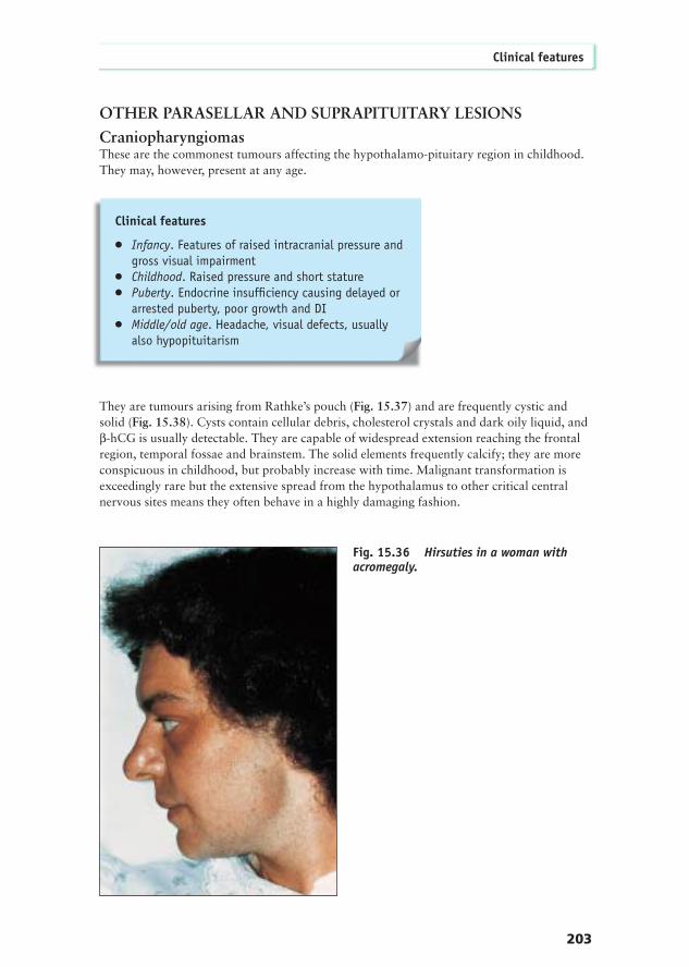
viscera● Prognathism and increased interdental spacing (Fig. 15.35)● Visual field defects● Obstructive sleep apnoea● Goitre, which is multinodular, and sometimes toxic● Hyperprolactinaemia, hypogonadism and galactorrhoea● Osteoporosis ● Osteoarthritis● Colonic polyps and carcinoma● Impaired glucose tolerance and diabetes mellitus● Carpal tunnel syndrome● Hirsutism (Fig. 15.36)● Hypopituitarism ● Myopathy

GONADOTROPHINOMASThese have only relatively recently been recognized as a distinct entity. Some claim theyconstitute the majority of ‘functionless’ pituitary tumours as judged by immunostaining and
200
15 Hypothalamo-Pituitary Disorders
a b
c
Fig. 15.30 Gigantism (a, b) with ribcage abnormality and (c) in a patientwith his mother.
secretion in cell culture after removal at surgery. However, the serum gonadotrophin levelsare normal and in addition this claim does not account for the well-recognized inclusion ofnormal pituitary tissue in many pituitary tumours. Even raised gonadotrophin levelscannot help diagnosis in postmenopausal women – hence the readier recognition oftumours hypersecreting gonadotrophins in men. They usually dominantly secrete FSHwith or without excess glycoprotein α-subunit production, LH, LHβ or indeed any othercombination. They have been characterized as benign tumours with low rates of duralinvasion which eventually present having grown large and causing primarily visualsymptoms as well as hypopituitarism. Against this concept of indolent growth is thecapacity of documented gonadotrophinomas to recur rapidly postoperatively accompaniedby deteriorating vision, which is only partially abrogated by radiotherapy. Men with thesetumours often have extremely large testicles caused by an increase in seminiferous tubules.
TSH-OMASThese are rare, causing thyrotoxicosis with goitre but no eye signs of Graves’ disease. Levelsof T4 and T3 are raised, accompanied by normal or raised TSH levels and often excessglycoprotein α-subunit production. Less frequently TSH-omas also secrete PRL, GH or
Clinical features
201
Fig. 15.31 Chronologicalsequence of photographsshowing slow developmentof acromegaly.
Fig. 15.32 Large spade-like fleshy handsin acromegaly (compared with normalhands).

gonadotrophins. They are frequently large at presentation with effects due to mass, as wellas thyrotoxic features. They need to be distinguished from thyroid hormone resistance,which is usually due to mutations in exons 9 and 10 of the thyroid hormone β-receptor, andin which there is no excessive α-subunit production and no association with pituitarytumours (see Ch. 17).
202
15 Hypothalamo-Pituitary Disorders
Fig. 15.33 Thick skin with deep creasesin acromegaly.
Fig. 15.34 Macroglossia in acromegaly.
Fig. 15.35 (a) Prognathism inacromegaly. (b) Leading to increasedinterdental spacing.
a b
OTHER PARASELLAR AND SUPRAPITUITARY LESIONS
CraniopharyngiomasThese are the commonest tumours affecting the hypothalamo-pituitary region in childhood.They may, however, present at any age.
They are tumours arising from Rathke’s pouch (Fig. 15.37) and are frequently cystic andsolid (Fig. 15.38). Cysts contain cellular debris, cholesterol crystals and dark oily liquid, andβ-hCG is usually detectable. They are capable of widespread extension reaching the frontalregion, temporal fossae and brainstem. The solid elements frequently calcify; they are moreconspicuous in childhood, but probably increase with time. Malignant transformation isexceedingly rare but the extensive spread from the hypothalamus to other critical centralnervous sites means they often behave in a highly damaging fashion.
Clinical features
203
Clinical features
● Infancy. Features of raised intracranial pressure andgross visual impairment
● Childhood. Raised pressure and short stature● Puberty. Endocrine insufficiency causing delayed or
arrested puberty, poor growth and DI ● Middle/old age. Headache, visual defects, usually
also hypopituitarism
Fig. 15.36 Hirsuties in a woman withacromegaly.

Germinomas are the commonest germ cell tumours of CNS and closely resemble gonadaltumours. They usually present in young teenage girls with headache, visual loss, anteriorpituitary failure especially affecting growth and diabetes insipidus, which should alert theclinician that this is not a routine pituitary tumour. Boys tend to have pineal tumours, oftenalso with suprasellar components. Secretion of β-HCG and α-fetoprotein into blood and CSFmay occur in germ cell tumours and seeding can occur down the neuraxis.
Other lesions such as optic nerve glioma and sphenoid ridge meningioma cause primarilyvisual disturbances, but astrocytomas and other tumours can cause hypothalamic dysfunction.Pituitary damage can follow trauma, especially in abused infants who can develop varying
204
15 Hypothalamo-Pituitary Disorders
Embryology of craniopharyngioma
Rathke's pouch
Stomodaeum
Forebrain
Pericardium
ForegutForegutMandibular arch
Notochord
NormallyReplaced by PROP-1 PIT 1in anterior pituitary GH, PRL, TSH cell lines
RPX =Rathke pouchHomeobox gene
RPX
Notochord
Interventricularforamen
Site of craniopharyngioma
Oralmembrane
Site of oralmembrane
anteriorpituitary lobe
posteriorpituitary lobe
PROP-1
PIT 1
Fig. 15.37 Embryology of craniopharyngioma arising from Rathke’s pouch.
Fig. 15.38 Cystic and calcifiedcraniopharyngioma.

degrees of damage including panhypopituitarism, which is often apparent only years later whenthere is growth failure. Note that psychosocial deprivation can also cause GH deficiency butthis often reverses rapidly on hospitalization or on being taken into care. Road traffic accidentscan cause pituitary stalk section, which classically causes DI but also anterior pituitary failure.Other consequences include cortical blindness, and emotional and cognitive difficulties.
VASCULAR DISORDERS OF THE PITUITARYAny of the following factors may lead to pituitary infarction:● Pregnancy● Diabetes mellitus● Anticoagulation● Pituitary tumours● Cerebral aneurysms● Coronary artery bypass grafting● Old age
Pituitary failure can occur after vascular catastrophes such as infarction or haemorrhage.Sheehan’s syndrome may follow massive ante- or postpartum haemorrhages withhypotensive shock; prompt blood transfusion should prevent this, however. Hyperplasia andhypertrophy of lactotrophs in pregnancy render the pituitary blood supply vulnerable.
Clinical symptoms occur only when more than 90% of the pituitary volume has beendestroyed.
Diabetes mellitus is said to enhance the risk of pituitary infarction but the incidence is verysmall, and this also applies to anticoagulation. Histological examination of pituitarytumours frequently reveals areas of infarction or haemorrhage of varying age that are usuallysymptomatically and hormonally insignificant.
Pituitary apoplexy (see p.194) may prove lethal as a result of subarachnoid haemorrhageor of easily overlooked adrenal insufficiency if the condition is not recognized. Morbidityincludes ophthalmoplegia as well as pituitary deficiency – which may include completeextirpation of a hypersecreting tumour such as acromegaly. When partial deficiencies arisethey do so unpredictably with, for example, ACTH or PRL deficiency but preservation ofgonadotrophin secretion. This feature is typical of vascular insults causing selective losses ofpituitary hormones and contrasts with the progressive pattern of growing pituitaryadenomas.
Ectatic loops of carotid may occupy and expand the pituitary fossa with consequentendocrine deficiency but their recognition is much more important if surgery is planned.Coronary artery bypass grafting involves non-pulsatile flow with anticoagulated blood,
Clinical features
205
Clinical features
● Failure of lactation (hypoprolactinaemia) ● Other features of anterior pituitary failure (e.g.
amenorrhoea)● ACTH and TSH deficiency, which may declare only
much later with patients classically becomingtorpid, bedbound and losing all interest andinitiative

which has been documented to produce major disturbances in pituitary hormone secretionand occasional early cases of pituitary apoplexy. Delayed presentation also occurs with lossof libido, secondary hypothyroidism and adrenal insufficiency, which is often indicated bypersistent hyponatraemia. Imaging of the pituitary shows marked loss of volume. The samepattern can occur idiopathically in old age when the presenting feature is often orthostatichypotension.
DEFICIENCY SYNDROMESAny of the following hypothalamic releasing factors may be lost:
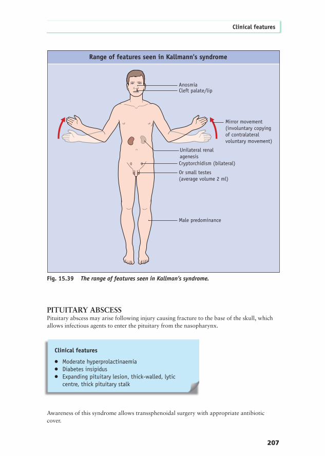
Deficiency of GnRH may be inherited as an isolated defect, or be due to a mutation in anadhesion molecule that leads to failure of migration of GnRH neurons from their origin inthe olfactory placode to the median eminence. If there is associated anosmia it is termedKallman’s syndrome, and may also include other midline defects such as cleft lip and palateand renal tract anomalies (Fig. 15.39). Acquired GnRH deficiency accompanies severeweight loss especially anorexia nervosa and stress; it may be mediated by endogenousopioids and may follow excessive physical activity (usually also causing loss of body fat).
Isolated GH deficiency is rarely total, and is more often due to congenital GHRHdeficiency – hence sufferers respond to exogenous GHRH, which limits its use in diagnosingGH deficiency but permits its use as a therapy (see Ch. 16). Isolated ACTH deficiency isusually acquired (presenting variously with hypoglycaemia, weight loss, hypotension andother features of cortisol deficiency). Alcohol abuse may be a factor. Most cases appear to bedue to primary pituitary ACTH deficiency but there are also well-recorded cases that areapparently due to CRH deficiency. TRH deficiency is rare but may occur as an isolatedacquired phenomenon. Dopamine deficiency is postulated as underlying ‘idiopathichyperprolactinaemia’ but the possibility of prolactinomas too small to be visualized cannotbe discounted. Somatostatin deficiency rarely presents since most pathological processessimultaneously affect GHRH. However, Alzheimer’s disease is notably associated withdeficiency of somatostatin amongst other neurotransmitters. Such patients often havemodestly elevated GH levels though this may reflect IGF-I deficiency occasioned by theirfrequent though ill-explained cachexia.
Multiple hypothalamic hormone deficiencies can occur congenitally or after trauma. Acommon cause is cranial irradiation, which causes dose- and time-dependent GH andgonadotrophin deficiencies that are secondary to releasing factor loss, andhyperprolactinaemia due to dopamine deficiency. Hypothalamo-pituitary–adrenal and–thyroid axes may also become deficient, but usually much later after radiotherapy, whichpoints to the need for continuing surveillance and repeated retesting over the years.
206
15 Hypothalamo-Pituitary Disorders
● GnRH (with anosmia: Kallman’s syndrome)● GHRH● ACTH● Thyrotropin-releasing hormone (TRH)● Dopamine● Somatostatin● Multiple deficiencies
PITUITARY ABSCESSPituitary abscess may arise following injury causing fracture to the base of the skull, whichallows infectious agents to enter the pituitary from the nasopharynx.
Awareness of this syndrome allows transsphenoidal surgery with appropriate antibioticcover.
Clinical features
207
Range of features seen in Kallmann's syndrome
AnosmiaCleft palate/lip
Mirror movement(involuntary copyingof contralateralvoluntary movement)
Unilateral renalagenesisCryptorchidism (bilateral)
Or small testes(average volume 2 ml)
Male predominance
Fig. 15.39 The range of features seen in Kallman’s syndrome.
Clinical features
● Moderate hyperprolactinaemia● Diabetes insipidus● Expanding pituitary lesion, thick-walled, lytic
centre, thick pituitary stalk

OTHER CONDITIONS
Granulomatous conditionsGranulomatous conditions such as neurosarcoid and syphilis can affect the pituitary orhypothalamus. Tuberculous meningitis causes a chronic basal meningitis that often calcifiesand is associated with profound pituitary failure. Idiopathic granulomatous hypophysitis hasalso been described.
Lymphocytic hypophysitisLymphocytic hypophysitis is most commonly associated with the third trimester ofpregnancy or the puerperium, in association with a pituitary mass and often initially ACTHand TSH deficiency. Diabetes insipidus is sometimes also seen. In such patients it may resolvespontaneously, whereas in older women and in the few men in whom it has been recordedthe disease seems more permanent and profound. Concomitant Hashimoto’s disease andpernicious anaemia occurring together with limited pathological material demonstratinglymphocytic infiltration suggests an autoimmune aetiology.
Langerhans cell histiocytosis This condition is associated with the following triad of clinical features:
Anterior pituitary deficiency also occurs, especially GH deficiency. Some patients developpresenile dementia.
208
15 Hypothalamo-Pituitary Disorders
Clinical features
● Punched-out skull lesions (Fig. 15.40)● Exophthalmos● DI
Fig. 15.40 Skull X-ray showing punched-out lesions due to Langerhans cellhistiocytosis.
Fig. 15.41 Cutaneous lesions ofxanthoma disseminatum.
Xanthoma disseminatumThis rare condition may present with cutaneous (Fig. 15.41) and mucosal xanthomatouslesions, anterior and posterior pituitary failure, deposits within the skull causing epilepsy andtracheal stenosis requiring tracheotomy.
Wegener’s granulomatosisThis condition, characteristically affecting the respiratory and renal tracts, may causepituitary involvement with mass effects and endocrine deficiency.
Metastatic diseaseMetastatic disease to the hypothalamus is especially common in carcinoma of the breast andbronchus. If there is concomitant DI then this usually presages an early demise. Autopsystudies frequently reveal pituitary metastases with no clinical symptoms. However,occasionally apparently solitary metastases in the pituitary cause chiasmal compression, forinstance in renal carcinoma (Fig. 15.42).
Iron overloadConditions associated with iron overload (see Ch. 19) such as haemochromatosis and β-thalassaemia following multiple transfusions characteristically deposit iron in gonadotrophscausing primary pituitary gonadotrophin deficiency.
MANAGEMENT
HYPOPITUITARISMIdeally each deficient hormone, or more frequently target organ hormones, should bereplaced in as physiological a manner as possible. This is largely limited by practicalconsiderations, however.
Adrenal insufficiencyAdrenal insufficiency, which is usually defined as an inability to respond with adequatecortisol secretion to stress, is usually treated by replacement doses of glucocorticoid. Thephysiological steroid hydrocortisone is preferred because its concentration can be checked byserial measurements. Conventionally, two-thirds of the total is given in the early morning toachieve a peak level approximating the time of maximum circadian secretion in normalindividuals, and the remainder is given at 4–6 p.m. (because administration late at night candisturb sleep and cause polyuria). The usual regimen of 20 mg and 10 mg respectively is now
Management
209
Fig. 15.42 Renal cell metastases in thepituitary gland.
considered excessive for many people, and carries the risk of osteoporosis and other side-effects. Some patients prefer a thrice-daily pattern, in which two small doses at lunch and teafollow the major morning dose. Prednisolone 2.5 mg twice daily, or 5 mg and 2.5 mg, maybe preferred especially if given in an enteric-coated form; in the US this is also significantlycheaper than hydrocortisone. Patients on anticonvulsants may clear steroids more quickly somay need higher and more frequent doses.
Secondary hypothyroidism is easily dealt with using levo thyroxine, building up over weeksto a final dose of 75–200 µg daily. Obviously TSH cannot be used to monitor therapy: thisshould be done clinically while aiming for thyroxine levels to be in the upper normal range.Measurement of T3 levels may be helpful. Thyroxine should not be given to ACTH-deficientpatients until they have been on replacement glucocorticoid for 48 hours; this avoids thetheoretical risk of precipitating an adrenal crisis.
HypogonadismMale patients with hypogonadism require testosterone to maintain libido, sexual functionand secondary sexual characteristics, but also for general vigour, maintenance of musclebulk and strength and prevention of osteoporosis. Oral testosterone undecanoate orsublingual testosterone preparations are rarely adequate. The main route is intramuscular,using testosterone esters usually administered every 2 weeks (the classical monthlytreatment often leaves the patient with subnormal levels for 2 weeks). There has been arevival of testosterone pellet implant treatment, in which a dose of 600 mg is deliveredsubcutaneously every 4–6 months. Recently transdermal patches have been introduced,which were originally applied to shaved scrotal skin, but more recently to body skin. Inwomen less than 50 years of age, hormone replacement therapy (HRT) is required, or alow dose oral contraceptive pill up to the age of 35 in cases where infertility cannot beassumed.
210
15 Hypothalamo-Pituitary Disorders
● It is essential that patients understand the need todouble or treble the dose for 2 to 3 days in theevent of stressful illnesses, such as fever due toinfection, trauma, surgery and myocardialinfarction. They must take the responsibility forinitiating the increase without feeling that thisabsolves them of the need to seek medical advice.They must clearly understand the need to secureparenteral hydrocortisone rapidly if vomiting orotherwise unable to absorb oral agents, and shouldkeep an ampoule of hydrocortisone sodiumsuccinate at home on standby. In addition tocarrying conventional steroid cards patients benefitfrom a clearly worded letter as many emergencydoctors seem unwisely reluctant or ignorant of theneed to raise steroids appropriately. Too often asmall increment is given too late but maintainedchronically with the development of Cushingoidside-effects
Growth hormone deficiencyGH is now widely available as a synthetic recombinant DNA product but this is veryexpensive and requires subcutaneous administration. In selected adult patients GH therapycan improve a number of the clinical features:
The dose per unit weight is much less than that required to optimize growth in children.Daily nocturnal subcutaneous injections are recommended but thrice-weekly injection iseffective. Very small doses are used initially to avoid rapid overcorrection of body water,which causes joint pains and carpal tunnel syndrome. Women tend to need higher doses. A 6 month trial using physical and QOL assessment is recommended. If no clear-cut objectiveor subjective improvement is shown after this period then treatment should be stopped.Serum IGF-I should be monitored and GH dosage adjusted to keep it within the age-relatednormal range.
If there is osteoporosis, GH is given in combination with sex steroids and optimalhydrocortisone and thyroxine dosage.
DIABETES INSIPIDUSFrank DI requires desmopressin, which is usually administered by a metered intranasal spraythat provides 10 µg per squirt. A dose of 10–20 µg is administered at bedtime but a seconddose may be needed in the morning. Overdosage leading to water intoxication must beavoided. This is particularly difficult in patients with hypothalamic damage to the thirstcentre in addition to DI. They require a strict regimen of fluid input, adjusted appropriatelyin hot weather and monitored by weighing at the same time daily, or twice daily in the eventof problems. Regular biochemical checks are needed and should be repeated when clinicallyrequired. Milder degrees of DI can be managed by oral DDAVP, which is especially useful inchildren or patients with cognitive and visual problems. In some patients medication withcarbamazepine will suffice.
Postoperative DI may require intramuscular DDAVP for some days, especiallyfollowing transsphenoidal surgery when nasal packs are in place. Postoperative DI is oftentemporary, lasting only days or weeks. Occasionally a triphasic response occurs (Fig. 15.43). It is advisable for patients on DDAVP after surgery to try without it every fewmonths at a time when sudden return of polyuria will not prove inconvenient. Ifreplacement is still needed they will experience unequivocal symptoms within 24 hours ofdiscontinuing DDAVP.
Some patients with hypothalamic disease, rather than DI, have disturbed thirst anddevelop primary polydipsia. This must not be mistaken for DI as antidiuretic treatmentcauses water intoxication. This has been particularly recorded in neurosarcoid conditions.
Management
211
Clinical features
● Truncal obesity● Lack of skeletal muscle and physical strength● Exercise capacity● Adverse lipid profile● Lack of vitality● Osteoporosis ● Reduced total body water

OTHER CAUSES OF POLYURIAProfound polyuria after surgery for aneurysm of the anterior communicating artery, andinfrequently for other intracranial conditions, may be due to cerebral salt wasting. Thisshould not be mistaken for DI and treated with DDAVP. In fact it causes greater loss of saltthan water, hence there is marked hyponatraemia, so there is no justification for confusion.Indeed it is frequently mistaken for the syndrome of inappropriate ADH secretion (SIADH)and treated by fluid restriction. However, the polyuria makes this diagnosis untenable andsuch management dangerous. The appropriate treatment is infusion of normal saline inquantities matching the previous, carefully measured, 24 hour urinary sodium losses. Thismust be continued for as long as the condition requires, and chronic cases may require oralsodium chloride supplementation. There have been suggestions that the underlying defect isexcessive secretion of brain antinatriuretic peptide; if these are substantiated this may lead tothe development of antagonist drugs.
Other causes of polyuria such as diabetes mellitus may occur and require appropriatetreatment. Hypercalciuria is common in acromegaly, especially if there is associatedhypercalcaemia from another cause; the most common of which is hyperparathyroidism dueto MEN-1. Hypercalcaemia should be treated appropriately (see Ch. 14). Hypercalciuriamay respond to low dose thiazide. This is also helpful in nephrogenic DI, in which, ifnecessary, amiloride and a prostaglandin synthetase inhibitor such as indomethacin shouldbe added.
PITUITARY TUMOURSSmall functionless tumours causing neither mass effects nor endocrine dysfunction may beincidentally discovered during CT or MRI scans performed for other reasons. Havingdocumented their innocuous nature no action is needed, but repeat MRI scans arerecommended every 2 to 5 years unless clinical features appear.
Functionless tumours causing visual disturbance or other mass effects require surgicalremoval with few exceptions. The transsphenoidal approach is preferred where possible,including in those tumours with extensive suprasellar extension, when the roof of the
212
15 Hypothalamo-Pituitary Disorders
Plas
ma
Osm
olal
ity
Mos
m/k
g
Plasma sodium
mosm
/LImmediate DI
Rx DDAVP
Surgery Days Post - Op
AVP Release from NecroticPosterior Pituitary SIADH
Permanent DIPost exhaustionof AVP Stores
Delayedrecognition
of DI andRx DDAVPlong-term
230
260
290
320
350
Early hospital discharge:danger period
120
130
140
150
160
2 4 6 8 10 12 14
Triphasic plasma osmolality responsefollowing pituitary surgery
Fi
Fig. 15.43 A triphasicplasma osmolality responsefollowing pituitary surgery.

resected tumour will be seen to descend. In contrast, tumours with eccentric lobulatedextensions and most parasellar lesions are approached transcranially. However, the classicalapproach involving a retraction of the right frontal lobe frequently gives rise to infarctionand cerebromalacia as well as serious psychological disabilities. Depending on thecompleteness of resection and whether there is any evidence of dural invasion (which isclinically difficult to assess) many patients require external radiotherapy, which is a skilledtechnique.
External radiotherapyA suggested regimen is:
Radiotherapy reduces the recurrence rate strikingly. Possible long-term side effects includesecond tumours in the field of irradiation (gliomas often responding poorly to treatment),progressive hypopituitarism, and vasculitis particularly affecting the visual pathways.Conventional radiotherapy cannot be repeated. If tumours recur then further surgery may be
Management
213
● Maximum tumour dose 4500 cGy● Maximum daily dose 180 cGy● Number of fractions 25, over 5 weeks● Use of three ports Rotating through each in
turn (Fig. 15.44)● Individual clear Moulded for each patient
Perspex mask (Fig. 15.45)
The title
30
100
95
80 70
30
RadiotherapyFig. 15.44 Radiationdosimetry using a three-port approach for externalpituitary radiotherapy(from Belchetz 1986Management of PituitaryDisease. Chapman & Hall,with permission).
required. Newer techniques under evaluation include γ- knife stereotactic radiotherapy andphotodynamic therapy.
Visual field defects improve in most patients, often completely, though in an appreciableminority they are unchanged and very rarely may worsen postoperatively. Severe headachetends to improve significantly following surgery. Pituitary function may improve after theremoval of small to moderate tumours (microadenomas ≤ 10 mm maximum diameter) butwith large macroadenomas there is usually no change or significant worsening of function.This implies full endocrine testing preoperatively.
A few centres recommend no steroid cover in patients with normal ACTH reserve butmost suggest the following regimen:
Patients should be discharged on the last dose with appropriate advice (see above). Approximately 6 weeks after surgery stop hydrocortisone for 24 hours and check levels of
ACTH and GH reserves using the ITT or glucagon stimulation test; also recheck electrolytes,PRL, and thyroid and gonadal status. If there are adequate ACTH reserves then stophydrocortisone immediately (with no tailing of doses). If the reserves are borderline thenconsider stopping but advise the patient to have a standby supply to cover times of stress.Whenever possible reduce the daily hydrocortisone to 20 mg daily (see above regimen). Makeadjustments to cover replacement requirements, and consider the need for GH therapy (seeabove). Patients will require follow-up scans, beginning within a year of surgery and 2–5 yearlythereafter. Endocrine surveillance, especially after radiotherapy, should continue indefinitely.
ACROMEGALYThe first-line treatment for most patients is transsphenoidal surgery. Cure rates with smalltumours approximate 80% but reduce markedly with increasing size. The aim should be anormal IGF-I and a mean GH level over 24 hours of less than 5 mU/L (2.5 ng/ml).
214
15 Hypothalamo-Pituitary Disorders
Fig. 15.45 A Perspex mould used toensure an accurate head position inpituitary irradiation.
● Hydrocortisone 100 mg i.v. with premedication● Hydrocortisone 100 mg i.v. 8 hourly for 24–48 hours● Hydrocortisone 50 mg i.v. 8 hourly for a further
24–48 hours● Hydrocortisone by mouth rapidly reducing to 15 mg
on waking, 5 mg at 6 p.m.
Medical treatment of acromegaly may suffice alone in mild cases, especially in elderlypatients as in these the disease appears more indolent than in the young. As the soletreatment or as an adjuvant to surgery, radiotherapy, or both, if there is an adequateresponse to bromocriptine then try up to 30 mg daily or cabergoline 0.25–1 mg once tothrice weekly. If the octreotide response is adequate then use 50–100 µg subcutaneouslyone to three times daily. Octreotide often causes transient abdominal pain or diarrhoeafor a week or so. Gall stones may develop but are seldom clinically troublesome. Watchfor diabetes mellitus developing or worsening. Headache may disappear within minutesof octreotide injection. Lanreotide (another somatostatin analogue) and octreotide depot preparations are now available, which act for 7–14 days and 28 days plusrespectively. Currently under trial is the use of the synthetic GH antagonist Pegvisomont,which may reduce IGF-I and improve clinical features in patients with resistantacromegaly.
PROLACTINOMADopamine agonist therapy (bromocriptine, quinagolide or cabergoline) reduces PRL levels inalmost all cases, often back to normal. Drugs are usually regarded as first-line therapy formost patients with micro- or macroprolactinomas for a number of reasons: ● Reversal of amenorrhoea usually occurs within months and in 90% after a year’s
treatment● Fertility responds rapidly and almost as satisfactorily● Galactorrhoea usually diminishes or disappears● Tumours usually shrink often strikingly with improvement in visual fields ● Drugs are usually withdrawn in pregnancy but bromocriptine is safe to continue● Pregnancy-associated tumour expansion can be rapidly reversed ● Impotence in men is usually corrected if testosterone is normalized● Cessation of therapy allows tumour re-expansion especially after short-term treatment
Management
215
In postoperative patients exceeding these values:● Consider reoperation if the scan suggests an
accessible remnant is present – but beware of earlynon-specific artefacts
● Consider external radiotherapy – but be aware thatthere may be a delay of up to 10 years before thefull benefit is apparent
● Check the response to bromocriptine – in 80% ofpatients the GH level falls, but often only modestly
● Check the response to octreotide – 80% of patientsrespond, often very markedly
Surgery may be considered in the following circumstances:● Rapid visual deterioration● Resistance to drugs (rare), resulting in inadequate PRL fall or tumour shrinkage● Intolerable side-effects, such as nausea, dizziness, headache, or white hands● A patient preference

However, surgery rarely cures macroprolactinomas and often involves hypopituitarism.The benefits of radiotherapy are delayed but may constrain tumour expansion in pregnancy.
CUSHING’S DISEASEWhen a secure diagnosis has been achieved (itself a major challenge), most patients aresubmitted to pituitary surgery. This is a highly specialized area, however, and so surgeryshould be confined to the few neurosurgeons with sufficient experience and expertise.Tumours are usually small – often only a few millimetres in diameter – and may not show upon MRI or CT scanning. At transsphenoidal surgery, serial sagittal cuts through the pituitarymay be needed to disclose the tumour. Sometimes nothing is found and then near-totalhypophysectomy may be required. Pathological examination may not demonstrate a tumour,simply hyperplasia or indeed no obvious abnormality, even when clinical and biochemicalevaluation postoperatively indicates a cure. The finding of undetectable morning cortisolwithin days of surgery (before the first dose of hydrocortisone) indicates a likely cure thoughlate relapses are well recognized. Low-normal cortisol levels are associated with higher andearlier relapse rates but can be followed by long-term remissions. A clear failure to improvebiochemistry can indicate the need for early further surgery. There is some dispute overwhether preoperative reduction in hypercortisolaemia by adrenal-blocking drugs such asmetyrapone and ketoconazole improves the perioperative course (see Ch. 18).
Bilateral adrenalectomy as primary therapy for Cushing’s disease has largely beenabandoned because of the risk of causing Nelson’s syndrome (Fig. 15.46). Most ‘cured’patients require corticosteroid replacement therapy for at least a year after pituitary surgery,and sometimes permanently. Radiotherapy has a role but is less often used than formerly.Rare macroadenomas are especially difficult to treat and may exceptionally metastasize.
216
15 Hypothalamo-Pituitary Disorders
a b
Fig. 15.46 (a, b) Nelson’s syndrome ina patient treated for Cushing’s disease bybilateral adrenalectomy.

CRANIOPHARYNGIOMASThese require surgery for mass effects and almost invariably require anterior and posteriorpituitary replacement therapy, especially after surgery. Though radical surgery is optimal forprevention of recurrence, incomplete resection is often all that can be safely achieved withoutdamaging critical neural centres, depending on the extent of the tumour. Radiotherapy isoften combined with surgery – which is usually transcranial and carries quite a highmorbidity. The radiotherapy dose is generally slightly higher than with pituitary tumours butdaily fractions are restricted to 180 cGy. Other problems are common includinghydrocephalus requiring ventriculo-peritoneal shunting. Hyperphagia, inertia, adipsia, visualand cognitive impairments can also seriously complicate management. Recurrent cysts maybe drained and 90Yttrium or bleomycin instilled. These tumours are commonly aggressiveand unsatisfactory to treat when, in most cases, they present early in life.
Management
217