hypoglycemia unawareness in insulinoma revealed with flash
TRANSCRIPT
3407
doi: 10.2169/internalmedicine.1173-18
Intern Med 57: 3407-3412, 2018
http://internmed.jp
【 CASE REPORT 】
Hypoglycemia Unawareness in Insulinoma Revealed withFlash Glucose Monitoring Systems
Taku Sugawa 1, Takaaki Murakami 1, Daisuke Yabe 1, Riko Kashima 1, Makiko Tatsumi 1,
Shinobu Ooshima 1, Erina Joo 1, Keiko Wada 1, Atsushi Yoshizawa 2, Toshihiko Masui 2,
Yuji Nakamoto 3, Yuki Yamauchi 4, Yuzo Kodama 4, Yoshiki Iemura 5, Masahito Ogura 1,
Akihiro Yasoda 1 and Nobuya Inagaki 1
Abstract:The delayed diagnosis of insulinoma remains a clinical issue. One of the main causes of such a delay is
hypoglycemia unawareness. A 53-year-old woman fell unconscious during postprandial exercises. Flash glu-
cose monitoring (FGM) systems revealed glucose profiles with fasting hypoglycemia, which facilitated the
clinical diagnosis of insulinoma even though she was unaware of her hypoglycemia. The preoperative com-
parison of the blood glucose values provided by FGM with those obtained from capillary blood were consis-
tent. Thus, FGM may have potential utility in revealing the presence of insulinoma-induced hypoglycemia.
Key words: insulinoma, flash glucose monitoring, continuous glucose monitoring, hypoglycemia, DOTATOC,
hypoglycemia unawareness
(Intern Med 57: 3407-3412, 2018)(DOI: 10.2169/internalmedicine.1173-18)
Introduction
Insulinoma is a rare neuroendocrine tumor (1), accounting
for 20.9% of all pancreatic endocrine tumors in epidemi-
ological studies across Japan (2). Fasting hypoglycemia is
known to be common in insulinoma and has diagnostic
value, as more than 80% of patients with insulinoma dem-
onstrate fasting hypoglycemia (3, 4). However, repeated and
prolonged hypoglycemic episodes can reduce the awareness
of neurogenic and neuroglycopenic hypoglycemic symptoms
in insulinoma (5), which delays the diagnosis of insuli-
noma (6, 7).
Continuous glucose monitoring (CGM) has been success-
fully used to detect hypoglycemia in insulinoma cases with
hypoglycemia unawareness (7, 8). However, currently avail-
able CGM systems in Japan require calibration by frequent
self-monitoring of capillary blood glucose (CBG) levels, and
patients without an insulin pump use can only view their
CGM values retrospectively in consultation with their physi-
cians (9). The flash glucose monitoring (FGM) systems
FreestyleⓇ Libre Pro and FreestyleⓇ Libre (Abbot Diabetes
Care, Oxon, UK) were recently approved for market in Ja-
pan. Due to their factory calibration, these two FGM sys-
tems require no CBG-based calibration, and the FreestyleⓇ
Libre even allows patients ready access to check their cur-
rent glucose levels and trends (10, 11). These features can
facilitate the detection of unnoticed hypoglycemic events
and the clinical diagnosis of insulinoma and will also im-
prove patients’ health-related quality of life by allowing pa-
tients to avoid episodes of hypoglycemia while awaiting sur-
gery.
We herein report a case of insulinoma presenting with hy-
poglycemic symptoms during postprandial exercise, in
which FGM facilitated the detection of the patient’s unno-
ticed hypoglycemia.
1Department of Diabetes, Endocrinology and Nutrition, Kyoto University Graduate School of Medicine, Japan, 2Department of Hepato-Biliary-
Pancreatic Surgery and Transplantation, Kyoto University Graduate School of Medicine, Japan, 3Department of Diagnostic Imaging and Nuclear
Medicine, Graduate School of Medicine, Kyoto University, Japan, 4Department of Gastroenterology and Hepatology, Graduate School of Medi-
cine, Kyoto University, Japan and 5Department of Diagnostic Pathology, Kyoto University Graduate School of Medicine, Japan
Received: March 12, 2018; Accepted: May 7, 2018; Advance Publication by J-STAGE: August 10, 2018
Correspondence to Dr. Nobuya Inagaki, [email protected]
Intern Med 57: 3407-3412, 2018 DOI: 10.2169/internalmedicine.1173-18
3408
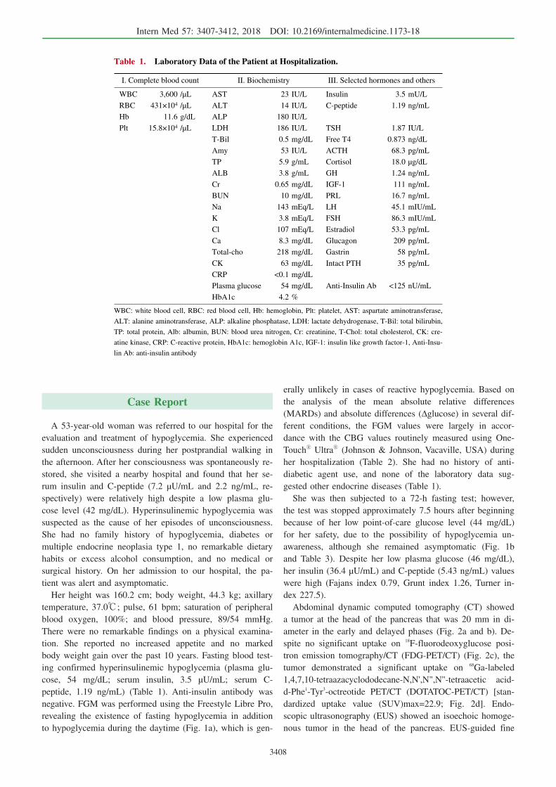
Table 1. Laboratory Data of the Patient at Hospitalization.
I. Complete blood count II. Biochemistry III. Selected hormones and others
WBC 3,600 /μL AST 23 IU/L Insulin 3.5 mU/L
RBC 431×104 /μL ALT 14 IU/L C-peptide 1.19 ng/mL
Hb 11.6 g/dL ALP 180 IU/L
Plt 15.8×10⁴ /μL LDH 186 IU/L TSH 1.87 IU/L
T-Bil 0.5 mg/dL Free T4 0.873 ng/dL
Amy 53 IU/L ACTH 68.3 pg/mL
TP 5.9 g/mL Cortisol 18.0 μg/dL
ALB 3.8 g/mL GH 1.24 ng/mL
Cr 0.65 mg/dL IGF-1 111 ng/mL
BUN 10 mg/dL PRL 16.7 ng/mL
Na 143 mEq/L LH 45.1 mIU/mL
K 3.8 mEq/L FSH 86.3 mIU/mL
Cl 107 mEq/L Estradiol 53.3 pg/mL
Ca 8.3 mg/dL Glucagon 209 pg/mL
Total-cho 218 mg/dL Gastrin 58 pg/mL
CK 63 mg/dL Intact PTH 35 pg/mL
CRP <0.1 mg/dL
Plasma glucose 54 mg/dL Anti-Insulin Ab <125 nU/mL
HbA1c 4.2 %
WBC: white blood cell, RBC: red blood cell, Hb: hemoglobin, Plt: platelet, AST: aspartate aminotransferase,
ALT: alanine aminotransferase, ALP: alkaline phosphatase, LDH: lactate dehydrogenase, T-Bil: total bilirubin,
TP: total protein, Alb: albumin, BUN: blood urea nitrogen, Cr: creatinine, T-Chol: total cholesterol, CK: cre-
atine kinase, CRP: C-reactive protein, HbA1c: hemoglobin A1c, IGF-1: insulin like growth factor-1, Anti-Insu-
lin Ab: anti-insulin antibody
Case Report
A 53-year-old woman was referred to our hospital for the
evaluation and treatment of hypoglycemia. She experienced
sudden unconsciousness during her postprandial walking in
the afternoon. After her consciousness was spontaneously re-
stored, she visited a nearby hospital and found that her se-
rum insulin and C-peptide (7.2 μU/mL and 2.2 ng/mL, re-
spectively) were relatively high despite a low plasma glu-
cose level (42 mg/dL). Hyperinsulinemic hypoglycemia was
suspected as the cause of her episodes of unconsciousness.
She had no family history of hypoglycemia, diabetes or
multiple endocrine neoplasia type 1, no remarkable dietary
habits or excess alcohol consumption, and no medical or
surgical history. On her admission to our hospital, the pa-
tient was alert and asymptomatic.
Her height was 160.2 cm; body weight, 44.3 kg; axillary
temperature, 37.0℃; pulse, 61 bpm; saturation of peripheral
blood oxygen, 100%; and blood pressure, 89/54 mmHg.
There were no remarkable findings on a physical examina-
tion. She reported no increased appetite and no marked
body weight gain over the past 10 years. Fasting blood test-
ing confirmed hyperinsulinemic hypoglycemia (plasma glu-
cose, 54 mg/dL; serum insulin, 3.5 μU/mL; serum C-
peptide, 1.19 ng/mL) (Table 1). Anti-insulin antibody was
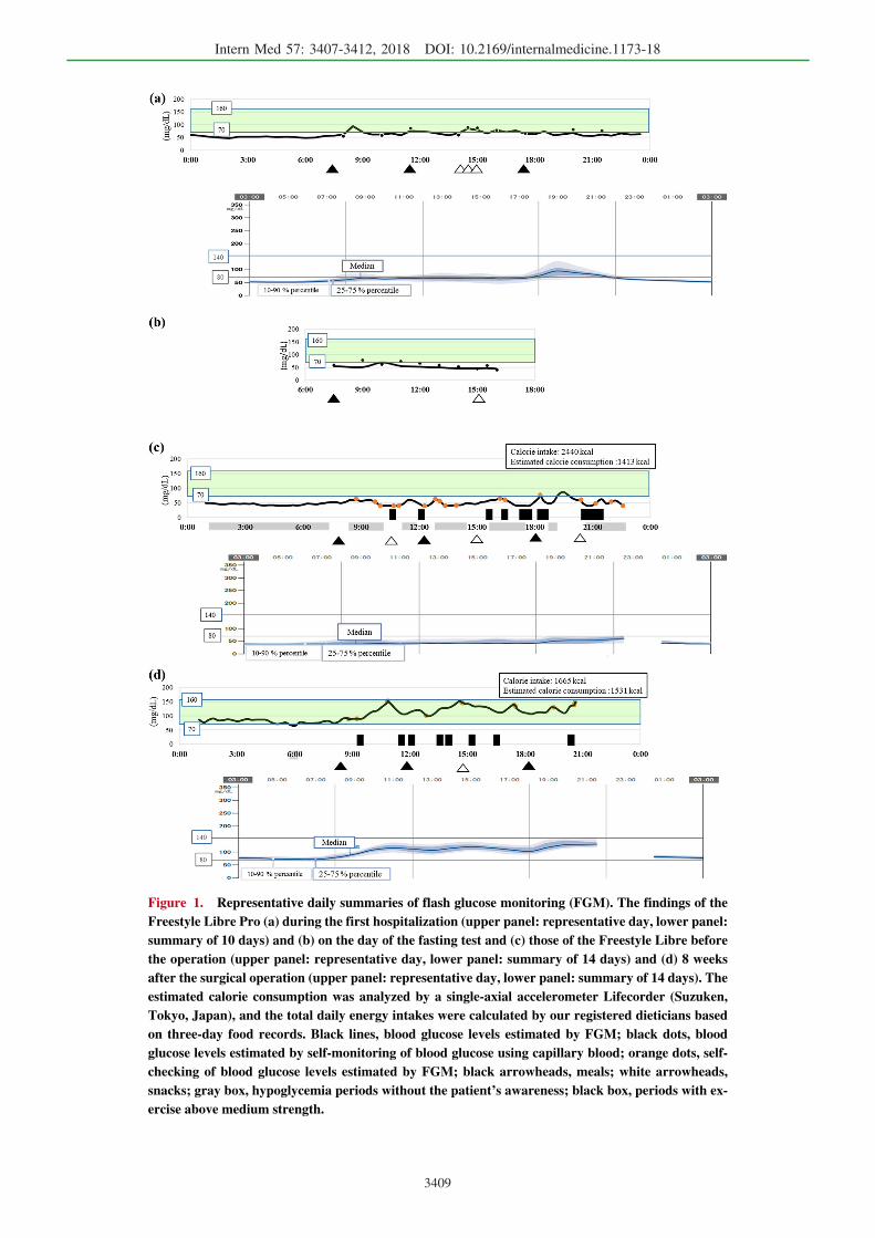
negative. FGM was performed using the Freestyle Libre Pro,
revealing the existence of fasting hypoglycemia in addition
to hypoglycemia during the daytime (Fig. 1a), which is gen-
erally unlikely in cases of reactive hypoglycemia. Based on
the analysis of the mean absolute relative differences
(MARDs) and absolute differences (Δglucose) in several dif-
ferent conditions, the FGM values were largely in accor-
dance with the CBG values routinely measured using One-
TouchⓇ UltraⓇ (Johnson & Johnson, Vacaville, USA) during
her hospitalization (Table 2). She had no history of anti-
diabetic agent use, and none of the laboratory data sug-
gested other endocrine diseases (Table 1).
She was then subjected to a 72-h fasting test; however,
the test was stopped approximately 7.5 hours after beginning
because of her low point-of-care glucose level (44 mg/dL)
for her safety, due to the possibility of hypoglycemia un-
awareness, although she remained asymptomatic (Fig. 1b
and Table 3). Despite her low plasma glucose (46 mg/dL),
her insulin (36.4 μU/mL) and C-peptide (5.43 ng/mL) values
were high (Fajans index 0.79, Grunt index 1.26, Turner in-
dex 227.5).
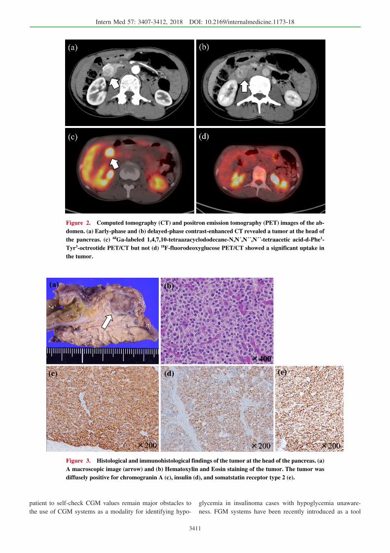
Abdominal dynamic computed tomography (CT) showed
a tumor at the head of the pancreas that was 20 mm in di-
ameter in the early and delayed phases (Fig. 2a and b). De-
spite no significant uptake on 18F-fluorodeoxyglucose posi-
tron emission tomography/CT (FDG-PET/CT) (Fig. 2c), the
tumor demonstrated a significant uptake on 68Ga-labeled
1,4,7,10-tetraazacyclododecane-N,N',N",N''-tetraacetic acid-
d-Phe1-Tyr3-octreotide PET/CT (DOTATOC-PET/CT) [stan-
dardized uptake value (SUV)max=22.9; Fig. 2d]. Endo-
scopic ultrasonography (EUS) showed an isoechoic homoge-
nous tumor in the head of the pancreas. EUS-guided fine
Intern Med 57: 3407-3412, 2018 DOI: 10.2169/internalmedicine.1173-18
3409
Figure 1. Representative daily summaries of flash glucose monitoring (FGM). The findings of the Freestyle Libre Pro (a) during the first hospitalization (upper panel: representative day, lower panel: summary of 10 days) and (b) on the day of the fasting test and (c) those of the Freestyle Libre before the operation (upper panel: representative day, lower panel: summary of 14 days) and (d) 8 weeks after the surgical operation (upper panel: representative day, lower panel: summary of 14 days). The estimated calorie consumption was analyzed by a single-axial accelerometer Lifecorder (Suzuken, Tokyo, Japan), and the total daily energy intakes were calculated by our registered dieticians based on three-day food records. Black lines, blood glucose levels estimated by FGM; black dots, blood glucose levels estimated by self-monitoring of blood glucose using capillary blood; orange dots, self-checking of blood glucose levels estimated by FGM; black arrowheads, meals; white arrowheads, snacks; gray box, hypoglycemia periods without the patient’s awareness; black box, periods with ex-ercise above medium strength.
Intern Med 57: 3407-3412, 2018 DOI: 10.2169/internalmedicine.1173-18
3410
Table 2. A Comparison between Flash Glucose Monitoring and Capillary Blood Glucose Values.
Number of time points analyzed MARD (%) ΔGlucose (mg/dL)
Fasting 9 3.5±15.2 -2.7±8.3
Post-meal 28 11.6±16.1 -10.2±12.3
Hypoglycemia 35 0.8±18.8 -0.4±10.0
Total 69 8.1±17.7 -7.6±12.0
The FGM values obtained by Freestyle Libre Pro 48 h after FGM sensor attachment were retrospec-
tively compared with a total of 69 CBG values obtained by the OneTouch® Ultra®. The mean abso-
lute relative difference (MARD) and ΔGlucose were calculated for not only all of the time points for
which CBG values were available (Total) but also fasting (Fasting) and two hours after meals (Post-
meal). MARD and ΔGlucose were also calculated for the time points at which the CBG values were
within the hypoglycemic range (≤70 mg/dL). MARD is defined as 100×|FGM value-CBG value|/
CBG value; ΔGlucose is defined as "FGM value-CBG value."
Table 3. Laboratory Data of the Patient at the End of the Fasting Test.
At the end of the fasting test 20 min after i.v. 1 mg glucagon
Plasma glucose (mg/dL) 46 95
Insulin (mU/L) 36.4 123.8
C-peptide (ng/mL) 5.43 8.35
Acetoacetic acid (μmol/L) 30.1 -
3-β-hydroxybutyric acid (μmol/L) 14.7 -
A 72-h fasting test was performed after admission to our hospital for the investigation of spontaneous hypoglyce-
mia. After confirming glucose levels <45 mg/dL using OneTouch® Ultra®, 1 mg glucagon was administered intra-
venously.
needle aspiration cytology revealed that the tumor was posi-
tive for insulin staining. Based on these findings, a clinical
diagnosis of insulinoma was made.
The patient was discharged from our hospital to await her
surgical operation. FGM using the Freestyle Libre system
revealed hypoglycemia lasting almost 24 hours with evident
hypoglycemia unawareness (Fig. 1c). Although the patient
checked her glucose levels using the Freestyle Libre system
over 15 times a day and consumed high-caloric foods and
occasional snacks, prolonged hypoglycemia without the pa-
tient’s awareness was still observed throughout the day
(Fig. 1c, Gray boxes). Despite our recommendations, she re-
fused any preoperative drug therapy, including diazoxide.
Thus, prompt surgery was planned in addition to dietary
guidance.
Six weeks after admission after the initial admission, the
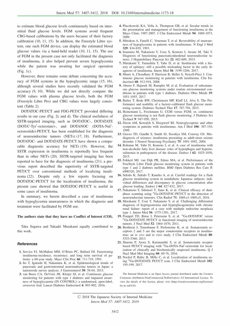
patient underwent pancreatoduodenectomy. A pathological
examination revealed a tumor 23 mm in diameter at the
head of pancreas (Fig. 3a and b). Immunohistochemical
studies showed that the tumor was positive for chromo-
granin A (Fig. 3c) and synaptophysin as well as insulin
(Fig. 3d) and somatostatin receptor (SSTR) type 2 (Fig. 3e)
but negative for gastrin and glucagon. The Ki67 proliferative
index of the tumor was 1.8%. The patient’s pathological
staging was T2N0M0 (stage IB) and T2N0M0 (stage IIA)
according to the AJCC/UICC and European neuroendocrine
tumor society TNM staging system, respectively. Blood test-
ing performed 2 hours after surgery showed the rapid reso-
lution of hyperinsulinemia (plasma glucose, 128 mg/dL; se-
rum insulin, 4.2 μU/mL). After the operation, the patient ex-
perienced no hypoglycemic symptoms even under a rela-
tively low calorie intake and more intensive exercise than
during the preoperative period (Fig. 1d). Eight weeks after
surgery, her fasting plasma glucose was 81 mg/dL.
Discussion
Insulinoma is a rare neuroendocrine tumor that produces
excess endogenous insulin, resulting in hypoglycemia (6).
The clinical diagnosis of insulinoma is established by both
the presence of hypoglycemia with inappropriate insulin se-
cretion and the identification of a tumor mass. Previous
studies have shown that the median time to a diagnosis is 24
months, with a range of up to 30 years (12). A delayed di-
agnosis of insulinoma can progressively induce coma or
death (7, 8, 13) and remains a clinical issue (6, 14).
One of the main causes of a delay in the diagnosis of in-
sulinoma is hypoglycemia unawareness. As previously
shown, a lack of neurogenic and neuroglycopenic symptoms
is not rare in patients with insulinoma (5). Repeated and
prolonged hypoglycemia events cause hypoglycemia un-
awareness, which can obscure a patient’s notice and a physi-
cian’s suspicion of possible hypoglycemia. In fact, the pre-
sent case developed sudden postprandial unconsciousness
without any evident hypoglycemic episodes at fasting.
CGM systems have been used in insulinoma cases with
hypoglycemia unawareness (7, 8). However, the requirement
of frequent CBG-based calibrations and the inability of the
Intern Med 57: 3407-3412, 2018 DOI: 10.2169/internalmedicine.1173-18
3411
Figure 2. Computed tomography (CT) and positron emission tomography (PET) images of the ab-domen. (a) Early-phase and (b) delayed-phase contrast-enhanced CT revealed a tumor at the head of the pancreas. (c) 68Ga-labeled 1,4,7,10-tetraazacyclododecane-N,N´,N´´,N´´-tetraacetic acid-d-Phe1-Tyr3-octreotide PET/CT but not (d) 18F-fluorodeoxyglucose PET/CT showed a significant uptake in the tumor.
Figure 3. Histological and immunohistological findings of the tumor at the head of the pancreas. (a) A macroscopic image (arrow) and (b) Hematoxylin and Eosin staining of the tumor. The tumor was diffusely positive for chromogranin A (c), insulin (d), and somatstatin receptor type 2 (e).
patient to self-check CGM values remain major obstacles to
the use of CGM systems as a modality for identifying hypo-
glycemia in insulinoma cases with hypoglycemia unaware-
ness. FGM systems have been recently introduced as a tool
Intern Med 57: 3407-3412, 2018 DOI: 10.2169/internalmedicine.1173-18
3412
to estimate blood glucose levels continuously based on inter-
stitial fluid glucose levels. FGM systems avoid frequent
CBG-based calibrations by the users because of their factory
calibration (10, 11, 15). In addition, the Freestyle Libre sys-
tem, one such FGM device, can display the estimated blood
glucose values via a hand-held reader (10, 11, 15). The use
of FGM in the present case not only facilitated the diagnosis
of insulinoma, it also helped prevent severe hypoglycemia
while the patient was awaiting her surgical operation
(Fig. 1c).
However, there remains some debate concerning the accu-
racy of FGM systems in the hypoglycemic range (15, 16),
although several studies have recently validated the FGM
accuracy (9, 10). While we did not directly compare the
FGM values with plasma glucose levels, both the FGM
(Freestyle Libre Pro) and CBG values were largely consis-
tent (Table 2).
DOTATOC-PET/CT and FDG-PET/CT provided differing
results in our case (Fig. 2c and d). The clinical usefulness of
SSTR-targeted imaging, such as DOTATOC-, DOTATATE
(DOTA0-Tyr3-octreotate)-, and DOTANOC (DOTA-Nal3-
octoreotide)-PET/CT, has been established for the diagnosis
of neuroendocrine tumors (NETs) (17, 18). Furthermore,
DOTATOC- and DOTATATE-PET/CT have shown a compa-
rable diagnostic accuracy for NETs (19). However, the
SSTR expression in insulinoma is reportedly less frequent
than in other NETs (20). SSTR-targeted imaging has been
reported to have for the diagnosis of insulinoma (21); a pre-
vious report described the superiority of SSTR-targeted
PET/CT over conventional methods of localizing insuli-
noma (22). Despite only a few reports focusing on
DOTATOC-PET/CT for the localization of insulinoma, the
present case showed that DOTATOC-PET/CT is useful in
some cases of insulinoma.
In summary, we herein described a case of insulinoma
with hypoglycemia unawareness in which the diagnosis and
treatment were facilitated by FGM use.
The authors state that they have no Conflict of Interest (COI).
Taku Sugawa and Takaaki Murakami equally contributed to
this work.
References
1. Service FJ, McMahon MM, O’Brien PC, Ballard DJ. Functioning
insulinoma-incidence, recurrence, and long term survival of pa-
tients: a 60-year study. Mayo Clin Proc 66: 711-719, 1991.
2. Ito T, Igarashi H, Nakamura K, et al. Epidemiological trends of
pancreatic and gastrointestinal neuroendocrine tumors in Japan: a
nationwide survey analysis. J Gastroenterol 50: 58-64, 2015.
3. van Beers CA, DeVries JH, Kleijer SJ, et al. Continuous glucose
monitoring for patients with type 1 diabetes and impaired aware-
ness of hypoglycaemia (IN CONTROL): a randomised, open-label,
crossover trial. Lancet Diabetes Endocrinol 4: 893-902, 2016.
4. Placzkowski KA, Vella A, Thompson GB, et al. Secular trends in
the presentation and management of functioning insulinoma at the
Mayo Clinic 1987-2007. J Clin Endocrinol Metab 94: 1069-1073,
2009.
5. Mitrakou A, Fanelli C, Veneman T, et al. Reversibility of unaware-
ness of hypoglycemia in patients with insulinomas. N Engl J Med
329: 834-839, 1993.
6. Imamura M, Nakamoto Y, Uose S, Komoto I, Awane M, Taki Y.
Diagnosis of functioning pancreaticoduodenal neuroendocrine tu-
mors. J Hepatobiliary Pancreat Sci 22: 602-609, 2015.
7. Murakami T, Yamashita T, Yabe D, et al. Insulinoma with a his-
tory of epilepsy: still a possible misleading factor in the early di-
agnosis of insulinoma. Intern Med 56: 3199-3204, 2017.
8. Munir A, Choudhary P, Harrison B, Heller S, Newell-Price J. Con-
tinuous glucose monitoring in patients with insulinoma. Clin En-
docrinol 68: 912-918, 2008.
9. Aberer F, Hajnsek M, Rumpler M, et al. Evaluation of subcutane-
ous glucose monitoring systems under routine environmental con-
ditions in patients with type 1 diabetes. Diabetes Obes Metab 19:
1051-1055, 2017.
10. Bailey T, Bode BW, Christiansen MP, Klaff LJ, Alva S. The Per-
formance and usability of a factory-calibrated flash glucose moni-
toring system. Diabetes Technol Ther 17: 787-794, 2015.
11. Heinemann L, Freckmann G. CGM versus FGM; or, continuous
glucose monitoring is not flash glucose monitoring. J Diabetes Sci
Technol 9: 947-950, 2015.
12. Dizon AM, Kowalyk S, Hoogwerf BJ. Neuroglycopenic and other
symptoms in patients with insulinomas. Am J Med 106: 307-310,
1999.
13. Graves TD, Gandhi S, Smith SJ, Sisodiya SM, Conway GS. Mis-
diagnosis of seizures: insulinoma presenting as adult-onset seizure
disorder. J Neurol Neurosurg Psychiatry 75: 1091-1092, 2004.
14. Rokutan M, Yabe D, Komoto I, et al. A case of insulinoma with
non-alcoholic fatty liver disease: roles of hyperphagia and hyperin-
sulinemia in pathogenesis of the disease. Endocr J 62: 1025-1030,
2015.
15. Fokkert MJ, van Dijk PR, Edens MA, et al. Performance of the
FreeStyle Libre Flash glucose monitoring system in patients with
type 1 and 2 diabetes mellitus. BMJ Open Diabetes Res Care 5:
e000320, 2017.
16. Sekido K, Sekido T, Kaneko A, et al. Careful readings for a flash
glucose monitoring system in nondiabetic Japanese subjects: indi-
vidual differences and discrepancy in glucose concentration after
glucose loading. Endocr J 64: 827-832, 2017.
17. Nakamoto Y, Ishimori T, Sano K, et al. Clinical efficacy of dual-
phase scanning using 68Ga-DOTATOC-PET/CT in the detection of
neuroendocrine tumours. Clin Radiol 71: 1069.e1-1069.e5, 2016.
18. Murakami T, Usui T, Nakamoto Y, et al. Challenging differential
diagnosis of hypergastremia and hyperglucagonemia with chronic
renal failure: report of a case with multiple endocrine neoplasia
type 1. Intern Med 56: 1375-1381, 2017.
19. Poeppel TD, Binse I, Petersenn S, et al. 68Ga-DOTATOC versus68Ga-DOTATATE PET/CT in functional imaging of neuroendocrine
tumors. J Nucl Med 52: 1864-1870, 2011.
20. Bertherat J, Tenenbaum F, Perlemoine K, et al. Somatostatin re-
ceptors 2 and 5 are the major somatostatin receptors in insulino-
mas: an in vivo and in vitro study. J Clin Endocrinol Metab 88:
5353-5360, 2013.
21. Sharma P, Arora S, Karunanithi S, et al. Somatostatin receptor
based PET/CT imaging with 68Ga-DOTA-Nal3-octreotide for local-
ization of clinically and biochemically suspected insulinoma. Q J
Nucl Med Mol Imaging 60: 69-76, 2016.
22. Nockel P, Babic B, Millo C, et al. Localization of insulinoma us-
ing 68Ga-DOTATATE PET/CT scan. J Clin Endocrinol Metab 102:
195-199, 2017.
The Internal Medicine is an Open Access journal distributed under the Creative
Commons Attribution-NonCommercial-NoDerivatives 4.0 International License. To
view the details of this license, please visit (https://creativecommons.org/licenses/
by-nc-nd/4.0/).
Ⓒ 2018 The Japanese Society of Internal Medicine
Intern Med 57: 3407-3412, 2018