hypertension nih public access metabolism, proliferation...
TRANSCRIPT
mTORC2 coordinates pulmonary artery smooth muscle cellmetabolism, proliferation and survival in pulmonary arterialhypertension
Dmitry A. Goncharov, BS1,7, Tatiana V. Kudryashova, PhD1,7, Houman Ziai, BS1, KaoriIhida-Stansbury, PhD2,3, Horace DeLisser, MD1,3, Vera P. Krymskaya, PhD, MBA1,3,5, RubinM. Tuder, MD6, Steven M. Kawut, MD, MS1,3,4, and Elena A. Goncharova, PhD1,3,7,*
1Pulmonary, Allergy & Critical Care Division, Perelman School of Medicine at the University ofPennsylvania, Philadelphia, PA2Dept of Pathology and Laboratory Medicine, Perelman School of Medicine at the University ofPennsylvania, Philadelphia, PA3Pulmonary Vascular Disease Program, Perelman School of Medicine at the University ofPennsylvania, Philadelphia, PA4Center for Clinical Epidemiology and Biostatistics, Perelman School of Medicine at the Universityof Pennsylvania, Philadelphia, PA5Abramson Cancer Center, Perelman School of Medicine at the University of Pennsylvania,Philadelphia, PA6Division of Pulmonary Sciences and Critical Care Medicine, University of Colorado Denver,Aurora, CO7Division of Pulmonary, Allergy and Critical Care Medicine, Vascular Medicine Institute, Universityof Pittsburgh Medical Center, Pittsburgh, PA
AbstractBackground—Enhanced proliferation, resistance to apoptosis and metabolic shift to glycolysisof pulmonary arterial vascular smooth muscle cells (PAVSMC) are key pathophysiologicalcomponents of pulmonary vascular remodeling in idiopathic pulmonary arterial hypertension(IPAH). The role of distinct mTOR complexes mTORC1 (mTOR-raptor) and mTORC2 (mTOR-rictor) in PAVSMC proliferation and survival in PAH and their therapeutic relevance is unknown.
Methods and Results—Immunohistochemical and immunoblot analyses revealed thatmTORC1 and mTORC2 pathways are markedly up-regulated in small remodeled PAs and isolateddistal PAVSMC from IPAH subjects that have increased ATP levels, proliferation and survivalthat depend on glycolytic metabolism. siRNA- and pharmacological-based analysis showed thatwhile both mTORC1 and mTORC2 contributing to proliferation, only mTORC2 is required forATP generation and survival of IPAH PAVSMC. mTORC2 down-regulated energy sensor AMPKallowing activation of mTORC1-S6 and increased proliferation, and deficiency of pro-apoptoticprotein Bim and IPAH PAVSMC survival. Nox4 protein levels were increased in IPAH PAVSMCthat was necessary for mTORC2 activation, proliferation and survival. Nox4 levels and mTORC2signaling were significantly up-regulated in small PAs from hypoxia-exposed rats at days 2-28 of
*Address for Correspondence: Elena A. Goncharova, PhD Vascular Medicine Institute BST E1259 University of Pittsburgh MedicalCenter 200 Lothrop Street Pittsburgh, PA 15261 Tel: 412-648-8474 Fax: 412-648-5980 [email protected].
Conflict of Interest Disclosures: None
NIH Public AccessAuthor ManuscriptCirculation. Author manuscript; available in PMC 2015 February 25.
Published in final edited form as:Circulation. 2014 February 25; 129(8): 864–874. doi:10.1161/CIRCULATIONAHA.113.004581.
NIH
-PA Author Manuscript
NIH
-PA Author Manuscript
NIH
-PA Author Manuscript
hypoxia. Treatment with the mTOR kinase inhibitor PP242 at days 15-28 suppressed mTORC2,but not Nox4, induced SM-specific apoptosis in small PAs and reversed hypoxia-inducedpulmonary vascular remodeling in rats.
Conclusions—These data provide a novel mechanistic link of Nox4-dependent activation ofmTORC2 via energy sensor AMPK to increased proliferation and survival of PAVSMC in PAHsuggesting a new potential pathway for the therapeutic interventions.
KeywordsmTORC2; AMPK; IPAH; pulmonary vascular remodeling; energy metabolism; pulmonaryvascular changes; proliferation; vascular smooth muscle; signal transduction
IntroductionPulmonary arterial hypertension (PAH) is a multi-factorial disease with a poor prognosisthat may be idiopathic, heritable, or associated with other diseases.1 All types of PAH sharesimilar pathological manifestations such as remodeling of the small muscular PAs leading toincreased right ventricular afterload and ultimately right heart failure and death.1 Increasedcell proliferation and survival in the intima and media of small muscular PAs are keycellular events leading to pathological components of pulmonary vascular remodeling.2-4
We propose that the underlying mechanisms involve an interplay of metabolic adaptationwith growth promoting cellular signals.
PA vascular smooth muscle cells (PAVSMC) in idiopathic PAH (IPAH) have increasedexpression of hypoxia-inducible factor 1-α (HIF1α) and metabolic glycolytic shift similar tothe “Warburg effect” seen in human tumors supporting cancer cell growth and survival.2,5
Several of these characteristics are also present in hypoxic PH and cellular responses due tohypoxia. The majority of non-transformed cells, including human airway smooth muscle,respond to chronic hypoxia by up-regulating AMP-activated protein kinase (AMPK) thatsuppresses cell proliferation via inhibiting mammalian target of rapamycin complex 1(mTORC1), a key positive regulator of protein, nucleotide and lipid synthesis.6 Cancer cellsovercome this translational block by mutational up-regulation of phosphatidylinositol 3kinase (PI3K)-Akt and Raf-extracellular signal-regulated kinases 1/2 (ERK1/2) pathwaysthat activate mTORC1 via inhibiting or counter-balancing AMPK, increase HIF1αexpression and/or transcriptional activity, stimulate glycolysis and protect cells fromapoptosis.7 PAVSMC respond to chronic hypoxia by Akt and mTORC1 activation that isrequired for increased proliferation and VSM remodeling8-11 without changes in PI3K andERK1/2 activities8 by the mechanism(s) that are currently unknown.
In addition to rapamycin-sensitive mTORC1, mTOR acts via functionally distinct mTORC2,which is rapamycin-resistant in most cell types including human VSMC8,12 andphosphorylates S-473 Akt.6-7 The only known activator of mTORC2 is growth factor- andinsulin-induced PI3Ksignaling. Alternative mechanisms of mTORC2 activation and itsfunction in pulmonary vasculature have not been studied. Several lines of evidence suggestthat mTORC2 may act as a coordinator of the metabolic shift with proliferation and survivalof PAVSMC in human PAH. First, chronic hypoxia induces PI3K-independent mTORC2activation that is required for PAVSMC proliferation.8 Second, mTORC2 stimulatesglycolysis and up-regulates expression of HIF1α in certain cell types.13-14 Third, NADPHoxidase Nox4, an important regulator of pulmonary vascular remodeling in PAH15,increases P-S473-Akt in human PAVSMC under chronic hypoxia16 and contributes toHIF1α expression in heart.17 Last, mTORC2 is required for cancer cell survival18-19
Goncharov et al. Page 2
Circulation. Author manuscript; available in PMC 2015 February 25.
NIH
-PA Author Manuscript
NIH
-PA Author Manuscript
NIH
-PA Author Manuscript

suggesting its possible role in regulating PAVSMC glycolytic metabolism, proliferation andsurvival in PAH.
In this study, we aimed to dissect the role of mTOR signaling in PAVSMC proliferation andsurvival in IPAH. Our data show a novel role for mTORC2 as a coordinator of PAVSMCenergy metabolism, proliferation and survival in PAH and provide a novel mechanistic linkof Nox4-dependent activation of mTORC2 via AMPK to increased proliferation andsurvival of PAVSMC in PAH suggesting a new potential pathway for the therapeuticinterventions.
MethodsMethods are expanded in Online Data Supplement.
Human tissues and cell culturesLung tissues from four non-diseased (control) and four IPAH female lungs were provided bythe Pulmonary Hypertension Breakthrough Initiative (PHBI) and National Disease ResearchInterchange (NDRI) under protocols approved by PHBI, NDRI, and the University ofPennsylvania and University of Colorado institutional review boards (Table S1). Distal(type III) PAVSMC (Table S1) were isolated and characterized as described (Online DataSupplement, Figures S1, S2). Each experiment was repeated using primary (3-8 passage)PAVSMC of the same passage from a minimum of three control and three IPAH subjects.LONZA media with 0.1% BSA used for serum-deprivation.
Immunohistochemical analysis was performed on lung tissue sections snap-frozen in OCTembedding compound (Tissue-Tek, Tokyo, Japan) as described.8
Transfection and immunoblot analysis were performed as described.8,19 siRNAs, pCMV6-Myc-DDK-Nox4 and pCMV6-Bim were purchased from Dharmacon (Lafayette, CO) andOriGene (Rockville, MD).
Apoptosis analysis was performed using In Situ Cell Death Detection Kit (Roche, Nutley,NJ) as described.19
DNA synthesis analysis was performed using BrdU incorporation assay as described.8,19
Cell growth and viability assayCells plated on 6-well cultured plates (180,000 cells/well) were placed in LONZA mediasupplemented with 0.1% BSA (day 0); cells were harvested at days 0, 5, and 10 and cellcounts or viability measured using a Countess automated cell counter (Invitrogen, GrandIsland, NY).
ATP analysisCell extracts were prepared as described in20. ATP colorimetric/fluorimetric assay (Abcam,Cambridge, UK) was performed according to manufacturer's protocol.
AnimalsAll animal procedures were in accordance with University of Pennsylvania Animal Care andUse Committee guidelines. 6-8-weeks-old male Sprague-Dawley rats randomly assigned tocontrol and experimental groups (n=6/group). Experimental groups were exposed to hypoxia(10% O2) for 2, 14, or 28 days, or treated with PP242 (20 mg/kg, IP 5 days/week) or vehicleat days 15-28. Controls included normoxia-maintained animals.8 Animals were euthanized
Goncharov et al. Page 3
Circulation. Author manuscript; available in PMC 2015 February 25.
NIH
-PA Author Manuscript
NIH
-PA Author Manuscript
NIH
-PA Author Manuscript

with pentobarbital overdose; the lungs were subjected to immunohistochemical or apoptosisanalysis or stained with hematoxylin and eosin. Images were taken using Nikon TE2000microscope; blinded morphometric analysis of PA medial wall thickness was performed asdescribed.21 The lumen area at the level of the basement membrane and total vascular areaat the adventitial border in muscular PA (25-150 μm outer diameter) per lung section wereoutlined, area sizes were measured using Image Pro-Plus 7. Medial wall thickness wascalculated as [(total vascular area - lumen area)/total vascular area]x100. Analysis offluorescent intensity in smooth muscle actin (SMA)-positive areas of small muscular PAwas performed using Image-Pro 7. For visualization of pulmonary vascular tree, the lungvasculature of rats randomly selected from experimental groups was rinsed with PBS,inflated with AltaBlu reagent, and microCT analysis was performed by Numira Biosciences.
Data analysisData expressed as mean±SE using StatView software. Statistical comparisons between twogroups were performed by the unpaired Student's t-test. Comparisons among ≥3 groups wereperformed with one-way, two-way, or three-way ANOVA without repeated measures asappropriate. Comparisons among ≥3 groups performed with one-way ANOVA followed byDunnett's post-hoc test. Comparisons among ≥3 groups performed with two- or three-wayANOVA followed by stratified independent t-test with Bonferroni corrections for multiplecomparisons. Statistical significance was defined as p≤0.05.
ResultsmTORC1 and mTORC2 pathways are activated in PAVSMC in PAH
Immunohistochemical analysis of lung tissues from four IPAH and four non-diseased(control) subjects revealed a marked increase in P-S2481-mTOR, a marker for mTORcatalytic activity22, mTORC1-specific P-S235/236-S66,8 and mTORC2-specific P-S473-Akt7,19 in SMA-positive areas in small muscular remodeled PAs (50-250 μM outerdiameter) in IPAH lungs (Figure 1A-D). Distal PAVSMC from IPAH patients demonstratedsignificant elevation of P-S2481-mTOR, P-S6 and P-S473-Akt in serum-replete conditions,which persisted after 48 h of serum-deprivation (Figure 2A-B) and associated withincreased DNA synthesis, growth and viability (Figure 2C-E). These data show thatPAVSMC from IPAH patients have activated mTORC1 and mTORC2 pathways and invitro elevated proliferation and survival without mitogenic stimuli.
Increased ATP generation, proliferation and survival of IPAH PAVSMC depend onglycolytic metabolism
Because glycolytic shift is proposed to play a role in pulmonary vascular cell proliferation inPAH5, we evaluated relative contributions of glycolytic versus mitochondrial metabolism toIPAH PAVSMC ATP generation, proliferation and survival. IPAH PAVSMC had ~2 foldhigher cellular ATP content than controls in both serum-replete and serum-depleteconditions that was markedly reduced by the glycolytic inhibitor 2-deoxy-D-glucose (2-DG), while the mitochondrial respiratory chain inhibitor rotenone had modest effect (Figure3A). 2-DG, but not rotenone, markedly decreased proliferation and promoted apoptosis inIPAH PAVSMC (Figure 3B,C). Contrarily, rotenone inhibited ATP levels, proliferation andsurvival of control PAVSMC while 2-DG had lesser effect (Figure 3A-C). These data showthat, in contrast to non-diseased cells, increased ATP generation, proliferation and survivalof IPAH PAVSMC depend predominantly on glycolytic metabolism.
Goncharov et al. Page 4
Circulation. Author manuscript; available in PMC 2015 February 25.
NIH
-PA Author Manuscript
NIH
-PA Author Manuscript
NIH
-PA Author Manuscript
mTORC2 is required for elevated ATP generation, proliferation and survival of IPAHPAVSMC
To determine the specific roles of mTORC1 and mTORC2 in PAVSMC remodeling, weselectively disrupted the complexes using siRNA-induced knock-down of their specificregulatory proteins, raptor and rictor.6 siRNA raptor reduced mTORC1-specific P-S6 andIPAH PAVSMC proliferation without effects on mTORC2-specific P-S473-Akt, HIF1αprotein levels, ATP content, and cell survival (Figures 4A-D, G, H). siRNA rictorsuppressed both mTORC2-specific P-S473-Akt and mTORC1-specific P-S6, reducedcellular ATP and HIF1α levels, decreased proliferation and induced apoptosis in IPAHPAVSMC (Figures 4A-F, 5C, S3). These data demonstrate that mTORC2 is required forincreased cellular ATP levels, mTORC1-S6 activity, proliferation and survival of IPAHPAVSMC, while mTORC1 contributes predominantly to increased proliferation.Comparison of the mTOR kinase inhibitor PP242 (inhibits both mTORC1 and mTORC2)and the allosteric mTORC1 inhibitor rapamycin8 revealed that both reduced proliferation,but only PP242 induced apoptosis in IPAH PAVSMC (Figure S4). Importantly, mTORC2inhibition did not significantly affect ATP, proliferation and apoptosis rates of controlPAVSMC (Figures 4B-D, S4) indicating specificity of mTORC2 up-regulation for diseasedPAVSMC phenotype.
mTORC2 down-regulates AMPK enabling mTORC1 activation and proliferation of IPAHPAVSMC
Because mTORC2 positively regulates mTORC1-S6, we next determined whethermTORC2 controls mTORC1 activation and cell proliferation via energy sensor AMPK.20
IPAH PAVSMC showed a significant reduction of P-T172-AMPK and AMPK-specific P-acetyl CoA-carboxylase (ACC) (Figure 5A, B) demonstrating that AMPK signaling isdown-regulated in IPAH PAVSMC. mTORC2 inhibition with siRNA rictor markedlyincreased P-ACC and P-AMPK while suppressing P-S6 (Figure 5C, D). siRNA-inducedAMPK knock-down in IPAH PAVSMC rescued siRNA rictor-dependent inhibition ofmTORC1-specific P-S6K1, P-S6 and proliferation (Figure 5E-G). In line with the role ofAMPK, its activator AICAR decreased P-S6K1 and P-S6 without affecting mTORC2-specific P-S473-Akt (Figure S5). In aggregate, these data indicate that mTORC2 inhibitionof AMPK leads to mTORC1 activation, which allows for proliferation of IPAH PAVSMC.
mTORC2 regulates IPAH PAVSMC survival via AMPK and BimTo determine the mechanisms by which mTORC2 stimulates IPAH PAVSMC survival, wetested whether mTORC2 regulates protein levels of Bim and Bcl2, its known downstreameffectors in cancer cells.19,23 IPAH PAVSMC showed deficiency of pro-apoptotic Bim andelevated levels of anti-apoptotic Bcl2 (Figure 6A, B). mTORC2 inhibition with siRNArictor, rather than mTORC1 suppression with siRNA raptor, markedly increased Bim levelswithout a significant effect on Bcl2 (Figure 6C-F). Since AMPK is shown to induceapoptosis via Bim24, we evaluated whether mTORC2 suppresses Bim levels and enhancesIPAH PAVSMC survival via the inhibition of AMPK. siRNA AMPK prevented siRNArictor-induced increase in Bim protein levels. Either siRNA AMPK or siRNA Bim rescuedsiRNA rictor-dependent apoptosis (Figures 6G-J, S6, S7). Bim overexpression inducedapoptosis in IPAH PAVSMC (Figure 6K). These data demonstrate that mTORC2-dependent down-regulation of AMPK promotes Bim deficiency that is required for IPAHPAVSMC survival.
Goncharov et al. Page 5
Circulation. Author manuscript; available in PMC 2015 February 25.
NIH
-PA Author Manuscript
NIH
-PA Author Manuscript
NIH
-PA Author Manuscript

Activation of mTORC2 signaling, proliferation and survival of IPAH PAVSMC depend onNox4
Nox4 contributes to PAH pathogenesis15 and to chronic hypoxia-induced S473-Aktphosphorylation in human PAVSMC.16 The link between Nox4 and mTORC2, however,have not been established. We found marked increase of Nox4 protein levels in humanIPAH compared to control PAVSMC (Figure 7A). siRNA Nox4 decreased P-S2481-mTOR, mTORC2-specific P-S473-Akt, and mTORC1-specific P-S6, up-regulated P-AMPK, AMPK-specific P-ACC and Bim levels without affecting Bcl2 (Figure 7B-D),reduced proliferation and increased apoptosis of IPAH PAVSMC (Figures 7E, S8). Nox4overexpression in non-diseased PAVSMC increased P-S473-Akt and P-S6, reduced P-ACCand Bim levels, and elevated cell proliferation (Figure 7F-H). These data show that Nox4acts as an upstream positive regulator of mTORC2 signaling, proliferation and survival ofIPAH PAVSMC.
mTOR is required for PAVSMC survival and pulmonary vascular remodeling in vivoTo evaluate the role of mTORC2 and Nox4 in the development of pulmonary vascularremodeling, we performed immunohistochemical analysis of lung tissues from rats withchronic hypoxia-induced pulmonary vascular remodeling. We found significant up-regulation of P-S4281-mTOR, P-S473-Akt and Nox4 in SMA-positive areas of smallmuscular PAs (25-150 μM outer diameter) at day 2 of hypoxia exposure with furtherincrease at day 14 (Figure 8A, B, D, E). Morphometric analysis under the same conditionsshowed significant SMC remodeling at day 14 of hypoxia with no changes in PA medialwall thickness at day 2 (Figure 8C). Thus, up-regulation of mTORC2 and Nox4 in distalPAVSMC occurs at the early stages of hypoxia preceding pulmonary vascular remodeling.Treatment with the mTOR kinase inhibitor PP242 at days 15-28 of hypoxia exposuremarkedly reduced P-S2481-mTOR and P-S473-Akt without significant effect on Nox4(Figures 8F, G, S9) supporting our observations that Nox4 acts upstream of mTORC2.PP242 induced apoptosis in SMA-positive cells in small muscular PAs, which wasassociated with increase in Bim protein levels (Figures 8H, S10), and decreased PA medialwall thickness to levels comparable to normoxia-exposed controls (Figure 8I). MicroCTanalysis showed improved pulmonary vascular density in PP242-treated rats compared tovehicle-treated animals (Figures 8J-K, Movies S1-S6). These data demonstrate that PP242inhibits mTORC2 that induces SMA-specific apoptosis in small muscular PAs and reversesexisting pulmonary vascular remodeling in vivo in a model relevant to pulmonaryhypertension in humans.
DiscussionIncreased proliferation and survival of PAVSMC in small PAs coupled with deregulatedexpression of HIF1α and 2α and glycolysis are critical components of the pathophysiologyof pulmonary vascular remodeling in PAH. This study identifies mTORC2 as an importantpositive regulator of glycolysis-dependent proliferation and survival of PAVSMC in IPAH.We report the novel mechanistic link from mTORC2 via AMPK to the activation ofmTORC1 signaling and increased proliferation, and Bim deficiency and survival of IPAHPAVSMC. We also show that Nox4 acts proximally to mTORC2-mediated effects topositively regulate IPAH PAVSMC proliferation and survival. Lastly, we demonstratebenefits of dual mTORC1/mTORC2 inhibition to reduce proliferation and promoteapoptosis in IPAH PAVSMC, and reverse hypoxia-induced pulmonary vascular remodelingin rats (Figure S11) suggesting attractiveness of mTORC2 as a potential target to treatderegulated proliferation and survival in human PAH.
Goncharov et al. Page 6
Circulation. Author manuscript; available in PMC 2015 February 25.
NIH
-PA Author Manuscript
NIH
-PA Author Manuscript
NIH
-PA Author Manuscript
Metabolic shift to glycolysis, similar to the “Warburg effect” in cancer, contributes toincreased PAVSMC proliferation and pulmonary vascular remodeling in PAH.2,5,8,25 Ourdata provide direct evidence that elevated ATP generation, proliferation and survival ofPAVSMC from subjects with IPAH depend on glycolytic metabolism and can occur withoutthe need of mitogenic stimuli indicating the critical role of glycolytic shift to PAVSMCproliferation and survival in IPAH.
Currently, the mechanisms coordinating the metabolic shift with increased vascular cellproliferation and survival in IPAH are not well understood. Recent studies in cancer indicatethat the maintenance of a glycolytically active proliferative cell phenotype requiresmutational activation of major proliferative pro-survival pathways, including PI3K-Akt andRaf-ERK1/2, that stimulate glycolysis, increased cell survival and enhanced mTORC1-dependent synthetic activity and cell growth.7,26 Our previous studies, however, have shownthat hypoxia-induced PAVSMC proliferation was not associated with changes in PI3Kactivity and/or ERK1/2 signaling, but requires activation of mTORC1 and mTORC2pathways.8 That led us to hypothesize about the critical role of mTORC2 in mediatingglycolytic metabolism, increased proliferation and survival of PAVSMC in PAH. Here, wedemonstrate that mTORC2 signaling is up-regulated in small remodeled PAs and inproliferative apoptosis-resistant distal IPAH PAVSMC without the need of exogenousmitogenic stimuli. Using both molecular and pharmacologic interventions, we subsequentlyshowed that suppression of mTORC2 reduces cellular ATP levels, decreases P-S473-Aktand protein levels of HIF1α, two confirmed stimulators of glycolysis in other cell types5,7,inhibits proliferation and promotes apoptosis in IPAH PAVSMC, strongly suggesting therole for mTORC2 as an upstream positive regulator of glycolytic metabolism and PAVSMCgrowth in human IPAH.
The paucity of the understanding the role of mTORC2 in normal and diseased PAVSMC islikely attributable to the exclusive use of allosteric mTORC1 inhibitor rapamycin or itsanalogs (rapalogs) in prior studies. Rapalogs in the doses used for clinical applications havetherapeutically proven cytostatic function with no appreciable pro-apoptotic effect inVSMC, including human PAVSMC.6,8,27 Indeed, we found that rapamycin inhibitsproliferation, but does not induce IPAH PAVSMC apoptosis. Rapamycin in clinically-relevant doses attenuates development of pulmonary vascular remodeling in experimentalPH, but has cytostatic effect on SM-like cells in clinical trials in pulmonarylymphangioleiomyomatosis and tuberous sclerosis, prevents apoptosis in rat carotid modelof vascular injury, and does not reverse existing monocrotaline-induced PH inrats.9-10,19,28-29 High doses of rapamycin, however, attenuate pulmonary vascularremodeling and down-regulate both mTORC1-specific S6 and mTORC2-specific P-S473-Akt in same experimental model.30 Although not pharmacologically applicable to humansdue to non-physiological doses of rapamycin, this study suggests potential link betweenmTORC2 and remodeling in experimental PH.
Our current findings show that mTORC1 pathway is activated in small PAs and inPAVSMC from IPAH lungs and is critical for IPAH PAVSMC proliferation. siRNA-basedapproach to selectively inhibit mTORC1 showed that mTORC1 inhibition does not affectcellular ATP levels and survival of IPAH PAVSMC. These data support the critical role ofmTORC1 for cell proliferation, but strongly suggest that mTORC1 acts downstream ofsignaling pathways regulating IPAH PAVSMC energy metabolism.
mTORC1 is a homeostatic energy-triggered molecular relay and is activated by increasedATP levels.20 Hypoxic stress suppresses mTORC1 and cell proliferation via the energysensor AMPK6 that has been recently identified as a key regulator of cardiovascularhomeostasis, the dysfunction of which underlies several cardiovascular pathologies.31 In
Goncharov et al. Page 7
Circulation. Author manuscript; available in PMC 2015 February 25.
NIH
-PA Author Manuscript
NIH
-PA Author Manuscript
NIH
-PA Author Manuscript

addition to growth-inhibitory effects, AMPK may act as a pro- or anti-apoptotic molecule inan isoform-specific manner32 by up-regulating p53 signaling or down-regulating the pro-apoptotic protein Bim.2433 Moreover, we found evidence of mTORC2 activation of Akt,which may also contribute to overall activation of mTORC1. Akt inhibits AMPK via up-regulating cellular ATP levels,34-35 and both mTORC2 and Akt stimulate cell survival viasuppressing Bim expression.19,36 These data raise the possibility that mTORC2 coordinatesglycolytic ATP generation, proliferation and survival of IPAH PAVSMC via AMPK.Indeed, we found that, in IPAH PAVSMC, mTORC2 acts as an upstream negative regulatorof AMPK signaling resulting in activation of mTORC1 and elevated proliferation, and indeficiency of Bim and increased cell survival.
The only confirmed regulator of mTORC2 is the PI3K/PTEN network.6 We did not detectsignificant differences in PI3K-specific mTORC2-independent T308-Akt phosphorylation aswell as in PTEN protein levels between control and IPAH PAVSMC (data not shown)suggesting another mechanism(s) of mTORC2 activation. NADPH oxidase Nox4 isoverexpressed in human IPAH lungs in the media of small PAs, and we recently reported anassociation of genetic variation in Nox4 with the risk of PAH in patients with portalhypertension.37-41 Hypoxia-induced Nox4 overexpression contributes to S473-Aktphosphorylation and increased proliferation of PAVSMC and pulmonary vascularremodeling in hypoxic PH.16,38-41 Our data indicate that Nox4 acts as an upstream positiveregulator of mTORC2 signaling, proliferation and survival in IPAH PAVSMC providing apotential mechanism of mitogen-independent mTORC2 activation, increased cellproliferation and survival in PAH.
Recognizing that human PAH is a multifactorial disease, we anticipate that other factorssuch as dysregulation of BMPRII, PPARγ signaling and mitogen exposure might furtherimpact on mTORC2-dependent regulation of proliferative apoptosis-resistant PAVSMCphenotype. Although there is no direct evidence exists linking BMPRII deficiency withmTORC2 activation, BMPRII downstream effector PPARγ inhibits mTORC1 signaling incancer cells42-43 providing the link between BMPRII and PPARγ deficiency and mTORactivation. Of note, a recent report from Green et al shows that Nox4 modulates chronichypoxia-induced expression of PPARγ and TGF-β144 suggesting potential cross-talkbetween Nox4, growth factors, BMPRII, PPARγ and mTORC2 signaling in human PAHthat requires further investigation.
Collectively, our study demonstrates that mTORC2 coordinates preferential energygeneration by glycolysis, increased proliferation and survival of PAVSMC in IPAH. Theattractiveness of mTORC2 as a potential therapeutic target is further supported by ourobservations that inhibition of mTORC2 signaling by siRNA rictor and mTOR kinaseinhibitor PP242 targets predominantly IPAH PAVSMC without significant effects on non-diseased cells. Similar selectivity of PP242 has already been demonstrated in a mouseleukemia model where PP242 showed improved therapeutic response compared torapamycin, but only mildly affected normal lymphocytes.45 Although our study has focusedon the IPAH PAVSMC and not other pulmonary vascular cell types, endothelial cells andadventitial fibroblasts in human IPAH also have a glycolytic phenotype,5,46 and hypoxia-induced endothelial cell proliferation requires constitutive mTORC2 activation.47 Werecognize that our study has the limitations associated with small human sample size thatarise from the nature of studied disease. The IPAH is rare disease that limits availability ofhuman lung tissue specimens and primary human cell cultures of early passage for themechanistic research of this type. However, given our current findings on pro-apoptoticeffects of PP242 on pulmonary VSMC from both IPAH subjects and chronic hypoxia-exposed rats and recent advances in the pharmacological use of PP242 in animal models of
Goncharov et al. Page 8
Circulation. Author manuscript; available in PMC 2015 February 25.
NIH
-PA Author Manuscript
NIH
-PA Author Manuscript
NIH
-PA Author Manuscript

cancer48-50, pre-clinical testing dual mTORC1/mTORC2 inhibitors on other human PAHcells and experimental PH models is worthy of further investigation.
Supplementary MaterialRefer to Web version on PubMed Central for supplementary material.
AcknowledgmentsThe authors wish to thank the PHBI and NDRI for human lung specimens; Numira Biosciences for their microCTimaging and image analysis of the pulmonary vasculature; statisticians from the University of Pittsburgh Clinicaland Translational Science Institute Design, Biostatistics and Epidemiology Core for their assistance with statisticalanalysis; Dr. John Blenis from the Harvard Medical School for productive discussion of data; and Mary McNicholfrom the University of Pennsylvana Pulmonary, Allergy and Critical Care Division for critical reading of themanuscript.
Funding Sources: This work is supported by NIH/National Heart, Lung and Blood Institute R01HL113178 (EAG),K24 HL103844 (SK), 1R01HL114085 (VPK) and UL1-TR-000005; and American Lung Association RG196551(EAG). PHBI is supported by Cardiovascular Medical Research and Educational Fund (HD, SK, KIS).
References1. Morrell NW, Adnot S, Archer SL, Dupuis J, Jones PL, MacLean MR, McMurtry IF, Stenmark KR,
Thistlethwaite PA, Weissmann N, Yuan JXJ, Weir EK. Cellular and Molecular Basis of PulmonaryArterial Hypertension. J Am Coll Cardiol. 2009; 54:S20–S31. [PubMed: 19555855]
2. Archer SL, Gomberg-Maitland M, Maitland ML, Rich S, Garcia JGN, Weir EK. Mitochondrialmetabolism, redox signaling, and fusion: a mitochondria-ROS-HIF-1α-Kv1.5 O2-sensing pathwayat the intersection of pulmonary hypertension and cancer. Am J Physiol Heart Circ Physiol. 2008;294:H570–H578. [PubMed: 18083891]
3. Masri FA, Xu W, Comhair SAA, Asosingh K, Koo M, Vasanji A, Drazba J, Anand-Apte B,Erzurum SC. Hyperproliferative apoptosis-resistant endothelial cells in idiopathic pulmonaryarterial hypertension. Am J Physiol Lung Cell Mol Physiol. 2007; 293:L548–L554. [PubMed:17526595]
4. Das M, Dempsey EC, Bouchey D, Reyland ME, Stenmark KR. Chronic Hypoxia InducesExaggerated Growth Responses in Pulmonary Artery Adventitial Fibroblasts. Potential Contributionof Specific Protein Kinase C Isozymes. Am J Respir Cell Mol Biol. 2000; 22:15–25. [PubMed:10615061]
5. Tuder RM, Davis LA, Graham BB. Targeting Energetic Metabolism. Am J Respir Crit Care Med.2012; 185:260–266. [PubMed: 22077069]
6. Zoncu R, Efeyan A, Sabatini DM. mTOR: from growth signal integration to cancer, diabetes andageing. Nat Rev Mol Cell Biol. 2011; 12:21–35. [PubMed: 21157483]
7. Hsu PP, Sabatini DM. Cancer Cell Metabolism: Warburg and Beyond. Cell. 2008; 134:703–707.[PubMed: 18775299]
8. Krymskaya VP, Snow J, Cesarone G, Khavin I, Goncharov DA, Lim PN, Veasey SC, Ihida-Stansbury K, Jones PL, Goncharova EA. mTOR is required for pulmonary arterial vascular smoothmuscle cell proliferation under chronic hypoxia. FASEB J. 2011; 25:1922–1933. [PubMed:21368105]
9. Paddenberg R, Stieger P, von Lilien AL, Faulhammer P, Goldenberg A, Tillmanns H, Kummer W,Braun-Dullaeus RC. Rapamycin attenuates hypoxia-induced pulmonary vascular remodeling andright ventricular hypertrophy in mice. Respir Res. 2007; 8:15. [PubMed: 17319968]
10. Nishimura T, Faul JL, Berry GJ, Veve I, Pearl RG, Kao PN. 40-O-(2-Hydroxyethyl)-rapamycinAttenuates Pulmonary Arterial Hypertension and Neointimal Formation in Rats. Am J Respir CritCare Med. 2001; 163:498–502. [PubMed: 11179130]
11. Luo C, Yi B, Bai L, Xia Y, Wang G, Qian G, Feng H. Suppression of Akt1 phosphorylation byadenoviral transfer of the PTEN gene inhibits hypoxia-induced proliferation of rat pulmonary
Goncharov et al. Page 9
Circulation. Author manuscript; available in PMC 2015 February 25.
NIH
-PA Author Manuscript
NIH
-PA Author Manuscript
NIH
-PA Author Manuscript
arterial smooth muscle cells. Biochem Biophys Res Commun. 2010; 397:486–492. [PubMed:20515660]
12. Goncharova EA. mTOR and vascular remodeling in lung diseases: current challenges andtherapeutic prospects. FASEB J. 2013; 27:1796–1807. [PubMed: 23355268]
13. Toschi A, Lee E, Gadir N, Ohh M, Foster DA. Differential Dependence of Hypoxia-inducibleFactors 1alpha and 2alpha on mTORC1 and mTORC2. J Biol Chem. 2008; 283:34495–34499.[PubMed: 18945681]
14. Hagiwara A, Cornu M, Cybulski N, Polak P, Betz C, Trapani F, Terracciano L, Heim MH, RüeggMA, Hall MN. Hepatic mTORC2 Activates Glycolysis and Lipogenesis through Akt,Glucokinase, and SREBP1c. Cell Metab. 2012; 15:725–738. [PubMed: 22521878]
15. Griffith B, Pendyala S, Hecker L, Lee PJ, Natarajan V, Thannickal VJ. NOX enzymes andpulmonary disease. Antioxid Redox Signal. 2009; 11:2505–2516. [PubMed: 19331546]
16. Ismail S, Sturrock A, Wu P, Cahill B, Norman K, Huecksteadt T, Sanders K, Kennedy T, Hoidal J.NOX4 mediates hypoxia-induced proliferation of human pulmonary artery smooth muscle cells:the role of autocrine production of transforming growth factor-β1 and insulin-like growth factorbinding protein-3. Am J Physiol Lung Cell Mol Physiol. 2009; 296:L489–L499. [PubMed:19036873]
17. Matsushima S, Kuroda J, Ago T, Zhai P, Ikeda Y, Oka S, Fong GH, Tian R, Sadoshima J. BroadSuppression of NADPH Oxidase Activity Exacerbates Ischemia/Reperfusion Injury ThroughInadvertent Downregulation of Hypoxia-inducible Factor-1α and Upregulation of PeroxisomeProliferator–activated Receptor-α. Circ Res. 2013; 112:1135–1149. [PubMed: 23476056]
18. Evangelisti C, Ricci F, Tazzari P, Tabellini G, Battistelli M, Falcieri E, Chiarini F, Bortul R,Melchionda F, Pagliaro P, Pession A, McCubrey JA, Martelli AM. Targeted inhibition ofmTORC1 and mTORC2 by active-site mTOR inhibitors has cytotoxic effects in T-cell acutelymphoblastic leukemia. Leukemia. 2011; 25:781–791. [PubMed: 21331075]
19. Goncharova EA, Goncharov DA, Li H, Pimtong W, Lu S, Khavin I, Krymskaya VP. mTORC2 isRequired for Proliferation and Survival of TSC2-Null Cells. Mol Cell Biol. 2011; 31:2484–2498.[PubMed: 21482669]
20. Dennis PB, Jaeschke A, Saitoh M, Fowler B, Kozma SC, Thomas G. Mammalian TOR: AHomeostatic ATP Sensor. Science. 2001; 294:1102–1105. [PubMed: 11691993]
21. Ma W, Han W, Greer PA, Tuder RM, Toque HA, Wang KKW, Caldwell RW, Su Y. Calpainmediates pulmonary vascular remodeling in rodent models of pulmonary hypertension, and itsinhibition attenuates pathologic features of disease. J Clin Invest. 2011; 121:4548–4566. [PubMed:22005303]
22. Soliman GA, Acosta-Jaquez HA, Dunlop EA, Ekim B, Maj NE, Tee AR, Fingar DC. mTORSer-2481 Autophosphorylation Monitors mTORC-specific Catalytic Activity and ClarifiesRapamycin Mechanism of Action. J Biol Chem. 2010; 285:7866–7879. [PubMed: 20022946]
23. Joha S, Nugues AL, Hetuin D, Berthon C, Dezitter X, Dauphin V, Mahon FX, Roche-Lestienne C,Preudhomme C, Quesnel B, Idziorek T. GILZ inhibits the mTORC2/AKT pathway in BCR-ABL+cells. Oncogene. 2012; 31:1419–1430. [PubMed: 21804606]
24. Concannon CG, Tuffy LP, Weisová P, Bonner HP, Dávila D, Bonner C, Devocelle MC, StrasserA, Ward MW, Prehn JHM. AMP kinase–mediated activation of the BH3-only protein Bim couplesenergy depletion to stress-induced apoptosis. J Cell Biol. 2010; 189:83–94. [PubMed: 20351066]
25. Sutendra G, Bonnet S, Rochefort G, Haromy A, Folmes KD, Lopaschuk GD, Dyck JRB,Michelakis ED. Fatty Acid Oxidation and Malonyl-CoA Decarboxylase in the VascularRemodeling of Pulmonary Hypertension. Sci Transl Med. 2010:2, 44ra58.
26. Kim D, Chung J. Akt: versatile mediator of cell survival and beyond. J Biochem Mol Biol. 2002;35:106–115. [PubMed: 16248975]
27. Marks AR. Sirolimus for the Prevention of In-Stent Restenosis in a Coronary Artery. New Engl JMed. 2003; 349:1307–1309. [PubMed: 14523135]
28. Reddy MK, Vasir JK, Sahoo SK, Jain TK, Yallapu MM, Labhasetwar V. Inhibition of ApoptosisThrough Localized Delivery of Rapamycin-Loaded Nanoparticles Prevented NeointimalHyperplasia and Reendothelialized Injured Artery. Circulation Cardiovasc Interv. 2008; 1:209–216.
Goncharov et al. Page 10
Circulation. Author manuscript; available in PMC 2015 February 25.
NIH
-PA Author Manuscript
NIH
-PA Author Manuscript
NIH
-PA Author Manuscript

29. McMurtry MS, Bonnet S, Michelakis ED, Bonnet S, Haromy A, Archer SL. Statin therapy, aloneor with rapamycin, does not reverse monocrotaline pulmonary arterial hypertension: therapamcyin-atorvastatin-simvastatin study. Am J Physiol Lung Cell Mol Physiol. 2007; 293:L933–L940. [PubMed: 17675370]
30. Houssaini A, Abid S, Mouraret N, Wan F, Rideau D, Saker M, Marcos E, Tissot CM, Dubois-Randé JL, Amsellem V, Adnot S. Rapamycin Reverses Pulmonary Artery Smooth Muscle CellProliferation in Pulmonary Hypertension. Am J Respir Cell Mol Biol. 2013; 48:568–577.[PubMed: 23470622]
31. Shirwany NA, Zou MH. AMPK in cardiovascular health and disease. Acta Pharmacol Sin. 2010;31:1075–1084. [PubMed: 20711221]
32. Ibe JCF, Zhou Q, Chen T, Tang H, Yuan JXJ, Raj JU, Zhou G. AMPK is Required for PulmonaryArtery Smooth Muscle Cell Survival and the Development of Hypoxic Pulmonary Hypertension.Am J Respir Cell Mol Biol. 2013; 49:609–618. [PubMed: 23668615]
33. Jones RG, Plas DR, Kubek S, Buzzai M, Mu J, Xu Y, Birnbaum MJ, Thompson CB. AMP-Activated Protein Kinase Induces a p53-Dependent Metabolic Checkpoint. Mol Cell. 2005;18:283–293. [PubMed: 15866171]
34. Kovacic S, Soltys CL, Barr AJ, Shiojima I, Walsh K, Dyck JR. Akt activity negatively regulatesphosphorylation of AMP-activated protein kinase in the heart. J Biol Chem. 2003; 278:39422–39427. [PubMed: 12890675]
35. Hahn-Windgassen A, Nogueira V, Chen CC, Skeen JE, Sonenberg N, Hay N. Akt Activates theMammalian Target of Rapamycin by Regulating Cellular ATP Level and AMPK Activity. J BiolChem. 2005; 280:32081–32089. [PubMed: 16027121]
36. Zhu S, Evans S, Yan B, Povsic TJ, Tapson V, Goldschmidt-Clermont PJ, Dong C. TranscriptionalRegulation of Bim by FOXO3a and Akt Mediates Scleroderma Serum–Induced Apoptosis inEndothelial Progenitor Cells. Circulation. 2008; 118:2156–2165. [PubMed: 18981303]
37. Roberts KE, Fallon MB, Krowka MJ, Brown RS, Trotter JF, Peter I, Tighiouart H, Knowles JA,Rabinowitz D, Benza RL, Badesch DB, Taichman DB, Horn EM, Zacks S, Kaplowitz Nl, KawutSM. Genetic Risk Factors for Portopulmonary Hypertension in Patients with Advanced LiverDisease. Am J Respir Crit Care Med. 2009; 179:835–842. [PubMed: 19218192]
38. Mittal M, Roth M, Konig P, Hofmann S, Dony E, Goyal P, Selbitz A-C, Schermuly RT, GhofraniHA, Kwapiszewska G, Kummer W, Klepetko W, Hoda MAR, Fink L, Hanze J, Seeger W,Grimminger F, Schmidt HHHW, Weissmann N. Hypoxia-Dependent Regulation ofNonphagocytic NADPH Oxidase Subunit NOX4 in the Pulmonary Vasculature. Circ Res. 2007;101:258–267. [PubMed: 17585072]
39. Lu X, Murphy TC, Nanes MS, Hart CM. PPARγ regulates hypoxia-induced Nox4 expression inhuman pulmonary artery smooth muscle cells through NF-κB. Am J Physiol Lung Cell MolPhysiol. 2010; 299:L559–L566. [PubMed: 20622120]
40. Touyz RM, Chen X, Tabet F, Yao G, He G, Quinn MT, Pagano PJ, Schiffrin EL. Expression of aFunctionally Active gp91phox-Containing Neutrophil-Type NAD(P)H Oxidase in Smooth MuscleCells From Human Resistance Arteries: Regulation by Angiotensin II. Circ Res. 2002; 90:1205–1213. [PubMed: 12065324]
41. Wingler K, Wünsch S, Kreutz R, Rothermund L, Paul M, Schmidt HHHW. Upregulation of thevascular NAD(P)H-oxidase isoforms Nox1 and Nox4 by the renin-angiotensin system in vitro andin vivo. Free Rad Biol Med. 2001; 31:1456–1464. [PubMed: 11728818]
42. Hansmann G, de Jesus Perez VA, Alastalo TP, Alvira CM, Guignabert C, Bekker JM, Schellong S,Urashima T, Wang L, Morrell NW, Rabinovitch M. An antiproliferative BMP-2/PPARgamma/apoE axis in human and murine SMCs and its role in pulmonary hypertension. J Clin Invest. 2008;118:1846–1857. [PubMed: 18382765]
43. Han S, Zheng Y, Roman J. Rosiglitazone, an Agonist of PPARgamma, Inhibits Non-Small CellCarcinoma Cell Proliferation In Part through Activation of Tumor Sclerosis Complex-2. PPARRes. 2007:29632. [PubMed: 17597835]
44. Green DE, Murphy TC, Kang BY, Kleinhenz JM, Szyndralewiez C, Page P, Sutliff RL, Hart CM.The Nox4 Inhibitor, GKT137831, Attenuates Hypoxia-Induced Pulmonary Vascular CellProliferation. Am J Respir Cell Mol Biol. 2012; 47:718–726. [PubMed: 22904198]
Goncharov et al. Page 11
Circulation. Author manuscript; available in PMC 2015 February 25.
NIH
-PA Author Manuscript
NIH
-PA Author Manuscript
NIH
-PA Author Manuscript
45. Janes MR, Limon JJ, So L, Chen J, Lim RJ, Chavez MA, Vu C, Lilly MB, Mallya S, Ong ST,Konopleva M, Martin MB, Ren P, Liu Y, Rommel C, Fruman DA. Effective and selectivetargeting of leukemia cells using a TORC1/2 kinase inhibitor. Nat Med. 2010; 16:205–213.[PubMed: 20072130]
46. Fijalkowska I, Xu W, Comhair SA, Janocha AJ, Mavrakis LA, Krishnamachary B, Zhen L, Mao T,Richter A, Erzurum SC, Tuder RM. Hypoxia inducible-factor1alpha regulates the metabolic shiftof pulmonary hypertensive endothelial cells. Am J Pathol. 2010; 176:1130–1138. [PubMed:20110409]
47. Li W, Petrimpol M, Molle KD, Hall MN, Battegay EJ, Humar R. Hypoxia-Induced EndothelialProliferation Requires Both mTORC1 and mTORC2. Circ Res. 2007; 100:79–87. [PubMed:17110594]
48. Thoreen CC, Kang SA, Chang JW, Liu Q, Zhang J, Gao Y, Reichling LJ, Sim T, Sabatini DM,Gray NS. An ATP-competitive Mammalian Target of Rapamycin Inhibitor Reveals Rapamycin-resistant Functions of mTORC1. J Biol Chem. 2009; 284:8023–8032. [PubMed: 19150980]
49. Feldman ME, Apsel B, Uotila A, Loewith R, Knight ZA, Ruggero D, Shokat KM. Active-SiteInhibitors of mTOR Target Rapamycin-Resistant Outputs of mTORC1 and mTORC2. PLoS Biol.2009; 7:e1000038.
50. Yu K, Toral-Barza L, Shi C, Zhang WG, Lucas J, Shor B, Kim J, Verheijen J, Curran K, MalwitzDJ, Cole DC, Ellingboe J, Ayral-Kaloustian S, Mansour TS, Gibbons JJ, Abraham RT, Nowak P,Zask A. Biochemical, Cellular, and In vivo Activity of Novel ATP-Competitive and SelectiveInhibitors of the Mammalian Target of Rapamycin. Canc Res. 2009; 69:6232–6240.
Goncharov et al. Page 12
Circulation. Author manuscript; available in PMC 2015 February 25.
NIH
-PA Author Manuscript
NIH
-PA Author Manuscript
NIH
-PA Author Manuscript
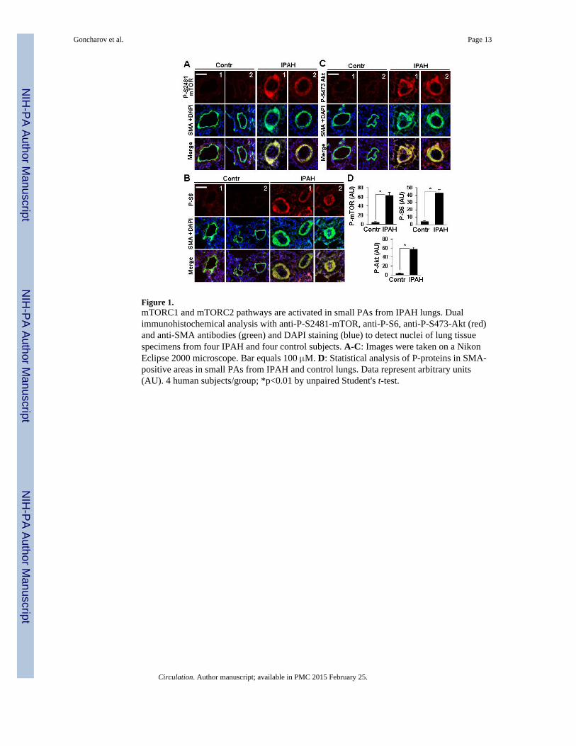
Figure 1.mTORC1 and mTORC2 pathways are activated in small PAs from IPAH lungs. Dualimmunohistochemical analysis with anti-P-S2481-mTOR, anti-P-S6, anti-P-S473-Akt (red)and anti-SMA antibodies (green) and DAPI staining (blue) to detect nuclei of lung tissuespecimens from four IPAH and four control subjects. A-C: Images were taken on a NikonEclipse 2000 microscope. Bar equals 100 μM. D: Statistical analysis of P-proteins in SMA-positive areas in small PAs from IPAH and control lungs. Data represent arbitrary units(AU). 4 human subjects/group; *p<0.01 by unpaired Student's t-test.
Goncharov et al. Page 13
Circulation. Author manuscript; available in PMC 2015 February 25.
NIH
-PA Author Manuscript
NIH
-PA Author Manuscript
NIH
-PA Author Manuscript
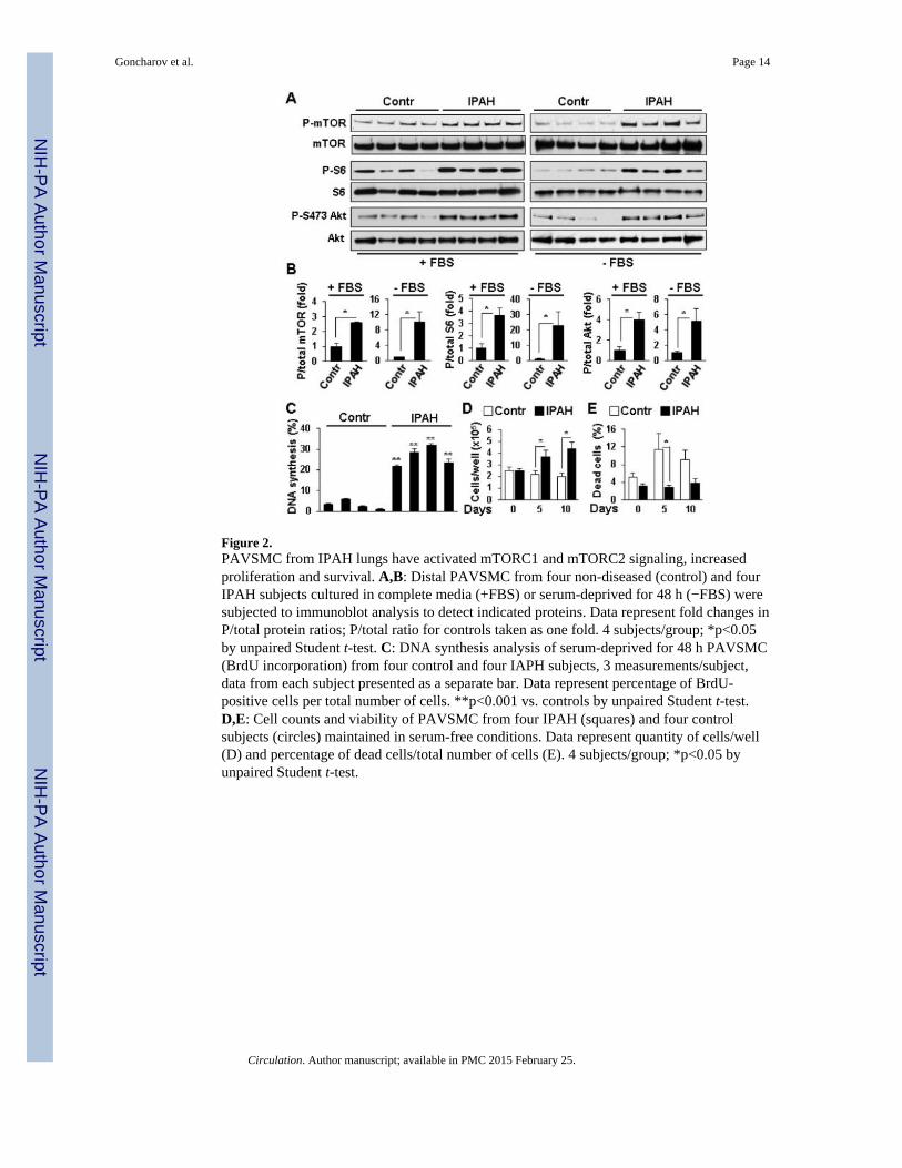
Figure 2.PAVSMC from IPAH lungs have activated mTORC1 and mTORC2 signaling, increasedproliferation and survival. A,B: Distal PAVSMC from four non-diseased (control) and fourIPAH subjects cultured in complete media (+FBS) or serum-deprived for 48 h (−FBS) weresubjected to immunoblot analysis to detect indicated proteins. Data represent fold changes inP/total protein ratios; P/total ratio for controls taken as one fold. 4 subjects/group; *p<0.05by unpaired Student t-test. C: DNA synthesis analysis of serum-deprived for 48 h PAVSMC(BrdU incorporation) from four control and four IAPH subjects, 3 measurements/subject,data from each subject presented as a separate bar. Data represent percentage of BrdU-positive cells per total number of cells. **p<0.001 vs. controls by unpaired Student t-test.D,E: Cell counts and viability of PAVSMC from four IPAH (squares) and four controlsubjects (circles) maintained in serum-free conditions. Data represent quantity of cells/well(D) and percentage of dead cells/total number of cells (E). 4 subjects/group; *p<0.05 byunpaired Student t-test.
Goncharov et al. Page 14
Circulation. Author manuscript; available in PMC 2015 February 25.
NIH
-PA Author Manuscript
NIH
-PA Author Manuscript
NIH
-PA Author Manuscript

Figure 3.Increased ATP levels, proliferation and survival of IPAH PAVSMC depend on glycolyticmetabolism. A: ATP assay performed on PAVSMC from three control and three IPAHsubjects maintained in complete media (+FBS) or serum-deprived for 48 h (−FBS) andtreated with 100 mM 2-DG, 10 μM rotenone or diluent for 24 h. ATP levels in controldiluent-treated cells in complete media are taken as 100%. 3 subjects/group; **p<0.01 by 2-way ANOVA with a post hoc stratified independent t-test with corrections for multiplecomparisons. B,C: DNA synthesis (B) and apoptosis analysis (C) of serum-deprived for 48h cells treated with 100 mM 2-DG, 10 μM rotenone or diluent. Data represent percentage ofBrdU-positive (B) (4 subjects/group) or TUNEL-positive cells (C) (3 subjects/group) pertotal number of cells. *p<0.05, **p<0.01 by 3-way ANOVA (IPAH/control, treatment, time)with a post hoc stratified independent t-test with corrections for multiple comparisons.
Goncharov et al. Page 15
Circulation. Author manuscript; available in PMC 2015 February 25.
NIH
-PA Author Manuscript
NIH
-PA Author Manuscript
NIH
-PA Author Manuscript
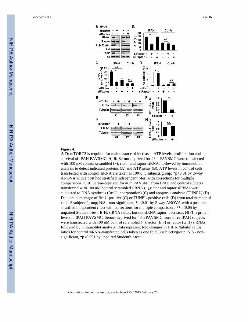
Figure 4.A-D: mTORC2 is required for maintenance of increased ATP levels, proliferation andsurvival of IPAH PAVSMC. A, B: Serum-deprived for 48 h PAVSMC were transfectedwith 100 nM control scrambled (−), rictor and raptor siRNAs followed by immunoblotanalysis to detect indicated proteins (A) and ATP assay (B). ATP levels in control cellstransfected with control siRNA are taken as 100%. 3 subjects/group; *p<0.01 by 2-wayANOVA with a post hoc stratified independent t-test with corrections for multiplecomparisons. C,D: Serum-deprived for 48 h PAVSMC from IPAH and control subjectstransfected with 100 nM control scrambled siRNA (−),rictor and raptor siRNAs weresubjected to DNA synthesis (BrdU incorporation) (C) and apoptosis analysis (TUNEL) (D).Data are percentage of BrdU-positive (C) or TUNEL-positive cells (D) from total number ofcells. 3 subjects/group; N/S - non-significant, *p<0.01 by 2-way ANOVA with a post hocstratified independent t-test with corrections for multiple comparisons; **p<0.05 byunpaired Student t-test. E-H: siRNA rictor, but not siRNA raptor, decreases HIF1 α proteinlevels in IPAH PAVSMC. Serum-deprived for 48 h PAVSMC from three IPAH subjectswere transfected with 100 nM control scrambled (−), rictor (E,F) or raptor (G,H) siRNAsfollowed by immunoblot analysis. Data represent fold changes in HIF1α/tubulin ratios;ratios for control siRNA-transfected cells taken as one fold. 3 subjects/group; N/S - non-significant, *p<0.001 by unpaired Student's t-test.
Goncharov et al. Page 16
Circulation. Author manuscript; available in PMC 2015 February 25.
NIH
-PA Author Manuscript
NIH
-PA Author Manuscript
NIH
-PA Author Manuscript
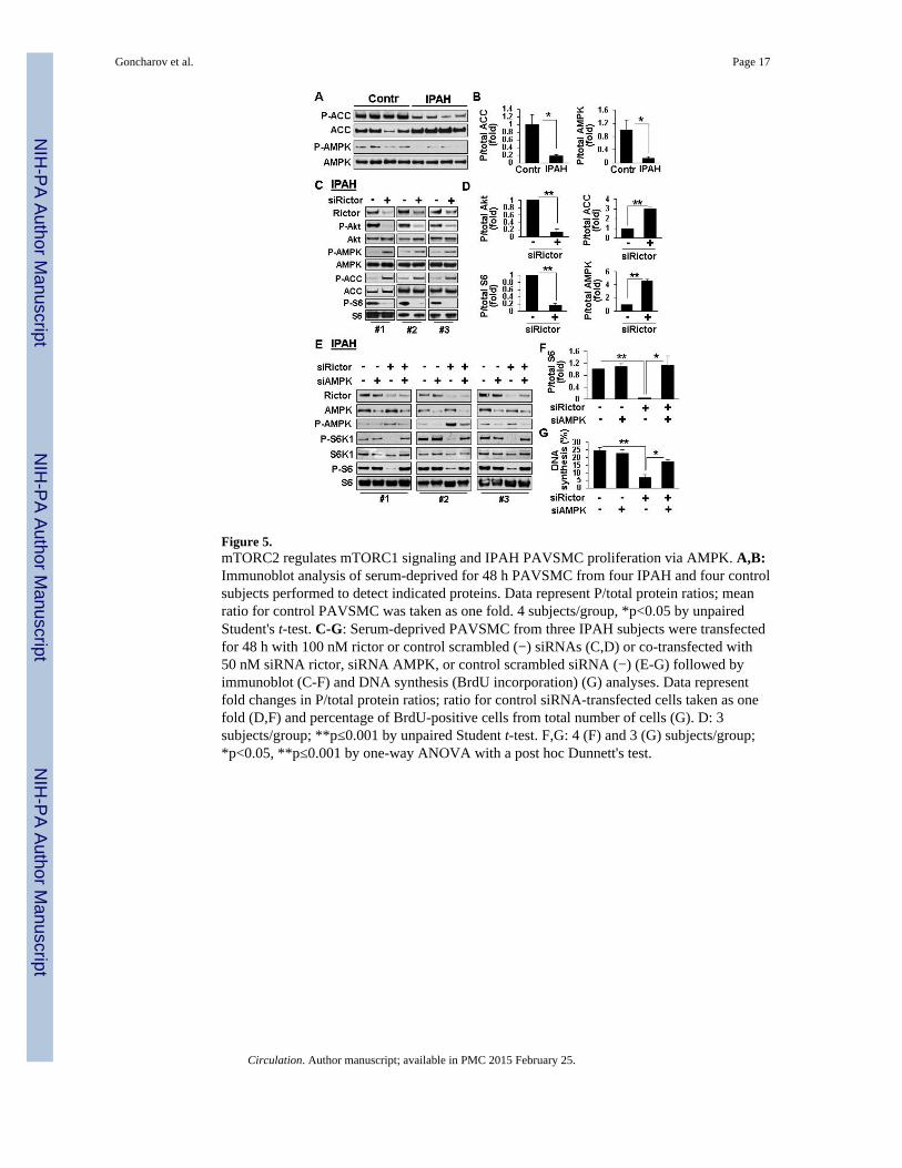
Figure 5.mTORC2 regulates mTORC1 signaling and IPAH PAVSMC proliferation via AMPK. A,B:Immunoblot analysis of serum-deprived for 48 h PAVSMC from four IPAH and four controlsubjects performed to detect indicated proteins. Data represent P/total protein ratios; meanratio for control PAVSMC was taken as one fold. 4 subjects/group, *p<0.05 by unpairedStudent's t-test. C-G: Serum-deprived PAVSMC from three IPAH subjects were transfectedfor 48 h with 100 nM rictor or control scrambled (−) siRNAs (C,D) or co-transfected with50 nM siRNA rictor, siRNA AMPK, or control scrambled siRNA (−) (E-G) followed byimmunoblot (C-F) and DNA synthesis (BrdU incorporation) (G) analyses. Data representfold changes in P/total protein ratios; ratio for control siRNA-transfected cells taken as onefold (D,F) and percentage of BrdU-positive cells from total number of cells (G). D: 3subjects/group; **p≤0.001 by unpaired Student t-test. F,G: 4 (F) and 3 (G) subjects/group;*p<0.05, **p≤0.001 by one-way ANOVA with a post hoc Dunnett's test.
Goncharov et al. Page 17
Circulation. Author manuscript; available in PMC 2015 February 25.
NIH
-PA Author Manuscript
NIH
-PA Author Manuscript
NIH
-PA Author Manuscript
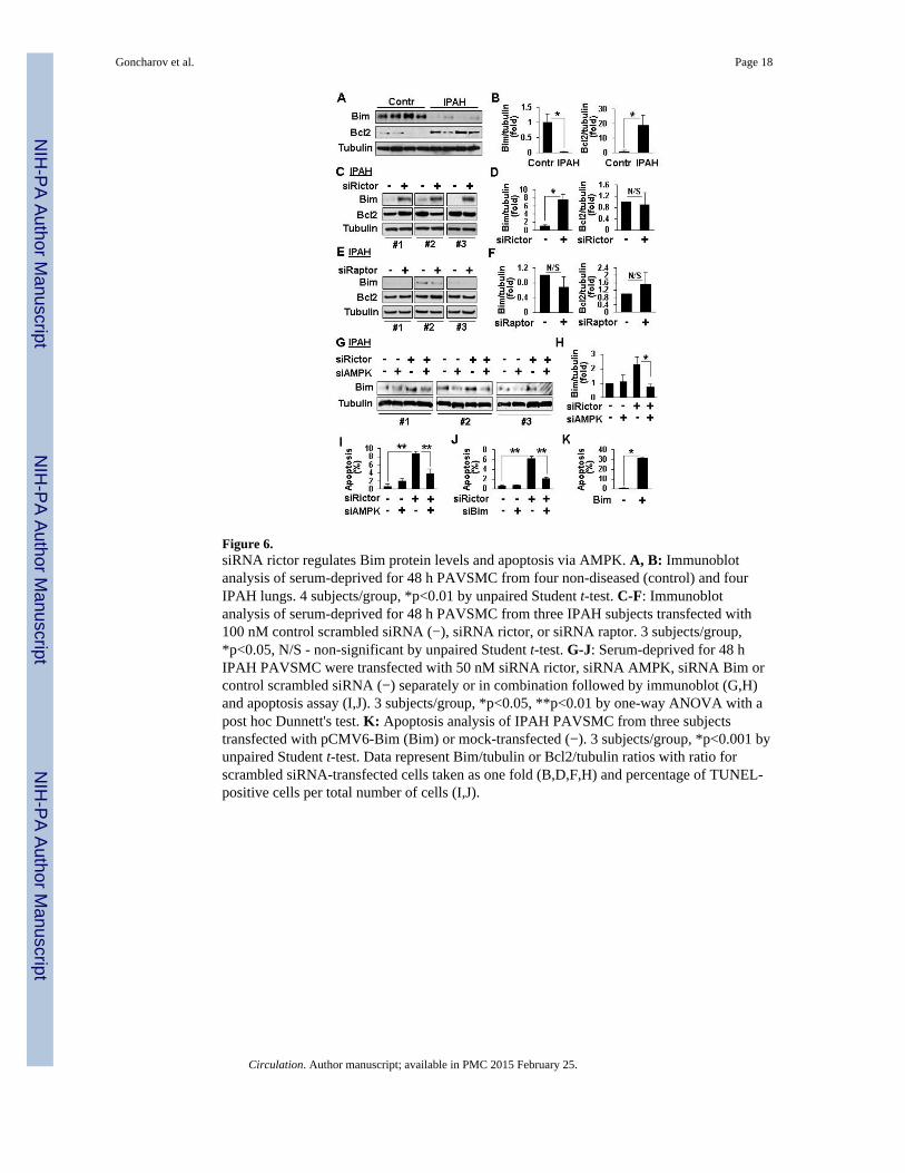
Figure 6.siRNA rictor regulates Bim protein levels and apoptosis via AMPK. A, B: Immunoblotanalysis of serum-deprived for 48 h PAVSMC from four non-diseased (control) and fourIPAH lungs. 4 subjects/group, *p<0.01 by unpaired Student t-test. C-F: Immunoblotanalysis of serum-deprived for 48 h PAVSMC from three IPAH subjects transfected with100 nM control scrambled siRNA (−), siRNA rictor, or siRNA raptor. 3 subjects/group,*p<0.05, N/S - non-significant by unpaired Student t-test. G-J: Serum-deprived for 48 hIPAH PAVSMC were transfected with 50 nM siRNA rictor, siRNA AMPK, siRNA Bim orcontrol scrambled siRNA (−) separately or in combination followed by immunoblot (G,H)and apoptosis assay (I,J). 3 subjects/group, *p<0.05, **p<0.01 by one-way ANOVA with apost hoc Dunnett's test. K: Apoptosis analysis of IPAH PAVSMC from three subjectstransfected with pCMV6-Bim (Bim) or mock-transfected (−). 3 subjects/group, *p<0.001 byunpaired Student t-test. Data represent Bim/tubulin or Bcl2/tubulin ratios with ratio forscrambled siRNA-transfected cells taken as one fold (B,D,F,H) and percentage of TUNEL-positive cells per total number of cells (I,J).
Goncharov et al. Page 18
Circulation. Author manuscript; available in PMC 2015 February 25.
NIH
-PA Author Manuscript
NIH
-PA Author Manuscript
NIH
-PA Author Manuscript
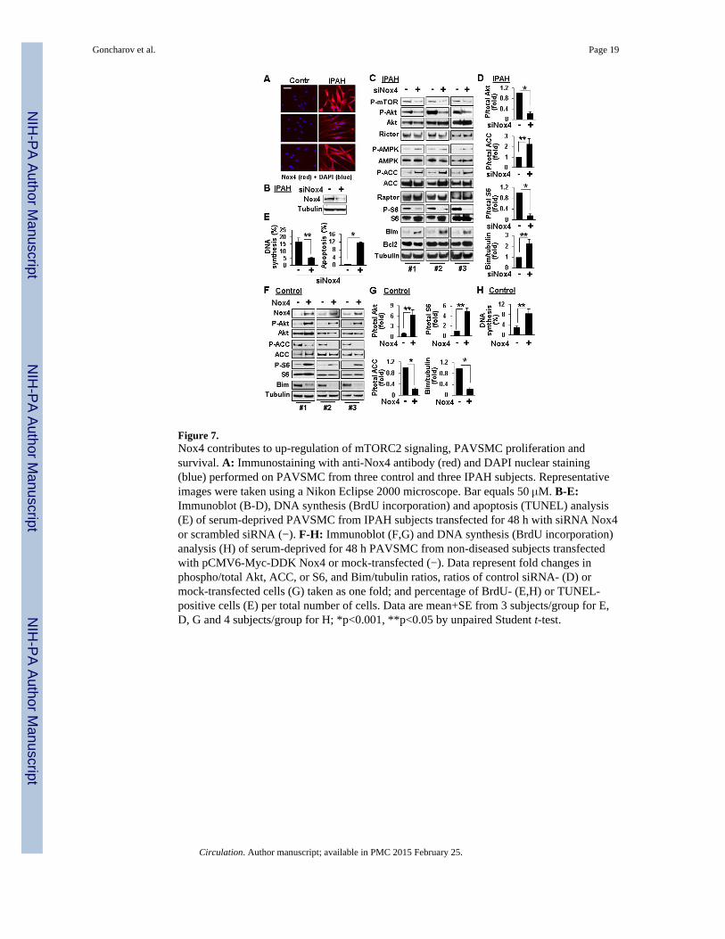
Figure 7.Nox4 contributes to up-regulation of mTORC2 signaling, PAVSMC proliferation andsurvival. A: Immunostaining with anti-Nox4 antibody (red) and DAPI nuclear staining(blue) performed on PAVSMC from three control and three IPAH subjects. Representativeimages were taken using a Nikon Eclipse 2000 microscope. Bar equals 50 μM. B-E:Immunoblot (B-D), DNA synthesis (BrdU incorporation) and apoptosis (TUNEL) analysis(E) of serum-deprived PAVSMC from IPAH subjects transfected for 48 h with siRNA Nox4or scrambled siRNA (−). F-H: Immunoblot (F,G) and DNA synthesis (BrdU incorporation)analysis (H) of serum-deprived for 48 h PAVSMC from non-diseased subjects transfectedwith pCMV6-Myc-DDK Nox4 or mock-transfected (−). Data represent fold changes inphospho/total Akt, ACC, or S6, and Bim/tubulin ratios, ratios of control siRNA- (D) ormock-transfected cells (G) taken as one fold; and percentage of BrdU- (E,H) or TUNEL-positive cells (E) per total number of cells. Data are mean+SE from 3 subjects/group for E,D, G and 4 subjects/group for H; *p<0.001, **p<0.05 by unpaired Student t-test.
Goncharov et al. Page 19
Circulation. Author manuscript; available in PMC 2015 February 25.
NIH
-PA Author Manuscript
NIH
-PA Author Manuscript
NIH
-PA Author Manuscript
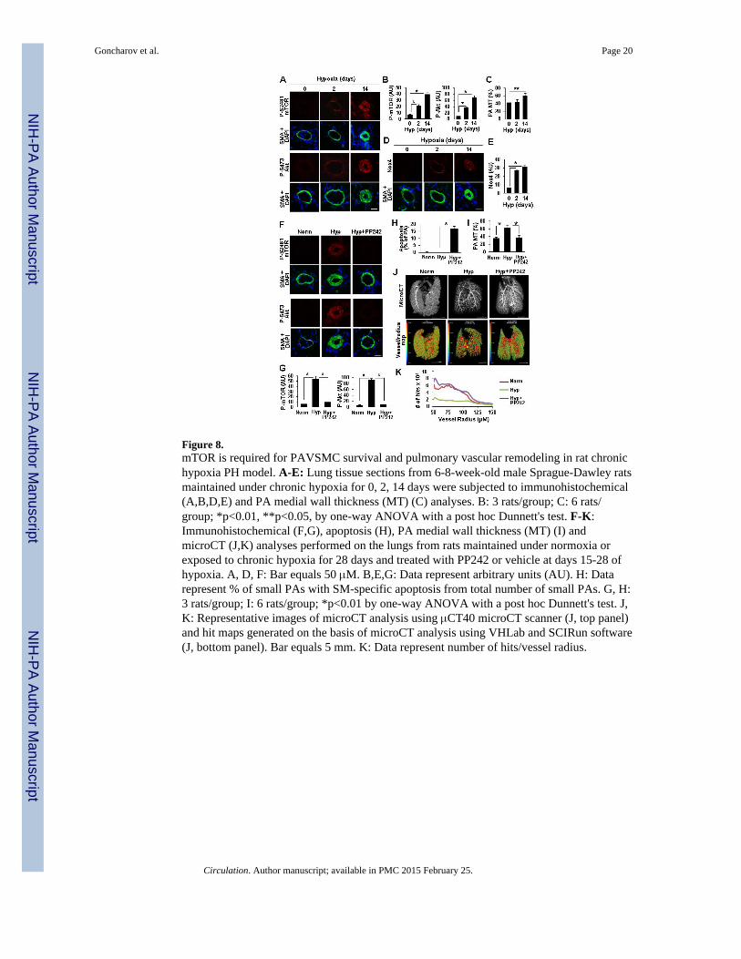
Figure 8.mTOR is required for PAVSMC survival and pulmonary vascular remodeling in rat chronichypoxia PH model. A-E: Lung tissue sections from 6-8-week-old male Sprague-Dawley ratsmaintained under chronic hypoxia for 0, 2, 14 days were subjected to immunohistochemical(A,B,D,E) and PA medial wall thickness (MT) (C) analyses. B: 3 rats/group; C: 6 rats/group; *p<0.01, **p<0.05, by one-way ANOVA with a post hoc Dunnett's test. F-K:Immunohistochemical (F,G), apoptosis (H), PA medial wall thickness (MT) (I) andmicroCT (J,K) analyses performed on the lungs from rats maintained under normoxia orexposed to chronic hypoxia for 28 days and treated with PP242 or vehicle at days 15-28 ofhypoxia. A, D, F: Bar equals 50 μM. B,E,G: Data represent arbitrary units (AU). H: Datarepresent % of small PAs with SM-specific apoptosis from total number of small PAs. G, H:3 rats/group; I: 6 rats/group; *p<0.01 by one-way ANOVA with a post hoc Dunnett's test. J,K: Representative images of microCT analysis using μCT40 microCT scanner (J, top panel)and hit maps generated on the basis of microCT analysis using VHLab and SCIRun software(J, bottom panel). Bar equals 5 mm. K: Data represent number of hits/vessel radius.
Goncharov et al. Page 20
Circulation. Author manuscript; available in PMC 2015 February 25.
NIH
-PA Author Manuscript
NIH
-PA Author Manuscript
NIH
-PA Author Manuscript