organ-specific lymphatic vasculature: from development to
TRANSCRIPT

Review
The Rockefeller University Press J. Exp. Med. 2018 Vol. 215 No. 1 35–49https://doi.org/10.1084/jem.20171868
The
Journ
al o
f Exp
erim
enta
l M
edic
ine
35
IntroductionRich lymphatic vessel (LV) networks supply the skin dermis and mucosal membranes covering major organs, including the respiratory tract, nasopharyngeal cavity, intestine, mes-entery, diaphragm, heart, and lung. LVs are lacking or very sparse in bone, bone marrow, adipose tissue, heart myocar-dium and skeletal muscles, and parenchymal tissues of brain, liver, kidney, and endocrine organs, such as the adrenal or thyroid gland. Presumably, these organs are devoid of LVs because of scarce interstitial fluid or the presence of an al-ternative drainage system, such as fenestrated blood vessels (BVs). Interstitial fluid is drained into specialized blind-ended lymphatic capillaries, which connect and converge into grad-ually larger collecting LVs and lymphatic ducts that empty into the subclavian vein. Lymphatic endothelial cells (LECs) of lymphatic capillaries are surrounded by a thin, discontin-uous basement membrane, lack perivascular cells, and have discontinuous “button-like” cell junctions (Baluk et al., 2007). They readily sense changes in interstitial pressure via special-ized anchoring filaments, which can modulate the opening of “flap valves” in-between the button junctions to allow fluid entry. It is also through these flap valves that immune cells enter lymphatic capillaries. Unidirectional lymph flow in collecting vessels is promoted by numerous intraluminal valves and coordinated contraction of LV smooth muscle cells (SMCs; Schulte-Merker et al., 2011; Sabine et al., 2016). LECs represent a distinct endothelial cell (EC) lineage, and
LVs are frequently distinguished from BVs based on their expression of the transcription factor prospero homeobox-1 (Prox1), transmembrane O-glycoprotein podoplanin (also known as gp38), vascular endothelial growth factor receptor 3 (VEG FR3; also known as Flt4), neuropilin-2, and lymphatic vessel endothelial hyaluronan receptor-1 (LYVE1; Tammela and Alitalo, 2010; Alitalo, 2011; Aspelund et al., 2016).
LVs have traditionally been regarded as passive conduits for fluid and some immune cells, but this perspective has been enormously updated with the discovery of novel structures, origins, and functions of LVs in several organs. Organ-specific lymphatic capillary LECs display remarkable heterogene-ity and plasticity, and acquire specialized functional proper-ties adapted to the local microenvironment. Understanding organotypic LEC differentiation and function can help in designing more effective therapeutic and regenerative strat-egies to cure a wider spectrum of common human diseases in which LVs have been shown to play major roles (Tam-mela and Alitalo, 2010; Alitalo, 2011; Aspelund et al., 2016). Advances in general physiology and pathology, development, lymphedema, and tumor lymphangiogenesis are already cov-ered by excellent recent reviews (Tammela and Alitalo, 2010; Alitalo, 2011; Mortimer and Rockson, 2014; Aspelund et al., 2016; Dieterich and Detmar, 2016; Ulvmar and Mäkinen, 2016; Potente and Mäkinen, 2017). In this review, we aim to summarize the latest results and their significance in under-
Recent discoveries of novel functions and diverse origins of lymphatic vessels have drastically changed our view of lymphatic vasculature. Traditionally regarded as passive conduits for fluid and immune cells, lymphatic vessels now emerge as active, tissue-specific players in major physiological and pathophysiological processes. Lymphatic vessels show remarkable plasticity and heterogeneity, reflecting their functional specialization to control the tissue microenvironment. Moreover, alternative developmental origins of lymphatic endothelial cells in some organs may contribute to the diversity of their functions in adult tissues. This review aims to summarize the most recent findings of organotypic differentiation of lymphatic endothelial cells in terms of their distinct (patho)physiological functions in skin, lymph nodes, small intestine, brain, and eye. We discuss recent advances in our understanding of the heterogeneity of lymphatic vessels with respect to the organ-specific functional and molecular specialization of lymphatic endothelium, such as the hybrid blood-lymphatic identity of Schlemm’s canal, functions of intestinal lymphatics in dietary fat uptake, and discovery of meningeal lymphatic vasculature and perivascular brain lym-phatic endothelial cells.
Organ-specific lymphatic vasculature: From development to pathophysiology
Tatiana V. Petrova1,2 and Gou Young Koh3,4
1Department of Fundamental Oncology, Ludwig Institute for Cancer Research, University of Lausanne, Epalinges, Switzerland2Division of Experimental Pathology, Vaud University Hospital Center, University of Lausanne, Lausanne, Switzerland3Center for Vascular Research, Institute for Basic Science and 4Graduate School of Medical Science and Engineering, Korea Advanced Institute of Science and Technology, Daejeon, Republic of Korea
© 2018 Petrova and Koh This article is distributed under the terms of an Attribution–Noncommercial–Share Alike–No Mirror Sites license for the first six months after the publication date (see http ://www .rupress .org /terms /). After six months it is available under a Creative Commons License (Attribution–Noncommercial–Share Alike 4.0 International license, as described at https ://creativecommons .org /licenses /by -nc -sa /4 .0 /).
Correspondence to Tatiana V. Petrova: [email protected]; Gou Young Koh: [email protected]
Dow
nloaded from http://rupress.org/jem
/article-pdf/215/1/35/1169329/jem_20171868.pdf by guest on 13 D
ecember 2021

Organotypic lymphatic vasculatures | Petrova and Koh36
standing organ-specific lymphatic vasculatures, and highlight the key characteristics and uniqueness of their structure and functions in development, homeostasis, and diseases.
Developmental origins of organ-specific LVsSince the discovery of VEGF-C and its receptor, VEG FR3, as the key lymphangiogenic growth factor pathway and the transcription factor Prox1 as the master of LEC specifica-tion, the development of lymphatic vasculature has been ex-tensively studied and characterized in mouse and zebrafish models (Escobedo and Oliver, 2016; Venero Galanternik et al., 2016). The majority of LECs are produced by transdiffer-entiation from venous endothelium, when a subpopulation of venous ECs expressing Prox1 is specialized into LECs (Es-cobedo and Oliver, 2016; Venero Galanternik et al., 2016). Importantly, recent lineage-tracing studies highlighted an un-expected diversity of LEC origins in several organs. In skin, cervical and thoracic LVs are formed by lymphangiogenic sprouting of Tie2-lineage+ venous-derived LEC progenitors (Martinez-Corral et al., 2015). In contrast, most lumbar LVs are formed via the process of coalescence and vessel net-work formation (so-called lymphovasculogenesis) by isolated Tie2-lineage− nonvenous LEC progenitors. Although the he-matopoietic origin of LECs in such clusters has been con-clusively ruled out (Martinez-Corral et al., 2015), the precise precursor cell type remains to be established. Similarly, during cardiac development, a proportion of LECs in the heart was shown to descend from Tie2-lineage− nonvenous LEC pro-genitors (Klotz et al., 2015). Moreover, mesenteric LVs orig-inate from two distinct LEC sources: Tie2+ venous ECs and isolated EC clusters arising from PDG FB+ and c-Kit+ he-mogenic ECs (Stanczuk et al., 2015). During lymph node (LN) development, all stromal cells, including LECs, have been suggested to arise from nestin+ precursors (Koning et al., 2016). Nestin is a marker of mesenchymal stem cells, but it is also expressed by a variety of other cells types, includ-ing ECs; therefore, the precise origin of LN LECs remains to be fully investigated.
Prox1 and VEGF-C/VEG FR3/ADAM metallopep-tidase with thrombospondin type 1 motif 3/collagen and calcium-binding EGF domains 1 are obligate molecular com-ponents that define LEC fate and LV expansion in all organs; however, there are important organ-specific differences in downstream pathways. For example, blockade of PI3K signal-ing, which acts downstream of activated VEG FR3, selectively affected the development of mesenteric, but not dermal, LVs (Stanczuk et al., 2015). Thus, the origins and heterogeneity of LECs in each organ are more diverse than previously ex-pected, and further exciting findings on this topic are antici-pated in the next decade.
Skin lymphatic vasculatureThe skin is the body’s first line of defense against a hostile environment, including pathogens that can enter upon skin injury. Although the epidermis is avascular, the skin dermis is
rich in BVs and LVs (Fig. 1 A). Dermal LVs are the main con-duits for pathogens and immune cells from the periphery to draining LNs. They also play a major role in reverse cholesterol transport by transporting high-density lipoprotein-bound cholesterol from peripheral tissues to systemic blood circula-tion to regulate cholesterol metabolism (Fig. 1 B; Lim et al., 2013; Martel et al., 2013; Randolph and Miller, 2014). The migration rate of dendritic cells (DCs) and T lymphocytes via dermal afferent LVs is dramatically increased during the peak of inflammation (Fig. 1 C; Kim et al., 2014; Hunter et al., 2016). The majority of immune cells in the lymph of af-ferent LVs are T cells (80–90%) and DCs (10–15%; Hunter et al., 2016). CD4+ T effector memory cells are the main subset that migrates via dermal afferent LVs at steady state, as well as in inflammation, and Foxp3+CD4+ regulatory T cells (T reg cells) constitute up to 25% of the intralymphatic T cell population (Brinkman et al., 2016; Hunter et al., 2016). Tis-sue T reg cells are important regulators of dermal lymphatic function and repair LVs in inflammation and lymphedema (Gousopoulos et al., 2016); it is unclear whether T reg cells also contribute to maintaining normal LV integrity.
Cellular and molecular events during immune cell mi-gration via dermal LVs have been extensively studied and re-viewed (Randolph et al., 2017). C-C motif chemokine ligand 21 (CCL21) produced by LECs is required for DC and T cell trafficking in skin LVs, and likely in most LVs of other or-gans (Girard et al., 2012; Randolph et al., 2017). LECs secrete CCL21 abluminally and luminally, and extracellular CCL21 gradients guide both recruitment and intraluminal directional crawling of DCs (Russo et al., 2016; Randolph et al., 2017). In addition, sphingosine-1 phosphate (S1P) signaling via S1P receptor 1 (S1PR1) regulates a T cell’s decision to egress or remain in tissue (Baeyens et al., 2015). It is unknown whether S1P production by dermal LECs is as important for regula-tion of tissue lymphocyte egress as shown previously for LN LECs (Pham et al., 2010). Vascular cell adhesion molecule 1 (VCAM-1), expressed by inflamed LECs, promotes T cell and DC entry into LVs (Brinkman et al., 2016; Randolph et al., 2017; Teijeira et al., 2017). T reg cells produce especially high levels of lymphotoxin-α2β1, which engages lymphotoxin receptor-β on LECs to promote cell transmigration (Brink-man et al., 2016). Subsequent intralymphatic T cell adhesion and crawling within capillaries are supported by intercellu-lar adhesion molecule 1 (ICAM-1) on LECs and integrin lymphocyte function-associated antigen-1 on T cells (Teijeira et al., 2017). In addition to CCL21, C-X3-C motif chemo-kine ligand 1 and C-X-C motif chemokine ligand 12 are secreted from LECs to guide DC and other innate immune cells trafficking into inflamed LVs (Fig. 1 C; Kabashima et al., 2007; Johnson and Jackson, 2013). The spectrum of chemok-ines and adhesion receptors produced by dermal LECs differ depending on the nature of inflammatory stimuli; for exam-ple, ICAM-1, CXCL9, and CXCL10 are strongly induced in LECs in a contact hypersensitivity model, but not during in-nate immune response elicited by complete Freund’s adjuvant
Dow
nloaded from http://rupress.org/jem
/article-pdf/215/1/35/1169329/jem_20171868.pdf by guest on 13 D
ecember 2021
37JEM Vol. 215, No. 1
(Vigl et al., 2011). Such differential LEC responses are likely important for temporal coordination of tissue retention ver-sus egress of specific immune cell populations during inflam-mation. Lately, a novel role of LYVE1 in DC transmigration has been uncovered (Johnson et al., 2017). In inflamed der-mal interstitium, DCs are coated with hyaluronan and interact with LYVE1 on lymphatic capillary LECs, mediating docking between the two cells within discrete “transmigratory cups” that envelop transiting DCs, thereby facilitating DC trans-migration into LVs (Fig. 1 C; Johnson et al., 2017). Bacterial hyaluronic acid capsule interaction with LEC LYVE1 is also involved in the spread of a common skin-resident pathogen group A streptococcus (GAS) from the infection site to the regional draining LN (Fig. 1 C; Lynskey et al., 2015). Lym-phatic spread of GAS can be detrimental for the host, as it causes lymphangitis and lymphadenitis. However, blocking LN accumulation of GAS in Lyve1−/− mice increased sys-temic dissemination of bacteria in blood circulation (Lynskey et al., 2015), indicating that high levels LYVE1 on LECs may be important for pathogen containment.
Neutrophil egress into dermal LVs is minimal both in the steady-state condition and in sterile inflammation. How-ever, during bacterial infection, neutrophils are the first to
migrate from inflamed skin to draining LN, where they stimulate lymphocyte proliferation (Hampton et al., 2015). Neutrophil migration relies on lymphocyte function–asso-ciated antigen 1, an integrin/complement receptor CD11b and C-X-C or C-C chemokine receptors CXCR4 and/or CCR7 (Fig. 1 C; Gorlino et al., 2014; Hampton et al., 2015; Arokiasamy et al., 2017). Dermis also contains an abundant population of macrophages, which contributes to local con-trol of lymphangiogenesis and lymphatic remodeling during development, inflammation, and wound healing (Harvey and Gordon, 2012; Kim et al., 2014). In adult skin, macro-phages play a unique role in the regulation of salt-sensitive hypertension: in response to increased local osmotic stress, skin macrophages activate nuclear factor of activated T cell 5– dependent production and secretion of VEGF-C (Fig. 1 C). The resulting expansion of cutaneous lymphatic network enhances interstitial electrolyte clearance, reduces peripheral tissue fluid pressure, and restores homeostasis (Machnik et al., 2010; Wiig et al., 2013).
Lymphatic transport is important for normal skin ho-meostasis and is implicated in multiple skin inflammatory diseases, including atopic and contact dermatitis and psori-asis. The density and function of dermal LVs gradually re-
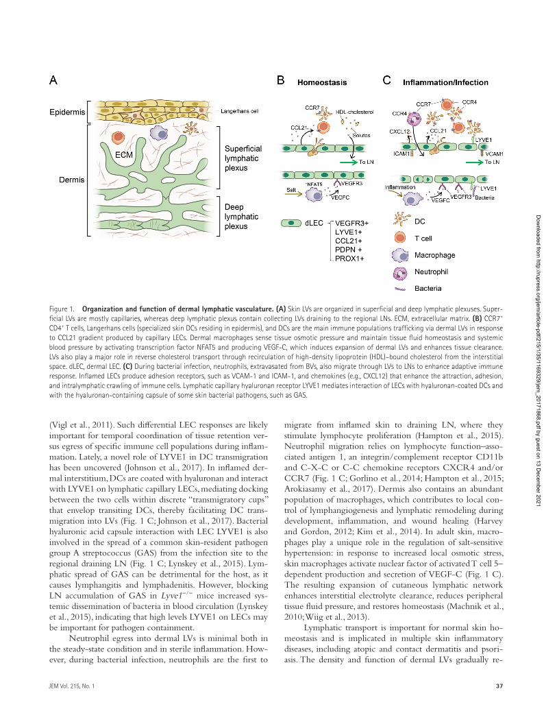
Figure 1. Organization and function of dermal lymphatic vasculature. (A) Skin LVs are organized in superficial and deep lymphatic plexuses. Super-ficial LVs are mostly capillaries, whereas deep lymphatic plexus contain collecting LVs draining to the regional LNs. ECM, extracellular matrix. (B) CCR7+ CD4+ T cells, Langerhans cells (specialized skin DCs residing in epidermis), and DCs are the main immune populations trafficking via dermal LVs in response to CCL21 gradient produced by capillary LECs. Dermal macrophages sense tissue osmotic pressure and maintain tissue fluid homeostasis and systemic blood pressure by activating transcription factor NFAT5 and producing VEGF-C, which induces expansion of dermal LVs and enhances tissue clearance. LVs also play a major role in reverse cholesterol transport through recirculation of high-density lipoprotein (HDL)–bound cholesterol from the interstitial space. dLEC, dermal LEC. (C) During bacterial infection, neutrophils, extravasated from BVs, also migrate through LVs to LNs to enhance adaptive immune response. Inflamed LECs produce adhesion receptors, such as VCAM-1 and ICAM-1, and chemokines (e.g., CXCL12) that enhance the attraction, adhesion, and intralymphatic crawling of immune cells. Lymphatic capillary hyaluronan receptor LYVE1 mediates interaction of LECs with hyaluronan-coated DCs and with the hyaluronan-containing capsule of some skin bacterial pathogens, such as GAS.
Dow
nloaded from http://rupress.org/jem
/article-pdf/215/1/35/1169329/jem_20171868.pdf by guest on 13 D
ecember 2021
Organotypic lymphatic vasculatures | Petrova and Koh38
duces with age, which may contribute to the development of age-related skin disorders (Karaman et al., 2015). Impairment of dermal LV function leads to lymphedema, hyperpigmen-tation, keratosis, and papillomatosis (Carlson, 2014). Studies of animal models of chronic skin inflammation demonstrated that blocking LV expansion exacerbates the disease, whereas stimulation of lymphangiogenesis by VEGF-C administration reduces it (Huggenberger et al., 2010).
Meningeal lymphatic vasculatureThe brain parenchyma lacks LVs and instead has an alternative but essential fluid drainage system, the so-called glymphatic system (Iliff et al., 2012). The glymphatic system is named for its dependence on glial cells for a draining function similar to the lymphatic system. The glymphatic system promotes waste clearance from the brain by expediting the flux of cerebrospi-nal fluid across the brain parenchyma, which is ∼60% more efficient during sleep (Iliff et al., 2012). However, recent re-discovery of the mouse brain meninges lymphatic networks raised exciting questions and intriguing concepts (Aspelund et al., 2015; Louveau et al., 2015), and similar structures were also found in autopsy specimens of human meninges (Absinta et al., 2017). Importantly, high-resolution, clinical magnetic resonance imaging technique has enabled the visualization of human meningeal lymphatic networks (Absinta et al., 2017). In fact, the existence of lymphatic vasculature on the sur-face of the human brain had been proposed two centuries ago and displayed in anatomical wax model collections (the Josephinum Wax Models Museum, Vienna, Austria; Lukić et al., 2003), but it has been mostly ignored. Most meningeal LVs are located on the meningeal layer (also called “dura mater”) and are phenotypically similar to lymphatic capillar-ies in other organs (Fig. 2; Aspelund et al., 2015). However, valves are present only in LVs near the base of the skull along-side the cranial nerves. Activation of VEG FR3 by VEGF-C increases the diameter of meningeal LVs, and blocking VEG FR3 signaling completely abrogates meningeal LVs, in-dicating that these vessels, like peripheral LVs, are regulated by VEG FR3 (Aspelund et al., 2015). A recent study (Antila et al., 2017) demonstrated that VEGF-C–VEG FR3 signaling is crucial not only for development but also for maintenance of meningeal LV integrity during adulthood. Functionally, some cerebrospinal fluid and interstitial fluid of brain parenchyma flow through glymphatic system, drain into meningeal LVs, and reach the deep cervical LNs. Intriguingly, a significant decline in cerebrospinal fluid outflow via meningeal LVs was observed in aged mice (Ma et al., 2017), implying that menin-geal lymphatic networks could be targeted for age-associated neurological conditions. Further research is required to map out the connections among this newly uncovered lymphatic circuit, the glymphatic system, and the rest of the lymphatic circuitry throughout the body (Louveau et al., 2017). It would also be intriguing to determine whether cerebrospinal fluid drainage into meningeal LVs also increases during sleep. Moreover, it is reasonable to speculate that impairment in this
lymphatic circuit could be related to brain edema, which is often detected during ischemia, concussions, inflammation, and brain tumors. Furthermore, it will be important to es-tablish whether meningeal LVs are involved in the clearance of amyloid β and other misfolded proteins from the brain parenchyma, which are frequently and highly accumulated in patients with neurodegenerative diseases such as Alzheimer’s and Parkinson’s diseases.
The brain has also been regarded as an immune-privileged organ, in part because of the dearth of LVs. It was thought that infiltrated T cells in the brain exit through venous rather than lymphatic circulation, which is the case in all other or-gans (Ransohoff and Engelhardt, 2012). However, a large population of T cells was observed in the lumen of meningeal LVs, raising the intriguing possibility of an alternative gate-way for T cells to egress from the brain to the deep cervical LNs (Louveau et al., 2015). Dissecting the lymphatic route for brain-resident T cells will advance our understanding of the communication between the central nervous system and the immune system and unveil new therapeutic targets for inflammatory neurological disorders, including encephalo-myelitis and multiple sclerosis (Louveau et al., 2017). Another question requiring further investigation is whether meningeal LVs can act as a route for brain cancer metastasis. Thus, the rediscovery of meningeal LVs reveals new concepts regarding brain functions and diseases.
Brain mural LECs (muLECs)In 2017, three research groups (Bower et al., 2017; van Lessen et al., 2017; Venero Galanternik et al., 2017) independently identified a novel population of isolated muLECs (also called mannose receptor-1+ perivascular cells) surrounding men-ingeal BVs using transgenic zebrafish models (Fig. 2). The muLECs express LEC markers such as LYVE1 and Prox1, and a perivascular macrophage marker, mannose receptor-1, but, unlike all other ECs, they do not form a lumenized vessel. muLECs are uniquely found on the inner side of the meninx, adjacent to the brain parenchyma. They originate from the lymphatic endothelium in the midbrain or the optic choroidal vascular plexus by sprouting lymphangiogenesis during brain development, and differentiate into a dispersed, nonlumenized mural lineage. Similar to other LECs, muLEC development requires signaling through the VEGF-C–VEGF-D–collagen and calcium-binding EGF domains 1–VEG FR3 pathway. Mature muLECs are relatively large mural cells that produce vascular growth factors and accumulate low-density lipopro-teins from the bloodstream. Although muLECs seem to be important for meningeal vascularization, they are dispens-able for the maintenance of vessel integrity. These studies identified a novel lymphatic lineage that differentiates into mural cell-like cells that are necessary for establishing normal meningeal blood vasculature, and have raised the possibility that an equivalent cell type exists in the mammalian brain (Bower et al., 2017; van Lessen et al., 2017; Venero Galan-ternik et al., 2017). Although the three groups did not have
Dow
nloaded from http://rupress.org/jem
/article-pdf/215/1/35/1169329/jem_20171868.pdf by guest on 13 D
ecember 2021

39JEM Vol. 215, No. 1
the same consensus opinion, it is tempting to speculate that muLECs may be the equivalent of lipid-laden cells, perivas-cular macrophages, fluorescent granular perithelial cells, or ‘Mato Cells’ (Mato et al., 1981, 1996; Williams et al., 2001) that take up lipids and low-density lipoproteins from men-ingeal BVs in mammals. In fact, muLECs become elongated and accumulate a larger amount of lipid droplets after acute high-cholesterol diet. Moreover, they are able to take up and clear macromolecules through mannose receptors (van Lessen et al., 2017; Venero Galanternik et al., 2017). Further compar-ative investigation between fish and mammals is required to understand the roles and significance of muLECs.
Schlemm's canal (SC): Lymphatic intermediate and glaucomaSC is an endothelium-lined channel that encircles the cornea and provides unique vascular route for aqueous humor out-
flow, which constantly refreshes the anterior chamber of eye (Fig. 3 A). Aqueous humor, continuously produced by the ciliary body, flows into the anterior chamber, and is drained into the aqueous and episcleral veins through trabecular mesh-work and SC. SC has morphological, molecular, and functional similarities with LVs. In 2014, several independent research groups (Aspelund et al., 2014; Kizhatil et al., 2014; Park et al., 2014; Truong et al., 2014) found that SC represents a new intermediate vessel type, which expresses LEC markers Prox1, VEG FR3, and integrin α9, but not LYVE1 and podoplanin. The primitive SC is formed by blood ECs sprouting from the choroidal vein, and SC ECs are postnatally respecified to acquire lymphatic phenotypes through up-regulation of Prox1, which also appears to act as a molecular biosensor for aqueous humor outflow (Park et al., 2014). Importantly, Tie2 is expressed before Prox1 in SC ECs and is maintained at a high level to critically regulate SC integrity during adulthood
Figure 2. Meningeal lymphatic vasculature and brain mural LECs. Localization and distribution of LVs in dura matter of mouse brain. Cerebrospinal fluid (CSF) and interstitial fluid of brain parenchyma drain into meningeal LVs and reach the deep cervical LNs. Trafficking of brain immune cells, including T cells, DCs, and macrophages, and clearance of neurotoxic misfolded proteins and peptides, such as amyloid β, are among the potential important functions of meningeal LVs. A unique, LV-derived population of muLECs surrounding brain BVs was recently found in zebrafish. muLECs express Prox1, LYVE1, and mannose receptor 1 (MR1), and are highly endocytotic. Unlike all other EC, muLECs do not form cell–cell junctions or lumenized structures. Whether muLECs are equivalent to perivascular Mato cells (LYVE1+, Prox1−, MR1+, and lipid droplet+), found in mammalian brain, remains to be established. Perivascular Mato cells are also detected in surrounding meningeal LVs.
Dow
nloaded from http://rupress.org/jem
/article-pdf/215/1/35/1169329/jem_20171868.pdf by guest on 13 D
ecember 2021

Organotypic lymphatic vasculatures | Petrova and Koh40
(Kim et al., 2017). Thus, SC ECs seem to originate from Tie2+ venous EC progenitors (Aspelund et al., 2014; Kizhatil et al., 2014; Park et al., 2014). Another important regulator of SC is VEGF-C/VEG FR-3 signaling, which is critical for the forma-tion and differentiation of SC. Surprisingly, supplementation of VEGF-C can also induce SC growth in adults, leading to a reduction of intraocular pressure in the aged glaucoma model (Aspelund et al., 2014).
Aqueous humor is rapidly drained transcellularly through SC ECs causing formation of giant vacuoles (Fig. 3, B and C). When SC is impaired, aqueous humor drainage is impeded and intraocular pressure is increased, ultimately leading to glaucoma (Jonas et al., 2017). In this regard, glau-coma induced by defective SC could also be regarded as ‘eye lymphedema’. However, the pathogenesis involving SC defects in glaucoma is still poorly understood. Intriguingly, primary congenital glaucoma phenotypes were detected in mouse models of double Angpt1/Angpt2 deletions or Tie2 deletion during postnatal periods, highlighting the impor-
tance of the angiopoietin (Angpt)-Tie2 system in SC de-velopment (Thomson et al., 2014). In fact, Tie2 mutations have been identified in patients with primary congeni-tal glaucoma (Souma et al., 2016). Nevertheless, although the incidence rate of primary congenital glaucoma is low, primary open-angle glaucoma is frequently observed in the elderly. A recent study (Kim et al., 2017) showed that double Angpt1/Angpt2 deletions or Tie2 deletion in adult mice severely impairs SC integrity and transcellular aque-ous humor fluid transcytosis, leading to elevated intraocular pressure, retinal neuron damage, and impairment of retinal ganglion cell function, which are all hallmarks of primary open-angle glaucoma. Accordingly, Tie2 reactivation using a Tie2 agonistic antibody relieved the phenotype in double Angpt1/Angpt2-deleted mice and rejuvenated the SC in aged mice (Kim et al., 2017). These findings provide not only a novel molecular pathway in understanding pathogenesis of primary open-angle glaucoma but also a new therapeutic avenue for its treatment.
Figure 3. Schlemm's canal. (A) SC is an endothelium-lined channel that encircles the cornea and provides an exit route for aqueous humor. (B) Aqueous humor is produced from the ciliary body and drained into aqueous and episcleral veins through the trabecular meshwork and SC. (C) Aqueous humor is drained transcellularly and transported from the basal to luminal side through SC ECs, causing formation of giant vacuoles. SC ECs have an intermediate blood-lymphatic EC phenotype and express Prox1, VEG FR3, Tie2, and integrin α9, but not LYVE1 or podoplanin. Angpt1+ stromal cells adjacent to the SC LECs may produce proteins of trabecular meshwork. When SC function is impaired, aqueous humor drainage is impeded and intraocular pressure is increased, ultimately leading to glaucoma. Angpt–Tie2 signaling maintains SC integrity, and loss of such signaling induces primary congenital and open-angle glau-coma. AHO, aqueous humor outflow; E & A vein, episcleral & aqueous vein; VEC, venous endothelial cell.
Dow
nloaded from http://rupress.org/jem
/article-pdf/215/1/35/1169329/jem_20171868.pdf by guest on 13 D
ecember 2021

41JEM Vol. 215, No. 1
Sinusoidal LVs in LNsLNs are highly dynamic secondary lymphoid organs where antigens, together with costimulatory signals, are delivered by afferent LVs (Fig. 4 A). LN LVs are extended lymphatic networks from peripheral afferent LVs, which continue to form the subcapsular sinus (SCS), stretch into the medullary sinus, and ultimately exit as efferent LVs. LVs traverse through densely packed aggregations of immune cells, predominantly B and T cells and such architecture facilitates intimate in-teraction between LN LVs and immune cells, directly influ-encing immune responses. Thus, LN LVs efficiently transport antigens and innate immune cells from various organs to naive lymphocytes in LNs, which is one of the crucial steps for the initiation and regulation of adaptive immune response as well as for the maintenance of immune tolerance (Junt et al., 2008; Randolph et al., 2017). During the acute phase of local tissue inflammation, robust lymphangiogenesis, stimu-lated by VEGF-A, C and D secreted from infiltrated, activated macrophages, occurs in the draining LN, and subcapsular LN LVs proliferate and penetrate deep into the cortex (Kataru et al., 2009). In this situation, activated B cells also contrib-ute to LN lymphangiogenesis to promote dendritic cell (DC) mobilization from the inflamed tissue to LN (Angeli et al., 2006). As shown in helminth infection model, VEGF-A and VEGF-C production by B cells and mesenteric LN lymph-angiogenesis relies on lymphotoxin-dependent feed-forward cross-talk of B-cells and surrounding follicular reticular cells (Dubey et al., 2017). Interferon-γ secreted from activated T cells can be a negative but balancing regulator that suppresses LN lymphangiogenesis during inflammation resolution (Kataru et al., 2011).
LN LECs produce several factors that regulate leuko-cyte trafficking, delivery of inflammatory signals, modulation of T cell activation, and development of secondary lymphoid organs. LECs regulate the exit of immune cells via efferent LVs by secreting S1P, generated by sphingosine kinases with the S1P lyase enzyme, creating an S1P gradient important for immune cell egress through efferent LVs (Fig. 4 A; Pham et al., 2010). In addition to its role in T cell trafficking, LN LEC-derived S1P promotes naive T cell survival by sustaining their mitochondrial function and oxidative phosphorylation (Mendoza et al., 2017). Integrin α9β1 on LECs is also neces-sary for LN lymphocyte egress, and the interaction between integrin α9β1 and its ligand tenascin-C has been shown to enhance the production of S1P by LECs (Ito et al., 2014). LN LECs are also an important source of interleukin-7, critical for lymphocyte expansion during LN remodeling (Onder et al., 2012). Interleukin-7 expression by LECs also provides a niche for memory T-helper cells in inducible bronchus-associated lymphoid tissues during allergic inflammation in lung (Shi-noda et al., 2016). LN LECs produce immunosuppressive factors, such as TGF-β, indoleamine-2,3-dioxygenase, and nitric oxide to maintain peripheral tolerance to self-antigen in lymph (Fig. 4 B; Lukacs-Kornek et al., 2011; Malhotra et al., 2012). Of note, LN LECs directly express peripheral
tissue antigens and induce deletional tolerance in CD8+ T cell via PD-1–PD-L1 or LAG-3–MHC-II pathways (Rou-hani et al., 2015). Moreover, a subset of LECs was shown to provide antigens to DCs, which then induced CD4+ T cell anergy (Rouhani et al., 2015). On the other hand, Dubrot et al. (2014) also reported that LECs can acquire preloaded pep-tide–MHC-II complexes from DCs to induce CD4+ T cell tolerance. Overall, LN LECs play multiple tolerogenic roles against self-antigens by directly presenting a variety of pe-ripheral tissue antigens for CD8+ T cell deletional tolerance, whereas they also induce CD4+ T cell tolerance via antigen transfer to or from DCs (Fig. 4 B).
Tumors produce lymphangiogenic growth factors, tumor antigens, and premetastatic signals to tumor draining LNs even before LN metastasis (Karaman and Detmar, 2014; Ogawa et al., 2014; Dieterich and Detmar, 2016). COX-2/PGE2-EP3–dependent induction of VEGF-C and VEGF-D in macrophages and DCs induces tumor-induced LN lymphan-giogenesis (Ogawa et al., 2014). Interestingly, LECs of tumor draining LNs are capable of scavenging and cross-presenting tumor antigens, which induce dysfunction of CD8+ T cells (Lund et al., 2012). Tumor and tumor draining LN–derived VEGF-C promotes these processes and accelerates metastasis (Lund et al., 2012). In addition to follicular DCs, proliferat-ing SCS LECs can capture or act as a reservoir for persistent viral antigens, contributing to the development of an effective memory CD8+ T cell pool with increased effector function and protective capacity (Fig. 4 B; Tamburini et al., 2014). This beneficial role of LN LECs can be exploited to improve effi-cacy of vaccines against viral infections.
SCS LECs and medullary sinusoidal LECs have distinct features including cellular organization, expression profiles, roles, and responses to inflammation (Fig. 4 A; Iftakhar-E-Khuda et al., 2016). For instance, macrophage scavenger receptor 1 is selectively expressed by SCS LECs and regulates the binding and transmigration of lymphocytes entering from peripheral tissues into LN parenchyma, whereas endomucin produced by medullary sinusoidal LECs presumably regu-lates lymphocyte trafficking via L-selectin (Iftakhar-E-Khuda et al., 2016). Medullary sinusoidal LECs also produced the highest levels of peripheral tissue antigens and PD-L1 and therefore play a key role in the deletion of alloreactive CD8+ T cells (Cohen et al., 2014). SCS LECs, but not peripheral LECs, express plasmalemma vesicle-associated protein (also known as MECA-32; Rantakari et al., 2015). The SCS floor allows the entrance of only small (<70 kD and <4 nm) sol-utes, which are then rapidly transported through the LN pa-renchyma to BVs by conduits, a specialized tubular meshwork formed by extracellular matrix from follicular reticular cells. Plasmalemma vesicle-associated protein plays a central role in such selective barrier function of subcapsular sinusoids by forming molecular “sieves” or diaphragms covering transen-dothelial channels in SCS LECs, which then prevents the passage of larger solutes to follicular reticular cell conduits (Fig. 4 A; Rantakari et al., 2015).
Dow
nloaded from http://rupress.org/jem
/article-pdf/215/1/35/1169329/jem_20171868.pdf by guest on 13 D
ecember 2021

Organotypic lymphatic vasculatures | Petrova and Koh42
A substantial degree of specialization is also present in the SCS LECs, where LECs facing the LN capsule (or “ceiling” LECs) express high levels of a CCL21/CCL19 decoy chemokine receptor CCRL1. Polarized CCRL1 ex-pression creates a CCL21 gradient, allowing the directional DC migration into the LN paracortex to reach T cells (Ulvmar et al., 2014). Ceiling LECs are LYVE1 negative and also produce CCL1 chemokine (Das et al., 2013), whereas floor LEC are LYVE1 positive. The floor LEC layer is interspersed with CD169+ SCS macrophages, which take up antigens and pathogens (Fig. 4 A). Interestingly, although DCs are able to transmigrate directly through the floor of the SCS, naive T cells use the medullary sinus to reach LN parenchyma; however, transmigrating DCs also induce
structural alterations in the SCS floor, which allows sub-sequent homing of T cells through the SCS (Braun et al., 2011). Onder et al. (2017) recently highlighted a new role of LECs in LN formation. Different from the prevailing LN formation model (Mebius, 2003; van de Pavert and Mebius, 2010), this study reports that that initiation of LN devel-opment requires lymphoid tissue inducer cell-mediated activation of LECs and that the engagement of mesenchy-mal stromal cells is a subsequent event. Mechanistically, LN initiation is mediated mainly through receptor activator of the NF-κB signaling–noncanonical NF-κB pathway in LVs and is driven by CCL21 and S1P receptor–dependent retention of lymphoid tissue inducer cells in the LN an-lage (Onder et al., 2017).
Figure 4. LN lymphatic vasculature. (A) Afferent LVs deliver lymph carrying antigens and immune cells to the LN SCS. From the SCS, lymph flows to the cortical and medullary sinuses and exits via efferent LVs. SCS “ceiling” LECs (cLECs) express the decoy CCL19/CCL21 receptor CCRL1, which creates a gradient of CCL21 enhancing transmigration of DCs into T zones. CD169+ SCS macrophages inserted in the “floor” LEC (fLEC) layer take up antigens and pathogens. The majority of lymph drains via LN LVs, whereas a proportion of small solutes is absorbed from lymph via specialized conduits, a network of collagen fibrils, surrounded by follicular reticular cells. Molecular diaphragms formed by plasmalemma vesicle-associated protein restrict access of larger substances into conduits. Similar to fLECs, medullary sinus LECs are in close contact with macrophages, which clear pathogens and antigens. S1P produced by medullary and cortical sinuses LECs induces egress of lymphocytes into efferent LVs and promotes T cell survival. (B) Specialized functions of LN LECs. LN LECs contribute to maintenance of tolerance against self-antigens through the expression of peripheral tissue antigens and deletion of self-reactive CD8+ T cells. LN LECs can tolerize CD4+ cells either by transferring peptide–MHC-II complexes to DCs or acquiring them from DCs. LN LECs also produce immunosuppressive nitric oxide (NO) and indoleamine-2,3-dioxygenase (IDO1), which restrains proliferation of activated T cells. During acute viral infection, proliferating LN LECs uptake and store viral antigens. During the LN contraction stage, such antigens are transferred by dying LECs to DCs, which cross-present them to T cells to promote CD8+ T cell memory responses. Ag, antigen; FRC, follicular reticular cell; PLV AP, plasmalemma vesicle–associated protein.
Dow
nloaded from http://rupress.org/jem
/article-pdf/215/1/35/1169329/jem_20171868.pdf by guest on 13 D
ecember 2021

43JEM Vol. 215, No. 1
Lacteals and lymphatic vasculature in the small intestineIn addition to the common task of all LVs in transporting interstitial fluid and immune cells, the intestinal lymphatic vasculature plays a major role in the uptake and transport of dietary fat. In mammals, dietary lipids are repackaged in enterocytes into large (200–1,000 nm) triglyceride-loaded particles or “chylomicrons,” which are secreted basally into intestinal stroma. In addition to triglycerides, chylomicrons can also incorporate fat-soluble vitamins and drugs, as well as some microbiota components, such as bacterial lipopoly-saccharides (Bernier-Latmani and Petrova, 2017). The single lymphatic capillary located at the center of each small intes-tinal villus, called a lacteal, takes up chylomicrons and other interstitial fluid components from villi and transports them to the submucosal and mesenteric collecting LVs (Fig. 5, A and B). The lymphatic vascular plexus, located in the intes-tinal muscular layer, drains independently to the mesenteric
collecting LVs. Intestinal lymph is transported to the mes-enteric LNs and thoracic duct and is subsequently released into the blood circulation (Fig. 5 A). Through these transport processes, lipid-soluble drugs can directly access the target or-gans while bypassing the liver, where most of drugs are de-graded (Trevaskis et al., 2015). The mechanism and selectivity of chylomicron uptake into lacteals are not entirely clear and may involve both intercellular transport through LEC junctions and intracellular transport across LECs in vesicles (Bernier-Latmani and Petrova, 2017). Periodic squeezing of lacteals, which is mediated by the contraction of surround-ing longitudinal smooth muscle cells in intestinal villi con-trolled by the autonomic nervous system, allows efficient drainage of absorbed lipids into the collecting vessel net-work (Choe et al., 2015).
Although the majority of adult LVs are quiescent, in-testinal lacteals exhibit low but detectable proliferation under
Figure 5. LVs of the small intestine. (A) Intestinal lacteals are positioned in the middle of intestinal villi. Smooth muscle cells in the villi are closely associated with lacteals, and their contractions promote lymph uptake and transport. Another lymphatic vascular plexus is located in the intestinal muscular layer. Lymph from both plexuses is drained to mesenteric collecting vessels, mesenteric LNs, and the thoracic duct and returned to the blood circulation. (B) Intestinal lacteals transport chylomicrons, cholesterol, gut hormones, and immune cells. Lymphatic trafficking of CD103+ DCs carrying food antigens and apoptotic intestinal epithelial cells to mesenteric LN drives the development of T reg cells and tolerance. The role of LVs trafficking in intestinal ILC3 function is not understood. (C) Role of villus SMCs in the maintenance of intestinal LVs. VEGF-C, produced by villus SMCs, and VEGF-C–dependent DLL4 signaling in LECs fuel continuous lacteal regeneration. Periodic contraction of villus SMCs, controlled by the autonomic nervous system, promotes lymph transport. Ag, antigen; IEC, intestinal epithelial cell; TD, thoracic duct.
Dow
nloaded from http://rupress.org/jem
/article-pdf/215/1/35/1169329/jem_20171868.pdf by guest on 13 D
ecember 2021

Organotypic lymphatic vasculatures | Petrova and Koh44
steady-state conditions and frequently harbor filopodia, in-dicating an ongoing lymphangiogenic response (Bernier- Latmani et al., 2015; Bernier-Latmani and Petrova, 2016). Mechanistically, maintenance of lacteal integrity and fat trans-port function requires continuous Notch and VEGF-C–VEG FR-3 signaling, with VEGF-C being supplied by the sur-rounding smooth muscle cells (Fig. 5 C; Bernier-Latmani et al., 2015; Nurmi et al., 2015). Lacteals are characterized by a mix of both continuous zipper junctions and discontinuous button-like junctions, and loss of Notch signaling impairs the formation of mature button-like junctions (Bernier-Latmani et al., 2015). Optimal junctional organization and transport function of lacteals also require adrenomedullin-calcitonin receptor signaling; lymphatic-specific inactivation of calci-tonin receptor results in intestinal lymphangiectasia and pro-tein-losing enteropathy (Davis et al., 2017). Identification of the specific lacteal transcriptome will be important to further understand the molecular mechanisms underlying the func-tional specialization of lacteals.
The intestine harbors a large population of immune cells whose task is to maintain and fine-tune the balance between immune tolerance to the myriad of commensal intestinal bac-teria and ingested harmless antigens and responses against po-tential pathogens. Similar to other tissues, CCR7-expressing DCs migrate into intestinal lymphatics in response to the CCL21 gradient generated by LECs. The transport of food antigens by CD103+ DCs to mesenteric LNs appears to be a key step in the establishment of oral tolerance through the induction of mesenteric LN T reg cells (Pabst and Mowat, 2012). Production of such T reg cells is also maintained by lymphatic trafficking of DCs after ingestion of apoptotic in-testinal epithelial cells, which are continuously produced as a result of rapid intestinal epithelium turnover (Cummings et al., 2016). The CCR7+Rorgt+ subset of intestinal innate lymphoid cells also egresses via intestinal LVs to mesenteric LNs, but the functional significance of such transport requires further investigation (Fig. 5 B; Mackley et al., 2015). Given the importance of T cell trafficking via dermal LVs, it would also be interesting to characterize lymphatic transport of in-testinal T cell subsets.
The role of LVs in intestinal pathological states, for ex-ample in inflammatory bowel disease (IBD), is attracting in-creasing attention (von der Weid et al., 2011; Bernier-Latmani and Petrova, 2017). Increased lymphangiogenesis and lym-phatic vascular dysfunction, such as lymphangiectasia and intralymphatic lymphocyte stasis, have long been described as pathological features of Crohn’s disease (von der Weid et al., 2011). Data from animal models suggest that damage to lymphatic vasculatures and its unproductive expansion may be contributing or perhaps even initiating factors in IBD (Bernier-Latmani and Petrova, 2017). Diphtheria toxin–me-diated conditional ablation of LVs in the intestine leads to rapid animal demise because of extreme disruption to the in-testinal mucosal barrier and septic shock (Jang et al., 2013). Furthermore, impaired intestinal lymphatic vascular func-
tion or lymphangiogenic response exacerbates intestinal in-flammation and worsens symptoms in experimental models of colitis (von der Weid et al., 2011; D’Alessio et al., 2014; Davis et al., 2017). Mice deficient in D6 decoy chemokine receptor, which is highly expressed by LECs and is neces-sary to restrict aberrant inflammatory leukocyte adhesion to LVs, develop more severe colitis, further underscoring a major role of LVs in resolution of intestinal inflammation (Vetrano et al., 2010; McKimmie et al., 2013). Conversely, even tran-sient inflammation during acute intestinal infection has been shown to induce a long-lasting damage of LV function and gut immunity, suggesting that cumulative immunological scarring of intestinal LVs could underlie the development of IBD (Fonseca et al., 2015). In contrast, prolymphangiogenic therapy using adenoviral delivery of VEGF-C markedly re-duces disease severity (D’Alessio et al., 2014). Further studies of molecular characteristics of intestinal lymphatic endothe-lium in homeostasis and diseases, together with identification of immune cell subsets that travel via LVs, will be import-ant to better understand the mechanisms behind responses against pathological insults.
Perspectives and open questionsThe discovery of lymphatic-specific molecular markers, growth factors and their receptors, and transcription factors transformed the field of lymphatic vascular biology and laid the foundations for further conceptual advances in under-standing the mechanisms and functions of organotypic lym-phatic vasculatures. Many questions are emerging in this rapidly developing field, some of which are outlined below.
Efficient in vitro LEC fate programming for tissue engi-neering. All tissue regeneration procedures, from wound healing to transplantation of engineered organs, demand ad-equate vascularization by both BVs and LVs. Therefore, reli-able methods for the in vitro production of LECs will likely improve the outcomes. Prox1 overexpression partially re-programs mature blood ECs toward the LEC lineage, and several stimuli, such as retinoic acid, WNT, or constitutively active ERK signaling, facilitate acquisition of LEC pheno-type (Marino et al., 2011; Deng et al., 2013; Bowles et al., 2014; Nicenboim et al., 2015). Furthermore, generation of fully differentiated LECs from human pluripotent stem cells is feasible (Lee et al., 2015). However, unlike for blood ECs (Orlova et al., 2014), questions regarding the best combina-tion of transcription factors, extracellular inputs, and intra-cellular signaling cascades for driving LEC fate commitment still require definitive answers.
Degree and functional significance of LEC heterogeneity. The discovery of the developmental heterogeneity of LECs has raised several further questions. What are the origins of LECs in other organs, and what is the reason for the existence of multiple LEC sources? Can LECs of different origins be dis-tinguished in mature vessels, and do such LECs participate
Dow
nloaded from http://rupress.org/jem
/article-pdf/215/1/35/1169329/jem_20171868.pdf by guest on 13 D
ecember 2021

45JEM Vol. 215, No. 1
equally in the growth and expansion of LVs during inflam-mation and regeneration? Single-cell sequencing approaches have already provided a host of important insights into the heterogeneity and population dynamics of immune and can-cer cells (Papalexi and Satija, 2017); therefore, it is anticipated that the application of this methodology to LECs will open new perspectives for high-resolution mapping of LEC sub-populations in different organs and associated phenotypes.
Organ-specific lymphangiocrine signaling. BVs not only de-liver oxygen and nutrients to tissues but also produce tis-sue-specific molecules that participate in organ repair and regeneration (Rafii et al., 2016; Augustin and Koh, 2017; Po-tente and Mäkinen, 2017). LECs are also capable of secreting distinguishable molecules, including growth factors, cyto-kines, and chemokines, which are defined as “lymphangioc-rine” molecules. Most of such lymphangiocrine signals to date have been linked to the regulation of immune responses, especially in LNs (e.g., S1P, which promotes migration and survival of T cells). Identifying lymphangiocrine molecules in other organs and understanding how they contribute to the organ-specific function are essential future tasks to accomplish.
Maintenance of organ-specific differentiation. The transcrip-tional profiles of cultured human intestinal LECs differ from those of dermal LECs, indicating that some organ-specific fea-tures are conserved in vitro (Norrmén et al., 2010). However, standard cell culture conditions introduce significant biases when analyzing the specialized phenotypes of ECs. Analyses and comparisons of various “omics” of LECs isolated (1) from different organs, (2) at different stages of development, and (3) during tissue regeneration or in pathological conditions will undoubtedly be useful when characterizing their organotypic properties and identifying tissue-specific markers of LVs and the mechanisms for their maintenance. The current boom in the development of “organ-on-chip” devices will provide an important new opportunity for modeling and studying hetero-typic interactions of LECs with other tissue components, in-cluding various epithelia, immune cell populations, and microbiota products.
ACkNOWLEDgMENTSWe apologize for not being able to cite all of the original research articles and related references due to space limitations. We thank Choong-kun Lee for figures and Ben Hogan, Intae Park, and Jeremiah Bernier-Latmani for reading the manuscript and useful discussions.
The work in the T.V. Petrova’s laboratory is supported by the Swiss National Science Foundation (31003A-156266 and CR32I3_166326), MED IC Foundation, the Emma Muschamp Foundation, Fondation Leenaards, the TheraLymph ERA-NET E-Rare Research Program (FNS 31ER30_160674), the Commission for Technology and Innovation, and the Swiss Cancer League (KLS 3406-02-2016). The work in the G.Y. Koh laboratory is supported by the Human Frontier Science Program (RGP0034/2016), and the Institute for Basic Science funded by the Ministry of Sci-ence and Information and Communications Technology, Republic of Korea (grant IBS-R025-D1).
The authors declare no competing financial interests.
Submitted: 12 October 2017
Revised: 27 November 2017
Accepted: 28 November 2017
REfERENCESAbsinta, M., S.K. Ha, G. Nair, P. Sati, N.J. Luciano, M. Palisoc, A. Louveau,
K.A. Zaghloul, S. Pittaluga, J. Kipnis, and D.S. Reich. 2017. Human and nonhuman primate meninges harbor lymphatic vessels that can be visualized noninvasively by MRI. eLife. 6:6. https ://doi .org /10 .7554 /eLife .29738
Alitalo, K. 2011. The lymphatic vasculature in disease. Nat. Med. 17:1371–1380. https ://doi .org /10 .1038 /nm .2545
Angeli, V., F. Ginhoux, J. Llodrà, L. Quemeneur, P.S. Frenette, M. Skobe, R. Jessberger, M. Merad, and G.J. Randolph. 2006. B cell-driven lymphangiogenesis in inflamed lymph nodes enhances dendritic cell mobilization. Immunity. 24:203–215. https ://doi .org /10 .1016 /j .immuni .2006 .01 .003
Antila, S., S. Karaman, H. Nurmi, M. Airavaara, M.H. Voutilainen, T. Mathivet, D. Chilov, Z. Li, T. Koppinen, J.H. Park, et al. 2017. Development and plasticity of meningeal lymphatic vessels. J. Exp. Med. 214:3645–3667. https ://doi .org /10 .1084 /jem .20170391
Arokiasamy, S., C. Zakian, J. Dilliway, W. Wang, S. Nourshargh, and M.B. Voisin. 2017. Endogenous TNFα orchestrates the trafficking of neutrophils into and within lymphatic vessels during acute inflammation. Sci. Rep. 7:44189. https ://doi .org /10 .1038 /srep44189
Aspelund, A., T. Tammela, S. Antila, H. Nurmi, V.M. Leppänen, G. Zarkada, L. Stanczuk, M. Francois, T. Mäkinen, P. Saharinen, et al. 2014. The Schlemm’s canal is a VEGF-C/VEG FR-3-responsive lymphatic-like vessel. J. Clin. Invest. 124:3975–3986. https ://doi .org /10 .1172 /JCI75395
Aspelund, A., S. Antila, S.T. Proulx, T.V. Karlsen, S. Karaman, M. Detmar, H. Wiig, and K. Alitalo. 2015. A dural lymphatic vascular system that drains brain interstitial fluid and macromolecules. J. Exp. Med. 212:991–999. https ://doi .org /10 .1084 /jem .20142290
Aspelund, A., M.R. Robciuc, S. Karaman, T. Makinen, and K. Alitalo. 2016. Lymphatic System in Cardiovascular Medicine. Circ. Res. 118:515–530. https ://doi .org /10 .1161 /CIR CRE SAHA .115 .306544
Augustin, H.G., and G.Y. Koh. 2017. Organotypic vasculature: From descriptive heterogeneity to functional pathophysiology. Science. 357:357. https ://doi .org /10 .1126 /science .aal2379
Baeyens, A., V. Fang, C. Chen, and S.R. Schwab. 2015. Exit Strategies: S1P Signaling and T Cell Migration. Trends Immunol. 36:778–787. https ://doi .org /10 .1016 /j .it .2015 .10 .005
Baluk, P., J. Fuxe, H. Hashizume, T. Romano, E. Lashnits, S. Butz, D. Vestweber, M. Corada, C. Molendini, E. Dejana, and D.M. McDonald. 2007. Functionally specialized junctions between endothelial cells of lymphatic vessels. J. Exp. Med. 204:2349–2362. https ://doi .org /10 .1084 /jem .20062596
Bernier-Latmani, J., and T.V. Petrova. 2016. High-resolution 3D analysis of mouse small-intestinal stroma. Nat. Protoc. 11:1617–1629. https ://doi .org /10 .1038 /nprot .2016 .092
Bernier-Latmani, J., and T.V. Petrova. 2017. Intestinal lymphatic vasculature: structure, mechanisms and functions. Nat. Rev. Gastroenterol. Hepatol. 14:510–526. https ://doi .org /10 .1038 /nrgastro .2017 .79
Bernier-Latmani, J., C. Cisarovsky, C.S. Demir, M. Bruand, M. Jaquet, S. Davanture, S. Ragusa, S. Siegert, O. Dormond, R. Benedito, et al. 2015. DLL4 promotes continuous adult intestinal lacteal regeneration and dietary fat transport. J. Clin. Invest. 125:4572–4586. https ://doi .org /10 .1172 /JCI82045
Bower, N.I., K. Koltowska, C. Pichol-Thievend, I. Virshup, S. Paterson, A.K. Lagendijk, W. Wang, B.W. Lindsey, S.J. Bent, S. Baek, et al. 2017. Mural lymphatic endothelial cells regulate meningeal angiogenesis in the zebrafish. Nat. Neurosci. 20:774–783. https ://doi .org /10 .1038 /nn .4558
Dow
nloaded from http://rupress.org/jem
/article-pdf/215/1/35/1169329/jem_20171868.pdf by guest on 13 D
ecember 2021

Organotypic lymphatic vasculatures | Petrova and Koh46
Bowles, J., G. Secker, C. Nguyen, J. Kazenwadel, V. Truong, E. Frampton, C. Curtis, R. Skoczylas, T.L. Davidson, N. Miura, et al. 2014. Control of retinoid levels by CYP26B1 is important for lymphatic vascular development in the mouse embryo. Dev. Biol. 386:25–33. https ://doi .org /10 .1016 /j .ydbio .2013 .12 .008
Braun, A., T. Worbs, G.L. Moschovakis, S. Halle, K. Hoffmann, J. Bölter, A. Münk, and R. Förster. 2011. Afferent lymph-derived T cells and DCs use different chemokine receptor CCR7-dependent routes for entry into the lymph node and intranodal migration. Nat. Immunol. 12:879–887. https ://doi .org /10 .1038 /ni .2085
Brinkman, C.C., D. Iwami, M.K. Hritzo, Y. Xiong, S. Ahmad, T. Simon, K.L. Hippen, B.R. Blazar, and J.S. Bromberg. 2016. Treg engage lymphotoxin beta receptor for afferent lymphatic transendothelial migration. Nat. Commun. 7:12021. https ://doi .org /10 .1038 /ncomms12021
Carlson, J.A. 2014. Lymphedema and subclinical lymphostasis (micro-lymphedema) facilitate cutaneous infection, inflammatory dermatoses, and neoplasia: A locus minoris resistentiae. Clin. Dermatol. 32:599–615. https ://doi .org /10 .1016 /j .clindermatol .2014 .04 .007
Choe, K., J.Y. Jang, I. Park, Y. Kim, S. Ahn, D.Y. Park, Y.K. Hong, K. Alitalo, G.Y. Koh, and P. Kim. 2015. Intravital imaging of intestinal lacteals unveils lipid drainage through contractility. J. Clin. Invest. 125:4042–4052. https ://doi .org /10 .1172 /JCI76509
Cohen, J.N., E.F. Tewalt, S.J. Rouhani, E.L. Buonomo, A.N. Bruce, X. Xu, S. Bekiranov, Y.X. Fu, and V.H. Engelhard. 2014. Tolerogenic properties of lymphatic endothelial cells are controlled by the lymph node microenvironment. PLoS One. 9:e87740. https ://doi .org /10 .1371 /journal .pone .0087740
Cummings, R.J., G. Barbet, G. Bongers, B.M. Hartmann, K. Gettler, L. Muniz, G.C. Furtado, J. Cho, S.A. Lira, and J.M. Blander. 2016. Different tissue phagocytes sample apoptotic cells to direct distinct homeostasis programs. Nature. 539:565–569. https ://doi .org /10 .1038 /nature20138
D’Alessio, S., C. Correale, C. Tacconi, A. Gandelli, G. Pietrogrande, S. Vetrano, M. Genua, V. Arena, A. Spinelli, L. Peyrin-Biroulet, et al. 2014. VEGF-C-dependent stimulation of lymphatic function ameliorates experimental inflammatory bowel disease. J. Clin. Invest. 124:3863–3878. https ://doi .org /10 .1172 /JCI72189
Das, S., E. Sarrou, S. Podgrabinska, M. Cassella, S.K. Mungamuri, N. Feirt, R. Gordon, C.S. Nagi, Y. Wang, D. Entenberg, et al. 2013. Tumor cell entry into the lymph node is controlled by CCL1 chemokine expressed by lymph node lymphatic sinuses. J. Exp. Med. 210:1509–1528. https ://doi .org /10 .1084 /jem .20111627
Davis, R.B., D.O. Kechele, E.S. Blakeney, J.B. Pawlak, and K.M. Caron. 2017. Lymphatic deletion of calcitonin receptor-like receptor exacerbates intestinal inflammation. JCI Insight. 2:e92465. https ://doi .org /10 .1172 /jci .insight .92465
Deng, Y., D. Atri, A. Eichmann, and M. Simons. 2013. Endothelial ERK signaling controls lymphatic fate specification. J. Clin. Invest. 123:1202–1215. https ://doi .org /10 .1172 /JCI63034
Dieterich, L.C., and M. Detmar. 2016. Tumor lymphangiogenesis and new drug development. Adv. Drug Deliv. Rev. 99(Pt B):148–160. https ://doi .org /10 .1016 /j .addr .2015 .12 .011
Dubey, L.K., P. Karempudi, S.A. Luther, B. Ludewig, and N.L. Harris. 2017. Interactions between fibroblastic reticular cells and B cells promote mesenteric lymph node lymphangiogenesis. Nat. Commun. 8:367. https ://doi .org /10 .1038 /s41467 -017 -00504 -9
Dubrot, J., F.V. Duraes, L. Potin, F. Capotosti, D. Brighouse, T. Suter, S. LeibundGut-Landmann, N. Garbi, W. Reith, M.A. Swartz, and S. Hugues. 2014. Lymph node stromal cells acquire peptide-MHC II complexes from dendritic cells and induce antigen-specific CD4+ T cell tolerance. J. Exp. Med. 211:1153–1166. https ://doi .org /10 .1084 /jem .20132000
Escobedo, N., and G. Oliver. 2016. Lymphangiogenesis: Origin, Specification, and Cell Fate Determination. Annu. Rev. Cell Dev. Biol. 32:677–691. https ://doi .org /10 .1146 /annurev -cellbio -111315 -124944
Fonseca, D.M., T.W. Hand, S.J. Han, M.Y. Gerner, A. Glatman Zaretsky, A.L. Byrd, O.J. Harrison, A.M. Ortiz, M. Quinones, G. Trinchieri, et al. 2015. Microbiota-Dependent Sequelae of Acute Infection Compromise Tissue-Specific Immunity. Cell. 163:354–366. https ://doi .org /10 .1016 /j .cell .2015 .08 .030
Girard, J.P., C. Moussion, and R. Förster. 2012. HEVs, lymphatics and homeostatic immune cell trafficking in lymph nodes. Nat. Rev. Immunol. 12:762–773. https ://doi .org /10 .1038 /nri3298
Gorlino, C.V., R.P. Ranocchia, M.F. Harman, I.A. García, M.I. Crespo, G. Morón, B.A. Maletto, and M.C. Pistoresi-Palencia. 2014. Neutrophils exhibit differential requirements for homing molecules in their lymphatic and blood trafficking into draining lymph nodes. J. Immunol. 193:1966–1974. https ://doi .org /10 .4049 /jimmunol .1301791
Gousopoulos, E., S.T. Proulx, S.B. Bachmann, J. Scholl, D. Dionyssiou, E. Demiri, C. Halin, L.C. Dieterich, and M. Detmar. 2016. Regulatory T cell transfer ameliorates lymphedema and promotes lymphatic vessel function. JCI Insight. 1:e89081. https ://doi .org /10 .1172 /jci .insight .89081
Hampton, H.R., J. Bailey, M. Tomura, R. Brink, and T. Chtanova. 2015. Microbe-dependent lymphatic migration of neutrophils modulates lymphocyte proliferation in lymph nodes. Nat. Commun. 6:7139. https ://doi .org /10 .1038 /ncomms8139
Harvey, N.L., and E.J. Gordon. 2012. Deciphering the roles of macrophages in developmental and inflammation stimulated lymphangiogenesis. Vasc. Cell. 4:15. https ://doi .org /10 .1186 /2045 -824X -4 -15
Huggenberger, R., S. Ullmann, S.T. Proulx, B. Pytowski, K. Alitalo, and M. Detmar. 2010. Stimulation of lymphangiogenesis via VEG FR-3 inhibits chronic skin inflammation. J. Exp. Med. 207:2255–2269. https ://doi .org /10 .1084 /jem .20100559
Hunter, M.C., A. Teijeira, and C. Halin. 2016. T Cell Trafficking through Lymphatic Vessels. Front. Immunol. 7:613. https ://doi .org /10 .3389 /fimmu .2016 .00613
Iftakhar-E-Khuda, I., R. Fair-Mäkelä, A. Kukkonen-Macchi, K. Elima, M. Karikoski, P. Rantakari, M. Miyasaka, M. Salmi, and S. Jalkanen. 2016. Gene-expression profiling of different arms of lymphatic vasculature identifies candidates for manipulation of cell traffic. Proc. Natl. Acad. Sci. USA. 113:10643–10648. https ://doi .org /10 .1073 /pnas .1602357113
Iliff, J.J., M. Wang, Y. Liao, B.A. Plogg, W. Peng, G.A. Gundersen, H. Benveniste, G.E. Vates, R. Deane, S.A. Goldman, et al. 2012. A paravascular pathway facilitates CSF flow through the brain parenchyma and the clearance of interstitial solutes, including amyloid β. Sci. Transl. Med. 4:147ra111. https ://doi .org /10 .1126 /scitranslmed .3003748
Ito, K., J. Morimoto, A. Kihara, Y. Matsui, D. Kurotaki, M. Kanayama, S. Simmons, M. Ishii, D. Sheppard, A. Takaoka, and T. Uede. 2014. Integrin α9 on lymphatic endothelial cells regulates lymphocyte egress. Proc. Natl. Acad. Sci. USA. 111:3080–3085. https ://doi .org /10 .1073 /pnas .1311022111
Jang, J.Y., Y.J. Koh, S.H. Lee, J. Lee, K.H. Kim, D. Kim, G.Y. Koh, and O.J. Yoo. 2013. Conditional ablation of LYVE-1+ cells unveils defensive roles of lymphatic vessels in intestine and lymph nodes. Blood. 122:2151–2161. https ://doi .org /10 .1182 /blood -2013 -01 -478941
Johnson, L.A., and D.G. Jackson. 2013. The chemokine CX3CL1 promotes trafficking of dendritic cells through inflamed lymphatics. J. Cell Sci. 126:5259–5270. https ://doi .org /10 .1242 /jcs .135343
Johnson, L.A., S. Banerji, W. Lawrance, U. Gileadi, G. Prota, K.A. Holder, Y.M. Roshorm, T. Hanke, V. Cerundolo, N.W. Gale, and D.G. Jackson. 2017. Dendritic cells enter lymph vessels by hyaluronan-mediated docking to the endothelial receptor LYVE-1. Nat. Immunol. 18:762–770. https ://doi .org /10 .1038 /ni .3750
Jonas, J.B., T. Aung, R.R. Bourne, A.M. Bron, R. Ritch, and S. Panda-Jonas. 2017. Glaucoma. Lancet. 390:2183–2193. https ://doi .org /10 .1016 /S0140 -6736(17)31469 -1
Dow
nloaded from http://rupress.org/jem
/article-pdf/215/1/35/1169329/jem_20171868.pdf by guest on 13 D
ecember 2021

47JEM Vol. 215, No. 1
Junt, T., E. Scandella, and B. Ludewig. 2008. Form follows function: lymphoid tissue microarchitecture in antimicrobial immune defence. Nat. Rev. Immunol. 8:764–775. https ://doi .org /10 .1038 /nri2414
Kabashima, K., N. Shiraishi, K. Sugita, T. Mori, A. Onoue, M. Kobayashi, J. Sakabe, R. Yoshiki, H. Tamamura, N. Fujii, et al. 2007. CXCL12-CXCR4 engagement is required for migration of cutaneous dendritic cells. Am. J. Pathol. 171:1249–1257. https ://doi .org /10 .2353 /ajpath .2007 .070225
Karaman, S., and M. Detmar. 2014. Mechanisms of lymphatic metastasis. J. Clin. Invest. 124:922–928. https ://doi .org /10 .1172 /JCI71606
Karaman, S., D. Buschle, P. Luciani, J.C. Leroux, M. Detmar, and S.T. Proulx. 2015. Decline of lymphatic vessel density and function in murine skin during aging. Angiogenesis. 18:489–498. https ://doi .org /10 .1007 /s10456 -015 -9479 -0
Kataru, R.P., K. Jung, C. Jang, H. Yang, R.A. Schwendener, J.E. Baik, S.H. Han, K. Alitalo, and G.Y. Koh. 2009. Critical role of CD11b+ macrophages and VEGF in inflammatory lymphangiogenesis, antigen clearance, and inflammation resolution. Blood. 113:5650–5659. https ://doi .org /10 .1182 /blood -2008 -09 -176776
Kataru, R.P., H. Kim, C. Jang, D.K. Choi, B.I. Koh, M. Kim, S. Gollamudi, Y.K. Kim, S.H. Lee, and G.Y. Koh. 2011. T lymphocytes negatively regulate lymph node lymphatic vessel formation. Immunity. 34:96–107. https ://doi .org /10 .1016 /j .immuni .2010 .12 .016
Kim, H., R.P. Kataru, and G.Y. Koh. 2014. Inflammation-associated lymphangiogenesis: a double-edged sword? J. Clin. Invest. 124:936–942. https ://doi .org /10 .1172 /JCI71607
Kim, J., D.Y. Park, H. Bae, D.Y. Park, D. Kim, C.K. Lee, S. Song, T.Y. Chung, D.H. Lim, Y. Kubota, et al. 2017. Impaired angiopoietin/Tie2 signaling compromises Schlemm’s canal integrity and induces glaucoma. J. Clin. Invest. 127:3877–3896. https ://doi .org /10 .1172 /JCI94668
Kizhatil, K., M. Ryan, J.K. Marchant, S. Henrich, and S.W. John. 2014. Schlemm’s canal is a unique vessel with a combination of blood vascular and lymphatic phenotypes that forms by a novel developmental process. PLoS Biol. 12:e1001912. https ://doi .org /10 .1371 /journal .pbio .1001912
Klotz, L., S. Norman, J.M. Vieira, M. Masters, M. Rohling, K.N. Dubé, S. Bollini, F. Matsuzaki, C.A. Carr, and P.R. Riley. 2015. Cardiac lymphatics are heterogeneous in origin and respond to injury. Nature. 522:62–67. https ://doi .org /10 .1038 /nature14483
Koning, J.J., T. Konijn, K.A. Lakeman, T. O’Toole, K.J. Kenswil, M.H. Raaijmakers, T.V. Michurina, G. Enikolopov, and R.E. Mebius. 2016. Nestin-Expressing Precursors Give Rise to Both Endothelial as well as Nonendothelial Lymph Node Stromal Cells. J. Immunol. 197:2686–2694. https ://doi .org /10 .4049 /jimmunol .1501162
Lee, S.J., C. Park, J.Y. Lee, S. Kim, P.J. Kwon, W. Kim, Y.H. Jeon, E. Lee, and Y.S. Yoon. 2015. Generation of pure lymphatic endothelial cells from human pluripotent stem cells and their therapeutic effects on wound repair. Sci. Rep. 5:11019. https ://doi .org /10 .1038 /srep11019
Lim, H.Y., C.H. Thiam, K.P. Yeo, R. Bisoendial, C.S. Hii, K.C. McGrath, K.W. Tan, A. Heather, J.S. Alexander, and V. Angeli. 2013. Lymphatic vessels are essential for the removal of cholesterol from peripheral tissues by SR-BI-mediated transport of HDL. Cell Metab. 17:671–684. https ://doi .org /10 .1016 /j .cmet .2013 .04 .002
Louveau, A., I. Smirnov, T.J. Keyes, J.D. Eccles, S.J. Rouhani, J.D. Peske, N.C. Derecki, D. Castle, J.W. Mandell, K.S. Lee, et al. 2015. Structural and functional features of central nervous system lymphatic vessels. Nature. 523:337–341. https ://doi .org /10 .1038 /nature14432
Louveau, A., B.A. Plog, S. Antila, K. Alitalo, M. Nedergaard, and J. Kipnis. 2017. Understanding the functions and relationships of the glymphatic system and meningeal lymphatics. J. Clin. Invest. 127:3210–3219. https ://doi .org /10 .1172 /JCI90603
Lukacs-Kornek, V., D. Malhotra, A.L. Fletcher, S.E. Acton, K.G. Elpek, P. Tayalia, A.R. Collier, and S.J. Turley. 2011. Regulated release of nitric oxide by nonhematopoietic stroma controls expansion of the activated T
cell pool in lymph nodes. Nat. Immunol. 12:1096–1104. https ://doi .org /10 .1038 /ni .2112
Lukić, I.K., V. Gluncić, G. Ivkić, M. Hubenstorf, and A. Marusić. 2003. Virtual dissection: a lesson from the 18th century. Lancet. 362:2110–2113. https ://doi .org /10 .1016 /S0140 -6736(03)15114 -8
Lund, A.W., F.V. Duraes, S. Hirosue, V.R. Raghavan, C. Nembrini, S.N. Thomas, A. Issa, S. Hugues, and M.A. Swartz. 2012. VEGF-C promotes immune tolerance in B16 melanomas and cross-presentation of tumor antigen by lymph node lymphatics. Cell Reports. 1:191–199. https ://doi .org /10 .1016 /j .celrep .2012 .01 .005
Lynskey, N.N., S. Banerji, L.A. Johnson, K.A. Holder, M. Reglinski, P.A. Wing, D. Rigby, D.G. Jackson, and S. Sriskandan. 2015. Rapid Lymphatic Dissemination of Encapsulated Group A Streptococci via Lymphatic Vessel Endothelial Receptor-1 Interaction. PLoS Pathog. 11:e1005137. https ://doi .org /10 .1371 /journal .ppat .1005137
Ma, Q., B.V. Ineichen, M. Detmar, and S.T. Proulx. 2017. Outflow of cerebrospinal fluid is predominantly through lymphatic vessels and is reduced in aged mice. Nat. Commun. 8:1434. https ://doi .org /10 .1038 /s41467 -017 -01484 -6
Machnik, A., A. Dahlmann, C. Kopp, J. Goss, H. Wagner, N. van Rooijen, K.U. Eckardt, D.N. Müller, J.K. Park, F.C. Luft, et al. 2010. Mononuclear phagocyte system depletion blocks interstitial tonicity-responsive enhancer binding protein/vascular endothelial growth factor C expression and induces salt-sensitive hypertension in rats. Hypertension. 55:755–761. https ://doi .org /10 .1161 /HYP ERT ENS ION AHA .109 .143339
Mackley, E.C., S. Houston, C.L. Marriott, E.E. Halford, B. Lucas, V. Cerovic, K.J. Filbey, R.M. Maizels, M.R. Hepworth, G.F. Sonnenberg, et al. 2015. CCR7-dependent trafficking of RORγ+ ILCs creates a unique microenvironment within mucosal draining lymph nodes. Nat. Commun. 6:5862. https ://doi .org /10 .1038 /ncomms6862
Malhotra, D., A.L. Fletcher, J. Astarita, V. Lukacs-Kornek, P. Tayalia, S.F. Gonzalez, K.G. Elpek, S.K. Chang, K. Knoblich, M.E. Hemler, et al. Immunological Genome Project Consortium. 2012. Transcriptional profiling of stroma from inflamed and resting lymph nodes defines immunological hallmarks. Nat. Immunol. 13:499–510. https ://doi .org /10 .1038 /ni .2262
Marino, D., V. Dabouras, A.W. Brändli, and M. Detmar. 2011. A role for all-trans-retinoic acid in the early steps of lymphatic vasculature development. J. Vasc. Res. 48:236–251. https ://doi .org /10 .1159 /000320620
Martel, C., W. Li, B. Fulp, A.M. Platt, E.L. Gautier, M. Westerterp, R. Bittman, A.R. Tall, S.H. Chen, M.J. Thomas, et al. 2013. Lymphatic vasculature mediates macrophage reverse cholesterol transport in mice. J. Clin. Invest. 123:1571–1579. https ://doi .org /10 .1172 /JCI63685
Martinez-Corral, I., M.H. Ulvmar, L. Stanczuk, F. Tatin, K. Kizhatil, S.W. John, K. Alitalo, S. Ortega, and T. Makinen. 2015. Nonvenous origin of dermal lymphatic vasculature. Circ. Res. 116:1649–1654. https ://doi .org /10 .1161 /CIR CRE SAHA .116 .306170
Mato, M., S. Ookawara, E. Aikawa, and K. Kawasaki. 1981. Studies on fluo-rescent granular perithelium (F.G.P.) of rat cerebral cortex - especially referring to morphological changes in aging. Anat. Anz. 149:486–501.
Mato, M., S. Ookawara, A. Sakamoto, E. Aikawa, T. Ogawa, U. Mitsuhashi, T. Masuzawa, H. Suzuki, M. Honda, Y. Yazaki, et al. 1996. Involvement of specific macrophage-lineage cells surrounding arterioles in barrier and scavenger function in brain cortex. Proc. Natl. Acad. Sci. USA. 93:3269–3274. https ://doi .org /10 .1073 /pnas .93 .8 .3269
McKimmie, C.S., M.D. Singh, K. Hewit, O. Lopez-Franco, M. Le Brocq, S. Rose-John, K.M. Lee, A.H. Baker, R. Wheat, D.J. Blackbourn, et al. 2013. An analysis of the function and expression of D6 on lymphatic endothelial cells. Blood. 121:3768–3777. https ://doi .org /10 .1182 /blood -2012 -04 -425314
Mebius, R.E. 2003. Organogenesis of lymphoid tissues. Nat. Rev. Immunol. 3:292–303. https ://doi .org /10 .1038 /nri1054
Dow
nloaded from http://rupress.org/jem
/article-pdf/215/1/35/1169329/jem_20171868.pdf by guest on 13 D
ecember 2021

Organotypic lymphatic vasculatures | Petrova and Koh48
Mendoza, A., V. Fang, C. Chen, M. Serasinghe, A. Verma, J. Muller, V.S. Chaluvadi, M.L. Dustin, T. Hla, O. Elemento, et al. 2017. Lymphatic endothelial S1P promotes mitochondrial function and survival in naive T cells. Nature. 546:158–161. https ://doi .org /10 .1038 /nature22352
Mortimer, P.S., and S.G. Rockson. 2014. New developments in clinical aspects of lymphatic disease. J. Clin. Invest. 124:915–921. https ://doi .org /10 .1172 /JCI71608
Nicenboim, J., G. Malkinson, T. Lupo, L. Asaf, Y. Sela, O. Mayseless, L. Gibbs-Bar, N. Senderovich, T. Hashimshony, M. Shin, et al. 2015. Lymphatic vessels arise from specialized angioblasts within a venous niche. Nature. 522:56–61. https ://doi .org /10 .1038 /nature14425
Norrmén, C., W. Vandevelde, A. Ny, P. Saharinen, M. Gentile, G. Haraldsen, P. Puolakkainen, E. Lukanidin, M. Dewerchin, K. Alitalo, and T.V. Petrova. 2010. Liprin (beta)1 is highly expressed in lymphatic vasculature and is important for lymphatic vessel integrity. Blood. 115:906–909. https ://doi .org /10 .1182 /blood -2009 -03 -212274
Nurmi, H., P. Saharinen, G. Zarkada, W. Zheng, M.R. Robciuc, and K. Alitalo. 2015. VEGF-C is required for intestinal lymphatic vessel maintenance and lipid absorption. EMBO Mol. Med. 7:1418–1425. https ://doi .org /10 .15252 /emmm .201505731
Ogawa, F., H. Amano, K. Eshima, Y. Ito, Y. Matsui, K. Hosono, H. Kitasato, A. Iyoda, K. Iwabuchi, Y. Kumagai, et al. 2014. Prostanoid induces premetastatic niche in regional lymph nodes. J. Clin. Invest. 124:4882–4894. https ://doi .org /10 .1172 /JCI73530
Onder, L., P. Narang, E. Scandella, Q. Chai, M. Iolyeva, K. Hoorweg, C. Halin, E. Richie, P. Kaye, J. Westermann, et al. 2012. IL-7-producing stromal cells are critical for lymph node remodeling. Blood. 120:4675–4683. https ://doi .org /10 .1182 /blood -2012 -03 -416859
Onder, L., U. Morbe, N. Pikor, M. Novkovic, H.W. Cheng, T. Hehlgans, K. Pfeffer, B. Becher, A. Waisman, T. Rulicke, et al. 2017. Lymphatic Endothelial Cells Control Initiation of Lymph Node Organogenesis. Immunity. 47:80–92. https ://doi .org /10 .1016 /j .immuni .2017 .05 .008
Orlova, V.V., F.E. van den Hil, S. Petrus-Reurer, Y. Drabsch, P. Ten Dijke, and C.L. Mummery. 2014. Generation, expansion and functional analysis of endothelial cells and pericytes derived from human pluripotent stem cells. Nat. Protoc. 9:1514–1531. https ://doi .org /10 .1038 /nprot .2014 .102
Pabst, O., and A.M. Mowat. 2012. Oral tolerance to food protein. Mucosal Immunol. 5:232–239. https ://doi .org /10 .1038 /mi .2012 .4
Papalexi, E., and R. Satija. 2017. Single-cell RNA sequencing to explore immune cell heterogeneity. Nat. Rev. Immunol. https ://doi .org /10 .1038 /nri .2017 .76
Park, D.Y., J. Lee, I. Park, D. Choi, S. Lee, S. Song, Y. Hwang, K.Y. Hong, Y. Nakaoka, T. Makinen, et al. 2014. Lymphatic regulator PROX1 determines Schlemm’s canal integrity and identity. J. Clin. Invest. 124:3960–3974. https ://doi .org /10 .1172 /JCI75392
Pham, T.H., P. Baluk, Y. Xu, I. Grigorova, A.J. Bankovich, R. Pappu, S.R. Coughlin, D.M. McDonald, S.R. Schwab, and J.G. Cyster. 2010. Lymphatic endothelial cell sphingosine kinase activity is required for lymphocyte egress and lymphatic patterning. J. Exp. Med. 207:17–27. https ://doi .org /10 .1084 /jem .20091619
Potente, M., and T. Mäkinen. 2017. Vascular heterogeneity and specialization in development and disease. Nat. Rev. Mol. Cell Biol. 18:477–494. https ://doi .org /10 .1038 /nrm .2017 .36
Rafii, S., J.M. Butler, and B.S. Ding. 2016. Angiocrine functions of organ-specific endothelial cells. Nature. 529:316–325. https ://doi .org /10 .1038 /nature17040
Randolph, G.J., and N.E. Miller. 2014. Lymphatic transport of high-density lipoproteins and chylomicrons. J. Clin. Invest. 124:929–935. https ://doi .org /10 .1172 /JCI71610
Randolph, G.J., S. Ivanov, B.H. Zinselmeyer, and J.P. Scallan. 2017. The Lymphatic System: Integral Roles in Immunity. Annu. Rev. Immunol. 35:31–52. https ://doi .org /10 .1146 /annurev -immunol -041015 -055354
Ransohoff, R.M., and B. Engelhardt. 2012. The anatomical and cellular basis of immune surveillance in the central nervous system. Nat. Rev. Immunol. 12:623–635. https ://doi .org /10 .1038 /nri3265
Rantakari, P., K. Auvinen, N. Jäppinen, M. Kapraali, J. Valtonen, M. Karikoski, H. Gerke, I. Iftakhar-E-Khuda, J. Keuschnigg, E. Umemoto, et al. 2015. The endothelial protein PLV AP in lymphatics controls the entry of lymphocytes and antigens into lymph nodes. Nat. Immunol. 16:386–396. https ://doi .org /10 .1038 /ni .3101
Rouhani, S.J., J.D. Eccles, P. Riccardi, J.D. Peske, E.F. Tewalt, J.N. Cohen, R. Liblau, T. Mäkinen, and V.H. Engelhard. 2015. Roles of lymphatic endothelial cells expressing peripheral tissue antigens in CD4 T-cell tolerance induction. Nat. Commun. 6:6771. https ://doi .org /10 .1038 /ncomms7771
Russo, E., A. Teijeira, K. Vaahtomeri, A.H. Willrodt, J.S. Bloch, M. Nitschké, L. Santambrogio, D. Kerjaschki, M. Sixt, and C. Halin. 2016. Intralymphatic CCL21 Promotes Tissue Egress of Dendritic Cells through Afferent Lymphatic Vessels. Cell Reports. 14:1723–1734. https ://doi .org /10 .1016 /j .celrep .2016 .01 .048
Sabine, A., C. Saygili Demir, and T.V. Petrova. 2016. Endothelial Cell Responses to Biomechanical Forces in Lymphatic Vessels. Antioxid. Redox Signal. 25:451–465. https ://doi .org /10 .1089 /ars .2016 .6685
Schulte-Merker, S., A. Sabine, and T.V. Petrova. 2011. Lymphatic vascular morphogenesis in development, physiology, and disease. J. Cell Biol. 193:607–618. https ://doi .org /10 .1083 /jcb .201012094
Shinoda, K., K. Hirahara, T. Iinuma, T. Ichikawa, A.S. Suzuki, K. Sugaya, D.J. Tumes, H. Yamamoto, T. Hara, S. Tani-Ichi, et al. 2016. Thy1+IL-7+ lymphatic endothelial cells in iBALT provide a survival niche for memory T-helper cells in allergic airway inflammation. Proc. Natl. Acad. Sci. USA. 113:E2842–E2851. https ://doi .org /10 .1073 /pnas .1512600113
Souma, T., S.W. Tompson, B.R. Thomson, O.M. Siggs, K. Kizhatil, S. Yamaguchi, L. Feng, V. Limviphuvadh, K.N. Whisenhunt, S. Maurer-Stroh, et al. 2016. Angiopoietin receptor TEK mutations underlie primary congenital glaucoma with variable expressivity. J. Clin. Invest. 126:2575–2587. https ://doi .org /10 .1172 /JCI85830
Stanczuk, L., I. Martinez-Corral, M.H. Ulvmar, Y. Zhang, B. Laviña, M. Fruttiger, R.H. Adams, D. Saur, C. Betsholtz, S. Ortega, et al. 2015. cKit Lineage Hemogenic Endothelium-Derived Cells Contribute to Mesenteric Lymphatic Vessels. Cell Reports. 10:1708–1721. https ://doi .org /10 .1016 /j .celrep .2015 .02 .026
Tamburini, B.A., M.A. Burchill, and R.M. Kedl. 2014. Antigen capture and archiving by lymphatic endothelial cells following vaccination or viral infection. Nat. Commun. 5:3989. https ://doi .org /10 .1038 /ncomms4989
Tammela, T., and K. Alitalo. 2010. Lymphangiogenesis: Molecular mechanisms and future promise. Cell. 140:460–476. https ://doi .org /10 .1016 /j .cell .2010 .01 .045
Teijeira, A., M.C. Hunter, E. Russo, S.T. Proulx, T. Frei, G.F. Debes, M. Coles, I. Melero, M. Detmar, A. Rouzaut, and C. Halin. 2017. T Cell Migration from Inflamed Skin to Draining Lymph Nodes Requires Intralymphatic Crawling Supported by ICAM-1/LFA-1 Interactions. Cell Reports. 18:857–865. https ://doi .org /10 .1016 /j .celrep .2016 .12 .078
Thomson, B.R., S. Heinen, M. Jeansson, A.K. Ghosh, A. Fatima, H.K. Sung, T. Onay, H. Chen, S. Yamaguchi, A.N. Economides, et al. 2014. A lymphatic defect causes ocular hypertension and glaucoma in mice. J. Clin. Invest. 124:4320–4324. https ://doi .org /10 .1172 /JCI77162
Trevaskis, N.L., L.M. Kaminskas, and C.J. Porter. 2015. From sewer to saviour - targeting the lymphatic system to promote drug exposure and activity. Nat. Rev. Drug Discov. 14:781–803. https ://doi .org /10 .1038 /nrd4608
Truong, T.N., H. Li, Y.K. Hong, and L. Chen. 2014. Novel characterization and live imaging of Schlemm’s canal expressing Prox-1. PLoS One. 9:e98245. https ://doi .org /10 .1371 /journal .pone .0098245
Ulvmar, M.H., and T. Mäkinen. 2016. Heterogeneity in the lymphatic vascular system and its origin. Cardiovasc. Res. 111:310–321. https ://doi .org /10 .1093 /cvr /cvw175
Dow
nloaded from http://rupress.org/jem
/article-pdf/215/1/35/1169329/jem_20171868.pdf by guest on 13 D
ecember 2021

49JEM Vol. 215, No. 1
Ulvmar, M.H., K. Werth, A. Braun, P. Kelay, E. Hub, K. Eller, L. Chan, B. Lucas, I. Novitzky-Basso, K. Nakamura, et al. 2014. The atypical chemokine receptor CCRL1 shapes functional CCL21 gradients in lymph nodes. Nat. Immunol. 15:623–630. https ://doi .org /10 .1038 /ni .2889
van de Pavert, S.A., and R.E. Mebius. 2010. New insights into the development of lymphoid tissues. Nat. Rev. Immunol. 10:664–674. https ://doi .org /10 .1038 /nri2832
van Lessen, M., S. Shibata-Germanos, A. van Impel, T.A. Hawkins, J. Rihel, and S. Schulte-Merker. 2017. Intracellular uptake of macromolecules by brain lymphatic endothelial cells during zebrafish embryonic development. eLife. 6:6. https ://doi .org /10 .7554 /eLife .25932
Venero Galanternik, M., A.N. Stratman, H.M. Jung, M.G. Butler, and B.M. Weinstein. 2016. Building the drains: the lymphatic vasculature in health and disease. Wiley Interdiscip. Rev. Dev. Biol. 5:689–710. https ://doi .org /10 .1002 /wdev .246
Venero Galanternik, M., D. Castranova, A.V. Gore, N.H. Blewett, H.M. Jung, A.N. Stratman, M.R. Kirby, J. Iben, M.F. Miller, K. Kawakami, et al. 2017. A novel perivascular cell population in the zebrafish brain. eLife. 6:e24369. https ://doi .org /10 .7554 /eLife .24369
Vetrano, S., E.M. Borroni, A. Sarukhan, B. Savino, R. Bonecchi, C. Correale, V. Arena, M. Fantini, M. Roncalli, A. Malesci, et al. 2010. The lymphatic system controls intestinal inflammation and inflammation-associated Colon Cancer through the chemokine decoy receptor D6. Gut. 59:197–206. https ://doi .org /10 .1136 /gut .2009 .183772
Vigl, B., D. Aebischer, M. Nitschké, M. Iolyeva, T. Röthlin, O. Antsiferova, and C. Halin. 2011. Tissue inflammation modulates gene expression of lymphatic endothelial cells and dendritic cell migration in a stimulus-dependent manner. Blood. 118:205–215. https ://doi .org /10 .1182 /blood -2010 -12 -326447
von der Weid, P.Y., S. Rehal, and J.G. Ferraz. 2011. Role of the lymphatic system in the pathogenesis of Crohn’s disease. Curr. Opin. Gastroenterol. 27:335–341. https ://doi .org /10 .1097 /MOG .0b013e3283476e8f
Wiig, H., A. Schröder, W. Neuhofer, J. Jantsch, C. Kopp, T.V. Karlsen, M. Boschmann, J. Goss, M. Bry, N. Rakova, et al. 2013. Immune cells control skin lymphatic electrolyte homeostasis and blood pressure. J. Clin. Invest. 123:2803–2815. https ://doi .org /10 .1172 /JCI60113
Williams, K., X. Alvarez, and A.A. Lackner. 2001. Central nervous system perivascular cells are immunoregulatory cells that connect the CNS with the peripheral immune system. Glia. 36:156–164. https ://doi .org /10 .1002 /glia .1105
Dow
nloaded from http://rupress.org/jem
/article-pdf/215/1/35/1169329/jem_20171868.pdf by guest on 13 D
ecember 2021