hypercoagulability in chronic kidney disease is associated with coagulation activation but not...
TRANSCRIPT
www.elsevier.com/locate/thromres
Thrombosis Research (2008) 123, 374–380
REGULAR ARTICLE
Hypercoagulability in chronic kidney disease isassociated with coagulation activation but notendothelial function
M.J. Adams a,b,⁎, A.B. Irish c, G.F. Watts d, R. Oostryck b, G.K. Dogra c,d
a School of Human Life Sciences, University of Tasmania, Launceston, Tasmania, Australiab School of Biomedical Sciences, Curtin University of Technology, Perth, Western Australiac Department of Nephrology, Royal Perth Hospital, Perth, Western Australia, Australiad School of Medicine and Pharmacology, University of Western Australia, Perth, Western Australia, Australia
Received 16 November 2007; received in revised form 24 February 2008; accepted 26 March 2008Available online 16 May 2008
Abbreviations: ATIII, Antithrombinaccident; CVD, Cardiovascular disease;fragment 1+2; GFR, Glomerular filtratiTotal protein S; PVD, Peripheral vasThrombomodulin; TF, Tissue factor; TF⁎ Corresponding author. School of Hu
Tel.: +61 3 6324 5483; fax: +61 3 6324E-mail address: Murray.Adams@uta
0049-3848/$ - see front matter © 200doi:10.1016/j.thromres.2008.03.024
KEYWORDSBlood coagulation;Chronic kidney disease;Endothelium;Thrombin generation;Tissue factor
Abstract
Introduction: Patients with chronic kidney disease exhibit features of a hypercoagul-able state and have endothelial dysfunction, which may contribute to their increasedcardiovascular risk. We examined the relationship between coagulation activationand vascular function in patients with chronic kidney disease.
Materials and Methods: We measured parameters of the tissue factor pathway of bloodcoagulation (tissue factor, factor VIIc and factor X); natural inhibitors (tissue factorpathway inhibitor, protein C, free and total protein S, antithrombin III) and markers ofcoagulation activation (thrombin-antithrombin complexes, prothrombin fragment 1+2) in66 stage 4&5 chronic kidney disease patients and 36 healthy controls. Their relationshipwith markers of vascular function (flow mediated dilatation, soluble E-selectin andthrombomodulin) and a mediator of inflammation (interleukin-6) was determined.Results: Up-regulation of the tissue factor pathway (increased tissue factor and factorVIIc), increased prothrombin fragment 1+2 and significant reductions in antithrombin IIIand the ratio of free protein S: total protein S were found in patients compared tohealthy controls. Increased tissue factor antigen was significantly and independentlycorrelated with creatinine and interleukin-6 (Pb0.001). Factor X and antithrombin IIIIII; CABG, Coronary artery bypass graft; CKD, Chronic kidney disease; CVA, CerebrovascularFMD, Flow mediated dilatation; FVIIc, Factor VII coagulant; FX, Factor X; F1+2, Prothrombin
on rate; IHD, Ischaemic heart disease; IL-6, Interleukin-6; PC, Protein C; PSf, Free protein S; PSt,cular disease; sE-Selectin, Soluble E-selectin; TAT, Thrombin-antithrombin complexes; TM,PI, Tissue factor pathway inhibitor.man Life Sciences, University of Tasmania, Locked Bag 1320, Launceston, Tasmania, Australia.3658.s.edu.au (M.J. Adams).
8 Elsevier Ltd. All rights reserved.

375Hypercoagulability in chronic kidney disease is associated with coagulation activation
were both reduced in chronic kidney disease and correlated (r=0.58; Pb0.001).Changes in coagulation and anti-coagulation were independent of all measures ofendothelial function.Conclusions: Significant activation of the TF pathway of coagulation and depletion orreduction of some natural anticoagulants in chronic kidney disease was correlatedwith the degree of renal dysfunction, but not correlated with the abnormalities ofvascular function. These data are consistent with a hypercoagulable state in chronickidney disease that may be independent of endothelial based regulation butassociated with an inflammatory state.© 2008 Elsevier Ltd. All rights reserved.
Chronic Kidney Disease (stages 4&5) (CKD) isassociated with alterations of coagulation that fa-vour a hypercoagulable or prothrombotic state [1,2]and thus an increased thrombotic risk that may con-tribute to an increase in cardiovascular morbidityand mortality [3]. These changes are observed inpatients with a reduced glomerular filtration rate(GFR) and may be further modulated by the mode ofdialysis [2]. Although it is well recognised that CKDpatients have increased atherothrombotic cardio-vascular complications, including myocardial infarc-tion [4], stroke [5] and peripheral vascular disease[6], with an excess of both “traditional” and non-traditional putative risk factors, causal mechanismsresulting in these thrombotic complications remainunclear.
The endothelium regulates the activation andinhibition of coagulation and fibrinolysis. In healththe endothelium is anti-coagulant preventing coa-gulation unless injury exposes sub-endothelial tis-sue factor (TF), which triggers activation ofcoagulation following binding with activated factorVII (FVIIa) [7]. The generation of thrombin occursrapidly, followed by the formation of fibrin to allowclot stabilisation in the presence of platelets. Theamplification of coagulation by thrombin triggers itsown regulation by the activation of protein C (PC)after thrombin binds with endothelial bound throm-bomodulin (TM). In addition, coagulation is tightlyregulated by the natural anticoagulants, tissuefactor pathway inhibitor (TFPI), proteins C (PC)and S (PS) and antithrombin III (ATIII). The extent ofactivation of coagulation can be measured bothby the amount of the product prothrombin fragment1+2 (F1+ 2) and the inactivation of thrombin bymeasuring its binding with ATIII as the thrombin-ATIIIproduct (TAT).
CKD patients have evidence of endothelial injury,measured by circulating markers of endothelialfunction (TM, E-Selectin) [8,9] and direct non-invasive vascular measurement using flow mediateddilatation (FMD) [10,11]. Endothelial dysfunctionis associated with systemic inflammation, insulin
resistance and hypertension in patients with CKD[10]. Impaired endothelial function is a feature ofatherosclerosis and abnormal vascular function isassociated with a prothrombotic phenotype withactivation of pro-coagulant activity and depressionof fibrinolysis.
It was therefore the aim of this study toinvestigate associations between; 1) markers ofthe major pathway of blood coagulation, the TFpathway (TF, FVIIc, FX and TFPI), 2) naturalinhibitors of coagulation (ATIII, PSt, PSf and PC),whose deficiencies in vivo are associated withincreased thrombotic tendency, and 3) measuresof endothelial function (TM, sE-Selectin, %FMD),that would allow us to evaluate potential linksbetween blood coagulation and the endothelium inCKD. In addition we measured the cytokine inter-leukin-6 (IL-6) which is pivotal in inflammation, todetermine whether inflammation is a common linkbetween endothelial dysfunction and coagulation inCKD. We performed a cross-sectional analysis ofmeasures of coagulation in 66 patients with CKD and36 healthy controls and examined their relationshipwith measures of vascular function. We hypothe-sised that the hypercoagulable state in CKD isassociated with enhanced coagulation and impairedanti-coagulation which may in part relate directlywith endothelial dysfunction, or indirectly via acommon mediator such as inflammation.
Materials and Methods
The study was approved by the Royal Perth Hospital EthicsCommittee and the Human Ethics Committee of Curtin Universityof Technology. All Volunteers gave informed, written consent andthe research was carried out in accordance with the Declarationof Helsinki of the World Medical Association.
Sixty six patients [37 with stage 4&5 CKD not receiving dialysisand 29 undergoing dialysis (haemodialysis 21, peritoneal 8)] and36 healthy controls were included in this study (Table 1). Thesepatients were age and sex matched and drawn consecutivelyfrom the baseline cohort of 105 patients with CKD enrolled in arandomised double blind study of gemfibrozil and atorvastatinupon endothelial function [10]. The diagnoses included
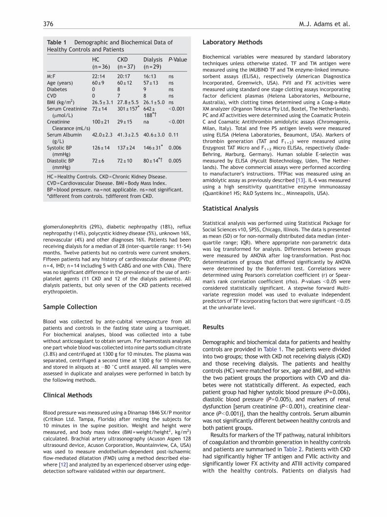
Table 1 Demographic and Biochemical Data ofHealthy Controls and Patients
HC(n=36)
CKD(n=37)
Dialysis(n=29)
P-Value
M:F 22:14 20:17 16:13 nsAge (years) 60±9 60±12 57±13 nsDiabetes 0 8 9 nsCVD 0 7 8 nsBMI (kg/m2) 26.5±3.1 27.8±5.5 26.1±5.0 nsSerum Creatinine(μmol/L)
72±14 301±157⁎ 642±188⁎†
b0.001
CreatinineClearance (mL/s)
100±21 29±15 na b0.001
Serum Albumin(g/L)
42.0±2.3 41.3±2.5 40.6±3.0 0.11
Systolic BP(mmHg)
126±14 137±24 146±31⁎ 0.006
Diastolic BP(mmHg)
72±6 72±10 80±14⁎† 0.005
HC=Healthy Controls. CKD=Chronic Kidney Disease.CVD=Cardiovascular Disease. BMI=Body Mass Index.BP=blood pressure. na=not applicable. ns=not significant.⁎different from controls. †different from CKD.
376 M.J. Adams et al.
glomerulonephritis (29%), diabetic nephropathy (18%), refluxnephropathy (14%), polycystic kidney disease (5%), unknown 16%,renovascular (4%) and other diagnoses 16%. Patients had beenreceiving dialysis for a median of 28 (inter-quartile range: 11-54)months. Twelve patients but no controls were current smokers.Fifteen patients had any history of cardiovascular disease (PVD;n=4, IHD; n=14 including 5 with CABG and one with CVA). Therewas no significant difference in the prevalence of the use of anti-platelet agents (11 CKD and 12 of the dialysis patients). Alldialysis patients, but only seven of the CKD patients receivederythropoietin.
Sample Collection
Blood was collected by ante-cubital venepuncture from allpatients and controls in the fasting state using a tourniquet.For biochemical analyses, blood was collected into a tubewithout anticoagulant to obtain serum. For haemostasis analysesone part whole blood was collected into nine parts sodium citrate(3.8%) and centrifuged at 1300 g for 10 minutes. The plasma wasseparated, centrifuged a second time at 1300 g for 10 minutes,and stored in aliquots at −80 °C until assayed. All samples wereassessed in duplicate and analyses were performed in batch bythe following methods.
Clinical Methods
Blood pressure was measured using a Dinamap 1846 SX/P monitor(Critikon Ltd. Tampa, Florida) after resting the subjects for10 minutes in the supine position. Weight and height weremeasured, and body mass index (BMI=weight/height2, kg/m2)calculated. Brachial artery ultrasonography (Acuson Aspen 128ultrasound device, Acuson Corporation, Mountainview, CA, USA)was used to measure endothelium-dependent post-ischaemicflow-mediated dilatation (FMD) using a method described else-where [12] and analyzed by an experienced observer using edge-detection software validated within our department.
Laboratory Methods
Biochemical variables were measured by standard laboratorytechniques unless otherwise stated. TF and TM antigen weremeasured using the IMUBIND TF and TM enzyme-linked immuno-sorbent assays (ELISA), respectively (American DiagnosticaIncorporated, Greenwich, USA). FVII and FX activities weremeasured using standard one stage clotting assays incorporatingfactor deficient plasmas (Helena Laboratories, Melbourne,Australia), with clotting times determined using a Coag-a-MateXM analyzer (Organon Teknica Pty Ltd, Boxtel, The Netherlands).PC and ATactivities were determined using the Coamatic ProteinC and Coamatic Antithrombin amidolytic assays (Chromogenix,Milan, Italy). Total and free PS antigen levels were measuredusing ELISA (Helena Laboratories, Beaumont, USA). Markers ofthrombin generation (TAT and F1 + 2) were measured usingEnzygnost TAT Micro and F1+2 Micro ELISAs, respectively (Dade-Behring, Marburg, Germany). Human soluble E-selectin wasmeasured by ELISA (Hycult Biotechnology, Uden, The Nether-lands). The above commercial assays were performed accordingto manufacturer's instructions. TFPIac was measured using anamidolytic assay as previously described [13]. IL-6 was measuredusing a high sensitivity quantitative enzyme immunoassay(Quantikine1 HS; R&D Systems Inc., Minneapolis, USA).
Statistical Analysis
Statistical analysis was performed using Statistical Package forSocial Sciences v10, SPSS, Chicago, Illinois. The data is presentedas mean (SD) or for non-normally distributed data median (inter-quartile range; IQR). Where appropriate non-parametric datawas log transformed for analysis. Differences between groupswere measured by ANOVA after log-transformation. Post-hocdeterminations of groups that differed significantly by ANOVAwere determined by the Bonferroni test. Correlations weredetermined using Pearson's correlation coefficient (r) or Spear-man's rank correlation coefficient (rho). P-values b0.05 wereconsidered statistically significant. A stepwise forward Multi-variate regression model was used to evaluate independentpredictors of TF incorporating factors that were significant b0.05at the univariate level.
Results
Demographic and biochemical data for patients and healthycontrols are provided in Table 1. The patients were dividedinto two groups; those with CKD not receiving dialysis (CKD)and those receiving dialysis. The patients and healthycontrols (HC) werematched for sex, age and BMI, and withinthe two patient groups the proportions with CVD and dia-betes were not statistically different. As expected, eachpatient group had higher systolic blood pressure (P=0.006),diastolic blood pressure (P=0.005), and markers of renaldysfunction [serum creatinine (Pb0.001), creatinine clear-ance (Pb0.001)], than the healthy controls. Serum albuminwas not significantly different between healthy controls andboth patient groups.
Results for markers of the TF pathway, natural inhibitorsof coagulation and thrombin generation in healthy controlsand patients are summarised in Table 2. Patients with CKDhad significantly higher TF antigen and FVIIc activity andsignificantly lower FX activity and ATIII activity comparedwith the healthy controls. Patients on dialysis had
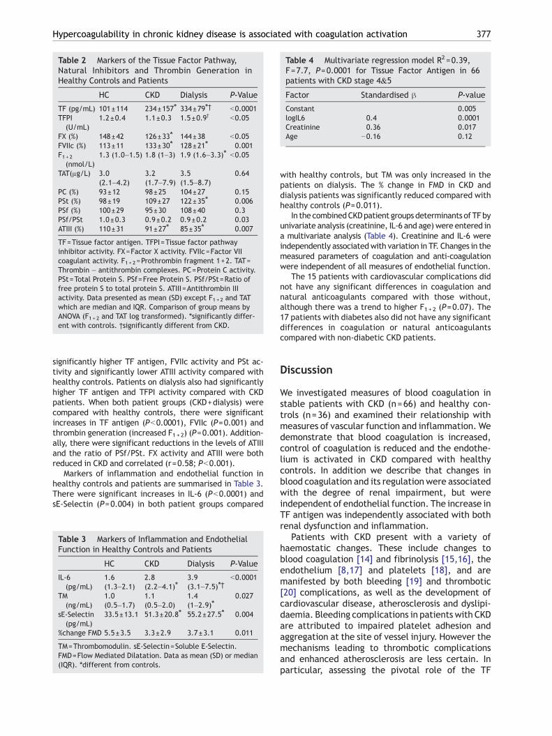
Table 2 Markers of the Tissue Factor Pathway,Natural Inhibitors and Thrombin Generation inHealthy Controls and Patients
HC CKD Dialysis P-Value
TF (pg/mL) 101±114 234±157⁎ 334±79⁎† b0.0001TFPI
(U/mL)1.2±0.4 1.1±0.3 1.5±0.9† b0.05
FX (%) 148±42 126±33⁎ 144±38 b0.05FVIIc (%) 113±11 133±30⁎ 128±21⁎ 0.001F1+ 2
(nmol/L)1.3 (1.0–1.5) 1.8 (1–3) 1.9 (1.6–3.3)⁎ b0.05
TAT(μg/L) 3.0(2.1–4.2)
3.2(1.7–7.9)
3.5(1.5–8.7)
0.64
PC (%) 93±12 98±25 104±27 0.15PSt (%) 98±19 109±27 122±35⁎ 0.006PSf (%) 100±29 95±30 108±40 0.3PSf/PSt 1.0±0.3 0.9±0.2 0.9±0.2 0.03ATIII (%) 110±31 91±27⁎ 85±35⁎ 0.007
TF=Tissue factor antigen. TFPI=Tissue factor pathwayinhibitor activity. FX=Factor X activity. FVIIc=Factor VIIcoagulant activity. F1 + 2=Prothrombin fragment 1+2. TAT=Thrombin – antithrombin complexes. PC=Protein C activity.PSt=Total Protein S. PSf=Free Protein S. PSf/PSt=Ratio offree protein S to total protein S. ATIII=Antithrombin IIIactivity. Data presented as mean (SD) except F1+ 2 and TATwhich are median and IQR. Comparison of group means byANOVA (F1+ 2 and TAT log transformed). ⁎significantly differ-ent with controls. †significantly different from CKD.
Table 4 Multivariate regression model R2=0.39,F=7.7, P=0.0001 for Tissue Factor Antigen in 66patients with CKD stage 4&5
Factor Standardised β P-value
Constant 0.005logIL6 0.4 0.0001Creatinine 0.36 0.017Age −0.16 0.12
377Hypercoagulability in chronic kidney disease is associated with coagulation activation
significantly higher TF antigen, FVIIc activity and PSt ac-tivity and significantly lower ATIII activity compared withhealthy controls. Patients on dialysis also had significantlyhigher TF antigen and TFPI activity compared with CKDpatients. When both patient groups (CKD+dialysis) werecompared with healthy controls, there were significantincreases in TF antigen (Pb0.0001), FVIIc (P=0.001) andthrombin generation (increased F1+2) (P=0.001). Addition-ally, there were significant reductions in the levels of ATIIIand the ratio of PSf/PSt. FX activity and ATIII were bothreduced in CKD and correlated (r=0.58; Pb0.001).
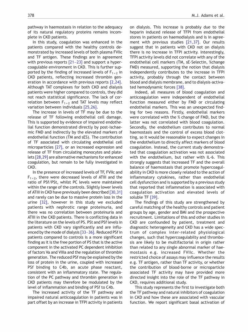
Markers of inflammation and endothelial function inhealthy controls and patients are summarised in Table 3.There were significant increases in IL-6 (Pb0.0001) andsE-Selectin (P=0.004) in both patient groups compared
Table 3 Markers of Inflammation and EndothelialFunction in Healthy Controls and Patients
HC CKD Dialysis P-Value
IL-6(pg/mL)
1.6(1.3–2.1)
2.8(2.2–4.1)⁎
3.9(3.1–7.5)⁎†
b0.0001
TM(ng/mL)
1.0(0.5–1.7)
1.1(0.5–2.0)
1.4(1–2.9)⁎
0.027
sE-Selectin(pg/mL)
33.5±13.1 51.3±20.8⁎ 55.2±27.5⁎ 0.004
%change FMD 5.5±3.5 3.3±2.9 3.7±3.1 0.011
TM=Thrombomodulin. sE-Selectin=Soluble E-Selectin.FMD=Flow Mediated Dilatation. Data as mean (SD) or median(IQR). ⁎different from controls.
with healthy controls, but TM was only increased in thepatients on dialysis. The % change in FMD in CKD anddialysis patients was significantly reduced compared withhealthy controls (P=0.011).
In the combinedCKDpatient groups determinants of TFbyunivariate analysis (creatinine, IL-6 and age) were entered ina multivariate analysis (Table 4). Creatinine and IL-6 wereindependently associatedwith variation in TF. Changes in themeasured parameters of coagulation and anti-coagulationwere independent of all measures of endothelial function.
The 15 patients with cardiovascular complications didnot have any significant differences in coagulation andnatural anticoagulants compared with those without,although there was a trend to higher F1+ 2 (P=0.07). The17 patients with diabetes also did not have any significantdifferences in coagulation or natural anticoagulantscompared with non-diabetic CKD patients.
Discussion
We investigated measures of blood coagulation instable patients with CKD (n=66) and healthy con-trols (n=36) and examined their relationship withmeasures of vascular function and inflammation. Wedemonstrate that blood coagulation is increased,control of coagulation is reduced and the endothe-lium is activated in CKD compared with healthycontrols. In addition we describe that changes inblood coagulation and its regulation were associatedwith the degree of renal impairment, but wereindependent of endothelial function. The increase inTF antigen was independently associated with bothrenal dysfunction and inflammation.
Patients with CKD present with a variety ofhaemostatic changes. These include changes toblood coagulation [14] and fibrinolysis [15,16], theendothelium [8,17] and platelets [18], and aremanifested by both bleeding [19] and thrombotic[20] complications, as well as the development ofcardiovascular disease, atherosclerosis and dyslipi-daemia. Bleeding complications in patients with CKDare attributed to impaired platelet adhesion andaggregation at the site of vessel injury. However themechanisms leading to thrombotic complicationsand enhanced atherosclerosis are less certain. Inparticular, assessing the pivotal role of the TF
378 M.J. Adams et al.
pathway in haemostasis in relation to the adequacyof its natural regulatory proteins remains incom-plete in CKD patients.
In this study, coagulation was enhanced in thepatients compared with the healthy controls de-monstrated by increased levels of both plasma FVIIcand TF antigen. These findings are in agreementwith previous reports [21–23] and support a hyper-coagulable environment in CKD. This is further sup-ported by the finding of increased levels of F1+ 2 inCKD patients, reflecting increased thrombin gen-eration in accordance with previous reports [2,24].Although TAT complexes for both CKD and dialysispatients were higher compared to controls, they didnot reach statistical significance. The lack of cor-relation between F1+ 2 and TAT levels may reflectvariation between individuals [25,26].
The increase in levels of TF may be due to therelease of TF following endothelial cell damage.This is supported by evidence of impaired endothe-lial function demonstrated directly by post-ischae-mic FMD and indirectly by the elevated markers ofendothelial function (TM and sES). The contributionof TF associated with circulating endothelial cellmicroparticles [27], or an increased expression andrelease of TF from circulating monocytes and plate-lets [28,29] are alternativemechanisms for enhancedcoagulation, but remain to be fully investigated inCKD.
In the presence of increased levels of TF, FVIIc andF1+2, there were decreased levels of ATIII and theratio of PSf/PSt, whilst PC levels were maintainedwithin the range of the controls. Slightly lower levelsof ATIII in CKD have previously been described [30,31]and rarely can be due to massive protein loss in theurine [32], however in this study we excludedpatients with nephrotic range proteinuria, andthere was no correlation between proteinuria andATIII in the CKD patients. There is conflicting data inthe literature on the levels of PS. PSt and PSf levels inpatients with CKD vary significantly and are influ-enced by themode of dialysis [33–36]. Reduced PSf inpatients compared to controls is a more significantfinding as it is the free portion of PS that is the activecomponent in the activated PC dependent inhibitionof factors Va and VIIIa and the regulation of thrombingeneration. The reduced PSfmay be explained by theloss of protein in the urine, coupled with increasedPSf binding to C4b, an acute phase reactant,consistent with an inflammatory state. The regula-tion of the PC pathway and thrombin generation inCKD patients may therefore be modulated by thelevel of inflammation and binding of PSf to C4b.
The increased activity of the TF pathway andimpaired natural anticoagulation in patients was inpart offset by an increase in TFPI activity in patients
on dialysis. This increase is probably due to theheparin induced release of TFPI from endothelialstores in patients on haemodialysis and is in agree-ment with previous studies [21,37]. Our resultssuggest that in patients with CKD not on dialysisthere is no increase in TFPI activity. Interestingly,TFPI activity levels did not correlate with any of theendothelial cell markers (TM, sE-Selectin, %changeFMD) measured, supporting the notion that dialysisindependently contributes to the increase in TFPIactivity, probably through the contact betweenblood and dialysis membrane, and to dialysis-activa-ted hemodynamic forces [38].
Indeed, all measures of blood coagulation andanticoagulation were independent of endothelialfunction measured either by FMD or circulatingendothelial markers. This was an unexpected find-ing for two reasons. Firstly, endothelial markerswere correlated with the % change of FMD, but thelatter was not correlated with blood coagulation.Secondly, the endothelium contributes to normalhaemostasis and the control of excess blood clot-ting, so it would be reasonable to expect changes inthe endothelium to directly affect markers of bloodcoagulation. Instead, the current study demonstra-ted that coagulation activation was not associatedwith the endothelium, but rather with IL-6. Thisstrongly suggests that increased TF and the overallbalance of haemostasis that promote hypercoagul-ability in CKD is more closely related to the action ofinflammatory cytokines, rather than endothelialcell dysfunction and is supported by a previous studythat reported that inflammation is associated withcoagulation activation and elevated levels ofsoluble TF [39].
The findings of this study are strengthened bycareful matching of the healthy controls and patientgroups by age, gender and BMI and the prospectiverecruitment. Limitations of this and other studies inCKD are confounded by patient, treatment anddiagnostic heterogeneity and CKD has a wide spec-trum of complex inter-related physiologicalchanges, such that hypercoagulabilty and thrombo-sis are likely to be multifactorial in origin ratherthan related to any single abnormal marker of hae-mostasis e.g. increased FVIIc. Whether therestricted choice of assays may influence the resultse.g. TF antigen, rather than TF activity, or whetherthe contribution of blood-borne or microparticleassociated TF activity may have provided moredirected insight into the role of the TF pathway inCKD, requires additional study.
This study represents the first to investigate boththe TF pathway and natural inhibitors of coagulationin CKD and how these are associated with vascularfunction. We report significant basal activation of
379Hypercoagulability in chronic kidney disease is associated with coagulation activation
the TF pathway of blood coagulation and depletionor reduction of some natural anticoagulants in CKD.These changes were correlated with the degree ofrenal dysfunction and inflammation but were notassociated with endothelial function. Further stu-dies are required in CKD patients to refine thestimulus and source of the increase in TF. Althoughstudies in patients without CKD suggest that hyper-coagulability contributes to both the developmentand progression of atherothrombotic disease, directevidence is lacking to support similar processes inCKD. The substantial burden of atherothrombosis inCKD therefore demands prospective studies to eluci-date the potential mechanisms and therapeuticinterventions thatmaybeof benefit to thesepatients,such as the HMG coA reductase inhibitors (statins).
Acknowledgements
Australian Kidney Foundation (Medical ResearchGrant S0102).
References
[1] Molino D, De Lucia D, Gaspare De Santo N. Coagulationdisorders in uremia. Semin Nephrol 2006;26:46–51.
[2] Ambuhl PM, Wuthrich RP, KorteW, Schmid L, Krapf R. Plasmahypercoagulability in haemodialysis patients: impact ofdialysis and anticoagulation. Nephrol Dial Transplant1997;12:2355–64.
[3] Llach F. Hypercoagulability, renal vein thrombosis, andother thrombotic complications of nephrotic syndrome.Kidney Int 1985;28:429–39.
[4] Charytan DM, Kuntz RE, Chhabra A, Cutlip DE. Relationship ofchronic kidney disease to cardiovascular death andmyocardialinfarction following coronary stenting. J Nephrol 2006;19:764–70.
[5] Koren-Morag N, Goldbourt U, Tanne D. Renal dysfunctionand risk of ischemic stroke or TIA in patients withcardiovascular disease. Neurology 2006;67:224–8.
[6] Stack AG. Coronary artery disease and peripheral vasculardisease in chronic kidney disease: an epidemiologicalperspective. Cardiol Clin 2005;23:285–98.
[7] McVey JH. Tissue factor pathway. Baillieres Clin Haematol1994;7:469–84.
[8] Takagi M, Wada H, Mukai K, Kihira H, Yano S, Minamikawa K,et al. Increased vascular endothelial cell markers in patientswith chronic renal failure on maintenance haemodialysis.Blood Coagul Fibrinolysis 1994;5:713–7.
[9] Rustom R, Leggat H, Tomura HR, Hay CR, Bone JM. Plasmathrombomodulin in renal disease: effects of renal functionand proteinuria. Clin Nephrol 1998;50:337–41.
[10] Dogra G, Irish A, Chan D, Watts G. Insulin resistance,inflammation, and blood pressure determine vasculardysfunction in CKD. Am J Kidney Dis 2006;48:926–34.
[11] Dogra GK, Herrmann S, Irish AB, Thomas MA, Watts GF. Insulinresistance, dyslipidaemia, inflammation and endothelialfunction in nephrotic syndrome. Nephrol Dial Transplant2002;17:2220–5.
[12] Woodman RJ, Playford DA, Watts GF, Cheetham C, Reed C,Taylor RR, et al. Improved analysis of brachial artery
ultrasound using a novel edge-detection software system.J Appl Physiol 2001;91:929–37.
[13] Adams M, Breckler L, Stevens P, Thom J, Baker R, OostryckR. Anti-tissue factor pathway inhibitor activity in subjectswith antiphospholipid syndrome is associated withincreased thrombin generation. Haematologica 2004;89:985–90.
[14] Sagripanti A, Cupisti A, Baicchi U, Ferdeghini M, Morelli E,Barsotti G. Plasma parameters of the prothrombotic state inchronic uremia. Nephron 1993;63:273–8.
[15] Canavese C, Stratta P, Pacitti A, Mangiarotti G, Racca M,Oneglio R, et al. Impaired fibrinolysis in uremia: partial andvariable correction by four different dialysis regimes. ClinNephrol 1982;17:82–9.
[16] Hong SY, Yang DH. Fibrinolytic activity in end-stage renaldisease. Nephron 1993;63:188–92.
[17] Gris JC, Branger B, Vecina F, al Sabadani B, Fourcade J,Schved JF. Increased cardiovascular risk factors andfeatures of endothelial activation and dysfunction indialyzed uremic patients. Kidney Int 1994;46:807–13.
[18] Escolar G, Diaz-Ricart M, Cases A. Uremic platelet dysfunc-tion: past and present. Curr Hematol Rep 2005;4:359–67.
[19] Remuzzi G. Bleeding in renal failure. Lancet 1988;1:1205–8.[20] Rabelink TJ, Zwaginga JJ, Koomans HA, Sixma JJ. Throm-
bosis and hemostasis in renal disease. Kidney Int1994;46:287–96.
[21] Matsuo T, Koide M, Kario K, Suzuki S, Matsuo M. Extrinsiccoagulation factors and tissue factor pathway inhibitor in end-stage chronic renal failure. Haemostasis 1997;27:163–7.
[22] Mezzano D, Tagle R, Pais E, Panes O, PerezM, Downey P, et al.Endothelial cell markers in chronic uremia: relationship withhemostatic defects and severity of renal failure. Thromb Res1997;88:465–72.
[23] Irish AB, Green FR. Factor VII coagulant activity (VIIc) andhypercoagulability in chronic renal disease and dialysis:relationship with dyslipidaemia, inflammation, and factorVII genotype. Nephrol Dial Transplant 1998;13:679–84.
[24] Irish AB, Thompson CH. The effects of gemfibrozil upon thehypercoagulable state in dyslipidaemic patients with chronicrenal failure. Nephrol Dial Transplant 1996;11:2223–8.
[25] Garcia-Avello A, Garcia-Frade LJ, Gandarias C, Ocana J,Cancelas JA, Lasso M. High F1.2 fragment of prothrombin,thrombin-antithrombin III complex (TAT) and soluble fibrinplasma levels demonstrate hypercoagulability inducedduring loco-regional thrombolytic therapy with rt-PA.Thromb Res 1994;73:109–15.
[26] Takahashi H, Wada K, Niwano H, Shibata A. Comparison ofprothrombin fragment 1+2 with thrombin-antithrombin IIIcomplex in plasma of patients with disseminated intravas-cular coagulation. Blood Coagul Fibrinolysis 1992;3:813–8.
[27] Faure V, Dou L, Sabatier F, Cerini C, Sampol J, Berland Y, et al.Elevation of circulating endothelial microparticles in patientswith chronic renal failure. J Thromb Haemost 2006;4:566–73.
[28] Al-Saady NM, Leatham EW, Gupta S, Kwan JT, Eastwood JB,Seymour CA. Monocyte expression of tissue factor andadhesion molecules: the link with accelerated coronaryartery disease in patients with chronic renal failure. Heart1999;81:134–40.
[29] Mercier E, Branger B, Vecina F, Al-Sabadani B, Berlan J,Dauzat M, et al. Tissue factor coagulation pathway andblood cells activation state in renal insufficiency. Hematol J2001;2:18–25.
[30] Alwakeel J, Gader AM, Hurieb S, al-Momen AK, Mitwalli A,Abu Aisha H. Coagulation inhibitors and fibrinolytic para-meters in patients on peritoneal dialysis and haemodialysis.Int Urol Nephrol 1996;28:255–61.
380 M.J. Adams et al.
[31] Tomura S, Nakamura Y, Tachibana K, Deguchi F, Ando R,Chida Y, et al. Enhanced coagulation and fibrinolysis duringtreatment with recombinant human erythropoietin inpatients undergoing chronic hemodialysis. Blood Purif1993;11:370–7.
[32] Vaziri ND, Paule P, Toohey J, Hung E, Alikhani S, Darwish R, etal. Acquired deficiency and urinary excretion of antithrombinIII in nephrotic syndrome. Arch Intern Med 1984;144:1802–3.
[33] Vaziri ND, Gonzales EC, Wang J, Said S. Blood coagulation,fibrinolytic, and inhibitory proteins in end-stage renal disease:effect of hemodialysis. Am J Kidney Dis 1994;23:828–35.
[34] Demicheli M, Contino L, Iberti M, Ortensia A, Finotto E,Lombardi A, et al. Protein C and protein S levels in uremicpatients before and after dialysis. Thromb Res 1992;68:451–7.
[35] Lai KN, Yin JA, Yuen PM, Li PK. Effect of hemodialysis onprotein C, protein S, and antithrombin III levels. Am J KidneyDis 1991;17:38–42.
[36] Lai KN, Yin JA, Yuen PM, Li PK. Protein C, protein S, andantithrombin III levels in patients on continuous ambulatoryperitoneal dialysis and hemodialysis. Nephron 1990;56:271–6.
[37] Kario K, Matsuo T, Yamada T, Matsuo M. Increased tissuefactor pathway inhibitor levels in uremic patients on regularhemodialysis. Thromb Haemost 1994;71:275–9.
[38] Naumnik B, Borawski J, Pawlak K, Mysliwiec M. Effect ofhemodialysis on plasma levels of vascular endothelialmarkers. Clin Appl Thromb Hemost 2002;8:245–50 OfficialJournal Of The International Academy Of Clinical AndApplied Thrombosis/Hemostasis.
[39] Szotowski B, Antoniak S, Poller W, Schultheiss HP, Rauch U.Procoagulant soluble tissue factor is released from endothe-lial cells in response to inflammatory cytokines. Circ Res2005;96:1233–9.