hybrid antimicrobial enzyme and silver nanoparticle ... · hybrid antimicrobial enzyme and silver...
TRANSCRIPT

AFRL-RX-TY-TP-2008-4630
HYBRID ANTIMICROBIAL ENZYME AND SILVER NANOPARTICLE COATINGS FOR MEDICAL INSTRUMENTS (POSTPRINT)
D. Mathew Eby and Heather R. Luckarift Universal Technology Corporation 1270 North Fairfield Road Dayton, OH 45432
Glenn R. Johnson Airbase Technologies Division Air Force Research Laboratory 139 Barnes Drive, Suite 2 Tyndall Air Force Base, FL 32403-5323
Contract No. FA4819-07-D-0001 December 2008
DISTRIBUTION A: Approved for release to the public; distribution unlimited.
This work is copyrighted. The United States has for itself and others acting on its behalf an unlimited, paid-up, nonexclusive, irrevocable worldwide license. Any other form of use is subject
to copyright restrictions.
AIR FORCE RESEARCH LABORATORY MATERIALS AND MANUFACTURING DIRECTORATE
Air Force Materiel Command United States Air Force Tyndall Air Force Base, FL 32403-5323

Standard Form 298 (Rev. 8/98)
REPORT DOCUMENTATION PAGE
Prescribed by ANSI Std. Z39.18
Form Approved OMB No. 0704-0188
The public reporting burden for this collection of information is estimated to average 1 hour per response, including the time for reviewing instructions, searching existing data sources, gathering and maintaining the data needed, and completing and reviewing the collection of information. Send comments regarding this burden estimate or any other aspect of this collection of information, including suggestions for reducing the burden, to Department of Defense, Washington Headquarters Services, Directorate for Information Operations and Reports (0704-0188), 1215 Jefferson Davis Highway, Suite 1204, Arlington, VA 22202-4302. Respondents should be aware that notwithstanding any other provision of law, no person shall be subject to any penalty for failing to comply with a collection of information if it does not display a currently valid OMB control number. PLEASE DO NOT RETURN YOUR FORM TO THE ABOVE ADDRESS. 1. REPORT DATE (DD-MM-YYYY) 2. REPORT TYPE 3. DATES COVERED (From - To)
4. TITLE AND SUBTITLE 5a. CONTRACT NUMBER
5b. GRANT NUMBER
5c. PROGRAM ELEMENT NUMBER
5d. PROJECT NUMBER
5e. TASK NUMBER
5f. WORK UNIT NUMBER
6. AUTHOR(S)
7. PERFORMING ORGANIZATION NAME(S) AND ADDRESS(ES) 8. PERFORMING ORGANIZATION REPORT NUMBER
9. SPONSORING/MONITORING AGENCY NAME(S) AND ADDRESS(ES) 10. SPONSOR/MONITOR'S ACRONYM(S)
11. SPONSOR/MONITOR'S REPORT NUMBER(S)
12. DISTRIBUTION/AVAILABILITY STATEMENT
13. SUPPLEMENTARY NOTES
14. ABSTRACT
15. SUBJECT TERMS
16. SECURITY CLASSIFICATION OF: a. REPORT b. ABSTRACT c. THIS PAGE
17. LIMITATION OF ABSTRACT
18. NUMBER OF PAGES
19a. NAME OF RESPONSIBLE PERSON
19b. TELEPHONE NUMBER (Include area code)
15-DEC-2008 Journal Article - POSTPRINT 01-APR-2007 -- 30-NOV-2008
Hybrid Antimicrobial Enzyme and Silver Nanoparticle Coatings for Medical Instruments (POSTPRINT)
FA4819-07-D-0001
0909999F
4918
L0
Q140LA62
*Eby, D. Matthew; *Luckarift, Heather R.; ^Johnson, Glenn R.
*Universal Technology Corporation 1270 North Fairfield Road Dayton, OH 45432
^Air Force Research Laboratory Materials and Manufacturing Directorate Airbase Technologies Division 139 Barnes Drive, Suite 2 Tyndall Air Force Base, FL 32403-5323
AFRL/RXQL
AFRL-RX-TY-TP-2008-4630
Distribution Statement A: Approved for public release; distribution unlimited.
Document contains color images. Ref Public Affairs Case # AFRL/RXQ 08-203. Published on http://pubs.acs.org | doi: 10.1021/am9002155.
We report a method for the synthesis of antimicrobial coatings on medical devices that combine the bacteriolytic activity of lysozyme and the biocidal properties of silver nanoparticles. Colloidal suspensions of lysozyme and silver nanoparticles were electrophoretically deposited onto the surface of stainless steel surgical blades and needles. The coatings retained the hydrolytic properties of lysozyme and exhibited antimicrobial activity against a range of bacterial species. In particular, coated blades demonstrated potent bactericidal activity, reducing cell viability by > log 10 within 1.5 h for Klebsiella pneumonia, Bacillus anthracis Sterne and Bacillus subtilis and within 3 h for Staphylococcus aureus. The results confirmed that complex antimicrobial coatings can be easily made using facile silver nanoparticle synthesis and electrodeposition methods.
antimicrobial, lysozyme, silver, nanocomposite, electrochemistry
U U U UU 8
Glenn R. Johnson
Reset

Hybrid Antimicrobial Enzyme and SilverNanoparticle Coatings for MedicalInstrumentsD. Matthew Eby,*,†,‡ Heather R. Luckarift,†,‡ and Glenn R. Johnson*,‡
Microbiology and Applied Biochemistry, Materials and Manufacturing Directorate, Air Force Research Laboratoryand Universal Technology Corporation, 139 Barnes Drive, Suite 2, Tyndall AFB, Florida 32403
ABSTRACT We report a method for the synthesis of antimicrobial coatings on medical instruments that combines the bacteriolyticactivity of lysozyme and the biocidal properties of silver nanoparticles. Colloidal suspensions of lysozyme and silver nanoparticleswere electrophoretically deposited onto the surface of stainless steel surgical blades and needles. Electrodeposited films firmly adheredto stainless steel surfaces even after extensive washing and retained the hydrolytic properties of lysozyme. The antimicrobial efficacyof coatings was tested by using blades and needles in an in vitro lytic assay designed to mimic the normal application of the instruments.Coated blades and needles were used to make incisions and punctures, respectively, into agarose infused with bacterial cells. Celllysis was seen at the contact sites, demonstrating that antimicrobial activity is transferred into the media, as well as retained on thesurface of the blades and needles. Blade coatings also exhibited antimicrobial activity against a range of bacterial species. In particular,coated blades demonstrated potent bactericidal activity, reducing cell viability by at least 3 log within 1.5 h for Klebsiella pneumoniae,Bacillus anthracis Sterne, and Bacillus subtilis and within 3 h for Staphylococcus aureus and Acinetobacter baylyi. The results confirmedthat complex antimicrobial coatings can be created using facile methods for silver nanoparticle synthesis and electrodeposition.
KEYWORDS: antimicrobial • lysozyme • silver • nanoparticle • electrochemistry • coating
INTRODUCTION
Infections originating from implanted devices (e.g., or-thopedic fixation hardware, artificial prosthetics, endo-vascular stents, and catheters) are a persistent and
serious health issue. The first step towards reducing theseinfections is inhibiting bacterial colonization on subcutane-ous device surfaces. Current antimicrobial coating methodsinvolve either modification of the physiochemical propertiesof the device surface or application of coatings that resistcell adhesion and biofilm formation (1). In order for coatingsto be effective, they must incorporate a direct, on-contactbiocide and also provide sustained antimicrobial activity, soto neutralize any initial contamination of the surface and alsoinhibit colonization over time from a wide range of op-portunistic microorganisms. These issues and the rise ofmicrobial resistance to commonly used antibiotics drive acontinuous search for new antibiotic formulations that areamenable to adhesion and immobilization on surfaces.
One strategy given considerable attention in recent yearsis the use of silver on medical device surfaces and in wounddressings (2-4). These coatings are particularly attractivebecause silver ions act against a broad range of bacteria andyet are relatively nontoxic to mammalian cells (5, 6). While
the antimicrobial effectiveness of silver coatings has beenshown in the laboratory, there have been mixed results inthe clinical setting (7). The past limitations of silver coatingsas prophylactic treatments most likely stem from the inef-fective release of silver ions from the surface and theunproductive adsorption and precipitation of silver ions inbody fluids (8, 9).
More recently, silver in nanoparticle form has beenshown to exhibit enhanced antimicrobial effects over previ-ous silver formulations. The high surface-to-volume ratio ofnanoparticles provides a substantial and sustained contactwith the bacterial cell that is not easily quenched by othersalts and proteins (10, 11). Numerous methods for silvernanoparticle synthesis and incorporation into materials arecurrently under investigation (12-20), and silver nanopar-ticle coatings are beginning to see more prevalent use inhealth care (1, 5, 8). In recent work, we discovered that thehydrolytic enzyme, lysozyme, catalyzes the reduction ofsilver, which yields a stable colloid of silver nanoparticlesand active lysozyme (21). The use of lysozyme in thesynthesis of silver nanoparticles is advantageous because itcombines two different biocidal mechanisms into one ma-terial; lysozyme primarily inhibits growth of the Gram-typepositive strains through its muramidase activity (22), whilesilver nanoparticles inhibit growth of both Gram-type nega-tive and positive strains by inhibiting membrane functionand enzyme activity (23). In a few cases, the synthesis ofbioactive coatings has been achieved by adding noble metalnanoparticles to enzyme solutions and electrodepositing themixture onto surfaces (24-27). Because of the uniqueproperties of silver nanoparticles, their utility as the precur-
* Corresponding author. Tel.: 850-283-6026. Fax: 850-283-6090. Email:[email protected] (D.M.E.), [email protected] (G.R.J.).Received for review March 30, 2009 and accepted June 16, 2009† Universal Technology Corporation.‡ Microbiology and Applied Biochemistry, Materials and Manufacturing Director-ate, Air Force Research Laboratory.DOI: 10.1021/am9002155
© 2009 American Chemical Society
ARTIC
LE
www.acsami.org VOL. 1 • NO. 7 • 1553–1560 • 2009 1553Published on Web 07/02/2009
Dow
nloa
ded
by A
IR F
OR
CE
RES
EAR
CH
LA
B o
n Ju
ly 3
1, 2
009
Publ
ishe
d on
July
2, 2
009
on h
ttp://
pubs
.acs
.org
| do
i: 10
.102
1/am
9002
155

sor for electrodeposited coatings may produce more effec-tive antimicrobial activities than previous methods based onconventional electrodeposition solutions (e.g., silver saltsolutions). Herein, we report a method to produce antimi-crobial coatings through electrophoretic deposition of silvernanoparticles formed from the lysozyme-mediated reduc-tion of silver ions. The antimicrobial effectiveness of coatedsurfaces was tested against a variety of microorganisms todemonstrate the retention of antimicrobial properties withincoatings.
EXPERIMENTAL SECTIONReagents and the Synthesis of Silver Nanoparticles. Hen
egg white lysozyme (∼95%, ∼50 000 units per mg of protein)and silver(I) acetate (99.99%) were obtained from Sigma-Aldrich (St. Louis, MO). Unless stated otherwise, all otherchemicals were obtained from either Sigma-Aldrich or FisherScientific (Pittsburgh, PA) and were of the highest purity avail-able. For experiments measuring the electrochemical propertiesof lysozyme, a lysozyme solution made from a stock powderwas dialyzed against two changes of ultrapure water (1:100, v/v)using a 5000 Da molecular weight cut-off dialysis membrane(Spectra/Por cellulose ester, Spectrum Medical Industries, Hous-ton, TX) over 24 h to remove residual buffer and salts and thenstored at -20 °C in 30 mg mL-1 aliquots. To generatelysozyme-silver nanoparticle solutions for electrophoretic depo-sition, undialyzed lysozyme solutions (20 mg mL-1) were madewith ultrapure water and then diluted with methanol (1:1, v/v).A silver acetate solution in 50% methanol (1 mM) was thenadded to the lysozyme solution to produce a final concentrationof 5 mg mL-1 lysozyme and 0.5 mM silver acetate in 50%methanol. The solution was then exposed to light for ap-proximately 24 h to allow the formation of silver nanoparticles.The resulting lysozyme-silver colloidal suspension was storedin the dark at 4 °C to inhibit precipitation until use.
Electrochemical Measurements. Electrochemical measure-ments were performed in a one-compartment, three-electrodecell at room temperature using a 757 VA Computrace Systempotentiostat (Metrohm AG, Herisau, Switzerland). A silver rotat-ing-disk electrode (2.0 mm disk diameter) was employed as theworking electrode in conjunction with a platinum rod auxiliaryelectrode (15 mm × 2 mm diameter, 10 mm submerged in cell)and a Ag/AgCl electrode in saturated KCl as the referenceelectrode. The working electrode was stationary during mea-surement. Before each measurement, the working and auxiliaryelectrodes were cleaned by polishing with 8000 grit sandpaperand then rinsed with ultrapure water. No additional electrolytewas added to the potentiostat cell. Current-potential curves(cyclic voltammograms) were generated by linear voltage sweepsbetween -1 and 1 V at 0.1 V s-1 (vs Ag/AgCl).
Electrophoretic Deposition of Lysozyme-Silver Nano-particles. Surfaces to accept electrophoretic deposition werestainless steel surgical blades (#10, Exelint International Co., LosAngeles, CA) and syringe needles (single-use, 22 Gauge, 1.5 in.length, Becton Dickinson Co., Franklin Lakes, NJ). Before depo-sition, metal surfaces were cleaned and etched, using a proce-dure similar to that described previously (28). Blades andneedles were cleaned by ultrasonication for 15 min each indichloromethane, acetone, and water. Clean surfaces were thenetched for 1 min in a solution containing 0.25% hydrochloricacid and 2.5% nitric acid. After etching, metal surface passiva-tion was completed in 40% nitric acid for 40 min. Blades andneedles were then extensively washed with ultrapure water,dried, and stored until use.
A two-electrode electrophoretic cell was constructed thatcontained either a blade or a needle as the working electrode.Approximately 1 cm of the blade or needle tip was submerged
into 5 mL of the lysozyme-silver nanoparticle depositionsolution. A platinum wire was also submerged approximately1 cm into the solution and served as the secondary electrode.The distance between each electrode was approximately 1 cm.A power source [1735 direct-current (dc) power supply, BKPrecision, Placentia, CA] applied a dc voltage (3 V) for 10 min.After electrodeposition, blades and needles were rinsed exten-sively with ultrapure water and stored at room temperature.
Three additional electrophoretic coating experiments werecompleted to determine the individual effects that lysozyme andsilver nanoparticles have on deposition and when lysozyme andsilver acetate were combined in the deposition solution butwithout prior formation of nanoparticles. For the first twocontrol experiments, lysozyme and silver nanoparticles wereused in separate electrodeposition solutions. For electrophoreticdeposition containing only lysozyme, the procedure was fol-lowed as described above, except a solution containing 5 mgmL-1 lysozyme in 50% methanol served as the depositionsolution. For electrophoretic deposition primarily composed ofsilver nanoparticles, a dialysis method was used to removesoluble lysozyme from the deposition solution that was nottightly associated with nanoparticles (21). Prior to deposition,a 5 mL solution of lysozyme-silver nanoparticles was placedinto a 25 000 Da molecular weight cut-off dialysis bag (Spectra/Por cellulose ester) and dialyzed against four changes of 1 L ofwater for 72 h to remove the majority of lysozyme. This resultedin a deposition solution that contained mostly silver nanopar-ticles but still contained small amounts of lysozyme. For thethird control experiment, freshly mixed solutions of lysozyme(10 mg mL-1) and silver acetate (1 mM) in 50% methanol (1:1,v/v) served as the electrodeposition solution and were usedimmediately in the electrophoretic cell in the absence of light,in order to prevent silver reduction and subsequent nanoparticleformation.
Physical and Spectroscopic Characterization. UV-visiblespectroscopy was completed using a Cary 3E spectrophotom-eter (Varian Inc., Palo Alto, CA) and 0.5 mL quartz cuvettes.Atomic force microscopy (AFM) images were obtained using aNanoscope V, equipped with a Multimode V scanning probemicroscope and a PicoForce stage (Veeco Instruments Inc.,Woodbury, NY). Blades were first dried under vacuum at 50 °Cfor 10 min before imaging in tapping mode, using an etchedphosphorous (n)-doped silicon cantilever probe (type RTESP,Veeco Instruments Inc.). The natural frequency of the cantileverwas in the 200 kHz range, and the radius of the probe tip was<10 nm. Transmission electron microscopy (TEM) was com-pleted using a 100 CX II electron microscope from JEOL Ltd.(Tokyo, Japan). Attenuated total reflectance Fourier transforminfrared (ATR FT-IR) spectroscopy was performed using aNicolet FT-IR 6700 spectrophotometer equipped with a SmartMiracle single-bounce diamond ATR accessory (Thermo FisherScientific, Waltham, MA). The data collection was completedusing OMNIC 2.1 software. Before measurement, coated anduncoated blades were washed with methanol and dried at 50°C for 30 min. For ATR FT-IR of the lysozyme stock powder,10 µL of a lysozyme powder slurry in methanol was dried onthe instrument crystal before measurement.
Measurement of the Lysozyme Activity. Qualitative mea-surement of the lysozyme activity in coatings was adapted fromthe Micrococcus lysodeikticus cell lysis assay in solid media, firstdescribed by Fleming (29). Blades and needles were first usedto cut and puncture, respectively, solid media plates containinga suspension of 0.5 mg mL-1 M. lysodeikticus cells (Sigma-Aldrich) in a 50 mM potassium phosphate buffer (pH 8) and 1%agarose before being placed on top of the plate surface. Plateswere incubated at 37 °C for 16 h and periodically inspected forcell lysis, which resulted in a clearing of the solid agar at theblade and needle contact points.
ARTIC
LE
1554 VOL. 1 • NO. 7 • 1553–1560 • 2009 Eby et al. www.acsami.org
Dow
nloa
ded
by A
IR F
OR
CE
RES
EAR
CH
LA
B o
n Ju
ly 3
1, 2
009
Publ
ishe
d on
July
2, 2
009
on h
ttp://
pubs
.acs
.org
| do
i: 10
.102
1/am
9002
155

Antimicrobial Activity Assays. The following strains used inthe antimicrobial assays were obtained from the American TypeCulture Collection (ATCC, Manassas, VA): Escherichia coli (ATCC25922), Staphylococcus aureus (ATCC 25923), Klebsiella pneu-moniae (ATCC 4352), Pseudomonas fluorescens (ATCC 13525),Candida albicans (ATCC 10231), and Staphylococcus epidermidis(ATCC 14990). Bacillus anthracis Sterne strain 34F2 was ob-tained from Colorado Serum Co., Denver, CO (30). Acinetobacterbaylyi strain ADP1 (31) was a gift from Dr. Ellen Neidle(University of Georgia, Athens, GA). Luria-Bertani and YeastMold (Difco Laboratories, Sparks, MD) were used to growbacterial strains and C. albicans, respectively, in liquid and onsolid media plates (plates also contained 1.5% agarose).
At the start of the antimicrobial assay, coated blades wereincubated in growth media containing approximately 105 cellsin the exponential growth phase (1 mL). Viable cells werequantitated after 0, 1.5, and 3 h of incubation by removing 100µL from the cultures, spreading all or a serial dilution of thealiquot onto solid media, and enumerating colony-forming units(cfu). After cultures were incubated for 24 h, blades wereremoved from cultures and placed into fresh, sterile media (1mL). In addition, the remaining growth media from the bladeculture was diluted into fresh, sterile media (1:10 dilution). Thetwo subcultures were incubated overnight and then visuallyinspected for growth. Results are the average of at least threereplicates. Uncoated, etched blades were also incubated inseparate cultures to ensure that cells grew in the absence of acoating material, and the etched stainless steel did not adverselyaffect the growth.
RESULTS AND DISCUSSIONSynthesis and Electrochemical Behavior of
Lysozyme-Silver Nanoparticles. In our recent work,we found that stable colloids of silver nanoparticles formedfrom the incubation of micrograms per milliliter quantitiesof lysozyme and silver acetate in 100% methanol (21). Thegrowth of silver nanoparticles was dependent upon exposureto light and only required lysozyme, which acted as thereductant and colloidal stabilizing agent. For electrophoreticdeposition, the previous formulation was optimized byincreasing the lysozyme concentration to 5 mg mL-1 andcompleting the nanoparticle synthesis reaction in an aque-ous solution containing 50% methanol (Figure 1). The finalsolution contained a sufficient concentration of ionic speciesto facilitate electrophoretic deposition without additionalelectrolytes, while also maintaining a stable suspension ofnanoparticles. When stored in the dark at ambient condi-tions, the lysozyme-silver nanoparticles can be maintainedas a colloid for several months.
The electrochemical properties of lysozyme were mea-sured using cyclic voltammetry with a silver disk electrode.Lysozyme was oxidized at 745 mV and underwent tworeductions at -640 and -800 mV (Figure 2A). Repeatedpotentiostat cycling resulted in successively smaller peakcurrents, which were indicative of irreversible deposition ofprotein on the electrode surface (32). When silver acetatewas added to lysozyme, voltammograms completed im-mediately after mixing show a shift in the reduction peaksof lysozyme toward a lower cathodic potential (Figure 2B).Similar voltammeric behavior has been previously reportedwhen protein-metal complexes form in the electrochemicalcell and change the electrochemical properties of the indi-
vidual species (33). Considering that silver acetate alone doesnot produce current peaks under our conditions (dashedgray line in Figure 2B), the shift in the peak potential oflysozyme after silver addition suggests that silver associateswith lysozyme and forms a new electrochemically activespecies. As seen with lysozyme alone, additional voltagecycles also showed that the lysozyme-silver complex wasirreversibly deposited and formed a coating on the electrodesurface. As silver nanoparticles developed in the lysozymeand silver acetate solution, cyclic voltammetry was com-pleted at intermittent time intervals during incubation (0.3,2, and 24 h). Samples were taken from the synthesis reactionfor voltammeric analysis and the electrodes were polishedbetween each use. In each case, there was no change in thepotential and intensity of the current peaks, which indicatedthat the electrochemical properties of lysozyme were notaltered during and after nanoparticle synthesis (results notshown). When silver nanoparticles were present in thepotentiostat cell, a resilient red film formed on the electrodesurface during repeated voltage cycling, which requiredremoval by physical abrasion using fine-grit sandpaper.Taken together, these observations showed that lysozyme-silver nanoparticles form a complex that was electrophoreti-cally deposited onto the electrode surface. The results ledus to investigate whether the antimicrobial properties oflysozyme and silver were retained within the coatings.
Electrophoretic Deposition of Lysozyme-Silver Nanoparticles onto Stainless Steel. Stainlesssteel surgical blades and syringe needles were investigatedas model surfaces for the coating. An electrodeposition cellwas constructed using a blade or needle as the workingelectrode, and the tip was submerged in the lysozyme-silver
FIGURE 1. Lysozyme-catalyzed silver nanoparticles (A). TEM imageof lysozyme-silver nanoparticles (B). UV-visible spectrum oflysozyme-silver nanoparticles showing a plasmon resonance ab-sorbance maximum at 420 nm (C).
ARTIC
LE
www.acsami.org VOL. 1 • NO. 7 • 1553–1560 • 2009 1555
Dow
nloa
ded
by A
IR F
OR
CE
RES
EAR
CH
LA
B o
n Ju
ly 3
1, 2
009
Publ
ishe
d on
July
2, 2
009
on h
ttp://
pubs
.acs
.org
| do
i: 10
.102
1/am
9002
155

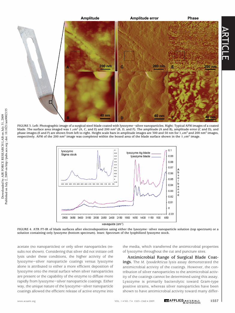
nanoparticle solution. After a constant dc potential wasapplied, a red film formed on the surface of the blades andneedles. Much of this coating was removed after extensivewashing with deionized water, but a thin coating remainedon the metal surfaces (Figure 3, left). The evenly dispersedand strongly adherent coating was not observed when anyof the control solutions were used in the electrophoreticdeposition assay. A uniform coating was not seen whenmost of the lysozyme was dialyzed from a silver nanoparticlesolution prior to electrophoresis or with solutions of freshlymixed lysozyme and silver acetate, which did not containany nanoparticles. A translucent coating was observed whenonly lysozyme was present in solutions, but the film waseasily washed off the blade surface.
The coatings from lysozyme-silver nanoparticle elec-trodeposition solutions retained lysozyme, as shown by ATRFT-IR (Figure 4). The spectrum of the coated blades wasidentical to the spectrum of blades that were coated withlysozyme only, as well as to the spectrum of the lysozymestock. The results confirmed that no major perturbations inthe lysozyme structure occurred as a result of silver nano-particle synthesis or electrophoretic deposition. Changes in
the protein secondary and tertiary structures will alterhydrogen bonding between the CO and NH groups in thepeptide backbone, which results in aberrations of the amineI, II, and III bands between 1200 and 1700 cm-1 (34). Nosuch significant shifts were observed in this IR regionbetween lysozyme-coated blades and the stock powder. Inaddition, coated blade surfaces were analyzed at the na-nometer scale using AFM (Figure 3). A layer of sphericalnanoparticles on the surface of coated blades was clearlyseen in images. The AFM images of etched, uncoated bladesdid not have these features (results not shown). The nano-particles visualized in the coating were similar in shape anddiameter to the silver nanoparticles observed in the TEMimages of the deposition solution (Figure 1). The nanopar-ticles were also readily seen in phase images (lighter areas,Figure 3E) and were surrounded by a material of contrastingphase (darker areas). Under closer inspection, edge enhance-ment illustrated in the 200 nm2 amplitude error image(Figure 3D) showed a veinlike texture between the nano-particles. In the corresponding phase image (Figure 3F),these features appeared in contrasting phase color to thenanoparticles. The contrast in the phase images can be usedto discern between an organic material, consisting of proteinor other hydrocarbon compounds, and metallic nanopar-ticles (35, 36). Lysozyme and crystalline silver would impartvery different interactions with the AFM tip, resulting in cleardifferences in the phase images. Considering that the elec-trodeposition buffer contained primarily silver and lysozyme,the phase-contrasting materials are most likely distinctregions of these two substances. The regions surroundingthe spherical protrusions on the blade surface are proposedto be areas of lysozyme packed between the silver nano-particles. Similar grafting features have been observed inAFM phase contrast images of surfaces after the electro-chemical deposition of silver nanoparticles and acrylate,where a sustained cathodic overpotential during electrodepo-sition forced the organic material off the silver nanoparticlesand onto the electrode surface (36).
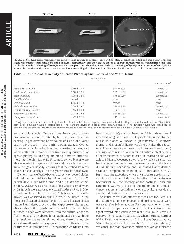
Evaluation of the Lysozyme Activity withinSurface Coatings. The hydrolytic activity of lysozymeretained within electrodeposited coatings on blades andneedles was investigated through an in vitro cell lysis assaydesigned to mimic the normal use of the instruments.Coated blades and needles were used to make incisions andpunctures, respectively, into agarose infused with M. lyso-deikticus cells. Within minutes, zones of clearing were seenat the contact site, which was indicative of cell lysis due tolysozyme hydrolytic activity (Figure 5). The spread of celllysis over time within the agar demonstrated that theantimicrobial activity not only was retained on the surfaceof the blades and needles but also was transferred into themedia. Blades subjected to electrodeposition using a solutioncontaining only lysozyme demonstrated a minimal zone ofclearing after incubation for 16 h, in comparison to bladescoated with lysozyme-silver nanoparticles. Cell lysis wasnot observed with blades and needles subjected to electro-phoretic deposition using solutions containing soluble silver
FIGURE 2. Cyclic voltammograms of pure lysozyme (A) and thelysozyme-silver nanoparticle electrophoresis solution (B) in 50%methanol. In each case, five voltammograms were recorded. Theinitial voltammogram is shown in black, and successive voltammo-grams are in increasingly lighter gray. The initial voltammogram ofpure lysozyme from part A is shown as a dashed black line in partB. The voltammogram of 0.5 mM silver acetate in 50% methanol isshown as a dashed gray line.
ARTIC
LE
1556 VOL. 1 • NO. 7 • 1553–1560 • 2009 Eby et al. www.acsami.org
Dow
nloa
ded
by A
IR F
OR
CE
RES
EAR
CH
LA
B o
n Ju
ly 3
1, 2
009
Publ
ishe
d on
July
2, 2
009
on h
ttp://
pubs
.acs
.org
| do
i: 10
.102
1/am
9002
155
acetate (no nanoparticles) or only silver nanoparticles (re-sults not shown). Considering that silver did not initiate celllysis under these conditions, the higher activity of thelysozyme-silver nanoparticle coatings versus lysozymealone is attributed to either a more efficient deposition oflysozyme onto the metal surface when silver nanoparticlesare present or the capability of the enzyme to diffuse morerapidly from lysozyme-silver nanoparticle coatings. Eitherway, the unique nature of the lysozyme-silver nanoparticlecoatings allowed the efficient release of active enzyme into
the media, which transferred the antimicrobial propertiesof lysozyme throughout the cut and puncture sites.
Antimicrobial Range of Surgical Blade Coat-ings. The M. lysodeikticus lysis assay demonstrated theantimicrobial activity of the coatings. However, the con-tribution of silver nanoparticles to the antimicrobial activ-ity of the coatings cannot be determined using this assay.Lysozyme is primarily bacterolytic toward Gram-typepositive strains, whereas silver nanoparticles have beenshown to have antimicrobial activity toward many differ-
FIGURE 3. Left: Photographic image of a surgical steel blade coated with lysozyme-silver nanoparticles. Right: Typical AFM images of a coatedblade. The surface area imaged was 1 µm2 (A, C, and E) and 200 nm2 (B, D, and F). The amplitude (A and B), amplitude error (C and D), andphase images (E and F) are shown from left to right. Height scale bars in amplitude images are 300 and 50 nm for 1 µm2 and 200 nm2 images,respectively. AFM of the 200 nm2 image was completed within the boxed area of the blade surface shown in the 1 µm2 image.
FIGURE 4. ATR FT-IR of blade surfaces after electrodeposition using either the lysozyme-silver nanoparticle solution (top spectrum) or asolution containing only lysozyme (bottom spectrum). Inset: Spectrum of the lyophilized lysozyme stock.
ARTIC
LE
www.acsami.org VOL. 1 • NO. 7 • 1553–1560 • 2009 1557
Dow
nloa
ded
by A
IR F
OR
CE
RES
EAR
CH
LA
B o
n Ju
ly 3
1, 2
009
Publ
ishe
d on
July
2, 2
009
on h
ttp://
pubs
.acs
.org
| do
i: 10
.102
1/am
9002
155
ent microbial species. To determine the range of antimi-crobial activity demonstrated by both components of thecoating, eight different bacterial strains and one yeaststrain were used in the antimicrobial assays. Coatedblades were incubated with actively-growing cultures, andviable cells that remained over time were quantitated byspread-plating culture aliquots on solid media and enu-merating the cfu (Table 1). Uncoated, etched blades werealso incubated in separate cultures and, in each case, cellsgrew to high cell density, ensuring that the etched stainlesssteel did not adversely affect the growth (results not shown).
Demonstrating effective bactericidal activity, coated bladesreduced the cell viability by >3 log within 1.5 h for K.pneumoniae, B. anthracis Sterne, and B. subtilis and within3 h for S. aureus. A lesser biocidal effect was observed whenA. baylyi cells were exposed to coated blades (∼3 log in 3 h).Growth inhibition lasted beyond 3 h because no visiblegrowth was observed in cultures when incubated in thepresence of coated blades for 24 h. To assess if coated bladesretained antimicrobial activity after exposure to cultures andalso inhibited the transfer of any viable cells on bladesurfaces, blades were removed from the culture, added tofresh media, and incubated for an additional 24 h. With thefive sensitive strains mentioned above, there was no ob-served growth in the subsequent cultures. Furthermore, theculture media from the first 24 h incubation was diluted into
fresh media (1:10) and incubated for 24 h to determine ifany remaining viable cells could proliferate in the absenceof coated blades. S. aureus, K. pneumoniae, B. anthracisSterne, and B. subtilis did not visibly grow after the subcul-ture. The two subsequent sets of cultures confirmed that (i)coatings were resilient and retained antimicrobial activityafter an extended exposure to cells, (ii) coated blades wereable to inhibit subsequent growth of any viable cells that mayhave attached to coated and uncoated areas of the bladeduring the first incubation, and (iii) coated blades demon-strated a complete kill in the initial culture after 24 h. A.baylyi was one exception, where one subculture grew to highcell density. We conclude that the effect on A. baylyi wasbactericidal, but the potency of the coatings under ourconditions was very close to the minimum bactericidalconcentration, and growth in the one subculture was due tostandard deviation or experimental error.
An initial, bactericidal effect was measured for E. coli, butthe strain was able to recover and turbid cultures wereobserved after 24 h incubation. Previous work demonstratedthat silver nanoparticles were an effective antimicrobialagent toward this particular strain of E. coli (16, 21). We didobserve higher bactericidal activity when the initial numberof E. coli cells was reduced to 104 in cultures (approximately3 log reduction in viable cells within 1.5 h; data not shown).We concluded that the concentration of silver ions released
FIGURE 5. Cell lysis assay measuring the antimicrobial activity of coated blades and needles. Coated blades (left and middle) and needles(right) were used to make incisions and punctures, respectively, and then placed on top of agarose infused with M. lysodeikticus cells. Thetop blade contains a coating of lysozyme-silver nanoparticles, while the lower blade has a coating of lysozyme only. Zones of cell lysis areseen at the incision and puncture sites, as well as surrounding the blades and needles after incubation at 37 °C for 30 min and 16 h.
Table 1. Antimicrobial Activity of Coated Blades against Bacterial and Yeast Strainslog reductiona
strain 1.5 h 3 h inhibition typeb
Acinetobacter baylyi 2.49 ( 1.48 2.98 ( 1.75 bactericidalBacillus anthracis Sterne 3.50 ( 1.25 3.93 ( 0.62 bactericidalBacillus subtilis 4.78 ( 0.50 4.78 ( 0.50 bactericidalCandida albicans growth growth noneEscherichia coli 1.56 ( 1.38 growth noneKlebsiella pneumoniae 3.29 ( 1.67 3.94 ( 1.14 bactericidalPseudomonas fluorescens 0.65 ( 0.24 0.26 ( 0.30 noneStaphylococcus aureus 2.01 ( 0.62 3.48 ( 0.53 bactericidalStaphylococcus epidermidis 0.47 ( 0.55 0.43 ( 0.24 bacteriostatic
a log reduction was calculated as [log of viable cells (cfu mL-1) before exposure to a coated blade] - [log of the viable cells (cfu mL-1) at a timepoint after incubation with a coated blade]. The standard deviation is from three separate assays. b The inhibition type was based on logreduction values and the viability of the subcultures made from the initial 24 h incubation with coated blades. See the text for details.
ARTIC
LE
1558 VOL. 1 • NO. 7 • 1553–1560 • 2009 Eby et al. www.acsami.org
Dow
nloa
ded
by A
IR F
OR
CE
RES
EAR
CH
LA
B o
n Ju
ly 3
1, 2
009
Publ
ishe
d on
July
2, 2
009
on h
ttp://
pubs
.acs
.org
| do
i: 10
.102
1/am
9002
155

into the culture by coated blades was below the minimuminhibitory concentration for E. coli under the conditions ofthe antimicrobial assay. Similar to E. coli, an initial, bacte-ricidal effect was also measured for P. fluorescens, but thesurviving population recovered and propagated after 24 h.When P. fluorescens cultures were transferred and grown onsolid media to enumerate viable cells, colonies originatingfrom coated blade cultures were nearly identical with colo-nies grown from uncoated blade cultures but did not producethe characteristic yellow-green pyoverdine pigments that arenormally observed with robust cells (37). The cause of thisphenotype is not clear although it suggested that residualsilver disrupted the pyoverdine synthesis and/or export fromthe cell.
The effect of coated blades on S. epidermidis was consid-ered to be bacteriostatic. On average, culture viability didnot change significantly in viability during the first 3 h afterexposure to coated blades and did not grow to visibleturbidity after 24 h. Upon transfer of the blades in freshmedia, cultures grew to a slight turbidity, suggesting thatblade coatings still inhibited S. epidermidis growth but hadlost some of their potency. When the initial culture wassubcultured into fresh media and incubated for an additional24 h in the absence of coated blades, the cells grew nor-mally. The subsequent cultures confirmed that the coatingexerted a bacteriostatic effect on S. epidermidis. Under theconditions of the assays, coated blades inhibited reproduc-tion and growth of S. epidermidis but did not completely killcells. Once removed from exposure to coated blades, cellsrecovered and grew.
There was no apparent effect against the representativefungal strain C. albicans. When cultures of this yeast wereexposed to coated blades, they grew at the same rate ascultures containing uncoated blades. In our previous study,the effectiveness of lysozyme-silver nanoparticle suspen-sions against C. albicans increased when lysozyme wasremoved from the solution (21). The results in this studywere consistent with our previous findings because thecoatings were created from nanoparticle suspensions thatcontained a considerably higher concentration of lysozymeand, as a result, were less effective against C. albicans cells.
CONCLUSIONSOur studies demonstrated that composite antimicrobial
coatings can be generated using methods for lysozyme-mediated silver nanoparticle synthesis and electrophoreticdeposition. The coating employs two different biocidalmechanisms: the antimicrobial activity of silver ions and themuramidase activity of lysozyme. Coatings that integratedifferent biocidal mechanisms are of particular interestbecause they can minimize the selection and proliferationof resistant strains. Silver nanoparticles have already beenshown to be effective in suppression of a wide range ofpathogens, especially against strains resistant to mainstreamantibiotics (6, 38). While the contribution of lysozyme hasbeen shown to minimally contribute to the antimicrobialactivity of the composite (21), the enzyme was essential toelectrophoretic deposition and promoted the formation of
homogeneous coatings on stainless steel surfaces. Thenature of the coating was such that the mixture of lysozymeand silver nanoparticles facilitated strong adsorption tostainless steel surfaces, yet the active components alsodiffused from the coating following contact with media. Theobservations indicate that the physical properties of thecoating are inherent to lysozyme, where the protein acts asa water-soluble adhesive and enables robust adsorption ofthe silver nanoparticles to the blade surface but will alsopermit diffusion of silver ions or release of the nanoparticlesthemselves once incubated in an aqueous environment.Hence, the effect could provide a persistent self-cleaningsurface for the medical instruments and allow transfer of theantimicrobial activity to the contact surface during use (e.g.,diffusion into the surrounding flesh following an incision,injection, or implant). With respect to implanted devices, theintegration of lysozyme within the composite may alsoincrease the biocompatibility of the coating and not hampertissue reconstruction, compared to other silver coatings thatare composed solely of inorganic material (39). Furthermore,the rapid coating method presented here employs reagentsalready accepted for use in the food and medical industries(40, 41), increasing its potential for transition to manufactur-ing for a diverse range of applications. While the coating wasnot effective against all strains tested in this study, furtheroptimization of nanoparticle synthesis and deposition willimprove its potency.
Acknowledgment. We thank Karen Kelly at the Univer-sity of Florida Interdisciplinary Center for BiotechnologyResearch, Electron Microscopy Core Lab, Gainsville, FL, forgenerating TEM images, Robert K. Nichols for helpful dis-cussionsonelectrophoreticdeposition,andKennethStrawheck-er for assistance in AFM techniques. This work was sup-ported through funding from the Air Force Materials andManufacturing Directorate (AFRL/RX) and the Joint ScienceTechnology Office for Chemical and Biological DefenseProject Code AA06CBT008 (Ilya Elashvili, Jennifer Becker,and Stephen Lee, Program Managers).
REFERENCES AND NOTES(1) Hetrick, E. M.; Schoenfisch, M. H. Chem. Soc. Rev. 2006, 35, 780–
789.(2) Babu, R.; Zhang, J.; Beckman, E. J.; Virji, M.; Pasculle, W. A.; Wells,
A. Biomaterials 2006, 27, 4304–4314.(3) Hardes, J.; Ahrens, H.; Gebert, C.; Streitbuerger, A.; Buerger, H.;
Erren, M.; Gunsel, A.; Wedemeyer, C.; Saxler, G.; Winkelmann,W.; Gosheger, G. Biomaterials 2007, 28, 2869–2875.
(4) Ong, S.-Y.; Wu, J.; Moochhala, S. M.; Tan, M.-H.; Lu, J. Biomaterials2008, 29, 4323–4332.
(5) Dowsett, C. Nurs. Stand. 2004, 19, 56–60.(6) Strohal, R.; Schelling, M.; Takacs, M.; Jurecka, W.; Gruber, U.;
Offner, F. J. Hosp. Infect. 2005, 60, 226–230.(7) Silver, S. FEMS Microbiol. Rev. 2003, 27, 341–353.(8) Lansdown, A. B. G. Curr. Prob. Derm. 2006, 33, 17–34.(9) Schierholz, J. M.; Lucas, L. J.; Rump, A.; Pulverer, G. J. Hosp. Infect.
1998, 40, 257–262.(10) Baker, C.; Pradhan, A.; Pakstis, L.; Pochan, D. J.; Shah, S. I. J.
Nanosci. Nanotechnol. 2005, 5, 244–249.(11) Pal, S.; Tak, Y. K.; Song, J. M. Appl. Environ. Microbiol. 2007, 73,
1712–1720.(12) Kim, Y. H.; Lee, D. K.; Cha, H. G.; Kim, C. W.; Kang, Y. S. J. Phys.
Chem. C 2007, 111, 3629–3635.(13) Kong, H.; Jang, J. Langmuir 2008, 24, 2051–2056.
ARTIC
LE
www.acsami.org VOL. 1 • NO. 7 • 1553–1560 • 2009 1559
Dow
nloa
ded
by A
IR F
OR
CE
RES
EAR
CH
LA
B o
n Ju
ly 3
1, 2
009
Publ
ishe
d on
July
2, 2
009
on h
ttp://
pubs
.acs
.org
| do
i: 10
.102
1/am
9002
155

(14) Lee, D.; Cohen, R. E.; Rubner, M. F. Langmuir 2005, 21, 9651–9659.
(15) Li, Y.; Leung, P.; Yao, L.; Song, Q. W.; Newton, E. J. Hosp. Infect.2006, 62, 58–63.
(16) Marini, M.; Niederhausern, S. D.; Iseppi, R.; Bondi, M.; Sabia, C.;Toselli, M.; Pilati, F. Biomacromolecules 2007, 8, 1246–1254.
(17) Sambhy, V.; MacBride, M. M.; Peterson, B. R.; Sen, A. J. Am. Chem.Soc. 2006, 128, 9798–808.
(18) Vigneshwaran, N.; Kathe, A. A.; Varadarajan, P. V.; Nachane,R. P.; Balasubramanya, R. H. Langmuir 2007, 23, 7113–7117.
(19) Wang, Q.; Yu, H.; Zhong, L.; Liu, J.; Sun, J.; Shen, J. Chem. Mater.2006, 18, 1988–1994.
(20) Zhang, Y.; Peng, H.; Huang, W.; Zhou, Y.; Zhang, X.; Yan, D. J.Phys. Chem. C 2008, 112, 2330–2336.
(21) Eby, D. M.; Schaeublin, N. M.; Farrington, K. E.; Hussain, S. M.;Johnson, G. R. ACS Nano 2009, 3, 984–994.
(22) Pellegrini, A.; Thomas, U.; von Fellenberg, R.; Wild, P. J. Appl.Bacteriol. 1992, 72, 180–187.
(23) Panaeek, A.; Kvıtek, L.; Prucek, R.; Kolar, M.; Vecerova, R.;Pizurova, N.; Sharma, V. K.; Nevecna, T.; Zboril, R. J. Phys. Chem.B 2006, 110, 16248–16253.
(24) Lim, S. H.; Wei, J.; Lin, J.; Li, Q.; Kuayou, J. Biosens. Bioelectron.2005, 20, 2341–2346.
(25) Luo, X. L.; Xu, J. J.; Du, Y.; Chen, H. Y. Anal. Biochem. 2004, 334,284–289.
(26) Wu, L. Q.; Lee, K.; Wang, X.; English, D. S.; Losert, W.; Payne,G. F. Langmuir 2005, 21, 3641–3646.
(27) Zhao, G.; Xu, J. J.; Chen, H. Y. Anal. Biochem. 2006, 350, 145–150.
(28) Spoerke, E. D.; Stupp, S. I. Biomaterials 2005, 26, 5120–5129.(29) Fleming, A. Proc. R. Soc. London, Ser. B 1922, 93, 306–317.(30) Ireland, J. A.; Hanna, P. C. Infect. Immun. 2002, 70, 5870–5872.(31) Juni, E.; Janik, A. J. Bacteriol. 1969, 98, 281–288.(32) Barton, A. C.; Collyer, S. D.; Davis, F.; Gornall, D. D.; Law, K. A.;
Lawrence, E. C.; Mills, D. W.; Myler, S.; Pritchard, J. A.; Thomp-son, M.; Higson, S. P. Biosens. Bioelectron. 2004, 20, 328–337.
(33) Pires de Castro, C. S.; Rodrigues Souza De, J.; Bloch Junior, C.J. Inorg. Biochem. 2003, 94, 365–371.
(34) Socrates, G. Infrared and raman chracteristic group frequencies. Tablesand charts, 3rd ed.; John Wiley and Sons, Ltd.: Chichester, U.K., 2001.
(35) Lin, S.; Lee, C. K.; Lee, S. Y.; Kao, C. L.; Lin, C. W.; Wang, A. B.;Hsu, S. M.; Huang, L. S. Cell. Microbiol. 2005, 7, 1763–1770.
(36) Voccia, S.; Ignatova, M.; Jerome, R.; Jerome, C. Langmuir 2006,22, 8607–8613.
(37) Meyer, J.-M. Arch. Microbiol. 2000, 174, 135–142.(38) Atiyeh, B. S.; Costagliola, M.; Hayek, S. N.; Dibo, S. A. Burns 2007,
33, 139–148.(39) Meyers, S. R.; Khoo, X.; Huang, X.; Walsh, E. B.; Grinstaff, M. W.;
Kenan, D. J. Biomaterials 2009, 30, 277–286.(40) Silver, S.; Phung, L. T.; Silver, G. J. Ind. Microbiol. Biotechnol. 2006,
33, 627–634.(41) U.S. Food and Drug Administration, Center for Food Safety and
Applied Nutrition, Office of Premarket Approval. Agency ResponseLetter Generally Regarded as Safe (GRAS) Notice No. GRN 000064;April 2, 2001.
AM9002155
ARTIC
LE
1560 VOL. 1 • NO. 7 • 1553–1560 • 2009 Eby et al. www.acsami.org
Dow
nloa
ded
by A
IR F
OR
CE
RES
EAR
CH
LA
B o
n Ju
ly 3
1, 2
009
Publ
ishe
d on
July
2, 2
009
on h
ttp://
pubs
.acs
.org
| do
i: 10
.102
1/am
9002
155