hyaluronate synthase: cloning and sequencing of the gene from
TRANSCRIPT

Biochem. J. (1993) 289,179-184 (Printed in Great Britain)
Hyaluronate synthase: cloning and sequencing of the gene fromStreptococcus sp.Manfred LANSING,*II Sabine LELLIG,* Andreas MAUSOLF,t Irene MARTINI,t Fabiana CRESCENZI4 Michael O'REGANtand Peter PREHM*§*Institut fur Physiologische Chemie und Pathobiochemie, Waldeyerstrasse 15, D-44 Munster, Federal Republic of Germany,tMax Planck Institut fur Biochemie, D-8033 Martinsried, Federal Republic of Germany,and tAdvanced Technology Division, FIDIA s.p.a., Via Ponte delta Fabrica 3/A, 1-35031 Abano Terme PD, Italy
The complete nucleotide sequence of hyaluronate synthase from
Streptococcus sp. and its flanking regions is presented. The gene
locus was designated has. Southern-blotting results suggestedthat the gene was conserved in hyaluronate-producingstreptococci. A putative translation-initiation codon was
identified and the open reading frame consists of 1566 bp,
INTRODUCTIONHyaluronate is a linear high-molecular-mass glycosaminoglycanwhich has been shown to be involved in many biologicalinteractions (Laurent and Fraser, 1992). Group A and Cstreptococci produce a hyaluronate capsule which amplifiesinfectious virulence, because it protects streptococci againstimmunological attacks of the infected host (Todd and Lancefield,1928; Seastone, 1939; Hirst, 1941; Whitnack et al., 1981; Wesselset al., 1991). Hyaluronate is produced by a synthase whichresides in protoblast membranes of streptococci (Markovitz etal., 1958; Markovitz and Dorfman, 1962; Stoolmiller andDorfman, 1969; Sugahara et al., 1979) and in plasma membranesof eukaryotic cells (Prehm, 1984). We have previously identifiedthe streptococcal synthase as a 52 kDa protein (Prehm andMausolf, 1986). The bacterial enzyme cross-reactsimmunologically with proteins in plasma membranes fromeukaryotic cells (Prehm 1989; Klewes et al., 1993) and is shedfrom growing streptococci into the culture media (Mausolf et al.,1990). In order to gain more information on the hyaluronatesynthase itself and on the genetics of hyaluronate synthesis, we
have undertaken the cloning and sequencing of the gene encodingthe hyaluronate synthase.
MATERIALS AND METHODS
Materials
DNA-modifying enzymes were obtained from Boehringer, NEBGibco/BRL or Pharmacia. Radiochemicals were obtained fromAmersham International, and other reagents from Sigma Chemi-cal Co. Agt-1 1 and pBS-SK were from Stratagene, pGEM-3Zfrom Promega, and pUC-18 from Boehringer. EcoRI-linker: 5'-d(CCGGAATTCCGG)-3' was from NEB. Oligonucleotideswere synthesized on an Applied Biosystems DNA Synthesizer380B. The primers used were as follows: primer 1, 5'-d(GGTGGCGACGACTCCTGGAGCCCG)-3'; primer 2,5'-d(TTGACACCAGACCAACTGGTAATG)-3'; primer 3,
specifying a protein of 56 kDa. Sequences resembling thepromoter and ribosome-binding site of Gram-positiveorganisms are found upstream of the synthase. The predictedamino-acid sequence reveals the presence of a 35-residue signalpeptide. The sequence has some similarity to bacterial peptide-binding proteins.
5'-d(AGGAATGTCACAACCTT)-3'; primer 4, 5'-d(CCCC-TAGAGAGTCTAGA)-3'. DNA manipulations were performedas described by Sambrook et al. (1989).
Bacterial strains
Streptococcus equisimilis strain D181 was from the RockefellerUniversity collection. Streptococcus equi 68222 and Streptococcuszooepidemicus 68270 were from the Belfanti SerotherapeuticInstitute, Milan. Streptococcus sanguis (Challis) was obtainedfrom Dr. Laplace, Jena, F.R.G. Escherichia coli SMR 10 was
from Dr. Meinhard, Miinster. E. coli Y1089 and Y1090 were
from Stratagene.
Isolation of streptococcal DNAGenomic DNA was isolated from streptococci by the method ofNida and Cleary (1983) with slight modifications. An overnightculture of streptococci (5 ml) growing in Todd-Hewitt brothcontaining 40 mM DL-threonine and 30 mM glucose at 37 °Cwas used to inoculate 100 ml of Todd-Hewitt broth containing40 mM DL-threonine. The culture was incubated at 37 'C. Onreaching a D600 of 0.03, cysteine was added to give a finalconcentration of 6 mM, and at a D600 of 0.1 testicular hyal-uronidase was added to give a final concentration of 3 units/ml.The bacteria were harvested, at a D600 value of 0.5, bycentrifugation at 5000 g for 10 min, washed twice with TE buffer(10 mM Tris/HCl/1 mM EDTA) at 0 'C, resuspended in 8.75 mlofTE and incubated with lysozyme (5 mg/ml) and hyaluronidase(1.5 units/ml) for 1 h at 37 'C. Proteinase K and Pronase E(250 jig/ml of each) were added and incubation was continuedfor 30 min at 37 'C. SDS was added to a final concentration of0.5 % and incubation was continued, until cell lysis was observed.TE was added to a final volume of 10 ml. NaCl andcetyltrimethylammonium bromide (CTAB) were added to finalconcentrations of 0.7 M and 1.130% (w/v) respectively. Thesolution was heated to 65 'C for 20 min and extracted with 1 vol.
Abbreviations used: FE, 10 mM Tris/HCI, 1 mM EDTA (pH 7.5); CTAB, cetyltrimethylammonium bromide.§ To whom correspondence should be addressed.11 Present address: Advanced Technology Division, FIDIA s.p.a., Via Ponte della Fabrica 3/A, 1-35031 Abano Terme PD, Italy.
179
180 M. Lansing and others
chloroform/isoamyl alcohol (24:1). The phases were separatedby centrifugation at 9000 g for 10 min at room temperature. Theupper, aqueous phase was withdrawn and mixed with 0.6 vol.propan-2-ol to precipitate DNA.
Genomic DNA libraryHigh-molecular-mass DNA from S. equisimilis D181 waspartially digested with AluI, and fragments of 2-5 kb wereisolated by sucrose gradient centrifugation (Weis, 1989). Theisolated AluI fragments were dephosphorylated, methylated withEcoRI methylase and ligated to EcoRl linkers (Wu et al., 1987).The linker-containing DNA was ligated to EcoRI-digested Agt-11 arms and packaged into infectious virions using the 'one-strain-packaging' system E. coli SMR1O according to the methodof Rosenberg et al. (1985). The genomic library was establishedusing E. coli Y1089 and amplified with E. coli Y1090.
Library screeningThe library was screened as described by Huynh et al. (1986) withpolyclonal antibodies raised against the synthase which hadpreviously been isolated in our laboratory (Prehm and Mausolf,1986). Cross-reactive antibodies to E. coli/Agt-11 lysates wereremoved by pseudoscreening as described by Sambrook et al.(1989).
Clone characterizationPlate lysates were prepared from positive A clones. The plateswere washed with 5 ml of phosphate-buffered saline and proteinswere precipitated by addition of polyethylene glycel 6000 to afinal concentration of 20% (w/v). The samples were incubated at4 °C for 2 h. After centrifugation at 35000 g for 30 min at 4 °Cthe pellet was resuspended in 0.5 ml of phosphate-buffered saline.Proteins were precipitated by the method of Wessel and Flugge(1984), separated on a 100% SDS/polyacrylamide gel andanalysed by Western blotting with antibodies raised against thehyaluronate synthase.
Inserts of the A clones were amplified using PCR by themethod of Saiki et al. (1988) using primers 1 and 2 adjacent tothe EcoRI cloning site. PCR was carried out in a Bio-medthermocycler 60. The thermocycling profile consisted of an initialdenaturation at 95 °C for 2 min, followed by 35 cycles at 95 °C,for 1 min; at 45 °C, for 1 min; and at 72 °C, for 5 min. PCRproducts were purified according to Both et al. (1991), digestedwith different restriction enzymes to map the inserts and analysedby agarose-gel electrophoresis.
Southern blotting was carried out essentially as described bySambrook et al. (1989). Probe preparation, hybridization anddetection were carried out with the non-radioactive DIG-DNAlabelling kit from Boehringer (Mannheim).
DNA-sequence analysisNucleotide sequences were determined by the dideoxy-chain-termination method according to Sanger et al. (1977) using theT7 DNA polymerase sequencing kit (Pharmacia) or the fmolsequencing kit (Promega). Double-stranded plasmid DNA waspurified using the plasmid preparation kit from Diagen andsequenced according to Chen and Seeburg (1985).A DNA was sequenced using the protocol of Snyder et al.
(1987) with slight modifications. DNA (5 jig) was double digestedwith KpnI and SacI, phenol extracted, ethanol precipitated, and
addition of 2 ,l of a solution containing 2 M NaOH and 2 mMEDTA for 10 min at 37 'C. Sodium acetate (13 pA; 1.5 M) wasadded and the DNA was precipitated with 2.5 vol. of ethanol at-20 'C, and the sample was placed in a methanol/solid CO2bath for 15 min. After centrifugation the DNA was resuspendedin 15 41l of 7.5 mM Tris/HCl/7.5 mM dithiothreitol/5 mMMgCl2, pH 7.5, containing 0.7 ng/al 32P-labelled primer 1 or 2and incubated for 15 min at 55 'C. Sequencing reactions werecarried out as for plasmid DNA sequencing.
PCR-amplified DNA from Agt-I 11/2LK-D was sequenced asdescribed by Both et al. (1991) with primers 3 and 4 which werededuced from the hyaluronate-synthase-gene sequence.
Agt-l 11/2LK-D DNA was isolated as described above andsequenced using the fmol sequencing kit (Promega) with 32P-end-labelled primer 2.DNA sequences were analysed with DNASTAR software
(DNASTAR Inc. Madison, U.S.A.), GENEPRO software(Riverside Scientific, Washington, U.S.A.) and CLUSTALsoftware (Higgins and Sharp, 1988).
Isolation of peptides and amino-acid-sequence analysisPurified hyaluronate synthase (200 /pg) was dissolved in 200 ,ul of6 M urea and diluted with 400 ,ul of 20 mM Tris/HC1, pH 7.8,and digested with 5% (w/v) trypsin or V8-protease for 24 h. Themixture was acidified with trifluoroacetic acid and peptides were
centrifuged at 10000 g for 3 min. The supernatant was applied toa C18 reverse-phase h.p.l.c. column (VYDAC 218 TP54). Peptideswere eluted with a linear gradient of 0.1 0% trifluoroacetic acid inwater to 0.1 trifluoroacetic acid in 70% acetonitrile in 30 minwith a flow rate of 1 ml/min. The elution was monitored at220 nm. The fractions were dried in a speedvac and analysed ina gas-phase sequencer (Applied Biosystems 120A).
RESULTSIsolation of Agt-11 clones expressing hyaluronate synthaseA Agt- genomic library was established from S. equisimilisD181 and screened with antibodies against the hyaluronatesynthase. Positive plaques were found on plates which had notbeen induced by isopropylthiogalactoside, indicating that thoserecombinants contained a promoter which was functional in E.coli. Ten positive clones were identified from 50000 plaques.These clones were purified and amplified.
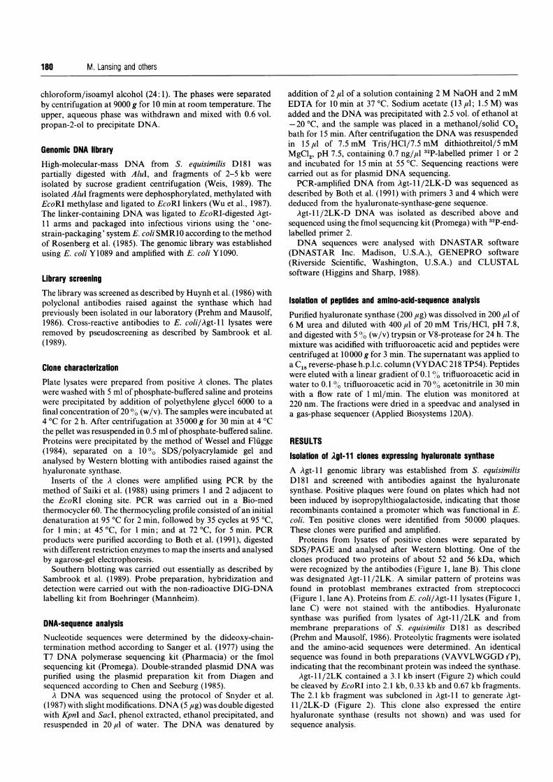
Proteins from lysates of positive clones were separated bySDS/PAGE and analysed after Western blotting. One of theclones produced two proteins of about 52 and 56 kDa, whichwere recognized by the antibodies (Figure 1, lane B). This clonewas designated Agt-1 1/2LK. A similar pattern of proteins wasfound in protoblast membranes extracted from streptococci(Figure 1, lane A). Proteins from E. coli/Agt-1 1 lysates (Figure 1,lane C) were not stained with the antibodies. Hyaluronatesynthase was purified from lysates of Agt-l 1/2LK and frommembrane preparations of S. equisimilis D 181 as described(Prehm and Mausolf, 1986). Proteolytic fragments were isolatedand the amino-acid sequences were determined. An identicalsequence was found in both preparations (VAVVLWGGD fP),indicating that the recombinant protein was indeed the synthase.
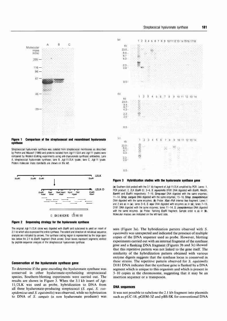
Agt-1 1/2LK contained a 3.1 kb insert (Figure 2) which couldbe cleaved by EcoRI into 2.1 kb, 0.33 kb and 0.67 kb fragments.The 2.1 kb fragment was subcloned in Agt- 11 to generate Agt-11 /2LK-D (Figure 2). This clone also expressed the entirehyaluronate synthase (results not shown) and was used for
resuspended in 20 1l of water. The DNA was denatured by sequence analysis.
Streptococcal hyaluronate synthase
(a)
A B C
Molecularmass(kDa)
205 -
116-96-
67-_-5
45- Ib)
1 2 3 4 5 6 7 8 9 10111213 141516 1718
kb23.09.46.74.3
2.32.0
0.5
1 2 3 4 5 6 7 8 9 10 11 12 13 14kb
23.09.4.6.74.3
2.32.0
29-
0.5
Figure 1 Comparison of the streptococcal and recombinant hyaluronatesynthase
Streptococcal hyaluronate synthase was isolated from streptococcal membranes as describedby Prehm and Mausolf (1986) and proteins isolated from Agt-11/2LK and Agt-11 lysates were
compared by Western-blotting experiments using anti-(hyaluronate synthase) antibodies. LaneA, streptococcal hyaluronate synthase; lane B, Agt-11/2LK lysate; lane C, Agt-11 lysate.Protein molecular mass standards are shown on the left.
(c)kb
23.09.46.74.3
2.32.0
1 2 3 4 5 6 7 8 9 10 11 12 13 14
0.5
EcoRI EcoRI EcoRl--A2LKEcoRI
lEcoRI Rsal Rsal Hpall Kpnl Pstl
Ndel Nhel Xbal Hpall
o 0laIioin0 A goom
Figure 2 Sequencing strategy for the hyaluronate synthase
The original Agt-11/2LK clone was digested with EcoRl and subcloned to yield an insert of2.1 kb which also expressed the entire synthase. The extent and direction of individual sequenceanalysis are indicated by arrows. The synthase coding region is represented by the large openbox below the 2.1 kb EcoRl fragment (thick arrow). Small boxes represent segments verifiedby peptide-sequence analysis of the streptococcal hyaluronate synthase.
Conservation of the hyaluronate synthase gene
To determine if the gene encoding the hyaluronate synthase was
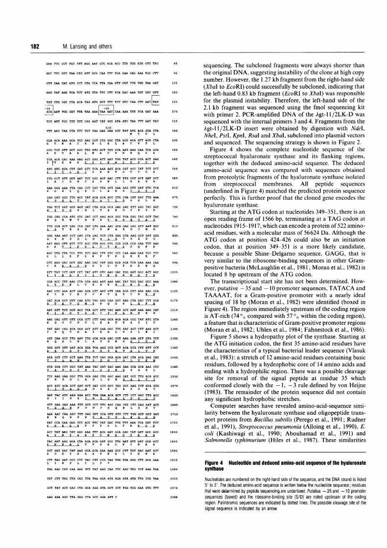
conserved in other hyaluronate-synthesizing streptococcalspecies, Southern-blotting experiments were carried out. Theresults are shown in Figure 3. When the 3.1 kb insert of Agt-11/2LK was used as probe, hybridization to DNA fromall three hyaluronate-producing streptococci (S. equi, S. zoo-
epidemicus and S. equisimilis) was observed, while no hybrizationto DNA of S. sanguis (a non hyaluronate producer) was
Figure 3 Hybridization studies with the hyaluronate synthase gene
(a) Southern blot probed with the 3.1 kb fragment of Agt-1 1/2LK amplified by PCR. Lanes: 1,o
A2LK-D PCR product; 2, 2LK EcoRl El; 3-6, S. equisimilis D181 DNA digested with EcoRl, Hindlll,
HpalR BamHl and EcoRV respectively; 7-10, Strepequi DNA digested with the same enzymes;11-14, Strep. sanguis DNA digested with the same enzymes; 15-18, Strep. zooepidemicusDNA digested with the same enzymes. (b) Probe: Xbal-Pstt internal has fragment. Lanes 1and 2 are as in (a); lanes 3-6, S. equi DNA digested with enzymes as in (a); lanes 7-10,D181 DNA digested with the same enzymes; lanes 11-14, S. zooepidemicus DNA digestedwith the same enzymes. (c) Probe: flanking EcoRl fragment. Sample order is as in (b).Molecular masses are indicated on the left-hand side.
seen (Figure 3a). The hybridization pattern observed with S.equisimilis was unexpected and indicated the presence of multiplecopies of the DNA sequence used as probe. However, blottingexperiments carried out with an internal fragment of the synthasegene and a flanking DNA fragment (Figures 3b and 3c) showedthat this repetitive pattern was not linked to the gene itself. Thesimilarity of the hybridization pattern obtained with variousenzyme digests suggests that the synthase locus is conserved inthese strains. The repetitive pattern observed for S. equisimilisDI 81 DNA indicates that the synthase gene is flanked by a DNAsegment which is unique to this organism and which is present in5-10 copies in the chromosome, suggesting that it may be an
insertion sequence or a transposon.
DNA sequences
It was not possible to subclone the 2.1 kb fragment into plasmidssuch as pUC-18, pGEM-3Z and pBS-SK for conventional DNA
181
M. Lansing and others
GAA TTC CCT TGT TTT AGG AAT GTC ACA ACC TTA TGG ATG GTT TAC
GGC TTC GTT TAA GTG ATT GCG CAA TTT TGA GAA CAG AAA TGC CTT
CTT CAA CAT ATG CCT CTA TCA TTA CAA GTT CGT TTG TGG TAA GAT
GGG TAT AGG TCA TCT ATG TTA TTC CTT TCA GAC AAA TGT GGT GTT
TAT CTG AGC CTA ACA TAG ATG GCT TTT TCT GTC TAA CTT AATAT
j AAT TGG GAT TTA TAA AAA [AA CAA AAA TTT TCA GAT AAA
TCT ATT TCC TTT TTT CAG AAT TAT GGT ATA ATC TAA TTT AAT TAT
S/DTTT AAC TAA CTA TTT TGT TAG GAG GAA GGT TAT ATG ACA GTA CTA
M T V L
GGA ACA AAA GCA TGT AAG CGT CTG GGC TTA GCG ACA GTT ACG CTA
G T K A C K R L G L A T V T L
GCC TCT GTT GCT GCC TTG ATG GCT TGT CCA AAT AAG CAA TCA GCG
A S V A A L M A C P N K Q S A
TCA ACA GAC AAA AAG AGT GAG ATT AAT TGG TAT ACG CCA ACT GAG
S tT D K K S E I N W Y T P T E
ATC ATC ACA CTT GAT ATT TCA AAA AAT ACA GAT ACC TAT TCT GCT
I I T L D I S K N T D T Y S A
TTG GCT ATT GGT AAT TCT GGC AGT AAC CTT TTG CGT GCT GAT GCT
L A I G N S G S N L L R A D A
AAA GGG AAA TTA CAG CCT GAT TTA GCT GAA AAG GTT GAT GTG TCA
K G K L Q P D L A E K V D V S
GAG GAT GGC TTG ACC TAT ACA GCA ACC CTG CGA CAT GGC TTG AAA
E D G L T Y T A T L R H G L K
TGG TCT GAT GGT AGT GAT CTA ACA GCA GAG GAC TTT GAG TAC AGT
W S D G S D L T A E D F E Y S
TGG CAG CGA ATG GTC GAT CCT AAG ACA GCC TCA GAG TAC GCT TAC
W Q R M V D P K T A S E Y A Y
TTG GCA ACT GAG TCA CAT GTG AAA AAC GCA GAG GAC ATT AAT AGC
L A T E S H v K N A E D I N S
GGG AAA AAT CCT GAT CTA GAC TCT CTA GGG GTA AAG GCT GAT GGG
G K N P D L D S L G V K A D G
AAT AAG GTT ATT TTT ACC TTA ACG GTG CCG GCA CCA CAA TTT AAGN K V I F T L T V P A P Q F K
AGC TTG CTA TCC TTC TCT AAC TTT GTC CCT CAA AAA GAA TCC TTTS L L S F S N F V P Q K E S F
GTC AAG GAC GCT GGC AAG GAC TAT GGG ACA ACA TCA GAA AAA CAA
V K D A G K D Y G T T S E K Q
ATT TAT TCT GGT CCT TAT ATT GTC AAG GAC TGG AAT GGC ACT AGC
I Y S G P Y I V K D W N G T S
GGA ACC TTT AAG CTA GTA AAG AAT AAA AAC TAT TGG GAC GCC AAA
G T F L V K N K N Y W D A K
AAC GTC AAA ACT GAG ACA GTT AAT GTT CAA ACG GTT AAA AAG CCAN V K T E T V N v Q T V K K P
GAT ACA GCT GTT CAA ATG TAC AAG CAA GGT AAG CTA GAC TTT GCA
D T A V 0 M Y K Q G K L D F
AAT ATT TCT GGT ACC TCA GCT ATT TAC AAT GCT AAT AAA AAG CATN I S G T S A I Y N A N K K H
AAG GAC GTT GTT CCA GTT CTT GAG GCA ACA ACA GCC TAT ATC GTAK D V V P V L E A T T A Y I V
TAT AAC CAG ACA GGA GCT ATT GAA GGC TTG AAC AGT CTT AAA ATT
Y N Q T G A I E G L N S L K I
CGT CAA GCC TTG AAT TTG GCA ACA GAC CGT AAG GGA ATT GTA TCT
R Q A L N L A T D R K G I V S
GCG GCT GTT GAT ACA GGA TCA AAG CCG GCT ACA GCG CTT GTT CCTA A V D T G S K P A T A L V P
ACA GGT CTT GCT AAA TTA TCT GAC GGA ACA GAT CTA ACA GAG CATT G L A K L S D G T D L T E H
GTA GCA CCT GGC TAT AAA TAC GAT GAC AAG GAG GCA GCA AAG CTC
V A P G Y K Y D D K I A A K L
TTC AAG GAA GGC TTA GCA GAG CTG GGC AAG GAT GCC TTG ACA ATC
F K E G L A E L G K D A L T I
ACC ATC ACA GCT GAT GCT GAT GCC GCC TGC GCC AAG TCT GCA GTGT I T A D A D A A C A K S A V
GAT TAC ATC AAG GAA ACC TGG GAA ACA GCT CTT CCT GGC TTG ACCD Y I K E T W E T A L P G L T
GTT GAA GAG AAA TTT GTT CCT TTC AGC CAA CGT CTT GAG GAT ACT
V E E F V P F S Q R L E D T
AAA AAC CAA AAC TTT GAG GTT GCA GTT GTT CTT TGG GGT GGT GATK N Q N F E V A V V L w G G D
TAT CCA GAA GGG TCT ACC TTC TAT GGC TTG TTT AAA TCA GGT TCT
Y P E C S T F Y G L F K S G S
GCT TAT AAC TAT GGC AAA TTT ACG AAT GCA GAC TAT GAT GCC GCC
A Y N Y 0 K F T N A D Y D A A
TAC AAT AAG GCA CTA ACA ACA GAT GCC TTG AAT ATT GAT GCG GCTY N K A T T D A L N I D A A
GCT GAT GAC TAT AAG GCA GCA GAA AAA GCC CTT TAT GAC AAT GCT
A D D Y K A A E K A L Y D N A
CTT TAC AAT CCC CTT TAC CTT CCG TAG TGG TGA GGG CTT GCA AAA
L Y N P L Y L P *
TGG AAG CAT CAA AGG TCT TAT AAG TAA TTC AAC TGG TCT AAA TGA
TGT CTT TAC TTA CGC TTA TAA GGA ATA ACA ATA ATA TTA TCG TAA
GCT TAT ACT GAC CTA GCA AGG ATA GCT CCT TGG TGG GAA GTG TTT
AAG AGC TTA GGG CTA GCC GGA ATT C
180
405
450
675
810
900
990
1080
1215
1485
1890
1980
2070
2098
sequencing. The subcloned fragments were always shorter thanthe original DNA, suggesting instability of the clone at high copynumber. However, the 1.27 kb fragment from the right-hand side(XbaI to EcoRI) could successfully be subcloned, indicating thatthe left-hand 0.83 kb fragment (EcoRI to XbaI) was responsiblefor the plasmid instability. Therefore, the left-hand side of the2.1 kb fragment was sequenced using the fmol sequencing kitwith primer 2. PCR-amplified DNA of the Agt-1 1/2LK-D wassequenced with the internal primers 3 and 4. Fragments from theAgt- 11/2LK-D insert were obtained by digestion with Ndel,NheI, PstI, KpnI, RsaI and XbaI, subcloned into plasmid vectorsand sequenced. The sequencing strategy is shown in Figure 2.
Figure 4 shows the complete nucleotide sequence of thestreptococcal hyaluronate synthase and its flanking regions,together with the deduced amino-acid sequence. The deducedamino-acid sequence was compared with sequences obtainedfrom proteolytic fragments of the hyaluronate synthase isolatedfrom streptococcal membranes. All peptide sequences(underlined in Figure 4) matched the predicted protein sequenceperfectly. This is further proof that the cloned gene encodes thehyaluronate synthase.
Starting at the ATG codon at nucleotides 349-351, there is anopen reading frame of 1566 bp, terminating at a TAG codon atnucleotides 1915-1917, which can encode a protein of 522 amino-acid residues, with a molecular mass of 56624 Da. Although theATG codon at position 424-426 could also be an initiationcodon, that at position 349-351 is a more likely candidate,because a possible Shine-Delgarno sequence, GAGG, that isvery similar to the ribosome-binding sequences in other Gram-positive bacteria (McLaughlin et al., 1981; Moran et al., 1982) islocated 8 bp upstream of the ATG codon.The transcriptional start site has not been determined. How-
ever, putative -35 and - 10 promoter sequences, TATACA andTAAAAT, for a Gram-positive promoter with a nearly idealspacing of 18 bp; (Moran et al., 1982) were identified (boxed inFigure 4). The region immediately upstream of the coding regionis AT-rich (74 (I compared with 570 within the coding region),a feature that is characteristic of Gram-positive promoter regions(Moran et al., 1982; Uhlen et al., 1984; Fahnestock et al., 1986).
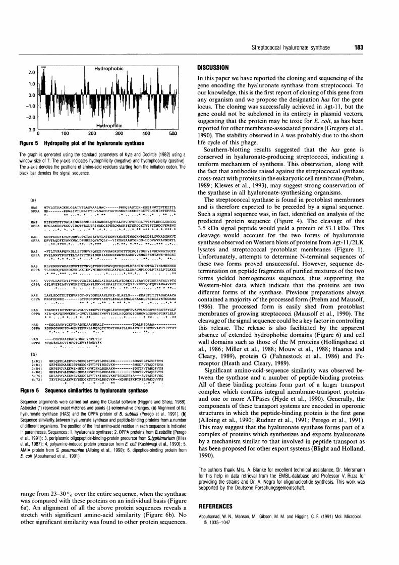
Figure 5 shows a hydropathy plot of the synthase. Starting atthe ATG initiation codon, the first 35 amino-acid residues havethe characteristics of a typical bacterial leader sequence (Vlasuket al., 1983): a stretch of 12 amino-acid residues containing basicresidues, followed by a hydrophobic core of 14 amino acids andending with a hydrophilic region. There was a possible cleavagesite for removal of the signal peptide at residue 35 whichconformed closely with the - 1, -3 rule defined by von Heijne(1983). The remainder of the protein sequence did not containany significant hydrophobic stretches.Computer searches have revealed amino-acid-sequence simi-
larity between the hyaluronate synthase and oligopeptide trans-port proteins from Bacillus subtilis (Perego et al., 1991; Rudneret al., 1991), Streptococcus pneumonia (Alloing et al., 1990), E.coli (Kashiwagi et al., 1990; Abouhamad et al., 1991) andSalmonella typhimurium (Hiles et al., 1987). These similarities
Figure 4 Nucleotide and deduced amino-acid sequence of the hyaluronatesynttase
Nucleotides are numbered on the right-hand side of the sequence, and the DNA strand is listed5' to 3'. The deduced amino-acid sequence is written-below the nucleotide sequence; residuesthat were determined by peptide sequencing are underlined. Putative -35 and -10 promotersequences (boxed) and the ribosome-binding site (S/D) are noted upstream of the codingregion. Palindromic sequences are indicated by dotted lines. The possible cleavage site of thesignal sequence is indicated by an arrow.
182
Streptococcal hyaluronate synthase 183
2.0 j; | HydrophobicDISCUSSIONIn this paper we have reported the cloning and sequencing of thegene encoding the hyaluronate synthase from streptococci. Toour knowledge, this is the first report of cloning of this gene fromany organism and we propose the designation has for the genelocus. The cloning was successfully achieved in Agt-l 1, but thegene could not be subcloned in its entirety in plasmid vectors,
ilicsuggesting that the protein may be toxic for E. coli, as has beenreported for other membrane-associated proteins (Gregory et al.,
0 100 200 300 400 5m0 1990). The stability observed in A was probably due to the short5 Hydropathy plot of the hyaluronate synthase life cycle of this phage.
Southern-blotting results suggested that the has gene isih is generated using the standard parameters of Kyte and Doolittle (1 982) using a conserved in hyaluronate-producing streptococci indicating a,ize of 7. The y-axis indicates hydrophilicity (negative) and hydrophobicity (positive). .i
isdenotes the positions of amino-acid residues starting from the initiation codon. The uniform mechanism of synthesis. This observation, along withrdenotes the signal sequence. the fact that antibodies raised against the streptococcal synthase
cross-react with proteins in the eukaryotic cell membrane (Prehm,1989; Klewes et al., 1993), may suggest strong conservation ofthe synthase in all hyaluronate-synthesizing organisms.The streptococcal synthase is found in protoblast membranes
S MTVLGTKACKRLGLATVTLASVAALMAC------PNKQSASTDK-KSEINWYTPTEIITL and is therefore expected to be preceded by a signal sequence.PA MK-------KRW6IVTLMLIFTLVLSACGFGGTGSNGEGKKDSKGKTTLNINIKTEPFSL
** *. *-
* ** .* .....* *. * ** ..* Such a signal sequence was, in fact, identified on analysis of theS DISKNTDTYSALAIGNSGSNLLRADAKGKLQPDLAEKVDVSEDGLTYTATLRHGLKWSDG predicted protein sequence (Figure 4). The cleavage of thisPA HPGLANDSVSGGVIRQTFEGLTRINADGEPEEGMASKIETSKDGKTYTFTIRDGVKWSNG... . .*. *. * * *.*. ..*.* .. *.* *.*.***.* 3.5 kDa signal peptide would yield a protein of 53.1 kDa. ThisS SULTAEDFEYSWQRMVDPKTASEYAYLATESHVKNAEUINSGKNPDLDSLtGVKADGNKVI cleavage would account for the two forms of hyaluronatePA DPVTAQDFEYAWKWALDPNNESQYAYQLY--YIKGAEAANTGKGS-LDDVAVKAVNDKTL*.*.** . *. ..*. *** . ~ *.*. * ..* *.. synthase observed on Western blots of proteuns from Agt-lI 1 /2LKS -FTLTVPAPQFKSLLSFSNFVPQKESFVKDAGKDYGTTSEKQIYSGPYIVKDWNGTSGTF lysates and streptococcal protoblast membranes (Figure 1).PA KVELNNPTPYFTELTAFYTYMPINEKIAEKNKKWNTNAGDDYVSNGPFKMTAWK-HSGSI*.* ..* ..... .......... Unfortunately, attempts to determine N-terminal sequences ofS KLVKNKNYWDAKNVKTETVNVQTVKKPDTAVQMYKQGJLDFANIS-G'rSAIYNANKKHKD these two forms proved unsuccessful. However, sequence de-PA TLEKNDQYWDKDKVKLKKIDMVMINNNNTELKKFQAGELDWAGMPLGQLPTESLPTLKKD termination on peptide fragments of purified mixtures of the two
S VVPVLEATTAYIVYNQTGAIEGLNSLKIRQALNLATDRKGIVSAAVDTGSKPATALVPTG forms yielded homogeneous sequences, thus supporting thePA GSLHVEPIAGVYWYKFNTEAKPLDNVNIRKALTYSLDRQSIVKNVTQGEQMPAMAAVPPT Western-blot data which indicate that the -proteins are two
S LAKLSDGTDLTEHVAPGY-KYDDKEAAKLFKE-GLAELG----KDALTITITADADAACA different forms of the synthase. Previous preparations always'PA MKGFEDNKE-------GYFKDNDVKTAKEYLEKGLKEMGLSKASDLPKIKLSYNTDDAHA contained a majority of the processed form (Prehm and Mausolf,
S KSAVDYIKETWETALPGLTVEEKFVPFSQRLEDTKNQNFEVAVVLWGGDYPEGSTFYGLF 1986). The processed form is easily shed from protoblast'PA KIA-QAVQEMWKKNL-GVDVELDNSEWNVYIDKLHSQDYQIGRMGWLGDFNDPINFLELF membranes of growing streptococci (Mausolf et al., 1990). The
TDALH... ..DAA.A.. cleavage of the signal sequence could be a key factor in controlling,S --KSGSAYNYGKFTNADYDAAYNKALT---------- ----TDALNIDAAA-----------'PA RDKNGGNNDTG-WENPEFKKLLNQSQTETDKTKRAELLKKAEGIFIDEMPVAPIYFYTDT this release. The release is also facilitated by the apparent
* .* .. *.. * * .. .... **
absence of extended hydrophobic domains (Figure 6) and cellS ----DDYKAAEKALYDNALYNPLYLP'PA WVQDENLKGVIMPGTGEVYFRNAYFK wall domains such as those of the M proteins (Hollingshead et
* * al., 1986; Miller et al., 1988; Mouw et al., 1988; Haanes andCleary, 1989), proteit G (Fahnestock et al., 1986) and Fc-
81) GKLQPDLAEKVDVSEDGLTYTATLRHGLKW--------SDGSDLTAEDFEYS81) GEPEEGMASKIETSKDGKTYTFTIRDGVKW--------SNGDPVTAQDFEYA receptor (Heath and Cleary, 1989).54) GHPSPGVAEKWE-NKDFKVWTFHLRENAKW--------SDGTPVTAHDFVYS80) GHPAPGVAESWO-NKDAKVWTFHLRKDAKW--------SDGTPVTAQDFVYS Significant amino-acid-sequence similarity was observed be-76) GNLAPAVAEDWEVSKDGLTYTYKIRKGVKWFTSDGEEYA---EVTAKDFVNG tween the synthase and a number of peptide-binding proteins.
..*.. .* .* .* f* S ,.*... All of these binding proteins form part of a larger transport6 Sequence similarities to hyaluronate synthase complex which contains integral membrane-transport proteins
e alignments were carried out using the Clustal software (Higgins and Sharp, 1988). and one or more ATPases (Hyde et al., 1990). Generally, the(*) represeRt exact maches and poiats (.) sonservative chenges. (a) Alignment of the components of these transport systems are encoded in operonic
ate synthase (HAS) and the OPPA protein of B. subtilis (Perego et al., 1991). (b) structures in which the peptide-binding protein is the first geneesini4arity.between hyaluronate synthase and peptide-binding proteins from a.number (Alloing et al, 1.990; Rudner et al., 1991; Perego et al., 1991).nt organisms. The position of the first amino-acid residue in each sequence is indicated This may suggest that the hyaluronate synthase forms part of aheses. Sequences: 1, hyaluronate synthase; 2, OPPA proteins from B.subtilis (Perego complex of proteins which synthesizes and exports hyaluronate91); 3, periplasmic oligopeptide-binding-protein precursor from Styphimurium (Hiles87); 4, polyamine-induced protein precursor from E. cofi (Kashiwagi et al., 1990); 5, by a mechanism simlar to that involved n peptlide transport asotein from S. pneumoniae (Alloing et al., 1990); 6, dipeptide-binding protein from has been proposed for other export systems (Blight and Holland,Abouhamad et al., 1991). 1990).
range from 23-30 00 over the entire sequence, when the synthasewas compared with these proteins on an individual basis (Figure6a). An alignment of all the above protein sequences reveals a
stretch with significant amino-acid similarity (Figure 6b). Noother significant similarity was found to other protein sequences.
The authors thaxrk Mrs. A. Blanke for excellent technical assistance, Dr. Mersmannfor his help in data retrieval from the EMBL-database and Professor V. -Rizza forproviding the strains and Dr. A. Negro for oligonucleotide synthesis. This work was
supported by the Deutsche Fofschungsgemeinschaft.
REFERENCESAbouhamad, W. N., Manson, M., Gibson, M. M. and Higgins, C F. (1991) Mol. Microbiol.
5,1035-1047
1.0
0.0
-1.0
-2.0
-3.0
Figure
The grapwindow sThe x-axiblack bar
(a)HA'OPI
HA'OP]
HA'OPI
HA'OPI
HA.OPI
HA'OPR
HAOPI
HA'OP:
HA'OP'
HA!OP:
(bl1)I2)(3)(4)(5 (6)
Figure
SequenceAsteLiskshyaluronaSequenceof diftererin parenttet al., 19!et al., 191AMIA prnE. cofi (A

184 M. Lansing and others
Alloing, G., Trombe, M. C. and Claverys J. P. (1990) Mol. Microbiol. 4, 633-644Blight, M. A. and Holland, I. B. (1990) Mol. Microbiol. 4, 873-880Both, B., Krupp, G. and Stackebrandt, E. (1991) Anal. Biochem. 199, 216-218Chen, E. Y. and Seeburg, P. H. (1985) DNA 4, 165-170Fahnestock, S. R., Alexander, P., Nagle, J. and Filpula, D. (1986) J. Bacteriol. 167,
870-880Gregory, R. J., Cheng, S. H., Rich, D. P., Marshall, J., Sucharita, P., Hehir, K., Ostegaard,
L., Klinger, K. W., Welsh, M. J. and Smith, A. E. (1990) Nature (London) 347, 382-386Haanes, E. and Cleary, P. P. (1989) J. Bacteriol. 171, 6397-6408Heath, D. G. and Cleary, P. P. (1989) Proc. Natl. Acad. Sci. U.S.A. 86, 4741-4745Higgins, D. G. and Sharp, P. M. (1988) Gene 73, 237-244Hiles, I. D., Gallagher, M. P., Jamieson, D. J. and Higgins, C. F. (1987) J. Mol. Biol. 195,
125-142Hirst, G. K. (1941) J. Exp. Med. 73, 493-506Hollingshead, S. K., Fishetti, V. A. and Scott, J. R. (1986) J. Biol. Chem. 261, 1677-1686Huynh, T. V., Young, R. A. and Davis, R. W. (1986) in DNA-Cloning: A Practical Approach,
(Glover, D. M., ed.), vol. 1, IRL Press, OxfordHyde, S. C., Emsley, P., Hartshorn, M. S., Mimmack, M. M., Gileadi, M., Pearce, S. R.,
Gallagher, M. P., Gill, D. R., Hubbard, R. E. and Higgins, C. F. (1990) Nature (London)346, 362-365
Kashiwagi, K., Yamaguchi, Y., Sakai, Y., Kobayashi, H. and Igarashi, K. (1990) J. Biol.Chem. 265, 8387-8391
Klewes, L., Turley, E. A. and Prehm, P. (1993) Biochem. J., in the pressKyte, J. and Doolittle, R. F. (1982) J. Mol. Biol. 157, 105-132Laurent, T. C. and Fraser, J. R. E. (1992) FASEB J. 6, 2397-2404Markovitz, A. and Dorfman, A. (1962) J. Biol. Chem. 237, 273-278Markovitz, A., Cifonelli, J. A. and Dorfman, A. (1958) J. Biol. Chem. 234, 2343-2350Mausolf, A., Jungmann, J., Robenek, H. and Prehm, P. (1990) Biochem. J. 267, 191-196McLaughlin, J. R., Morray, C. L. and Rabinowitz, J. C. (1981) J. Biol. Chem. 256,
11283-11291Miller, L., Gray, L., Beachey, E. and Kehoe, M. (1988) J. Biol. Chem. 263, 5668-5673Moran, C. P., Lang, N., LeGrice, S. F. L., Lee, G., Stephens, M., Sonenshein, P., Pero, J.
and Losick, R. (1982) Mol. Gen. Genet. 186, 339-346Mouw, A. R., Beachey, E. H. and Burdett, V. (1988) J. Bacteriol. 170, 676484
Nida, K. and Cleary, P. (1983) J. Bacteriol. 155, 1156-1161Perego, M., Higgens, C. F., Pearce, S. R., Gallagher, M. P. and Hoch, J. A. (1991) Mol.
Microbiol. 5, 173-185Prehm, P. (1984) Biochem. J. 220, 597-600Prehm, P. (1989) in CIBA Found. Symp. 143, 21-40Prehm, P. and Mausolf, A. (1986) Biochem. J. 235, 887489Rosenberg, S. M., Stahl, M. M., Kobayashi, I. and Stahl, F. W. (1985) Gene 38, 165-175Rudner, D. Z., LeDeaux, J. R., Ireland, K. and Grossman, A. D. (1991) J. Bacteriol. 173,
1388-1 398Saiki, R. K., Gelfand, P. H., Stoffel, S., Scharf, S. J., Higuci, R., Horn, G. T., Mullis, K. B.
and Erlich, H. A. (1988) Science 239, 487-491Sambrook, J., Fritsch, E. F. and Maniatis, T. (1989) Molecular Cloning, 2nd edn. Cold
Spring Harbor Laboratory Press, Cold Spring HarborSanger, F., Nicklen, S. and Coulson, A. R. (1977) Proc. Natl. Acad. Sci. U.S.A. 74,
5463-5467Seastone, C. V. (1939) J. Exp. Med. 70, 361-378Snyder, M., Elledge, S., Sweeter, D., Young, R. A. and Davis, R. W. (1987) Methods
Enzymol. 154, 107-128Stoolmiller, A. C. and Dorfman, A. (1969) J. Biol. Chem. 244, 236-246Sugahara, K., Schwartz, N. B. and Dorfman, A. (1979) J. Biol. Chem. 254, 6252-6261Todd, E. W. and Lancefield, R. C. (1928) J. Exp. Med. 48, 751-767Uhlen, M., Guss, B., Nilsson, B., Batenback, S., Philipson, L. and Lindberg, M. (1984)
J. Biol. Chem. 259, 1695-1702Vlasuk, G. P., Inouye, S., Ito, H., Itakura, K. and Inouye, M. (1983) J. Biol. Chem. 258,
7141-7148von Heijne, G. (1983) Eur. J. Biochem. 133, 17-21Weis J. H. (1989) in Current Protocols in Molecular Biology (Ausubel, F. M., Brent, R.,
Kingston, R. E., Moore, D. D., Seidman, J. G., Smith, J. A. and Struhl, K., eds.), Wiley &Sons, New York
Wessel, D. and Flugge, U. I. (1984) Anal. Biochem. 138, 141-143Wessels, M. R., Moses, A. E., Goldberg, J. B. and DiCesare, T. J. (1991) Proc. Natl. Acad.
Sci. U.S.A. 88, 8317-8321Whitnack, E., Bisno, A. L. and Beachey, E. H. (1981) Infect. Immun. 31, 985-991Wu, R., Wu, T. and Ray, A. (1987) in Guide to Molecular Cloning Techniques, (Berger, S.
and Kimmel, A. R., eds.), pp. 343-349, Academic Press, San Diego
Received 19 June 1992/10 August 1992; accepted 14 August 1992