geneticorganizationofthe cellulose synthase operonin ... · geneticorganizationofthe cellulose...
TRANSCRIPT
Proc. Nati. Acad. Sci. USAVol. 87, pp. 8130-8134, October 1990Genetics
Genetic organization of the cellulose synthase operon inAcetobacter xylinum
(cellulose biogenesis/cloning/sequencing)
HING C. WONG*t, ANNA LISA FEAR*, ROGER D. CALHOONt, GREGORY H. EICHINGER*, RAPHAEL MAYER§,DORIT AMIKAM§, MOSHE BENZIMAN§, DAVID H. GELFAND*, JAMES H. MEADE*, ANNE W. EMERICK*,ROBERT BRUNER¶, ARIE BEN-BASSAT¶, AND RONY TAL*Departments of *Microbial Genetics, tProtein Chemistry and IFermentation, Cetus Corporation, Emeryville, CA 94608; and §Department of BiologicalChemistry, Institute of Life Science, The Hebrew University of Jerusalem, Jerusalem, Israel
Communicated by H. 0. Smith, July 13, 1990 (received for review May 9, 1990)
ABSTRACT An operon encoding four proteins requiredfor bacterial cellulose biosynthesis (bcs) in Acetobacter xylinumwas isolated via genetic complementation with strains lackingcellulose synthase activity. Nucleotide sequence analysis indi-cated that the cellulose synthase operon is 9217 base pairs longand consists of four genes. The four genes-bcsA, bcsB, bcsC,and bcsD-appear to be translationally coupled and tran-scribed as a polycistronic mRNA with an initiation site 97 basesupstream of the coding region of the first gene (besA) in theoperon. Results from genetic complementation tests and genedisruption analyses demonstrate that all four genes in theoperon are required for maximal bacterial cellulose synthesis inA. xylinum. The calculated molecular masses of the proteinsencoded by bcsA, bcsB, bcsC, and bcsD are 84.4, 85.3, 141.0,and 17.3 kDa, respectively. The second gene in the operon(bcsB) encodes the catalytic subunit of cellulose synthase. Thefunctions of the bcsA, bcsC, and bcsD gene products areunknown. Bacterial strains mutated in the bcsA locus werefound to be deficient in cellulose synthesis due to the lack ofcellulose synthase and diguanylate cyclase activities. Mutantsin the bcsC and bcsD genes were impaired in cellulose produc-tion in vivo, even though they had the capacity to make all thenecessary metabolic precursors and cyclic diguanylic acid, theactivator of cellulose synthase, and exhibit cellulose synthaseactivity in vitro. When the entire operon was present on amulticopy plasmid in the bacterial cell, both cellulose synthaseactivity and cellulose biosynthesis increased. When the pro-moter of the cellulose synthase operon was replaced on thechromosome by E. coli tac or lac promoters, cellulose produc-tion was reduced in parallel with decreased cellulose synthaseactivity. These observations suggest that the expression of thebcs operon is rate-limiting for cellulose synthesis in A. xylinum.
Cellulose is the most abundant biopolymer in nature and is anindispensable raw material for many industries. In spite oftheimportance of cellulose, its mechanism of biosynthesis is stillpoorly understood (1). Since the bacterium Acetobacterxylinum synthesizes a cellulose that is structurally similar tothe cellulose produced by plants, it has been a model systemof choice for the study of cellulose biogenesis.The enzymatic pathway for cellulose synthesis in A. xyli-
num has been extensively investigated and four essentialenzymatic steps have been identified (2). Glucose is trans-ported through the bacterial membrane and phosphorylatedto glucose 6-phosphate by glucose kinase. Glucose 6-phos-phate is isomerized by phosphoglucomutase to glucose1-phosphate. Glucose 1-phosphate is converted to uridine5'-diphosphate glucose (UDPG) by UDPG pyrophosphory-
lase. Finally, UDPG is polymerized to cellulose by cellulosesynthase.
Cellulose synthesis is an energy-consuming and nonrevers-ible process. Cellulose synthase is the only enzyme known tobe unique to the cellulose synthetic pathway. Therefore, onewould expect that cellulose synthase activity is under strictregulatory control. It is known that cellulose synthase isspecifically activated by the unique nucleotide cyclic digua-nylic acid (3). This nucleotide is synthesized from GTP bydiguanylate cyclase and is degraded by phosphodiesterases Aand B (3). It is likely that cellulose synthase is furtherregulated at the genetic level.
In this paper, we describe the cloning, sequencing, andcharacterization of the cellulose synthase operon as a nec-essary step toward a better understanding of the regulation ofcellulose production in A. xylinum. 11
MATERIALS AND METHODSBacterial and Bacteriophage Strains, Plasmids, and Media.
A. xylinum 1306-3 is an isolate from A. xylinum B42 (NorthRegional Research Laboratories, Peoria, IL). A. xylinum 824was obtained from the Cetus Master Culture Collection. A.xylinum 1306-21 is a glucose dehydrogenase mutant of 1306-3obtained through ethyl methanesulfonate mutagenesis (4).
Escherichia coli MM294 (thi-1, hsdR17 endAl supE44),DG98 [thi-J, hsdRI7 endAl supE44 lacIq lacZ M15proC::TnJO(F' lacIq lacZ M15 proC+)], and K802 recA(lacYl sup E44 galK gal722 rtbDI metBI hsdR2) (5) werekindly provided by D. Gelfand (Cetus). BacteriophageM13mplO and M13mpll and plasmids pUC18, pBR322,pACYC184, pKT230, and pRK2013 have been described (6,7). The cosmid pKT230cos5 was constructed by cloning the1.85-kilobase (kb) Bgl II fragment carrying the A cos site fromplasmid pVK100 (8) into the BamHI site of pUC19. The cossite was then reisolated as an Sma I/HindIII fragment andsubcloned into the Sma I/HindIII sites of pKT230. Plasmidsfrom E. coli and double-stranded DNA of M13 phages wereprepared according to Katz et al. (9). Plasmids from A.xylinum were purified as described (10).The R-70-2 medium contains 7.3 mM KH2PO4 (pH 5.0), 25
mM (NH4)2SO4, 4.0 mM sodium citrate, 1.0 mM MgSO4, 0.1mM CaC12, 0.01 mM FeCl3, 0.001 mM Na2MoO4, 0.005 mMZnSO4, 0.005 mM MnSO4, 0.001 mM CuS04, 0.001 mMCoC12, 0.001 mM NiCl2, and 2% or 4% (wt/vol) glucose. TheR-20 medium contains 5 g of Na2HPO4 (pH 5.0) per liter, 5 gof Bacto-peptone per liter, 5 g of yeast extract per liter, 1.15g of citric acid per liter, and 2% (wt/vol) glucose.
Abbreviation: UDPG, uridine 5'-diphosphate glucose.tTo whom reprint requests should be addressed.'The sequence reported in this paper has been deposited in theGenBank data base (accession no. M37202).
8130
The publication costs of this article were defrayed in part by page chargepayment. This article must therefore be hereby marked "advertisement"in accordance with 18 U.S.C. §1734 solely to indicate this fact.
Proc. Natl. Acad. Sci. USA 87 (1990) 8131
Chromosomal DNA Isolation, Gene Bank Construction, andConjugation. High molecular weight chromosomal DNAfrom A. xylinum 1306-3 was partially digested with Sau3Aand fragments of -30 kb were isolated from a sucrosegradient. Approximately 1 Ag of 27- to 38-kb DNA wasligated into BamHI-cleaved and dephosphorylatedpKT230cosS vector, and packaged into A phage particles asdescribed (11). The phage particles were used to infect E. coliK802 recA. Cosmids from the individual transformants weretransferred into A. xylinum via conjugation. Conjugation wasperformed on an agar surface essentially as described (12)except that mobilization of the cosmid was provided by an E.coli strain host carrying the helper plasmid pRK2013.Primer Extension and Nucleotide Sequence Analysis. Total
cellular RNA from A. xylinum cells was extracted andpurified as described (13). Primer extension was carried outby the procedure ofJones et al. (14) with the oligonucleotide-containing sequence 5'-TGCGCGATAAGTGCACA-3'. Se-quences of both strands of an 11-kb region containing the bcsoperon were determined by the chain-termination method(15) with specifically synthesized primers.Promoter Replacement and Gene Disruption in A. xylinum.
A 2.5-kb HindIII restriction fragment containing the bcsoperon promoter and its flanking regions was cloned intoM13mplO. An Sst I/Sma I/BamHI linker was then intro-duced into the operon promoter 59 base pairs (bp) 3' of thetranscriptional initiation site by site-specific mutagenesis (6).This mutated fragment was subcloned into the HindIlI sitesof pACYC184 to construct the plasmid pACYC184:Pcs-10.The E. coli tac and lac promoter fragments were synthe-
sized with an EcoRI site at the 5' end and BamHI/HindIII atthe 3' end. Both synthetic fragments carry sequence fromnucleotide -45 to nucleotide +3 of its corresponding pro-moter. These fragments were cloned into the EcoRI/HindIIIsite of pALF20, a derivative of pBR322 with the AlwNI sitechanged to an Sst I site. The Sst I/BamHI fragments con-taining the promoter and the ,-lactamase genes from theresultant plasmids were subcloned into the BamHI/Sst I siteof pACYC184:Pcs-10. The resultant plasmids were digestedwith Xba I and the linearized fragments were used to trans-form A. xylinum 1306-21. Ampicillin-resistant transformantswere isolated and their chromosomal DNA was used inSouthern blot analysis to confirm the identity of the trans-formants. The ampicillin-resistant colonies that were ob-tained reflect a recombination between the plasmid's cellu-lose synthase regions, which flanked the marker-promotersequence, and homologous regions in the chromosome. Sincethe heterologous promoter precedes the ribosome bindingsite of the bcsA gene, the transcription of the bcs operon wasunder the control of the inserted promoter.To construct a bcsD mutant, a 3.7-kb Sma I/BamHI
fragment carrying the 3' portion of bcsC and the entire codingregion of bcsD was cloned into the EcoRV/BamHI site ofpACYC184. This plasmid was designated pALF23. The Tn3,B-lactamase gene was then isolated as an EcoRI/AlwNIfragment from pBR322. The ends of the fragment wererepaired and it was subcloned into the EcoRV site ofpALF23. The resultant plasmid was digested with Xba I andthe linearized plasmid was used to transform A. xylinum1306-21.Other methods used for DNA manipulations were de-
scribed by Sambrook et al. (11).Transformation ofA. xylinum. Cells were grown in R20-2 at
30°C with shaking in the presence of 0.1% (vol/vol) cellulase.When the cultures reached midlogarithmic phase growth(ODw0 = 0.5), cells were harvested, extensively washed with10% (vol/vol) glycerol, concentrated 300-fold in 10% (vol/vol) glycerol, and stored frozen at -700C until use. They werethawed on ice and mixed with DNA in a 2-mm Bio-Radelectroporation cuvette. Electroporation was carried out at a
field force of 9.0 kV and a pulse duration of 25 AtF/750 fQ. Thetransformed cells were grown in R20-2 medium for 3 hr beforethey were plated on selective plates. Plasmid DNA forelectroporation was purified by CsCl ultracentrifugation andextensively dialyzed against distilled water before use.
Assay for Cellulose Production and Cell Growth. Cells weregrown in R70-2 medium containing 10 ,uM FeC13, 25 mM3,3-dimethylglutaric acid (pH 5.0), 2% (wt/vol) corn steepliquor, and 1% (wt/vol) fructose (for strain 1306-3) or 1%(wt/vol) glucose (for strain 1306-21). Cell growth and cellu-lose production were analyzed after 3 days of incubationwhen all the carbon source was consumed. The cultures (25ml) were harvested by centrifugation, washed with salinetwice, and resuspended in 15 ml of 0.1 M NaOH andincubated at 60'C with mild agitation for 60 min. The heat-treated samples were centrifuged. The concentration of pro-tein in the supernatants was determined and used to calculatethe cell concentration in the culture assuming a cell with 65%protein content. The dry weight of the precipitate representsthe amounts of cellulose in the sample.
Purification and N-Terminal Amino Acid Sequencing ofCellulose Synthase. Exponentially growing cells were brokenin a French press and the membrane fraction was isolated asdescribed (16). The membrane preparation [10 mg of cells(dry wt) per ml] was then treated with trypsin (80 pug/ml) inbuffer containing 100 mM Tris HCI (pH 8.0), 20% (wt/vol)sucrose at 40C for 1 hr. The digestion was terminated byaddition of 80 Ag of trypsin inhibitor per ml. The trypsin-treated membranes were then solubilized in 2% (wt/vol)digitonin by sonication at 4°C for 2 min. Cellulose synthasewas further purified from the soluble proteins by entrapmentin cellulose as adapted from the procedure developed forchitin synthase (17). The entrapped enzyme was subjected toSDS/PAGE analysis (18). After Coomassie staining, themajor bands were cut out and eluted from the gel. N-terminalamino acid sequences were determined by the Edman deg-radation method (19).Enzymatic Assay. Exponentially growing cultures were
harvested and passed through cheesecloth to remove thecellulose from the cells. The cells were washed twice in 50mM KH2PO4 buffer (pH 6.0), resuspended to 20 mg (dry wt)per ml and sonicated in the appropriate assay buffers. Thecellulose synthase, diguanylate cyclase, phosphoglucomu-tase, and UDPG pyrophosphorylase were assayed as de-scribed (3, 16, 20). Cyclic diguanylic acid was prepared asdescribed by Ross et al. (3).
RESULTSIsolation and Characterization of A. xylinum Strains Defi-
cient in Cellulose Synthesis. Cellulose-producing (Cel+) A.xylinum strains form small rough and dry colonies, whereascellulose nonproducers (Cel-) form large flat and shinycolonies on agar plates. This phenotypic difference permitsisolation of Cel- mutants from A. xylinum strain 1306-3 aftermutagenesis with ethyl methanesulfonate. These Cel- mu-tants can be biochemically classified into five groups. GroupI mutants are deficient in one of the enzymes involved in theconversion of glucose to UDPG. The group II and group IIImutants lack the cellulose synthase or diguanylate cyclaseactivities, respectively. Group IV mutants are defective inboth diguanylate cyclase and cellulose synthase activities.The group V mutants have normal levels of all the knownenzymes involved in the conversion of glucose to cellulose.Thus, the mutants in this group are deficient in the gene(s)that is essential for cellulose synthesis in vivo but thatremains undefined. One of the group II mutants, 1306-24, washost in the genetic complementation experiments used toisolate the cellulose synthase operon.
Genetics: Wong et al.
Proc. Natl. Acad. Sci. USA 87 (1990)
G A T C 1
A B C _ D
I1kbI
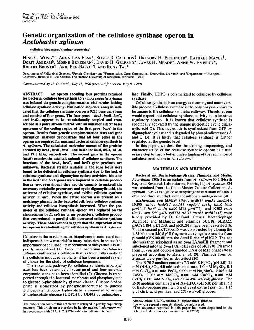
FIG. 1. Physical map of the bcs locus. The extent of the codingregions of the bcs genes and their transcription direction are indi-cated by the arrows. Hatched bar indicates the sequenced region.
Cloning of the Cellulose Synthase Operon. The broad hostrange plasmid pKT230 (7) was found to be stably maintainedin A. xylinum (data not shown). We constructed an A.xylinum gene library in a cosmid vector derived from theplasmid pKT230 in E. coli. A total of 2010 individual strep-tomycin-resistant transformants were picked and their re-
combinant cosmids were mobilized with the helper plasmidpRK2013 and transferred into the cellulose synthase-defi-cient mutant 1306-24 via the tripartite mating method.Cosmid T19G9 was found to restore the Cel+ phenotype ofstrain 1306-24. A physical map of the region containing thebcs operon on the A. xylinum chromosome was constructed(Fig. 1) based on restriction analysis of this cosmid. Themolecular organization of the bcs locus was also confirmedby genomic Southern analysis (data not shown).
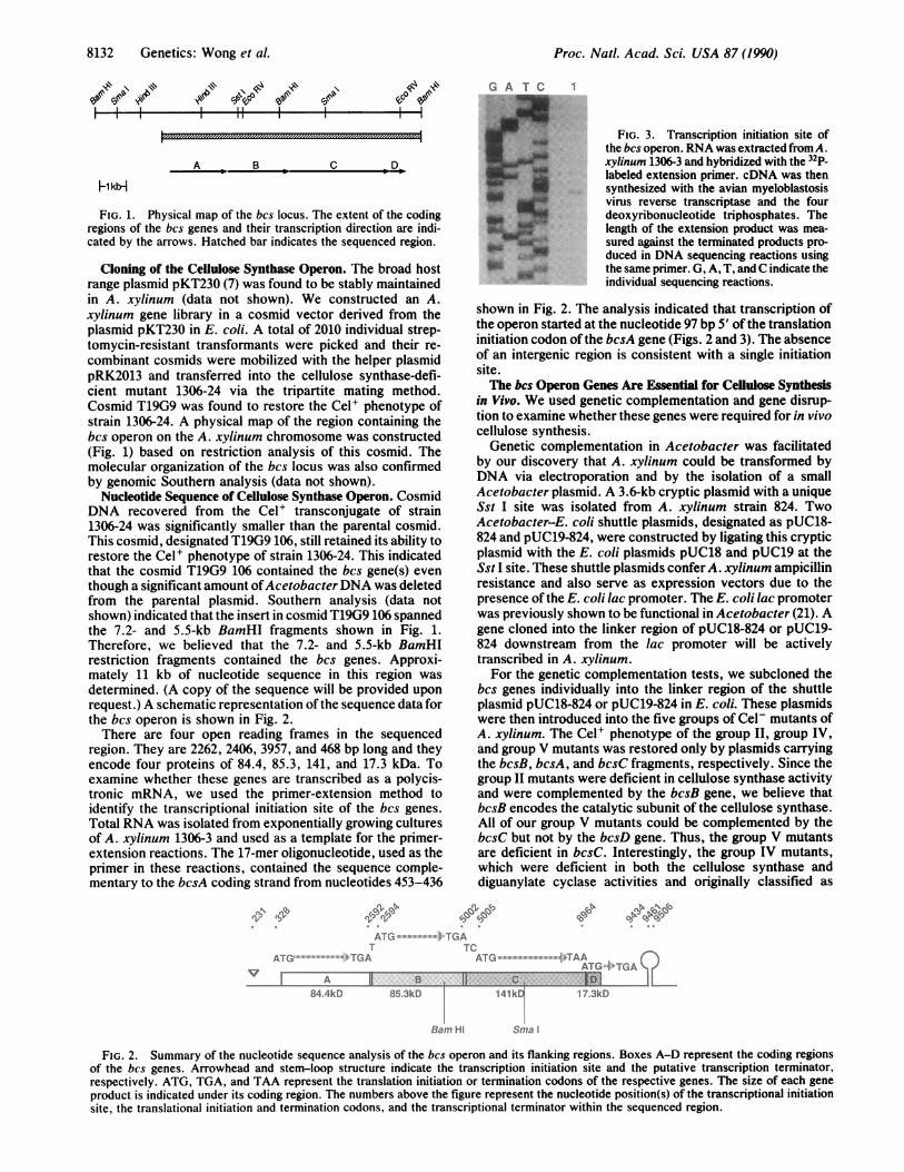
Nucleotide Sequence of Cellulose Synthase Operon. CosmidDNA recovered from the Cel+ transconjugate of strain1306-24 was significantly smaller than the parental cosmid.This cosmid, designated T19G9 106, still retained its ability torestore the Cel' phenotype of strain 1306-24. This indicatedthat the cosmid T19G9 106 contained the bcs gene(s) eventhough a significant amount ofAcetobacter DNA was deletedfrom the parental plasmid. Southern analysis (data notshown) indicated that the insert in cosmid T19G9 106 spannedthe 7.2- and 5.5-kb BamHI fragments shown in Fig. 1.Therefore, we believed that the 7.2- and 5.5-kb BamHIrestriction fragments contained the bcs genes. Approxi-mately 11 kb of nucleotide sequence in this region wasdetermined. (A copy of the sequence will be provided uponrequest.) A schematic representation of the sequence data forthe bcs operon is shown in Fig. 2.There are four open reading frames in the sequenced
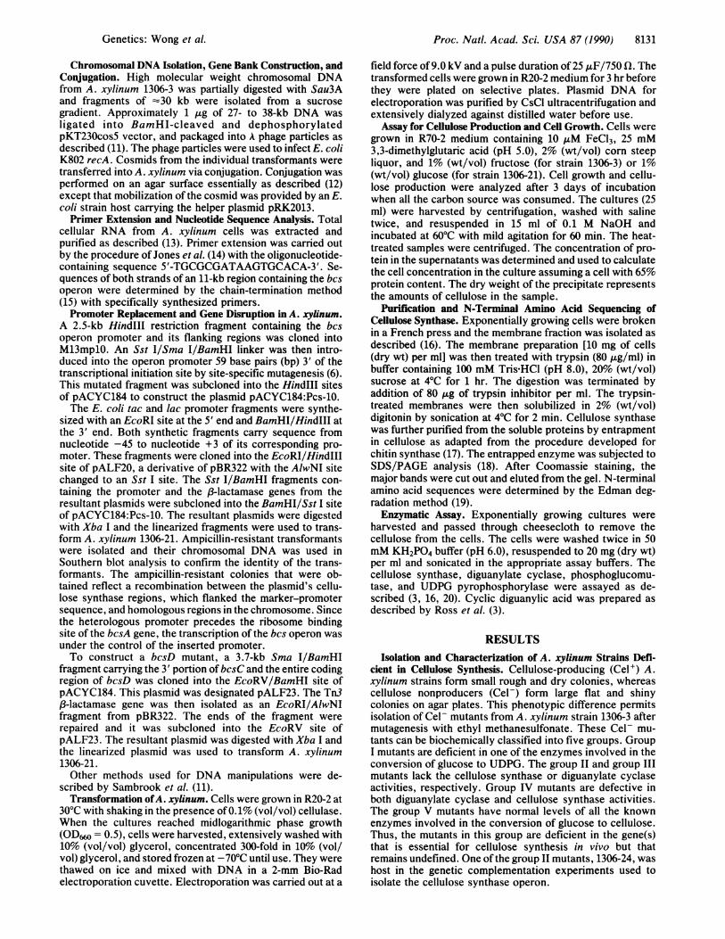
region. They are 2262, 2406, 3957, and 468 bp long and theyencode four proteins of 84.4, 85.3, 141, and 17.3 kDa. Toexamine whether these genes are transcribed as a polycis-tronic mRNA, we used the primer-extension method toidentify the transcriptional initiation site of the bcs genes.Total RNA was isolated from exponentially growing culturesof A. xylinum 1306-3 and used as a template for the primer-extension reactions. The 17-mer oligonucleotide, used as theprimer in these reactions, contained the sequence comple-mentary to the bcsA coding strand from nucleotides 453-436
FIG. 3. Transcription initiation site ofthe bcs operon. RNA was extractedfromA.xylinum 1306-3 and hybridized with the 32P-labeled extension primer. cDNA was thensynthesized with the avian myeloblastosisvirus reverse transcriptase and the fourdeoxyribonucleotide triphosphates. Thelength of the extension product was mea-sured against the terminated products pro-duced in DNA sequencing reactions usingthe same primer. G, A, T, and C indicate theindividual sequencing reactions.
shown in Fig. 2. The analysis indicated that transcription ofthe operon started at the nucleotide 97 bp 5' of the translationinitiation codon of the bcsA gene (Figs. 2 and 3). The absenceof an intergenic region is consistent with a single initiationsite.The bcs Operon Genes Are Essential for Cellulose Synthesis
in Vivo. We used genetic complementation and gene disrup-tion to examine whether these genes were required for in vivocellulose synthesis.
Genetic complementation in Acetobacter was facilitatedby our discovery that A. xylinum could be transformed byDNA via electroporation and by the isolation of a smallAcetobacter plasmid. A 3.6-kb cryptic plasmid with a uniqueSst I site was isolated from A. xylinum strain 824. TwoAcetobacter-E. coli shuttle plasmids, designated as pUC18-824 and pUC19-824, were constructed by ligating this crypticplasmid with the E. coli plasmids pUC18 and pUC19 at theSst I site. These shuttle plasmids confer A. xylinum ampicillinresistance and also serve as expression vectors due to thepresence of the E. coli lac promoter. The E. coli lac promoterwas previously shown to be functional in Acetobacter (21). Agene cloned into the linker region of pUC18-824 or pUC19-824 downstream from the lac promoter will be activelytranscribed in A. xylinum.For the genetic complementation tests, we subcloned the
bcs genes individually into the linker region of the shuttleplasmid pUC18-824 or pUC19-824 in E. coli. These plasmidswere then introduced into the five groups of Cel- mutants ofA. xylinum. The Cel' phenotype of the group II, group IV,and group V mutants was restored only by plasmids carryingthe bcsB, bcsA, and bcsC fragments, respectively. Since thegroup II mutants were deficient in cellulose synthase activityand were complemented by the bcsB gene, we believe thatbcsB encodes the catalytic subunit of the cellulose synthase.All of our group V mutants could be complemented by thebcsC but not by the bcsD gene. Thus, the group V mutantsare deficient in bcsC. Interestingly, the group IV mutants,which were deficient in both the cellulose synthase anddiguanylate cyclase activities and originally classified as
IC\b
f.li @4'ATG
T TCATG :!! !"'; ;; l-"!'!l -:TGA ATG li~i,, liili!i :'ll i.i'' ,TAPGATGTiGATGA
84.4kD 85.3kD 141ko 17.3kD
Bam Hi Sma
FIG. 2. Summary of the nucleotide sequence analysis of the bcs operon and its flanking regions. Boxes A-D represent the coding regionsof the bcs genes. Arrowhead and stem-loop structure indicate the transcription initiation site and the putative transcription terminator,respectively. ATG, TGA, and TAA represent the translation initiation or termination codons of the respective genes. The size of each geneproduct is indicated under its coding region. The numbers above the figure represent the nucleotide position(s) of the transcriptional initiationsite, the translational initiation and termination codons, and the transcriptional terminator within the sequenced region.
ve Aft e\v 0Xv §
8132 Genetics: Wong et al.
0le
io 'AM&
.Ul: :11
Proc. Natl. Acad. Sci. USA 87 (1990) 8133
1 2 3 4
-92.5-69
Y3>H 46
- 30
-21.5
- 14.3
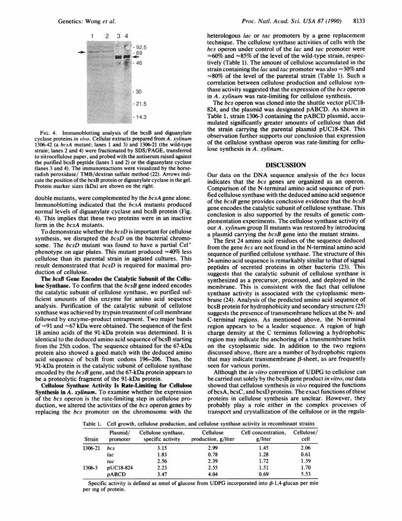
FIG. 4. Immunoblotting analysis of the bcsB and diguanylatecyclase proteins in vivo. Cellular extracts prepared from A. xylinum1306-42 (a bcsA mutant; lanes 1 and 3) and 1306-21 (the wild-typestrain; lanes 2 and 4) were fractionated by SDS/PAGE, transferredto nitrocellulose paper, and probed with the antiserum raised againstthe purified bcsB peptide (lanes 1 and 2) or the diguanylate cyclase(lanes 3 and 4). The immunoreactions were visualized by the horse-radish peroxidase/ TMB/dextran sulfate method (22). Arrows indi-cate the position of the bcsB protein or diguanylate cyclase in the gel.Protein marker sizes (kDa) are shown on the right.
double mutants, were complemented by the bcsA gene alone.Immunoblotting indicated that the bcsA mutants producednormal levels of diguanylate cyclase and bcsB protein (Fig.4). This implies that these two proteins were in an inactiveform in the bcsA mutants.To demonstrate whether the bcsD is important for cellulose
synthesis, we disrupted the bcsD on the bacterial chromo-some. The bcsD mutant was found to have a partial Cel'phenotype on agar plates. This mutant produced -40% lesscellulose than its parental strain in agitated cultures. Thisresult demonstrated that bcsD is required for maximal pro-duction of cellulose.The bcsB Gene Encodes the Catalytic Subunit of the Cellu-
lose Synthase. To confirm that the bcsB gene indeed encodesthe catalytic subunit of cellulose synthase, we purified suf-ficient amounts of this enzyme for amino acid sequenceanalysis. Purification of the catalytic subunit of cellulosesynthase was achieved by trypsin treatment of cell membranefollowed by enzyme-product entrapment. Two major bandsof -91 and =67 kDa were obtained. The sequence of the first18 amino acids of the 91-kDa protein was determined. It isidentical to the deduced amino acid sequence ofbcsB startingfrom the 25th codon. The sequence obtained for the 67-kDaprotein also showed a good match with the deduced aminoacid sequence of bcsB from codons 196-206. Thus, the91-kDa protein is the catalytic subunit of cellulose synthaseencoded by the bcsB gene, and the 67-kDa protein appears tobe a proteolytic fragment of the 91-kDa protein.
Cellulose Synthase Activity Is Rate-Limiting for CelluloseSynthesis in A. xylinum. To examine whether the expressionof the bcs operon is the rate-limiting step in cellulose pro-duction, we altered the activities of the bcs operon genes byreplacing the bcs promoter on the chromosome with the
heterologous lac or tac promoters by a gene replacementtechnique. The cellulose synthase activities of cells with thebcs operon under control of the lac and tac promoter were
-60% and -85% of the level of the wild-type strain, respec-tively (Table 1). The amount of cellulose accumulated in thestrain containing the lac and tac promoter was also -30% and-80% of the level of the parental strain (Table 1). Such a
correlation between cellulose production and cellulose syn-thase activity suggested that the expression of the bcs operonin A. xylinum was rate-limiting for cellulose synthesis.The bcs operon was cloned into the shuttle vector pUC18-
824, and the plasmid was designated pABCD. As shown inTable 1, strain 1306-3 containing the pABCD plasmid, accu-mulated significantly greater amounts of cellulose than didthe strain carrying the parental plasmid pUC18-824. Thisobservation further supports our conclusion that expressionof the cellulose synthase operon was rate-limiting for cellu-lose synthesis in A. xylinum.
DISCUSSIONOur data on the DNA sequence analysis of the bcs locusindicates that the bcs genes are organized as an operon.Comparison of the N-terminal amino acid sequence of puri-fied cellulose synthase with the deduced amino acid sequenceof the bcsB gene provides conclusive evidence that the bcsBgene encodes the catalytic subunit of cellulose synthase. Thisconclusion is also supported by the results of genetic com-plementation experiments. The cellulose synthase activity ofour A. xylinum group II mutants was restored by introducinga plasmid carrying the bcsB gene into the mutant strains.The first 24 amino acid residues of the sequence deduced
from the gene bcs are not found in the N-terminal amino acidsequence of purified cellulose synthase. The structure of this24-amino acid sequence is remarkably similar to that of signalpeptides of secreted proteins in other bacteria (23). Thissuggests that the catalytic subunit of cellulose synthase issynthesized as a precursor, processed, and deployed in themembrane. This is consistent with the fact that cellulosesynthase activity is associated with the cytoplasmic mem-brane (24). Analysis of the predicted amino acid sequence ofbcsB protein for hydrophobicity and secondary structure (25)suggests the presence oftransmembrane helices at the N- andC-terminal regions. As mentioned above, the N-terminalregion appears to be a leader sequence. A region of highcharge density at the C terminus following a hydrophobicregion may indicate the anchoring of a transmembrane helixon the cytoplasmic side. In addition to the two regionsdiscussed above, there are a number of hydrophobic regionsthat may indicate transmembrane p-sheet, as are frequentlyseen for various porins.Although the in vitro conversion of UDPG to cellulose can
be carried out solely by the bcsB gene product in vitro, our datashowed that cellulose synthesis in vivo required the functionsofbcsA, bcsC, and bcsD proteins. The exact functions oftheseproteins in cellulose synthesis are unclear. However, theyprobably play a role either in the complex processes oftransport and crystallization of the cellulose or in the regula-
Table 1. Cell growth, cellulose production, and cellulose synthase activity in recombinant strains
Plasmid/ Cellulose synthase, Cellulose Cell concentration, Cellulose/Strain promoter specific activity production, g/liter g/liter cell
1306-21 bcs 3.15 2.99 1.45 2.06lac 1.83 0.78 1.28 0.61tac 2.56 2.39 1.72 1.39
1306-3 pUC18-824 2.23 2.55 1.51 1.70pABCD 3.47 4.04 0.69 5.53
Specific activity is defined as nmol of glucose from UDPG incorporated into P-1,4-glucan per minper mg of protein.
Genetics: Wong et al.
Proc. Natl. Acad. Sci. USA 87 (1990)
tion of cellulose synthase activity. In A. xylinum, the biosyn-thesis of high molecular weight 3-1,4-glucan sub-elementaryfibrils by cellulose synthase is believed to occur at the cyto-plasmic membrane. These subelementary fibrils are extrudedfrom pores located in the outer membrane and then crystal-lized (by hydrogen bonding) into microfibrils. The microfibrilsthen assemble into bundles. Interestingly, polymerization andcrystallization processes have been shown to be coupled in A.xylinum (26). It is possible that these processes are actuallycatalyzed by a single enzyme complex, and that the bcs geneproducts are part ofthis complex. If this is the case, one wouldexpect overexpression of an individual gene product to havelittle impact on the production of cellulose in A. xylinum. Insupport of this, we found that the simultaneous and individualincrease in the level of the bcsA and bcsB gene products didnot improve the cellulose production of A. xylinum (data notshown). Furthermore, our nucleotide sequence analysis of thebcs operon indicated that the bcs genes were translationallycoupled. Translational coupling leads to a coordination ofgeneexpression and may be used by A. xylinum to maintain thecorrect balance or appropriate architecture of the multiproteincellulose synthase complex. The deduced amino acid se-quence of bcsC at its N terminus is similar to signal sequencesof secreted proteins in other bacteria. This may indicate thatthe bcsC gene product is a membrane protein and a componentof the membrane-bound cellulose synthase complex. On theother hand, the bcs gene products may be involved in regu-lating the activity of cellulose synthase in A. xylinum. Mutantsdeficient in bcsA were found to lack cellulose synthase anddiguanylate cyclase activities. Immunoblotting of these mu-tants indicated that they actually contained normal levels ofbcsB and diguanylate cyclase proteins (Fig. 4). This impliesthat the bcsB gene product and diguanylate cyclase requireposttranslational modification/activation and that the bcsAprotein is involved in such a modification process.Among E. coli promoters, two highly conserved stretches
of sequences centered at the -35 and -10 regions are easilyidentified (27). These regions have proven to be the contactpoints between the promoter and RNA polymerase. Thealdehyde dehydrogenase (ald) and the aktohol dehydroge-nase (adh) genes from Acetobacter acetii have been isolatedand their nucleotide sequences determined (28, 29). Compar-ison of the 5' untranslated regions of these genes with thepromoter sequence of the bcs operon indicated that theycontain scattered regions of homology among them. Thehomology between the ald and bcs promoter sequence isparticularly striking. For instance, the sequence CATCGCTG located between nucleotides -11 and -4 in the bcspromoter is quite similar to the sequence CATGGCTG po-sitioned 84 bp 5' of the initiation codon of ald. Since thetranscriptional initiation sites of ald and adh promoters havenot been identified, the positional significance of such ho-mologous regions between the three promoters is unclear.However, comparison of the sequences adjacent to theinitiation codons of the bcs, ald, and adh genes indicated thatthe sequence GGACGNG is highly conserved approximately2-6 bases 5' of the ATG codon. This sequence may representthe ribosome binding site of Acetobacter mRNAs.Computer analysis of the DNA sequence 3' of the bcsD
gene reveals a region with an inverted repeat sequence. Thissequence has the potential of forming a stable stem-loopstructure and AG = -29.1 kcal (1 cal = 4.184 J) at the end ofa mRNA, which is often used as a transcription terminationsignal in bacteria (26). This region is positioned 26 bp 3' of thetermination codon of the bcsD gene and could be the tran-scriptional termination of the bcs operon.We do not know of another case in which the genetic
organization of a cellulose synthase locus has been disclosed.Immunochemical and enzymatic evidence suggests that sim-ilar systems operate in other cellulose-producing bacteria (30)
and plants (31). Thus, this work should greatly facilitate thefurther understanding of cellulose biogenesis.
We thank Shing Chang for valuable comments on the manuscript,the Cetus Organic Synthesis Group for providing the oligonucleo-tides used in this work, and Robin Kurka and Joan Murphy forpreparation of the manuscript. This work was supported by theWeyerhaeuser Company.
1. Delmer, D. R. (1987) Annu. Rev. Plant Physiol. 38, 259-290.2. Swissa, M., Aloni, Y., Weinhouse, H. & Benziman, M. (1980)
J. Bacteriol. 143, 1142-1150.3. Ross, P., Weinhouse, H., Aloni, Y., Michaeli, D., Weinberger-
Ohana, P., Mayer, R., Braun, S., de Vroom, E., van der Marel,G., van Boom, J. H. & Benziman, M. (1987) Nature (London)325, 279-281.
4. Miller, J. H. (1972) in Experiments in Molecular Genetics (ColdSpring Harbor Lab., Cold Spring Harbor, NY), p. 138.
5. Lawyer, F. C., Stoffel, S., Saiki, R. K., Myambo, K., Drum-mond, R. & Gelfand, D. H. (1989) J. Biol. Chem. 264, 6427-6437.
6. Wittman, V. & Wong, H. C. (1988) J. Bacteriol. 170, 3206-3212.
7. Schmidhauser, T. J., Ditta, G. & Helinski, D. R. (1988) inVectors: A Survey of Molecular Cloning Vectors and TheirUses, eds. Rodriguez, R. L. & Denhardt, D. T. (Butterworth,Boston), pp. 287-332.
8. Knauf, V. C. & Nester, E. W. (1982) Plasmids 8, 45-54.9. Katz, L., Kingsbury, D. & Helinski, D. R. (1973) J. Bacteriol.
114, 577-591.10. Guerry, P., LeBlanc, D. J. & Falkow, S. (1973) J. Bacteriol.
116, 1064-1066.11. Sambrook, J., Fritsch, E. J. & Maniatis, T. (1989) Molecular
Cloning:A Laboratory Manual (Cold Spring Harbor Lab., ColdSpring Harbor, NY).
12. Ditta, G., Stanfield, S., Corbin, D. & Helinski, D. R. (1980)Proc. Natl. Acad. Sci. USA 77, 7347-7351.
13. Wong, H. C. & Chang, S. (1985) Proc. Natl. Acad. Sci. USA83, 3233-3237.
14. Jones, K. A., Yamamoto, K. R. & Tjian, R. (1985) Cell 42,559-572.
15. Sanger, F., Nicklen, S. & Coulson, A. R. (1977) Proc. NatI.Acad. Sci. USA 74, 5463-5467.
16. Aloni, Y., Delmer, D. P. & Benziman, M. (1982) Proc. Natl.Acad. Sci. USA 79, 6448-6452.
17. Kang, M. S., Elango, N., Mattia, E., Au-Young, J., Robbins,P. W. & Cabib, E. (1984) J. Biol. Chem. 259, 14966-14972.
18. Laemmli, U. K. (1970) Nature (London) 227, 680-685.19. Hewick, R. M., Hunkapiller, M. W., Hood, L. E. & Dreyer,
W. J. (1981) J. Biol. Chem. 256, 7990-7997.20. Hansen, R. G., Albrecht, G. J., Bass, S. T. & Seifert, L. L.
(1966) Methods Enzymol. 8, 248-253.21. Kukaya, M., Tayama, K., Tamaki, T., Tagami, H., Okumura,
H., Kawamura, Y. & Beppu, T. (1989) Appl. Environ. Micro-biol. 55, 171-176.
22. McLaughlin, J. R., Wong, H. C., Ting, Y. E., Van Arsdell,J. N. & Chang, S. (1986) J. Bacteriol. 167, 952-959.
23. Oliver, D. (1985) Annu. Rev. Microbiol. 39, 615-648.24. Bureau, T. E. & Brown, R. M., Jr. (1987) Proc. Natl. Acad.
Sci. USA 84, 6985-6989.25. Devereux, J., Haeberli, P. & Smithies, 0. (1984) Nucleic Acids
Res. 12, 387-395.26. Benziman, M., Haigler, C. H., Brown, R. M., Jr., White, A. R.
& Cooper, K. M. (1980) Proc. Natl. Acad. Sci. USA 77,6678-6682.
27. Rosenberg, M. & Court, D. (1979) Annu. Rev. Genet. 13,319-353.
28. Tamaki, T., Horinouchi, S., Fukaya, M., Okumura, H., Kawa-mura, Y. & Beppu, T. (1989) J. Biochem. 106, 541-544.
29. Inoue, T., Sunagawa, M., Mori, A., Imai, C., Fukuda, M.,Takagi, M. & Yano, K. (1989) J. Bacteriol. 171, 3115-3122.
30. Amikam, D. & Benziman, M. (1989) J. Bacteriol. 171, 6649-6655.
31. Mayer, R., Ross, P., Weinhouse, H., Amikam, D., Vollman,G., Ohana, P. & Benziman, M. (1990) in Proceedings of theFifth Cell Wall Meeting, eds. Frg, F. C., Brett, C. D. & Reid,J. C. G., in press.
8134 Genetics: Wong et al.