humanization of antibodies
TRANSCRIPT
[Frontiers in Bioscience 13, 1619-1633, January 1, 2008]
1619
Humanization of antibodies Juan C. Almagro1, Johan Fransson2 1 Centocor R&D, Inc. 145 King of Prussia Rd. Radnor, PA 19087,2 Centocor Discovery Research, San Diego. 3210 Merryfield Row. San Diego, CA 92121 TABLE OF CONTENTS 1. Abstract 2. Introduction 3. Antibody structure and the antigen-binding site 4. Rational approaches to humanize antibodies
4.1. CDR grafting 4.1.1. Regions determining the antibody specificity 4.1.2. Human FRs 4.1.3. Back mutations to retain or restore affinity
4.2. Resurfacing 4.3. Superhumanization 4.4. Human String Content
5. Empirical approaches to humanize antibodies 5.1. FR libraries 5.2. Guided Selection 5.3. FR shuffling 5.4. Humaneering
6. Perspectives 7. Acknowledgement 8. References 1. ABSTRACT
Humanization has played a fundamental role in
the remarkable progress of antibodies as therapeutic reagents. Here we have reviewed the publications on antibody humanization since the first report on CDR grafting in the second half of the 1980’s up to June 2007. We describe the two main trends in the field: rational and empirical methods to humanize antibodies. Rational methods rely on the so-called design cycle. It consists of generating a small set of variants, which are designed based on the antibody structure and/or sequence information, and assessing their binding or any other characteristic of interest. Rational methods include CDR grafting, Resurfacing, Superhumanization and Human String Content Optimization. In contrast to rational methods, empirical methods are based on generating large combinatorial libraries and selecting the desired variants by enrichment technologies such as phage, ribosome or yeast display, or by high throughput screening techniques. The latter methods rest on selection rather than making assumptions on the impact of mutations on the antibody structure. These methods include Framework Libraries, Guided Selection, Framework Shuffling and Humaneering.
2. INTRODUCTION
In the last ten years, more than 20 antibodies
have been approved by the Food and Drug Administration (FDA) for therapeutic applications in humans and yet, almost one thousand clinical trials related to antibodies are currently in progress (http://www.clinicaltrials.gov/ct). All therapeutic settings involving antibodies so far described require large amounts and multiple doses and hence, immunogenicity is a critical concern when developing an antibody-based drug (1-3). In vitro discovery of human antibodies via enrichment technologies such as phage display (4, 5) or immunization of transgenic mice bearing the antibody human gene repertoire (6) have provided powerful means to generate human antibodies. Nevertheless, humanization methods have been diversified during the last decade and the number of humanized antibodies has shown a continuous steady growth.
Antibody humanization methods are designed to
produce a molecule with minimal immunogenicity when applied to humans, while retaining the specificity and affinity of the parental non-human antibody. Humanization began with chimerization (7), a method developed during the first half of the 1980’s, which consisted of combining

Antibody humanization
1620
the variable (V) domains of murine antibodies with human constant (C) domains to generate molecules with ~70% of human content. Chimeric antibodies successfully retained the mouse parent antibody specificity and diminished its immunogenicity but still elicited a human anti-chimeric antibody (HACA) response (3).
In the second half of the 1980’s, Greg Winter’s
group (8) envisioned complementarity-determining region (CDR) grafting as a method to further minimize immunogenicity. As proof of concept, the CDRs from heavy (H) chain of the human myeloma protein NEWM were substituted for the corresponding CDRs of the murine anti-hapten antibody B1-8. The hybrid product was then combined with the B1-8 mouse light (L) chain and the resulting antibody acquired B1-8 antibody specificity. Two years later (9), a similar procedure was followed to successfully humanize the anti-protein antibody D1.3, thus generalizing CDR grafting to antibodies that recognize protein antigens.
In 1988 (10), CDR grafting of both H and L
chains from an antibody with therapeutic value in treatment of B-cell chronic lymphocytic leukemia, currently marketed as CamPath® (generic name: Alemtuzumab), was reported. One year later, Queen et al (11) humanized the first FDA approved antibody for therapeutic use in United States, termed Zenapax® (generic name: Daclizumab), with indication in transplantation and treatment of asthma, autoimmunity, inflammation and multiple sclerosis. Zenapax® was generated by selecting the human framework regions (FRs) to maximize homology with the murine antibody sequence. Guided by a computer model of the mouse antibody, several murine amino acids outside the CDRs were identified as to interact with the CDRs or antigen and were back mutated in the humanized antibody to improve binding. Altogether, these pioneering works laid foundation for CDR grafting, the paradigm of humanization and current standard in the field.
During the 1990’s and the present decade,
humanization methods based on different paradigms such as Resurfacing (12), Superhumanization (13) and Human String Content Optimization (14) have been developed. As CDR grafting, these methods rely on analyses of the antibody structure and sequence comparison of the non-human and human antibodies in order to evaluate the potential impact of the humanization process into the final product. These methods have in common the generation of few humanized variants to be tested for binding or any other property of interest. If the designed variants prove to be unsatisfactory, a new cycle of design and binding assessment is initiated. Therefore, these methods can be classified as rational strategies to humanize antibodies.
Also during the 1990’s, phage display and
high-throughput screening (HTS) techniques emerged as efficient tools to explore combinatorial libraries of millions and billions of antibody variants and select those of interest (15-17). These techniques were then applied to antibody humanization protocols stimulating
the creation of methods that rested on selection rather than on the design cycle (18, 19). One of these methods, called Guided Selection (18), combined the V domain from the heavy chain (VH) of the rodent antibody Mab32 against human tumor necrosis factor alpha (TNF-α) with a library of human VL domains. The chimeric library was displayed on phage and panned against TNF-α. The human VL thus selected was cloned into a library of human VH domains and then panned again against TNF-α, resulting in the first human antibody approved by the FDA (20) called Humira® (Adalimumab) for treatment of rheumatoid arthritis and Crohn’s disease. Guided Selection and other humanization strategies relying on selection of large combinatorial libraries make few assumptions on the impact of mutations on the final humanized product and accordingly, we have called these techniques empirical methods to humanize antibodies.
Here, we review the publications on antibody
humanization since the pioneering works of Greg Winter’s group in the 1980’s up until June 2007, including both rational and empiric approaches to humanize antibodies. To provide context for describing these methods, we first briefly describe the structure of the antibody molecule as well as the location and anatomy of the antigen-binding site. Next, the rational methods to humanize antibodies are reviewed, followed by the empirical methods. The final section includes a summary of the main trends in the field and highlights its perspectives.
3. ANTIBODY STRUCTURE AND THE ANTIGEN-BINDING SITE
IgG isotypes are products of an immune response
maturation and thus in general are highly specific and high affinity antibodies. IgGs are composed of two identical polypeptide H and L chains (Figure 1A). Each H chain has one VH domain and three C domains, CH1 - CH3, counted from the amino terminal. The L chain has one VL domain at the amino terminus and only one C domain, CL. V domains are composed of two β-sheets and loops connecting the β-strands (21). Three of the loops are highly diverse in length and/or amino acid composition and are referred to as hypervariable (HV) (21, 22). The remaining regions of the V domain contain the two β-sheets and non-HV loops, and are referred to as FR. HV loops, denoted H1, H2 and H3 for VH, and L1, L2 and L3 for VL, are brought together by non-covalent association of VH and VL to accommodate the antigen-binding site at the end-terminal region of the Fv fragment (Figure 1B).
Prior to the resolution of the first antibody-
antigen complex (23), Wu and Kabat (24) identified the regions involved in antigen recognition, or CDRs, as the regions with highest variability values in a multiple alignment of antibody sequences. Variability was defined as the number of different amino acids at a given position divided by the frequency of the most common amino acid at that position. Therefore, CDR is a sequence-based definition, whereas HV is a structural definition and in some instances, HV and CDRs do not coincide (Figure 2).

Antibody humanization
1621
Figure 1. Antibody structure. A. IgG structure (PDB code: 1IGT (109)) showing the two identical H chains (blue) and two identical L chains (gray). H chains have four domains, three C domains (CH1 to 3) and one VH. L chains have one CL domain and one VL domain. The Fv fragment, indicated by a blue square, is the portion of the molecule that interacts with the antigen. It is composed by non-covalent pairing of VL and VH. B. Fv fragment shown from the antigen view indicating the placement of the HV loops (yellow).
Figure 2. Definitions of the regions determining the antigen recognition. Numbering and placement of gaps as in Chothia and Lesk (22). Residues defining HV loops (22), CDRs (25) or aCDRs (26) are shown with the letter “o” and are highlighted in red. Only the regions involved in the antigenic recognition are depicted.
Kabat et al. (25) also noticed that some positions within CDRs have low variability values and hypothesized that such residues could play a structural role, whereas highly variable residues should carry out the predominant role in antigen recognition. Subsequent analysis of antigen-antibody complexes by Padlan (26) indicated that only one third of the CDR residues are involved in the interaction
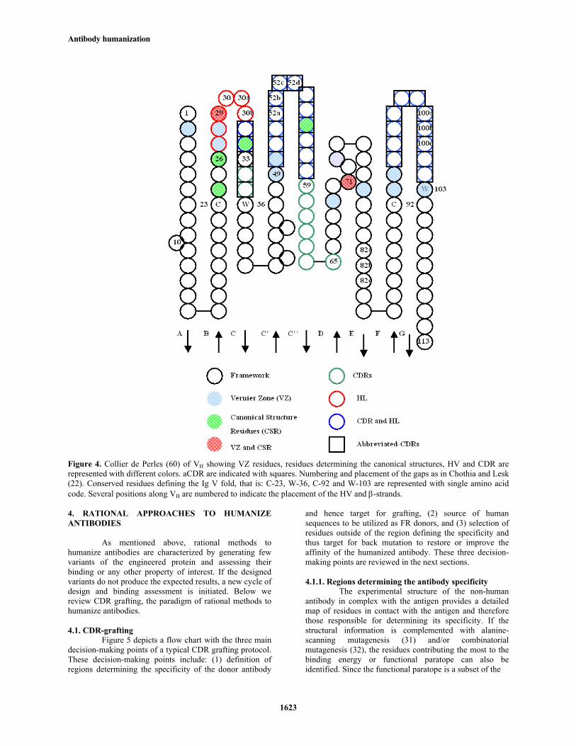
with antigen. Comparison of the residues in contact with antigens and sequence variability values pinpointed residues in contact with the antigen as the most variable ones. These residues were defined as Specificity-Determining Residues (SDRs) and the regions containing the SDRs were termed abbreviated-CDRs (aCDRs) (Figure 2).
25 30 35 50 55 90 95 ..|....|abcdef....|. ..|....|.. ..|....|ab...
HV ----oooooooooooo---- --ooo----- --ooooooooo--CDR -ooooooooooooooooo-- --ooooooo- -ooooooooooo-aCDR --------------oooo-- --oooooo-- -oooooooooo–
L1 L2 L3
25 30 35 50 55 60 65 95 100 ..|....|ab....|... ....|..abcd..|....|....|. .|....|abcdefgh...
HV ---ooooooooo------ ---------oooooooo----------- -oooooooooooooooo-CDR ----------ooooo--- ----oooooooooooooooooooo- -oooooooooooooooo-aCDR ----------ooooo--- ----ooooooooooooo-------- -oooooooooooooooo-
H1 H2 H3

Antibody humanization
1622
Figure 3. Collier de Perles (60) of VL showing VZ residues, residues determining the canonical structures, HV and CDR are represented with different colors. aCDR are indicated with squares. Numbering and placement of the gaps as in Chothia and Lesk (22). Conserved residues defining the Ig V fold, that is: C-23, W-35, C-88 and F-98 are represented with single amino acid code. Several positions along VL are numbered to indicate the placement of the HV and β-strands.
Chothia and Lesk (22) compared the first antibodies of known structure and found that all the HV loops, with the exception of H3, exhibit a small number of main-chain conformations or canonical structures. Canonical structures are determined by the HV length and conserved residues in the HV and FR regions (22, 27-29). Analyses of functional germline genes, mature antibody sequences and pseudogenes, based on these rules relating the amino acid sequence with the three-dimensional structure, have corroborated the existence of canonical structures in the vast majority of antibodies.
Foote and Winter (30) estimated that 30 residues underlying the CDRs, 16 in VH and 14 in VL, are responsible for stabilizing the HV loop structure as well as modifying their positioning. Since these residues fine-tune the antibody affinity, they called this region Vernier Zone (VZ). As expected, some VZ residues coincide with residues responsible for maintaining the canonical structures (22, 27, 28). Figures 3 and 4 map onto VH and VL, respectively, the VZ and residues defining the canonical structures. Also, HV loops, CDRs, and aCDRs are depicted in the figures as reference for the next sections.
Antibody humanization
1623
Figure 4. Collier de Perles (60) of VH showing VZ residues, residues determining the canonical structures, HV and CDR are represented with different colors. aCDR are indicated with squares. Numbering and placement of the gaps as in Chothia and Lesk (22). Conserved residues defining the Ig V fold, that is: C-23, W-36, C-92 and W-103 are represented with single amino acid code. Several positions along VH are numbered to indicate the placement of the HV and β-strands. 4. RATIONAL APPROACHES TO HUMANIZE ANTIBODIES
As mentioned above, rational methods to
humanize antibodies are characterized by generating few variants of the engineered protein and assessing their binding or any other property of interest. If the designed variants do not produce the expected results, a new cycle of design and binding assessment is initiated. Below we review CDR grafting, the paradigm of rational methods to humanize antibodies.
4.1. CDR-grafting
Figure 5 depicts a flow chart with the three main decision-making points of a typical CDR grafting protocol. These decision-making points include: (1) definition of regions determining the specificity of the donor antibody
and hence target for grafting, (2) source of human sequences to be utilized as FR donors, and (3) selection of residues outside of the region defining the specificity and thus target for back mutation to restore or improve the affinity of the humanized antibody. These three decision-making points are reviewed in the next sections.
4.1.1. Regions determining the antibody specificity
The experimental structure of the non-human antibody in complex with the antigen provides a detailed map of residues in contact with the antigen and therefore those responsible for determining its specificity. If the structural information is complemented with alanine-scanning mutagenesis (31) and/or combinatorial mutagenesis (32), the residues contributing the most to the binding energy or functional paratope can also be identified. Since the functional paratope is a subset of the

Antibody humanization
1624
Figure 5. Flow chart of the decision-making points in a typical CDR grafting protocol.
residues in contact (33), grafting only the functional paratope would reduce the number of non-human residues in the humanized product.
Unfortunately, only in rare occasions are the
experimental structure of the antigen-antibody complex and/or functional paratope available at the beginning of a humanization protocol. In absence of a precise definition of residues responsible for a given antibody specificity, CDRs (Figure 2) have often been employed as regions defining the specificity (10, 30, 34, 35). Also, a combination of CDR and HV loop (36, 37) has been targeted for grafting, in particular at CDR-1 of VH. All CDRs with exception of the CDR-1 of VH contain the HV loops (Figures 2, 3 and 4) and thus, by grafting CDRs, all HV loops but the CDR-1 of VH are transferred onto the human FR. For the CDR-1 of VH, a consensus of the CDR and HV is grafted (37). Since the N-terminal region of H1 contains several VZ residues (Figures 2 and 4), transferring this region could help to preserve binding. The downside of using a CDR/HV consensus at CDR-1 of VH instead of straight CDR grafting is that it might increase the potential immunogenicity, as the non-human region grafted onto the human FR is larger.
To reduce the number of residues to be grafted
onto the human FRs, Tamura et al (38) humanized the anti-tumor-associated glycoprotein-72 mAb CC49 by SDR grafting. The resulting humanized CC49 retained binding of the parental antibody, while reactivity with the sera of patients was only minimal. De Pascalis et al. (39) compared CDR-grafted and aCDR-grafted (Figures 2, 3
and 4) variants of the murine antibody COL-1 specific for carcinoembryonic antigen (CEA). No significant differences were found in binding to CEA between the CDR-grafted and SDR-grafted variants. Compared with CDR-grafted, the SDR-grafted COL-1 variant showed, however, lower reactivity to sera of patients carrying anti-V region antibodies to COL-1. A detailed description of the SDR grafting method has recently been published by Kashmiri et al (40).
Wilson et al. (41) have shown that antibodies do
not use L2 to contact haptens and peptides, whereas the recognition of proteins involves the six HV loops (42). A systematic analysis conducted by MacCallum et al. (43) with 26 antibodies of known three-dimensional structure indicated that large antigens like proteins contact the most apical region of the HV loops, in contrast to small antigens, i.e. haptens that are recognized by residues in the extremes of the HV loops. Almagro (44) studied 59 unique antigen-antibody complexes and found that the number and distribution of SDRs for different types of antigens or SDR usage (SDRU) correlate well with the size of the antigen. For instance, antibodies use five additional SDRs (2 in VL and 3 in VH) to contact peptides and proteins as compared to the number of SDRs used to recognize haptens. It has been suggested (44) that the number of non-human residues to graft onto human FR could be reduced by SDRU grafting.
4.1.2. Source of human FRs
Once the non-human antibody regions targeted for grafting onto the human FR have been defined, the second step in a humanization process is to identify the source of human FR donors (Figure 5). Initial works (8, 10) utilized FRs of human antibodies of known structure, regardless of their homology to the non-human antibody. Subsequent works (11) reported the use of human sequences with the highest homology to the non-human antibody. The former strategy has been called fixed FR, whereas the latter is termed best fit.
Graziano et al. (45) compared the fixed FR and
best fit strategies and found that the latter results in humanized antibodies with higher affinity. This result is consistent with the fact that in most cases the difference between the non-human CDRs and human FRs, and thus potential conflicts, is minimized when the FR is chosen on the basis of sequence homology. Nevertheless, affinity is not the only variable to be considered during a humanization protocol, as other parameters such as low immunogenicity and production yields should be taken into account when choosing a given FR for humanization. Consequently, combination of fixed FR in VL and best fit in VH have also been reported (37, 46-48). Still another strategy of selecting human FRs as template for humanization is by generating consensus sequences (49, 50).
Regardless of the method chosen to select the
human FRs, two sources of human sequences have been utilized: mature and germline gene sequences. Mature sequences, which are products of immune responses, carry

Antibody humanization
1625
somatic mutations generated by random processes (51) and are not under the species selection, resulting in potential immunogenic residues. Thus, to avoid immunogenic residues, human germline genes have increasingly been utilized as source of FR donors (13, 51-59).
Germline genes have two additional advantages
over mature sequences as FR donors. First, the physical map of the human H and L chains loci and the functional germline gene repertoire they encode have been thoroughly characterized; see The International ImMunoGeneTics information system® (IMGT (60); URL: http://imgt.cines.fr/) for a full description of the human germline gene tables. This information has opened up the possibility of reliable analyses of the human germline gene repertoire evolution (61-64), precise estimation of germline gene and somatic mutation patterns (65), and accurate estimation of the germline gene usage in vivo (66) and in vitro (67). This accumulated knowledge has been useful to rank humanized variants with FRs generated by different germline genes during humanization protocols (36, 63).
A second potential advantage of using germline
genes as FR donors is that comparison of germline gene and mature antibody x-ray crystallography structures in bound and free ligand stages have shown that the former molecules are more flexible (68, 69). Such plasticity might in theory accommodate diverse CDRs with fewer or no back mutations into the FR to restore the affinity of the humanized antibody. In fact, successful humanization of murine antibodies with CDRs selected solely on the basis of their homology with germline gene CDRs, regardless of FR homology, have been reported (see Superhumanization below).
4.1.3. Back mutations to retain or restore affinity
Commonly, affinity decreases after CDR grafting as a consequence of incompatibilities between non-human CDRs and human FRs. Therefore, the third step in a typical CDR grafting protocol (Figure 5) is to define mutations that would restore or prevent affinity losses. A substantial body of evidences accumulated during the more than two decades of CDR grafting (70) indicates that the number of back mutations and the type of amino acid replacements to introduce into the humanized antibody in order to restore binding depend on each particular case. Thus, back mutations should be carefully designed based on the structure or a model of the humanized antibody and tested experimentally.
Antibody modeling has been highly facilitated by
discovery of the canonical structures (22, 27, 29, 71) as well as by the large number of antibody structures available in the Protein Data Bank [PDB (72); URL: http://www.rcsb.org/], amounting 848 entries as of April 30th, 2007; see 3D repertoire (73) at IMGT. A web site for automated antibody modeling called WAM (74) can be found at the URL: http://antibody.bath.ac.uk. WAM builds antibody models based on the canonical structure patterns. The CDR-H3 structure is modeled using CONGEN (75). WAM and other modeling software such as Modeller (76) (URL: http://salilab.org/modeller/ modeller.html) and
Swiss PDB Viewer (77) predict the VL conformation and most of the VH domain with reasonable precision. Accurate prediction of the CDR-H3 loop conformation is still troublesome.
In absence of structural guidance, Bodao et al.
(37) recently created human/mouse hybrids of the entire FRs and assessed expression and binding of the variants, as they humanized the murine monoclonal antibody HIRMAb for use as a drug-delivery system across the blood-brain barrier and found that myeloma cells poorly secreted the initial humanized antibody. One of the hybrid molecules, in which the human FR-3 region was replaced by the corresponding murine FR-3, showed 27% decreased affinity compared to the murine HIRMAb but restored expression. Since this variant introduced five back mutations into the humanized antibody, a concern when using this experimental strategy is the potential large number of murine residues back mutated into the humanized molecule, which might increase the immunogenicity of the engineered antibody.
4.2. Resurfacing
Resurfacing (12) emerged in the early 1990’s as an alternative method to CDR grafting. This method shares with CDR grafting the two first decision-making points of the design cycle (Figure 5). Different from CDR grafting, however, resurfacing retains the non-exposed residues of the non-human antibody. Only surface residues in the non-human antibody are changed to human residues. Since resurfacing does not change the residues in the core of the V domains, the expectation is to eliminate potential B-cell epitopes, while minimizing the perturbation of residues determining the specificity of the antibody.
The initial resurfacing method was developed by
Pedersen et al. (78). These authors analyzed 12 antibodies of known structure and identified a set of residues with ≥ 30% solvent accessible surface area in the FRs. Next, human and murine sequences were compared and the pattern of residues to resurface any given murine antibody was derived. Staelens et al. (79) suggested a modification of the method, in which the non-human surface accessible FR residues are determined in a model and residues to mutate are selected by comparison to the human antibody with highest sequence identity. Several discrepancies with Pedersen’s (78) assignments were found, suggesting that the new approach is more accurate. Fontayne et al. (80) resurfaced an anti-GPIbα antibody termed B4-Fab using Staelens’ et al. (79) method. The resurfaced version, called h6B4-Fab, showed the same in vitro bioactivity in different systems where it was compared to the original murine monoclonal antibody. Injections of h6B4-Fab in baboons indicated a behavior comparable to what was previously observed with the murine Fab.
Several antibodies have been resurfaced in the
last years (81-84). The affinity of the parental antibodies has been retained but no clinical data on resurfaced antibodies has been published (79). Therefore, a systematic assessment of the immunogenicity of resurfaced antibodies is not available.

Antibody humanization
1626
4.3. Superhumanization CDR grafting relies on the FR comparison of the
non-human and human sequences. Tan et al (13) proposed an alternative paradigm based on comparison of CDRs, called Superhumanization, in which FR homology is irrelevant. Using this strategy, Tan et al. (13) superhumanized the murine anti-human CD28 antibody 9.3. The method consists of comparison of the non-human sequence with the functional human germline gene repertoire. Those genes encoding the same or closely related canonical structures to the murine sequences are then selected. Next, within the genes sharing the canonical structures with the non-human antibody, those with highest homology within the CDRs are chosen as FR donors. Finally, the non-human CDRs are grafted onto these FRs. Application of this method to 9.3 as the first case study yielded a superhumanized 9.3 VL with insignificant loss in affinity. The loss in affinity of superhumanized VH was 20-fold. The fully superhumanized antibody lost 30-fold affinity but retained biological activity.
Hwang et al. (55) published a detailed description
of the superhumanization method using D1.3 as a second case study. The affinity loss of superhumanized D1.3 was six-fold as compared to 70-fold loss in affinity of a CDR-grafted version of D1.3 (30). Another example published by Hu et al. (54), in which they superhumanized the murine antibody 1A4A1, a neutralizing antibody against the Venezuelan equine encephalitis virus (VEEV), demonstrated retention of antigen-binding specificity and neutralizing activity.
4.4. Human String Content Optimization
Recently, Lazar et al. (14) have introduced a new method for antibody humanization based on a metric of antibody humanness termed Human String Content (HSC). This method compares the mouse sequence with the repertoire of human germline genes and the differences are scored as HSC. The target sequence is then humanized by maximizing its HSC rather than using a global identity measure to generate multiple diverse humanized variants. The method was applied to humanize four antibodies with different antigen specificities showing better binding signals than or comparable to those of the parent non-human antibodies.
5. EMPIRICAL APPROACHES TO HUMANIZE ANTIBODIES
In contrast to the rational methods to humanize antibodies reviewed above, empirical methods rest on the generation of large libraries of humanized variants and selection of the best clones using enrichment technologies or HTS techniques. Thus, empirical methods are dependent on a reliable selection and/or screening system that should be able to search through a vast space of antibody variants. In vitro display technologies, such as phage and ribosome display, meet these needs and have proven very useful in antibody humanization efforts. Efficient isolation of antibody variants from phage, ribosome and yeast display libraries as well as bacterial colony screening has been extensively reported and reviewed (4, 85-87).
5.1. FR libraries In the FR library approach, a collection of residue
variants are introduced at specific positions in the FR followed by panning of the library to select the FR that best supports the grafted CDR. Rosok et al. (57) reported for the first time the use of a FR library to humanize antibodies. Their method resembled CDR grafting, but instead of creating few back mutations in the FR, a combinatorial library of 124 variants was constructed. The residues targeted for mutagenesis were buried residues critical to maintain the canonical structures (Figure 3 and 4). As FR donors, human germline genes closely related to the murine BR26 M1 antibody (88) anti-Lewis Y antigen were chosen. By using a M13 derived phage vector and plaque lift assay as selection method, several FR variants with antigen binding within 2-fold of the parent antibody were isolated.
Subsequently, Baca et al. (19) grafted the CDRs
of the anti-vascular endothelial growth factor (anti-VEGF) monoclonal antibody A4.6.1 onto a human VL1 (κ), VH3 FR and a combinatorial library was built in 11VZ residues (30) (Figures 3 and 4). Following phage display panning, an optimized FR was selected that increased the affinity 125-fold compared to the clone with no FR mutations. Son et al. (89) used an analogous approach to Rosok (57) to humanize the anti-human 4-1BB monoclonal antibody but employed phage display as selection method like Baca et al. (19). The most homologous germline genes were identified (VH1-46, VL A14) and diversity was introduced in buried residues that might affect the HV loop conformation. Selection on phage resulted in clones with 10-100 folds loss in affinity but the functionality, measured as proliferation of peripheral blood mononuclear cells (PBMC) in mixed lymphocyte reaction (MLR), was retained.
Also by phage display, Rader et al. (90)
humanized a rabbit antibody. Similarly as Baca (19) and Rosok (57), a human acceptor FR was chosen using the best fit approach, followed by CDR grafting of all 6 rabbit CDRs into the human framework. A Fab phage library was created with diversity in VZ residues and panned against the human A33 colon cancer antigen. The best clone matched the affinity of the rabbit parent antibody.
5.2. Guided Selection
In contrast to the FR library approach described above, in which the CDRs remain non-human, guided selection allows for the isolation of completely human antibodies. By using this method, Humira® (Adalimumab), the first human antibody approved by the FDA, was isolated (20). This method is the paradigm of empirical methods, as no assumptions on the antibody structure are made.
The strategy was developed by Jespers et al. (18)
and consists of combining the VH or VL domain of a given non-human antibody specific for a particular epitope with a human VH or VL library and specific human V domains are selected against the antigen of interest. In the pivotal study (20), a rodent antibody against human TNF-α was humanized by first combining the rodent VH with a library
Antibody humanization
1627
of human L chains. The library was then selected against TNF-α by phage display and the selected VL was cloned into a library of human VH chains and selected against TNF-α. As a result, a fully human antibody with similar affinity as the rodent antibody was isolated.
Isolating de novo human antibodies from phage
libraries toward cell surface antigens has proven to be a challenging task. On the contrary, antibodies derived from rodents targeting membrane receptors have been successfully engineered into some of the most important antibodies on the therapeutic market, e.g. Herceptin® (trastuzumab, anti-erbB2) and Rituxan® (rituximab, anti-CD20) (91). Examples of guided selection to humanize mouse antibodies towards cell surface antigens include the folate-binding protein present on ovarian cancer cells (92) and CD147, which is highly expressed on hepatocellular carcinoma (93).
A potential disadvantage of the guided selection
approach is that shuffling of one antibody chain while keeping the other constant could result in epitope drift (17, 94, 95). Indeed, several studies have reported the isolation of binders with a change in fine-specificity (96, 97). Kuepper et al. (96) humanized a murine antibody binding to the SAI/II antigen of Streptococcus mutans by guided selection, resulting in the isolation of several VLs; some binding the same epitope, whereas others showed a epitope drift. This was solved by implementation of an inhibition ELISA, which allowed for the selection of clones recognizing the same epitope as the parent antibody. In another study by Wang et al. (98), the VH and VL domains of the mouse anti-human TNF-α antibody were shuffled in parallel (98). Although successful isolation of a humanized antibody retaining recognition of the epitope was reported, the study also generated several polyreactive antibodies, again demonstrating the risk of epitope drift when guided selection is employed.
In order to maintain the epitope recognized by
the non-human antibody, CDR retention can be applied (99, 100). In this method, the non-human CDR-H3 is commonly retained, as this CDR is at the center of the antigen-binding site (Figure 1B) and has proven to be the most important region of the antibody for antigen recognition (101-103). Nevertheless, retention of both CDR-H3 and CDR-L3 (104) as well as CDR-H3, CDR-L3 and CDR-L2 (105) of the non-human antibody have also been reported. Furthermore, implementation of the CDR-H3 retention approach has given some general clues to the significance of VL usage in antibody-antigen interaction. Beiboer et al. (100) reported that the selected human VLs originated from the VL3κ family, whereas the mouse VL had higher homology to the VL2κ family. Hence, the VL sequence homology was not that important, consistent with the proposition that VH has a predominant role in the antigen recognition.
Krinner et al. (106) used guided selection to
humanize a rat antibody binding granulocyte-macrophage colony stimulating factor (GM-CSF). When the rat
monoclonal antibody was constructed and produced as a single chain Fragment variable (scFv) in E. coli, it resulted in loss of functionality. However, after human VL replacement by phage library selections, rat/human hybrid scFv’s were selected that expressed well and retained functionality. Thus, VL selection could stabilize scFv’s while maintaining the binding characteristics. In fact, Almagro et al. (107) designed a synthetic VH repertoire based on the SDRU of anti-protein and anti-peptide antibodies and combined it with the VL of D1.3. The repertoire was displayed on phage and panned against Hen-Egg White Lysozyme (HEL). A diverse panel of scFv’s was isolated, with a pattern of conservation that resembled the functional D1.3 epitope (33).
5.3. FR shuffling
Dall’Acqua et al. (58) introduced the FR shuffling approach. This approach differs from the FR library method described above in which selected residues are variegated. In FR shuffling whole FRs are combined with the non-human CDRs instead of creating combinatorial libraries of selected residue variants. Using FR shuffling, Dall’Acqua and co-workers humanized the murine antibody B233, which bind the potential cancer target receptor tyrosine kinase EphAa2. All six murine CDRs of B233 were cloned into a library of human germline gene FRs. The libraries were screened for binding in a two-step selection process, first humanizing VL, followed by VH. In another study (52), a one-step FR shuffling process showed to be more efficient than the two-step screening, as the resulting antibodies exhibited improved biochemical and physico-chemical properties including enhanced expression, increased affinity and thermal stability.
5.4. Humaneering
Lastly, within the most recent empirical approaches to humanize antibodies, Humaneering was presented at Cambridge Healthtech Institute’s Protein Engineering Summit 2007 in Boston (108). Humaneering allows for isolation of antibodies that are 91-96 % homologous to human germline gene antibodies. The method is based on experimental identification of essential minimum specificity determinants (MSDs) and is based on sequential replacement of non-human fragments into libraries of human FRs and assessment of binding. It begins with regions of the CDR-3 of non-human VH and VL chains and progressively replaces other regions of the non-human antibody into the human FRs, including the CDR-1 and CDR-2 of both VH and VL. This methodology typically results in epitope retention and identification of antibodies from multiple sub-classes with distinct human V-segment CDRs.
6. PERSPECTIVES
Fueled by the FDA approval of more than 20 antibodies for therapeutic use in humans and the list of almost one thousand clinical trails involving antibodies, humanization has shown a steady growth and several humanization methods have been generated in the last few years. These methods can be classified in two main trends:
Antibody humanization
1628
(1) rational methods, which rely on the so-called design cycle and (2) empirical methods, which rest on the generation of large combinatorial libraries of humanized variants and selection of the desired variant via enrichment technologies or HTS techniques.
The rational methods have evolved toward
minimizing the content of non-human residues in the human context such as SDR and aCDR grafting and simplifying the design cycle, such as Superhumanization or HSC Optimization. The empirical methods have evolved from FR libraries, which resemble CDR grafting but rest on selection of large combinatorial libraries of selected residues, toward Guided Selection and Humaneering, where the assumptions on the antibody structure are minimal.
The increasing number of antibodies in
evaluation by the FDA should provide data on the efficacy and immunogenicity of the antibodies humanized using the different humanization methods here reviewed. This knowledge would be complemented with the study of the increasing number of antibody structures as well as further characterization of the human germline gene repertoire and gene usage in physiological conditions. Altogether, it should provide a framework to guide future antibody humanization protocols toward engineering molecules with minimal or none immunogenicity, enhanced physico-chemical profile and superior biological efficacy. 7. ACKNOWLEDGEMENTS
We would like to thank Dr. Biplab Bose for his
kind invitation to prepare this review. To Jefferson Foote, Gary Gilliland and Miguel Barbosa for reading the manuscript and valuable comments and discussions. 8. REFERENCES 1. Schellekens, H.: Immunogenicity of therapeutic proteins: clinical implications and future prospects. Clin. Ther., 24, 1720-40 (2002) 2. Schellekens, H., D. Crommelin & W. Jiskoot: Immunogenicity of Antibody Therapeutics. In: Handbook of therapeutic antibodies. Ed: Dubel S. Wiley-VCH, Weinheim (2007) 3. Hwang, W. Y. & J. Foote: Immunogenicity of engineered antibodies. Methods. , 36, 3-10 (2005) 4. Hoogenboom, H. R.: Selecting and screening recombinant Selecting and screening recombinant antibody libraries. Nat. Biotechnol., 23, 1105-16 (2005) 5. Hust, M., L. Toleikis & S. Dubel: Selection Strategies II: Antibody phage display. In: Handbook of therapeutic antibodies. Ed: Dubel S. Wiley-VCH, Weinheim (2007) 6. Bruggermann, M., J. A. Smith, M. J. Osborn & X. Zou: Selection Strategies III: Transgenic Mice. In: Handbook of
therapeutic antibodies. Ed: Dubel S. Wiley-VCH, Weinheim (2007) 7. Morrison, S. L., M. J. Johnson, L. A. Herzenberg & V. T. Oi: Chimeric human antibody molecules: mouse antigen-binding domains with human constant region domains. Proc. Natl. Acad. Sci. USA., 81, 6851-5 (1984) 8. Jones, P. T., P. H. Dear, J. Foote, M. S. Neuberger & G. Winter: Replacing the complementarity-determining regions in a human antibody with those from a mouse. Nature., 321, 522-5 (1986) 9. Verhoeyen, M., C. Milstein & G. Winter: Reshaping human antibodies: grafting an antilysozyme activity. Science., 239, 1534-6 (1988) 10. Riechmann, L., M. Clark, H. Waldmann & G. Winter: Reshaping human antibodies for therapy. Nature., 332, 323-7 (1988) 11. Queen, C., W. P. Schneider, H. E. Selick, P. W. Payne, N. F. Landolfi, J. F. Duncan, N. M. Avdalovic, M. Levitt, R. P. Junghans & T. A. Waldmann: A humanized antibody that binds to the interleukin 2 receptor. Proc. Natl. Acad. Sci. USA., 86, 10029-33 (1989) 12. Padlan, E. A.: A possible procedure for reducing the immunogenicity of antibody variable domains while preserving their ligand-binding properties. Mol. Immunol., 28, 489-98 (1991) 13. Tan, P., D. A. Mitchell, T. N. Buss, M. A. Holmes, C. Anasetti & J. Foote: "Superhumanized" antibodies: reduction of immunogenic potential by complementarity-determining region grafting with human germline sequences: application to an anti-CD28. J. Immunol., 169, 1119-25 (2002) 14. Lazar, G. A., J. R. Desjarlais, J. Jacinto, S. Karki & P. W. Hammond: A molecular immunology approach to antibody humanization and functional optimization. Mol. Immunol., 44, 1986-98 (2007) 15. McCafferty, J., A. D. Griffiths, G. Winter & D. J. Chiswell: Phage antibodies: filamentous phage displaying antibody variable domains. Nature., 348, 552-4 (1990) 16. Winter, G., A. D. Griffiths, R. E. Hawkins & H. R. Hoogenboom: Making antibodies by phage display technology. Annu. Rev. Immunol., 12, 433-55 (1994) 17. Kang, A. S., T. M. Jones & D. R. Burton: Antibody redesign by chain shuffling from random combinatorial immunoglobulin libraries. Proc. Natl. Acad. Sci. USA., 88, 11120-3 (1991) 18. Jespers, L. S., A. Roberts, S. M. Mahler, G. Winter & H. R. Hoogenboom: Guiding the selection of human antibodies from phage display repertoires to a single epitope of an antigen. Biotechnology (NY). 12, 899-903 (1994)

Antibody humanization
1629
19. Baca, M., L. G. Presta, S. J. O'Connor & J. A. Wells: Antibody Humanization Using Monovalent Phage Display. J. Biol. Chem., 272, 10678-84 (1997) 20. Osbourn, J., M. Groves & T. Vaughan: From rodent reagents to human therapeutics using antibody guided selection. Methods., 36, 61-8 (2005) 21. Amzel, L. M. & R. J. Poljak: Three-Dimensional Structure of Immunoglobulins. Ann. Rev. Biochem., 48, 961-97 (1979) 22. Chothia, C. & A. M. Lesk: Canonical structures for the hypervariable regions of immunoglobulins. J. Mol. Biol., 196, 901-17 (1987) 23. Segal, D. M., E. A. Padlan, G. H. Cohen, S. Rudikoff, M. Potter & D. R. Davies: The three-dimensional structure of a phosphorylcholine-binding mouse immunoglobulin Fab and the nature of the antigen binding site. Proc. Natl. Acad. Sci. USA., 71, 4298-302 (1974) 24. Wu, T. T. & E. A. Kabat: An analysis of the sequences of the variable regions of Bence Jones proteins and myeloma light chains and their implications for antibody complementarity. J. Exp. Med., 132, 211-50 (1970) 25. Kabat, E. A., T. T. Wu & H. Bilofsky: Unusual distributions of amino acids in complementarity-determining (hypervariable) segments of heavy and light chains of immunoglobulins and their possible roles in specificity of antibody-combining sites. J. Biol. Chem., 252, 6609-16 (1977) 26. Padlan, E. A., C. Abergel & J. P. Tipper: Identification of specificity-determining residues in antibodies. FASEB J., 9, 133-9 (1995) 27. Chothia, C., A. M. Lesk, A. Tramontano, M. Levitt, S. J. Smith-Gill, G. Air, S. Sheriff, E. A. Padlan, D. Davies, W. R. Tulip, P. M. Colman, S. Spinelli, P. M. Alzari & R. J. Poljak: Conformations of immunoglobulin hypervariable regions. Nature., 342, 877-83 (1989) 28. Tramontano, A., C. Chothia & A. M. Lesk: Framework residue 71 is a major determinant of the position and conformation of the second hypervariable region in the VH domains of immunoglobulins. J. Mol. Biol., 215, 175-82 (1990) 29. Al-Lazikani, B., A. M. Lesk & C. Chothia: Standard conformations for the canonical structures of immunoglobulins. J. Mol. Biol., 273, 927-48 (1997) 30. Foote, J. & G. Winter: Antibody framework residues affecting the conformation of the hypervariable loops. J. Mol. Biol., 224, 487-99 (1992) 31. Fuh, G., P. Wu, W. C. Liang, M. Ultsch, C. V. Lee, B. Moffat & C. Wiesmann: Structure-function studies of two synthetic anti-vascular endothelial growth factor Fabs and comparison with the Avastin Fab. . J. Biol. Chem., 281, 6625-31 (2006)
32. Pál, G., J. L. Kouadio, D. R. Artis, A. A. Kossiakoff & S. S. Sidhu: Comprehensive and quantitative mapping of energy landscapes for protein-protein interactions by rapid combinatorial scanning. J. Biol. Chem., 281, 22378-85 (2006) 33. Dall'Acqua, W., E. R. Goldman, W. Lin, C. Teng, D. Tsuchiya, H. Li, X. Ysern, B. C. Braden, Y. Li, S. J. Smith-Gill & R. A. Mariuzza: A mutational analysis of binding interactions in an antigen-antibody protein-protein complex. Biochemistry. , 37, 7981-7991 (1998) 34. Kettleborough, C. A., J. Saldanha, V. J. Heath, C. J. Morrison & M. M. Bendig: Humanization of a mouse monoclonal antibody by CDR-grafting: the importance of framework residues on loop conformation. Protein Eng., 4, 773-83. (1991) 35. Asano, R., Y. Sone, K. Makabe, K. Tsumoto, H. Hayashi, Y. Katayose, M. Unno, T. Kudo & I. Kumagai: Humanization of the bispecific epidermal growth factor receptor x CD3 diabody and its efficacy as a potential clinical reagent. Clin. Cancer Res., 12, 4036-42 (2006) 36. Tempest, P. R., P. White, M. Buttle, F. J. Carr & W. J. Harris: Identification of framework residues required to restore antigen binding during reshaping of a monoclonal antibody against the glycoprotein gB of human cytomegalovirus. Int. J. Biol. Macromol., 17, 37-42 (1995) 37. Boado, R. J., Y. Zhang, Y. Zhang & W. M. Pardridge: Humanization of anti-human insulin receptor antibody for drug targeting across the human blood-brain barrier. Biotechnol. Bioeng., 96, 381-91 (2007) 38. Tamura, M., D. E. Milenic, M. Iwahashi, E. Padlan, J. Schlom & S. V. Kashmiri: Structural correlates of an anticarcinoma antibody: identification of specificity-determining residues (SDRs) and development of a minimally immunogenic antibody variant by retention of SDRs only. J. Immunol., 164, 1432-41 (2000) 39. De Pascalis, R., M. Iwahashi, M. Tamura, E. A. Padlan, N. R. Gonzales, A. D. Santos, M. Giuliano, P. Schuck, J. Schlom & S. V. Kashmiri: Grafting of "abbreviated" complementarity-determining regions containing specificity-determining residues essential for ligand contact to engineer a less immunogenic humanized monoclonal antibody. J. Immunol., 169, 3076-84 (2002) 40. Kashmiri, S. V., R. De Pascalis, N. R. Gonzales & J. Schlom: SDR grafting - a new approach to antibody humanization. Methods., 36, 25-34 (2005) 41. Wilson, I. A., J. B. Ghiara & R. L. Stanfield: Structure of anti-peptide antibody complexes. Res. Immunol., 145, 173-178 (1994) 42. Davies, D. R., E. A. Padlan & S. Sheriff: Antibody-antigen complexes. Annu. Rev. Biochem., 59 439-473 (1990)

Antibody humanization
1630
43. MacCallum, R. M., A. C. Martin & J. M. Thornton: Antibody-antigen interactions: contact analysis and binding site topography. J. Mol. Biol., 262 732-745 (1996) 44. Almagro, J. C.: Identification of differences in the specificity-determining residues of antibodies that recognize antigens of different size: implications for the rational design of antibody repertoires. J. Mol. Recognit., 17, 132-43 (2004) 45. Graziano, R. F., P. R. Tempest, P. White, T. Keler, Y. Deo, H. Ghebremariam, K. Coleman, L. C. Pfefferkorn, M. W. Fanger & P. M. Guyre: Construction and characterization of a humanized anti-gamma-Ig receptor type I (Fc gamma RI) monoclonal antibody. J. Immunol., 155, 4996-5002 (1995) 46. Bourne, P. C., S. S. Terzyan, G. Cloud, N. F. Landolfi, M. Vasquez & A. B. Edmundson: Three-dimensional structures of a humanized anti-IFN-gamma Fab (HuZAF) in two crystal forms. Acta. Crystallogr. D Biol. Crystallogr., 60, 1761-9 (2004) 47. Gorman, S. D., M. R. Clark, E. G. Routledge, S. P. Cobbold & H. Waldmann: Reshaping a therapeutic CD4 antibody. Proc. Natl. Acad. Sci. USA., 88, 4181-5 (1991) 48. Hamilton, A. A., D. M. Manuel, J. E. Grundy, A. J. Turner, S. I. King, J. R. Adair, P. White, F. J. Carr & W. J. Harris: A humanized antibody against human cytomegalovirus (CMV) gpUL75 (gH) for prophylaxis or treatment of CMV infections. J. Infect. Dis., 176, 59-68 (1997) 49. Carter, P., L. Presta, C. M. Gorman, J. B. Ridgway, D. Henner, W. L. Wong, A. M. Rowland, C. Kotts, M. E. Carver & H. M. Shepard: Humanization of an anti-p185HER2 antibody for human cancer therapy. Proc. Natl. Acad. Sci. USA., 89, 4285-9 (1992) 50. Presta, L. G., S. J. Lahr, R. L. Shields, J. P. Porter, C. M. Gorman, B. M. Fendly & P. M. Jardieu: Humanization of an antibody directed against IgE. J. Immunol., 151, 2623-32 (1993) 51. Neuberger, M. S. & C. Milstein: Somatic hypermutation. Curr. Opin. Immunol. , 7, 248-254 (1995) 52. Damschroder, M. M., L. Widjaja, P. S. Gill, V. Krasnoperov, W. Jiang, W. F. Dall'Acqua & H. Wu: Framework shuffling of antibodies to reduce immunogenicity and manipulate functional and biophysical properties. Mol. Immunol., 44, 3049-60 (2007) 53. Gonzales, N. R., E. A. Padlan, R. De Pascalis, P. Schuck, J. Schlom & S. V. Kashmiri: SDR grafting of a murine antibody using multiple human germline templates to minimize its immunogenicity. Mol. Immunol., 41, 863-72 (2004) 54. Hu, W. G., D. Chau, J. Wu, S. Jager & L. P. Nagata: Humanization and mammalian expression of a murine
monoclonal antibody against Venezuelan equine encephalitis virus. Vaccine., 25, 3210-4 (2007) 55. Hwang, W. Y., J. C. Almagro, T. N. Buss, P. Tan & J. Foote: Use of human germline genes in a CDR homology-based approach to antibody humanization. Methods., 36, 35-42 (2005) 56. Mazor, Y., I. Keydar & I. Benhar: Humanization and epitope mapping of the H23 anti-MUC1 monoclonal antibody reveals a dual epitope specificity. Mol. Immunol., 42, 55-69 (2005) 57. Rosok, M. J., D. E. Yelton, L. J. Harris, J. Bajorath, K.-E. Hellstrom, I. Hellstrom, G. A. Cruz, K. Kristensson, H. Lin, W. D. Huse & S. M. Glaser: A Combinatorial Library Strategy for the Rapid Humanization of Anticarcinoma BR96 Fab. J. Biol. Chem., 271, 22611-18 (1996) 58. Dall'Acqua, W. F., M. M. Damschroder, J. Zhang, R. M. Woods, L. Widjaja, J. Yu & H. Wu: Antibody humanization by framework shuffling. Methods., 36, 43-60 (2005) 59. Routledge, E. G., S. D. Gorman & M. Clark: Protein engineering of antibody molecules for prophylactic and therapeutic applications in man. Ed: Clark M., Academic Titles, Nottingham.13-44 (1993) 60. Lefranc, M. P., V. Giudicelli, Q. Kaas, E. Duprat, J. Jabado-Michaloud, D. Scaviner, C. Ginestoux, O. Clement, D. Chaume & G. Lefranc: IMGT, the international ImMunoGeneTics information system. Nucleic Acids Res., 33, D593-7 (2005) 61. Ota, T. & M. Nei: Divergent evolution and evolution by the birth-and-death process in the immunoglobulin IGHV gene family. Mol. Biol. Evol. , 11, 469-482 (1994) 62. Sitnikova, T. & C. Su: Coevolution of immunoglobulin heavy- and light-chain variable-region gene families. Mol. Biol. Evol. , 15, 617-25. (1998) 63. Almagro, J. C., I. Hernandez, M. del Carmen Ramirez & E. Vargas-Madrazo: The differences between the structural repertoires of IGHV germ-line gene segments of miceand humans: implication for the molecular mechanism of the immune response. Mol. Immunol., 34, 1199-1214 (1997) 64. Almagro, J. C., I. Hernandez, M. C. Ramirez & E. Vargas-Madrazo: Structural differences between the repertoires of mouse and human germline genes and their evolutionary implications. Immunogenetics., 47, 355-363 (1998) 65. Tomlinson, I. M., G. Walter, P. T. Jones, P. H. Dear, E. L. Sonnhammer & G. Winter: The imprint of somatic hypermutation on the repertoire of human germline V genes. J. Mol. Biol., 256, 813-17 (1996)

Antibody humanization
1631
66. de Wildt, R. M., R. M. Hoet, W. J. van Venrooij, I. M. Tomlinson & G. Winter: Analysis of heavy and light chain pairings indicates that receptor editing shapes the human antibody repertoire. J. Mol. Biol., 285, 895-901 (1999) 67. Griffiths, A. D., S. C. Williams, O. Hartley, I. M. Tomlinson, P. Waterhouse, W. L. Crosby, R. E. Kontermann, P. T. Jones, N. M. Low, T. J. Allison, P. T.D., H. H.R., N. A., C. J.P.L., H. J.L., Z. M., G. E. & W. G.: Isolation of high affinity human antibodies directly from large synthetic repertoires. EMBO J., 13, 3245-60 (1994) 68. Wedemayer, G. J., P. A. Patten, L. H. Wang, P. G. Schultz & R. C. Stevens: Structural insights into the evolution of an antibody combining site. Science., 276, 1665-9 (1997) 69. Zimmermann, J., E. L. Oakman, I. F. Thorpe, X. Shi, P. Abbyad, C. L. r. Brooks, S. G. Boxer & F. E. Romesberg: Antibody evolution constrains conformational heterogeneity by tailoring protein dynamics. Proc. Natl. Acad. Sci. USA. , 103, 13722-7 (2006) 70. Saldhana, J. W.: Molecular Engineering I: Humanization. In: Handbook of therapeutic antibodies. Ed: Dubel S. Wiley-VCH, Weinheim (2007) 71. Tramontano, A., C. Chothia & A. M. Lesk: Structural determinants of the conformations of medium-sized loops in proteins. Proteins., 6, 382-94 (1989) 72. Berman, H. M., J. Westbrook, Z. Feng, G. Gilliland, T. N. H. Bhat, I. N. Weissig, P. E. Shindyalov & P. E. Bourne: The Protein Data Bank. Nucleic Acids Res., 28, 235-242 (2000) 73. Kaas, Q., M. Ruiz & M. P. Lefranc: IMGT/3Dstructure-DB and IMGT/StructuralQuery, a database and a tool for immunoglobulin, T cell receptor and MHC structural data. Nucleic Acids Res., 32, D208-10 (2004) 74. Whitelegg, N. R. & A. R. Rees: WAM: an improved algorithm for modelling antibodies on the WEB. Protein Eng., 13, 819-24 (2000) 75. Bruccoleri, R. E. & M. Karplus: Prediction of the folding of short polypeptide segments by uniform conformational sampling. Biopolymers., 26, 137-68 (1987) 76. Sali, A. & T. L. Blundell: Comparative protein modelling by satisfaction of spatial restraints. J. Mol. Biol., 234, 779-815. (1993) 77. Guex, N. & M. C. Peitsch: SWISS-MODEL and the Swiss-PdbViewer: an environment for comparative protein modeling. Electrophoresis., 18, 2714-23 (1997) 78. Pedersen, J. T., A. H. Henry, S. J. Searle, B. C. Guild, M. Roguska & A. R. Rees: Comparison of surface accessible residues in human and murine immunoglobulin Fv domains. Implication for humanization of murine antibodies. J. Mol. Biol., 235, 959-73 (1994)
79. Staelens, S., J. Desmet, T. H. Ngo, S. Vauterin, I. Pareyn, P. Barbeaux, I. Van Rompaey, J. M. Stassen, H. Deckmyn & K. Vanhoorelbeke: Humanization by variable domain resurfacing and grafting on a human IgG4, using a new approach for determination of non-human like surface accessible framework residues based on homology modelling of variable domains. Mol. Immunol., 43, 1243-57 (2006) 80. Fontayne, A., K. Vanhoorelbeke, I. Pareyn, I. Van Rompaey, M. Meiring, S. Lamprecht, J. Roodt, J. Desmet & H. Deckmyn: Rational humanization of the powerful antithrombotic anti-GPIbalpha antibody: 6B4. Thromb. Haemost., 96, 671-84 (2006) 81. O'Connor, S. J., Y. G. Meng, A. R. Rezaie & L. G. Presta: Humanization of an antibody against human protein C and calcium-dependence involving framework residues. Protein Eng., 11, 321-8 (1998) 82. Roguska, M. A., J. T. Pedersen, C. A. Keddy, A. H. Henry, S. J. Searle, J. M. Lambert, V. S. Goldmacher, W. A. Blattler, A. R. Rees & B. C. Guild: Humanization of murine monoclonal antibodies through variable domain resurfacing. Proc. Natl. Acad. Sci. USA., 91, 969-73 (1994) 83. Roguska, M. A., J. T. Pedersen, A. H. Henry, S. M. Searle, C. M. Roja, B. Avery, M. Hoffee, S. Cook, J. M. Lambert, W. A. Blattler, A. R. Rees & B. C. Guild: A comparison of two murine monoclonal antibodies humanized by CDR-grafting and variable domain resurfacing. Protein Eng., 9, 895-904 (1996) 84. Delagrave, S., J. Catalan, C. Sweet, G. Drabik, A. Henry, A. Rees, T. P. Monath & F. Guirakhoo: Effects of humanization by variable domain resurfacing on the antiviral activity of a single-chain antibody against respiratory syncytial virus. Protein Eng., 12 357-62 (1999) 85. Dufner, P., L. Jermutus & R. R. Minter: Harnessing phage and ribosome display for antibody optimisation. Trends Biotechnol., 24, 523-9 (2006) 86. Feldhaus, M. J., R. W. Siegel, L. K. Opresko, J. R. Coleman, J. M. Feldhaus, Y. A. Yeung, J. R. Cochran, P. Heinzelman, D. Colby, J. Swers, C. Graff, H. S. Wiley & K. D. Wittrup: Flow-cytometric isolation of human antibodies from a nonimmune Saccharomyces cerevisiae surface display library. Nat. Biotechnol., 21, 163-70 (2003) 87. Schlapschy, M., H. Gruber, O. Gresch, C. Schafer, C. Renner, M. Pfreundschuh & A. Skerra: Functional humanization of an anti-CD30 Fab fragment for the immunotherapy of Hodgkin's lymphoma using an in vitro evolution approach. Protein Eng. Des. Sel., 17, 847-60 (2004) 88. Yelton, D. E., M. J. Rosok, G. Cruz, W. L. Cosand, J. Bajorath, I. \Hellström, K. E. Hellström, W. D. Huse & S. M. Glaser: Affinity maturation of the BR96 anti-carcinoma antibody by codon-based mutagenesis. J. Immunol., 155, 1994-2004 (1995)

Antibody humanization
1632
89. Son, J. H., U. H. Lee, J. J. Lee, B. Kwon, B. S. Kwon & J. W. Park: Humanization of agonistic anti-human 4-1BB monoclonal antibody using a phage-displayed combinatorial library. J. Immunol. Methods., 286, 187-201 (2004) 90. Rader, C., G. Ritter, S. Nathan, M. Elia, I. Gout, A. A. Jungbluth, L. S. Cohen, S. Welt, L. J. Old & C. F. r. Barbas: The rabbit antibody repertoire as a novel source for the generation of therapeutic human antibodies. J. Biol. Chem., 275, 13668-76 (2000) 91. Brekke, O. H. & I. Sandlie: Therapeutic Antibodies for Human Diseases at the Dawn of the Twenty-first Century. Nat. Rev. Drug Discovery., 2, 52-62 (2003) 92. Figini, M., L. Obici, D. Mezzanzanica, A. Griffiths, M. I. Colnaghi, G. Winter & S. Canevari: Panning Phage Antibody Libraries on Cells: Isolation of Human Fab Fragments against Ovarian Carcinoma Using Guided Selection. Cancer Res., 58, 991-996 (1998) 93. Bao, G. Q., Y. Li, Q. J. Ma, X. L. He, J. L. Xing, X. M. Yang & Z. N. Chen: Isolating human antibody against human hepatocellular carcinoma by guided-selection. Cancer Biol. Ther., 4, 1374-80 (2005) 94. Ohlin, M., H. Owman, M. Mach & C. A. Borrebaeck: Light chain shuffling of a high affinity antibody results in a drift in epitope recognition. Mol. Immunol., 33, 47-56 (1996) 95. Zebedee, S. L., C. F. Barbas, 3rd, Y. L. Hom, R. H. Caothien, R. Graff, J. DeGraw, J. Pyati, R. LaPolla, D. R. Burton, R. A. Lerner & et al.: Human combinatorial antibody libraries to hepatitis B surface antigen. Proc. Natl. Acad. Sci. USA., 89, 3175-9 (1992) 96. Kuepper, M. B., M. Huhn, H. Spiegel, J. K. Ma, S. Barth, R. Fischer & R. Finnern: Generation of human antibody fragments against Streptococcus mutans using a phage display chain shuffling approach. BMC. Biotechnol., 5, 4 (2005) 97. Watzka, H., K. Pfizenmaier & D. Moosmayer: Guided selection of antibody fragments specific for human interferon γ receptor 1 from a human VH- and VL-gene repertoire. Immunotechnology., 3, 279-291 (1998) 98. Wang, Z., Y. Wang, Z. Li, J. Li & Z. Dong: Humanization of a mouse monoclonal antibody neutralizing TNF-alpha by guided selection. J. Immunol. Methods., 241, 171-84 (2000) 99. Klimka, A., B. Matthey, R. C. Roovers, S. Barth, J. W. Arends, A. Engert & H. R. Hoogenboom: Human anti-CD30 recombinant antibodies by guided phage antibody selection using cell panning. Br. J. Cancer., 83, 252-60 (2000) 100. Beiboer, S. H., A. Reurs, R. C. Roovers, J. W. Arends, N. R. Whitelegg, A. R. Rees & H. R. Hoogenboom: Guided
selection of a pan carcinoma specific antibody reveals similar binding characteristics yet structural divergence between the original murine antibody and its human equivalent. J. Mol. Biol., 296, 833-49 (2000) 101. Tonegawa, S.: Somatic generation of antibody diversity. Nature., 14, 575-81 (1983) 102. Kabat, E. A. & T. T. Wu: Identical V region amino acid sequences and segments of sequences in antibodies of different specificities. Relative contributions of VH and VL genes, minigenes, and complementarity-determining regions to binding of antibody-combining sites. J. Immunol., 147, 1709-19 (1991) 103. Zemlin, M., M. Klinger, J. Link, C. Zemlin, K. Bauer, J. A. Engler, H. W. Schroeder & P. M. Kirkham: Expressed murine and human CDR-H3 intervals of equal length exhibit distinct repertoires that differ in their amino acid composition and predicted range of structures. J. Mol. Biol., 334, 733-49 (2003) 104. Rader, C., D. A. Cheresh & C. F. r. Barbas: A phage display approach for rapid antibody humanization: designed combinatorial V gene libraries. Proc. Natl. Acad. Sci. USA., 95, 8910-5 (1998) 105. Steinberger, P., J. K. Sutton, C. Rader, M. Elia & C. F. r. Barbas: Generation and Characterization of a Recombinant Human CCR5-specific Antibody. A phage display approach rabbit antibody humanization. J. Biol. Chem., 275, 36073-36078 (2000) 106. Krinner, E. M., J. Hepp, P. Hoffmann, S. Bruckmaier, L. Petersen, S. Petsch, L. Parr, I. Schuster, S. Mangold, G. Lorenczewski, P. Lutterbuse, S. Buziol, I. Hochheim, J. Volkland, M. Molhoj, M. Sriskandarajah, M. Strasser, C. Itin, A. Wolf, A. Basu, K. Yang, D. Filpula, P. Sorensen, P. Kufer, P. Baeuerle & T. Raum: A highly stable polyethylene glycol-conjugated human single-chain antibody neutralizing granulocyte-macrophage colony stimulating factor at low nanomolar concentration. Protein Eng. Des. Sel., 19, 461-70 (2006) 107. Almagro, J. C., V. Quintero-Hernandez, M. Ortiz-Leon, A. Velandia, S. L. Smith & B. Becerril: Design and validation of a synthetic VH repertoire with tailored diversity for protein recognition. J. Mol. Recognit., 19, 413-22 (2006) 108. Alfenito, M.: Conversion from non-human to human, near-germline therapeutic antibodies via Humaneering. Cambridge Healthtech Institute's Third Annual PEGS, The Protein Engineering Summit, (2007). 109. Harris, L. J., S. B. Larson, K. W. Hasel, J. Day, A. Greenwood & A. Mcpherson: The three-dimensional structure of an intact monoclonal antibody for canine lymphoma. Nature., 360, 369-70 (1992) Key Words: Immunoglobulins, Igs, Hypervariable Loops, Framework, FR, Complementarity Determining Regions, CDR, SDR, Canonical Structure, CDR grafting, SDR

Antibody humanization
1633
grafting, Resurfacing, Superhumanization, Guided Selection, Humaneering, Framework Shuffling, Review. Send correspondence to: Juan C. Almagro, Ph.D., Centocor R&D, Inc., 145 King of Prussia Rd., Radnor, PA 19087, Tel: 610-651-6782, Fax: 610-240-8150, E-mail: [email protected] http://www.bioscience.org/current/vol13.htm