human haematopoietic stem cell development: …...enriched for primitive erythrocytesthat express...
TRANSCRIPT

REVIEW
Human haematopoietic stem cell development: from the embryoto the dishAndrejs Ivanovs1,2, Stanislav Rybtsov1, Elizabeth S. Ng3,4, Edouard G. Stanley3,4,5, Andrew G. Elefanty3,4,5,*and Alexander Medvinsky1,*
ABSTRACTHaematopoietic stem cells (HSCs) emerge during embryogenesisand give rise to the adult haematopoietic system. Understandinghow early haematopoietic development occurs is of fundamentalimportance for basic biology and medical sciences, but ourknowledge is still limited compared with what we know of adultHSCs and their microenvironment. This is particularly true for humanhaematopoiesis, and is reflected in our current inability to recapitulatethe development of HSCs from pluripotent stem cells in vitro. In thisReview, we discuss what is known of human haematopoieticdevelopment: the anatomical sites at which it occurs, the differenttemporal waves of haematopoiesis, the emergence of the firstHSCs and the signalling landscape of the haematopoietic niche.We also discuss the extent to which in vitro differentiation of humanpluripotent stem cells recapitulates bona fide human developmentalhaematopoiesis, and outline some future directions in the field.
KEY WORDS: HSC, Human, Embryo, hPSC, Reprogramming
IntroductionIn mammals, the first transient waves of blood cells arise in the yolksac (extra-embryonic haematopoiesis) and serve the immediateneeds of the growing embryo (Silver and Palis, 1997). They includethe most primitive wave, which consists mainly of large nucleatederythrocytes that is rapidly followed by a second wave of erythro-myeloid progenitors (EMPs) and lymphoid progenitors thattransiently seed the foetal liver (Böiers et al., 2013; McGrathet al., 2015). The third wave, which includes self-renewinghaematopoietic stem cells (HSCs) that give rise to the permanentadult haematopoietic system, emerges later inside the body of theembryo (intra-embryonic haematopoiesis) in the aorta-gonad-mesonephros (AGM) region, which is evolutionarily conserved inmany vertebrates (Medvinsky et al., 1993; Medvinsky andDzierzak, 1996). The process of HSC development in vertebratesis driven by largely conserved, although not entirely identical,molecular mechanisms (Medvinsky et al., 2011; Ciau-Uitz et al.,2016). The endothelial origin of haematopoietic progenitors andHSCs has been established by their emergence from the aorticendothelial layer (Garcia-Porrero et al., 1995; Tavian et al., 1996;
Jaffredo et al., 1998), lineage tracing in vivo using genetic labelling(Zovein et al., 2008; Chen et al., 2009; Bertrand et al., 2010; Kissaand Herbomel, 2010) and observations of embryonic stem cells(ESCs) differentiating in vitro (Eilken et al., 2009; Lancrin et al.,2009). HSCs and non-self-renewing haematopoietic progenitorcells that emerge in the AGM region are organised within intra-aortic haematopoietic clusters (IAHCs). These bud predominantlyfrom the endothelial floor of the dorsal aorta, which accordingly co-express endothelial and haematopoietic markers. The search formarkers that would accurately identify developing HSCs andseparate them from non-self-renewing progenitors is an importantgoal towards understanding the mechanisms that underlie HSCdevelopment (Rybtsov et al., 2014; Zhou et al., 2016). Importantevents that occur downstream of HSC emergence includecolonisation and expansion of HSCs in the foetal liver andsubsequent lodging in the adult bone marrow.
Our current understanding of the mechanisms that drive HSCdevelopment has come from analysis of various model organisms.We do not intend here to provide detailed overview of non-humanmodels, as this extensive topic has been reviewed recently by us andby others (Medvinsky et al., 2011; Kim et al., 2014; Ciau-Uitz et al.,2016; Kauts et al., 2016; Robertson et al., 2016; Crisan andDzierzak, 2016). Owing to the high accessibility of modelorganisms for experimentation and the genetic variability andrarity of human embryonic material, the analysis of humanhaematopoietic development has lagged behind these well-established experimental models and for a long time was limitedto immunohistological and in vitro studies. More recently, thedevelopment of highly efficient xenograft mouse models hasallowed human HSCs to be tested functionally (Shultz et al., 2012).Despite the discovery of major commonalities between mouse andhuman HSC development, significant differences remain that needto be characterised in order to understand human HSC development.
Attempts to recapitulate haematopoiesis development viadirected differentiation of pluripotent stem cells (PSCs) – bothESCs and induced PSCs (iPSCs) – have provided a valuable sourceof information, although current protocols appear to replicate extra-embryonic, yolk sac-like, rather than intra-embryonic, adult-likehaematopoiesis. Indeed, it may be for this reason that efforts togenerate bona fide HSCs from PSCs in vitro have not beensuccessful, undermining expectations for the generation of HSCsfor clinical needs. This, in turn, has stimulated the quest foralternative methodologies, such as direct cell reprogramming(Riddell et al., 2014; Sandler et al., 2014). In this Review, we firstdiscuss in vivo human haematopoietic development, focussing onthe different anatomical sites where haematopoiesis takes place, aswell as the molecular and functional characterisation of humanhaematopoietic stem and progenitor cells as they emerge in aspatiotemporal manner. Based on what is known regarding thedevelopment of the human haematopoietic system, we consider the
1Institute for Stem Cell Research, MRC Centre for Regenerative Medicine,University of Edinburgh, Edinburgh EH16 4UU, UK. 2Institute of Anatomy andAnthropology, Riga Stradins University, Riga LV-1007, Latvia. 3Murdoch ChildrensResearch Institute, The Royal Children’s Hospital, Parkville, Victoria 3052, Australia.4Department of Anatomy and Developmental Biology, Faculty of Medicine, Nursingand Health Sciences, Monash University, Clayton, Victoria 3800, Australia.5Department of Paediatrics, Faculty of Medicine, Dentistry and Health Sciences,University of Melbourne, Parkville, Victoria 3052, Australia.
*Authors for correspondence ([email protected];[email protected])
A.M., 0000-0002-6071-7447
2323
© 2017. Published by The Company of Biologists Ltd | Development (2017) 144, 2323-2337 doi:10.1242/dev.134866
DEVELO
PM
ENT

extent to which attempts to generate HSCs in vitro recapitulate invivo development, and discuss the prospect of generating true fullyfunctional HSCs in the dish – cells that would be suitable fortransplantations in clinical settings. The generation of HSCs in vitroin the absence of genetic modifications would represent thestrongest indication that the key basic mechanisms that underpinHSC development in the human embryo have become largelyunderstood.
The anatomy of haematopoietic developmentIn the mouse, developmental stages can be distinguished bymorphological changes occurring over less than a day. Humanembryo development, however, takes a significantly longer time.Stages of human development, known as Carnegie stages (CS), aredefined by external morphological characteristics and normallycover several days each (O’Rahilly and Muller, 1987). Stagingaccording to the Carnegie classification is more accurate than usinggestational age or crown-rump length, which is important whenanalysing human HSC development (Ivanovs et al., 2013). Forconsistency, where days post-conception (dpc) were used in originalpublications, corresponding CS are presented in parallel in thisReview. Despite differences in developmental timescales,substantial parallels between mouse and human haematopoieticdevelopment can readily be observed (Fig. 1).
The yolk sacThe first wave of human haematopoiesis originates in the yolk sac, astructure that develops differently in human and mouse. Whereas inthe mouse conceptus the yolk sac surrounds the body of the embryo,in the human it develops in front of the embryo body in a balloon-like form (Fig. 2A). Tight groups of ‘mesenchymal’ cells, ofmesoderm origin, that are adjacent to the endoderm, differentiateinto haematopoietic cells surrounded by endothelial cells, whichlater remodel to form the yolk sac vascular plexus. Large primitivenucleated erythrocytes represent the major haematopoietic outputfrom the yolk sac at CS 7-8 (16-18.5 dpc), with the occasionalpresence of primitive macrophages and megakaryocytes (Bloomand Bartelmez, 1940; Fukuda, 1973; Luckett, 1978). By CS 10(21-22 dpc), the first primitive erythroblasts can be observed insidethe cardiac cavity, marking the onset of blood circulation, followedby appearance of the first CD45+ (PTPRC+) cells, (Tavian et al.,1999), similar to analyses in the mouse (Ghiaur et al., 2008).Although a yolk sac EMP wave similar to the mouse has not beenformally characterised during human development, early studiesindicate that this wave may emerge in the human yolk sac during CS13-15 (28-35 dpc) (Migliaccio et al., 1986).
The AGM regionThe appearance of IAHCs on the ventral wall of the human dorsalaorta in the AGM region, reported early last century (Minot, 1912),marks the onset of the intra-embryonic, permanent adulthaematopoietic wave in the vertebrate embryo. Spatiotemporalanalysis revealed that IAHCs emerge at CS 13 (27 dpc), exclusivelyat the floor of the embryonic dorsal aorta and disappear by CS 17(39-42 dpc) (Tavian et al., 1996, 1999). By contrast, in the mousesome IAHCs are also observed in the aortic roof (Taoudi andMedvinsky, 2007; Taylor et al., 2010; Yokomizo and Dzierzak,2010). In the human embryo, IAHC formation covers the pre-umbilical area of the floor of the dorsal aorta and penetrates the entryto the vitelline artery (Fig. 2B,C). Early mesoderm and theendothelial compartment in early post-gastrulation embryos ismarked by KDR (also known as FLK1) expression (Cortés et al.,
1999). The endothelial lining of the human dorsal aorta upregulatesCD34, which later marks adult HSCs, from CS 9 (19 dpc) (Tavianet al., 1996, 1999, 2001; Oberlin et al., 2002). By CS 13 (27 dpc),the first CD34+CD45+ cells emerge in the pre-umbilical region ofthe dorsal aorta, and their number reaches several hundred by CS 15(33 dpc).
Although it is broadly accepted that IAHCs are formed frommesodermal precursors through endothelial intermediates, in theabsence of direct tracking, various scenarios of IAHC formation canbe considered (Medvinsky et al., 2011) (see also ‘The endothelialorigin of human haematopoiesis’, below). Developing humanIAHCs sometimes appear to penetrate through the aortic endotheliallining (Tavian et al., 1999). This could reflect a number of scenarios,including ‘excessive’ endothelial-to-haematopoietic transition (EHT)activity in situ, transendothelial cell migration towards the aorticlumen into the bloodstream (Bertrand et al., 2005; Rybtsov et al.,2011), or migration of EHT-undergoing cells in the oppositedirection, towards the venous system, as described in zebrafish(Bertrand et al., 2010; Kissa and Herbomel, 2010). Expression ofangiotensin converting enzyme (ACE), which labels human foetalliver HSCs (Jokubaitis et al., 2008), identifies scattered ACE+CD34−
cells beneath the dorsal aorta (Sinka et al., 2012). These have beenpostulated to represent IAHC precursors, which upregulate CD34 andafter integration into the CD34+ endothelial lining form ACE+CD34+
IAHCs (Sinka et al., 2012). Identification of the exact migrationpathways during HSC specification in the mammalian AGM regionremains one of the least explored issues in the field.
The liverThe liver rudiment emerges as a diverticulum from the floor of theembryonic gut at early CS 10 (21 dpc). From late CS 10 (22 dpc),the liver rudiment contains primitive yolk sac-derived erythrocytesand CD45+ cells, likely of monocytic/macrophage lineage. FromCS13 (27-29 dpc), the liver is seeded by growing numbers ofCD34+CD45+ cells (Tavian et al., 1999), which likely representyolk sac-derived cells similar to mouse EMPs (McGrath et al.,2015). Although direct experimental evidence is lacking, thesemight be the cells that progressively replace primitive erythroblastsin the human embryo bloodstream during liver colonisation fromCS13-14 (30-33 dpc) onwards (Migliaccio et al., 1986). Concurrentemergence of IAHCs and CD34+CD45+ cells in the liver at CS 13suggests that some of these cells also represent colonisation ofthe foetal liver by AGM-derived cells, despite a delay inthe appearance of HSCs in the liver (see ‘Spatiotemporal HSCdevelopment in the early human embryo’, below). The liver remainsan important niche for haematopoietic differentiation and HSCexpansion until birth. Although some analysis of the developingliver as the haematopoietic niche was conducted on the mouse, thisissue in human remains a largely unexplored territory.
The placentaIn the mouse placenta, definitive HSCs are detected at the samedevelopmental age as in the AGM region. For this reason, theplacenta is considered to be one of the HSC sources in the mouseembryo (Gekas et al., 2005; Ottersbach and Dzierzak, 2005). Atleast from week 5-6 of development, the human placenta containshigh numbers of CD34+ cells, represented by immatureCD34++CD45lo cells that lack CD38 and contain high colony-forming units-culture (CFU-C) numbers, and CD34+CD45lo cellscommitted to erythroid and myeloid differentiation containingfewer CFU-Cs (Barcena et al., 2009). At this stage, the placenta isalso a site of extensive erythroid maturation: placental villi are
2324
REVIEW Development (2017) 144, 2323-2337 doi:10.1242/dev.134866
DEVELO
PM
ENT

enriched for primitive erythrocytes that express embryonic ζ-globinin association with macrophages, which may facilitate theirenucleation (Van Handel et al., 2010). Although CD34+ cellsappear in the human placenta as early as at week 5 of gestation,true HSCs can be detected there only after week 9 of gestationor even later, as determined by xenotransplantation intoimmunocompromised mice (Robin et al., 2009; Muench et al.,2017). Although the CD34++CD45lo population that contains HSCs
is embedded within a vimentin-positive stromal environment orrarely in close association with blood vessels (Muench et al., 2017),localisation of the exact HSC fraction and their regulatorymicroenvironment in the human placenta is currently unavailable.
The bone marrowThe development of the definitive bone marrow niche is closelylinked to the invasion of cartilaginous bone by blood vessels and
15 20 25 30 35 40
Days
Yolk sac blood islands
Intra-aortic haematopoietic cell clusters
HSCs in the AGM region
HSCs in the yolk sac
280
HSCs in the placenta
CS 7 CS 8 CS 9 CS 10 CS 11 CS 12 CS 13 CS 14 CS 15 CS 16 CS 17
HSCs in the liver
63
Onset of bloodcirculation
Haematogenic endothelial cells in the dorsal aorta
HSCs in the BM
56
Yolk sac-derived erythroid and CD45+ cells in the embryonic circulation
End of embryonic period/beginning of foetal period
Birth
CD34+ CD45+ cells in the liver
HSC expansion
Pre-HSC IIPre-HSC I
Aorta
9.5 10.5 11.5 12.5
Endothelial cell Pro-HSC HSC
Mouse HSC emergence
HSC emergencePro/Pre-HSC?Endothelialprecursor
Mesodermalprecursor
21
Birth
Days
Foetalliver
Key
Fig. 1. Chronology of human haematopoietic development. The first human haematopoietic cells, primitive erythroid cells and monocytes/macrophages areproduced in the yolk sac during CS 7 and 8 (16-18.5 days). With the onset of cardiac contractions at CS 10 (day 21) and blood circulation, yolk sac-derivedhaematopoietic cells are disseminated throughout the developing embryo. CD34+CD45+ intra-aortic haematopoietic clusters appear in the vitelline artery and onthe ventral wall of the dorsal aorta at CS 13 (day 27). The clusters disappear by CS 16 (35-38 days). HSCs in the AGM region persist from CS 14 (day 30) until atleast CS 17 (day 42; latest stage tested). HSC activity in the AGM region precedes that in the yolk sac (CS 16; 35-38 days), liver (CS 17; 39-42 days) and placenta(ca. 63 days). The haematopoietic lineage derives from a mesodermal precursor (grey) through the intermediate of the haematogenic endothelium beforeexpanding. For the sake of comparison, the step-wise emergence of mouse HSCs through immature haematopoietic precursors (pro-HSC, pre-HSC I, pre-HSCII) is shown at the bottom of the figure. The existence of similar precursors in human haematopoietic development has not been functionally shown, whichis indicated by a question mark for human pro/pre-HSC. Red, bona fide HSCs; light red, haematopoietic lineages which may or may not be related to HSCdevelopment; yellow, yolk sac haematopoietic differentiation. Fading of coloured bubbles to blue represents extinction of the process. White striped linesrepresent a change in time scale (omission of several days for the mouse or weeks for the human).
2325
REVIEW Development (2017) 144, 2323-2337 doi:10.1242/dev.134866
DEVELO
PM
ENT

bone ossification. Vascular invasion facilitates seeding of bonemarrow with haematopoietic progenitors and HSCs. Bone marrowformation arbitrarily marks the end of the human embryonic period(CS 23; 56 dpc) (O’Rahilly and Muller, 1987). The initialCD34−CD45+ haematopoietic cells that invade the cartilaginousbone include mostly CD68+ monocytes/macrophages possiblyparticipating in chondrolysis. This is followed by colonisationwith CD34+CD45+ progenitors and HSCs (Charbord et al., 1996),although the time of onset of HSC activity in the human bonemarrow remains unknown.
Understanding haematopoietic development through in vitroapproachesA semi-solid in vitro (methylcellulose) assay allows an assessmentof the presence of CD34+ CFU-Cs in the early human embryo(Tavian et al., 1999). However, this assay does not reveal thepotential of embryonic tissues to generate CFU-Cs or HSCs. In themouse, this issue was addressed by pre-culturing embryonic tissuesas explants to allow their further development ex vivo beforeassessing their haematopoietic potential (Cumano et al., 1996;Medvinsky and Dzierzak, 1996). This approach was adopted forhuman studies and revealed that the human yolk sac and para-aorticsplanchnopleura (P-Sp; the precursor of the AGM region) isolatedprior to the onset of circulation at CS 10 (21 dpc; to exclude cross-seeding) possess differing haematopoietic potentials (Tavian et al.,2001). The yolk sac could generate only myeloid and natural killer(NK) cells, whereas the P-Sp showed a broader spectrum ofhaematopoietic differentiation including lymphoid B- and T-cell
lineages starting from CS 11 (24 dpc), suggesting that it could giverise to multipotent haematopoietic progenitors in vivo (Tavian et al.,2001). A subsequent study revealed that only the dorsal aorta andnot the yolk sac contained so-called long-term cobblestone area-forming cells, which in bone marrow correlate with the presence ofHSCs (Oberlin et al., 2002). Although these results are consistentwith analyses in the mouse embryo, the human lympho-myeloidprecursors revealed in culture may not be equal to true HSCs.Identification of the very few HSCs in the early human embryorequired development of a robust in vivo long-term repopulationassay, which became possible only with the later advent of highlyreceptive xenograft mouse models.
The endothelial origin of human haematopoiesisVarious non-human experimental models have shown that duringdevelopment haematopoietic cells emerge from the embryonicendothelium (Jaffredo et al., 2000; de Bruijn et al., 2002; Bertrandet al., 2010; Kissa and Herbomel, 2010; Chen et al., 2011).KDR+CD34− cells observed in the early human embryo markmesodermal precursors that are likely to develop into endothelialprecursors (Cortés et al., 1999). CD34, which initially marks humanaortic endothelium, at CS 13 (27 dpc) has also been shown to labelIAHCs (Tavian et al., 1999). In a more recent study, CD34+CD45−
populations purified from CS 13-18 (28-44 dpc) dorsal aortas andvitelline arteries were shown to give rise to myeloid and lymphoidpopulations after co-culture with stromal cells (Oberlin et al., 2002).This activity of the CD34+CD45− population (defined ashaematogenic endothelium) temporally correlated with IAHC
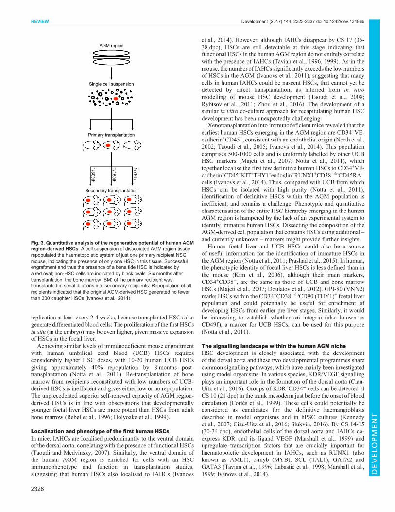
Fig. 2. Human embryonic haematopoietic tissues. (A) The location of the AGM region, yolk sac, liver, vitelline and umbilical arteries and placenta in aCS 15 human embryo. The location of the amnion, eye, and upper and lower limbs are also indicated for reference. Note that HSCs at this stage are localised to theAGM region and will appear in the yolk sac and liver slightly later. (B) Human IAHC development and the stage-specific spatial localisation of CD34+CD45+
cell clusters on the ventral wall of the dorsal aorta [indicated in red; modified from Tavian and Peault (2005)]. (C) VE-cadherin+CD45+RUNX1+ human IAHCs onthe ventral wall of the dorsal aorta of a CS 16 human embryo. Whole-mount antibody staining (left) and sagittal confocal section of the boxed area (right). Thelargest clusters are usually localised close to the entry of the vitelline artery. Scale bars: 1 mm in A; 0.05 mm in C.
2326
REVIEW Development (2017) 144, 2323-2337 doi:10.1242/dev.134866
DEVELO
PM
ENT
formation, supporting the idea that clonogenic haematopoieticprogenitors reside within these structures. The CD34+CD45−
population, however, is heterogeneous and may contain not onlyendothelial cells. Indeed, the endothelial-to-haematopoietictransition (EHT) is a multistep maturation process, which includescell intermediates fully committed to the haematopoietic fate(Taoudi et al., 2008; Medvinsky et al., 2011; Rybtsov et al., 2014).In the mouse embryo and differentiating ESCs, the earliesthaematopoietic progenitors express the haematopoietic markerCD41 (ITGA2B), and only later become CD45+ (Ferkowiczet al., 2003; Mikkola et al., 2003; Eilken et al., 2009; Lancrinet al., 2009). Similarly, the developing mouse HSC lineagesequentially upregulates CD41, CD43 and finally CD45, whichtogether define the different stages of HSC maturation (Rybtsovet al., 2014). Importantly, although these cell intermediates expressperhaps the most specific endothelial marker, VE-cadherin(cadherin 5), they are devoid of endothelial activity and are fullycommitted to the haematopoietic fate. During human ESCdifferentiation in vitro, CD43 is the first marker of haematopoieticspecification (Vodyanik et al., 2006). Therefore, apart fromendothelial cells, the human AGM-derived CD34+CD45−
population might also include haematopoietic progenitors, whichcould be responsible for haematopoietic activity in culture (Oberlinet al., 2002). Although some molecular mechanisms underlyingEHT transition have been described (Ciau-Uitz et al., 2016), exactlyhow haematopoietic progenitors and HSCs emerge from theembryonic endothelium, especially in the human, remains anissue for further investigation.
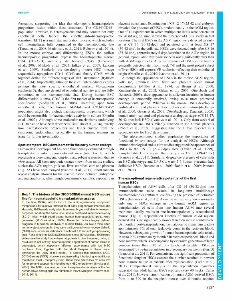
SpatiotemporalHSCdevelopment in theearly humanembryoHuman HSC development has been functionally evaluated throughtransplantation into immunodeficient mice (see Box 1), whichrepresents a more stringent, long-term and robust assessment than invitro assays. All haematopoietic tissues known from mouse studies,such as the AGM region, yolk sac, liver, umbilical cord and placenta(Fig. 2A) have been assayed (Ivanovs et al., 2011). Short tandemrepeat analysis allowed for the discrimination between embryonicand maternal cells, which might contaminate samples, especially in
placenta transplants. Examination of CS 12-17 (25-42 dpc) embryosrevealed the presence of HSCs predominantly in the AGM region.Out of 11 experiments in which multipotent HSCs were detected inthe AGM region, nine showed the presence of HSCs solely in thatlocation. The first HSCs in the AGM region were detected as earlyas at CS 14 (30-32 dpc) and persisted until at least CS 17(39-42 dpc). In the yolk sac, HSCs were detected only after CS 16(35-38 dpc), approximately 5 days later than in the AGM region. Ingeneral, repopulation with yolk sac cells was significantly rarer thanwith AGM region cells. A robust presence of HSCs in the liver isgenerally detected later, from week 7-8 and the most potent subsetof liver HSCs still express VE-cadherin, reflecting their endothelialorigin (Oberlin et al., 2010; Ivanovs et al., 2011).
Although the appearance of HSCs in the mouse AGM region,yolk sac, umbilical cord, liver and placenta occur almostconcurrently (Müller et al., 1994; de Bruijn et al., 2000;Kumaravelu et al., 2002; Gekas et al., 2005; Ottersbach andDzierzak, 2005), their appearance in different locations in humanembryo is temporally resolved, owing to a more protracteddevelopmental period. Whereas in the mouse HSCs develop inumbilical cord and placenta prior to liver colonisation (de Bruijnet al., 2000; Gekas et al., 2005; Ottersbach and Dzierzak, 2005),human umbilical cord and placenta at analogous stages (CS 14-17;30-42 dpc) lack HSCs (Ivanovs et al., 2011). Only from week 9 ofdevelopment are HSCs reliably detected in the human placenta(Robin et al., 2009), suggesting that the human placenta is asecondary site for HSC development.
The aforementioned studies emphasise the importance offunctional in vivo assays for the analysis of HSCs. Althoughimmunohistological and in vitro studies suggested the appearance ofHSCs in the CS 13 (27-29 dpc) liver (Tavian et al., 1999),transplantable HSCs appear there only after CS 17 (39-42 dpc)(Ivanovs et al., 2011). Similarly, despite the presence of cells withan HSC phenotype and CFU-Cs, week 5-6 human placentas lackdefinitive HSCs (Barcena et al., 2009; Robin et al., 2009; Ivanovset al., 2011).
The exceptional regenerative potential of the firsthuman HSCsTransplantation of AGM cells after CS 14 (30-32 dpc) intoimmunodeficient mice results in long-term multilineagehaematopoietic engraftment, confirming the presence of definitiveHSCs (Ivanovs et al., 2011). As in the mouse, very few – normallyonly one – HSCs emerge in the human AGM region, astransplantation of cells from one human AGM into severalrecipients usually results in one haematopoietically reconstitutedmouse (Fig. 3). Repopulation kinetics of human AGM region-derived HSCs are significantly slower than their mouse counterparts.By month 3 post-transplantation, haematopoietic chimerism reachesapproximately 1% of total leukocyte count in the recipient blood.However, subsequent growth of human haematopoietic cells resultsin up to 90% chimerism by month 8 in recipient peripheral blood andbone marrow, which is accompanied by extensive generation of largenumbers (more than 300) of fully functional daughter HSCs, asdetermined by re-transplantation into secondary recipients (Fig. 3)(Ivanovs et al., 2011). For comparison, this significant production offunctional daughter HSCs exceeds the number required to preventbone marrow failure in patients after myeloablation (Catlin et al.,2011). Computational analysis of the repopulation dynamicssuggested that adult human HSCs replicate every 40 weeks (Catlinet al., 2011). However, amplification of human AGM-derived HSCsfrom 1 to 300 in the recipient mouse over 6 months requires
Box 1. The history of the (NOD/SCID/Gamma) NSG mouseline for haematopoietic transplantation assaysIn the late 1980s, introduction of the antiprogestational compoundmifepristone for elective termination of early pregnancies (Vervest andHaspels, 1985) made early intact human embryos available for researchpurposes. At about the same time, severe combined immunodeficiency(SCID) mice, which could accept human haematopoietic grafts, weregenerated (McCune et al., 1988). These two factors largely definedprogress in functional analysis of human HSCs. As SCID mice oftenimmunoreject xenografts, they were backcrossed to non-obese diabetic(NOD)mice, which are deficient in functional T, B and antigen-presentingcells. For a long time, NOD/SCID recipient mice (Shultz et al., 1995) werewidely used to study human haematopoiesis in vivo. However, owing toresidual NK cell activity, haematopoietic engraftment of human HSCs isattenuated, which especially affected experiments with low HSCnumbers. This, together with the short lifespan of these mice,diminishes the utility of the model. To address this limitation, NOD/SCID/Gamma (NSG) mice were engineered by introducing an additionalmutation in the IL2 receptor γ chain. Thesemice, which lack NK cells, livefor longer and support high-level human HSC engraftment (Shultz et al.,2005). The NSG mice later permitted transplantation analysis of the firsthumanHSCs emerging in low numbers in the AGM region (Ivanovs et al.,2014, 2011).
2327
REVIEW Development (2017) 144, 2323-2337 doi:10.1242/dev.134866
DEVELO
PM
ENT
replication at least every 2-4 weeks, because transplanted HSCs alsogenerate differentiated blood cells. The proliferation of the first HSCsin situ (in the embryo) may be even higher, given massive expansionof HSCs in the foetal liver.Achieving similar levels of immunodeficient mouse engraftment
with human umbilical cord blood (UCB) HSCs requiresconsiderably higher HSC doses, with 10-20 human UCB HSCsgiving approximately 40% repopulation by 8 months post-transplantation (Notta et al., 2011). Re-transplantation of bonemarrow from recipients reconstituted with low numbers of UCB-derived HSCs is inefficient and gives either low or no repopulation.The unprecedented superior self-renewal capacity of AGM region-derived HSCs is in line with observations that developmentallyyounger foetal liver HSCs are more potent than HSCs from adultbone marrow (Rebel et al., 1996; Holyoake et al., 1999).
Localisation and phenotype of the first human HSCsIn mice, IAHCs are localised predominantly to the ventral domainof the dorsal aorta, correlating with the presence of functional HSCs(Taoudi and Medvinsky, 2007). Similarly, the ventral domain ofthe human AGM region is enriched for cells with an HSCimmunophenotype and function in transplantation studies,suggesting that human HSCs also localised to IAHCs (Ivanovs
et al., 2014). However, although IAHCs disappear by CS 17 (35-38 dpc), HSCs are still detectable at this stage indicating thatfunctional HSCs in the human AGM region do not entirely correlatewith the presence of IAHCs (Tavian et al., 1996, 1999). As in themouse, the number of IAHCs significantly exceeds the low numbersof HSCs in the AGM (Ivanovs et al., 2011), suggesting that manycells in human IAHCs could be nascent HSCs, that cannot yet bedetected by direct transplantation, as inferred from in vitromodelling of mouse HSC development (Taoudi et al., 2008;Rybtsov et al., 2011; Zhou et al., 2016). The development of asimilar in vitro co-culture approach for recapitulating human HSCdevelopment has been unexpectedly challenging.
Xenotransplantation into immunodeficient mice revealed that theearliest human HSCs emerging in the AGM region are CD34+VE-cadherin+CD45+, consistent with an endothelial origin (North et al.,2002; Taoudi et al., 2005; Ivanovs et al., 2014). This populationcomprises 500-1000 cells and is uniformly labelled by other UCBHSC markers (Majeti et al., 2007; Notta et al., 2011), whichtogether localise the first few definitive human HSCs to CD34+VE-cadherin+CD45+KIT+THY1+endoglin+RUNX1+CD38−/loCD45RA−
cells (Ivanovs et al., 2014). Thus, compared with UCB from whichHSCs can be isolated with high purity (Notta et al., 2011),identification of definitive HSCs within the AGM population isinefficient, and remains a challenge. Phenotypic and quantitativecharacterisation of the entire HSC hierarchy emerging in the humanAGM region is hampered by the lack of an experimental system toidentify immature human HSCs. Dissecting the composition of theAGM-derived cell population that contains HSCs using additional –and currently unknown – markers might provide further insights.
Human foetal liver and UCB HSCs could also be a sourceof useful information for the identification of immature HSCs inthe AGM region (Notta et al., 2011; Prashad et al., 2015). In human,the phenotypic identity of foetal liver HSCs is less defined than inthe mouse (Kim et al., 2006), although their main markers,CD34+CD38−, are the same as those of UCB and bone marrowHSCs (Majeti et al., 2007; Doulatov et al., 2012). GPI-80 (VNN2)marks HSCs within the CD34+CD38−/loCD90 (THY1)+ foetal liverpopulation and could potentially be useful for enrichment ofdeveloping HSCs from earlier pre-liver stages. Similarly, it wouldbe interesting to establish whether α6 integrin (also known asCD49f), a marker for UCB HSCs, can be used for this purpose(Notta et al., 2011).
The signalling landscape within the human AGM nicheHSC development is closely associated with the developmentof the dorsal aorta and these two developmental programmes sharecommon signalling pathways, which have mainly been investigatedusing model organisms. In various species, KDR/VEGF signallingplays an important role in the formation of the dorsal aorta (Ciau-Uitz et al., 2016). Groups of KDR+CD34− cells can be detected atCS 10 (21 dpc) in the trunk mesoderm just before the onset of bloodcirculation (Cortés et al., 1999). These cells could potentially beconsidered as candidates for the definitive haemangioblastsdescribed in model organisms and in hPSC cultures (Kennedyet al., 2007; Ciau-Uitz et al., 2016; Slukvin, 2016). By CS 14-15(30-34 dpc), endothelial cells of the dorsal aorta and IAHCs co-express KDR and its ligand VEGF (Marshall et al., 1999) andupregulate transcription factors that are crucially important forhaematopoietic development in IAHCs, such as RUNX1 (alsoknown as AML1), c-myb (MYB), SCL (TAL1), GATA2 andGATA3 (Tavian et al., 1996; Labastie et al., 1998; Marshall et al.,1999; Ivanovs et al., 2014).
AGM region
Single cell suspension
1/75th
1/150th
1/300th
Secondary transplantation
Primary transplantation
Fig. 3. Quantitative analysis of the regenerative potential of human AGMregion-derived HSCs. A cell suspension of dissociated AGM region tissuerepopulated the haematopoietic system of just one primary recipient NSGmouse, indicating the presence of only one HSC in this tissue. Successfulengraftment and thus the presence of a bona fide HSC is indicated bya red oval; non-HSC cells are indicated by black ovals. Six months aftertransplantation, the bone marrow (BM) of the primary recipient wastransplanted in serial dilutions into secondary recipients. Repopulation of allrecipients indicated that the original AGM-derived HSC generated no fewerthan 300 daughter HSCs (Ivanovs et al., 2011).
2328
REVIEW Development (2017) 144, 2323-2337 doi:10.1242/dev.134866
DEVELO
PM
ENT

Ventral polarisation in IAHC and HSC development in modelorganisms is at least partly driven by spatial asymmetry of molecularsignalling in the AGM region (Wilkinson et al., 2009; Souilholet al., 2016), suggesting similar mechanisms in human. From CS 13(28 dpc), BMP4 expression is observed ventrally in a thinsubendothelial mesenchymal layer of the dorsal aorta, whichtransiently expands and subsequently disappears by CS 16 (38 dpc).At this stage, functionally defined HSCs are still present in theAGM region (Ivanovs et al., 2011). BMP4 is a negative regulator ofHSCmaturation: although it is involved in the formation of the HSCniche, its direct action on HSCs is likely limited by BMP antagonists(Souilhol et al., 2016). Notably, BMP4 can induce expression oftenascin C and fibronectin (Molloy et al., 2008), two extracellularmatrix molecules that are also ventrally polarised in the humanAGM region (Marshall et al., 1999). Tenascin C promoteshaematoendothelial development of hESCs (Uenishi et al., 2014)and its deficiency impairs mouse bone marrow cultures, which canbe rescued by fibronectin (Ohta et al., 1998). TGFβ, another well-known modulator of the extracellular matrix, is expressed in closeproximity to IAHCs at the same time as BMP4 expression isdownregulated. It has been proposed that TGFβ could participate inrearrangement of the niche and restriction of HSC expansion prior tofoetal liver colonisation (Marshall et al., 2000).Signalling through KIT is important for HSC maturation and its
ligand, SCF (KITL in mouse; KITLG in human), is ventrallypolarised in the mouse AGM region (Rybtsov et al., 2014; Souilholet al., 2016). KIT is expressed in IAHCs and in the first functionalHSCs emerging in the human embryo (Labastie et al., 1998;Ivanovs et al., 2014). BMP4 can upregulate KIT in various tissuesand, through upregulation of KIT in human aortic endothelium, itmay potentially facilitate HSC initiation (Marshall et al., 2007).However, further HSC development may require suppression ofBMP4 signalling, as described in the mouse (Souilhol et al., 2016).FLT3, which marks mouse embryonic HSC precursors (Boyer et al.,2011), and its ligand are also expressed in IAHCs and surroundingendothelium, and may be involved in human HSC maturation(Marshall et al., 1999).
Adhesion and migrationHSC development is associated with cell migration and IAHCs aremarked by a range of adhesion molecules that potentially play a rolein this process. CD34 and its receptor L-selectin (CD62L; SELL)are involved in cell adhesion processes including lymphocyte-endothelial interactions (Rosen, 2004), and are expressed inhuman embryonic endothelium, IAHCs and functional HSCs(Tavian et al., 1996; Ivanovs et al., 2014). The first lineage-restricted haematopoietic marker, SPN, appears during hPSCdifferentiation (Vodyanik et al., 2006) and can mediate adhesionthrough E-selectin receptor (SELE) (Matsumoto et al., 2005). CD34and CD43 can cooperatively define adhesive behaviour ofhaematopoietic cells (Drew et al., 2005). Other surface moleculesinvolved in cell adhesion – ALCAM, VE-cadherin, CD44, CD164and VCAM1 – are also expressed in IAHCs, endothelial cells of thedorsal aorta and sometimes the mesenchymal cells underneathIAHCs (Cortés et al., 1999;Watt et al., 2000;Marshall, 2006; Ivanovset al., 2014). Given the role of the TNF/lymphotoxin/NFκB pro-inflammatory pathway in early HSC development described in modelorganisms (Espín-Palazón et al., 2014; Li et al., 2014) and its impacton VCAM1, L- and E-selectin expression (Suna et al., 2008), it isconceivable that complex and finely regulated adhesion dynamicsplay important roles in human HSC development. However, it islikely that additional signalling pathways described in other model
organisms (Ciau-Uitz et al., 2016) also play a role in human HSCdevelopment. Modelling HSC development in vitro using PSCs andother approaches could shed light on this issue.
Recapitulation of humanhaematopoietic development usingpluripotent stem cellsThis section examines parallels between the in vitro haematopoieticdifferentiation of hPSCs and aspects of haematopoietic developmentin the human embryo. Rather than replicating recent reviews thatcomprehensively discuss protocols for the differentiation of hPSCsintoHSCs or progenitors (Ditadi et al., 2016; Slukvin, 2016;Wahlsterand Daley, 2016), we have structured this section to highlight theextent to which in vitro differentiation reflects human haematopoieticdevelopment (Fig. 4). We argue that ongoing efforts to generateHSCs from PSCs in vitro are both informed by and add to ourunderstanding of the signalling events that underpin in vivo humanhaematopoietic development.
Mesoderm induction and patterningIn the mammalian embryo, ingression of cells through the primitivestreak during gastrulation generates mesoderm, which colonises theyolk sac (extra-embryonic mesoderm) and intra-embryonic sitesincluding paraxial, intermediate and lateral plate mesoderm (Tamand Behringer, 1997). Whereas extra-embryonic mesoderm givesrise to transient yolk sac haematopoiesis, lateral plate mesodermadjacent to the paraxial mesoderm (the splanchnopleura) formsbilateral mesodermal strips that converge to the midline where theycoalesce to form the dorsal aorta. The growth factors involved inmesoderm specification, dorsal aorta formation and haematopoieticdevelopment include members of the FGF, WNT, TGFβ, retinoicacid, Hedgehog and Notch signalling pathways (Lawson et al.,2001; Gering and Patient, 2005; Chanda et al., 2013; Lizama et al.,2015; Ciau-Uitz et al., 2016).
In culture, as in vivo, mesoderm induction and pattering ismediated by the cooperative actions of highly conserved FGF,BMP4, activin and canonical WNT signals (Schier and Shen, 2000;Kimelman, 2006; Nostro et al., 2008; Woll et al., 2008; Wang andNakayama, 2009; Bernardo et al., 2011; Yu et al., 2011). Mesodermpatterned to a haematopoietic fate is marked by the expression ofprimitive streak genes, such as the transcription factor genesbrachyury (T), MIXL1, FOXF1, as well as cell surface receptorsKDR and PDGFRA (Davis et al., 2008; Ditadi et al., 2016; Slukvin,2016). An early study showed that the most efficient production ofhaematopoietic cells resulted from the exposure of the emergingmesoderm to activin and BMP4 signalling (Nostro et al., 2008),analogous to the posterior primitive streak from whence the firstyolk sac haematopoietic progenitors are derived. Arguably, thismight explain why the haematopoietic output also resembled thatof the yolk sac and did not include long-term HSCs. It was onlylater that a series of studies shed light on the role of mesodermpatterning for both the suppression of primitive haematopoiesisand for the support of definitive haematopoietic lineages.Following an initial period of BMP4-based mesoderm induction,brief inhibition of activin (Kennedy et al., 2012), or provision of aWNT agonist (Gertow et al., 2013) were sufficient to inhibit theerythroid-biased primitive programme. Importantly, activininhibition (Kennedy et al., 2012), WNT stimulation (Sturgeonet al., 2014) or a combination of the two (Ng et al., 2016) from days2-4 of in vitro differentiation also resulted in a bias towardsdefinitive haematopoietic lineages, as defined by the capacity togenerate T lymphocytes (Kennedy et al., 2012; Sturgeon et al.,2014).
2329
REVIEW Development (2017) 144, 2323-2337 doi:10.1242/dev.134866
DEVELO
PM
ENT

The selective expression of HOXA genes in human foetal liverand UCB-derived haematopoietic progenitor and stem cells clearlydistinguished them from PSC-derived haematopoietic progenitorsgenerated using conventional mesoderm induction protocols(Dou et al., 2016; Ng et al., 2016). HOX genes mark axialposition during development and are first expressed at theprimitive streak stage (Deschamps and van Nes, 2005).Therefore, it was relevant that activin inhibition and WNTstimulation during mesoderm patterning, alone and incombination, increased the expression of the CDX genes,leading to sustained expression of their target HOXA genes andmore closely mimicking the HOXA expression signature seen infoetal liver and cord blood cells (Ng et al., 2016). These datasupport the premise that allocation of cells to extra- and intra-embryonic haematopoietic fates is initiated during mesodermpatterning, and that this is reflected in the selective expression ofHOXA genes in definitively patterned mesoderm (Fig. 4).
The generation of yolk sac-like haematopoietic lineagesThe emergence of FGF2- and VEGF-dependent blast colonies fromBMP4-induced mesoderm after 2-4 days of differentiation marksthe onset of haematopoiesis in human hPSC cultures (Kennedyet al., 2007; Davis et al., 2008; Vodyanik et al., 2010). As well as thesurface markers KDR and PDGFRα, blast colony-forming cells(BL-CFCs) also express the apelin receptor (Vodyanik et al., 2010;Choi et al., 2012; Yu et al., 2012), and CD235a (glycophorin A)(Sturgeon et al., 2014). As in the mouse (Huber et al., 2004), humanblast colonies give rise to haematopoietic, endothelial and smoothmuscle lineages (Yu et al., 2012). Haematopoietic differentiation ofblast colonies is limited to primitive erythrocytes, megakaryocytesand macrophages, and, as in the mouse (Lancrin et al., 2009),probably occurs via a haematogenic endothelial cell precursor.
Further differentiation of hPSCs along a yolk sac-type path can beachieved by co-culture with mouse OP9 stromal cells (Vodyaniket al., 2006), as embryoid bodies (Pick et al., 2007; Nostro et al.,2008) or monolayer cultures (Uenishi et al., 2014) in serum-freemedia supplemented with growth factors, usually including VEGF,SCF and FGF2. Endothelial and haematopoietic cell surface markersare sequentially acquired, starting with CD34 after 4-6 days, andfollowed by CD43 at day 6-8, the expression of which firstdistinguishes haematopoietic from endothelial lineages (Vodyaniket al., 2006). The formation of these blood cells from an endothelialintermediate indicates that the endothelial-to-haematopoietictransition is a common mechanism for blood formation both in theembryo and in differentiating hPSCs and is not limited to a particulardevelopmental stage.
The CD34+CD43+ haematopoietic progenitors give rise to abroad range of erythroid and myeloid lineages (Choi et al., 2012;Kennedy et al., 2012; Ng et al., 2016), reminiscent of the broadpotential of the yolk sac-derived erythro-myeloid progenitor (EMP)described in the mouse that transiently seeds the foetal liver(McGrath et al., 2015). Although EMP-type haematopoiesis has notbeen formally characterised in human embryos, kinetic studiesreport a rapid transition of clonogenic cells from the yolk sac to thefoetal liver at around 5 weeks of gestation (Migliaccio et al., 1986).Given that at this time the human foetal liver does not yet possessrepopulating ability (Ivanovs et al., 2011), the cells seeding the earlyfoetal liver are likely to include yolk sac-derived EMPs, and the yolksac-like erythroid and myeloid cells derived from hPSCs may betheir human in vitro equivalent. Whether these EMP-type cells aredefined as a second ‘primitive’ or a first ‘definitive’ haematopoieticwave is somewhat moot. For the moment, the descriptor ‘EMP’maybe sufficient. We do point out, however, that in contrast tohaematopoietic cells derived from the AGM region, foetal liver or
HOXA+HOXA–
Intra-embryonichaematopoiesis
B* progenitors
B lymphocytes
Yolk sac EMPs
T progenitors
Foetalerythroid andmyeloid cells
T lymphocytes
Pre-HSCs
HSCs
B-1 and B-2progenitors
T progenitors Multipotent
myeloidprogenitors
Pluripotentstem cells
Mesodermd2-4 SB
CHIR BL-CFCs
Embryonic erythrocytes andmegakaryocytes
Macrophages
Yolk sac-like HEd7-9 AGM EMPs
Extra-embryonichaematopoiesis
AGM-like HEd9-18
Foetalerythroid andmyeloid cells
Fig. 4. Model of in vitro haematopoieticdifferentiation from human pluripotent stemcells. Separation between extra-embryonic, yolksac-like haematopoiesis (blue shading and arrows)and intra-embryonic AGM-like haematopoiesis(red shading and arrows) initially occurs at thetime of mesoderm patterning [days (d) 2-4 ofdifferentiation]. WNT agonists (CHIR99021,denoted CHIR) and activin antagonists(SB431542, denoted SB) orchestrate this switch,although other molecules, such as retinoic acid,may also play a role. A key difference between theextra- and intra-embryonic programmes is theexpression of a specific combination of HOXAgenes that is restricted to the endothelia andhaematopoietic progenitors of intra-embryonichaematopoiesis. The extra-embryonic programmegenerates blast colony-forming cells (BL-CFCs),which are suppressed in the intra-embryonicprogramme. In both programmes, haemogenicendothelia (HE) generate erytho-myeloidprogenitors (EMPs) and T lymphocytes. Thus far, Bcells have been generated from an extra-embryonictype protocol although it is anticipated that similar Bcells can be derived from AGM-like HE (dashed redarrow). The asterisk indicates that it is not knownwhether the B cells generated from hPSCs are B-1or B-2 type. The intra-embryonic AGM-like HEyields cells that have pre-HSC characteristics.Although HSCs and their progeny are predicted(greyed arrows and images), these have yet tobe formally shown.
2330
REVIEW Development (2017) 144, 2323-2337 doi:10.1242/dev.134866
DEVELO
PM
ENT

other embryonic sites, hPSC-derived EMP-like cells do not expressHOXA genes (Dou et al., 2016; Ng et al., 2016).
The generation of AGM-like haematopoietic lineages fromhaematogenic endotheliumAs discussed above, the appearance of IAHCs in the AGM region,which arise from a transient haematogenic endothelium in the dorsalaorta, predicts the emergence of the first stem cells that acquirerepopulating activity in the vertebrate embryo. In contrast tonon-haematogenic vascular cells, hPSC-derived haematogenicendothelium lacks expression of CD73 (NT5E) (Choi et al., 2012;Ditadi et al., 2015), CXCR4 and DLL4 (Ditadi et al., 2015). Also,both mouse (North et al., 1999; Thambyrajah et al., 2016) andhuman (Choi et al., 2012; Ng et al., 2016) haematogenicendothelium are distinguished by their expression of the keytranscription factors RUNX1 and GFI1, which are required for theendothelial to haematopoietic transition. Interestingly, hPSCsdifferentiated with a protocol that induces HOXA gene expressionproduce SOX17-expressing endothelial cells that bear similarity atthe transcriptional level to developing dorsal aorta endothelium, anda SOX17dull haematogenic endothelial population that efficientlygenerates haematopoietic cells (Ng et al., 2016). The low level ofSOX17 in haematogenic cells is consistent with literature reportingthe downregulation of SOX17 during the EHT (Clarke et al., 2013;Nobuhisa et al., 2014). Similarly, single-cell analysis of hPSC-derived cells documented a downregulation of SOX17 at the EHTpoint (Guibentif et al., 2017).It has not been possible to identify cell surface markers that
distinguish true or ‘definitive’ AGM-derived HSCs from yolk sac-type, non-engrafting haematopoietic progenitors (Ditadi et al.,2016; Slukvin, 2016). For this reason, considerable attention hasbeen paid to the generation of lymphocytes from hPSCs in vitro,arguing that these lineages represent definitive haematopoiesis andmay indicate culture conditions that support the development ofAGM-like HSCs. Many laboratories have generated T cells fromhPSCs, in a Notch-dependent fashion, from endothelial precursorspresent at 7-9 days of differentiation (Timmermans et al., 2009;Kennedy et al., 2012; Uenishi et al., 2014; Ditadi et al., 2015; Douet al., 2016; Ng et al., 2016). The dependence of AGM-type, but notyolk sac-type haematopoiesis on Notch signals in the mouse embryo(Kumano et al., 2003; Hadland et al., 2004) has been used to furtherstrengthen the case that the hPSC-derived T cells are progeny of anintra-embryonic haematopoietic progenitor. An important caveat,however, is the realisation that, in the mouse, both T- and B-cellpotential is present in the pre-AGM stage yolk sac (Yoshimoto et al.,2011, 2012; McGrath et al., 2015). The dependence of human yolksac-derived EMP or lymphoid cells on Notch signals is not known.Unlike the relative ease in which T cells can be derived, B-cell
development from hPSCs has been infrequently reported. Onelaboratory described the generation of cells with a CD19+CD10(MME)+ pre-B-cell phenotype, some of which matured to surfaceIgM+ immature B cells (French et al., 2015).To place this in context, during mouse development there are
several waves of B-cell production (Herzenberg and Herzenberg,1989). The first innate type B cells, called B-1 cells, ariseindependently from haematogenic endothelia in both the yolk sacand the AGM region, prior to HSCs, and seed the foetal liver(Yoshimoto, 2015). Subsequent waves of HSC-derived B-1 andadaptive B-2 lymphocytes arise later in the foetal liver (Yoshimoto,2015) and a wave of predominantly HSC-derived B-2 lymphocytesemerges in the bone marrow (Montecino-Rodriguez and Dorshkind,2012). The identification of B-1 lymphocytes in humans (Rothstein
and Quach, 2015), and their observation in the 10-11 week foetalliver (Bueno et al., 2016), argues for conservation of the innateimmune system between species. Given that human B-1 cells differimmunophenotypically from B-2 lymphocytes (Rothstein andQuach, 2015), it should therefore be possible to discern whetherB cells derived from human hPSCs include B-2 cells, and are thusunequivocally descended from HSCs.
The pattern of globin chain expression by hPSC-derived erythoidcells has also served as an indicator of developmental stage.Erythroid cells differentiated from human PSCs usually expressε- and γ-globins (Chang et al., 2006; Lu et al., 2008; Hatzistavrouet al., 2009; Dias et al., 2011), although prolonged culture in humanplasma or on stromal layers has yielded predominantly γ- or γ- andβ-globin-expressing cells (Qiu et al., 2008; Lapillonne et al., 2010;Yang et al., 2014; Fujita et al., 2016). Importantly, the switch toβ-globin synthesis occurs perinatally, and thus is not expected atthe AGM or foetal liver stages of haematopoiesis. Rather,focus is on the anticipated downregulation of embryonic ε- andζ-globins that signifies exit from primitive haematopoiesis. As afurther complication, yolk sac EMP-derived erythroid precursors inmice carrying a single copy of the human β-globin locusdownregulate ε-globin and express low levels of β-globin whenmatured in vitro (McGrath et al., 2011). As with the generation oflymphoid cells, these findings indicate that it is not easy to excludeyolk sac EMP haematopoiesis as a source of erythroid cells thatappear to derive from foetal liver because they have suppressedembryonic globins.
Finally, although EMPs are traditionally associated with the yolksac, cells with a similar restricted developmental capacity couldconceivably derive from the AGM haematogenic endotheliumalso. Consistent with this hypothesis, clonogenic progenitors(approximately 90 colonies per embryo) are present in the VE-cadherin+CD45+ population in the human AGM region, the samefraction harbouring less frequent pre-HSCs and HSCs (Ivanovset al., 2014). These clonogenic cells might also seed the early foetalliver, and might only be distinguishable from yolk sac-derivedEMPs by their expression of HOXA genes. Although furtherexperiments will be required as proof, there is support for this idea ina recent report of ε- to γ-globin switching and downregulation of ζ-globin in clonogenic cells derived from HOXA+ cultures (Ng et al.,2016).
Transcriptional comparisons of cells emerging from hPSC-derived HOXA+ haematogenic endothelium and AGM-derivedstem progenitors has revealed a high degree of similarity across abroad range of haematopoietic cell surface markers, transcriptionfactors and signalling molecules (Ng et al., 2016). Nevertheless,the hPSC-derived progenitors lack some essential characteristicsthat enable long-term haematopoietic repopulation. Althoughmesoderm patterning with modulation of WNT and activinsignals increases HOXA expression, the pattern of expression inthe hPSC-derived progenitors differs subtly from that observed inhuman AGM samples, with reduced expression of anterior HOXAgenes and elevated levels of the most posterior HOXA genes(Ng et al., 2016).
Alternative strategies to explore human haematopoieticdevelopmentTeratoma formation as a model for human HSC developmentIn vivo teratoma formation may offer valuable insights into thegeneration of transplantable HSCs from hPSCs. Teratomas, whichcontain derivatives of all three germ layers, can be formed afterectopic engraftment of PSCs into a host animal. The mixture of cell
2331
REVIEW Development (2017) 144, 2323-2337 doi:10.1242/dev.134866
DEVELO
PM
ENT

types in these tumours can preserve some developmentallyimportant inductive interactions, leading to the formation ofdisorganised tissues and organs, including bone marrow (Hentzeet al., 2009; Amabile et al., 2013). Teratomas formed from hPSCsengrafted into immunodeficient mice, in some cases, generatehuman CD45+ cells that home to bone marrow, circulate in therecipient peripheral blood and colonise lymphoid tissues (Amabileet al., 2013; Suzuki et al., 2013). Transplantation of total bonemarrow or sorted human CD34+CD45+ bone marrow cellsfrom primary into secondary recipients shows donor-derivedmultilineage peripheral blood chimerism, comparable to UCBrepopulation (Amabile et al., 2013; Suzuki et al., 2013). Teratoma-derived B and T cells appear functionally normal, and erythrocytesexpress adult haemoglobins, which is not usually achievable duringin vitro hPSC differentiation. It is conceivable that in vivo teratomaformation might more faithfully recapitulate some of the processesthat occur during embryo development and, in some cases, thiscould include the specification of an AGM-like region and lead tothe production of HSCs. However, an extended period ofobservation is required to determine whether these HSC-like cellshave a truly long-term haematopoietic potential (Ivanovs et al.,2011; Notta et al., 2011).
Direct cell lineage conversion: programming and reprogrammingto bloodAlthough enforced gene expression has been shown to be apowerful tool for altering cell fate for several decades (Davis et al.,1987; Kulessa et al., 1995), the real boost to the generation of bloodcells using this approach was precipitated by reprogramming studiesin which fibroblasts were reverted back to pluripotency using fourtranscription factors (Takahashi and Yamanaka, 2006; Pereira et al.,2012). These studies spawned quests for combinations oftranscription factors, overexpression of which could generateclinically relevant cell types, including HSCs. Groups choseeither ESCs (Kyba et al., 2002; Elcheva et al., 2014), humanendothelial cells (Sandler et al., 2014), haematopoietic cells(Doulatov et al., 2013) or fibroblasts (Pereira et al., 2013; Battaet al., 2014) as the starting material. Comparative transcriptomics ofHSC and progenitor cell populations often provided lists ofcandidate factors to overexpress. Although many of the factorsused in reprogramming studies have previously been implicated indevelopmental haematopoiesis, their true ‘haematopoietic potency’becomes unveiled when they are expressed in a foreign cellularcontext, such as when reprogramming fibroblasts (Pereira et al.,2013; Batta et al., 2014) or undifferentiated PSCs (Elcheva et al.,2014; Vereide et al., 2014) (see Fig. 5 for details).Using an hPSC differentiation model, the potency of two pairs of
transcription factors, ETV2/GATA2 and GATA2/SCL, has beenshown in activating pan-myeloid or erythro-megakaryocyticprogrammes, respectively (Elcheva et al., 2014). These resultsreflect the importance of these factors as haematopoietic fate-determining transcription factors (Elcheva et al., 2014). Althoughthis system failed to generate long-term repopulating cells, it is apowerful tool for the dissection of haematopoietic developmentalpathways. In separate studies, a HOXA9/ERG/RORA/SOX4/MYBcombination of transcription factors was identified by comparing ahuman HSC-enriched population with more mature progenitorsfrom UCB (Doulatov et al., 2010; Laurenti et al., 2013).Overexpression of this combination of transcription factors inhPSC-derived haematopoietic progenitors conferred transientin vivo engraftment capacity (Doulatov et al., 2013). The datasuggested that HOXA9, ERG, RORA, SOX4 and MYB belong to
the same molecular regulatory network; this is supported by theobservation that HOXA9 occupies Erg, Sox4 and Myb promoters(Huang et al., 2012). The interest in analysis of HOX genes reflectstheir strong positive impact on expansion of mouse bone marrowHSCs (Antonchuk et al., 2002; Argiropoulos and Humphries,2007). Moreover, collinear activation of HOX genes is involved intemporal regionalisation of the mesoderm (Iimura and Pourquié,2006), which may underlie functional differences in sequentiallyemerging haematopoietic waves. For example, HoxB4overexpression along with adjusted Notch signalling confersmultilineage long-term repopulation ability to mouse ESCs (Luet al., 2016). Downregulation of HOXA5 and HOXA7 in humanfoetal liver HSCs supresses their capacity to self-renew andrepopulate NSG recipients. Although, forced expression ofHOXA5, HOXA7 and HOXA9 alone or in combination inHOXA-negative hPSC-derived progenitors was insufficient toconvert the progenitors to HSCs (Dou et al., 2016), a recent studyreported a greater degree of success by using a larger cohort of genes(Sugimura et al., 2017). Induced expression of seven transcriptionfactors, notably including three HOXA genes, successfullyconverted hPSC-derived haematogenic endothelium into long-term repopulating HSCs (Sugimura et al., 2017). Finally, expressionof four transcription factors (FOSB, GFI1, RUNX1 and SPI1) inhuman umbilical vein and dermal microvascular endothelial cells,or in adult mouse endothelial cells, were also sufficient to generateimmunocompetent HSCs (Sandler et al., 2014; Lis et al., 2017). Inall these studies, using transcription factors to manipulate cellularidentity has provided insights into the regulation of normalhaematopoietic development (Fig. 5). For example, successfulreprogramming of fibroblasts into blood allowed, through obtaininga molecular signature, identification of precursors of long-termrepopulating cells in the mouse placenta (Pereira et al., 2016).
Future directions and challengesUnderstanding the development of the human haematopoieticsystem is of fundamental biological and clinical importance.Various haematopoietic diseases originate prenatally (Hungeret al., 1998; Greaves, 2005; Hong et al., 2008; Roy et al., 2012;Barrett et al., 2016), and oncogenic mutations in developinghaematopoietic progenitors or stem cells can lead to pre-leukaemicchanges that may not be manifested if the oncogene is activated inthe adult counterparts (Barrett et al., 2016). The molecularmechanisms that regulate development and maintenance ofhaematopoietic progenitor and stem cells in the embryo differsignificantly from those in the adult, and the functionalheterogeneity that appears within the emerging HSC populationduring development further complicates our understanding of HSCregulation (Copley et al., 2012; Crisan et al., 2015; Beaudin et al.,2016). Although the impetus to study human haematopoieticdevelopment is clear, poor availability and heterogeneity of humanembryonic material makes the analyses challenging. Directextrapolation of results obtained from model organisms is notalways appropriate owing to species-specific differences, althoughmajor genetic networks and sites of haematopoiesis are significantlyconserved. Furthermore, for ethical and practical reasons, studyingthe period of early implantation in the human is extremely difficult.Thus, this highly relevant developmental stage remains largelyinaccessible and so cannot inform protocols for derivation of HSCsfrom PSCs. Additionally, until recently, PSC differentiation wasbiased towards transitory yolk sac haematopoiesis, resulting in thefailure of this in vitromodel to fully inform us of the mechanisms ofHSC development in vivo. Thus, deficiency in our in vivo
2332
REVIEW Development (2017) 144, 2323-2337 doi:10.1242/dev.134866
DEVELO
PM
ENT

knowledge has not yet been compensated for by information fromthe in vitro system, and vice versa.In our opinion, two approaches may be taken to address this
problem. Notwithstanding their limitations, it is important to continueto use model organisms, which remain virtually unrestricted resourcesin which to pursue detailed analyses of HSC development. Althoughnot free from ethical and practical considerations, research using non-human primates may also provide valuable information that will helpto bridge the evolutionary gap between the mouse and human(D’Souza et al., 2016).A second approach is the continued use of reprogramming to
investigate key determinants of cell fate. Although reprogrammingseems like a bold approach to generating cells of interest, it couldpotentially inform us about events that occur in vivo, provided that itgoes beyond describing acquisition of markers and extends tofunctional characteristics. Research in this direction should aim toidentify the best cell sources and factors for producing normal,functional HSCs. To date, the selection of genes for haematopoieticreprogramming has been based on genes known to be essential forhaematopoietic development and genes that are differentiallyexpressed in bone marrow/foetal liver/UCB HSCs compared withprogenitor cells. However, the genes that drive the formation of thefirst exceptionally potent HSCs that emerge in the human AGMregion (Ivanovs et al., 2011) remain unknown. Identification of
these genes in human requires more detailed reconstruction of thedeveloping HSC hierarchy, which is currently limited by lack of anappropriate experimental system in human, but could be facilitatedby further in-depth analysis of model organisms. Although the datashow a lack of definitive HSCs in human placenta and large extra-embryonic arteries, they should not be excluded from considerationas they could harbour immature HSCs.
The presence of one or more transgenes in the genome ofreprogrammed human HSCs represents a clinical risk, and furthermakes it difficult to assess the entire scale of molecular changes inthe reprogrammed cells. Reprogramming methods that do notinvolve transgene integration, such as non-integrating vectors andRNA, will therefore be highly desirable. Whether this approach,which has proved suitable for iPSC generation, will work forreprogramming into HSCs will depend on the activation of crucialendogenous genes that act as ‘molecular switches’ to produce aphenotypically stable cell type, long after the non-integrating factorsare degraded or diluted. This is particularly important for conferringstemness to progenitor cells, but so far it is not clear whether thisapproach will be successful. Ultimately, the safest HSCs generatedin the dish would likely be genetically unaltered cells, producedby exposure to external factors equivalent to the embryonic cuesthat drive HSC development. Here, the bottleneck seems to beidentifying the combinations of soluble and cell-based factors and
ESCs/iPSCs Mesoderm
Haematogenicendothelium
iHSCs
MPPs
CMPs CLPs
GATA2, SOX17,SCL, LMO2, MYCN,
PITX2
HOXA9, ERG,RORA, SOX4,
MYB
Pro-B cells
GATA2, ETV2
GATA2,RUNX1C,
SCL,LMO2, ERG
GATA2,FOS,ETV6,GFI1B Adult
fibroblasts
HOXB4,low Notch
Haemangioblastsg
i
HUVECs
RUNX1,FOSB,GFI1, SPI1
+ E4-ORF1 cells
RUNX1, SCL,LMO2, BMI1
SOX2,MIR125B
Macrophages
ETO, LMO2,HLF,PBX1,
ZFP37,PRDM5,
MYCN, MEIS1
1
2
3
4
5
6
78
9
10
RUNX1, HOXA5,HOXA9, HOXA10,ERG, LCOR, SPI1 12
Adultendothelium
11
Haemangioblast
Haematogenicendothelium
iHSCs
RUNX1,FOSB,GFI1, SPI1
+ E4-ORF1 cells
Key
Fig. 5. Programming and reprogramming towards haematopoietic fate. In vitro generation of haematopoietic cells usually occurs via either directeddifferentiation of PSCs (green arrows), or via transcription factor-based reprogramming of a variety of different starting cell types (blue boxes). These includeESCs and iPSCs, human umbilical vein endothelial cells (HUVECs), adult fibroblasts, haematogenic endothelium cells, and pro-B cells. Transcription factorcombinations used for reprogramming as well as the direction of reprogramming are shown in red (human) and purple (mouse). ETO is also known as RUNX1T1.References are indicated: (1) (Vereide et al., 2014); (2) (Kyba et al., 2002; Lu et al., 2016); (3) (Doulatov et al., 2013); (4) (Elcheva et al., 2014); note that GATA2/SCL combination induces erythro-megakaryocytic differentiation (not shown); (5) (Sandler et al., 2014); (6) (Riddell et al., 2014); (7) (Pereira et al., 2013);(8) (Batta et al., 2014); (9) (Cheng et al., 2016); (10) (Pulecio et al., 2014); (11) (Lis et al., 2017); (12) (Sugimura et al., 2017). CLPs, common lymphoid progenitors;CMPs, common myeloid progenitors; MPPs, multipotent progenitors.
2333
REVIEW Development (2017) 144, 2323-2337 doi:10.1242/dev.134866
DEVELO
PM
ENT

inhibitors that will produce bona fide long-term repopulating humanHSCs and again, analysis of mouse and other model organisms aswell as reprogramming studies may shed light on this problem. Thegeneration of reporter hPSC lines will continue to be a powerfulmethodology for analysing human HSC development (Rafii et al.,2013; Jung et al., 2016; Ng et al., 2016). Ultimately, the depth ofour understanding of human HSC development will be measured byour ability to faithfully recapitulate in vitro the embryonic pathwaythat leads to HSC formation. This is what will pave the way toinformative in vitro modelling of childhood haematologicalpathologies, and to the robust generation of bona fide HSCs forpossible clinical use.
AcknowledgementsThe authors would like to thank Prof. I. Slukvin (Madison) for helpful commentson hPSC differentiation; Dr D. Paruzina for providing images for Fig. 2C;Dr S. Gordon-Keylock for helpful comments and assistance during preparationof this manuscript.
Competing interestsThe authors declare no competing or financial interests.
FundingThis work was supported by Bloodwise (formerly LLR) and Medical ResearchCouncil (UK). E.G.S. and A.G.E. are Research Fellows of the National Health andMedical Research Council (Australia). Work in the laboratories of E.G.S. and A.G.E.is supported by the National Health and Medical Research Council (Australia), StemCells Australia and the Children’s Cancer Foundation.
ReferencesAmabile, G., Welner, R. S., Nombela-Arrieta, C., D’Alise, A. M., Di Ruscio, A.,Ebralidze, A. K., Kraytsberg, Y., Ye, M., Kocher, O., Neuberg, D. S. et al.(2013). In vivo generation of transplantable human hematopoietic cells frominduced pluripotent stem cells. Blood 121, 1255-1264.
Antonchuk, J., Sauvageau, G. and Humphries, R. K. (2002). HOXB4-inducedexpansion of adult hematopoietic stem cells ex vivo. Cell 109, 39-45.
Argiropoulos, B. and Humphries, R. K. (2007). Hox genes in hematopoiesis andleukemogenesis. Oncogene 26, 6766-6776.
Barcena, A., Muench, M. O., Kapidzic, M. and Fisher, S. J. (2009). A new role forthe human placenta as a hematopoietic site throughout gestation.Reprod. Sci. 16,178-187.
Barrett, N. A., Malouf, C., Kapeni, C., Bacon, W. A., Giotopoulos, G., Jacobsen,S. E. W., Huntly, B. J. and Ottersbach, K. (2016). Mll-AF4 confers enhancedself-renewal and lymphoid potential during a restricted window in development.Cell Rep. 16, 1039-1054.
Batta, K., Florkowska, M., Kouskoff, V. and Lacaud, G. (2014). Directreprogramming of murine fibroblasts to hematopoietic progenitor cells. Cell Rep.9, 1871-1884.
Beaudin, A. E., Boyer, S. W., Perez-Cunningham, J., Hernandez, G. E.,Derderian, S. C., Jujjavarapu, C., Aaserude, E., MacKenzie, T. andForsberg, E. C. (2016). A transient developmental hematopoietic stem cellgives rise to innate-like B and T cells. Cell Stem Cell 19, 768-783.
Bernardo, A. S., Faial, T., Gardner, L., Niakan, K. K., Ortmann, D., Senner, C. E.,Callery, E. M., Trotter, M. W., Hemberger, M., Smith, J. C. et al. (2011).BRACHYURY and CDX2 mediate BMP-induced differentiation of human andmouse pluripotent stem cells into embryonic and extraembryonic lineages. CellStem Cell 9, 144-155.
Bertrand, J. Y., Giroux, S., Golub, R., Klaine, M., Jalil, A., Boucontet, L., Godin, I.and Cumano, A. (2005). Characterization of purified intraembryonichematopoietic stem cells as a tool to define their site of origin. Proc. Natl. Acad.Sci. USA 102, 134-139.
Bertrand, J. Y., Chi, N. C., Santoso, B., Teng, S., Stainier, D. Y. R. and Traver, D.(2010). Haematopoietic stem cells derive directly from aortic endothelium duringdevelopment. Nature 464, 108-111.
Bloom, B. and Bartelmez, G. W. (1940). Hematopoiesis in young human embryos.Am. J. Anat. 67, 21-53.
Boiers, C., Carrelha, J., Lutteropp, M., Luc, S., Green, J. C. A., Azzoni, E., Woll,P. S., Mead, A. J., Hultquist, A., Swiers, G. et al. (2013). Lymphomyeloidcontribution of an immune-restricted progenitor emerging prior to definitivehematopoietic stem cells. Cell Stem Cell 13, 535-548.
Boyer, S. W., Schroeder, A. V., Smith-Berdan, S. and Forsberg, E. C. (2011). Allhematopoietic cells develop from hematopoietic stem cells through Flk2/Flt3-positive progenitor cells. Cell Stem Cell 9, 64-73.
Bueno, C., van Roon, E. H. J., Mun oz-Lopez, A., Sanjuan-Pla, A., Juan, M.,Navarro, A., Stam, R. W. andMenendez, P. (2016). Immunophenotypic analysis
and quantification of B-1 and B-2 B cells during human fetal hematopoieticdevelopment. Leukemia 30, 1603-1606.
Catlin, S. N., Busque, L., Gale, R. E., Guttorp, P. and Abkowitz, J. L. (2011). Thereplication rate of human hematopoietic stem cells in vivo. Blood 117, 4460-4466.
Chanda, B., Ditadi, A., Iscove, N. N. and Keller, G. (2013). Retinoic acid signalingis essential for embryonic hematopoietic stem cell development. Cell 155,215-227.
Chang, K.-H., Nelson, A. M., Cao, H., Wang, L., Nakamoto, B., Ware, C. B. andPapayannopoulou, T. (2006). Definitive-like erythroid cells derived from humanembryonic stem cells coexpress high levels of embryonic and fetal globins withlittle or no adult globin. Blood 108, 1515-1523.
Charbord, P., Tavian, M., Humeau, L. and Peault, B. (1996). Early ontogeny of thehumanmarrow from long bones: an immunohistochemical study of hematopoiesisand its microenvironment. Blood 87, 4109-4119.
Chen, M. J., Yokomizo, T., Zeigler, B. M., Dzierzak, E. and Speck, N. A. (2009).Runx1 is required for the endothelial to haematopoietic cell transition but notthereafter. Nature 457, 887-891.
Chen, M. J., Li, Y., De Obaldia, M. E., Yang, Q., Yzaguirre, A. D.,Yamada-Inagawa, T., Vink, C. S., Bhandoola, A., Dzierzak, E. and Speck,N. A. (2011). Erythroid/myeloid progenitors and hematopoietic stem cellsoriginate from distinct populations of endothelial cells. Cell Stem Cell 9, 541-552.
Cheng, H., Ang, H. Y.-K., El Farran, C. A., Li, P., Fang, H. T., Liu, T. M.,Kong, S. L., Chin, M. L., Ling, W. Y., Lim, E. K. H. et al. (2016). Reprogrammingmouse fibroblasts into engraftable myeloerythroid and lymphoid progenitors.Nat. Commun. 7, 13396.
Choi, K.-D., Vodyanik, M. A., Togarrati, P. P., Suknuntha, K., Kumar, A.,Samarjeet, F., Probasco, M. D., Tian, S., Stewart, R., Thomson, J. A. et al.(2012). Identification of the hemogenic endothelial progenitor and its directprecursor in human pluripotent stem cell differentiation cultures. Cell Rep. 2,553-567.
Ciau-Uitz, A., Patient, R. and Medvinsky, A. (2016). Ontogeny of thehaematopoietic system. In Encyclopedia of Immunobiology, Vol. 1 (ed. M. J. H.Ratcliffe), pp. 1-14. Oxford: Academic Press.
Clarke, R. L., Yzaguirre, A. D., Yashiro-Ohtani, Y., Bondue, A., Blanpain, C.,Pear, W. S., Speck, N. A. and Keller, G. (2013). The expression of Sox17identifies and regulates haemogenic endothelium. Nat. Cell Biol. 15, 502-510.
Copley, M. R., Beer, P. A. and Eaves, C. J. (2012). Hematopoietic stem cellheterogeneity takes center stage. Cell Stem Cell 10, 690-697.
Cortes, F., Debacker, C., Peault, B. and Labastie, M.-C. (1999). Differentialexpression of KDR/VEGFR-2 and CD34 during mesoderm development of theearly human embryo. Mech. Dev. 83, 161-164.
Crisan, M. and Dzierzak, E. (2016). The many faces of hematopoietic stem cellheterogeneity. Development 143, 4571-4581.
Crisan, M., Kartalaei, P. S., Vink, C. S., Yamada-Inagawa, T., Bollerot, K.,van IJcken, W., van der Linden, R., de Sousa Lopes, S. M. C., Monteiro, R.,Mummery, C. et al. (2015). BMP signalling differentially regulates distincthaematopoietic stem cell types. Nat. Commun. 6, 8040.
Cumano, A., Dieterlen-Lievre, F. andGodin, I. (1996). Lymphoid potential, probedbefore circulation in mouse, is restricted to caudal intraembryonicsplanchnopleura. Cell 86, 907-916.
Davis, R. L., Weintraub, H. and Lassar, A. B. (1987). Expression of a singletransfected cDNA converts fibroblasts to myoblasts. Cell 51, 987-1000.
Davis, R. P., Ng, E. S., Costa, M., Mossman, A. K., Sourris, K., Elefanty, A. G.and Stanley, E. G. (2008). Targeting a GFP reporter gene to the MIXL1 locus ofhuman embryonic stem cells identifies human primitive streak-like cells andenables isolation of primitive hematopoietic precursors. Blood 111, 1876-1884.
de Bruijn, M. F. T. R., Speck, N. A., Peeters, M. C. E. and Dzierzak, E. (2000).Definitive hematopoietic stem cells first develop within themajor arterial regions ofthe mouse embryo. EMBO J. 19, 2465-2474.
de Bruijn, M. F. T. R., Ma, X., Robin, C., Ottersbach, K., Sanchez, M.-J. andDzierzak, E. (2002). Hematopoietic stem cells localize to the endothelial cell layerin the midgestation mouse aorta. Immunity 16, 673-683.
Deschamps, J. and vanNes, J. (2005). Developmental regulation of the Hox genesduring axial morphogenesis in the mouse. Development 132, 2931-2942.
Dias, J., Gumenyuk, M., Kang, H. J., Vodyanik, M., Yu, J., Thomson, J. A. andSlukvin, I. I. (2011). Generation of red blood cells from human induced pluripotentstem cells. Stem Cells Dev. 20, 1639-1647.
Ditadi, A., Sturgeon, C. M., Tober, J., Awong, G., Kennedy, M., Yzaguirre, A. D.,Azzola, L., Ng, E. S., Stanley, E. G., French, D. L. et al. (2015). Human definitivehaemogenic endothelium and arterial vascular endothelium represent distinctlineages. Nat. Cell Biol. 17, 580-591.
Ditadi, A., Sturgeon, C. M. and Keller, G. (2016). A view of human haematopoieticdevelopment from the Petri dish. Nat. Rev. Mol. Cell Biol. 18, 56-67.
Dou, D. R., Calvanese, V., Sierra, M. I., Nguyen, A. T., Minasian, A., Saarikoski,P., Sasidharan, R., Ramirez, C. M., Zack, J. A., Crooks, G. M. et al. (2016).Medial HOXA genes demarcate haematopoietic stem cell fate during humandevelopment. Nat. Cell Biol. 18, 595-606.
Doulatov, S., Notta, F., Eppert, K., Nguyen, L. T., Ohashi, P. S. and Dick, J. E.(2010). Revised map of the human progenitor hierarchy shows the origin of
2334
REVIEW Development (2017) 144, 2323-2337 doi:10.1242/dev.134866
DEVELO
PM
ENT

macrophages and dendritic cells in early lymphoid development. Nat. Immunol.11, 585-593.
Doulatov, S., Notta, F., Laurenti, E. and Dick, J. E. (2012). Hematopoiesis: ahuman perspective. Cell Stem Cell 10, 120-136.
Doulatov, S., Vo, L. T., Chou, S. S., Kim, P. G., Arora, N., Li, H., Hadland, B. K.,Bernstein, I. D., Collins, J. J., Zon, L. I. et al. (2013). Induction of multipotentialhematopoietic progenitors from human pluripotent stem cells via respecification oflineage-restricted precursors. Cell Stem Cell 13, 459-470.
Drew, E., Merzaban, J. S., Seo, W., Ziltener, H. J. and McNagny, K. M. (2005).CD34 and CD43 inhibit mast cell adhesion and are required for optimal mast cellreconstitution. Immunity 22, 43-57.
D’Souza, S. S., Maufort, J., Kumar, A., Zhang, J., Smuga-Otto, K., Thomson,J. A. and Slukvin, I. I. (2016). GSK3beta inhibition promotes efficient myeloid andlymphoid hematopoiesis from non-human primate-induced pluripotent stem cells.Stem Cell Rep. 6, 243-256.
Eilken, H. M., Nishikawa, S.-I. and Schroeder, T. (2009). Continuous single-cellimaging of blood generation from haemogenic endothelium. Nature 457, 896-900.
Elcheva, I., Brok-Volchanskaya, V., Kumar, A., Liu, P., Lee, J.-H., Tong, L.,Vodyanik, M., Swanson, S., Stewart, R., Kyba, M. et al. (2014). Direct inductionof haematoendothelial programs in human pluripotent stem cells by transcriptionalregulators. Nat. Commun. 5, 4372.
Espın-Palazon, R., Stachura, D. L., Campbell, C. A., Garcıa-Moreno, D., Del Cid,N., Kim, A. D., Candel, S., Meseguer, J., Mulero, V. and Traver, D. (2014).Proinflammatory signaling regulates hematopoietic stem cell emergence. Cell159, 1070-1085.
Ferkowicz, M. J., Starr, M., Xie, X., Li, W., Johnson, S. A., Shelley, W. C.,Morrison, P. R. and Yoder, M. C. (2003). CD41 expression defines the onset ofprimitive and definitive hematopoiesis in the murine embryo. Development 130,4393-4403.
French, A., Yang, C.-T., Taylor, S., Watt, S. M. and Carpenter, L. (2015). Humaninduced pluripotent stem cell-derived B lymphocytes express sIgM and can begenerated via a hemogenic endothelium intermediate. Stem Cells Dev. 24,1082-1095.
Fujita, A., Uchida, N., Haro-Mora, J. J., Winkler, T. and Tisdale, J. (2016).β-Globin-expressing definitive erythroid progenitor cells generated fromembryonic and induced pluripotent stem cell-derived sacs. Stem Cells 34,1541-1552.
Fukuda, T. (1973). Fetal hemopoiesis. I. Electron microscopic studies on humanyolk sac hemopoiesis. Virchows Arch. B Cell Pathol. 14, 197-213.
Garcia-Porrero, J. A., Godin, I. E. and Dieterlen-Lievre, F. (1995). Potentialintraembryonic hemogenic sites at pre-liver stages in the mouse. Anat. Embryol192, 425-435.
Gekas, C., Dieterlen-Lievre, F., Orkin, S. H. and Mikkola, H. K. A. (2005). Theplacenta is a niche for hematopoietic stem cells. Dev. Cell 8, 365-375.
Gering, M. and Patient, R. (2005). Hedgehog signaling is required for adult bloodstem cell formation in zebrafish embryos. Dev. Cell 8, 389-400.
Gertow, K., Hirst, C. E., Yu, Q. C., Ng, E. S., Pereira, L. A., Davis, R. P., Stanley,E. G. and Elefanty, A. G. (2013). WNT3A promotes hematopoietic ormesenchymal differentiation from hESCs depending on the time of exposure.Stem Cell Rep. 1, 53-65.
Ghiaur, G., Ferkowicz, M. J., Milsom, M. D., Bailey, J., Witte, D., Cancelas, J. A.,Yoder, M. C. and Williams, D. A. (2008). Rac1 is essential for intraembryonichematopoiesis and for the initial seeding of fetal liver with definitive hematopoieticprogenitor cells. Blood 111, 3313-3321.
Greaves, M. (2005). In utero origins of childhood leukaemia. Early Hum. Dev. 81,123-129.
Guibentif, C., Ronn, R. E., Boiers, C., Lang, S., Saxena, S., Soneji, S., Enver, T.,Karlsson, G. and Woods, N.-B. (2017). Single-cell analysis identifies distinctstages of human endothelial-to-hematopoietic transition. Cell Rep. 19, 10-19.
Hadland, B. K., Huppert, S. S., Kanungo, J., Xue, Y., Jiang, R., Gridley, T.,Conlon, R. A., Cheng, A. M., Kopan, R. and Longmore, G. D. (2004). Arequirement for Notch1 distinguishes 2 phases of definitive hematopoiesis duringdevelopment. Blood 104, 3097-3105.
Hatzistavrou, T., Micallef, S. J., Ng, E. S., Vadolas, J., Stanley, E. G. andElefanty, A. G. (2009). ErythRED, a hESC line enabling identification of erythroidcells. Nat. Methods 6, 659-662.
Hentze, H., Soong, P. L., Wang, S. T., Phillips, B. W., Putti, T. C. and Dunn, N. R.(2009). Teratoma formation by human embryonic stem cells: evaluation ofessential parameters for future safety studies. Stem Cell Res. 2, 198-210.
Herzenberg, L. A. and Herzenberg, L. A. (1989). Toward a layered immunesystem. Cell 59, 953-954.
Holyoake, T. L., Nicolini, F. E. and Eaves, C. J. (1999). Functional differencesbetween transplantable human hematopoietic stem cells from fetal liver, cordblood, and adult marrow. Exp. Hematol. 27, 1418-1427.
Hong, D., Gupta, R., Ancliff, P., Atzberger, A., Brown, J., Soneji, S., Green, J.,Colman, S., Piacibello, W., Buckle, V. et al. (2008). Initiating and cancer-propagating cells in TEL-AML1-associated childhood leukemia. Science 319,336-339.
Huang, Y., Sitwala, K., Bronstein, J., Sanders, D., Dandekar, M., Collins, C.,Robertson, G., MacDonald, J., Cezard, T., Bilenky, M. et al. (2012).
Identification and characterization of Hoxa9 binding sites in hematopoietic cells.Blood 119, 388-398.
Huber, T. L., Kouskoff, V., Fehling, H. J., Palis, J. and Keller, G. (2004).Haemangioblast commitment is initiated in the primitive streak of the mouseembryo. Nature 432, 625-630.
Hunger, S. P., McGavran, L., Meltesen, L., Parker, N. B., Kassenbrock, C. K. andBitter, M. A. (1998). Oncogenesis in utero: fetal death due to acute myelogenousleukaemia with an MLL translocation. Br. J. Haematol. 103, 539-542.
Iimura, T. and Pourquie, O. (2006). Collinear activation of Hoxb genes duringgastrulation is linked to mesoderm cell ingression. Nature 442, 568-571.
Ivanovs, A., Rybtsov, S., Welch, L., Anderson, R. A., Turner, M. L. andMedvinsky, A. (2011). Highly potent human hematopoietic stem cells firstemerge in the intraembryonic aorta-gonad-mesonephros region. J. Exp. Med.208, 2417-2427.
Ivanovs, A., Rybtsov, S., Anderson, R. A., Turner, M. L. and Medvinsky, A.(2013). Postmenstrual gestational age should be used with care in studies of earlyhuman hematopoietic development. Blood 121, 3051-3052.
Ivanovs, A., Rybtsov, S., Anderson, R. A., Turner, M. L. and Medvinsky, A.(2014). Identification of the niche and phenotype of the first human hematopoieticstem cells. Stem Cell Rep. 2, 449-456.
Jaffredo, T., Gautier, R., Eichmann, A. and Dieterlen-Lievre, F. (1998). Intraaortichemopoietic cells are derived from endothelial cells during ontogeny.Development 125, 4575-4583.
Jaffredo, T., Gautier, R., Brajeul, V. and Dieterlen-Lievre, F. (2000). Tracing theprogeny of the aortic hemangioblast in the avian embryo.Dev. Biol. 224, 204-214.
Jokubaitis, V. J., Sinka, L., Driessen, R., Whitty, G., Haylock, D. N., Bertoncello,I., Smith, I., Peault, B., Tavian, M. and Simmons, P. J. (2008). Angiotensin-converting enzyme (CD143) marks hematopoietic stem cells in humanembryonic, fetal, and adult hematopoietic tissues. Blood 111, 4055-4063.
Jung, H. S., Uenishi, G., Kumar, A., Park, M. A., Raymond, M., Fink, D., McLeod,E. and Slukvin, I. (2016). A human VE-cadherin-tdTomato and CD43-greenfluorescent protein dual reporter cell line for study endothelial to hematopoietictransition. Stem Cell Res. 17, 401-405.
Kauts, M.-L., Vink, C. S. and Dzierzak, E. (2016). Hematopoietic (stem) celldevelopment-how divergent are the roads taken? FEBS Lett. 590, 3975-3986.
Kennedy, M., D’Souza, S. L., Lynch-Kattman, M., Schwantz, S. and Keller, G.(2007). Development of the hemangioblast defines the onset of hematopoiesis inhuman ES cell differentiation cultures. Blood 109, 2679-2687.
Kennedy, M., Awong, G., Sturgeon, C. M., Ditadi, A., LaMotte-Mohs, R.,Zun iga-Pflucker, J. C. and Keller, G. (2012). T lymphocyte potential marks theemergence of definitive hematopoietic progenitors in human pluripotent stem celldifferentiation cultures. Cell Rep. 2, 1722-1735.
Kim, I., He, S., Yilmaz, O. H., Kiel, M. J. and Morrison, S. J. (2006). Enhancedpurification of fetal liver hematopoietic stem cells using SLAM family receptors.Blood 108, 737-744.
Kim, A. D., Stachura, D. L. and Traver, D. (2014). Cell signaling pathways involvedin hematopoietic stem cell specification. Exp. Cell Res. 329, 227-233.
Kimelman, D. (2006). Mesoderm induction: from caps to chips. Nat. Rev. Genet. 7,360-372.
Kissa, K. and Herbomel, P. (2010). Blood stem cells emerge from aorticendothelium by a novel type of cell transition. Nature 464, 112-115.
Kulessa, H., Frampton, J. and Graf, T. (1995). GATA-1 reprograms avianmyelomonocytic cell lines into eosinophils, thromboblasts, and erythroblasts.Genes Dev. 9, 1250-1262.
Kumano, K., Chiba, S., Kunisato, A., Sata, M., Saito, T., Nakagami-Yamaguchi,E., Yamaguchi, T., Masuda, S., Shimizu, K., Takahashi, T. et al. (2003). Notch1but not Notch2 is essential for generating hematopoietic stem cells fromendothelial cells. Immunity 18, 699-711.
Kumaravelu, P., Hook, L., Morrison, A. M., Ure, J., Zhao, S., Zuyev, S., Ansell, J.and Medvinsky, A. (2002). Quantitative developmental anatomy of definitivehaematopoietic stem cells/long-term repopulating units (HSC/RUs), role of theaorta-gonad-mesonephros (AGM) region and the yolk sac in colonisation of themouse embryonic liver. Development 129, 4891-4899.
Kyba, M., Perlingeiro, R. C. R. and Daley, G. Q. (2002). HoxB4 confers definitivelymphoid-myeloid engraftment potential on embryonic stem cell and yolk sachematopoietic progenitors. Cell 109, 29-37.
Labastie, M. C., Cortes, F., Romeo, P. H., Dulac, C. and Peault, B. (1998).Molecular identity of hematopoietic precursor cells emerging in the humanembryo. Blood 92, 3624-3635.
Lancrin, C., Sroczynska, P., Stephenson, C., Allen, T., Kouskoff, V. andLacaud, G. (2009). The haemangioblast generates haematopoietic cells througha haemogenic endothelium stage. Nature 457, 892-895.
Lapillonne, H., Kobari, L., Mazurier, C., Tropel, P., Giarratana, M.-C.,Zanella-Cleon, I., Kiger, L., Wattenhofer-Donze, M., Puccio, H., Hebert, N.et al. (2010). Red blood cell generation from human induced pluripotent stemcells: perspectives for transfusion medicine. Haematologica 95, 1651-1659.
Laurenti, E., Doulatov, S., Zandi, S., Plumb, I., Chen, J., April, C., Fan, J.-B. andDick, J. E. (2013). The transcriptional architecture of early human hematopoiesisidentifies multilevel control of lymphoid commitment. Nat. Immunol. 14, 756-763.
2335
REVIEW Development (2017) 144, 2323-2337 doi:10.1242/dev.134866
DEVELO
PM
ENT

Lawson, N. D., Scheer, N., Pham, V. N., Kim, C. H., Chitnis, A. B.,Campos-Ortega, J. A. and Weinstein, B. M. (2001). Notch signaling is requiredfor arterial-venous differentiation during embryonic vascular development.Development 128, 3675-3683.
Li, Y., Esain, V., Teng, L., Xu, J., Kwan, W., Frost, I. M., Yzaguirre, A. D., Cai, X.,Cortes, M., Maijenburg, M. W. et al. (2014). Inflammatory signaling regulatesembryonic hematopoietic stem and progenitor cell production. Genes Dev. 28,2597-2612.
Lis, R., Karrasch, C. C., Poulos, M. G., Kunar, B., Redmond, D., Duran, J. G. B.,Badwe, C. R., Schachterle, W., Ginsberg, M., Xiang, J. et al. (2017).Conversion of adult endothelium to immunocompetent haematopoietic stemcells. Nature 545, 439-445.
Lizama, C. O., Hawkins, J. S., Schmitt, C. E., Bos, F. L., Zape, J. P., Cautivo,K. M., Borges Pinto, H., Rhyner, A. M., Yu, H., Donohoe, M. E. et al. (2015).Repression of arterial genes in hemogenic endothelium is sufficient forhaematopoietic fate acquisition. Nat. Commun. 6, 7739.
Lu, S.-J., Feng, Q., Park, J. S., Vida, L., Lee, B.-S., Strausbauch, M., Wettstein,P. J., Honig, G. R. and Lanza, R. (2008). Biologic properties and enucleation ofred blood cells from human embryonic stem cells. Blood 112, 4475-4484.
Lu, Y.-F., Cahan, P., Ross, S., Sahalie, J., Sousa, P. M., Hadland, B. K., Cai, W.,Serrao, E., Engelman, A. N., Bernstein, I. D. et al. (2016). Engineered murineHSCs reconstitute multi-lineage hematopoiesis and adaptive immunity. Cell Rep.17, 3178-3192.
Luckett, W. P. (1978). Origin and differentiation of the yolk sac and extraembryonicmesoderm in presomite human and rhesus monkey embryos. Am. J. Anat. 152,59-97.
Majeti, R., Park, C. Y. and Weissman, I. L. (2007). Identification of a hierarchy ofmultipotent hematopoietic progenitors in human cord blood. Cell Stem Cell 1,635-645.
Marshall, C. (2006). Intraembyonic development of hematopoietic stem cells duringhuman ontogeny. In Hematopoietic Stem Cell Development (ed. A. Cumanoand I. Godin), pp. 142-153. Berlin, Germany: Springer.
Marshall, C. J., Moore, R. L., Thorogood, P., Brickell, P. M., Kinnon, C. andThrasher, A. J. (1999). Detailed characterization of the human aorta-gonad-mesonephros region reveals morphological polarity resembling a hematopoieticstromal layer. Dev. Dyn. 215, 139-147.
Marshall, C. J., Kinnon, C. and Thrasher, A. J. (2000). Polarized expression ofbone morphogenetic protein-4 in the human aorta-gonad-mesonephros region.Blood 96, 1591-1593.
Marshall, C. J., Sinclair, J. C., Thrasher, A. J. and Kinnon, C. (2007). Bonemorphogenetic protein 4 modulates c-Kit expression and differentiation potentialin murine embryonic aorta-gonad-mesonephros haematopoiesis in vitro.Br. J. Haematol. 139, 321-330.
Matsumoto, M., Atarashi, K., Umemoto, E., Furukawa, Y., Shigeta, A.,Miyasaka, M. and Hirata, T. (2005). CD43 functions as a ligand for E-Selectinon activated T cells. J. Immunol. 175, 8042-8050.
McCune, J. M., Namikawa, R., Kaneshima, H., Shultz, L. D., Lieberman, M. andWeissman, I. L. (1988). The SCID-hu mouse: murine model for the analysis ofhuman hematolymphoid differentiation and function. Science 241, 1632-1639.
McGrath, K. E., Frame, J. M., Fromm, G. J., Koniski, A. D., Kingsley, P. D., Little,J., Bulger, M. and Palis, J. (2011). A transient definitive erythroid lineage withunique regulation of the beta-globin locus in the mammalian embryo. Blood 117,4600-4608.
McGrath, K. E., Frame, J. M., Fegan, K. H., Bowen, J. R., Conway, S. J.,Catherman, S. C., Kingsley, P. D., Koniski, A. D. and Palis, J. (2015). Distinctsources of hematopoietic progenitors emerge before HSCs and provide functionalblood cells in the mammalian embryo. Cell Rep. 11, 1892-1904.
Medvinsky, A. and Dzierzak, E. (1996). Definitive hematopoiesis is autonomouslyinitiated by the AGM region. Cell 86, 897-906.
Medvinsky, A. L., Samoylina, N. L., Muller, A. M. and Dzierzak, E. A. (1993). Anearly pre-liver intraembryonic source of CFU-S in the developing mouse. Nature364, 64-67.
Medvinsky, A., Rybtsov, S. and Taoudi, S. (2011). Embryonic origin of the adulthematopoietic system: advances and questions. Development 138, 1017-1031.
Migliaccio, G., Migliaccio, A. R., Petti, S., Mavilio, F., Russo, G., Lazzaro, D.,Testa, U., Marinucci, M. and Peschle, C. (1986). Human embryonichemopoiesis. Kinetics of progenitors and precursors underlying the yolksac——liver transition. J. Clin. Invest. 78, 51-60.
Mikkola, H. K. A., Fujiwara, Y., Schlaeger, T. M., Traver, D. and Orkin, S. H.(2003). Expression of CD41 marks the initiation of definitive hematopoiesis in themouse embryo. Blood 101, 508-516.
Minot, C. S. (1912). The origin of the angioblast and the development of the blood. InManual of Human Embryology, Vol. 2 (ed. F. Keibel and F. P. Mall), pp. 498-534.Philadelphia: J. B. Lippincott Company.
Molloy, E. L., Adams, A., Moore, J. B., Masterson, J. C., Madrigal-Estebas, L.,Mahon, B. P. and O’Dea, S. (2008). BMP4 induces an epithelial-mesenchymaltransition-like response in adult airway epithelial cells. Growth Factors 26, 12-22.
Montecino-Rodriguez, E. andDorshkind, K. (2012). B-1 B cell development in thefetus and adult. Immunity 36, 13-21.
Muench, M. O., Kapidzic, M., Gormley, M., Gutierrez, A. G., Ponder, K. L.,Fomin, M. E., Beyer, A. I., Stolp, H., Qi, Z., Fisher, S. J. et al. (2017). The humanchorion contains definitive hematopoietic stem cells from the 15th week ofgestation. Development 144, 1399-1411.
Muller, A. M., Medvinsky, A., Strouboulis, J., Grosveld, F. and Dzierzakt, E.(1994). Development of hematopoietic stem cell activity in the mouse embryo.Immunity 1, 291-301.
Ng, E. S., Azzola, L., Bruveris, F. F., Calvanese, V., Phipson, B., Vlahos, K.,Hirst, C., Jokubaitis, V. J., Yu, Q. C., Maksimovic, J. et al. (2016). Differentiationof human embryonic stem cells to HOXA+ hemogenic vasculature that resemblesthe aorta-gonad-mesonephros. Nat. Biotechnol. 34, 1168-1179.
Nobuhisa, I., Osawa, M., Uemura, M., Kishikawa, Y., Anani, M., Harada, K.,Takagi, H., Saito, K., Kanai-Azuma, M., Kanai, Y. et al. (2014). Sox17-mediatedmaintenance of fetal intra-aortic hematopoietic cell clusters. Mol. Cell. Biol. 34,1976-1990.
North, T., Gu, T. L., Stacy, T., Wang, Q., Howard, L., Binder, M., Marin-Padilla, M.and Speck, N. A. (1999). Cbfa2 is required for the formation of intra-aortichematopoietic clusters. Development 126, 2563-2575.
North, T. E., de Bruijn, M. F. T. R., Stacy, T., Talebian, L., Lind, E., Robin, C.,Binder, M., Dzierzak, E. and Speck, N. A. (2002). Runx1 expression marks long-term repopulating hematopoietic stem cells in the midgestation mouse embryo.Immunity 16, 661-672.
Nostro, M. C., Cheng, X., Keller, G. M. and Gadue, P. (2008). Wnt, activin, andBMP signaling regulate distinct stages in the developmental pathway fromembryonic stem cells to blood. Cell Stem Cell 2, 60-71.
Notta, F., Doulatov, S., Laurenti, E., Poeppl, A., Jurisica, I. and Dick, J. E. (2011).Isolation of single human hematopoietic stem cells capable of long-termmultilineage engraftment. Science 333, 218-221.
Oberlin, E., Tavian, M., Blazsek, I. and Peault, B. (2002). Blood-forming potentialof vascular endothelium in the human embryo. Development 129, 4147-4157.
Oberlin, E., Fleury, M., Clay, D., Petit-Cocault, L., Candelier, J.-J., Mennesson,B., Jaffredo, T. and Souyri, M. (2010). VE-cadherin expression allowsidentification of a new class of hematopoietic stem cells within humanembryonic liver. Blood 116, 4444-4455.
Ohta, M., Sakai, T., Saga, Y., Aizawa, S. and Saito, M. (1998). Suppression ofhematopoietic activity in tenascin-C-deficient mice. Blood 91, 4074-4083.
O’Rahilly, R. and Muller, F. (1987). Developmental Stages in Human Embryos.Washington: Carnegie Institution of Washington.
Ottersbach, K. and Dzierzak, E. (2005). The murine placenta containshematopoietic stem cells within the vascular labyrinth region.Dev. Cell8, 377-387.
Pereira, C.-F., Lemischka, I. R. and Moore, K. (2012). Reprogramming cell fates:insights from combinatorial approaches. Ann. N. Y. Acad. Sci. 1266, 7-17.
Pereira, C.-F., Chang, B., Qiu, J., Niu, X., Papatsenko, D., Hendry, C. E.,Clark, N. R., Nomura-Kitabayashi, A., Kovacic, J. C., Ma’ayan, A. et al. (2013).Inductionof a hemogenic program inmouse fibroblasts.Cell StemCell13, 205-218.
Pereira, C.-F., Chang, B., Gomes, A., Bernitz, J., Papatsenko, D., Niu, X., Swiers,G., Azzoni, E., de Bruijn, M. F. T. R., Schaniel, C. et al. (2016). Hematopoieticreprogramming in vitro informs in vivo identification of hemogenic precursors todefinitive hematopoietic stem cells. Dev. Cell 36, 525-539.
Pick, M., Azzola, L., Mossman, A., Stanley, E. G. and Elefanty, A. G. (2007).Differentiation of human embryonic stem cells in serum-free medium revealsdistinct roles for bonemorphogenetic protein 4, vascular endothelial growth factor,stem cell factor, and fibroblast growth factor 2 in hematopoiesis. Stem Cells 25,2206-2214.
Prashad, S. L., Calvanese, V., Yao, C. Y., Kaiser, J., Wang, Y., Sasidharan, R.,Crooks, G., Magnusson, M. and Mikkola, H. K. (2015). GPI-80 defines self-renewal ability in hematopoietic stem cells during human development. Cell StemCell 16, 80-87.
Pulecio, J., Nivet, E., Sancho-Martinez, I., Vitaloni, M., Guenechea, G., Xia, Y.,Kurian, L., Dubova, I., Bueren, J., Laricchia-Robbio, L. et al. (2014).Conversionof human fibroblasts intomonocyte-likeprogenitorcells.StemCells32, 2923-2938.
Qiu, C., Olivier, E. N., Velho, M. and Bouhassira, E. E. (2008). Globin switches inyolk sac-like primitive and fetal-like definitive red blood cells produced from humanembryonic stem cells. Blood 111, 2400-2408.
Rafii, S., Kloss, C. C., Butler, J. M., Ginsberg, M., Gars, E., Lis, R., Zhan, Q.,Josipovic, P., Ding, B.-S., Xiang, J. et al. (2013). Human ESC-derivedhemogenic endothelial cells undergo distinct waves of endothelial tohematopoietic transition. Blood 121, 770-780.
Rebel, V. I., Miller, C. L., Eaves, C. J. and Lansdorp, P. M. (1996). Therepopulation potential of fetal liver hematopoietic stem cells in mice exceeds thatof their liver adult bone marrow counterparts. Blood 87, 3500-3507.
Riddell, J., Gazit, R., Garrison, B. S., Guo, G., Saadatpour, A., Mandal, P. K.,Ebina, W., Volchkov, P., Yuan, G.-C., Orkin, S. H. et al. (2014). Reprogrammingcommitted murine blood cells to induced hematopoietic stem cells with definedfactors. Cell 157, 549-564.
Robertson, A. L., Avagyan, S., Gansner, J. M. and Zon, L. I. (2016).Understanding the regulation of vertebrate hematopoiesis and blood disorders -big lessons from a small fish. FEBS Lett. 590, 4016-4033.
Robin, C., Bollerot, K., Mendes, S., Haak, E., Crisan, M., Cerisoli, F., Lauw, I.,Kaimakis, P., Jorna, R., Vermeulen, M. et al. (2009). Human placenta is a potent
2336
REVIEW Development (2017) 144, 2323-2337 doi:10.1242/dev.134866
DEVELO
PM
ENT

hematopoietic niche containing hematopoietic stem and progenitor cellsthroughout development. Cell Stem Cell 5, 385-395.
Rosen, S. D. (2004). Ligands for L-selectin: homing, inflammation, and beyond.Annu. Rev. Immunol. 22, 129-156.
Rothstein, T. L. and Quach, T. D. (2015). The human counterpart of mouse B-1cells. Ann. N. Y. Acad. Sci. 1362, 143-152.
Roy, A., Cowan, G., Mead, A. J., Filippi, S., Bohn, G., Chaidos, A., Tunstall, O.,Chan, J. K. Y., Choolani, M., Bennett, P. et al. (2012). Perturbation of fetal liverhematopoietic stem and progenitor cell development by trisomy 21. Proc. Natl.Acad. Sci. USA 109, 17579-17584.
Rybtsov, S., Sobiesiak, M., Taoudi, S., Souilhol, C., Senserrich, J.,Liakhovitskaia, A., Ivanovs, A., Frampton, J., Zhao, S. and Medvinsky, A.(2011). Hierarchical organization and early hematopoietic specification of thedeveloping HSC lineage in the AGM region. J. Exp. Med. 208, 1305-1315.
Rybtsov,S., Batsivari, A., Bilotkach,K., Paruzina, D., Senserrich, J., Nerushev,O.andMedvinsky, A. (2014). Tracing the origin of the HSC hierarchy reveals an SCF-dependent, IL-3-independent CD43(−) embryonic precursor. Stem Cell Rep. 3,489-501.
Sandler, V. M., Lis, R., Liu, Y., Kedem, A., James, D., Elemento, O., Butler, J. M.,Scandura, J. M. and Rafii, S. (2014). Reprogramming human endothelial cells tohaematopoietic cells requires vascular induction. Nature 511, 312-318.
Schier, A. F. and Shen, M. M. (2000). Nodal signalling in vertebrate development.Nature 403, 385-389.
Shultz, L. D., Schweitzer, P. A., Christianson, S. W., Gott, B., Schweitzer, I. B.,Tennent, B., McKenna, S., Mobraaten, L., Rajan, T. V., Greiner, D. L. (1995).Multiple defects in innate and adaptive immunologic function in NOD/LtSz-scidmice. J. Immunol. 154, 180-191.
Shultz, L. D., Brehm, M. A., Garcia-Martinez, J. V. and Greiner, D. L. (2012).Humanized mice for immune system investigation: progress, promise andchallenges. Nat. Rev. Immunol. 12, 786-798.
Silver, L. and Palis, J. (1997). Initiation of murine embryonic erythropoiesis: aspatial analysis. Blood 89, 1154-1164.
Sinka, L., Biasch, K., Khazaal, I., Peault, B. and Tavian, M. (2012). Angiotensin-converting enzyme (CD143) specifies emerging lympho-hematopoieticprogenitors in the human embryo. Blood 119, 3712-3723.
Slukvin, I. I. (2016). Generating human hematopoietic stem cells in vitro -exploringendothelial to hematopoietic transition as a portal for stemness acquisition.FEBS Lett. 590, 4126-4143.
Souilhol, C., Gonneau, C., Lendinez, J. G., Batsivari, A., Rybtsov, S.,Wilson, H.,Morgado-Palacin, L., Hills, D., Taoudi, S., Antonchuk, J. et al. (2016). Inductiveinteractions mediated by interplay of asymmetric signalling underlie developmentof adult haematopoietic stem cells. Nat. Commun. 7, 10784.
Sturgeon, C. M., Ditadi, A., Awong, G., Kennedy, M. and Keller, G. (2014). Wntsignaling controls the specification of definitive and primitive hematopoiesis fromhuman pluripotent stem cells. Nat. Biotechnol. 32, 554-561.
Sugimura, R., Jha, D. K., Han, A., Soria-Valles, C., da Rocha, E. L., Lu, Y.-F.,Goettel, J. A., Serrao, E., Rowe, R. G., Malleshaiah, M. et al. (2017).Haematopoietic stem and progenitor cells from human pluripotent stem cells.Nature 545, 432-438.
Suna, S., Sakata, Y., Sato, H., Mizuno, H., Nakatani, D., Shimizu, M., Usami, M.,Takashima, S., Takeda, H. and Hori, M. (2008). Up-regulation of cell adhesionmolecule genes in human endothelial cells stimulated by lymphotoxin alpha: DNAmicroarray analysis. J. Atheroscler. Thromb. 15, 160-165.
Suzuki, N., Yamazaki, S., Yamaguchi, T., Okabe, M., Masaki, H., Takaki, S., Otsu, M.and Nakauchi, H. (2013). Generation of engraftable hematopoietic stem cells frominduced pluripotent stem cells bywayof teratoma formation.Mol. Ther. 21, 1424-1431.
Takahashi, K. and Yamanaka, S. (2006). Induction of pluripotent stem cells frommouse embryonic and adult fibroblast cultures by defined factors.Cell 126, 663-676.
Tam, P. P. L. and Behringer, R. R. (1997). Mouse gastrulation: the formation of amammalian body plan. Mech. Dev. 68, 3-25.
Taoudi, S. andMedvinsky, A. (2007). Functional identification of the hematopoieticstem cell niche in the ventral domain of the embryonic dorsal aorta. Proc. Natl.Acad. Sci. USA 104, 9399-9403.
Taoudi, S., Morrison, A. M., Inoue, H., Gribi, R., Ure, J. and Medvinsky, A.(2005). Progressive divergence of definitive haematopoietic stem cells from theendothelial compartment does not depend on contact with the foetal liver.Development 132, 4179-4191.
Taoudi, S., Gonneau, C., Moore, K., Sheridan, J. M., Blackburn, C. C., Taylor, E.and Medvinsky, A. (2008). Extensive hematopoietic stem cell generation in theAGM region via maturation of VE-cadherin+CD45+ pre-definitive HSCs. Cell StemCell 3, 99-108.
Tavian, M. and Peault, B. (2005). Embryonic development of the humanhematopoietic system. Int. J. Dev. Biol. 49, 243-250.
Tavian, M., Coulombel, L., Luton, D., Clemente, H. S., Dieterlen-Lievre, F. andPeault, B. (1996). Aorta-associated CD34+ hematopoietic cells in the earlyhuman embryo. Blood 87, 67-72.
Tavian, M., Hallais, M. F. and Peault, B. (1999). Emergence of intraembryonichematopoietic precursors in the pre-liver human embryo. Development 126,793-803.
Tavian, M., Robin, C., Coulombel, L. and Peault, B. (2001). The human embryo,but not its yolk sac, generates lympho-myeloid stem cells: mapping multipotenthematopoietic cell fate in intraembryonic mesoderm. Immunity 15, 487-495.
Taylor, E., Taoudi, S. andMedvinsky, A. (2010). Hematopoietic stem cell activity inthe aorta-gonad-mesonephros region enhances after mid-day 11 of mousedevelopment. Int. J. Dev. Biol. 54, 1055-1060.
Thambyrajah, R., Mazan, M., Patel, R., Moignard, V., Stefanska, M.,Marinopoulou, E., Li, Y., Lancrin, C., Clapes, T., Moroy, T. et al. (2016).GFI1 proteins orchestrate the emergence of haematopoietic stem cells throughrecruitment of LSD1. Nat. Cell Biol. 18, 21-32.
Timmermans, F., Velghe, I., Vanwalleghem, L., De Smedt, M., Van Coppernolle,S., Taghon, T., Moore, H. D., Leclercq, G., Langerak, A. W., Kerre, T. et al.(2009). Generation of T cells from human embryonic stem cell-derivedhematopoietic zones. J. Immunol. 182, 6879-6888.
Uenishi, G., Theisen, D., Lee, J.-H., Kumar, A., Raymond, M., Vodyanik, M.,Swanson, S., Stewart, R., Thomson, J. and Slukvin, I. (2014). Tenascin Cpromotes hematoendothelial development and T lymphoid commitment from humanpluripotent stem cells in chemically defined conditions.StemCell Rep. 3, 1073-1084.
Van Handel, B., Prashad, S. L., Hassanzadeh-Kiabi, N., Huang, A., Magnusson,M., Atanassova, B., Chen, A., Hamalainen, E. I. and Mikkola, H. K. A. (2010).The first trimester human placenta is a site for terminal maturation of primitiveerythroid cells. Blood 116, 3321-3330.
Vereide, D. T., Vickerman, V., Swanson, S. A., Chu, L.-F., McIntosh, B. E. andThomson, J. A. (2014). An expandable, inducible hemangioblast state regulatedby fibroblast growth factor. Stem Cell Rep. 3, 1043-1057.
Vervest, H. A. and Haspels, A. A. (1985). Preliminary results with theantiprogestational compound RU-486 (mifepristone) for interruption of earlypregnancy. Fertil. Steril. 44, 627-632.
Vodyanik, M. A., Thomson, J. A. and Slukvin, I. I. (2006). Leukosialin (CD43)defines hematopoietic progenitors in human embryonic stem cell differentiationcultures. Blood 108, 2095-2105.
Vodyanik, M. A., Yu, J., Zhang, X., Tian, S., Stewart, R., Thomson, J. A. andSlukvin, I. I. (2010). A mesoderm-derived precursor for mesenchymal stem andendothelial cells. Cell Stem Cell 7, 718-729.
Wahlster, L. and Daley, G. Q. (2016). Progress towards generation of humanhaematopoietic stem cells. Nat. Cell Biol. 18, 1111-1117.
Wang, Y. and Nakayama, N. (2009). WNT and BMP signaling are both required forhematopoietic cell development from human ES cells. Stem Cell Res. 3, 113-125.
Watt, S. M., Butler, L. H., Tavian, M., Buhring, H. J., Rappold, I., Simmons, P. J.,Zannettino, A. C., Buck, D., Fuchs, A., Doyonnas, R. et al. (2000). Functionallydefined CD164 epitopes are expressed on CD34(+) cells throughout ontogeny butdisplay distinct distribution patterns in adult hematopoietic and nonhematopoietictissues. Blood 95, 3113-3124.
Wilkinson, R. N., Pouget, C., Gering, M., Russell, A. J., Davies, S. G., Kimelman,D. and Patient, R. (2009). Hedgehog and Bmp polarize hematopoietic stem cellemergence in the zebrafish dorsal aorta. Dev. Cell 16, 909-916.
Woll, P. S., Morris, J. K., Painschab, M. S., Marcus, R. K., Kohn, A. D., Biechele,T. L., Moon, R. T. and Kaufman, D. S. (2008). Wnt signaling promoteshematoendothelial cell development from human embryonic stem cells. Blood111, 122-131.
Yang, C.-T., French, A., Goh, P. A., Pagnamenta, A., Mettananda, S., Taylor, J.,Knight, S., Nathwani, A., Roberts, D. J.,Watt, S. M. et al. (2014). Human inducedpluripotent stem cell derived erythroblasts canundergo definitive erythropoiesis andco-express gamma and beta globins. Br. J. Haematol. 166, 435-448.
Yokomizo, T. and Dzierzak, E. (2010). Three-dimensional cartography ofhematopoietic clusters in the vasculature of whole mouse embryos.Development 137, 3651-3661.
Yoshimoto, M. (2015). The first wave of B lymphopoiesis develops independently ofstem cells in the murine embryo. Ann. N. Y. Acad. Sci. 1362, 16-22.
Yoshimoto, M., Montecino-Rodriguez, E., Ferkowicz, M. J., Porayette, P.,Shelley, W. C., Conway, S. J., Dorshkind, K. and Yoder, M. C. (2011).Embryonic day 9 yolk sac and intra-embryonic hemogenic endotheliumindependently generate a B-1 and marginal zone progenitor lacking B-2potential. Proc. Natl. Acad. Sci. USA 108, 1468-1473.
Yoshimoto, M., Porayette, P., Glosson, N. L., Conway, S. J., Carlesso, N.,Cardoso, A. A., Kaplan, M. H. and Yoder, M. C. (2012). Autonomous murine T-cell progenitor production in the extra-embryonic yolk sac before HSCemergence.Blood 119, 5706-5714.
Yu, P., Pan, G., Yu, J. and Thomson, J. A. (2011). FGF2 sustains NANOG andswitches the outcome of BMP4-induced human embryonic stem celldifferentiation. Cell Stem Cell 8, 326-334.
Yu, Q. C., Hirst, C. E., Costa, M., Ng, E. S., Schiesser, J. V., Gertow, K., Stanley,E. G. and Elefanty, A. G. (2012). APELIN promotes hematopoiesis from humanembryonic stem cells. Blood 119, 6243-6254.
Zhou, F., Li, X.,Wang,W., Zhu, P., Zhou, J., He,W., Ding,M., Xiong, F., Zheng, X.,Li, Z. et al. (2016). Tracing haematopoietic stem cell formation at single-cellresolution. Nature 533, 487-492.
Zovein, A. C., Hofmann, J. J., Lynch, M., French, W. J., Turlo, K. A., Yang, Y.,Becker, M. S., Zanetta, L., Dejana, E., Gasson, J. C. et al. (2008). Fate tracingreveals the endothelial origin of hematopoietic stem cells.Cell Stem Cell 3, 625-636.
2337
REVIEW Development (2017) 144, 2323-2337 doi:10.1242/dev.134866
DEVELO
PM
ENT