host-specific and tissue-dependent orchestration of
TRANSCRIPT

REGULAR ARTICLE
Host-specific and tissue-dependent orchestrationof microbiome community structure in traditional ricepaddy ecosystems
Weijuan Huang & Sarah Gilbert & Alexander Poulev &
Kenneth Acosta & Sarah Lebeis & Chunlin Long & EricLam
Received: 29 November 2019 /Accepted: 14 May 2020# The Author(s) 2020
AbstractBackground and aim Rice and duckweed are twomonocotyledonous plants that naturally coexist in pad-dy fields. While the presence of duckweed in paddyfields significantly improves rice productivity, the inter-play between soil microbes and the two plant hosts inthis agroecosystem remains unexplored.Methods We compared the bacterial community structurebetween duckweed, rice and soil from multiple ricepaddies. We also isolated bacteria from these communities
and characterized their modes of bacterial colonization andplant growth-promotion using model plants.Results Our data indicate that host-specific and tissue-dependent factors reproducibly orchestrate the bacterialcommunity structure associated with their plant hosts. Thisis corroborated by results from culture-dependent ap-proaches in which the dominant genus Pantoea isolatedfrom rice aerial tissues can strongly attach and colonizegnotobiotic duckweed in spite of the low representation ofthis bacterial genus in the natural duckweed microbiome.Our study identified a core of 254 bacterial taxa that areconsistently found in all rice and duckweed tissue samplesfrom rice paddy fields. Furthermore, characterizing auxin-producing bacteria isolates from both plant species identi-fied potential plant growth-promoting bacteria that mayimprove growth for both duckweed and rice in paddy fields.Conclusions Results from this work provide evidencefor the importance of the host tissue and species contextin determining plant colonization by microbes in thepaddy field system. The resources generated in thisstudy could facilitate the agronomic deployment of mi-crobes for more sustainable rice production.
Keywords Paddy field . Lemna aequinoctialis .Oryzasativa . Microbial community . Pantoea . Bacterialcolonization
Introduction
Paddy field farming became the dominant form of ricecultivation across the globe fromAsia to South America
https://doi.org/10.1007/s11104-020-04568-3
Responsible Editor: Birgit Mitter.
Electronic supplementary material The online version of thisarticle (https://doi.org/10.1007/s11104-020-04568-3) containssupplementary material, which is available to authorized users.
W. Huang :C. Long (*)College of Life and Environmental Sciences, Minzu University ofChina, Beijing 100081, Chinae-mail: [email protected]
W. Huang : S. Gilbert :A. Poulev :K. Acosta :E. Lam (*)Department of Plant Biology, Rutgers the State University of NewJersey, New Brunswick, NJ 08901, USAe-mail: [email protected]
S. LebeisDepartment of Microbiology, University of Tennessee, Knoxville,TN 37996, USA
Present Address:W. HuangGuangdong Bioengineering Institute (Guangzhou SugarcaneIndustry Research Institute), Guangzhou 510316, China
Plant Soil (2020) 452:379–395
/ Published online: 2 June 2020

during the 20th Century. However, the large amount ofpesticides and fertilizers typically used in modern inten-sive rice production not only increases the cost of agri-culture, but also leads to soil compaction, pesticideresidues, environmental pollution and a decline in farm-land biodiversity (Benton et al. 2003; Gu et al. 2015).Recent studies demonstrate that integration of multiplespecies with complementary roles into traditional agri-cultural systems could minimize the environmentalproblems associated with intensive rice production(Feng et al. 2016; Zhang et al. 2016; Sha et al. 2017;Yang et al. 2018). The presence of duckweed in floodedrice fields is a common phenomenon (Kumura 2005)and inclusion of duckweed (Lemna minor) in rice paddyagroecosystems has been reported to reduce nitrogenloss from 20–54% (Li et al. 2009). Furthermore, ureacombined with the duckweed Spirodela polyrhiza in-creased rice yields by 9–10%, indicating that duckweedcan serve as a green fertilizer to increase yield whilesimultaneously reducing nitrogen loss in rice production(Yao et al. 2017). In addition to enhancing rice produc-tion, duckweed may also suppress rice diseases, reducethe greenhouse gas footprint associated with rice culti-vation and remediate heavy metal contaminants in pad-dy fields (Wang et al. 2015; Ng et al. 2017).
Rice is surrounded by complex microbiota that caninfluence its growth, nutrition, and health. As ecologicalenvironments that combine aquatic and terrestrial com-ponents, rice paddy fields contain multiple microbialniches that coexist in one ecologically linked systemcomprising soil, water, and plant tissues. In addition,each of these niches may also display dynamic behav-iors by changing in response to the environment or otherbiotic factors or both. The composition and structure ofthe rice microbiome can be affected by factors includingdrought stress, cultivation patterns and fertilizer appli-cations (Zhu et al. 2016; Santos-Medellín et al. 2017).Microbiomes also vary between different rice tissuesand their developmental stages (Edwards et al. 2015).Only limited information is available on how the pres-ence of duckweed can affect community assembly ofthe rice microbiome. The potential beneficial interplaybetween multiple plant species, together with the asso-ciated microbiomes, provides a basis for the integratedmanagement of traditional agricultural systems such asrice cultivation and raises the possibility that duckweed,together with its associated microbiota, could minimizethe environmental problems associated with intensiverice production (Lansing and Kremer 2011; Finkel et al.
2017; Pascale et al. 2020). Bacterial communities ofduckweed (Lemna and Spirodela) have recently beencharacterized (Acosta et al. 2020), and 37 of 47culturable bacterial strains tested by the Salkowskimethod were revealed to be potential IAA (indole-3-acetic acid) producing bacteria (Gilbert et al. 2018).These studies indicate that duckweed and its associatedbacterial microbiome could be important components ofthe rice paddy field ecosystem and may impact ricehealth and productivity.
In this work, we set out to systematically characterizeand compare the bacterial microbiota in both duckweedand rice plants from multiple paddy fields in order togain a better understanding of how host tissues and theenvironment impact their respective microbial commu-nities. The questions that we wish to address are: 1)How do rice and duckweed bacterial communities com-pare with their shared environment in paddy field soils?2) Which bacterial taxa are common between the twoplant hosts within and between different paddy environ-ments? 3) Does host selection operate at the stage ofbacterial attachment to the plant or does it occur at thelevel of more complex community dynamics within aparticular host? 4) Can microbes isolated from a paddyfield environment modulate physiological traits of theplant hosts? Answers to these questions provide a foun-dation for understanding microbiota dynamics in paddyfields while the characterized resources from this workcould pave the way for designing new plant co-cultivation strategies to manipulate and manage themicrobial component of paddy field systems for im-proving rice production.
Materials and methods
Sample collection, processing and duckweedidentification
Samples of soil, rice plants (landraces of rice, Oryzasativa) and duckweed were collected randomly fromtraditional rice paddy fields located in each of threecounties in the province of Guizhou, China (Fig. 1).Rice pants were sampled for aerial tissues consistingof leaf blade and stalk pieces far away from the watersurface (RA) and roots (RR); duckweed (DW) samplesused combined whole fronds with roots due to theirsmall size. Soil samples (SL) from flooded fields aroundrice roots were also collected from each of the sites to
380 Plant Soil (2020) 452:379–395

survey the microbial diversity of the local environment.To maintain all samples fresh while transporting to thelab for further process, duckweed was stored in 50 mLsterile Falcon tubes along with paddy water, and ricewas stored in a sterile pot containing paddy soil. Ninesamples were collected for each type of material (threereplicates at each of three locations), with some of theplant tissue samples rinsed with either water alone orwith a salt and detergent solution as well, thus resultingin 63 total samples (Table S1). Plant samples treatedwith two different washes separately included one treat-ment that was washed with sterile water while the otherwas washed with salt/detergent solution (137 mMNaCl,2.7 mM KCl, 10 mM Na2HPO4, 1.8 mM KH2PO4,0.5 mM MgSO4, 1 mM CaCl2, 0.1% Triton-X100, pH7.4), followed by a second wash with sterile water.Species identification of the duckweed samples wascarried out by the two-barcode (atpF-atpH and psbK-psbI) approach described in Borisjuk et al. (2014).
Culture-independent bacterial 16S rRNA gene profilingof duckweed, rice and corresponding soil samples
DNA from soil samples and processed plant tissues(duckweed, rice root & rice aerial tissues) were isolatedusing the Soil Microbe DNA Kit (TIANGEN, China)and the 2x CTAB method, respectively. DNA concen-tration and purity were quantified using Nanodrop 2000and were also checked for quality by electrophoresis on0.5% agarose gels. The V4 region of the bacterial 16SrRNA gene was amplified using primers 515F (5 -GTGCCAGCMGCCGCGGTAA-3 ) and 806R (5 -GGACTACHVGGGTWTCTAAT-3 ). All PCR reac-tions were carried out in 30 µL reaction mixture with15 µL of Phusion® High-Fidelity PCR Master Mix(New England Biolabs, USA), 0.2 µM of forward andreverse primers, and approximately 10 ng templateDNA. Thermal cycling consisted of initial denaturationat 98 for 1 min, followed by 30 cycles of denaturation at98 for 10 s, annealing at 50 for 30 s, elongation at 72for 30 s and final heating at 72 for 5 min. An equalvolume of 1 × loading buffer (containing SYB green)was mixed with the PCR products and analyzed byelectrophoresis on a 2% agarose gel for detection. PCRproducts were purified using the GeneJETTM Gel Ex-traction Kit (Thermo Fisher Scientific, USA). IlluminaMiSeq DNA sequencing of 16S rRNA gene ampliconswas carried out using by the EMSL-JGI (DOE JointGenome Institute, USA) program and by Novogene
Corp. Inc. (CA, USA). Raw reads from this work havebeen deposited in the Short Read Archive of NCBIunder project no. PRJNA545325.
Culturable bacteria isolated from rice and duckweed
Plant-associated bacteria were isolated from surface-sterilized tissues or from plant tissues washed with thesalt/detergent solution in an effort to remove looselybound microbes from the surface. Culturable bacteriaassociated with rice (labeled Aw and Rt, representingrice aerial parts and rice root tissues, respectively) andduckweed (labeled as Lm) were collected from onelocation of each site and classified (Table S2). Planttissues were washed with a salt and detergent containingsolution for 20 min and/or a subsequent wash in 10%bleach for 1 min (targeting putative endophytes) andtransferred to TSA plates for culture overnight or forup to 2 days at 28 °C to allow bacteria within tissues togrow. The 39 bacterial colonies were then subjected tosingle colony isolation on subsequent TSA plates. Col-onies with different morphological characteristics werepurified by repeated streaking and were cryopreservedat -80 °C in 500 µL of LB broth supplemented with 500µL of 80% glycerol.
Bacterial genotyping and fingerprinting
Cultured bacterial strains were genotyped by standardPCR-based methods using primers targeting the V1-V5region of 16S rRNA gene (forward primer e9f: 5’-GAGTTTGATCCTGGCTCAG-3’; reverse primere926r: 5’-CCGTCAATTCCTTTGAGTTT-3’). ThePCR reaction mixture contained 2.5 µL of 10 × buffer,0.2mMdNTPs, 0.8 µMof each primer, 0.5 µL of 5 U/µLTaq polymerase (Denville Scientific, NJ, USA), ~ 100 ngof template DNA, and filter sterilized water to make atotal of 25 µL. The amplification program consisted of5 min at 95 °C, 25 cycles of 1 min at 95 °C, 30 s at 50 °C,and 1 min at 72 °C, followed by 5 min at 72 °C. PCRproducts were subjected to electrophoresis in 1% agarosegels for 20 to 30 min and stained with ethidium bromidefor visualization. PCR products were purified usingExoSAP-IT™ (Thermo Fisher Scientific, USA) and thensubjected to electrophoresis in 1% agarose gels to checkfor the presence of a single band, before sending toGENEWIZ (NJ, USA) for sequencing. The raw 16SrRNA gene sequences obtained were initially processedby Geneious (https://www.geneious.com/) and Serial
Plant Soil (2020) 452:379–395 381

Cloner (http://serialbasics.free.fr /Serial_Cloner.html)then compared to sequences available in the Genbankdatabase using BLAST for identification.
The 39 strains of cultured bacteria from this workwere further grouped by fingerprinting using PCR am-plification of the intergenic spacer (IGS) region in theconserved 16S-23S rRNA operon. Amplification of the16S-23S rRNA IGS regions was performed using theforward primer 16S-e1390f (5’-TGYACACACCGCCCGTCA-3’) and the reverse primer 23S-e130r(5’-GGGTTBCCCCATTCRG-3’) (Gürtler andStanisich 1996). The same PCR reaction mixture was
used as for the 16S rRNA gene above. The amplificationprogram consisted of 5 min at 95 °C, 30 cycles of 15 s at95 °C, 30 s at 60 °C, and 90 s at 72 °C, followed by5 min at 72 °C. PCR products were checked followingthe methods above.
Screening for potential IAA-producing strainsin bacteria collection and validation by LC/MS analysis
Detection of indole-related compounds, such as IAA,was performed using the Salkowski method adoptedfrom Gordon and Weber (1951). In this assay, the free,
Site 1: Leishan
Site 2: Congjiang
Site 3: Liping
b
c
Qiandongnan
0 100km
a
0 500 km
Fig. 1 Map of field sites. a Guizhou Province (red), China. bQiandongnan field site locations in Guizhou Province: Leishan(Site 1, square), Congjiang (Site 2, triangle), and Liping county
(Site 3, circle). c Typical environment appearance. Rice aerialparts, rice roots, duckweed and soil were collected from each site(Table S1).
382 Plant Soil (2020) 452:379–395

purified IAA standard results in a compound that has anabsorption peak at 530 nm. Bacteria that are found toproduce products with similar absorption characteristicsare considered to be candidate strains for free IAAproduction. From a glycerol stock, bacterial cultureswere grown in 5 mL of LB supplemented with 5 mML-tryptophan. They were cultured at 28ºC, shaken at250 rpm for 2 days or until the bacterial culture becameturbid. 1 ml of bacterial culture was centrifuged at10,000 rpm for 5 min. In duplicate, 100 µL of bacterialsupernatant, and 100 µL of each free IAA standard wereplaced into a 96-well microtiter plate. 200 µL ofSalkowski reagent was added to each sample. Sampleswere incubated for 30 minutes at room temperature.Samples were then read at OD530 nm (optical density)value using a microplate reader (Synergy HT, BioTek,USA), and the absorption peak between 450 nm and550 nm was recorded. LC/MS was then carried out aspreviously described (Gilbert et al. 2018) to validate theproduction of IAA in the bacterial culture medium.
Assay for Arabidopsis root developmentand for microbial impact
About 60Arabidopsis seeds (Arabidopsis thaliana, Col-0) in a microcentrifuge tube were sterilized by adding1 ml of 50% bleach (3% NaClO) and shaken for 1 min,then washed four times with sterile water. Sterilizedseeds were spread onto germination plates [1/2 MS(0.5xMurashige & SkoogModified BasalMediumwithGamborg Vitamins), 1% Sucrose, pH 5.7, 0.25%Phytagel (Sigma, USA)], and stored at 4 °C in the darkfor two days, then moved to a growth chamber andgrown vertically at 22 °C under 100 µmol m− 2 s− 1
12 h light until use. Candidate strains of IAA-producing bacteria were cultured on liquid LB medium.Controls included culturing without added bacteria, orwith the addition of E. coli (Escherichia coliDH5alpha), and strain RU1A. The RU1A bacterialstrain was previously isolated from L. minor (strainDWC112, Landolt ID 5576) and showed a strong inhi-bition of Arabidopsis main root elongation along with ahigh level of free IAA production in vitro (described inGilbert et al. (2018)). For the Arabidopsis root develop-ment assay, 5 days after plant growth, the bacterialsuspension OD600 was adjusted to 0.7 and 100 µL (~3.85 × 10⁷ cells) of the culture spread onto a squareexperimental plate (100 mm x 15 mm) with growthmedia [1/2 MS, pH 5.7, 0.5% Gellan Gum Powder
(PhytoTech Lab, USA)] and dried in a laminar flowhood. For each treatment condition, 6 seedlings weretransferred onto a single plate and sealed with microporetape. After transferring Arabidopsis seedlings ontoplates with or without bacteria, the plates werephotographed and then placed vertically in the growthchamber under 16 hr light/8 h dark, at 25oC. One weeklater, the plates were photographed again to compareArabidopsis root development with and without bacteriainoculation on plates. In this assay, sterile water andE. coli were used as negative controls. Experimentswere repeated 3 times with similar results.
Plant-bacteria attachment assay
For the duckweed attachment assay, a baby jar(CultureJar™G9, cat.# C1770; PhytoTechnology Labs,KS, USA) containing 50 mL of sterile 0.5x SH (Schenk& Hildebrandt Basal Salt Mixture, PhytoTech Lab,USA) was inoculated with bacterial cultures to anOD600 of 0.2 and then covered with a thin layer ofduckweed Lm5576. In addition, we set up 50 mL ofsterile 0.5x SH media in a baby jar covered with a thinlayer of duckweed as a negative control. For theArabidopsis attachment assay, similar treatments onplates as described for the root development assay abovewere used.
Duckweed or Arabidopsis samples were harvestedafter 2- and 7-days post inoculation onto mediumwith or without bacteria. Plant tissues were thensubjected to three different types of chemical treat-ment: 1) Two washes with sterile water for 1 min; 2)One wash in sterile salt and detergent solution for 20min, followed by rinsing with sterile water; 3) Onewash in sterile salt and detergent solution for 20 min,followed by washing in 5% bleach for 2 min, thenNa2S2O3 solution wash. For nucleic acid isolation, 5~ 10 washed duckweed fronds or two washed seed-lings of Arabidopsis were transferred to 2 mL pre-filled Bead-Beater (OPS Diagnostics LLC, NJ, USA)DNA extraction tubes (0.5 g 100 µm silica beads,0.5 g 1.7 mm zirconium beads, a single 14 mm silicabead), then mixed with 2x CTAB buffer. Tissueswere homogenized by bead beating for 3 min at4000 rpm using an HT Mini machine (OPS Diagnos-tics LLC, Lebanon, NJ, USA). Nucleic acid was thencollected by ethanol precipitation.
Plant Soil (2020) 452:379–395 383

Bacterial attachment detection by PCRand by fluorescence microscopy
To compare the mode of attachment to plants by theisolated bacteria strains, we applied a PCR-based assaytargeting the 16S-23S intergenic spacer region of the con-served rRNA gene operon to monitor the presence ofbacteria after rinsing the inoculated plants (gnotobioticallygrown Lemna minor 5576 (Lm5576) and Arabidopsisthaliana Col-0) with different wash solutions: water, saltand detergent mixture, and bleach. We first used twoknown strains of Azospirillum PGPBs originally isolatedfrom wheat, epiphyte Sp7 and endophyte Sp245 (Jain andPatriquin 1984), as controls to establish the validity of thismethod and supported the PCR-based results by fluores-cence microscopy. Bacterial attachment PCR was per-formed on plant-bacteria nucleic acid samples using theprotocol of bacterial IGS fingerprinting except that 200 ngwas used as template for each reaction mix. For normali-zation and as a quality control for DNA preparation andintegrity, a single-copy plant gene was used in each case.For duckweed, we use a single-copy gene LEAFY (Weigelet al. 1993) as the target from the L. minor genome, and forArabidopsis we use a unique region of theMetacaspase 7gene (Lam and Zhang 2012). LEAFY gene amplificationof Lm5576 was performed using the forward primerLmLFY2-F (5’-CACCCGTTCATCGTCACAGA-3’)and the reverse primer LmLFY2-R (5’-AGAAGATGTACGCTACGCCG-3’). Metacaspase 7 amplificationfor Arabidopsis was performed using the forward primerValMC7A-F (5’-CCCTAATTTCAGAAACAAACC-3’)and the reverse primer ValMC7B-R (5’-GATCCTATAATGGATGTTTTTC-3’). The PCR reaction mixtureincluded 2.5 µL of 10 × buffer, 0.2 mM dNTPs, 0.4 µMof each primer, 0.4 µL of 5 U/µL Taq polymerase, ~200 ng of template DNA, and filter sterilized water tomake a total of 25 µL. The amplification program forLm5576 consisted of 1 min at 95 °C, 28 cycles of 15 s at95 °C, 15 s at 60 °C, and 45 s at 72 °C, followed by 5 minat 72 °C. The amplification program for Arabidopsisconsisted of 3 min at 95 °C, 26 cycles of 30 s at 95 °C,30 s at 50 °C, and 45 s at 72 °C, followed by 5 min at 72°C. For fluorescence microscopy, bacteria were stainedusing Syto9 (Gilbert et al. 2018). After co-cultivation withadded bacteria as described above, duckweed tissues wereharvested for microscopy studies after day 2 or day 7,depending on the attachment time. The fluorescence mi-croscopy method was adopted from that described inGilbert et al. (2018).
Statistical analysis
DNA sequence analysis using the Next-Generation Se-quencing (NGS) methods was performed with the soft-ware QIIME (version 1.9.0) (Caporaso et al. 2010).Adaptors and primers were removed usingAdapterRemoval (Lindgreen 2012). Phix contaminationwas removed using the program DeconSeq (Schmiederand Edwards 2011). Reads were merged and filtered bysize (according to primer set) and quality (Phred qualityscore > 2). The sequences were then clustered into op-erational taxonomic units (OTUs) using an open refer-ence strategy based on 97% identity with GreenGenesDatabase (13_5 release) (DeSantis et al. 2006) as refer-ence. Taxonomy was assigned with the RDP classifier(Wang et al. 2007) retrained with GreenGenes 16SrRNA database (13_5 release). OTUs assigned to chlo-roplast and mitochondria were filtered out. Plots andfigures were generated with R (version 3.2.1) usingpackages vegan (Oksanen et al. 2016), plyr (Wickham2011), and ggplot (Wickham 2016). For bacterial 16SrRNA gene sequences, phylogenetic analyses were per-formed using MEGA version 6 after multiple align-ments of the data by Clustal W. Clustering was per-formed using the neighbor-joining method. The statisti-cal confidence of the nodes was estimated using 1000replications of bootstrap.
Results
Variation in OTU abundance profiles for different planttissues and soil samples from rice paddy fields
A total of 57 samples (8,788,158 reads) produced suffi-ciently high-quality reads for further analysis while sixsamples, including two from soil and four from rice root,failed to produce sufficient quality reads and were notincluded in downstream analyses. Rarefaction analysisindicated that the sequencing depth for the three planttissue samples (RA, RR, and DW) was sufficient tocapture the bacterial diversity in their respective tissues(Fig. S1c) while the microbial diversity in the SL samplescould be significantly higher than our data indicated sincemicrobes present at a low abundance in soil samplesmight not be detectable at the sequencing depth achievedhere. For the four sample types, we note that while RAsamples showed the highest number of reads (> 2.9 M)per sample, they had the lowest number of distinct OTUs
384 Plant Soil (2020) 452:379–395

(Table 1). The opposite is observed for SL, where in spiteof the relatively low number of reads (~ 0.69 M) persample, these samples had the highest number of OTUs.This is extended by a more systematic investigation ofwithin- and between-sampleα-diversity for the four sam-ple types from which it was observed that bacterial di-versity and relative species abundance decreased fromSL, RR, DW, and RA, respectively (Fig. 2, Table 2).Significant differences (p < 0.0001), using one-wayANOVA with Tukey’s HSD post-hoc tests, were foundbetween most samples except between DW and RRsamples. Taking geographical effects into consideration,the Shannon diversity of each sample type between thethree sites was also compared and revealed significantdifferences (ANOVA; p < 0.05) between RA, DW, RR,and SL samples of site 3 (Fig. 2c). Significant differences(p < 0.05) were also found between RA, DW or RR, andSL samples within site 1 and site 2, respectively.
Comparing the distribution of bacterial phyla be-tween the three sites revealed a highly reproduciblepattern that corresponded with sample types ratherthan their site of origin (Fig. S2). In all three sites,the soil microbiota is the most complex communitywith the majority of the bacteria represented byProteobacteria (50–60%) and Acidobacteria (10–15%) (Figs. S2 and S4). Nevertheless, whileProteobacteria become even more dominant in riceroots (up to 80% in site 1), there are fewer OTUs forAcidobacteria, Nitrospirae, Chloroflexi andVerrucomicrobia in rice roots than in the soil, withthe last three phyla being almost absent from riceroots collected from two of the three sites. In con-trast, the Firmicutes and Actinobacteria are presentin both roots and soil samples, indicating that theycan associate more effectively with rice roots thant h e o t h e r f ou r phy l a o f ba c t e r i a . Wh i l eProteobacteria is still the dominant phylum in theother plant tissue types, fewer distinct OTUs werefound in DW and RA tissues when compared to RR.The remaining phyla present in these plant samplesare members of Bacteroidetes and Actinobacteria,with a few remaining OTUs classified as membersof the phylum Firmicutes (Figs. S2 and S3). Com-pared to the SL and RR samples, there appears to bea significant enrichment of these four bacterial phylain RA and DW that is qualitatively similar to theknown core microbiota found in model plants(Lundberg et al. 2012; Durán et al. 2018; Walterset al. 2018).
Bacterial community structure in plant hosts appearsto be more strongly determined by plant tissue type thanthe local environment of paddy fields
Hierarchical clustering using the Bray-Curtis distance(Bray and Curtis 1957) revealed that host tissue is themajor source of variation in determining microbial com-munity structure (Fig. 3a). Clustering all 57 sets ofmicrobiota data from various samples collected in mul-tiple locations within the three sites revealed the exis-tence of four predominant clusters, defined by theirsample type: SL, DW, RR, and RA. While the replicatesamples from a single site usually cluster closer togeth-er, indicating reproducibility of our experimental sys-tem, differences between sites are less significant thansample type. Rinsing the plant tissues with either wateror a salt and detergent solution also produced a smallerbut noticeable difference in the data obtained (Fig. S3).
PCoA (principal coordinate analysis) using theweighted UniFrac distance showed that the microbialcommunities of SL, RR, RA, and DW were separatedfrom each other with no significant correlation to theparticular sampling site (Fig. 3b). The difference inmicrobial phyla distribution between SL and DW wassmaller, compared to that in RR, and the difference waseven larger between those of SL and RA. The β-diversity analysis results revealed that the phylogeneticdata obtained for the four sample types is robust, sincethe deduced community structure is highly congruentboth between and within sites. We thus grouped all datafrom similar tissue samples obtained at each of the threesites for subsequent analyses to increase the depth ofdata for comparative analysis.
Enrichment of common and distinct bacterial taxabetween soil, duckweed (Lemna aequinoctialis), riceroots, and rice aerial tissues from paddy fields
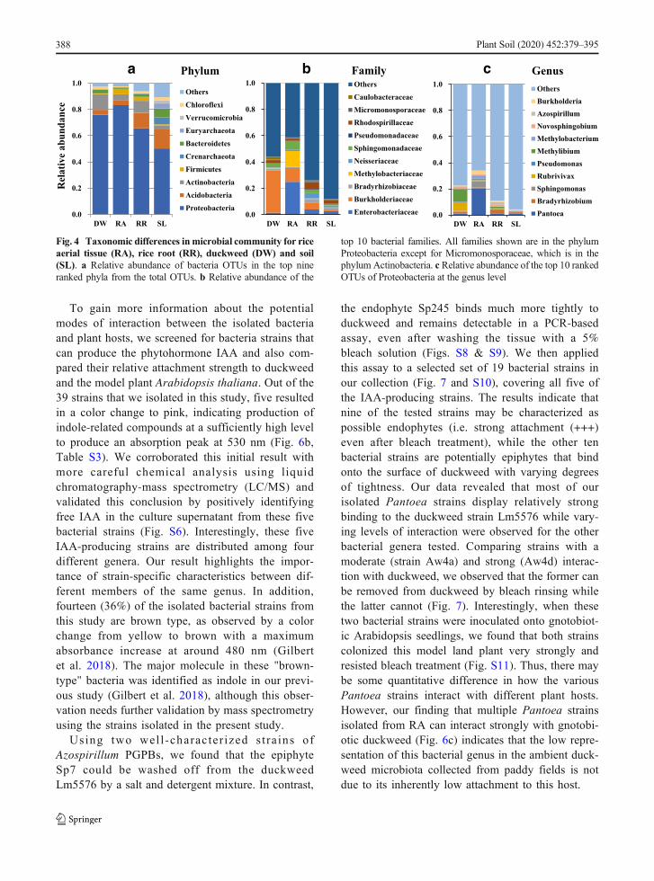
As noted above, the most dramatic changes observedbetween soil and plant tissue types were an increase inthe percentage of Proteobacteria along with a largedecrease in Acidobac te r ia , Crenarchaeo ta ,Euryarchaeota, Bacteroidetes, Verrucomicrobia andChloroflexi, in some cases to almost undetectable levels(Fig. 4a, Fig. S4a). At the family level (Fig. 4b, Fig.S4b), DW had the highest relative abundance inComamonadaceae (recently merged with theBurkholderiaceae (Parks et al. 2018)) at 32% of theOTUs identified, followed by Sphingomonadaceae
Plant Soil (2020) 452:379–395 385

(3%) and Rhodospirillaceae (2%). In contrast, Entero-bacteriaceae was the dominant family in RA with rela-tive abundance of about 25% (Fig. 4b). In addition, RRsamples were found to be rich in Burkholderiaceae(5%), Rhodospirillaceae (5%), and Neisseriaceae(14%), consistent with their known association with rootmicrobiota in plants (Santos-Medellín et al. 2017) andtheir potential function in nutrient acquisition. Interest-ingly, the family of Methylobacteriaceae (12%) wasenriched in RA compared to either RR or DW fromthe same paddy fields. The overall distribution of mi-croorganisms at the family level was relatively even inthe SL samples, where no dominant family with highrelative abundance was evident (Fig. 4b, Fig. S4b). It isworth noting that nine out of the top 10 families are inthe Proteobacteria phylum, with Micromonosporaceaerepresenting the only family from the Actinobacteriaphylum. Since Proteobacteria comprised more than80% of the bacterial microbiota in plant tissue samples,
we carried out a more detailed comparative analysisbetween our sample types at the genus level for thisphylum (Fig. 4c). Comparing the top three generaamong Proteobacteria that appear to dominate each ofthe three plant sample types, we found a distinct repre-sentation in each case: Rubrivivax, Methylibium andNovosphingobium in DW; Pantoea, Sphingomonasand Methylobacterium for RA; and Bradyrhizobium,Azospirillum and Burkholderia in RR (Fig. 4c). Fromthe apparent enrichment of distinct bacterial genera ineach of these sample types, there appears to be bothplant species-specific and tissue-specific enrichment ofthese genera of Proteobacteria in the paddy fieldecosystem.
As a first step in estimating the possible differences inmetabolic potential between the microbiome of duck-weed and rice, we used PICRUSt as an algorithm thatleveraged the large database of microbial genomes topredict gene functions/pathways that each microbiome
RA_WRA_
SDDW_WDW
_SDRR_W
0
1
2
3
4
5
RR_SD
a
a
aa
bb
Sh
ann
on
div
ersi
ty
RR SLRA DW0
2
4
6a
bb
c
a b
2
3
4
5
DW RR SL RARA DW RR SL RA DW RR SL
a a ab
c
ccc
c
dd
d
c Site 1 Site 3Site 2
Fig. 2 Within- and between-sample microbiome diversitymeasurements. a Shannon diversity, tissues with same letter arenot significantly different (ANOVA; p < 0.0001). b Shannondiversity, tissues and treatments with same letter are not signifi-cantly different (ANOVA; p < 0.05). c Shannon diversity, uniquetissues at different sites with the same letter are not significantly
different (ANOVA; p < 0.05). Abbreviations throughout are asfollows: rice aerial tissue (RA), rice root (RR), duckweed (DW),soil (SL), sterile water (W), salt and detergent (SD). Boxes are firstand third quartile (box bar is median), whiskers are 1.5x interquar-tile range, outliers plotted independently (dot)
Table 1 Average sequence reads for itags and OTU numbers
SampleType
SampleNumbers
Raw ReadsAvg.
CleanTagsAvg.
Effective TagsAvg.
Taxon TagsAvg.
Length Avg.(bp)
OTU NumbersAvg.
Observed_Species Avg.
RA 18 3,943,505 2,953,715 2,953,163 2,843,815 298 99 31
RR 14 1,528,213 1,117,424 1,116,565 785,745 299 939 169
DW 18 2,366,694 1,755,103 1,752,970 1,498,651 298 492 98
SL 7 949,746 691,183 690,754 186,397 299 3420 527
avg., average; OTU operational taxonomic unit, RA Rice aerial tissue, RR Rice root, DW Duckweed, SL Soil
386 Plant Soil (2020) 452:379–395

likely encodes (Langille et al. 2013). The results indicatethat the bacterial microbiota from duckweed tissues maybemore enriched in metabolic pathways associated withxenobiotic biodegradation and amino acid metabolismwhereas for the bacterial community present in riceaerial tissues, the metabolic pathways for energy as wellas vitamins and enzyme cofactors are significantly moreabundant (Fig. S5). These results suggest that the dis-tinct microbial communities in these plants may reflectpotentially different roles that they play in providingmetabolic capabilities for their associated host plants inthe rice paddy ecosystem.
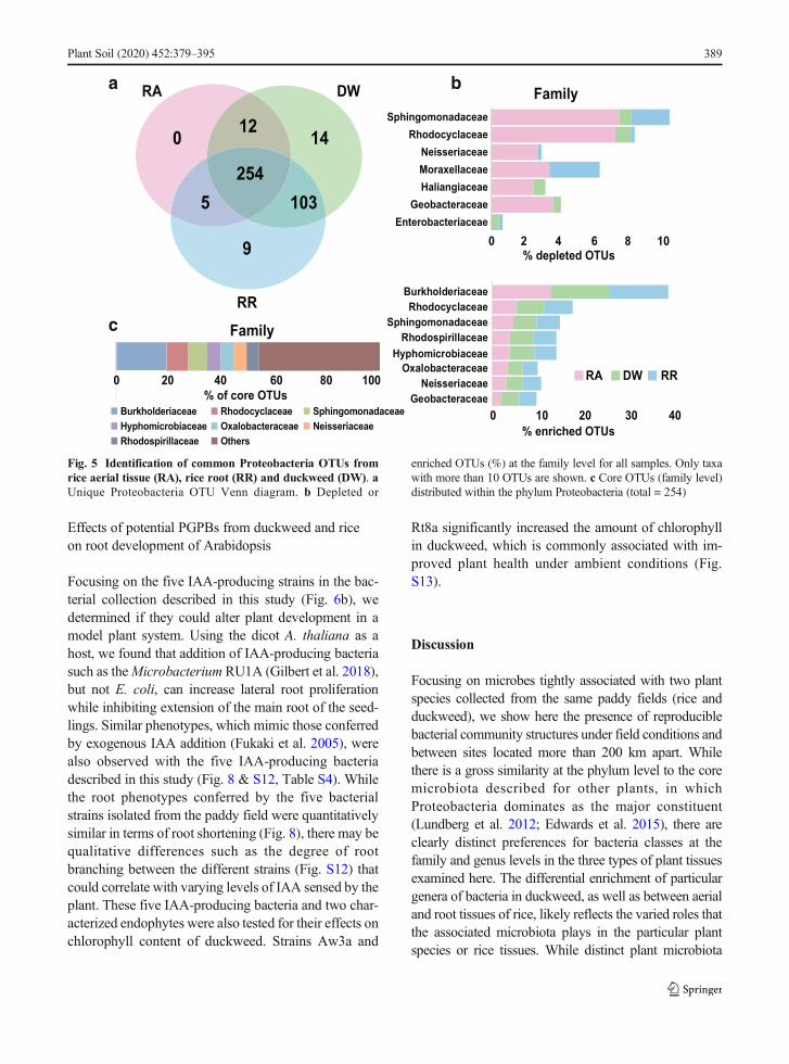
To identify common bacterial OTUs among samplesfrom the 3 tissue types, we carried out a Venn analysis ofthe 397 OTUs that resided within phylum Proteobacteriaand were found among the microbial samples derivedfrom the various plant samples. Of these, 254 OTUs werepresent in all plant sample types, and the core OTUcollection we found in all three tissue types comprisedmainlymembers of the Burkholderiaceae (> 20%), whichwere also highly enriched in RA, DW, and RR samples
(Fig. 5).We found about 10% of the Sphingomonadaceaeand Rhodocyclaceae were significantly underrepresentedin RA compared with those in RR and DW (p < 0.05)(Fig. 5b). In contrast, the genera that are preferentiallyenriched in each plant sample types that we noted abovemay reflect their differential importance in each particularplant species or tissue type.
Characterization of bacterial isolates from riceand duckweed tissues
To characterize the mode of growth-promoting actionfor plant-associated bacteria in the paddy field ecosys-tem, we isolated 39 bacteria strains from the three planttissue types used in this study. Using 16S rRNA genesequencing, we first determined the most likely genusfor each of the isolated bacteria (Fig. 6d, Table S2).Consistent with our culture-independent analysis of themicrobiome composition in these samples, most of theisolated bacteria from plants in the paddy fields belongto the Proteobacteria phylum (Pantoea, Enterobacter,Aeromonas, Acinetobacter and Pseudomonas) andsome from the Firmicutes (Bacillus, Lysinibacillus andStaphylococcus). Perhaps the most striking observation isthat all the isolated Pantoea strains were from RA tissues,where it was also most abundant, representing about 20%of all the Proteobacteria OTUs (Fig. 4c). This observationindicates that Pantoea species may be present at a higherpercentage in terms of total bacterial counts in RA. Incontrast, under our culture conditions RR appears to havea higher abundance of Bacillus and Enterobacter species,while there is a relatively even distribution of three differ-ent genera (Enterobacter, Aeromonas, Pseudomonas)among strains isolated from DW (Fig. 6).
Table 2 Average α-diversity indices for rice aerial tissue (RA),rice root (RR), duckweed (DW) and soil (SL)
SampleType
SampleNumbers
ShannonAvg.
SimpsonAvg.
Chao1Avg.
ACEAvg.
RA 18 2.015c 0.213c 175.501d 192.195c
RR 14 3.828b 0.057ab 451.213c 466.876a
DW 18 3.457b 0.076b 368.727b 362.956b
SL 7 5.016a 0.011a 473.595a 459.677a
Significant differences between different sample types were eval-uated using a one-way ANOVA and post-hoc comparisons withTukey’s correction. Different letters indicate the values are signif-icantly different at P < 0.05
RASite 1 Site 2 Site 3 DW RR SL
0.1
Soil Duckweed Rice root Rice aerial part
a b
Fig. 3 Beta-diversity of rice paddy environmental samples. aBray-Curtis distance metric phylogeny. Symbol shape indicatessite and symbol color indicates sample type (rice aerial tissue, RA;rice root, RR; duckweed, DW; and soil, SL). b Weighted Unifrac
distance Principle Component Analysis. Symbol color as in panela indicates sample type. Diamonds, rice aerial tissues; squares, riceroots; triangles, duckweed; inverted triangles, soil. Each pointrepresents a sample
Plant Soil (2020) 452:379–395 387
To gain more information about the potentialmodes of interaction between the isolated bacteriaand plant hosts, we screened for bacteria strains thatcan produce the phytohormone IAA and also com-pared their relative attachment strength to duckweedand the model plant Arabidopsis thaliana. Out of the39 strains that we isolated in this study, five resultedin a color change to pink, indicating production ofindole-related compounds at a sufficiently high levelto produce an absorption peak at 530 nm (Fig. 6b,Table S3). We corroborated this initial result withmore careful chemical analysis using liquidchromatography-mass spectrometry (LC/MS) andvalidated this conclusion by positively identifyingfree IAA in the culture supernatant from these fivebacterial strains (Fig. S6). Interestingly, these fiveIAA-producing strains are distributed among fourdifferent genera. Our result highlights the impor-tance of strain-specific characteristics between dif-ferent members of the same genus. In addition,fourteen (36%) of the isolated bacterial strains fromthis study are brown type, as observed by a colorchange from yellow to brown with a maximumabsorbance increase at around 480 nm (Gilbertet al. 2018). The major molecule in these "brown-type" bacteria was identified as indole in our previ-ous study (Gilbert et al. 2018), although this obser-vation needs further validation by mass spectrometryusing the strains isolated in the present study.
Using two wel l -charac ter ized s t ra ins ofAzospirillum PGPBs, we found that the epiphyteSp7 could be washed off from the duckweedLm5576 by a salt and detergent mixture. In contrast,
the endophyte Sp245 binds much more tightly toduckweed and remains detectable in a PCR-basedassay, even after washing the tissue with a 5%bleach solution (Figs. S8 & S9). We then appliedthis assay to a selected set of 19 bacterial strains inour collection (Fig. 7 and S10), covering all five ofthe IAA-producing strains. The results indicate thatnine of the tested strains may be characterized aspossible endophytes (i.e. strong attachment (+++)even after bleach treatment), while the other tenbacterial strains are potentially epiphytes that bindonto the surface of duckweed with varying degreesof tightness. Our data revealed that most of ourisolated Pantoea strains display relatively strongbinding to the duckweed strain Lm5576 while vary-ing levels of interaction were observed for the otherbacterial genera tested. Comparing strains with amoderate (strain Aw4a) and strong (Aw4d) interac-tion with duckweed, we observed that the former canbe removed from duckweed by bleach rinsing whilethe latter cannot (Fig. 7). Interestingly, when thesetwo bacterial strains were inoculated onto gnotobiot-ic Arabidopsis seedlings, we found that both strainscolonized this model land plant very strongly andresisted bleach treatment (Fig. S11). Thus, there maybe some quantitative difference in how the variousPantoea strains interact with different plant hosts.However, our finding that multiple Pantoea strainsisolated from RA can interact strongly with gnotobi-otic duckweed (Fig. 6c) indicates that the low repre-sentation of this bacterial genus in the ambient duck-weed microbiota collected from paddy fields is notdue to its inherently low attachment to this host.
Phylum Family c Genusa becnadnuba
evit aleR
0.0
0.2
0.4
0.6
0.8
1.0
DW RA RR SL
OthersCaulobacteraceaeMicromonosporaceaeRhodospirillaceaePseudomonadaceaeSphingomonadaceaeNeisseriaceaeMethylobacteriaceaeBradyrhizobiaceaeBurkholderiaceaeEnterobacteriaceae0.0
0.2
0.4
0.6
0.8
1.0
DW RA RR SL
OthersChloroflexiVerrucomicrobiaEuryarchaeotaBacteroidetesCrenarchaeotaFirmicutesActinobacteriaAcidobacteriaProteobacteria
0.0
0.2
0.4
0.6
0.8
1.0
DW RA RR SL
OthersBurkholderiaAzospirillumNovosphingobiumMethylobacteriumMethylibiumPseudomonasRubrivivaxSphingomonasBradyrhizobiumPantoea
Fig. 4 Taxonomic differences inmicrobial community for riceaerial tissue (RA), rice root (RR), duckweed (DW) and soil(SL). a Relative abundance of bacteria OTUs in the top nineranked phyla from the total OTUs. b Relative abundance of the
top 10 bacterial families. All families shown are in the phylumProteobacteria except for Micromonosporaceae, which is in thephylum Actinobacteria. c Relative abundance of the top 10 rankedOTUs of Proteobacteria at the genus level
388 Plant Soil (2020) 452:379–395
Effects of potential PGPBs from duckweed and riceon root development of Arabidopsis
Focusing on the five IAA-producing strains in the bac-terial collection described in this study (Fig. 6b), wedetermined if they could alter plant development in amodel plant system. Using the dicot A. thaliana as ahost, we found that addition of IAA-producing bacteriasuch as theMicrobacterium RU1A (Gilbert et al. 2018),but not E. coli, can increase lateral root proliferationwhile inhibiting extension of the main root of the seed-lings. Similar phenotypes, which mimic those conferredby exogenous IAA addition (Fukaki et al. 2005), werealso observed with the five IAA-producing bacteriadescribed in this study (Fig. 8 & S12, Table S4). Whilethe root phenotypes conferred by the five bacterialstrains isolated from the paddy field were quantitativelysimilar in terms of root shortening (Fig. 8), there may bequalitative differences such as the degree of rootbranching between the different strains (Fig. S12) thatcould correlate with varying levels of IAA sensed by theplant. These five IAA-producing bacteria and two char-acterized endophytes were also tested for their effects onchlorophyll content of duckweed. Strains Aw3a and
Rt8a significantly increased the amount of chlorophyllin duckweed, which is commonly associated with im-proved plant health under ambient conditions (Fig.S13).
Discussion
Focusing on microbes tightly associated with two plantspecies collected from the same paddy fields (rice andduckweed), we show here the presence of reproduciblebacterial community structures under field conditions andbetween sites located more than 200 km apart. Whilethere is a gross similarity at the phylum level to the coremicrobiota described for other plants, in whichProteobacteria dominates as the major constituent(Lundberg et al. 2012; Edwards et al. 2015), there areclearly distinct preferences for bacteria classes at thefamily and genus levels in the three types of plant tissuesexamined here. The differential enrichment of particulargenera of bacteria in duckweed, as well as between aerialand root tissues of rice, likely reflects the varied roles thatthe associated microbiota plays in the particular plantspecies or rice tissues. While distinct plant microbiota
0
9
1412
5 103254
RA DW
RR
100
0 2 4 6 8 10Enterobacteriaceae
GeobacteraceaeHaliangiaceaeMoraxellaceaeNeisseriaceae
RhodocyclaceaeSphingomonadaceae
% depleted OTUs
Family
RA DW RR
% enriched OTUs0 10 20 30 40
GeobacteraceaeNeisseriaceae
OxalobacteraceaeHyphomicrobiaceae
Rhodospirillaceae
RhodocyclaceaeBurkholderiaceae
SphingomonadaceaeFamily
0 20 40 60 80% of core OTUs
Burkholderiaceae Rhodocyclaceae SphingomonadaceaeHyphomicrobiaceae Oxalobacteraceae NeisseriaceaeRhodospirillaceae Others
a
c
b
Fig. 5 Identification of common Proteobacteria OTUs fromrice aerial tissue (RA), rice root (RR) and duckweed (DW). aUnique Proteobacteria OTU Venn diagram. b Depleted or
enriched OTUs (%) at the family level for all samples. Only taxawith more than 10 OTUs are shown. c Core OTUs (family level)distributed within the phylum Proteobacteria (total = 254)
Plant Soil (2020) 452:379–395 389

community structures have been reported before(Larousse et al. 2017; Walters et al. 2018), the mecha-nism for the differential enrichment of particular generain various species or tissue context remains unclear.There are at least two levels of selective enrichment thatcan operate to generate the apparent diversity from acommon microbial reservoir: 1) differences in microbe-plant tissue interaction may affect the initial steps ofattachment and subsequent colonization by the microbe,and 2) differences in host cell physiology and response tomicrobial colonization can exert constraints on the typesof bacteria that can thrive in a particular niche.
We examined the first possibility by testing Pantoeastrains cultured from the aerial tissues of rice on duck-weed for their ability to attach to plant tissues underlaboratory conditions. Using a PCR-based attachmentassay, we showed that the majority of the Pantoeastrains isolated can attach strongly to duckweed. Con-sequently, the near absence of this genus of bacteria inthe ambient duckweed microbiota from paddy fields is
unlikely to be a result of their inability to attach to thisplant host. Similarly, the very low levels of Pantoeaspecies in the rice root microbiota is also unlikely to bedue to their inability to interact with root tissues, sincetwo IAA-producing Pantoea strains from rice are able tobind strongly to Arabidopsis and alter root developmentunder laboratory conditions. Thus, at least for thePantoea genus, our data would be more consistent witha post-attachment selection model in which the localtissue-specific context of the host would determine itssteady state abundance relative to other microbes pres-ent in the same niche. This could involve both plant-microbe as well as microbe-microbe interactions withineach niche that will ultimately determine the success ofcolonization by a particular microbe.
One possible way that plant-associated bacteria maypromote improved plant functions is through the pro-duction of phytohormones, such as the auxin IAA, thatcan help reprogram host-encoded pathways for abioticstress tolerance, nutrient acquisition and disease
a b c dFig. 6 Structural andfunctional characteristics ofculturable bacteria isolatedfrom duckweed and rice. aNeighbor Joining tree constructedbased on bacterial 16S rRNAgene sequence. Symbol shapeindicates site and symbol colorindicates sample type (rice aerialtissue, RA; rice root, RR;duckweed, DW; and soil, SL). bBacterial producers of indolerelated compounds where tick (√)indicates high and cross (×)indicates low or undetectablelevels (Table S3). LC-MS massspectrum validation is shown inFig. S6 and Arabidopsis rootgrowth assay is shown in Fig.S12. c Duckweed attachment as-say attachment strength: strong(+++), moderate (++), weak (+)and no significant attachment (-);absence of symbol indicates strainwas not tested. Attachment resultsfor the Pantoea genus are shownin Fig. 7; all others are shown inFig. S10. d Bacteria classificationat genus level. The scale bar in-dicates the average numberof substitutions per site
390 Plant Soil (2020) 452:379–395

resistance (Berg et al. 2015). In addition to phytohor-mones that could be produced by plant-associated bac-teria, there are likely additional signaling molecules that
mediate communication between the host plant anddifferent microbes that can further modify the pheno-typic outcome of the interaction (Hassani et al. 2018;
ba
NoTC
+Aw4a
Lm5576
ControlsAw4aW SD BL
-Lm5576
W SD BL NoTC
+Aw4d
Lm5576
ControlsAw4d
W SD BL
Lm5576
IGS
Day 2
Day 7
LFYDay 2
Day 7
Aw4aW SD BL
-Lm5576
W SD BL
Aw4dW SD BL No
TC+
Aw4aLm
5576
Controls+
Aw4d
NoTC
+Aw4a
Lm5576
ControlsAw4aW SD BL
-Lm5576
W SD BL NoTC
+Aw4d
Lm5576
ControlsAw4d
W SD BL
Lm5576
IGS
Day 2
Day 7
LFYDay 2
Day 7
Aw4aW SD BL
-Lm5576
W SD BL
Aw4dW SD BL No
TC+
Aw4aLm
5576
Controls+
Aw4d
Fig. 7 Attachment of two Pantoea strains isolated from riceon duckweed. a Pantoea strain Aw4a attachment profile harvest-ed on day 2 and 7. b Pantoea strain Aw4d attachment profileharvested on day 2 and 7. Abbreviations are as follows: Lemnaminor 370-DWC112 (Lm5576), bacterial 16S-23S rRNA gene
intergenic spacer region (IGS, ~1.5 kb), Lm5576 LEAFY gene(LFY, ~500 bp), sterile water (W), salt and detergent (SD), SDwith 5% bleach (BL), gnotobiotic Lm5576 (-). Controls used forPCR were No TC: no template control, (+ strain ID): bacteriaDNA, and Lm5576: gnotobiotic Lm5576
Root
Leng
th(m
m)
Root
Leng
th(m
m)
Fig. 8 Effects on Arabidopsis root development by treatmentwith auxin-producing bacteria from rice and duckweed onpaddy fields. Increase in primary root length in millimeters(mm) after 7 days. Bacteria include E. coli as a negative control
and Microbacterium sp. RU1A (1A) as a positive control.Arabidopsis inoculated with different bacteria were compared tosterile group using Wilcoxon test with results indicated on the top(p-value ≤ 0.05 = *, p-value ≤ 0.0001 = ****)
Plant Soil (2020) 452:379–395 391

Stringlis et al. 2018). Nevertheless, the results from ourfunctional characterization of five IAA-producing bac-teria strains in this study are consistent with their poten-tial role as PGPBs that could enhance lateral root devel-opment in land plants, as well as forming stable associ-ations either as epiphytes or endophytes on duckweed.
In addition to the distinctive bacterial communitystructures described here for duckweed and the two ricetissues from multiple paddy fields, we also identified254 shared OTUs that were found in all tissue samplesexamined. Interestingly, the large majority of thesehighly conserved OTUs maps to the Comamonadaceaefamily, which has recently been merged with theBurkholderiaceae family (Parks et al. 2018). While thisgroup of microbes is dominant in duckweed, they areless prominent in rice tissues (Fig. 4b, Fig. S4b). Nev-ertheless, a recent functional study with synthetic com-munities in Arabidopsis has identified this family as oneof the key players in orchestrating anti-fungal activitiesin association with more robust plant health (Durán et al.2018). As we have found in this study, the conservedassociation and prominent representation ofComamonadaceae genera within plant tissues of bothrice and duckweed from paddy fields suggest the possi-ble importance of this bacteria family for plant health inthe field as well. For duckweed, the high representationof this family of bacteria may indicate its increasedimportance for the lifestyle of this aquatic macrophyteand may correlate with the predicted increase of meta-bolic potential in the bacteria microbiota of duckweedfor xenobiotic degradation and amino acid metabolism(Fig. S5).
One potential confounding issue that may influencethe interpretation of our results from soil is the necessityof using a specialized DNA extraction kit for nucleicacid isolation from these environmental samples, whichmay potentially result in differential extraction of DNAfrom the different bacterial phyla present. The difficultyin performing DNA isolation from soil, relative to planttissues, is exemplified by the low yields in sequencereads from these samples. Consequently, the derivedratios between the respective bacterial phyla within thesoil are likely to be estimates at best. Nevertheless, thesimilar patterns observed from soil samples of the threesites would indicate that the relative levels for the majorphyla revealed from our data analysis are likely to bereasonable approximations.
Microbial attachment to plants was investigated inthis study using a generic bacterial primer set for 16S-
23S intergenic spacer region to detect the presence ofbacteria on the plant tissue while a single-copied LEAFYgene of the plant was used as a reference gene tonormalize for the quantity of DNA used. This providesa relatively scalable assay that can be systematicallydeployed to examine plant-microbe interactions undera variety of conditions. In contrast, the most commonlyused approaches for measuring microbial attachment toplant tissues involve microscopy and/or colonycounting, both of which are labor intensive and timeconsuming. Previous studies have shown that certainconcentrations of ethanol, NaClO (i.e. bleach) orTween-20 can sterilize the surface of roots and seeds(Koeppel and Wu 2013). Our PCR-based method usesgnotobiotic duckweed and Arabidopsis as model plantsand uses chemical treatments to define surface accessi-bility and differentiate between plant-microbe interac-tions of different strengths. With this approach, ourpresent work has provided evidence that the selectionfor stable colonization of Pantoea species in rice aerialtissues and not in duckweed most likely occurs after theattachment of members of this bacterial genus onto theplant and may be the result of the host tissue context aswell as competition with other microbes in the sameniche.
In this work, we have begun to establish rice root-andleaf-derived microbiota culture collections with strainsrepresenting the majority of bacterial species that arereproducibly detected by culture-independent commu-nity sequencing. This will augment the collection ofduckweed-associated bacteria described previously(Gilbert et al. 2018). In parallel, we are systematicallycataloguing their potential for IAA production and abil-ity to attach to plants, using gnotobiotic Arabidopsis andduckweed as our model systems. While it would havebeen more ideal to use rice plants as a test system for themicrobes isolated from rice tissues, the small size andrapid growth of the model plant systems such as duck-weed and Arabidopsis make them more amenable forlarge-scale studies. Nevertheless, it is important to fur-ther test our model in the future by using rice plantsgrown under laboratory conditions to directly examinethe tissue-dependent attachment and colonization effi-ciency of these strains. As the recent studies with syn-thetic communities in Arabidopsis have illustrated (Baiet al. 2015), a robust bacterial community that can bestably established in the target plant would set the stagefor testing whether their presumed beneficial roles canbe reproduced under laboratory conditions and
392 Plant Soil (2020) 452:379–395

eventually transferred to their hosts under field condi-tions. The resources and data produced by our work herelaid the foundation for this approach to improve riceproduction via management of the microbiota in paddyfields.
Acknowledgements Duckweed research at the Lam Lab is sup-ported in part by a grant from the Department of Energy (DE-SC0018244) and a Hatch project (#12116) from the New JerseyAgricultural Experiment Station at Rutgers University. We aregrateful to Prof. Qiyi Lei of Kaili University, and the villagers ofthree counties in Qiandongnan Prefecture for assisting field sur-veys and sample collection. The work in the Long Lab wassupported by the National Natural Science Foundation of China(31761143001 & 31870316), Ministry of Ecology and Environ-ment of China (2019HJ2096001006), Key Laboratory of Ethno-medicine (Minzu University of China) of the Ministry of Educa-tion of China (KLEM-ZZ201906 & KLEM-ZZ201806), MinzuUniversity of China (YLDXXK201819), the Ministry of Educa-tion of China and State Administration of Foreign Experts Affairsof China (B08044), and the China Scholarship Council (CSC No.201606390030). This work was also supported in part by theGuangdong Academy of Sciences’ special project to W.H.(2019GDASYL-0103031). We thank Ilya Raskin (Rutgers Uni-versity, New Brunswick, NJ, USA) for providing access to themass spectrometry instrument in his laboratory as well as forproviding LC-MS related reagents and also thankMichael Lawton(Rutgers University) for editorial assistance to improve the man-uscript. K.A. acknowledges the Rutgers University SGS Excel-lence Fellowship for providing student support for part of thiswork. The Lebeis Lab thanks Elizabeth Denison for performingthe quality control for the DNA samples prior to 16S rDNAamplicon sequencing and acknowledges the start-up funds provid-ed to S.L. from the University of Tennessee, Knoxville. Thecontribution by the Facilities Integrating Collaborations for UserScience (FICUS) initiative under Contract Nos. DE-AC02-05CH11231 (JGI) and DE-AC05-76RL01830 (EMSL) to thesequencing of the duckweed microbiome work is also gratefullyacknowledged.
Author Contribution E.L. and C.L. designed the project ap-proach;W.H. performedmost of the experiments in the study; E.L.isolated the bacteria strains; C.L. led the field surveys and samplecollections; S.G. and A.P. performed the mass spectrometry stud-ies; K.A. and S.G. assisted in the set up of the bacteria attachmentand Salkowski assays; S.L. assisted in data analysis and drafting ofthe manuscript; W.H. and E.L. drafted the manuscript with helpfrom all other authors.
Data Accessibility Raw reads have been deposited in the ShortRead Archive of NCBI under project no. PRJNA545325.
Open Access This article is licensed under a Creative CommonsAttribution 4.0 International License, which permits use, sharing,adaptation, distribution and reproduction in anymedium or format,as long as you give appropriate credit to the original author(s) andthe source, provide a link to the Creative Commons licence, andindicate if changes were made. The images or other third partymaterial in this article are included in the article's Creative
Commons licence, unless indicated otherwise in a credit line tothe material. If material is not included in the article's CreativeCommons licence and your intended use is not permitted bystatutory regulation or exceeds the permitted use, you will needto obtain permission directly from the copyright holder. To view acopy of this l icence, vis i t ht tp: / /creat ivecommons.org/licenses/by/4.0/.
References
Acosta K, Xu J, Gilbert S, Denison E, Brinkman T, Lebeis S, LamE (2020) Duckweed hosts a taxonomically similar bacterialassemblage as the terrestrial leaf microbiome. PLoS One15(2):e0228560. https://doi.org/10.1371/journal.pone.0228560
Bai Y,Müller DB, Srinivas G, Garrido-Oter R, Potthoff E, RottM,Dombrowski N, Münch PC, Spaepen S, Remus-EmsermannM, Hüttel B, McHardy AC, Vorholt JA, Schulze-Lefert P(2015) Functional overlap of the Arabidopsis leaf and rootmicrobiota. Nature 528:364–369. https://doi.org/10.1038/nature16192
Berg G, Rybakova D, Grube M, Köberl M (2015) The plantmicrobiome explored: implications for experimental botany.J Exp Bot 67:995–1002. https://doi.org/10.1093/jxb/erv466
Benton TG, Vickery JA,Wilson JD (2003) Farmland biodiversity:is habitat heterogeneity the key? Trends Ecol Evol 18:182–188. https://doi.org/10.1016/s0169-5347(03)00011-9
Borisjuk N, Chu P, Gutierrez R, Zhang H, Acosta K, Friesen N,Sree KS, Garcia C, Appenroth KJ, Lam E (2014)Assessment, validation and deployment strategy of a two-barcode protocol for facile genotyping of duckweed species.Plant Biol 17:42–29. https://doi.org/10.1111/plb.12229
Bray JR, Curtis JT (1957) An ordination of the upland forestcommunities of southern Wisconsin. Ecol Monogr 27:326–349. https://doi.org/10.2307/1942268
Caporaso JG, Kuczynski J, Stombaugh J, Bittinger K, BushmanFD, Costello EK, Fierer N, Peña AG, Goodrich JK, GordonJI, Huttley GA, Kelley ST, Knights D, Koenig JE, Ley RE,Lozupone CA, McDonald D, Muegge BD, Pirrung M,Reeder J, Sevinsky JR, Turnbaugh PJ, Walters WA,Widmann J, Yatsunenko T, Zaneveld J, Knight R (2010)QIIME allows analysis of high-throughput community se-quencing data. Nat Methods 7:335–336. https://doi.org/10.1038/nmeth.f.303
DeSantis TZ, Hugenholtz P, Larsen N, RojasM, Brodie EL, KellerK, Huber T, Dalevi D, Hu P, Andersen GL (2006)Greengenes, a chimera-checked 16S rRNA gene databaseand workbench compatible with ARB. Appl EnvironMicrobiol 72:5069–5072. https://doi.org/10.1128/AEM.03006-05
Durán P, Thiergart T, Garrido-Oter R, Agler M, Kemen E,Schulze-Lefert P, Hacquard S (2018) Microbialinterkingdom interactions in roots promote Arabidopsis sur-vival. Cell 175:973–983. https://doi.org/10.1101/354167
Edwards J, Johnson C, Santos-Medellín C, Lurie E, PodishettyNK, Bhatnagar S, Eisen JA, Sundaresan V (2015) Structure,variation, and assembly of the root-associated microbiomes
Plant Soil (2020) 452:379–395 393

of rice. Proc Natl Acad Sci USA 112:E911–E920. https://doi.org/10.1073/pnas.1414592112
Feng J, Li F, Zhou X, Xu C, Fang F (2016) Nutrient removalability and economical benefit of a rice-fish co-culture systemin aquaculture pond. Ecol Eng 94:315–319. https://doi.org/10.1016/j.ecoleng.2016.06.002
Finkel OM, Castrillo G, Herrera Paredes S, Salas González I,Dangl JL (2017) Understanding and exploiting plant benefi-cial microbes. Curr Opin Plant Biol 38:155–163. https://doi.org/10.1016/j.pbi.2017.04.018
Fukaki H, Nakao Y, Okushima Y, Theologis A, Tasaka M (2005)Tissue-specific expression of stabilized SOLITARY-ROOT/IAA14 alters lateral root development in Arabidopsis. Plant J44:382–395. ht tps: / /doi .org/10.1111/j .1365-313x.2005.02537.x
Gilbert S, Xu J, Acosta K, Poulev A, Lebeis S, Lam E (2018)Bacterial production of indole related compounds revealstheir role in association between duckweeds and endophytes.F r o n t Ch em 6 : 2 6 5 . h t t p s : / / d o i . o r g / 1 0 . 3 3 8 9/fchem.2018.00265
Gordon SA, Weber RP (1951) Colorimetric Estimation ofIndoleacetic Acid. Plant Physiol 26:192–195. https://doi.org/10.1104/pp.26.1.192
Gu BJ, Ju XT, Chang J, Ge Y, Vitousek PM (2015) Integratedreactive nitrogen budgets and future trends in China. ProcNatl Acad Sci USA 112:8792–8797. https://doi.org/10.1073/pnas.1510211112
Gürtler V, Stanisich VA (1996) New approaches to typing andidentification of bacteria using the 16S-23S rDNA spacerregion. Microbiology 142:3–16. https://doi.org/10.1099/13500872-142-1-3
Hassani MA, Durán P, Hacquard S (2018) Microbial interactionswithin the plant holobiont. Microbiome 6(1):58. https://doi.org/10.1186/s40168-018-0445-0
Jain DK, Patriquin DG (1984) Root hair deformation, bacterialattachment, and plant growth in wheat-azospirillum associa-tions. Appl Environ Microbiol 48:1208–1213. https://doi.org/10.1016/0141-4607(84)90089-1
Koeppel AF, Wu M (2013) Surprisingly extensive mixed phylo-genetic and ecological signals among bacterial operationaltaxonomic units. Nucleic Acids Res 41:5175–5188.https://doi.org/10.1093/nar/gkt241
Kumura M (2005) Populations, community composition and bio-mass of aquatic organisms in the flood water of rice fields andeffects of field management. Soil Sci Plant Nutr 51:159–181.https://doi.org/10.1111/j.1747-0765.2005.tb00021.x
Lam E, Zhang Y (2012) Regulating the reapers: activatingmetacaspases for programmed cell death. Trends Plant Sci17:487–494. https://doi.org/10.1016/j.tplants.2012.05.003
LangilleMGI, Zaneveld J, Caporaso JG,McDonald D, Knights D,Reyes JA, Clemente JC, Burkepile DE, Vega Thurber RL,Knight R, Beiko RG, Huttenhower C (2013) Predictive func-tional profiling of microbial communities using 16S rRNAmarker gene sequences. Nat Biotechnol 31:814–821.https://doi.org/10.1038/nbt.2676
Lansing JS, Kremer JN (2011) Rice, fish, and the planet. Proc NatlAcad Sci U S A 108(50):19841–19842. https://doi.org/10.1073/pnas.1117707109
Larousse M, Rancurel C, Syska C, Palero F, Etienne C, Industri B,Nesme X, Bardin M, Galiana E (2017) Tomato root micro-biota and Phytophthora parasitica-associated disease.
Microbiome 5:56. https://doi.org/10.1186/s40168-017-0273-7
Li H, Liang XQ, Lian YF, Xu L, Chen YX (2009) Reduction ofammonia volatilization from urea by a floating duckweed inflooded rice fields. Soil Sci Soc Am J 73:1890. https://doi.org/10.2136/sssaj2008.0230
Lindgreen S (2012) AdapterRemoval: easy cleaning of next-generation sequencing reads. BMC Res Notes 5:337.https://doi.org/10.1186/1756-0500-5-337
Lundberg D, Lebeis SL, Paredes SH, Yourstone S, Gehring J,Malfatti S, Tremblay J, Engelbrektson A, Kunin V, Del RioTG, Edgar RC, Eickhorst T, Ley RE, Hugenholtz P, TringeSG, Dangl JL (2012) Defining the core Arabidopsis thalianaroot microbiome. Nature 488:86–90. https://doi.org/10.1038/nature11237
Ng CA, Wong LY, Lo PK, Bashir MJK, Chin SJ, Tan SP, ChongCY, Yong LK (2017) Performance of duckweed and effec-tive microbes in reducing arsenic in paddy and paddy soil.AIP Conference Proceedings 1828:020031. https://doi.org/10.1063/1.4979402
Oksanen JF, Blanchet G, Friendly M, Kindt R, Legendre P,McGlinn PR, O’Hara RB, Simpson GL, Solymos P,Stevens M, Wagner H (2016) Vegan: Community EcologyPackage, Version 2.5–3
Parks DH, Chuvochina M, Waite DW, Rinke C, Skarshewski A,Chaumeil PA, Hugenholtz P (2018) A standardized bacterialtaxonomy based on genome phylogeny substantially revisesthe tree of life. Nat Biotechnol 36:996–1004. https://doi.org/10.1038/nbt.4229
Pascale A, Proietti S, Pantelides IS, Stringlis IA (2020)Modulation of the root microbiome by plant molecules: thebasis for targeted disease suppression and plant growth pro-motion. Front Plant Sci 10:1741. https://doi.org/10.3389/fpls.2019.01741
Santos-Medellín C, Edwards J, Liechty Z, Nguyen B, SundaresanV (2017) Drought stress results in a compartment-specificrestructuring of the rice root-associated microbiomes. mBio8:e00764–e00717. https://doi.org/10.1128/mBio.00764-17
Schmieder R, Edwards R (2011) Quality control and preprocess-ing of metagenomic datasets. Bioinformatics 27:863–864.https://doi.org/10.1093/bioinformatics/btr026
Sha Z, Chu Q, Zhao Z, Yue Y, Lu L, Yuan J, Cao L (2017)Variations in nutrient and trace element composition of ricein an organic rice-frog coculture system. Sci Rep 7(1):15706.https://doi.org/10.1038/s41598-017-15658-1
Stringlis IA, Zhang H, Pieterse CMJ, Bolton MD, de Jonge R(2018) Microbial small molecules - weapons of plant subver-sion. Nat Prod Rep 35(5):410–433. https://doi.org/10.1039/C7NP00062F
Walters WA, Jin Z, Youngblut N, Wallace JG, Sutter J, ZhangW,González-Peña A, Peiffer J, Koren O, Shi Q, Knight R,Glavina Del Rio T, Tringe SG, Buckler ES, Dangl JL, LeyRE (2018) Large-scale replicated field study of maize rhizo-sphere identifies heritable microbes. Proc Natl Acad Sci USA115:7368–7373. https://doi.org/10.1073/pnas.1800918115
Wang Q, Garrity GM, Tiedje JM, Cole JR (2007) Naive Bayesianclassifier for rapid assignment of rRNA sequences into thenew bacterial taxonomy. Appl Environ Microbiol 73:5261–5267. https://doi.org/10.1128/aem.00062-07
Wang C, Li S, Lai DYF, Wang W, Ma Y (2015) The effect offloating vegetation on CH4 and N2O emissions from
394 Plant Soil (2020) 452:379–395

subtropical paddy fields in China. Paddy Water Environ 13:425–431. https://doi.org/10.1007/s10333-014-0459-6
Weigel D, Alvarez J, Smyth DR, Yanofsky MF, Meyerowitz EM(1993) LEAFY controls floral meristem identity inArabidopsis. Cell 69:843–859. https://doi.org/10.1007/978-1-4757-9607-0_17
Wickham H (2011) The split-apply‐combine strategy for dataanalysis. J Stat Softw 40:1–29. https://doi.org/10.18637/jss.v040.i01
Wickham H (2016) ggplot2. Elegant graphics for data analysis,2nd edn. Springer International Publishing, Cham
Yang H, YuD, Zhou J, Zhai S, Bian X,Weih M (2018) Rice-duckco-culture for reducing negative impacts of biogas slurryapplication in rice production systems. J Environ Manage2 1 3 : 1 4 2 – 1 5 0 . h t t p s : / / d o i . o r g / 1 0 . 1 0 1 6 / j .jenvman.2018.02.077
Yao Y, Zhang M, Tian Y, Zhao M, Zhang B, Zhao M, Zeng K,Yin B (2017) Duckweed (Spirodela polyrhiza) as green
manure for increasing yield and reducing nitrogen loss inrice production. Field Crop Res 214:273–282. https://doi.org/10.1016/j.fcr.2017.09.021
Zhang J, Hu L, Ren W, Guo L, Tang J, Shu M, Chen X (2016)Rice-soft shell turtle coculture effects on yield and its envi-ronment. Agr Ecosyst Environ 224:116–122
Zhu YG, Su JQ, Cao ZH, Xue K, Quensen J, Guo GX, Yang YF,Zhou JZ, Chu HY, Tiedje JM (2016) A buried Neolithicpaddy soil reveals loss of microbial functional diversity aftermodern rice cultivation. Sci Bull 61:1052–1060. https://doi.org/10.1007/s11434-016-1112-0
Publisher’s note Springer Nature remains neutral with regard tojurisdictional claims in published maps and institutionalaffiliations.
Plant Soil (2020) 452:379–395 395