homocysteine modulates the cd40/cd40l system · homocysteine modulates the cd40/cd40l system ......
TRANSCRIPT

Egrpbmlcctd
FCMAaM(
2
Journal of the American College of Cardiology Vol. 49, No. 22, 2007© 2007 by the American College of Cardiology Foundation ISSN 0735-1097/07/$32.00P
Homocysteine Modulates the CD40/CD40L System
Cesaria Prontera, PHD,* Nicola Martelli, TECH,† Virgilio Evangelista, MD,† Etrusca D’Urbano, TECH,‡Stefano Manarini, TECH,† Antonio Recchiuti, PHD,* Alfredo Dragani, MD,§ Cecilia Passeri, MD,§Giovanni Davì, MD,* Mario Romano, MD†
Chieti, San Maria Imbaro, and Pescara, Italy
Objectives This study evaluated the impact of hyperhomocysteinemia (HHcy) on the CD40/CD40 ligand (CD40L) dyad invivo and in vitro.
Background Hyperhomocysteinemia is associated with an increased incidence of atherothrombosis, although the molecularmechanisms of this association are incompletely defined. The CD40L pair triggers inflammatory signals in cellsof the vascular wall, representing a major pathogenetic pathway of atherosclerosis.
Methods We used a commercially available enzyme-linked immunosorbent assay kit to evaluate circulating levels of solu-ble (s) CD40L in 24 patients with HHcy and 24 healthy subjects. We also used real-time polymerase chain reac-tion and flow cytometry to determine expression levels of CD40 and vascular cell adhesion molecule (VCAM)-1 inhuman umbilical vein endothelial cells (HUVECs) and of CD40L in human platelets.
Results The sCD40L levels were significantly increased in HHcy patients (median [interquartile range] 8.0 [0.7 to 10.5]ng/ml vs. 2.1 [1.9 to 2.3] ng/ml, p � 0.0001). Positive correlations were noted between log sCD40L and loghomocysteine (Hcy) (R � 0.68, p � 0.0001) or log sVCAM-1 (R � 0.41, p � 0.005). Homocysteine significantlystimulated CD40 mRNA expression in HUVECs (p � 0.033). Consistently, 24-h exposure to Hcy increased thepercentage of CD40-expressing cells (p � 0.00025). Homocysteine also significantly enhanced CD40L expres-sion in platelets (p � 0.025) to a comparable extent as that of thrombin. Notably, Hcy increased VCAM-1 proteinexpression induced by CD40L in HUVECs (p � 0.0046).
Conclusions The present results uncover a potential molecular target of Hcy, namely the CD40/CD40L dyad. Collectively, theyindicate that upregulation of CD40/CD40L signaling may represent a link between HHcy and an increased riskof cardiovascular disease. (J Am Coll Cardiol 2007;49:2182–90) © 2007 by the American College of Cardiol-ogy Foundation
ublished by Elsevier Inc. doi:10.1016/j.jacc.2007.02.044
CBmgcskcc1o(itr
bl
levated homocysteine (Hcy) concentration, determined byenetic or dietary factors, is recognized as an independentisk factor for cardiovascular disease (1). Homocysteine mayromote vascular damage and atherothrombosis by a num-er of mechanisms, including release of proinflammatoryediators, induction of oxidative and endoplasmic reticu-
um stress, and activation of apoptotic pathways in vascularells (2). However, the relative contribution of these pro-esses to the causal relationship between hyperhomocys-einemia (HHcy) and atherothrombosis is still underebate.
rom the *Department of Medicine, Gabriele d’Annunzio University Foundation,hieti-Pescara, Italy; †Laboratory of Vascular Biology and Pharmacology, Consorzioario Negri Sud, San Maria Imbaro, Italy; ‡Department of Biomedical Sciences andging Research Center, Gabriele d’Annunzio University Foundation, Chieti, Italy;
nd §Civil Hospital, Pescara, Italy. Supported in part by a grant from the Italianinistry of Research to the Center of Excellence on Aging of the University of Chieti
to Drs. Davì and Romano).
cManuscript received January 31, 2006; revised manuscript received December 20,
006; accepted February 9, 2007.
Accumulating evidence supports the involvement ofD40/CD40 ligand (CD40L) signaling in atherosclerosis.oth CD40 and CD40L are expressed by vascular cells,acrophages, and platelets (3,4). The CD40/CD40L en-
agement on the surface of endothelial cells, smooth muscleells, or macrophages triggers a potent inflammatory re-ponse, characterized by the release of inflammatory cyto-ines (interleukins 1�, 6, 8, 12) and chemokines (monocytehemoattractant protein-1), expression of adhesion mole-ules (E-selectin, vascular cell adhesion molecule [VCAM]-, intercellular adhesion molecule-1, P-selectin), activationf matrix metalloproteinases, and procoagulant tissue factor3–7). Antibody blockade or genetic disruption of CD40Ln Apolipoprotein-E�/� mice provides direct evidence ofhe involvement of CD40/CD40L signaling in atheroscle-osis progression (8,9).
A soluble form of CD40L (sCD40L) is rapidly releasedy T cells and activated platelets (10,11). The sCD40Levels are increased in a number of pathological conditions
haracterized by cardiovascular damage, i.e., unstable angina
((mp
mpssuc
M
Rio((ItIBUeapa(�Pstathrmhcav4di(hl
Bcp
osb
sfm5(stm�s(swmcmiwCeffmUcgpmp�(3H(mwc
v2tupHEsCRw(6McA
2183JACC Vol. 49, No. 22, 2007 Prontera et al.June 5, 2007:2182–90 Homocysteine and the CD40/CD40L Dyad
11), acute coronary syndromes (12), hypercholesterolemia13,14), arterial hypertension (15), and diabetes (16). Thus,easurement of sCD40L is now regarded as an index of
latelet activation and inflammatory vascular damage.Because HHcy is associated with signs of vascular inflam-ation and platelet activation (17,18), we tested the hy-
othesis that Hcy may up-regulate the CD40/CD40Lystem. Here we provide the first evidence of increasedCD40L circulating levels in HHcy patients and of CD40p-regulation by Hcy in human umbilical vein endothelialells (HUVECs) and of CD40L in platelets.
ethods
eagents. Rabbit anti-CD40 and anti-CD40L polyclonalmmunoglobulin (Ig) G were from Santa Cruz Biotechnol-gy (Santa Cruz, California). Mouse anti-VCAM-1CD106) phycoerythrin-conjugated was from BioLegendSan Diego, California). Anti-rabbit fluorescein-conjugategG was from Calbiochem (Milan, Italy). The spin/vacuumotal RNA isolation system was from Promega (Milan,taly). Reagents for real-time analysis were from Appliediosystems (Milan, Italy). Human thrombin (2,000 NIH/mg protein); N-2 hydroxyethyl piperazine-N 1-2-
thanesulfonic acid (HEPES); ethylene glycol-bis (b-minoethyl ether)-N, N, N=, N=,-tetraacetic acid (EGTA);rostaglandin E1 (PGE1); DL-homocysteine; L-cysteine;nd all other chemicals were purchased from Sigma-AldrichMilan, Italy). Thrombin was dissolved in saline at 20mol/l (50 U/ml) and stored at �20°C until use.atients, genotyping, and measurements. We selected 24
ubjects carrying the 5,10 methylene tetrahydrofolate reduc-ase (MTHFR) C677T genotype with HHcy (�15 �mol/l)nd 24 age- and gender-matched subjects, also expressinghe MTHFR C677T polymorphism, but with normalomocysteinemia (�15 �mol/l). Exclusion criteria wereepresented by a recent history of thrombotic events (�6onths), pregnancy or delivery in the previous 6 months,
ypercholesterolemia, diabetes, current medication for birthontrol or hormone replacement therapy, and recent use ofspirin, ticlopidine, clopidogrel, anti-inflammatory drugs,itamin supplements, or anticoagulant agents. Among the8 patients, 21 (44%) had clinical evidence of vascularisease, in particular, angina pectoris (n � 4), myocardial
nfarction (n � 3), transient ischemic attack (n � 2), stroken � 1), and peripheral artery disease (n � 2). Nine patientsad suffered deep venous thrombosis or pulmonary embo-
ism. Patient characteristics are summarized in Table 1.Subjects were studied as outpatients after a 12-h fast.
lood samples were obtained in the morning. Informedonsent was obtained from each subject after approval of therotocol by the local institutional ethics committee.Analysis of the MTHFR C677T mutation was carried
ut by digestion with the restriction enzyme Hinf I. Theection of the gene containing the mutation was amplified
y polymerase chain reaction as described previously (19). tFasting plasma total Hcy (theum of free and protein-boundorms plus cysteine-homocysteineixed disulfide) was measured in
1 ethylenediaminetetraacetic acidEDTA)-anticoagulated bloodamples immediately refrigeratedo prevent in vitro total Hcy for-
ation. Plasma was stored at80°C. The total Hcy was mea-
ured using the Imx Hcy assayAbbott Park, Illinois). PlasmaCD40L and sVCAM-1 levelsere measured using specific im-unoassays (R & D Systems) ac-
ording to the instructions of theanufacturer. Intra-assay and
nter-assay coefficients of variationere �6%.ells. Human umbilical vein
ndothelial cells were isolatedrom umbilical cords obtainedrom randomly selected healthyothers delivering at the Chietiniversity Hospital, using 0.1%
ollagenase at 37°C. Cells wererown on 1.5% gelatin-coatedlates in medium Dulbecco’sodified Eagle’s medium (D-MEM)/M-199 (50:50) sup-
lemented with 20% heat-inactivated fetal calf serum, 10g/ml heparin, 50 �g/ml endothelial cell growth factor
ECGF), 50 mg/ml penicillin/streptomycin in 5% CO2 at7°C, and used within 4 passages. Before treatment,UVECs were made quiescent with D-MEM/M-199
50:50) medium supplemented with 1% bovine serum albu-in (BSA) and 50 �g/ml ECGF for 20 h. Homocysteineas delivered to cells in D-MEM/F12 (50:50) with 2% fetal
alf serum and ECGF.For platelet isolation, blood was collected from healthy
olunteers who had not received any medication for at leastweeks. Platelet-rich plasma was prepared by centrifuga-
ion at 200g for 15 min. Platelets were isolated by centrif-gation at 1,100g for 15 min, after addition to platelet-richlasma of 1 �mol/l PGE1. The pellet was suspended inEPES-tyrode containing 1 �mol/l PGE1 and 5 mmol/lGTA and centrifuged at 1,100g for 10 min. Platelets were
uspended with HEPES-tyrode buffer containing 1 mmol/la2� at the concentration of 1 � 108/ml.eal-time polymerase chain reaction. Total cellular RNAas extracted using the SV total RNA Isolation System
Promega). Polyadenosine RNA was reverse-transcribed for0 min at 42°C with StrataScript II (50 U/ml) (Stratagene,ilan, Italy). Real-time measurements of CD40 were
arried out using the Assay-on-Demand Hs00374176 frompplied Biosystems, in the ABI PRISM 7900 HT appara-
Abbreviationsand Acronyms
BSA � bovine serumalbumin
CD40L � CD40 ligand
EDTA � ethylenediaminetetra-acetic acid
Hcy � homocysteine
HEPES � N-2 hydroxyethylpiperazine-N 1-2-ethanesulfonic acid
HHcy �
hyperhomocysteinemia
HUVECs � human umbilicalvein endothelial cells
Ig � immunoglobulin
MFI � mean fluorescenceintensity
MTHFR � 5,10 methylenetetrahydrofolate reductase
NF � nuclear factor
PBS � phosphate-bufferedsaline
s � soluble
VCAM � vascular celladhesion molecule
us, according to the instructions
of the manufacturer.
Gt2RfFwPaffl4l(twi1SapbitsHBgwwla
B(ilioMstWs(ndt3cwaSp�Wo2(ascgc
ensity l
2184 Prontera et al. JACC Vol. 49, No. 22, 2007Homocysteine and the CD40/CD40L Dyad June 5, 2007:2182–90
lyceraldehyde-3-phosphate dehydrogenase was used ashe housekeeping gene. Data were elaborated with the SDS.0 software (Applied Biosystems, Foster City, California).elative gene expression was evaluated using the 2���Ct
ormula.low cytometric analysis. The HUVECs were harvestedith phosphate-buffered saline (PBS)/EDTA, washed withBS/0.5% BSA, and incubated with a polyclonal antibodygainst human CD40 (1:25 dilution, 1 �g for 5 � 105 cells)or 30 min at 4°C. Cells were then exposed to a secondaryuorescein-conjugate anti-rabbit antibody for 30 min at°C. Samples were washed with PBS/0.5% BSA and ana-yzed in a Becton Dickinson FACScan flow cytometerBecton Dickinson, Milan, Italy). Data were analyzed usinghe CellQuest software. Washed platelets (1 � 108/ml)ere exposed to stimuli for 5 min at 37°C, and then
ncubated with anti-CD40L polyclonal IgG (1 �g for 5 �06 cells) or control rabbit polyclonal IgG for 30 min at 4°C.amples were then exposed to a fluorescein-conjugate goatnti-rabbit IgG antibody (4°C, 30 min), fixed with 2%araformaldehyde for 1 h at room temperature and analyzedy FACScan. The CD40L specific mean fluorescencentensity (MFI) was calculated by subtracting the value ofhe isotype-matched control antibody from that of thepecific antibody. For evaluation of VCAM-1 expression,UVECs were grown in EGM-2MV medium (Cambrexio Science, Walkersville, Maryland) supplemented withrowth factors. At subconfluence, cells were washed twiceith PBS and incubated with vehicle or Hcy for 24 h. Cellsere then exposed to sCD40L (Bender MedSystem, Bur-
ingame, California) for 6 h. The HUVECs were harvested
Clinical Characteristics of MTHFR 677 C¡T CaHomocysteine >15 �mol/l Compared With Tho
Table 1 Clinical Characteristics of MTHFR 6Homocysteine >15 �mol/l Compar
Variable
Homoc
>15 �mol/
Gender, M/F 12/
Age, yrs (mean � SD) 40 �
Homocysteine, �mol/l (mean � SD) 23.6 �
Subjects with
1 additional cardiovascular risk factor 5
2 additional cardiovascular risk factors 2
3 additional cardiovascular risk factors 1
Vascular disease (%)
Arterial 6
Venous 4
Smoking 4
Hypertension 3
BMI �28 kg/m2 2
Total cholesterol, mg/dl (mean � SD) 176 �
HDL, mg/dl (mean � SD) 49 �
LDL, mg/dl (mean � SD) 102 �
Triglycerides, mg/dl (mean � SD) 116 �
Renal disease 0
Diabetes 1
BMI � body mass index; HDL � high-density lipoprotein; LDL � low-d
nd washed as above, suspended in buffer containing PBS � t
SA (0.5%), and incubated with anti-human VCAM-1CD106) conjugated with phycoerythrin (20 �l/106 cells) orsotype-matched IgG1�-phycoerythrin (Sigma Aldrich, Mi-an, Italy) in the dark for 30 min at 4°C. Cells were analyzedn a Becton Dickinson FacsCalibur flow cytometer. A totalf 10,000 events were acquired. The VCAM-1–specificFI was calculated using the CellQest analysis software by
ubtracting the value of the isotype-matched antibody fromhat of the specific antibody.
estern blot analysis. Proteins were separated by 10%odium dodecyl sulfate polyacrylamide gel electrophoresisSDS/PAGE) under reducing conditions and blotted ontoitrocellulose membranes. These were exposed, in 10%efatted dry milk, tris-buffered saline 1�, 0.1% tween-20,o a rabbit polyclonal anti-CD40 IgG (1:200 dilution for
h), followed by a secondary anti-rabbit IgG peroxidase-onjugate antibody (1:5,000 dilution for 1 h). Immunoblotsere visualized by the enhanced chemoluminescence system,
nd CD40 protein levels were normalized with �-actin.tatistical analysis. Differences between individuals withlasma Hcy �15 �mol/l and individuals with Hcy �15mol/l were evaluated using the 2-sample t test, Mann-hitney U test, and chi-square test, as appropriate. In view
f the skewed distribution of homocysteine (skewness �.9; after log transformation skewness � 0.6), sVCAM-1skewness � 1.3; after log transformation skewness � 0.7)nd sCD40L (skewness � 1.2; after log transformationkewness � �0.6), these variables were log-transformed fororrelation analysis and for subsequent multiple-linear re-ression analysis. For correlation analysis, the Pearsonorrelation coefficient was calculated (nonparametrically dis-
With Plasmaith Homocysteine <15 �mol/l
¡T Carriers With Plasmaith Those With Homocysteine <15 �mol/l
e Homocysteine p Value(Student t Test)24) <15 �mol/l (n � 24)
12/12 NS
41 � 13 NS
8.9 � 2.3 0.0001
4 NS
2 NS
0 NS
6
5
2
2
1
169 � 24 NS
48 � 7 NS
100 � 15 NS
124 � 41 NS
0
1
ipoprotein; MTHFR � 5,10 methylene tetrahydrofolate reductase.
rriersse W
77 Ced W
ystein
l (n �
12
12
13.8
22
8
14
38
ributed variables examined after log transformation). Data

arrapSI
ap
R
Hst
gbtifhw[1sp
w(0
2185JACC Vol. 49, No. 22, 2007 Prontera et al.June 5, 2007:2182–90 Homocysteine and the CD40/CD40L Dyad
re presented as mean (�SD) or as median and interquartileange (25th, 75th percentile). Only values of p � 0.05 wereegarded as statistically significant. All tests were 2-tailed,nd analyses were performed using a computer softwareackage (Statistica 6, StatSoft Inc., Tulsa, Oklahoma, ortatistical Package for the Social Sciences, version 13, SPSSnc., Chicago, Illinois).
In vitro results are reported as mean � SD. Statisticalnalyses were performed using the Student t test. Values of� 0.05 were considered statistically significant.
esults
Hcy is associated with increased levels of sCD40L andVCAM-1. To determine whether the CD40/CD40L sys-
Figure 1 Impact of Homocysteinemiaon sCD40L and sVCAM-1 Levels
The sCD40L (A) and sVCAM-1 (B) levels in patients carrying the MTHFR C677Tgenotype with hyperhomocysteinemia (�15 �mol/l) or normal homocysteine-mia (�15 �mol/l). Dots represent individual measurements. Horizontal linesrepresent median value. Differences between the 2 groups were determinedusing the Mann-Whitney U test. MTHFR � 5,10 methylene tetrahydrofolatereductase; sCD40L � soluble CD40 ligand; sVCAM � soluble vascular celladhesion molecule.
em is regulated by Hcy levels, we measured sCD40L in 2
roups of subjects carrying the MTHFR C677T mutation,ut with different plasma Hcy levels. As shown in Table 1,he incidence of cardiovascular risk factors was comparablen the 2 groups, thus excluding potentially confoundingactors. Individuals with plasma Hcy �15 �mol/l hadigher sCD40L concentrations compared with subjectsith Hcy �15 �mol/l (median [interquartile range]: 8.0
6.7 to 10.5] vs. 2.1 [1.9 to 2.3] ng/ml, p � 0.0001) (Fig.A). The HHcy patients also showed increased levels ofVCAM-1 (644 [567 to 767] vs. 519 [477 to 625] ng/ml,� 0.02) (Fig. 1B).Consistently, plasma log Hcy levels positively correlated
ith both log sCD40L (R � 0.68, R2 � 0.45, p � 0.0001)Fig. 2A) and log sVCAM-1 (R � 0.30, R2 � 0.09, p �.04) (results not shown). A weak correlation was also noted
Figure 2 Relationship Between SCD40Land Homocysteine or sVCAM-1
Correlation between plasma levels of homocysteine (A) or sVCAM-1 (B) andsCD40L in all included subjects. Dots represent individual measurements. Forcorrelation analysis, the Pearson correlation coefficient was calculated. Theinsight scatterplots represent the correlation of untransformed variables. Loghomocysteine � logarithmic transformation of homocysteine; Log sCD40L �
logarithmic transformation of plasma sCD40L; Log sVCAM-1 � logarithmictransformation of sVCAM-1; other abbreviations as in Figure 1.
b0
scymoHaHlcseu(aHl1rbcorreac0uHVu
aH(bhcMVpi1sfirCcotcaiFoLHp4ipsHHBll
ion mo
2186 Prontera et al. JACC Vol. 49, No. 22, 2007Homocysteine and the CD40/CD40L Dyad June 5, 2007:2182–90
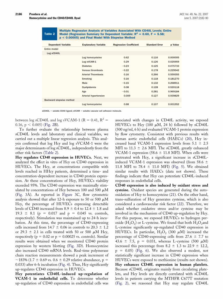
etween log sCD40L and log sVCAM-1 (R � 0.41, R2 �.16, p � 0.005) (Fig. 2B).To further evaluate the relationship between plasma
CD40L levels and laboratory and clinical variables, wearried out a multiple linear regression analysis. This anal-sis confirmed that log Hcy and log sVCAM-1 were theajor determinants of log sCD40L, independently from the
ther risk factors (Table 2).cy regulates CD40 expression in HUVECs. Next, we
nalyzed the effect in vitro of Hcy on CD40 expression inUVECs. The Hcy, at concentrations compatible with
evels reached in HHcy patients, determined a time- andoncentration-dependent increase in CD40 protein expres-ion. At these concentrations of Hcy, HUVECs viabilityxceeded 95%. The CD40 expression was maximally stim-lated by concentrations of Hcy between 100 and 500 �MFig. 3A). As reported in Figure 3C, flow cytometricnalysis showed that after 12-h exposure to 50 or 500 �Mcy, the percentage of HUVECs expressing detectable
evels of CD40 increased from 8.9 � 0.4 to 12.4 � 1.8 and9.3 � 8.1 (p � 0.017 and p � 0.045 vs. controls,espectively). Stimulation was maintained up to 24-h incu-ation. At this time, the percentage of CD40-expressingells increased from 14.7 � 0.06 in controls to 20.3 � 1.2r 29.3 � 2.1 in cells treated with 50 or 500 �M Hcy,espectively (p � 0.02 or p � 0.00025 vs. controls). Similaresults were obtained when we monitored CD40 proteinxpression by western blotting (Fig. 3D). Homocysteinelso increased CD40 mRNA levels. Real-time polymerasehain reaction measurements showed a peak increment of183% (1.7 � 0.69 vs. 0.6 � 0.29 relative abundance, p �
.033) after 6-h incubation (Fig. 4). Thus, Hcy significantlyp-regulates CD40 expression in HUVECs.cy potentiates CD40L-induced up-regulation of
CAM-1 in endothelial cells. To determine whether
Multiple Regression Analysis of Variables AssocModel (Regression Summary for Dependent Varip < 0.00005) and Final Model With Stepwise M
Table 2Multiple Regression Analysis of VarModel (Regression Summary for Dep < 0.00005) and Final Model With
Dependent Variable Explanatory Variable
Entire model
Log sCD40L
Log homocysteine
Log sVCAM-1
Diabetes
Venous thrombosis
Arterial Thrombosis
Smoking
Male
Dyslipidemia
Age
Arterial hypertension
Backward stepwise method
Log homocysteine
sCD40L � soluble CD40 ligand; sVCAM � soluble vascular cell adhes
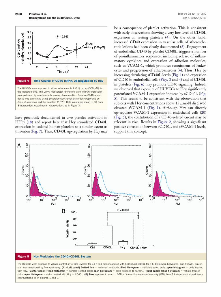
p-regulation of CD40 expression in endothelial cells was (
ssociated with changes in CD40L activity, we exposedUVECs to Hcy (100 �M, 24 h) followed by sCD40L
500 ng/ml, 6 h) and evaluated VCAM-1 protein expressiony flow cytometry. Consistent with previous results withuman aortic endothelial cells (HAECs) (20), Hcy in-reased basal VCAM-1 expression levels from 5.1 � 2.5
FI to 11.3 � 2.6 MFI. The sCD40L greatly enhancedCAM-1 expression (58.6 � 11.8 MFI). When cells wereretreated with Hcy, a significant increase in sCD40L-nduced VCAM-1 expression was observed (from 58.6 �1.8 MFI to 78.4 � 11.0 MFI) (Fig. 5). We obtainedimilar results with HAECs (data not shown). Thesendings indicate that Hcy can potentiate CD40L-inducedesponses in endothelial cells.
D40 expression is also induced by oxidant stress andysteine. Oxidant species are generated during the auto-xidation of Hcy to homocysteine (21). On the other hand,rans-sulfuration of Hcy generates cysteine, which is alsoonsidered a cardiovascular risk factor (22). Therefore, wesked whether oxidative stress and/or cysteine may benvolved in the mechanism of CD40 up-regulation by Hcy.or this purpose, we exposed HUVECs to hydrogen per-xide (H2O2) or L-cysteine. We found that both H2O2and-cysteine significantly up-regulated CD40 expression inUVECs. In particular, H2O2 (300 �M) increased the
ercentage of CD40-expressing cells from 27.6 � 7.7 to3.6 � 7.5, p � 0.031, whereas L-cysteine (500 �M)ncreased this percentage from 8.2 � 1.3 to 22.9 � 12.2,
� 0.05) (Fig. 6). We also observed a modest, nottatistically significant increase in CD40 expression whenUVECs were exposed to methionine (results not shown).cy enhances CD40L expression in human platelets.ecause sCD40L originates mainly from circulating plate-
ets, and Hcy levels are directly correlated with sCD40Levels in patients carrying the MTHFR C677T mutation
With CD40L Levels; EntireR2 � 0.60, F � 5.58;d
s Associated With CD40L Levels; Entireent Variable: R2 � 0.60, F � 5.58;pwise Method
gression Coefficient Standard Error p Value
0.62 0.116 0.000005
0.29 0.126 0.025959
�0.23 0.125 0.075733
�0.22 0.178 0.229542
0.16 0.266 0.559355
0.16 0.118 0.181270
�0.15 0.132 0.268911
0.08 0.128 0.520118
�0.01 0.281 0.969184
0.009 0.127 0.939829
0.68 0.107 0.001552
lecule.
iatedable:etho
iablepend
Ste
Re
Fig. 2), we reasoned that Hcy may regulate CD40L

ewHCi3bCtic
D
IsH
ImC(pbv
i(wcoal
2187JACC Vol. 49, No. 22, 2007 Prontera et al.June 5, 2007:2182–90 Homocysteine and the CD40/CD40L Dyad
xpression in platelets. To verify this hypothesis, we exposedashed human platelets to an increasing concentration ofcy for varying times. Homocysteine up-regulated plateletD40L expression, with a maximum when platelets were
ncubated with 500 �M Hcy for 5 min (14.1 � 4.8 MFI to1.4� 6.2 MFI (p � 0.025) (Fig. 7). Remarkably, throm-in, which is considered a potent inducer of plateletD40L, stimulated CD40L expression to a similar ex-
ent as Hcy (33.3 � 5.0 MFI, p � 0.044) (Fig. 7),ndicating that results with Hcy may be pathophysiologi-ally relevant.
iscussion
n the present report, we present the first evidence of aignificant increase in sCD40L levels in individuals with
Figure 3 Impact of Hcy on CD40 Protein Expression in HUVECs
(A) The left panel shows untreated cells. The dashed histogram represents the irright panels show HUVECs exposed to respectively 50 �M or 500 �M Hcy for 24cells. Each histogram is representative of at least 3 experiments and shows cell nIncrease in CD40 expression by Hcy in HUVECs is concentration dependent. Cells12 h, and CD40 expression was evaluated by flow cytometry. Results are from a rexperiments with HUVECs exposed to the indicated concentrations of Hcy for 12 hblot analysis. The HUVECs were exposed to the indicated concentrations of Hcy foSD of densitometric analyses from 3 separate experiments. Results were normalizHUVECs � human umbilical vein endothelial cells.
Hcy expressing the MTHFR C677T genotype (Fig. 1A). (
n addition, we show that Hcy, at concentrations thatay be reached in patients with HHcy, up-regulatesD40 expression in HUVECs and CD40L in platelets
Figs. 3, 4, and 7). These results unravel a novel potentialathogenetic mechanism of HHcy-associated atherothrom-osis, namely the up-regulation of CD40 signaling inascular cells.
The involvement of the CD40/CD40L dyad in vascularnflammation and atherogenesis is now widely recognized23). Increased sCD40L levels have been found in patientsith a variety of cardiovascular disorders (11–16). In these
onditions, activated platelets may represent a major sourcef sCD40L. It has been documented that sCD40L levelsre reduced by antiplatelet drugs (24), and positively corre-ate with 11d-TXB2, an index of in vivo platelet activation
t antibody; the green histogram depicts the anti-CD40 antibody. The middle anden histograms indicate untreated cells, open histograms depict Hcy-treatedrs (y axis) versus log fluorescence intensity (x axis) for 6.000 viable cells. (B)xposed to increasing concentrations of Hcy (10, 25, 50, 100, 500 �M) forntative experiment. (C) The graph summarizes results from 3 to 5 differenth. Data are expressed as mean � SD of percent of total events. (D) Western
. The upper panel shows a representative blot. The lower panel depicts mean �
�-actin. Ctrl � control; FL1 � fluorescent channel 1; Hcy � homocysteine;
relevanh. Greumbewere eepreseor 24
r 12 hed for
25). This also may be the case with HHcy subjects. We
hHet
bweiroomscioiwp5seu(rps
2188 Prontera et al. JACC Vol. 49, No. 22, 2007Homocysteine and the CD40/CD40L Dyad June 5, 2007:2182–90
ave previously documented in vivo platelet activation inHcy (18) and report here that Hcy stimulated CD40L
xpression in isolated human platelets to a similar extent ashrombin (Fig. 7). Thus, CD40L up-regulation by Hcy may
Figure 4 Time Course of CD40 mRNA Up-Regulation by Hcy
The HUVECs were exposed to either vehicle control (Ctrl) or Hcy (500 �M) forthe indicated time. The CD40 messenger ribonucleic acid (mRNA) expressionwas evaluated by real-time polymerase chain reaction. Relative CD40 abun-dance was calculated using glyceraldehyde-3-phosphate dehydrogenase asgene of reference and the equation 2���Ct. Data points are mean � SD from3 independent experiments. Abbreviations as in Figure 3.
Figure 5 Hcy Modulates the CD40/CD40L System
The HUVECs were exposed to vehicle control or to 100 �M Hcy for 24 h and thension was measured by flow cytometry. (A) (Left panel) Dotted line � irrelevanwith Hcy. (Center panel) Filled histogram � vehicle-treated cells; open histogrcells; open histogram � cells treated with Hcy � CD40L. (B) Bars represent mAbbreviations as in Figures 1 and 3.
e a consequence of platelet activation. This is consistentith early observations showing a very low level of CD40L
xpression in resting platelets (4). On the other hand,ncreased CD40 expression in vascular cells of atheroscle-otic lesions had been clearly documented (8). Engagementf endothelial CD40 by platelet CD40L triggers a numberf proinflammatory responses, including release of inflam-atory cytokines and expression of adhesion molecules,
uch as VCAM-1, which promotes recruitment of leuko-ytes and progression of atherosclerosis (4). Thus, Hcy byncreasing circulating sCD40L levels (Fig. 1) and expressionf CD40 in endothelial cells (Figs. 3 and 4) and of CD40Ln platelets (Fig. 6) may promote CD40 signaling. Indeed,e observed that exposure of HUVECs to Hcy significantlyotentiated VCAM-1 expression induced by sCD40L (Fig.). This seems to be consistent with the observation thatubjects with Hcy concentrations above 15 �mol/l displayedlevated sVCAM-1 (Fig. 1). Although Hcy can directlyp-regulate VCAM-1 expression in endothelial cells (20)Fig. 5), the contribution of a CD40-related circuit may beelevant in vivo. Results in Figure 2, showing a significantositive correlation between sCD40L and sVCAM-1 levels,upport this concept.
ted with 500 ng/ml CD40L for 6 h. Cells were harvested, and VCAM-1 expres-ody; filled histogram � vehicle-treated cells; open histogram � cells treatedcells exposed to CD40L. (Right panel) Filled histogram � vehicle-treatedSEM of mean fluorescence intensity (MFI) from 3 independent experiments.
incubat antibam �
ean �

mhiOoaHdi(
HalTrpitcfHrtgpcr(pwtsHmcoHu
2189JACC Vol. 49, No. 22, 2007 Prontera et al.June 5, 2007:2182–90 Homocysteine and the CD40/CD40L Dyad
Homocysteine may alter endothelial cell functions byultiple and still incompletely elucidated mechanisms. We
ave recently reported that severe HHcy is associated withncreased peroxidation of arachidonic acid in vivo (18).
thers have shown that exposure to Hcy triggers generationf reactive oxygen species (26,27) and that Hcy impairs thentioxidant potential of endothelial cells (28). Thus, somecy bioactions may be related to the induction of oxidative
amage. Our present results, showing that hydrogen perox-de significantly increases CD40 expression in HUVECs
Figure 6 Effect of H2O2 and L-cysteine onCD40 Glycoprotein Expression in HUVECs
(A) The upper left panel shows untreated cells (the open histogram representsthe irrelevant antibody, whereas the blue histogram depicts the anti-CD40 anti-body). The upper right panel shows a comparison between untreated cells(blue histogram) and cells treated with H2O2 (300 �M) for 30 min (open histo-gram). Cells were washed and cultured in normal medium for 24 h before flowcytometric analysis (upper panels). The graph in the lower panel shows mean� SD from 3 different experiments. (B) The upper left panel shows untreatedcells (the open histogram represents the irrelevant antibody, whereas the bluehistogram depicts the anti-CD40 antibody). The upper right panel shows acomparison between untreated cells (blue histogram) and cells treated withL-cysteine (500 �M) for 12 h. The lower panel shows mean � SD from 3 sep-arate experiments. Ctrl � control; other abbreviations as in Figure 3.
Fig. 5), suggest that oxidative events may be involved in
cy-induced CD40 up-regulation. This is an intriguingspect. It is, in fact, known that CD40 activation by itsigand stimulates production of reactive oxygen species (29).hus, up-regulation of CD40 by oxidant damage may
epresent a mechanism of amplification of CD40L-inducedroinflammatory responses. However, the molecular eventsnvolved in CD40 stimulation by Hcy in HUVECs remaino be fully elucidated. Because CD40 expression in vascularells is under the control of the transcription factor nuclearactor (NF)-� (30) and Hcy stimulates NF-� activity in
UVECs via generation of superoxide anion (27), it may beeasoned that CD40 up-regulation by Hcy may proceedhrough NF-�B activation. On the other hand, productsenerated during Hcy metabolism also may play a role in theathogenesis of atherothrombosis. In particular, cysteine isonsidered a cardiovascular risk factor (22), whereas a recenteport emphasizes the role of methionine in atherogenesis31). In our experimental conditions, L-cysteine was asotent as Hcy at inducing CD40 expression (Fig. 5),hereas results with methionine were less consistent, al-
hough some increase in CD40 expression could be ob-erved. Thus, it may be hypothesized that at least part of thecy effects on CD40 expression may be related to itsetabolic conversion to cysteine. On the other hand,
ysteine is known to potentiate the capability of Hcy toxidate low-density lipoprotein (32), suggesting that bothcy and cysteine may converge on pro-oxidant pathways to
p-regulate CD40.
Figure 7 Homocysteine Stimulates CD40LExpression in Washed Human Platelets
Platelets (1 � 108 /ml) were exposed to Homocysteine (Hcy) (500 �M) orthrombin (Thr) (0.2 U/ml) for 5 min at 37°C and analyzed by flow cytometry.(A) Filled histograms represent untreated cells, whereas open histograms rep-resent cells exposed to Hcy or Thr. Staining with irrelevant nonbinding antibodyis indicated by dotted histograms. (B) Bars represent mean � SD of meanfluorescence intensity (MFI) from 3 independent experiments. Ctrl � control;other abbreviations as in Figure 3.

uCta
AT
RDD
R
1
1
1
1
1
1
1
1
1
1
2
2
2
22
2
2
2
2
2
3
3
3
2190 Prontera et al. JACC Vol. 49, No. 22, 2007Homocysteine and the CD40/CD40L Dyad June 5, 2007:2182–90
In conclusion, our present results showing a previouslynappreciated relationship between Hcy and the CD40/D40L dyad in vivo and in vitro provide novel evidence of
he role that HHcy may play in the pathogenesis oftherothrombosis.
cknowledgmenthe authors thank Angela Falco for assistance with patients.
eprint requests and correspondence: Dr. Mario Romano,ipartimento di Scienze Biomediche, Ce.S.I., Universitá G.’Annunzio, 66013 Chieti, Italy. E-mail: [email protected].
EFERENCES
1. Clarke R, Daly L, Robinson K, et al. Hyperhomocysteinemia: anindependent risk factor for vascular disease. N Engl J Med 1991;324:1149–55.
2. Austin RC, Lentz SR, Werstuck GH. Role of hyperhomocysteinemiain endothelial dysfunction and atherothrombotic disease. Cell DeathDiffer 2004;11:S56–64.
3. Mach F, Schonbeck U, Sukhova GK, et al. Functional CD40 ligand isexpressed on human vascular endothelial cells, smooth muscle cells,and macrophages: implications for CD40-CD40 ligand signaling inatherosclerosis. Proc Natl Acad Sci U S A 1997;94:1931–6.
4. Henn V, Slupsky JR, Grafe M, et al. CD40 ligand on activatedplatelets triggers an inflammatory reaction of endothelial cells. Nature1998;391:591–4.
5. Dechanet J, Grosset C, Taupin JL, et al. CD40 ligand stimulatesproinflammatory cytokine production by human endothelial cells.J Immunol 1997;159:5640–7.
6. Horton DB, Libby P, Schonbeck U. Ligation of CD40 on vascularsmooth muscle cells mediates loss of interstitial collagen via matrixmetalloproteinase activity. Ann N Y Acad Sci 2001;947:329–36.
7. Schonbeck U, Mach F, Sukhova GK, et al. CD40 ligation inducestissue factor expression in human vascular smooth muscle cells. Am JPathol 2000;156:7–14.
8. Mach F, Schonbeck U, Sukhova GK, Atkinson E, Libby P. Reductionof atherosclerosis in mice by inhibition of CD40 signaling. Nature1998;394:200–3.
9. Lutgens E, Gorelik L, Daemen MJAP, et al. Requirement for CD154in the progression of atherosclerosis. Nat Med 1999;5:1313–6.
0. Graf D, Muller S, Korthauer U, vanKooten C, Weise C, Kroczek RA.A soluble form of TRAP (CD40 ligand) is rapidly released after T cellactivation. Eur J Immunol 1995;25:1749–54.
1. Aukrust P, Muller F, Ueland T, et al. Enhanced levels of soluble andmembrane-bound CD40 ligand in patients with unstable angina.Possible reflection of T lymphocyte and platelet involvement in thepathogenesis of acute coronary syndromes. Circulation 1999;100:614 –20.
2. Heeschen C, Dimmeler S, Hamm CW, et al., CAPTURE StudyInvestigators. Soluble CD40 ligand in acute coronary syndromes.N Engl J Med 2003;348:1104–11.
3. Garlichs CD, John S, Schmeisser A, et al. Upregulation of CD40 and
CD40 ligand (CD154) in patients with moderate hypercholesterol-emia. Circulation 2001;104:2395–400.4. Cipollone F, Mezzetti A, Porreca E, et al. Association betweenenhanced soluble CD40L and prothrombotic state in hypercholester-olemia: effects of statin therapy. Circulation 2002;106:399–402.
5. Damas JK, Otterdal K, Yndestad A, et al. Soluble CD40 ligand inpulmonary arterial hypertension: possible pathogenic role of theinteraction between platelets and endothelial cells. Circulation 2004;110:999–1005.
6. Varo N, Vicent D, Libby P, et al. Elevated plasma levels of theatherogenic mediator soluble CD40 ligand in diabetic patients: a noveltarget of thiazolidinediones. Circulation 2003;107:2664–9.
7. Welch GN, Loscalzo JN. Homocysteine and atherothrombosis.N Engl J Med 1998;338:1042–50.
8. Davi G, DiMinno G, Coppola A, et al. Oxidative stress and plateletactivation in homozygous homocystinuria. Circulation 2001;104:1124–8.
9. Frosst P, Blom HJ, Milos R, et al. A candidate genetic risk factor forvascular disease: a common mutation in methylenetetrahydrofolatereductase. Nat Genet 1995;10:111–3.
0. Silverman MD, Tumuluri RJ, Davis M, Lopez G, Rosenbaum JT,Lelkes PI. Homocysteine upregulates vascular cell adhesionmolecule-1 expression in cultured human aortic endothelial cells andenhances monocyte adhesion. Arterioscler Thromb Vasc Biol 2002;22:587–92.
1. Loscalzo J. The oxidant stress of hyperhomocyst(e)inemia. J ClinInvest 1996;98:5–7.
2. El-Khairy L, Ueland PM, Refsum H, Graham IM, Vollset SE,European Concerted Action Project. Plasma total cysteine as a riskfactor for vascular disease: the European Concerted Action Project.Circulation 2001;103:2544–9.
3. Libby P. Inflammation in atherosclerosis. Nature 2002;420:868–74.4. Nannizzi-Alaimo L, Alves VL, Phillips DR. Inhibitory effects of
glycoprotein IIb/IIIa antagonists and aspirin on the release of solubleCD40 ligand during platelet stimulation. Circulation 2003;107:1123–8.
5. Santilli F, Davi G, Consoli A, et al. Thromboxane-dependent CD40ligand release in type 2 diabetes mellitus. J Am Coll Cardiol 2006;47:391–7.
6. Zeng X, Dai J, Remick DG, Wang X. Homocysteine mediatedexpression and secretion of monocyte chemoattractant protein-1 andinterleukin-8 in human monocytes. Circ Res 2003;93:311–20.
7. Au-Yeung KK, Woo CW, Sung FL, Yip JC, Siow YL, O K.Hyperhomocysteinemia activates nuclear factor-kappa B in endothelialcells via oxidative stress. Circ Res 2004;94:28–36.
8. Weiss N, Zhang YY, Heydrick S, Bierl C, Loscalzo J. Overexpressionof cellular glutathione peroxidase rescues homocyst(e)ine-inducedendothelial dysfunction. Proc Natl Acad Sci U S A 2001;98:12503–8.
9. Urbich C, Dernbach E, Aicher A, Zeiher AM, Dimmeler S. CD40ligand inhibits endothelial cell migration by increasing production ofendothelial reactive oxygen species. Circulation 2002;106:981–6.
0. Wagner AH, Gebauer M, Pollok-Kopp B, Hecker M. Cytokine-inducible CD40 expression in human endothelial cells is mediated byinterferon regulatory factor-1. Blood 2002;99:520–5.
1. Troen AM, Lutgens E, Smith DE, Rosenberg IH, Selhub J. Theatherogenic effect of excess methionine intake. Proc Natl Acad SciU S A 2003;100:15089–94.
2. Pfanzagl B, Tribl F, Koller E, Moslinger T. Homocysteine stronglyenhances metal-catalyzed LDL oxidation in the presence of cystine
and cysteine. Atherosclerosis 2003;168:39–48.