locally delivered cd40 agonist antibody …...research article locally delivered cd40 agonist...
TRANSCRIPT
Research Article
Locally Delivered CD40 Agonist Antibody Accumulates inSecondary Lymphoid Organs and Eradicates ExperimentalDisseminated Bladder Cancer
Linda C. Sandin1, Anna Orlova3, Erika Gustafsson1, Peter Ellmark4,5, Vladimir Tolmachev2,Thomas H. T€otterman1, and Sara M. Mangsbo1
AbstractImmunotherapy with intratumoral injection of adenoviral vectors expressing CD40L has yielded positive
results in experimental and clinical bladder cancer.We therefore hypothesized that anti-CD40 antibody would beeffective in this setting. Agonistic CD40 antibodies were developed as vaccine adjuvants but have later been usedas treatment of advanced solid tumors and hematologic cancers. Systemic anti-CD40 therapy has been associatedwith immune-related adverse events, such as cytokine release syndrome and liver toxicity, and local delivery is anattractive approach that could reduce toxicity. Herein, we compared local and systemic anti-CD40 antibodydelivery to evaluate efficacy, toxicity, and biodistribution in the experimental MB49 bladder cancer model.Antitumor effects were confirmed in the B16 model. In terms of antitumor efficacy, local anti-CD40 antibodystimulation was superior to systemic therapy at an equivalent dose and CD8T cells were crucial for tumor growthinhibition. Both administration routes were dependent on host CD40 expression for therapeutic efficacy. In vivobiodistribution studies revealed CD40-specific antibody accumulation in the tumor-draining lymph nodes andthe spleen, most likely reflecting organs with frequent target antigen-expressing immune cells. Systemicadministration led to higher antibody concentrations in the liver and blood compared with local delivery, andwas associated with elevated levels of serum haptoglobin. Despite the lack of a slow-release system, local anti-CD40 therapy was dependent on tumor antigen at the injection site for clearance of distant tumors. Tosummarize, local low-dose administration of anti-CD40 antibody mediates antitumor effects in murine modelswith reduced toxicity and may represent an attractive treatment alternative in the clinic. Cancer Immunol Res;2(1); 80–90. �2013 AACR.
IntroductionIn 2008, approximately 380,000 patients worldwide were
diagnosed with bladder cancer and 150,000 succumbed to thedisease (1). The majority of these patients present with super-ficial transitional cell carcinoma (Tis, Ta, or T1) that is treatedwith transurethral resection followed by intravesical chemo-therapy, or for high-risk tumors local Bacillus Calmette-Gu�erin(BCG) immunotherapy (2, 3). BCG treatment induces remis-sion in a majority of patients but is associated with adverse
events and 30% to 50% of the patients ultimately fail to respond(4). Radical cystectomy for patients with muscle-invasivebladder cancer results in initial tumor control, but providesa 5-year survival rate of only 40% to 60% due to the presence ofmicrometastatic disease (2). We have previously shown thatlocal adenoviral CD40L (AdCD40L) as well as CpG therapy caninduce systemic antitumor responses in experimental bladdercancer (5, 6). AdCD40L therapy has proven efficient and safe inboth humans (7) and dogs (8). Consequently, we wanted toinvestigate whether local low-dose agonistic CD40 antibodyinjection could clear experimental bladder cancer. CD40 isexpressed by various cell types in the myeloid cell lineage, i.e.,dendritic cells, macrophages and monocytes, B cells as well asendothelial cells (9), and dendritic cells are recognized asimportant targets for anti-CD40 antibody in cancer immuno-therapy. Activated dendritic cells can efficiently engulf, pro-cess, and present tumor antigen to T cells, resulting in powerfulantitumor responses (10, 11). In addition to the antigen-pre-senting cell (APC)–activating properties of anti-CD40 anti-body, CD40 ligation on CD40-expressing tumor cells initiatesprogrammed cell death (12), antibody-dependent cellular cyto-toxicity (13), antibody-dependent cellular phagocytosis (14), orcomplement-mediated cytotoxicity (15), depending on theimmunoglobulin G (IgG) subclass.
Authors' Affiliations: 1Department of Immunology, Genetics and Pathol-ogy, Division of Clinical Immunology, 2Department of Radiology, Oncologyand Radiation Sciences, Division of Biomedical Radiation Sciences;3Department of Medical Chemistry, Preclinical PET Platform, UppsalaUniversity, Uppsala; 4Alligator Bioscience AB; and 5Department of Immu-notechnology, Lund University, Lund, Sweden
Note: Supplementary data for this article are available at Cancer Immu-nology Research Online (http://cancerimmunolres.aacrjournals.org/).
Corresponding Author: Sara Mangsbo, Department of Immunology,Genetics and Pathology, Uppsala University, Rudbeck Laboratory C11,Dag Hammarskj€olds v€ag 20, SE-751 85 Uppsala, Sweden. Phone: þ46(0)18-6119181; Fax: þ46(0)18-6110222; E-mail: [email protected]
doi: 10.1158/2326-6066.CIR-13-0067
�2013 American Association for Cancer Research.
CancerImmunology
Research
Cancer Immunol Res; 2(1) January 201480
on February 1, 2020. © 2014 American Association for Cancer Research. cancerimmunolres.aacrjournals.org Downloaded from
Published OnlineFirst October 21, 2013; DOI: 10.1158/2326-6066.CIR-13-0067

Several CD40 agonistic antibodies have been developedfor clinical use by the intravenous administration route(16–18). The resulting systemic immune activation has beenassociated with considerable toxicity, including grade 1 to2 cytokine release syndrome and transient lymphopenia(16, 19). This study extends the observations of Fransen andcolleagues, who compared local low-dose anti-CD40 therapyin combination with the slow-release adjuvant Montanideof virally transformedmurine tumors. Local low-dose admin-istration was as effective as systemic high-dose therapy,but with reduced liver toxicity (20). In the current study,we use an experimental bladder cancer model, a tumor wellsuited for local immunotherapy for which novel therapiesare needed to target disseminated disease. We wanted toinvestigate whether locally administered anti-CD40 antibodycould be as efficient as an equivalent dose delivered sys-temically, and to establish whether the alternative admin-istration route could reduce toxicity. Subsequently, weaimed to pinpoint the effector mechanisms behind thesuccessful local CD40-stimulating immunotherapy for blad-der cancer and, lastly, to compare the in vivo biodistributionbetween the two administration routes.
Materials and MethodsCell lines and reagentsThe murine bladder transitional cell carcinoma cancer
cell line mouse bladder-49 (MB49; a kind gift from Dr. K.Esuvaranathan, National University of Singapore, Singaporein 1996) was cultured in DMEMþ GlutaMax supplementedwith 10% FBS, 0.1 mmol/L sodium pyruvate, 100 U/mLpenicillin–streptomycin (PEST) at 37�C and 5% CO2. Lewislung cell carcinoma-1 (LLC-1;American Type Culture Col-lection) was kept in the same culture medium as MB49. TheD1 cell line (21) is a growth factor–dependent immaturesplenic mouse dendritic cell line cultured in Iscove's mod-ified Dulbecco's medium (IMDM) supplemented with 10%FBS, 100 U/mL PEST, 100 mmol/L b-mercaptoethanol (Invi-trogen), and 20 ng/mL recombinant murine granulocytemacrophage colony–stimulating factor (GM-CSF; NordicBiosite) at 37�C and 5% CO2. All cell lines tested negativefor Mycoplasma but were not authenticated in our labora-tory. Agonistic rat-anti-mouse CD40 (clone: FGK4.5) and ratIgG2a (clone: 2A3) were purchased from BioXCell anddiluted in PBS. Depletion of CD8þ cells was performed byinjecting 20 mg/g body weight rat-anti-mouse CD8a (clone:53.6.72, BioXCell) intraperitoneally on days 0, 1, 2, 6, 10, and14. CD8þ T-cell depletion was confirmed by flow cytometryby staining with clone 53-5.8 (data not shown).
AnimalsC57BL/6 mice were obtained from Taconic M&B. CD40�/�
knockout C57BL/6 mice were obtained from The JacksonLaboratory (B6.129P2-Cd40tm1Kik/J). Animals were housed atthe Rudbeck Animal Facility and cared for by the staff accord-ing to regional regulation. All animal experiments wereapproved by the Uppsala Animal Ethics Committee (Dnr:C303/9, C21/10, C11/11, and C38/11).
In vivo experimental designSeveral variants of subcutaneous MB49 tumor models were
used in this study. In themajority of experiments, 2.5� 105 cellswere injected in the right flank of C57BL/6 mice with therapyconducted on days 7, 10, and 13. For the biodistribution study,2� 105MB49 cells were inoculated in the right and leftflank onday 0. Ten days later, 30 mg of radioactively labeled 125I-CD40antibody and 131I-rat IgG2a were mixed and injected onceintravenously or peritumorally at the right side of the tumor.Animals were sacrificed 4, 24, 48, and 72 hours after injection,organs were isolated, and their radioactivity was measured.Radioactivity of 125I-CD40 antibody was measured in theenergy window of 3 to 6 keV and 131I-rat IgG2a of 100 to380 keV. In the last tumor model investigating the systemiceffects of low-dose anti-CD40 therapy, 2.5 � 105 MB49 cellswere injected in the right flank on day 0 and close to the leftshoulder on day 1. Therapy was injected peritumorally at theprimary tumor, subcutaneously in the nontumor flank, orintravenously every third day for a total of three times. Tokeep a similar distance between injection site and thedistant tumor as well as to reduce passive diffusion ofantibodies in the void space of the skin, animals injectedwith subcutaneous anti-CD40 antibody in the nontumorflank had both tumors inoculated on the same side of theanimal, opposite to the injection side (Fig. 2E). Antibodysolution was administered in 100 mL. Tumor growth andsurvival were monitored throughout the experiment using acaliper and tumor size was calculated by the ellipsoidvolume formula: ¼ 4/3�p�a (radius of length) � b (radiusof width) � c (radius of depth). Mice were sacrificed if thetumor exceeded 1 cm3 or if ulcers developed. Tumor rechal-lenge, by injection of 2.5 � 105 MB49 (contralateral flank)and LLC-1 cells (right foreleg), was performed on mice thathad been tumor free for over 100 days.
Labeling of antibodies125I and 131I were purchased from PerkinElmer. Chloramine-
T and sodium metabisulfite were from Sigma Chemical Com-pany. Chloramine-T and sodium metabisulfite solutions wereprepared immediately before use. The radiochemical purity ofthe labeled antibody construct was analyzed using instant thinlayer chromatography (ITLC) on 150-771 DARK GREEN, Tec-Control Chromatography Strips from Biodex Medical System.Distribution of radioactivity along the ITLC strips was mea-sured using a Cyclone Storage phosphor system and analyzedwith the OptiQuant image analysis software (PerkinElmer).Size-exclusion chromatography was performed on disposableNAP-5 columns (Amersham Pharmacia Biotech AB) accordingto the manufacturer's instructions. The radioactivity wasmeasured using an automated gamma-counter with a 3-inchNaI (Tl) detector (1480 Wizard; Wallac Oy). Monoclonal anti-bodies were labeled using Chloramine-T as an oxidant accord-ing to the following protocol. Anti-CD40 antibody was labeledwith 125I. An antibody solution in PBS (40 mg, 4–2 mL) wasmixed with radioiodine stock solution (2–5 mL, 5–10MBq) and40 mL PBS. The reaction was initiated by adding Chloramine-T(20 mg, 1mg/mL in PBS). After 2-minute incubation at ambienttemperature, the reaction was terminated by adding sodium
Biodistribution and Efficacy of Local CD40 Antibody Injection
www.aacrjournals.org Cancer Immunol Res; 2(1) January 2014 81
on February 1, 2020. © 2014 American Association for Cancer Research. cancerimmunolres.aacrjournals.org Downloaded from
Published OnlineFirst October 21, 2013; DOI: 10.1158/2326-6066.CIR-13-0067

metabisulfite (40 mg, 2 mg/mL in PBS). Rat IgG2a was labeledwith 131I in a similar way. Radiolabeled antibodies were puri-fied from unreacted radioiodine and low-molecular-weightcomponents of the reactive mixture using size-exclusion chro-matography on disposable NAP-5 columns. Radiochemicalpurity of the labeled antibodies was determined by radio-ITLCeluted with acetone:water (8:2) mixture. The yields were in therange of 65% to 75%. After size-exclusion purification, theradiochemical purity of the antibodies was more than 99.5%.
Trichloroacetic acid precipitationTrichloroacetic acid (TCA) precipitates high-molecular-
weight molecules. Precipitation was carried out accordingto the protocol described previously (22). Briefly, on top of300 mL of carrier solution (PBS/0.02% BSA, w/v), 200 mL ofplasma was added. Then, 500 mL of ice-cold 20% TCA/H2O(w/v) solution was added to precipitate high-molecular-weight molecules in the plasma. Tubes were centrifugedand separated before individual measurement for radioac-tivity using an automated gamma-counter with a 3-inch NaI(Tl) detector.
Internalization of anti-CD40 antibodyAnti-CD40 antibody was conjugated with Alexa Fluor 488
protein-labeling kit according to the manufacturer's protocol(Invitrogen). The functionality of the labeled antibody wasverified by staining of A20 cells (data not shown). D1 cells wereseeded in 96-well plates and stimulated overnight with 1mg/mL lipopolysaccharide (LPS; Sigma) to upregulate theexpression of the CD40 receptor. Cells were incubated with0.1 mg/mL Alexa Fluor 488-conjugated anti-CD40 antibodyfor 15 or 60 minutes at 37�C or 4�C or left untreated beforebeing fixed in 3%PFA/PBS on ice, and then washed twice in0.5%PFA/PBS. Trypan blue (Invitrogen) was added just beforeflow-cytometric analysis to quench any surface-bound anti-body. Samples were run in triplicates.
In vivo CD40 expressionTo evaluate which cell populations could potentially act as
targets for anti-CD40 therapy, na€�ve animals were injectedsubcutaneously with 30 mg of anti-CD40 one, two, or threetimes at 3-day intervals. Four hours postinjection the spleenand (pooled) inguinal lymph nodes were harvested anddigested with Liberase TL (Roche) for 15 minutes at 37�C.The Liberase-treated tissue was passed through a MESHmembrane, blocked for FcgR (TruStain fcX), and subsequentlystained for CD11c (clone: N418), CD11b (clone: M1/70), F4/80(CI:A3-1), B220 (RA3-6B2), and CD40 (3/23; all antibodies arefrom BioLegend). Staining with antibody clone 3/23 was nothampered by the presence of the therapeutic clone FGK4.5 oncells (data not shown). The following cell types were investi-gated: B cells (B220þ, CD11b�, CD11c�, and F4/80�), conven-tional dendritic cells (cDC; CD11chi, CD11bþ/�, and B220�),medullarymacrophage (M�; CD11clow/int, CD11bþ, B220�, andF4/80þ), and subcapsular sinus M� (CD11clow/int, CD11bþ,B220�, and F4/80�). Samples were analyzed in a FACSCanto IIcytometer (BD Biosciences). Data analysis was performed withFlowJo software (TreeStar).
Haptoglobin and anti-rat IgG2a antibodymeasurementsBlood was collected by tail vein incision and serum was
stored at �80�C. Mouse haptoglobin was detected by ELISAaccording to the manufacturer's protocol (Life Diagnostics,Inc.). Quantification of circulating anti-CD40 antibody wasperformed by ELISA. Briefly, wells were coated with 1.25 mg/mL anti-rat IgG2a (clone: MARG2a-1; Serotec) and blockedwith 3% milk powder before the addition of samples (1:10–1:75 dilution). Horseradish peroxidase (HRP)–conjugatedmouse-a-rat k/l light chain (MARK-1/MARL-15; Serotec)was used for detection. Substrate (Super Signal pico che-moluminescence; Pierce) was added before the lumines-cence was measured with an ELISA reader (Fluostar optimal;LabVision). Samples were run in duplicates.
Statistical analysisSurvival data were plotted by the Kaplan–Meier method and
analyzed by the log-rank test. When applicable, the D'Agostinoand Pearson omnibus normality test was used before selectionof statistical test. Where indicated, the difference betweengroups was evaluated using unpaired t test or paired t test.P values less than 0.05 were considered significant. Asterisksindicate the confidence interval (�, P < 0.05; ��, P < 0.01; ��� ,P < 0.001). Statistical analyses were performed using GraphPadprism software (GraphPad Software, Inc.).
ResultsLocal low-dose anti-CD40 therapy results in improvedsurvival and reduced toxicity compared with systemictreatment
To our knowledge, a direct comparison between local andsystemic administration of equal doses of anti-CD40 therapyhas not been performed. Ten and 30 mg of peritumorallydelivered antibody prolonged survival compared with con-trol animals, whereas systemic therapy of the same doses didnot (Fig. 1A, top and middle). Only the highest systemic dose(100 mg) resulted in prolonged survival that was similar tothat in the local therapy-treated animals (Fig. 1A, bottom).
Several immune-related side effects have been observedwith systemic anti-CD40 antibody therapy in both preclin-ical models (23) and patients (19). In our current study,levels of circulating therapeutic antibody were measured 4hours after each injection (Fig. 1B). For all doses andadministration routes, the serum maximum level of anti-CD40 antibody occurred after the second injection, with thehighest concentrations in animals treated systemically with30 or 100 mg of antibodies. Local immunotherapy led toreduced serum levels of antibody in all groups comparedwith that of the equivalent systemic dose. The initial reduc-tion in systemic antibody levels could translate to reducedtoxicity. In the current studies, we used haptoglobin, anacute phase protein produced mainly by the liver, as amarker for systemic inflammation (24–26). Serum levels ofhaptoglobin were measured 24 hours after the first dose, andelevated levels were detected in mice treated with intrave-nous injections (Fig. 1C). Local injection of the same dosedid not elevate haptoglobin levels to the same degree, and
Sandin et al.
Cancer Immunol Res; 2(1) January 2014 Cancer Immunology Research82
on February 1, 2020. © 2014 American Association for Cancer Research. cancerimmunolres.aacrjournals.org Downloaded from
Published OnlineFirst October 21, 2013; DOI: 10.1158/2326-6066.CIR-13-0067

neither peritumorally nor intravenously administeredirrelevant antibody affected haptoglobin levels (data notshown).The local low-dose strategy was also assessed using the fast-
growing B16-F10 tumor model where tumor growth inhibitionwas evident compared with control animals (SupplementaryFig. S1A). Furthermore, weekly dosing with peritumorally low-dose anti-CD40 was evaluated in a high MB49-tumor burdenmodel. Both the weekly and the 3-day interval treatmentschedule restrained tumor growth. However, elevated levelsof ADA were detected in the serum already after the secondinjection of anti-CD40 antibody on a weekly schedule, whichwas not seen using the 3-day interval protocol (data notshown).
CD8þT cells, host CD40 expression, and tumor antigen atthe injection site are all required for full therapeuticefficacy
CD40 is widely expressed on different cell types in vivo,and a variety of cells have been proposed to be effectors inCD40 agonistic therapy. Macrophages (27, 28), B cells (29),natural killer (NK) cells indirectly (30), and T cells (20) viadendritic cell activation have been suggested to play crucialroles in tumor eradication. We asked whether our local low-dose administration of anti-CD40 antibody would depend onthe presence of CD8þ T cells by using cell-depletion experi-ments. CD8þ cells were necessary for optimal antitumoreffects as depleted mice showed no survival benefit com-pared with controls (Fig. 2A). Furthermore, depletion of
100
80
60
40
20
0
25
20
15
10
5
0
Seru
m levels
rat
IgG
2a (
mg/m
L)
Seru
m levels
rat
IgG
2a (
mg/m
L)
Seru
m levels
rat
IgG
2a (
mg/m
L)
Seru
m h
ap
tog
lob
in (
ng
/mL
)
Seru
m levels
rat
IgG
2a (
mg/m
L)
50
40
30
20
10
0
50
40
30
20
10
0
50
40
30
20
10
0
0 20 40 60
Days
0 5
Intravenous
Peritumoral
Intravenous
Peritumoral
Intravenous3×1006
2×1006
2×1006
1×1006
5×1005
0
Peritumoral
Subcutaneous
10 15Days
0 5 10 15Days
0 5 10 15Days
0 5 10 15Days
80 100
0 20 40 60
Days
80 100
0 20 40 60
Days
80 100
Anti-CD40 10 μg p.t. (3/8)
Anti-CD40 10 mgB
C
A Anti-CD40 30 mg
Anti-CD40 100 mg
Anti-CD40 15 mg
Anti-CD40 5 mg
PBS
Haptoglobin ELISA
Anti-CD40 10 μg i.v. (1/8)
Rat IgG2a 30 μg p.t. (0/8)
PBS p.t. (0/8)
Rat IgG2a 30 μg i.v. (0/8)
Rat IgG2a 30 mg
Anti-CD40 30 μg p.t. (4/8)
Anti-CD40 30 μg i.v. (2/8)
Rat IgG2a 30 μg p.t. (0/8)
PBS p.t. (0/8)
Rat IgG2a 30 μg i.v. (0/8)
Anti-CD40 100 μg p.t. (6/8)
Anti-CD40 100 μg i.v. (5/8)
Rat IgG2a 30 μg p.t. (0/8)
PBS p.t. (0/8)
Rat IgG2a 30 μg i.v. (0/8)
Pe
rce
nta
ge
su
rviv
al
100
80
60
40
20
0
Pe
rce
nta
ge
su
rviv
al
100
80
60
40
20
0
Perc
en
tag
e s
urv
ival
Figure 1. Local low-dose anti-CD40 therapy is superior to systemic treatment and reduces circulating antibodies and haptoglobin levels. A, C57BL/6mice were treated as described in Materials and Methods. Survival data are shown for 8 mice/group (log-rank test). B, blood was collectedfor kinetic analysis 4 hours after every treatment occasion from mice in A and analyzed with ELISA. Data are shown as mean � SEM of 6 mice/groupand time point. Samples were run in duplicates. C, serum was isolated 24 hours after the first therapy occasion of mice in Fig. 2F andanalyzed for haptoglobin levels with ELISA. Data are shown as mean � SD of 9 mice/group and time point (Student t test; �, P < 0.05;��, P < 0.01; ���, P < 0.001). p.t., peritumorally.
Biodistribution and Efficacy of Local CD40 Antibody Injection
www.aacrjournals.org Cancer Immunol Res; 2(1) January 2014 83
on February 1, 2020. © 2014 American Association for Cancer Research. cancerimmunolres.aacrjournals.org Downloaded from
Published OnlineFirst October 21, 2013; DOI: 10.1158/2326-6066.CIR-13-0067

CD4þ T cells yielded variable results as 6 of 10 animalsexperienced rapidly growing tumors and 3 of 10 had recur-rence of tumors after initial remission (data not shown).Removal of regulatory T cells (Treg) cannot be excluded.Previous studies have shown that the CD40–CD40L inter-action not only activates APCs but generates a direct anti-tumor effect in CD40-expressing tumors (12, 31). Through-out this study, MB49 cells presented a negligible amount ofCD40 expression when staining with the therapeutic anti-body clone (data not shown). Despite this result, we wantedto exclude tumor cell death induced directly by the antibody
and therefore subjected tumor-bearing C57BL/6 wild-type(WT) and CD40�/� knockout mice to local or systemictreatment. Figure 2B shows that host CD40 expression isrequired for effective anti-CD40 therapy regardless ofadministration route. The knockout mice had elevated levelsof circulating anti-CD40 antibody 4 hours after the lasttreatment compared with WT animals (Figs. 2C and 1B).
To establish tumor-specific memory after local anti-CD40therapy, animals exhibiting complete tumor regression wererechallenged with MB49 cells and LLC-1 (Fig. 2D). LLC-1tumors grew progressively in both na€�ve and cured mice,
100
80
60
40
20
0
Se
rum
lev
els
ra
t Ig
G2
a (
mg/m
L)
0 20 40 60
Days MB49
KOWT KOWT
LLC-1MB49
LLC-180 100
0 20 40 60
Days
Blood kinetics
80 100
7/8
1/51/8
4/5
Anti-CD40 30 μg p.t.
Anti-CD40 p.t.
by tumor
Anti-CD40 s.c.
nontumor side
2nd tumor (day 1)
1st tumor (day 0)
2nd tumor
1st tumor
2nd tumor
1st tumor
2nd tumor
1st tumor
2nd tumor
1st tumor
2nd tumor
1st tumor
2nd tumor
1st tumor
Anti-CD40 i.v.
Anti-CD40 30 μg p.t.
Anti-CD40 30 μg i.v.
WT anti-CD40 30 μg p.t.
WT anti-CD40 30 μg i.v.
WT no therapy
KO anti-CD40 30 μg p.t.
KO anti-CD40 30 μg i.v.
KO no therapy
Anti-CD40 10 μg p.t. (n=5)
Anti-CD40 15 μg p.t. by tumor
Anti-CD40 15 μg s.c. nontumor side
Anti-CD40 15 μg i.v.
Naïve (n=4)
Anti-CD40 30 μg p.t. (n=8)
Anti-CD40 30 μg p.t. + CD8 dep
B
C
A
E
F
D
Rat IgG2a 30 μg p.t. PBS p.t.
PBS p.t.
by tumor
Pe
rce
nta
ge
su
rviv
al
100
80
60
40
20
0
100
80
60
40
20
0
Pe
rce
nta
ge
su
rviv
al
100
80
60
40
20
0
Nu
mb
er
of
reg
res
sin
g t
um
ors
(%
)
9
6
3
0
Nu
mb
er
of
reg
res
se
d t
um
ors
9
6
3
0Nu
mb
er
of
gro
win
g t
um
ors
100
80
60
40
20
0
Nu
mb
er
of
gro
win
g t
um
ors
(%
)
Survival CD8 depletion
Survival WT and KO
Rechallenge
Figure 2. Successful peritumoral (p.t.) anti-CD40 therapy is dependent on CD8þ cells, host CD40 expression, and tumor antigen by the injectionsite. C57BL/6 mice were treated as described in Materials and Methods. A, one therapy group was depleted of CD8þ cells. Survival data of 7to 10 mice/group are shown. B, anti-CD40 therapy was evaluated in wild-type (WT) and CD40 knockout (KO) C57BL/6 animals. Survival curve isshown for 8 mice/group (log-rank test). C, blood kinetic analysis of serum anti-CD40 antibody 4 hours after the last treatment was measuredwith ELISA. Data are shown as mean � SD of 5 mice/group (Student t test). D, complete responders to local anti-CD40 therapy were rechallengedwith 2.5 � 105 MB49 and LLC-1 cells to evaluate tumor-specific memory. Naïve mice, not age-matched, were inoculated with both cell lineson the right and left flank, respectively. Data are visualized as percentage of regressing or progressively growing tumors. E, MB49 cells wereinjected on day 0 and on day 1 of individual mice and treated with 15 mg of anti-CD40 antibody (black arrow) every third day starting onday 7 according to the illustration. F, tumor growth of animals presented as number of regressed or growing tumors per treatment group (n ¼ 9;���, P < 0.001).
Sandin et al.
Cancer Immunol Res; 2(1) January 2014 Cancer Immunology Research84
on February 1, 2020. © 2014 American Association for Cancer Research. cancerimmunolres.aacrjournals.org Downloaded from
Published OnlineFirst October 21, 2013; DOI: 10.1158/2326-6066.CIR-13-0067

whereas progressive growth of MB49 tumors was only seen inna€�ve animals.Because low but detectable levels of anti-CD40 antibody
were found in the blood after local administration, we wantedto investigate whether distant tumors could be cleared andwhether tumor antigens are required at the injection site, asdemonstrated earlier by Fransen and colleagues (20). Tominimize systemic antibody dissemination, 15 mg of anti-CD40antibody was used but without a slow-release formulation. Atwin-tumor model was set up by engrafting MB49 cells in theright flank on day 0 and close to the left shoulder on day 1 (Fig.2E). As visualized in Fig. 2F, anti-CD40 antibody needs to beinjected in close proximity to a tumor (or systemically) to affectboth the treated site as well as the distant tumor. Therapyinjected solely in the skin (nontumor side) did not inhibittumor growth. For individual tumor growth, see Supplemen-tary Fig. S1B.We determined the level of effector cytokines in the serum
and registered a dose-dependent elevation of serum IFN-g , IP-10, and TNF-a 4 hours after the second dose using all admin-istration routes (data not shown). In agreement with theclinical observation using the humanized CP-870,893 antibody(16, 19), we registered a transient B-cell depletion after a single
injection of anti-CD40 antibody (data not shown), which wasless pronounced after local administration.
Local and systemic anti-CD40 antibody accumulates insecondary lymphoid organs
To investigate the in vivo biodistribution of anti-CD40agonist or control antibody upon local versus systemic admin-istration, wemeasured antibody accumulation in target organsafter a single injection. For this purpose, anti-CD40 antibodywas labeled with 125I and the irrelevant isotype with 131I toallow simultaneous measurement in the same animal bygamma-spectrometry. The binding-specificity of the 125I-CD40-specific antibody was confirmed before experimentalinitiation (Supplementary Fig. S2A). An equal dosage mixtureof 125I-CD40–specific and 131I-rat IgG2a control antibodieswas injected into mice carrying a tumor in each flank eitherperitumorally at the right side tumor or intravenously. At 4, 24,48, and 72 hours after injection, animals were sacrificed, organsisolated, and radioactivity measured. A high accumulationof 125I-CD40–specific antibody was detected in the spleen 4hours after injection followed by a decrease over time (Fig. 3A).131I-rat IgG2a control antibody uptake in the spleen wassignificantly lower.
25
20
15
10
5
00 20 40 60
Time after injection (h)
80 0 20 40 60
Time after injection (h)
80
0 20 40 60
Time after injection (h)
80 0 20 40 60
Time after injection (h)
80
125I-CD40 antibody 30 μg i.v.
B
C
A
D
131-I rat IgG2a 30 μg i.v.
125I-CD40 antibody 30 μg p.t.
131-I rat IgG2a 30 μg p.t.
Up
take
, %
ID
/g
8
6
4
2
0
Up
take
, %
ID
/g
40
30
20
10
0
Up
take
, %
ID
/g
80
60
40
20
0
Up
take
, %
ID
/g
Spleen
Liver Blood
TDLN right (injected side)
Figure 3. CD40 agonistaccumulates in secondarylymphoid organs. C57BL/6 micewere treated as described inMaterials and Methods. Targetorgans (A–D) were isolated 4, 24,48, and 72 hours posttreatment,measured for accumulatedradioactivity, and visualized aspercentage injected dose/g tissue(%ID/g). Statistical significance iscalculated between intravenousand peritumoral (p.t.)administrations (mean � SD of4 mice/group; Student pairedt test. �, P < 0.05; ��, P < 0.01;���, P < 0.001).
Biodistribution and Efficacy of Local CD40 Antibody Injection
www.aacrjournals.org Cancer Immunol Res; 2(1) January 2014 85
on February 1, 2020. © 2014 American Association for Cancer Research. cancerimmunolres.aacrjournals.org Downloaded from
Published OnlineFirst October 21, 2013; DOI: 10.1158/2326-6066.CIR-13-0067

The highest amount of locally administered anti-CD40antibody was found in the spleen at 24 hours, followed by adecline. The uptake of 131I-rat IgG2a control antibody in thesame animal was significantly lower than that of the 125I-CD40–specific antibody (Fig. 3A). The spleen accumulationwas significantly lower after peritumoral administration thanafter intravenous injection at 4 and 24 hours. At subsequenttime points, the radioactivity uptakes were equal for bothadministration routes. Anti-CD40 antibody uptake in the righttumor-draining lymph node (TDLN) after peritumoral injec-tion was much higher at 4 hours as compared with that ofsystemic administration (Fig. 3B). Systemic delivery of anti-CD40 antibody induced a more pronounced accumulation inthe liver comparedwith peritumoral injection at 4 and 24 hours(Fig. 3C). Furthermore, local administration resulted in a lowerinitial antibody concentration in the blood at 4 and 24 hours(Fig. 3D), in agreement with our ELISA results. We found thatthe concentration of radioactivity in the plasma was higherthan in whole blood, indicating minor (if any) binding of 125I-CD40 to blood cells (Supplementary Fig. S2B). In summary,peritumoral delivery significantly reduced anti-CD40 antibodyconcentrations in the spleen, liver, and blood compared withintravenous injection at both 4 and 24 hours after injection.
Figure 4 shows antibody uptake for selected target organsat 4 (Fig. 4A) and 24 hours (Fig. 4B). Results from the 48- and72-hour time points are visualized in Supplementary Fig. S3.Statistical differences between the anti-CD40 and the con-
trol antibody and between the two administration routes inall organs investigated are summarized in SupplementaryTables S1–S4.
Repeated anti-CD40 therapymodulates CD40 expressionas well as immune cell distribution in lymphoid tissue
The anti-CD40–specific antibody showed a more rapidclearance from the blood than the control antibody. To verifyhow much of the blood-borne radioactivity was still conjugat-ed, we performed a TCA precipitation assay. Although almostall 131I was conjugated to rat IgG2a in both treatment groupsduring the entire study period, a decrease of activity in thehigh-molecular-weight form was detected for the 125I-CD40–specific antibody conjugate (Fig. 5A). This suggests that theanti-CD40 antibody was efficiently sequestered from thecirculation due to specific interaction followed by subse-quent internalization and catabolism. This could be byactive uptake in cells and we therefore incubated fluoro-phore-conjugated anti-CD40 antibody with CD40-expressingdendritic cells for 15 or 60 minutes and subsequentlyquenched surface-bound antibodies. Our findings imply thatanti-CD40 antibody is internalized within 15 minutes (Fig.5B). As several cell populations in lymphoid organs, includ-ing B cells, CD8þ, CD8� cDCs, macrophages, NK cells, NKTcells, and activated CD4þ T cells, can act as potential targetsfor CD40-activating therapy, we investigated the expressionof CD40 on immune cells from the inguinal lymph nodes
300
250
20080
60
40
20
0
40
30
20
10
0
BloodLiver
Spleen
TDLN right (i
njected side)
TDLN left (noninjected side)
Tumor right
Tumor left
125I-CD40 antibody 30 μg i.v.
B
A
131I-rat IgG2a 30 μg i.v.125I-CD40 antibody 30 μg p.t.131I-rat IgG2a 30 μg p.t.
125I-CD40 antibody 30 μg i.v.131I-rat IgG2a 30 μg i.v.125I-CD40 antibody 30 μg p.t.131I-rat IgG2a 30 μg p.t.
Up
take
, %
ID
/gU
pta
ke
, %
ID
/g
In vivo biodistribution 4 h
In vivo biodistribution 24 h
Figure 4. In vivo biodistribution oftarget organs 4 and 24 hours afterinjection of 125I-CD40–specific and131I-rat IgG2a control antibody.Radioactive uptake of targetorgans (A) 4 and (B) 24 hours afterinjection of animals in Fig. 3. Dataare presented as percentageinjected dose/g tissue (%ID/g,mean � SD of 4 mice/group).Statistical differences betweenanti-CD40 and rat IgG2a andbetween the two administrationroutes are depicted inSupplementary Table S1 (4 hours)and S2 (24 hours). p.t.,peritumorally.
Sandin et al.
Cancer Immunol Res; 2(1) January 2014 Cancer Immunology Research86
on February 1, 2020. © 2014 American Association for Cancer Research. cancerimmunolres.aacrjournals.org Downloaded from
Published OnlineFirst October 21, 2013; DOI: 10.1158/2326-6066.CIR-13-0067
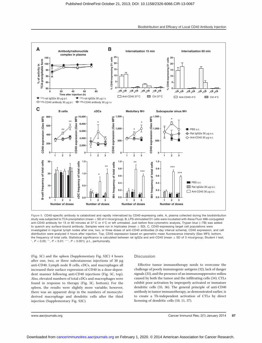
(Fig. 5C) and the spleen (Supplementary Fig. S2C) 4 hoursafter one, two, or three subcutaneous injections of 30 mganti-CD40. Lymph node B cells, cDCs, and macrophages allincreased their surface expression of CD40 in a dose-depen-dent manner following anti-CD40 injections (Fig. 5C, top).Also, elevated numbers of total cDCs and macrophages werefound in response to therapy (Fig. 5C, bottom). For thespleen, the results were slightly more variable; however,there was an apparent drop in the numbers of monocyte-derived macrophage and dendritic cells after the thirdinjection (Supplementary Fig. S2C).
Discussion
Effective tumor immunotherapy needs to overcome thechallenge of poorly immunogenic antigens (32), lack of dangersignals (33), and the presence of an immunosuppressive milieucaused by both the tumor and the infiltrating cells (34). CTLsexhibit poor activation by improperly activated or immaturedendritic cells (35, 36). The general principle of anti-CD40antibody in tumor immunotherapy, as demonstrated earlier, isto create a Th-independent activation of CTLs by directlicensing of dendritic cells (10, 11, 37).
105
100
95
90
85
80
800
600
400
200
0
30
20
10
0
1.5
1.0
0.5
0.0
1.0
0.8
0.6
0.4
0.2
0.0
1.0
0.8
0.6
0.4
0.2
0.0
10,000
8,000
6,000
4,000
2,000
0
1,500
1,000
500
0
1,500
1,000
500
0
125I-CD40 antibody 30 μg i.v.
Anti-CD40 37°C
Anti-CD40 30 μg s.c.
Anti-CD40 4°CCtrl 37°C Ctrl 4°C
BA
C
131I-rat IgG2a 30 μg i.v.131I-rat IgG2a 30 μg p.t.
Rat IgG2a 30 μg s.c.
PBS s.c.
Anti-CD40 30 μg s.c.
Rat IgG2a 30 μg s.c.
PBS s.c.
125I-CD40 antibody 30 μg p.t.
% o
f a
cti
vit
y in
TC
A-p
rec
ipit
ab
le f
orm
25
20
15
10
5
0% C
D40-e
xp
ressin
g D
1 c
ell
s
CD
40 e
xp
ressio
n (
Ge
o M
FI)
B-c
ell
fre
qu
en
cy o
f to
tal
cell
s
cD
C f
req
uen
cy o
f to
tal
cell
s
MΦ
fre
qu
en
cy o
f to
tal
cell
s
MΦ
fre
qu
en
cy o
f to
tal
cell
s
CD
40 e
xp
ressio
n (
Ge
o M
FI)
CD
40 e
xp
ressio
n (
Ge
o M
FI)
CD
40 e
xp
ressio
n (
Ge
o M
FI)
25
20
15
10
5
0% C
D40-e
xp
ressin
g D
1 c
ell
s
Antibody/radionuclide
complex in plasma
Internalization 15 min
B cells cDCs
Number of doses Number of doses Number of doses Number of doses
Medullary MΦ Subcapsular sinus MΦ
Internalization 60 min
0 20 40
1 2 3 1 2 3 1 2 3 1 2 3
1 2 3 1 2 3 1 2 3 1 2 3
Time after injection (h)
60 80 –TB+TB –TB
+TB –TB+TB –TB
+TB–TB
+TB –TB+TB –TB
+TB –TB+TB
Figure 5. CD40-specific antibody is catabolized and rapidly internalized by CD40-expressing cells. A, plasma collected during the biodistributionstudy was subjected to TCA precipitation (mean � SD of 4 mice/group). B, LPS-stimulated D1 cells were incubated with Alexa Fluor 488–conjugatedanti-CD40 antibody for 15 or 60 minutes at 37�C or 4�C or left untreated. Just before flow-cytometric analysis, Trypan blue (þTB) was addedto quench any surface-bound antibody. Samples were run in triplicates (mean � SD). C, CD40-expressing target cell populations wereinvestigated in inguinal lymph nodes after one, two, or three doses of anti-CD40 antibodies (3-day interval scheme), CD40 expression, and celldistribution were analyzed 4 hours after injection. Top, CD40 expression based on geometric mean fluorescence intensity (Geo MFI); bottom,the frequency of total cells. Statistical significance is calculated between rat IgG2a and anti-CD40 (mean � SD of 3 mice/group; Student t test;�, P < 0.05; ��, P < 0.01; ���, P < 0.001). p.t., peritumorally.
Biodistribution and Efficacy of Local CD40 Antibody Injection
www.aacrjournals.org Cancer Immunol Res; 2(1) January 2014 87
on February 1, 2020. © 2014 American Association for Cancer Research. cancerimmunolres.aacrjournals.org Downloaded from
Published OnlineFirst October 21, 2013; DOI: 10.1158/2326-6066.CIR-13-0067

We used peritumoral injection as the local administrationroute in this study because this route confersminimalmechan-ical stimulation of the tumor. A comparison with intratumoralinjection in tumors using the same treatment protocol asin Fig. 1 showed that there was no advantage to this approachover peritumoral delivery (data not shown).
A survival benefit was observed for mice treated with locallow-dose anti-CD40 antibody (10 and 30 mg) compared withthe same dose delivered systemically, which indicates thatsystemic injections in this dose range result in a low concen-tration of antibody in the tumor/TDLN axis that is insufficientfor CTL activation. As no difference in survival was noticed atthe 100 mg dose between the two administration routes, a highlevel of circulating antibody could potentially overcome thisproblem butwith increased side effects.We registered elevatedlevels of serum haptoglobin as a sign of systemic inflammation(24–26) already at low intravenous anti-CD40 antibody doses.Furthermore, liver exposure to the antibody was appreciablyhigher after intravenous injection compared with peritumoralinjection. These data suggest that local low-dose antibodydelivery can reduce liver toxicity compared with systemictherapy, which could be valuable in the clinic as patients oftendisplay metastatic disease that could be targeted by recruitingimmune cells rather than the anti-CD40 antibody itself.
Our finding that CD8þ T cells are important for the antitu-mor effects of anti-CD40 therapy is consistent with data fromother groups (23). However, CD40 activation can target manyother cell types, such as macrophages, which could participatein tumor eradication. The differences observedmay be dose- oranimal model-related. CD40-stimulating therapy can have adirect antitumor effect on CD40-expressing tumors by induc-ing apoptosis (12, 31). Throughout this study, MB49 cellspresented a negligible level of CD40 expression based onstaining with the therapeutic antibody. Notably, when stainingwith the anti-CD40 antibody clone 3/23 on MB49 cells, CD40expression could be visualized. To confirm that locally deliv-ered therapy was fully dependent on host CD40 expression, wemade use of CD40�/� knockout animals. Endogenous CD40expression was required for full therapeutic effect regardless ofinjection site. We also confirmed the antitumor effects in theCD40-negative B16 melanoma model in WT mice. In contrastwith our MB49 model, human bladder cancer cells usuallypresent CD40 expression compared with the CD40-negativeurothelium (38). Also, CD40 ligation in the form of cell-surfacebound CD40L or Fcg receptor cross-linked anti-CD40 antibodyinduces apoptosis in transitional bladder carcinoma (39).Engaging both the direct- and indirect-killing mechanisms forthe treatment of bladder cancer could be even more efficient.These observations, together with the convenient intravesicalaccess of bladder cancer, make this tumor a suitable target forlocalized anti-CD40 therapy.
The presence of tumor antigen in close proximity to theanti-CD40 antibody injection for systemic tumor eradicationwas demonstrated by Fransen and colleagues (20). Our datastrengthen these results and extended them further byshowing substantiated levels of the antibody in the regionallymph nodes, both at the injection site and at the nontreatedside. These results suggest that tumor-localized anti-CD40
therapy can be effective for targeting disseminated bladdercancer.
We performed in vivo biodistribution studies and detectedhigh initial levels of anti-CD40 antibody in the spleen afterintravenous injection and in the right TDLN after peritumoraldelivery. However, at later time points, accumulation of anti-CD40–specific antibody was observed in these lymphoidorgans with both administration routes, which may reflectthe abundance of CD40-positive target cells. The drainage ofboth anti-CD40 antibody and tumor antigens to the sameTDLN could increase the possibility of tumor-specific T-celltriggering by locally activated APCs as indicated by Fransenand colleagues (20). Therefore, our demonstration of a highconcentration of anti-CD40 antibody in the TDLN seemscrucial for optimal anti-CD40 antibody therapy. In addition,our in vivo study revealed increased CD40 expression andexpansion of target APCs in the lymph node after repeatedanti-CD40 injections, likely reflecting that the lymph node is akey player in local anti-CD40 therapy.
The kinetics of peritumorally and intravenously deliveredanti-CD40 antibody in the blood was determined by ELISA andby radiolabeled antibody. Locally injected antibody reached itsmaximum after 24 hours followed by exponential decrease. Asubsequent injection caused even higher serum levels for bothadministration routes, but after the third treatment (day 13),anti-CD40 antibody levels were low or nondetectable. Drugelimination could be due to the induction of ADA, the inabilityof ELISA to measure ADA complexes, and/or increased cell/tissue expression of CD40 leading to rapid antibody clearance.ADA-dependent serum clearance has also been suggested inpreclinical studies using cynomolgus monkeys (40). Further-more, elimination of 125I-CD40–specific antibody from thecirculation was faster than that for the control, which likelyreflects specific binding outside the blood (and not ADA-associated elimination) as these animals received only a singleinjection. We found higher amounts of circulating anti-CD40antibody inCD40 knockoutmice comparedwithWT indicativeof target-specific sequestering of the antibody. These findingscorrelate well with the clinical findings of CD40-agonisticantibodies, such as the CP-870,893 antibody (19), and the weakCD40 agonist SGN-40 antibody (41), both display relativelyshort half-life. This suggests that there may be a target-specificclearance from the blood in humans, and it has been specu-lated that this reflects a large sink of CD40 molecules in vivo(16). Our results demonstrate that, in mice, this sink is pri-marily located in the secondary lymphoid organs. In the lymphnode and spleen, the CD40-expressing cells are B cells, CD8þ,CD8� cDCs, a variety of macrophages, NK cells, NKT cells, andactivated CD4þ T cells. Interestingly, we found that macro-phages aswell as dendritic cells both increased in numbers andupregulated their CD40 expression subsequent to repeatedCD40 agonist injections, whereas B cells only upregulatedCD40 expression in the lymph node. The result was slightlymore variable for spleen cells, but the drop in monocyte-derived macrophages as well as dendritic cells could reflecta recruitment of APCs into the lymph node and/or tissues.Furthermore, our data showed that anti-CD40 antibody wasrapidly (15minutes) internalized byCD40-expressing dendritic
Sandin et al.
Cancer Immunol Res; 2(1) January 2014 Cancer Immunology Research88
on February 1, 2020. © 2014 American Association for Cancer Research. cancerimmunolres.aacrjournals.org Downloaded from
Published OnlineFirst October 21, 2013; DOI: 10.1158/2326-6066.CIR-13-0067

cells, most likely contributing to reduced serum levels of theantibody. In our hands, tumor-targeting seems minimal as nomajor specific accumulation of radiolabeled anti-CD40 couldbe detected. This is also in agreement with the lack of CD40-staining on MB49 cells by flow-cytometric analyses using thelabeled therapeutic antibody. The correlation between bloodand tumor, where late time points (48 and 72 hours) demon-strate specific accumulation of irrelevant antibody, furthersupports the target-specific sequestering of antibodies.Systemic anti-CD40 therapy has been used extensively in the
clinic (16, 17, 19, 41) and in preclinical models, with immune-related adverse events such as systemic cytokine releasesyndrome and liver toxicity. Lately, the focus has shiftedtoward local administration, through which doses and conse-quently toxicity can be reduced, but potentially retaining theefficacy of the systemic antitumor effects (20, 23, 29, 42–44).In summary, this is to our knowledge thefirst study exploring
local and systemic anti-CD40 therapy side by side in a dose-comparison study and including an in-depth analysis of anti-body biodistribution in vivo. Our data support the use of locallow-dose delivery of anti-CD40 antibody as an alternative tosystemic high-dose therapy. This should be a relevant immu-notherapeutic approach in combinatorial treatments withcurrent immunomodulatory agents for localized and dissem-inated bladder cancer, and possibly other tumor types as well.
Disclosure of Potential Conflicts of InterestL.C. Sandin, P. Ellmark, and S.M. Mangsbo have ownership interest in a
patent. T.H. Tötterman has an ownership interest in a patent and is a consultant/
advisory board member for Alligator Bioscience, Inc. and Immuvent Inc. Nopotential conflicts of interest were disclosed by the other authors.
Authors' ContributionsConception and design: L.C. Sandin, P. Ellmark, V. Tolmachev, T.H.T€otterman, S.M. MangsboDevelopment of methodology: L.C. Sandin, V. Tolmachev, T.H. T€otterman,S.M. MangsboAcquisition of data (provided animals, acquired and managed patients,provided facilities, etc.): L.C. Sandin, A. Orlova, E. Gustafsson, V. Tolmachev,T.H. T€otterman, S.M. MangsboAnalysis and interpretation of data (e.g., statistical analysis, biostatistics,computational analysis): L.C. Sandin, A. Orlova, E. Gustafsson, V. Tolmachev,T.H. T€otterman, S.M. MangsboWriting, review, and/or revision of themanuscript: L.C. Sandin, P. Ellmark,V. Tolmachev, T.H. T€otterman, S.M. MangsboAdministrative, technical, or material support (i.e., reporting or orga-nizing data, constructing databases): L.C. Sandin, A. Orlova, E. Gustafsson,V. Tolmachev, T.H. T€otterman, S.M. MangsboStudy supervision: V. Tolmachev, T.H. T€otterman, S.M. Mangsbo
AcknowledgmentsThe authors thank Ann-Charlotte Hellstr€om (IGP) and Anna Ros�en (Alligator
Bioscience, Inc.) for excellent technical assistance.
Grant SupportThis study was supported by The Swedish Cancer Society (to T.H.
T€otterman), the Swedish Research Council (to T.H. T€otterman), and FP7MCA-ITN 317445 (to S.M. Mangsbo). P. Ellmark was sponsored by theSwedish Research Council (VR-IFA hosted by Lund University).
The costs of publication of this article were defrayed in part by the payment ofpage charges. This article must therefore be hereby marked advertisement inaccordance with 18 U.S.C. Section 1734 solely to indicate this fact.
Received May 31, 2013; revised October 16, 2013; accepted October 16, 2013;published OnlineFirst October 21, 2013.
References1. JemalA,BrayF,CenterMM,Ferlay J,WardE, FormanD.Global cancer
statistics. CA Cancer J Clin 2011;61:69–90.2. KaufmanDS.Challenges in the treatment of bladder cancer. AnnOncol
2006;17(Suppl 5):v106–12.3. Brincks EL, Risk MC, Griffith TS. PMN and anti-tumor immunity—the
case of bladder cancer immunotherapy. Semin Cancer Biol 2013;23:183–9.
4. Kawai K, Miyazaki J, Joraku A, Nishiyama H, Akaza H. BacillusCalmette-Guerin (BCG) immunotherapy for bladder cancer: currentunderstanding and perspectives on engineered BCG vaccine. CancerSci 2013;104:22–7.
5. Lindqvist C, Sandin LC, Fransson M, Loskog A. Local AdCD40L genetherapy is effective for disseminated murine experimental cancer bybreaking T-cell tolerance and inducing tumor cell growth inhibition.J Immunother 2009;32:785–92.
6. Mangsbo SM, Sandin LC, Anger K, Korman AJ, Loskog A,Totterman TH. Enhanced tumor eradication by combining CTLA-4 or PD-1 blockade with CpG therapy. J Immunother 2010;33:225–35.
7. Malmstrom PU, Loskog AS, Lindqvist CA, Mangsbo SM, FranssonM, Wanders A, et al. AdCD40L immunogene therapy for bladdercarcinoma—the first phase I/IIa trial. Clin Cancer Res 2010;16:3279–87.
8. von Euler H, Sadeghi A, Carlsson B, Rivera P, Loskog A, Segall T, et al.Efficient adenovector CD40 ligand immunotherapy of canine malig-nant melanoma. J Immunother 2008;31:377–84.
9. van Kooten C, Banchereau J. CD40-CD40 ligand. J Leukoc Biol2000;67:2–17.
10. Bennett SR, Carbone FR, Karamalis F, Flavell RA, Miller JF, HeathWR.Help for cytotoxic-T-cell responses is mediated by CD40 signalling.Nature 1998;393:478–80.
11. Schoenberger SP, Toes RE, van der Voort EI, Offringa R, Melief CJ. T-cell help for cytotoxic T lymphocytes is mediated by CD40–CD40Linteractions. Nature 1998;393:480–3.
12. HessS, EngelmannH. A novel function ofCD40: induction of cell deathin transformed cells. J Exp Med 1996;183:159–67.
13. Clynes RA, Towers TL, Presta LG, Ravetch JV. Inhibitory Fc receptorsmodulate in vivo cytotoxicity against tumor targets. Nat Med2000;6:443–6.
14. Oflazoglu E, Stone IJ, Brown L, Gordon KA, van Rooijen N, Jonas M,et al. Macrophages and Fc-receptor interactions contribute to theantitumour activities of the anti-CD40 antibody SGN-40. Br J Cancer2009;100:113–7.
15. Di Gaetano N, Cittera E, Nota R, Vecchi A, Grieco V, Scanziani E, et al.Complement activation determines the therapeutic activity of ritux-imab in vivo. J Immunol 2003;171:1581–7.
16. Ruter J, Antonia SJ, Burris HA, Huhn RD, Vonderheide RH. Immunemodulationwithweekly dosing of an agonist CD40 antibody in a phaseI study of patients with advanced solid tumors. Cancer Biol Ther2010;10:983–93.
17. Advani R, Forero-Torres A, Furman RR, Rosenblatt JD, Younes A, RenH, et al. Phase I study of the humanized anti-CD40 monoclonalantibody dacetuzumab in refractory or recurrent nonHodgkin's lym-phoma. J Clin Oncol 2009;27:4371–7.
18. Bensinger W, Maziarz RT, Jagannath S, Spencer A, Durrant S, BeckerPS, et al. A phase 1 study of lucatumumab, a fully human anti-CD40antagonist monoclonal antibody administered intravenously topatients with relapsed or refractory multiple myeloma. Br J Haematol2012;159:58–66.
19. Vonderheide RH, Flaherty KT, Khalil M, Stumacher MS, BajorDL, Hutnick NA, et al. Clinical activity and immune modulationin cancer patients treated with CP-870,893, a novel CD40
Biodistribution and Efficacy of Local CD40 Antibody Injection
www.aacrjournals.org Cancer Immunol Res; 2(1) January 2014 89
on February 1, 2020. © 2014 American Association for Cancer Research. cancerimmunolres.aacrjournals.org Downloaded from
Published OnlineFirst October 21, 2013; DOI: 10.1158/2326-6066.CIR-13-0067

agonist monoclonal antibody. J Clin Oncol 2007;25:876–83.
20. FransenMF, SluijterM,MorreauH, ArensR,Melief CJ. Local activationof CD8 T cells and systemic tumor eradication without toxicity via slowrelease and local delivery of agonistic CD40 antibody. Clin Cancer Res2011;17:2270–80.
21. Winzler C, Rovere P, RescignoM,Granucci F, PennaG, Adorini L, et al.Maturation stages ofmouse dendritic cells in growth factor-dependentlong-term cultures. J Exp Med 1997;185:317–28.
22. Sundin J, Tolmachev V, Koziorowski J, Carlsson J, Lundqvist H, WeltS, et al. High yield direct 76Br-bromination of monoclonal antibodiesusing chloramine-T. Nucl Med Biol 1999;26:923–9.
23. vanMierloGJ, denBoer AT,MedemaJP, vander Voort EI, FransenMF,Offringa R, et al. CD40 stimulation leads to effective therapy of CD40(�) tumors through induction of strong systemic cytotoxic T lympho-cyte immunity. Proc Natl Acad Sci U S A 2002;99:5561–6.
24. Melgar S, Karlsson A, Michaelsson E. Acute colitis induced by dextransulfate sodium progresses to chronicity in C57BL/6 but not in BALB/cmice: correlation between symptoms and inflammation. Am J PhysiolGastrointest Liver Physiol 2005;288:G1328–38.
25. Duan X, Yarmush DM, Berthiaume F, Jayaraman A, Yarmush ML. Amouse serum two-dimensional gel map: application to profiling burninjury and infection. Electrophoresis 2004;25:3055–65.
26. Wait R, Chiesa G, Parolini C, Miller I, Begum S, Brambilla D, et al.Reference maps of mouse serum acute-phase proteins: changes withLPS-induced inflammation and apolipoprotein A-I andA-II transgenes.Proteomics 2005;5:4245–53.
27. Van De Voort TJ, Felder MA, Yang RK, Sondel PM, Rakhmilevich AL.Intratumoral delivery of low doses of anti-CD40 mAb combined withmonophosphoryl lipid a induces local and systemic antitumor effectsin immunocompetent and T cell–deficient mice. J Immunother2013;36:29–40.
28. Beatty GL, Chiorean EG, Fishman MP, Saboury B, Teitelbaum UR,Sun W, et al. CD40 agonists alter tumor stroma and show efficacyagainst pancreatic carcinoma in mice and humans. Science2011;331:1612–6.
29. Jackaman C, Cornwall S, Graham PT, Nelson DJ. CD40-activated Bcells contribute to mesothelioma tumor regression. Immunol Cell Biol2011;89:255–67.
30. Turner JG, Rakhmilevich AL, Burdelya L, Neal Z, Imboden M, SondelPM, et al. Anti-CD40 antibody induces antitumor and antimetastaticeffects: the role of NK cells. J Immunol 2001;166:89–94.
31. Eliopoulos AG, Davies C, Knox PG, Gallagher NJ, Afford SC, AdamsDH, et al. CD40 induces apoptosis in carcinoma cells through acti-
vation of cytotoxic ligands of the tumor necrosis factor superfamily.Mol Cell Biol 2000;20:5503–15.
32. Speiser DE, Miranda R, Zakarian A, BachmannMF, McKall-Faienza K,Odermatt B, et al. Self antigens expressed by solid tumors do notefficiently stimulate naive or activated T cells: implications for immu-notherapy. J Exp Med 1997;186:645–53.
33. Fuchs EJ, Matzinger P. Is cancer dangerous to the immune system?Semin Immunol 1996;8:271–80.
34. Kareva I, Hahnfeldt P. The emerging "Hallmarks" of metabolic repro-gramming and immune evasion: distinct or linked?Cancer Res2013;73:2737–42.
35. Kurts C, Robinson BW, Knolle PA. Cross-priming in health and dis-ease. Nat Rev Immunol 2010;10:403–14.
36. McDonnell AM, Robinson BW, Currie AJ. Tumor antigen cross-pre-sentation and the dendritic cell: where it all begins? Clin Dev Immunol2010;2010:539519.
37. Ridge JP, Di Rosa F,Matzinger P. A conditioned dendritic cell can be atemporal bridge between a CD4þ T-helper and a T-killer cell. Nature1998;393:474–8.
38. Cooke PW, James ND, Ganesan R, Wallace M, Burton A, Young LS.CD40 expression in bladder cancer. J Pathol 1999;188:38–43.
39. Bugajska U, Georgopoulos NT, Southgate J, Johnson PW, Graber P,Gordon J, et al. The effects of malignant transformation on suscep-tibility of human urothelial cells to CD40-mediated apoptosis. J NatlCancer Inst 2002;94:1381–95.
40. Kelley SK, Gelzleichter T, Xie D, Lee WP, Darbonne WC, Qureshi F,et al. Preclinical pharmacokinetics, pharmacodynamics, and activity ofa humanized anti-CD40 antibody (SGN-40) in rodents and non-humanprimates. Br J Pharmacol 2006;148:1116–23.
41. Hussein M, Berenson JR, Niesvizky R, Munshi N, Matous J, SobecksR, et al. A phase I multidose study of dacetuzumab (SGN-40; human-ized anti-CD40 monoclonal antibody) in patients with multiple mye-loma. Haematologica 2010;95:845–8.
42. Todryk SM, Tutt AL, Green MH, Smallwood JA, Halanek N, DalgleishAG, et al. CD40 ligation for immunotherapy of solid tumours. J ImmunolMethods 2001;248:139–47.
43. Khong A, Brown MD, Vivian JB, Robinson BW, Currie AJ. Agonisticanti-CD40 antibody therapy is effective against postoperative cancerrecurrence and metastasis in a murine tumor model. J Immunother2013;36:365–72.
44. Fransen MF, van der Sluis TC, Ossendorp F, Arens R, Melief CJ.Controlled local delivery ofCTLA-4blockingantibody inducesCD8þT-cell–dependent tumor eradication and decreases risk of toxic sideeffects. Clin Cancer Res 2013;19:5381–9.
Sandin et al.
Cancer Immunol Res; 2(1) January 2014 Cancer Immunology Research90
on February 1, 2020. © 2014 American Association for Cancer Research. cancerimmunolres.aacrjournals.org Downloaded from
Published OnlineFirst October 21, 2013; DOI: 10.1158/2326-6066.CIR-13-0067

2014;2:80-90. Published OnlineFirst October 21, 2013.Cancer Immunol Res Linda C. Sandin, Anna Orlova, Erika Gustafsson, et al. Disseminated Bladder CancerSecondary Lymphoid Organs and Eradicates Experimental Locally Delivered CD40 Agonist Antibody Accumulates in
Updated version
10.1158/2326-6066.CIR-13-0067doi:
Access the most recent version of this article at:
Material
Supplementary
http://cancerimmunolres.aacrjournals.org/content/suppl/2013/10/21/2326-6066.CIR-13-0067.DC1
Access the most recent supplemental material at:
Cited articles
http://cancerimmunolres.aacrjournals.org/content/2/1/80.full#ref-list-1
This article cites 44 articles, 15 of which you can access for free at:
Citing articles
http://cancerimmunolres.aacrjournals.org/content/2/1/80.full#related-urls
This article has been cited by 7 HighWire-hosted articles. Access the articles at:
E-mail alerts related to this article or journal.Sign up to receive free email-alerts
Subscriptions
Reprints and
To order reprints of this article or to subscribe to the journal, contact the AACR Publications Department
Permissions
Rightslink site. Click on "Request Permissions" which will take you to the Copyright Clearance Center's (CCC)
.http://cancerimmunolres.aacrjournals.org/content/2/1/80To request permission to re-use all or part of this article, use this link
on February 1, 2020. © 2014 American Association for Cancer Research. cancerimmunolres.aacrjournals.org Downloaded from
Published OnlineFirst October 21, 2013; DOI: 10.1158/2326-6066.CIR-13-0067