hiv and lungs
TRANSCRIPT

HIV and LungsDr. Manjit S. TendolkarDept of Chest Medicine,
Seth G. S. Medical College & K. E. M. Hospital.

“HIV and Lung Diseases”
Significance
• Pulmonary complaints are often the sentinel event that leads to the diagnosis of HIV infection in
persons who are unaware of their HIV status.
•Respiratory complications remain a common cause of adverse outcomes in HIV-infected
individuals.

Introduction to HIV
• Single Stranded RNA
Virus
• Retroviridae Family
• Lentivirus Genus
• Two Species - HIV 1 &
HIV 2 (HIV 1 being more
common and more
virulent)

Respiratory Complains in HIV
• Bacterial Pneumonia- Typically acute
• Pneumocystis Pneumonia- Subacute to
Chronic, most prominent symptoms being
Fever an d Shortness of breath. Chest
Tightness.
• Fever, Night Sweats, Weight Loss ——
mycobacterial disease, fungal infection, or
non-infectious conditions such as non-
Hodgkin’s lymphoma

Past Medical History• History of opportunistic infections - Pneumocystis Pneumonia,
Tuberculosis, as there is a higher risk of recurrence.
• History of encapsulated bacterial infections like Streptococcus and H.
influenza, since some patients with HIV have more prominent B cell
dysfunction. — recurrent hospitalisations for bacterial pneumonia, as
well as episodes of otitis media, bronchitis, and other bacterial
respiratory infections.
• HIV-infected persons with active injection drug usage are more likely
to develop invasive bacterial infections, including pneumonia,
compared with HIV-positive individuals who acquired HIV from other
routes.
• Relative risk of pneumonia increases as CD4 count declines

Medication History
• Patients receiving trimethoprim sulfamethoxazole for
Pneumocystis prophylaxis rarely suffer breakthroughs of
PCP if they are compliant with therapy.
• Trimethoprim sulfamethoxazole has the added benefit of
reducing the risk of all bacterial infections, including
community pneumonia as well as less common HIV-
associated opportunistic infections such as nocardia and
toxoplasmosis. High rates of pneumococcal resistance to
trimethoprim sulfamethoxazole and macrolides must be
taken into consideration

Physical Examination• Alteration in mental status- Cryptococcus, as it can
produce both respiratory complains and meningitis.
• Auscultation of the lung fields is commonly normal in
diseases such as PCP but evidence of focal
consolidation can be a clue to bacterial pneumonia.
• prominent dermatologic findings include disseminated
fungal infections (cryptococcus and blastomycosis in
particular) and Kaposi’s sarcoma hepatosplenomegaly
or peripheral lymphadenopathy may be seen in
disseminated mycobacterial disease, fungal disease,
and non-Hodgkin’s lymphoma

Diagnostic Tests- In All Cases
• CBC- If neutropenia give empirical cover for Pseudomonas.
• LDH- Elevated in most cases of PCP, nonspecific.
• Blood Cultures- Bacterial Pneumonia, especially Streptococcus.
• Expectorated Sputum
• Induced sputum for PCP, AFB and Culture
If No Response to Empirical
Treatment
• Fiberoptic Bronchoscopy with BAL +/- Transbronchial Biopsy- since BAL is highly sensitive for PCP, Kaposis Sarcoma shows characteristic purple patch (NO BIOPSY- as risk of Bleeding), biopsy often necessary to establish alternative diagnosis such as CMV, aspergillus, or lymphocytic interstitial pneumonitis
• VAT Biopsy
• Serum Cryptococcal Antigen - 100% Sensitive. If positive CSF examination is mandatory to r/o meningitis.
• Urinary histoplasmosis antigen
• HRCT- If normal, PCP is unlikely
• Gallium Scan- If negative, rules out PCP. Positive test is non specific.

Specific Pathogens

Bacterial Pneumonias• Injection drug use more than doubles the risk of
bacterial lower respiratory track infections
compared with those who acquired HIV through
sexual exposure.
• Since encapsulated bacteria (in particular
Streptococcus. pneumoniae) are more intrinsically
virulent than the opportunistic infections of
advanced HIV disease, these infections may occur
at any stage of HIV disease and hence be the
sentinel opportunistic process in a person
otherwise unaware that he or she has HIV infection

• Neither the concomitant use of trimethoprim-
sulfamethoxazole for PCP prophylaxis nor a
history of having received immunization
against Streptococcus pneumoniae are
sufficiently protective against bacterial
pneumonia

• The most common identified pathogen in HIV-related
bacterial pneumonia is Streptococcus pneumoniae,
generally followed by Haemophilus influenzae
• Gram-negative bacilli and Staphylococcus aureus assume
increasing importance as immunosuppression worsens,
presumably due both to neutrophil dysfunction and
selective pressure of other antimicrobials
• Pseudomonas aeruginosa has been associated with
neutropenia, prior treatment with cephalosporins, and CD4
cell counts less than 50 cells/mm (Like individuals with
structural lung disease, pseudomonal respiratory infections
in HIV-infected patients have a tendency to relapse and
chronic colonization typically ensues, with relapse rates
after therapy between 25 and 86 percent.)
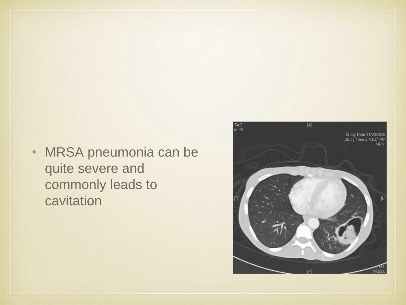
• MRSA pneumonia can be
quite severe and
commonly leads to
cavitation

• Pneumococcal vaccine is recommended for all
HIV patients who have a CD4 cell count
greater than 200 cells/mm3, and the influenza
vaccine should be given annually to all
patients, regardless of CD4 cell count
Pneumocystis jiroveci
• PCP in patients with HIV infection usually presents subacutely with progressive dyspnea,
non-productive cough, and fever. Pleuritic chest pain or acute worsening of symptoms is an
unusual manifestation of PCP in patients with HIV unless complicated by the development of
a pneumothorax.
• lungs are generally clear on auscultation.
• Diagnosis: Induced sputum - 55% sensitivity
BAL - 95% sensitivity
• Persistence of organisms through and even after therapy is common, and does not reflect
failure of therapy.
• Newer diagnostic techniques under investigation include polymerase chain reaction of saliva
or sputum, as well as blood tests measuring levels of S-adenosylmethionine, a metabolite
used exclusively by Pneumocystis jiroveci and hence lowered in patients with active disease.
• Chest imaging may be normal but more typically demonstrates bilateral diffuse interstitial
abnormalities
• Patients with PCP typically worsen after 2 to 3 days of therapy.

Endemic Fungi
• Histoplasmosis, coccidioidomycosis,
blastomycosis, and penicilliosis are dimorphic
fungi that are endemic to distinct regions but
that may reactivate
• rarely causes disease in patients with HIV until
the patient is significantly immunosuppressed
(CD4 less than 250 cells/mm3).

Histoplasma capsulatum
• histoplasmosis most commonly presents with disseminated disease in an individual with a CD4 cell count less than 150 cells/mm3.
• fever, weight loss, adenopathy, diarrhea, andmucosal lesions.
• occasionally occurs in person with HIV and preserved CD4 counts (greater than 350 cells/mm3)
• Histoplasma capsulatum can produce a sepsis syndrome as well with fever, hypotension, and multiorgan system failure.
• The presence of hilar or mediastinal adenopathy may help distinguish histoplasmosis from Pneumocystis jiroveci, as the clinical presentation in susceptible hosts overlap significantly, although these are uncommon findings for Pneumocystis.
• Diagnosis: detection of polysaccharide antigen in urine or blood, where urine testing has been shown to be positive in up to 95 percent of patients with disseminated disease and AIDS.
• Treatment: Amphotericin is currently considered the drug of choice for severe or disseminated histoplasmosis in HIV-positive patients and should be continued for 3 to 10 days until clear clinical improvement has occurred before patients are switched to itraconazole to complete a total of 3 months of therapy. Patients with HIV infection be maintained on suppressive doses of itraconazole indefinitely.
Coccidioides immitis
• The presence of hilar adenopathy, eosinophilia, or lack of response to conventional antibiotics should prompt consideration of pulmonary coccidiomycosis in patients from endemic areas
• Radiology: diverse, and may include reticulonodular disease, alveolar infiltrates, nodules, adenopathy, cavities, and pleural effusions.
• Diagnosis: Iolation in respiratory secretions (Bronchoscopy)
• Coccidioides immitis serology has a more established role to play in monitoring response to therapy. (sensitivity in diagnosis is 70%)
• DOC: (Without meningeal involvement) Amphotericin B. Treatment should be continued until clear clinical improvement has been demonstrated, at which time patients may be switched over to itraconazole or fluconazole.
• (With meningeal involvement): high-dose fluconazole, therapy should be continued indefinitely.

Penicillium marneffi
• Penicillium marneffei is generally seen in patients with advanced AIDS(CD4 less than 50 cell/mm3), where it commonly presentswith extrapulmonary symptoms (fevers, weight loss, sweats, hepatosplenomegaly, and hematologic abnormalities).
• cutaneous findings, particularly umbilical papules that may be confused with either cryptococcosis or molluscum contagiosum
• much more commonly associated with systemic disease, then isolated pneumonia.
• Chest radiographs most demonstrate interstitial changes though nodules, cavitation and pleural disease have all been reported previously.
• diagnosis requires isolation of the organism from blood, skin,bonemarrow, or lymphnodes.
• In patients with disseminated infection, amphotericin B should be used to control the infection. Patients who have demonstrated clear clinical improvement can be switched to twice-daily itraconazole to complete a 3- month treatment course. secondary prophylaxis with daily itraconazole should be maintained until patients have achieved a CD4 count of greater than 200 cells/mm3 for at least 6 months.
• Fluconazole does not have activity against Penicillium marneffei.
Aspergillosis• exclusively in those with advanced immunosuppression(CD4 cell
count less than 50 cells/mm3.
• Patients with AIDS who are diagnosed with invasive disease often
have other risk factors for aspergillosis, such as receipt of
corticosteroids or neutropenia.
• Aspergillus fumigatus, and to a lesser extent Aspergillus niger are the
most common causes of invasive aspergillosis.
• Respiratory tract disease is the most common manifestation of
aspergillosis and both tracheitis and invasive pneumonitis may
develop
• The diagnosis is established when endoscopic examination reveals
an exudative pseudomembrane adherent to the tracheal wall.

• Radiographic abnormalities in both of these forms
overlap, and can show diffuse infiltrates, cavities,
and focal wedge-shaped abnormalities reflecting
pulmonary infarction.
• Despite the predilection for the organism to invade
blood vessel walls and disseminate, focal CNS
disease due to aspergillus in AIDS patients
appears to arise more commonly from contiguous
spread from the sinuses, orbits, and ears.
• It is important to note, however, that aspergillus is
a common colonizer of diseased airways, so
definitive diagnosis requires documentation of
tissue invasion

• Voriconazole as the agent of choice for invasive
aspergillosis, although potential drug-drug
interactions in patients receiving antiretroviral
medications should be closely monitored.
• There are no formal recommendations for length
of treatment or prophylaxis (secondary or
primary), although every effort should be made
to provide effective antiretroviral therapy, to
correct neutropenia by discontinuing offending
agents or by using stimulatory cytokines, and to
eliminate exogenous immunosuppression by
discontinuing corticosteroids.

Viral Pneumonia
• Although many respiratory viruses produce
disease in patients with HIV, only
cytomegalovirus is an AIDS-defining
opportunistic infection
Cytomegalovirus• Asymptomatic persons with CMV infection excrete the virus in
saliva, respiratory secretions, urine, and semen.
• Clinically significant HIV-related CMV infection is associated with
severe immunosuppression (CD4 less than 50 cells/mm3).
• CMV-associated syndromes most commonly seen in patients with
AIDS are retinitis and enteritis (colitis or esophagitis).
• A variety of CMV-related neurologic complications have been
described, including polyradiculitis, ventriculitis, and mononeuritis
multiplex.
• When disease is limited to the chest, definitive diagnosis requires
demonstration of cytopathic change on histopathology (usually
obtained via transbronchial biopsy or VATS).

• In most studies, patients with positive BAL
cultures for CMV have evidence of a more
likely alternative diagnosis (especially PCP or
bacterial pneumonia), and may improve
2without specific therapy directed at CMV.
• Nonetheless, in a patient with advanced HIV
disease (CD4 cell count less than 50
cells/mm3), interstitial infiltrates on chest
radiograph, and no alternative organism
isolated, CMV pneumonitis may be the sole
responsible pathogen.

• The agents of choice for the treatment of
established CMV pneumonitis is oral
valganciclovir.
• For refractory disease, foscarnet would be an
appropriate alternative, although its use is
associated with high rates of renal dysfunction
and other metabolic abnormalities.
• Unlike CMV retinitis, there is no standard
secondary prophylaxis for CMV pneumonitis.
However, as with other opportunistic infections,
the antiretroviral therapy with restoration of the
CD4 cell count plays a critical role in preventing
recurrences.

• all patients with HIV infection should be
encouraged to get annual vaccination.
Although declines in CD4 count and rises in
HIV viral load may be seen after influenza
vaccination, these adverse laboratory events
are generally transient and have not been
shown to be associated with adverse clinical
events

Parasitic Infections• Toxoplasma gondii most common.
• CD4 less than 50 cells/mm3.
• The clinical presentation of pulmonary toxoplasmosis is similar to that of PCP, but
unlike PCP it may be accompanied by a sepsis-like syndrome with hypotension.
• Radiographic abnormalities are diverse- interstitial infiltrates, but also potentially
nodules, effusions, or mass lesions.
• As with PCP, an elevated LDH is a commonly reported laboratory finding. Diagnosis
may be established by identification of toxoplasma tachyzoites on Giemsa stain of BAL
fluid or tissue obtained on biopsy.
• Treatment- Pyrimethamine plus sulfadiazine is the first-line treatment x 6 weeks, and
secondary prophylaxis until their CD4 count is greater than 200 cell/mm3 for over 6
months.
• Prophylaxis with double strength TMP/SMX once daily in HIV-positive patients with
CD4 counts less than 100 cell/mm3

Non Infectious Pulmonary
Complications of HIV

Kaposis Sarcoma• Endothelial cells latently infected with HHV8 are activated by HIV, which in turn
drive the angiogenesis characteristic of this vascular malignancy
• KS may develop in patients with preserved CD4 counts, although the pulmonary
involvement is most commonly seen in advanced disease (CD4 less than 100
cells/mm3
• Pulmonary KS may be asymptomatic even in patients with extensive
abnormalities on chest radiograph.
• Lesions tend to show peribronchovascular distribution
• Diagnosis is generally made through direct visualisation of characteristic purplish
plaques on bronchoscopy
• Chemotherapy is indicated for symptomatic pulmonary disease
• However, as with cutaneous KS, potent antiretroviral therapy can induce
significant improvement in pulmonary KS, as well as sustained remissions.

Lung Cancer
• Lung cancer is the most frequently diagnosed
non-AIDS–related malignancy, with reported
rates of three- to eightfold higher than those
without identified infection
• While CD4 count at the time of diagnosis of
lung cancer may be depressed, it is more
commonly preserved.

Pulmonary Hypertension
• The most common hypothesis for the association of HIV and
pulmonary HTN is an alteration in pulmonary cytokine profile that
increases expression of vasoactive substances such as endothelin-1
• HIV-related pulmonary hypertension (HRPH) is a diagnosis of
exclusion and can only be made after other secondary causes of
pulmonary hypertension have been excluded.
• Management of pulmonary hypertension is as for HIV-negative hosts,
with prostaglandin agonists (epoprostenol), diuretics, and
anticoagulation. Sildenafil may also have a role, but it is associated
with potential drug-drug interactions in patients receiving with
protease inhibitors
• reports have not shown a consistent beneficial response of this
condition to antiretroviral therapy.

Lymphocytic Interstitial
Pneumonia• More commonly in HIV-infected children, adults may
rarely develop this complication as well. LIP is
characterised by polyclonal inflammatory lymphoid
proliferation of bronchus associated lymphoid tissue
(BALT).
• Diffuse reticulonodular or interstitial infiltrates are seen
on chest imaging, making differentiation from PCP
difficult
• Several case reports have demonstrated that
antiretroviral therapy may lead to substantial
improvement in LIP
• For unresponsive cases, corticosteroids may be
effective.

COPD
• greater risk for the development of
emphysema
• Investigators have postulated that the higher
proportion of CD8+ T lymphocytes seen in
BAL fluid from patients with HIV compared
with uninfected controls may help explain this
difference.

Thromboembolism
• Patients with HIV infection are at two- to
tenfold increased risk of thromboembolic
disease

Abacavir Hypersensitivity
• 5% individuals
• Symptoms of the abacavir HSR usually appear in the first 6 weeks of
treatment and include fever, rash, malaise, GI upset, myalgia, and
arthralgias. Cough and dyspnea also may occur and may mimic
bronchitis or pneumonia.
• It is critical that clinicians evaluating patients who have recently been
started on abacavir who present with fever and respiratory symptoms
consider abacavir hypersensitivity in the differential diagnosis, as
continued abacavir treatment in the face of this reaction can be fatal
• Symptoms resolve after cessation of drug.
• Never resume abacavir, as there might be immediate and life
threatening recurrence of hypersensitivity


Thank You