history of studies on mammalian middle ear evolution: a ... of studies on mammalian middle ear...
TRANSCRIPT
History of Studies on MammalianMiddle Ear Evolution: AComparative Morphological andDevelopmental Biology PerspectiveMASAKI TAKECHI� AND SHIGERU KURATANIEvolutionary Morphology Research Group, Center for Developmental Biology, RIKEN, Kobe, Japan
The mammalian middle ear represents one of the most fundamental morphological features thatdefine this class of vertebrates. Its skeletal pattern differs conspicuously from those of otheramniotes and has attracted the attention of comparative zoologists for about 200 years. Toreconcile this morphological inconsistency, early comparative morphologists suggested that themammalian middle ear was derived from elements of the jaw joint of nonmammalian amniotes.Fossils of mammalian ancestors also implied a transition in skeletal morphology that resulted inthe mammalian state. During the latter half of the 20th century, developmental mechanismscontrolling the formation of the jaw skeleton became the subject of studies in developmentalbiology and molecular genetics. Mammalian middle ear evolution can now be interpreted as aseries of changes in the developmental program of the pharyngeal arches. In this review, wesummarize the history of middle ear research, highlight some of the remaining problems, andsuggest possible future directions. We propose that to understand mammalian middle earevolution, it is essential to identify the critical developmental events underlying the particularmammalian anatomy and to describe the evolutionary sequence of changes in developmental andmolecular terms. We also discuss the degree of consistency between the developmentalexplanation of the mammalian middle ear based on molecular biology and morphological changesin the fossil record. J. Exp. Zool. (Mol. Dev. Evol.) 314B 2010. & 2010 Wiley-Liss, Inc.
How to cite this article: Takechi M, Kuratani S. 2010. History of studies on mammalian middleear evolution: a comparative morphological and developmental biology perspective. J. Exp.Zool. (Mol. Dev. Evol.) 314B:[page range].
Unlike nonmammalian amniotes, which have only one ossicle,
the columella auris (Fig. 1A, C, E), the mammalian middle ear has
three ossicles, the malleus, incus, and stapes (Fig. 1B, D, F). The
evolutionary origins of this complex and its homology have been
regarded as among the most formidable conundrums in
vertebrate comparative morphology. Why is a difference in the
number of the ossicles a problem? It is problematic because
animals are usually unable to generate entirely new anatomical
elements de novo in evolution. Geoffroy Saint-Hilaire, a French
anatomist, pointed out in 1818 that equivalent sets of skeletal
elements are connected in an identical order in all animals, and
proposed that every animal skeletal type was derived from
changes to a common skeletal pattern. This principe des
connexions remains one of the simplest definitions for morpho-
logical homology (reviewed by Hall, ’98). According to this law,
two of the three ossicles in the mammalian middle ear must have
their homologues in the nonmammalian skull, a phenomenon
that has puzzled morphologists for many years.
From the perspective of comparative morphology, the
vertebrate head exhibits a series of equivalent modules called
the pharyngeal arches (Fig. 1G). The first, the mandibular arch,
Published online in Wiley InterScience (www.interscience.wiley.com).
DOI: 10.1002/jez.b.21347
Received 4 December 2009; Revised 3 March 2010; Accepted 3 March
2010
Grant Sponsor: Grant-in-Aid for Young Scientists; Grant number: 21770259.�Correspondence to: Masaki Takechi, Evolutionary Morphology Research
Group, Center for Developmental Biology, RIKEN, 2-2-3 Minatojima-minami,
Chuo, Kobe, Hyogo 650-0047, Japan. E-mail: [email protected]
ABSTRACT
J. Exp. Zool. (Mol.Dev. Evol.) 314B2010
& 2010 WILEY-LISS, INC.
REVIEW ARTICLE
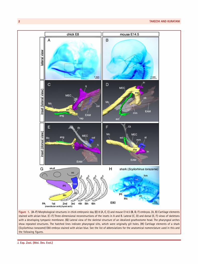
Figure 1. (A–F) Morphological structures in chick embryonic day (E) 8 (A, C, E) and mouse E14.5 (B, D, F) embryos. (A, B) Cartilage elements
stained with alcian blue. (C–F) Three-dimensional reconstructions of the insets in A and B. Lateral (C, D) and dorsal (E, F) views of skeletons
with a developing tympanic membrane. (G) Lateral view of the skeletal structure of an idealized gnathostome head. The pharyngeal arches
show repeated structures. The hatched lines indicate pharyngeal slits, which were originally gill holes. (H) Cartilage elements of a shark
(Scyliorhinus torazame) E80 embryo stained with alcian blue. See the list of abbreviations for the anatomical nomenclature used in this and
the following figures.
TAKECHI AND KURATANI2
J. Exp. Zool. (Mol. Dev. Evol.)
comprises the upper and lower jaws, called the palatoquadrate
and Meckel’s cartilage, respectively (Fig. 1G). The second, the
hyoid arch, is also subdivided dorsoventrally. The dorsal moiety
is called the hyomandibular and the ventral moiety the
ceratohyal (Fig. 1G), although an intercalated element between
the two elements, such as stylohyal, is often seen in many
vertebrates (Goodrich, ’30). The simplest configuration of the
latter arch is evident in elasmobranchs (Fig. 1H). In comparative
morphology, all the visceral arches are regarded as serial
homologues, and Geoffroy Saint-Hilaire first drew the conclusion
that the mammalian middle ear ossicles are homologous with the
opercular bones in teleosts (Geoffroy Saint-Hilaire, 1818;
reviewed by Appel, ’87).
Subsequently, several famous morphologists such as Meckel
(1820), Huschke (1824), Rathke (1832), and Burdach (1837)
addressed this issue, but only Reichert (1837) was able to
formulate a hypothesis that survives today. He dissected pig
embryos with needles under a microscope and realized that two
of the mammalian ossicles, the malleus and incus, were derived
from cartilages equivalent to the lower and upper jaw elements in
other amniotes: the articular and quadrate. Modern histological
observations have also shown that the incus arises from the
posterior part of the palatoquadrate as a primordium connected
to the basal part of the ala temporalis (the ascending process of
the palatoquadrate) by a thread of connective tissue (Presley and
Steel, ’76), which confirms Reichert’s interpretation. The con-
nective tissue thread often chondrifies to form a cartilaginous
element of the first arch domain when one of the genes expressed
in the first arch ectomesenchyme is knocked out (reviewed by
Smith and Schneider, ’98). Thus, the connection between the
incus and the rest of the upper jaw has been elucidated.
Geoffroy’s concept of homology was also applied to muscles
and nerves, and a consistent branchiomeric scheme in the
mammalian middle ear was confirmed: m. tensor tympani and
m. tensor veli palatini are derivatives of the first arch and are
innervated by the trigeminal nerve that innervates first arch
derivatives exclusively. Similarly, m. stapedius is a second arch
derivative innervated by the facial nerve (Rabl, 1887). The
German comparative morphologist Gaupp (’11a,b, ’12) examined
a huge number of skulls of various animals and published a 400-
page monograph titled ‘‘Die Reichert Theorie’’ (Gaupp, ’12). He
pointed out that, unlike the nonmammalian gnathostome jaw, in
which the ‘‘primary jaw joint’’ consists of the quadrate and the
articular, mammals have a unique ‘‘secondary jaw joint’’ between
two dermal elements, squamosal and dentary (Gaupp, ’11a,b, ’12).
Gaupp’s morphological insight influenced subsequent evolu-
tionary studies on the middle ear, based on the fossil record and
embryology (see below).
Reichert (1837) suggested that the stapes is homologous to the
columella auris in nonmammalians as an element homologous to
the hyomandibular (Fig. 1C–G). As the embryonic second arch
arises at the level of the inner ear, the dorsal end of the
hyomandibular must abut the otic capsule of the neurocranium
(Fig. 1G). This topographical relationship seems to have
facilitated the subsequent evolution of the hyomandibular. As a
device that anchors the upper jaw, this skeletal element is often
present in primitive gnathostomes, including extant elasmo-
branchs (Fig. 1H). It thus connects the quadrate (or jaw joint) and
the neurocranium. In this type of jaw suspension, called a
hyostyly, the upper jaw is fixed to the neurocranium via the
hyomandibular (Huxley, 1876; reviewed by Goodrich, ’30). Thus,
the mammalian middle ear exhibits a pattern topographically
identical to that of the hyostylic skull. By contrast, in most extant
tetrapods the quadrate is attached firmly to the cranium to
establish an autostylic jaw suspension that does not involve the
hyomandibular (Huxley, 1876). The latter suspension was
thought to have evolved in primitive tetrapods, in which the
hyomandibular assumed a hearing function (Goodrich, ’09, ’30;
de Beer, ’37; but see also Clack, ’89, ’93, 2002a,b; Clack et al.,
2003; Brazeau and Ahlberg, 2006). Thus, mammalian middle ear
evolution was tightly coupled with the evolution of jaw
suspension (reviewed by Clack, 2002a).
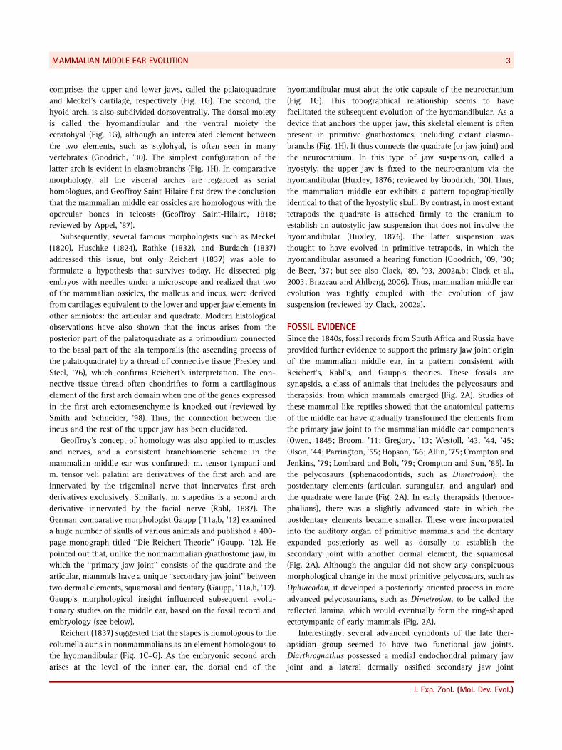
FOSSIL EVIDENCESince the 1840s, fossil records from South Africa and Russia have
provided further evidence to support the primary jaw joint origin
of the mammalian middle ear, in a pattern consistent with
Reichert’s, Rabl’s, and Gaupp’s theories. These fossils are
synapsids, a class of animals that includes the pelycosaurs and
therapsids, from which mammals emerged (Fig. 2A). Studies of
these mammal-like reptiles showed that the anatomical patterns
of the middle ear have gradually transformed the elements from
the primary jaw joint to the mammalian middle ear components
(Owen, 1845; Broom, ’11; Gregory, ’13; Westoll, ’43, ’44, ’45;
Olson, ’44; Parrington, ’55; Hopson, ’66; Allin, ’75; Crompton and
Jenkins, ’79; Lombard and Bolt, ’79; Crompton and Sun, ’85). In
the pelycosaurs (sphenacodontids, such as Dimetrodon), the
postdentary elements (articular, surangular, and angular) and
the quadrate were large (Fig. 2A). In early therapsids (theroce-
phalians), there was a slightly advanced state in which the
postdentary elements became smaller. These were incorporated
into the auditory organ of primitive mammals and the dentary
expanded posteriorly as well as dorsally to establish the
secondary joint with another dermal element, the squamosal
(Fig. 2A). Although the angular did not show any conspicuous
morphological change in the most primitive pelycosaurs, such as
Ophiacodon, it developed a posteriorly oriented process in more
advanced pelycosaurians, such as Dimetrodon, to be called the
reflected lamina, which would eventually form the ring-shaped
ectotympanic of early mammals (Fig. 2A).
Interestingly, several advanced cynodonts of the late ther-
apsidian group seemed to have two functional jaw joints.
Diarthrognathus possessed a medial endochondral primary jaw
joint and a lateral dermally ossified secondary jaw joint
MAMMALIAN MIDDLE EAR EVOLUTION 3
J. Exp. Zool. (Mol. Dev. Evol.)
(Crompton, ’63, ’72; Fig. 2B). This condition is highly suggestive
of a transitional state in mammalian evolution and implies that,
starting from a double-joint state, the laterally situated dermal
secondary jaw joint came to serve for feeding, releasing the
medially located primary jaw joint elements from their original
function, permitting them to move medially into the middle ear.
A developmental phenomenon also supports the above scenario.
Thus, in marsupial embryos, the angular shows a conspicuous
Figure 2. Paleontological evidence for mammalian middle ear evolution. (A) Diagrams of lateral views of jaw skeletal elements showing
modifications leading to the mammalian condition (after Allin, ’75). The geological record and occurrence of each animal are indicated on the
left. For clarity of comparison, no teeth are shown. Note that a set of postdentary elements (articular, surangular, and angular) and the upper
jaw elements (quadrate and quadratojugal), indicated by gray, became separated from the dentary and reduced in size during the transition
from pelycosaurs to mammals. The sequence of changes in the fossil record does not represent a true ancestor–descendent relationship, but
only structural grades. (B) Changes in jaw articulation during mammalian evolution. In a pelycosaur, Dimetrodon (top), the quadrate and
articular formed a functional jaw joint (black arrow). In an ‘‘advanced’’ cynodont, Diarthrognathus (middle), an additional jaw joint was
observed between the squamosal and dentary (white arrow). In an extant marsupial, Didelphis (bottom), the functional jaw joint has been
taken over only by the squamosal and dentary.
TAKECHI AND KURATANI4
J. Exp. Zool. (Mol. Dev. Evol.)
similarity to that of a cynodont before it takes the typical shape
of the ectotympanic (Palmer, ’13). Interestingly, the offspring of
marsupials initially use the primary jaw joint to suck the mother’s
nipples in the pouch, because their squamosal and dentary bones
are too premature to form a functional joint at this stage of
development (Crompton and Parker, ’78; Maier, ’87a; but see
Filan, ’91). Marsupials are thus in a sense born as ‘‘reptiles’’
before they become true mammals in the mother’s pouch.
However, it remains to be determined whether the skeletal pattern
of marsupial pouch young truly recapitulates the ancestral
condition or whether it is merely a secondary adaptation
prompted by a need for early suckling. In this context, it should
be noted that the mammalian developmental sequence does not
simply recapitulate the Diarthrognathus-like condition, in which
double joints are arranged mediolaterally. Instead, the primary
and secondary joints seem anteroposteriorly, as has been pointed
out earlier (see, for example, Fuchs, ’05, ’31; Jarvik, ’80; Fig. 2B).
The driving force for the shift from the primary to secondary jaw
joints has also been controversial. One explanation would be that
the increase in masticatory capabilities associated with expansion
of the dentary was impelled by selective forces, and only when
postdentary elements became vestigial did they assume an
auditory role and enter the middle ear (Parrington, ’79). Another
possibility is that the postdentary elements served as middle ear
components in the early mammalian ancestors and auditory
adaptation was an important factor in this sequence of
morphological evolution (Allin, ’75).
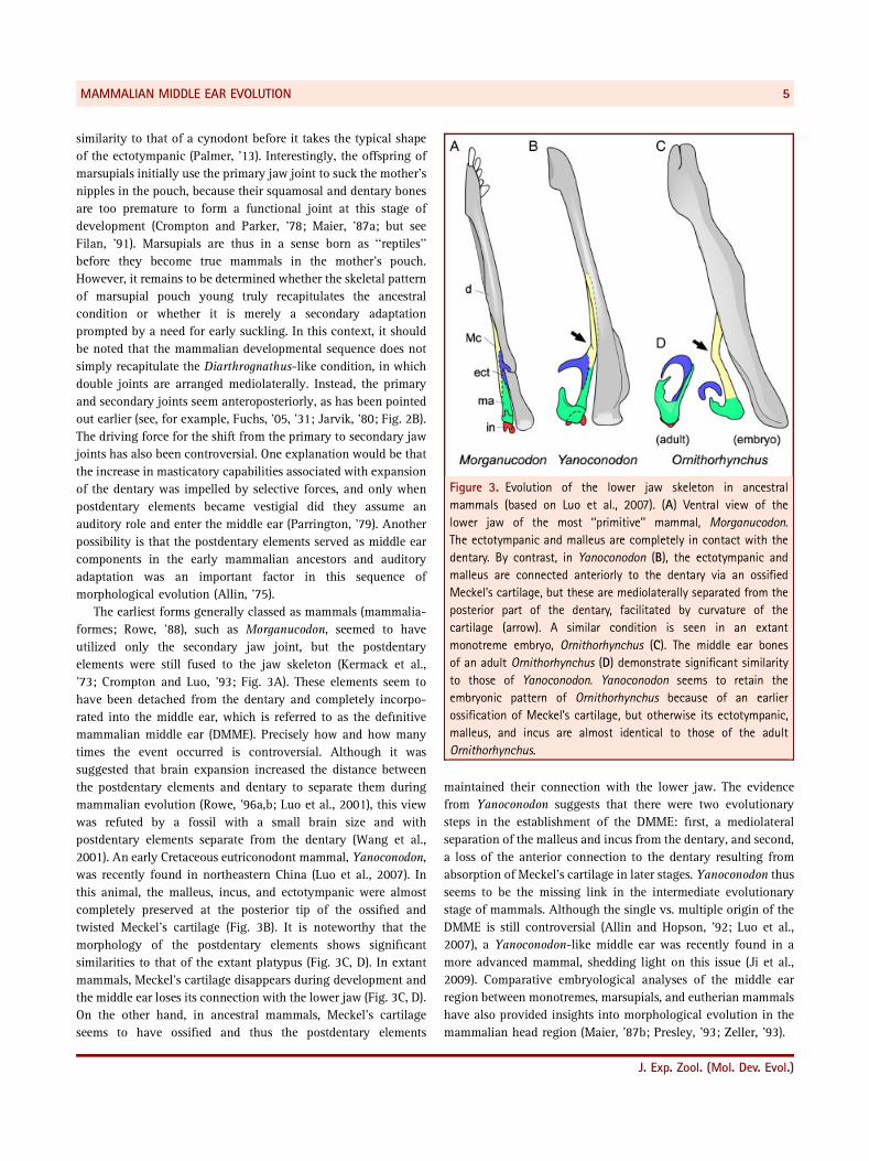
The earliest forms generally classed as mammals (mammalia-
formes; Rowe, ’88), such as Morganucodon, seemed to have
utilized only the secondary jaw joint, but the postdentary
elements were still fused to the jaw skeleton (Kermack et al.,
’73; Crompton and Luo, ’93; Fig. 3A). These elements seem to
have been detached from the dentary and completely incorpo-
rated into the middle ear, which is referred to as the definitive
mammalian middle ear (DMME). Precisely how and how many
times the event occurred is controversial. Although it was
suggested that brain expansion increased the distance between
the postdentary elements and dentary to separate them during
mammalian evolution (Rowe, ’96a,b; Luo et al., 2001), this view
was refuted by a fossil with a small brain size and with
postdentary elements separate from the dentary (Wang et al.,
2001). An early Cretaceous eutriconodont mammal, Yanoconodon,
was recently found in northeastern China (Luo et al., 2007). In
this animal, the malleus, incus, and ectotympanic were almost
completely preserved at the posterior tip of the ossified and
twisted Meckel’s cartilage (Fig. 3B). It is noteworthy that the
morphology of the postdentary elements shows significant
similarities to that of the extant platypus (Fig. 3C, D). In extant
mammals, Meckel’s cartilage disappears during development and
the middle ear loses its connection with the lower jaw (Fig. 3C, D).
On the other hand, in ancestral mammals, Meckel’s cartilage
seems to have ossified and thus the postdentary elements
maintained their connection with the lower jaw. The evidence
from Yanoconodon suggests that there were two evolutionary
steps in the establishment of the DMME: first, a mediolateral
separation of the malleus and incus from the dentary, and second,
a loss of the anterior connection to the dentary resulting from
absorption of Meckel’s cartilage in later stages. Yanoconodon thus
seems to be the missing link in the intermediate evolutionary
stage of mammals. Although the single vs. multiple origin of the
DMME is still controversial (Allin and Hopson, ’92; Luo et al.,
2007), a Yanoconodon-like middle ear was recently found in a
more advanced mammal, shedding light on this issue (Ji et al.,
2009). Comparative embryological analyses of the middle ear
region between monotremes, marsupials, and eutherian mammals
have also provided insights into morphological evolution in the
mammalian head region (Maier, ’87b; Presley, ’93; Zeller, ’93).
Figure 3. Evolution of the lower jaw skeleton in ancestral
mammals (based on Luo et al., 2007). (A) Ventral view of the
lower jaw of the most ‘‘primitive’’ mammal, Morganucodon.
The ectotympanic and malleus are completely in contact with the
dentary. By contrast, in Yanoconodon (B), the ectotympanic and
malleus are connected anteriorly to the dentary via an ossified
Meckel’s cartilage, but these are mediolaterally separated from the
posterior part of the dentary, facilitated by curvature of the
cartilage (arrow). A similar condition is seen in an extant
monotreme embryo, Ornithorhynchus (C). The middle ear bones
of an adult Ornithorhynchus (D) demonstrate significant similarity
to those of Yanoconodon. Yanoconodon seems to retain the
embryonic pattern of Ornithorhynchus because of an earlier
ossification of Meckel’s cartilage, but otherwise its ectotympanic,
malleus, and incus are almost identical to those of the adult
Ornithorhynchus.
MAMMALIAN MIDDLE EAR EVOLUTION 5
J. Exp. Zool. (Mol. Dev. Evol.)
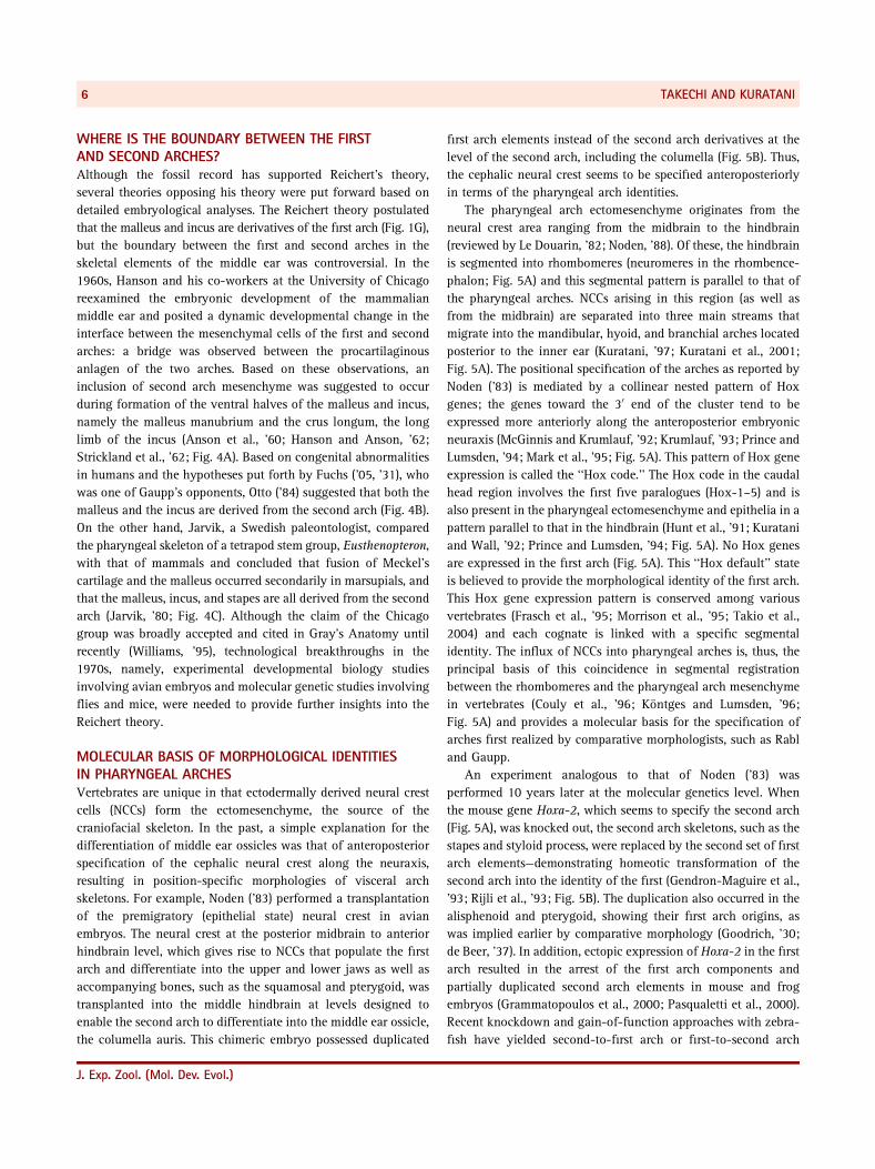
WHERE IS THE BOUNDARY BETWEEN THE FIRSTAND SECOND ARCHES?Although the fossil record has supported Reichert’s theory,
several theories opposing his theory were put forward based on
detailed embryological analyses. The Reichert theory postulated
that the malleus and incus are derivatives of the first arch (Fig. 1G),
but the boundary between the first and second arches in the
skeletal elements of the middle ear was controversial. In the
1960s, Hanson and his co-workers at the University of Chicago
reexamined the embryonic development of the mammalian
middle ear and posited a dynamic developmental change in the
interface between the mesenchymal cells of the first and second
arches: a bridge was observed between the procartilaginous
anlagen of the two arches. Based on these observations, an
inclusion of second arch mesenchyme was suggested to occur
during formation of the ventral halves of the malleus and incus,
namely the malleus manubrium and the crus longum, the long
limb of the incus (Anson et al., ’60; Hanson and Anson, ’62;
Strickland et al., ’62; Fig. 4A). Based on congenital abnormalities
in humans and the hypotheses put forth by Fuchs (’05, ’31), who
was one of Gaupp’s opponents, Otto (’84) suggested that both the
malleus and the incus are derived from the second arch (Fig. 4B).
On the other hand, Jarvik, a Swedish paleontologist, compared
the pharyngeal skeleton of a tetrapod stem group, Eusthenopteron,
with that of mammals and concluded that fusion of Meckel’s
cartilage and the malleus occurred secondarily in marsupials, and
that the malleus, incus, and stapes are all derived from the second
arch (Jarvik, ’80; Fig. 4C). Although the claim of the Chicago
group was broadly accepted and cited in Gray’s Anatomy until
recently (Williams, ’95), technological breakthroughs in the
1970s, namely, experimental developmental biology studies
involving avian embryos and molecular genetic studies involving
flies and mice, were needed to provide further insights into the
Reichert theory.
MOLECULAR BASIS OF MORPHOLOGICAL IDENTITIESIN PHARYNGEAL ARCHESVertebrates are unique in that ectodermally derived neural crest
cells (NCCs) form the ectomesenchyme, the source of the
craniofacial skeleton. In the past, a simple explanation for the
differentiation of middle ear ossicles was that of anteroposterior
specification of the cephalic neural crest along the neuraxis,
resulting in position-specific morphologies of visceral arch
skeletons. For example, Noden (’83) performed a transplantation
of the premigratory (epithelial state) neural crest in avian
embryos. The neural crest at the posterior midbrain to anterior
hindbrain level, which gives rise to NCCs that populate the first
arch and differentiate into the upper and lower jaws as well as
accompanying bones, such as the squamosal and pterygoid, was
transplanted into the middle hindbrain at levels designed to
enable the second arch to differentiate into the middle ear ossicle,
the columella auris. This chimeric embryo possessed duplicated
first arch elements instead of the second arch derivatives at the
level of the second arch, including the columella (Fig. 5B). Thus,
the cephalic neural crest seems to be specified anteroposteriorly
in terms of the pharyngeal arch identities.
The pharyngeal arch ectomesenchyme originates from the
neural crest area ranging from the midbrain to the hindbrain
(reviewed by Le Douarin, ’82; Noden, ’88). Of these, the hindbrain
is segmented into rhombomeres (neuromeres in the rhombence-
phalon; Fig. 5A) and this segmental pattern is parallel to that of
the pharyngeal arches. NCCs arising in this region (as well as
from the midbrain) are separated into three main streams that
migrate into the mandibular, hyoid, and branchial arches located
posterior to the inner ear (Kuratani, ’97; Kuratani et al., 2001;
Fig. 5A). The positional specification of the arches as reported by
Noden (’83) is mediated by a collinear nested pattern of Hox
genes; the genes toward the 30 end of the cluster tend to be
expressed more anteriorly along the anteroposterior embryonic
neuraxis (McGinnis and Krumlauf, ’92; Krumlauf, ’93; Prince and
Lumsden, ’94; Mark et al., ’95; Fig. 5A). This pattern of Hox gene
expression is called the ‘‘Hox code.’’ The Hox code in the caudal
head region involves the first five paralogues (Hox-1–5) and is
also present in the pharyngeal ectomesenchyme and epithelia in a
pattern parallel to that in the hindbrain (Hunt et al., ’91; Kuratani
and Wall, ’92; Prince and Lumsden, ’94; Fig. 5A). No Hox genes
are expressed in the first arch (Fig. 5A). This ‘‘Hox default’’ state
is believed to provide the morphological identity of the first arch.
This Hox gene expression pattern is conserved among various
vertebrates (Frasch et al., ’95; Morrison et al., ’95; Takio et al.,
2004) and each cognate is linked with a specific segmental
identity. The influx of NCCs into pharyngeal arches is, thus, the
principal basis of this coincidence in segmental registration
between the rhombomeres and the pharyngeal arch mesenchyme
in vertebrates (Couly et al., ’96; Kontges and Lumsden, ’96;
Fig. 5A) and provides a molecular basis for the specification of
arches first realized by comparative morphologists, such as Rabl
and Gaupp.
An experiment analogous to that of Noden (’83) was
performed 10 years later at the molecular genetics level. When
the mouse gene Hoxa-2, which seems to specify the second arch
(Fig. 5A), was knocked out, the second arch skeletons, such as the
stapes and styloid process, were replaced by the second set of first
arch elements—demonstrating homeotic transformation of the
second arch into the identity of the first (Gendron-Maguire et al.,
’93; Rijli et al., ’93; Fig. 5B). The duplication also occurred in the
alisphenoid and pterygoid, showing their first arch origins, as
was implied earlier by comparative morphology (Goodrich, ’30;
de Beer, ’37). In addition, ectopic expression of Hoxa-2 in the first
arch resulted in the arrest of the first arch components and
partially duplicated second arch elements in mouse and frog
embryos (Grammatopoulos et al., 2000; Pasqualetti et al., 2000).
Recent knockdown and gain-of-function approaches with zebra-
fish have yielded second-to-first arch or first-to-second arch
TAKECHI AND KURATANI6
J. Exp. Zool. (Mol. Dev. Evol.)
Figure 4. Theories opposed to Reichert’s (1837) theory. (A) The first and second arch contributions to the middle ear ossicles based on
Hanson and Anson (’62). Key: mad, mandibular arch derivative; hyd, hyoid arch derivative; otd, otic capsule derivative. (B) The origin of the
mammalian middle ear according to Otto’s theory (’84). Medial views of reptilian (top), therapsidian (middle), and human (bottom) middle
ears. Otto emphasized that human congenital abnormalities implied that jaw and middle ear development are not closely related to each
other. He also emphasized Fuchs’s theory (’05, ’31) that the reptilian jaw joint elements are equivalent to the secondary cartilage between the
squamosal and dentary in mammals. Based on these theories, Otto assumed that the malleus and incus were derived from the extracolumella
(ecol; dark gray). (C) Homology of skeletal elements derived from the second arch between Eusthenopteron (left) and mammals (right)
according to Jarvik (’80). Each presumed homologous bone is indicated by identical shading. Jarvik assumed that all the mammalian middle
ear ossicles are derived from the second arch.
MAMMALIAN MIDDLE EAR EVOLUTION 7
J. Exp. Zool. (Mol. Dev. Evol.)
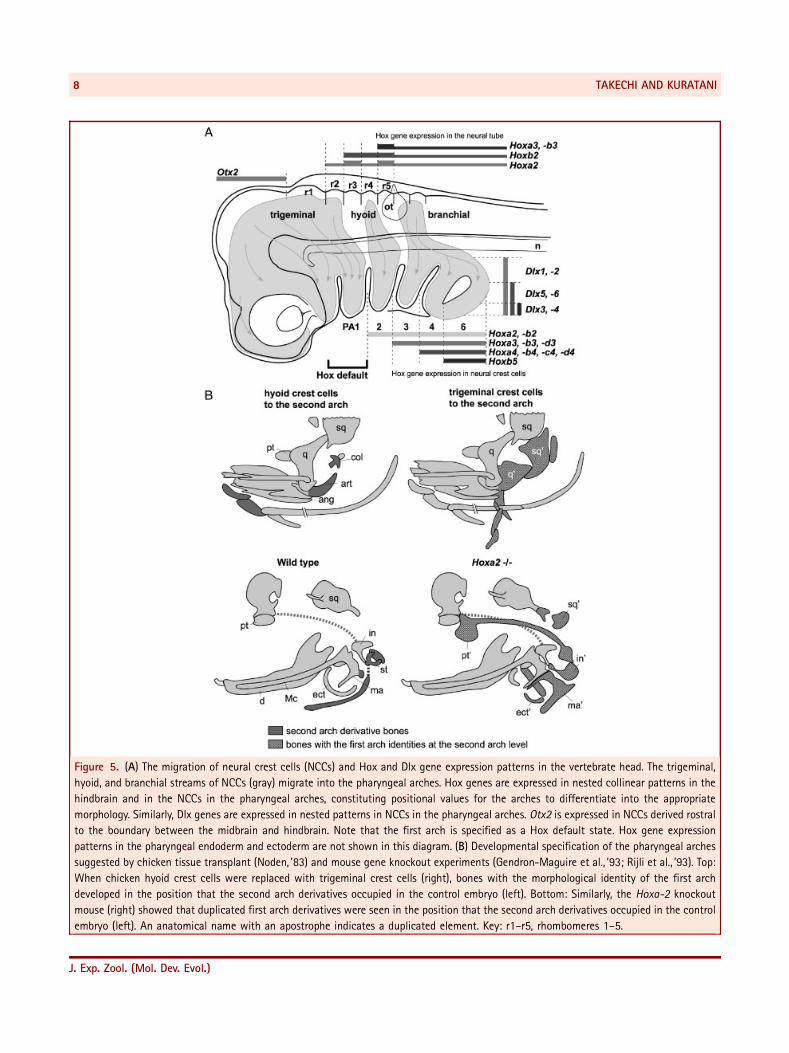
Figure 5. (A) The migration of neural crest cells (NCCs) and Hox and Dlx gene expression patterns in the vertebrate head. The trigeminal,
hyoid, and branchial streams of NCCs (gray) migrate into the pharyngeal arches. Hox genes are expressed in nested collinear patterns in the
hindbrain and in the NCCs in the pharyngeal arches, constituting positional values for the arches to differentiate into the appropriate
morphology. Similarly, Dlx genes are expressed in nested patterns in NCCs in the pharyngeal arches. Otx2 is expressed in NCCs derived rostral
to the boundary between the midbrain and hindbrain. Note that the first arch is specified as a Hox default state. Hox gene expression
patterns in the pharyngeal endoderm and ectoderm are not shown in this diagram. (B) Developmental specification of the pharyngeal arches
suggested by chicken tissue transplant (Noden, ’83) and mouse gene knockout experiments (Gendron-Maguire et al., ’93; Rijli et al., ’93). Top:
When chicken hyoid crest cells were replaced with trigeminal crest cells (right), bones with the morphological identity of the first arch
developed in the position that the second arch derivatives occupied in the control embryo (left). Bottom: Similarly, the Hoxa-2 knockout
mouse (right) showed that duplicated first arch derivatives were seen in the position that the second arch derivatives occupied in the control
embryo (left). An anatomical name with an apostrophe indicates a duplicated element. Key: r1–r5, rhombomeres 1–5.
TAKECHI AND KURATANI8
J. Exp. Zool. (Mol. Dev. Evol.)
homeotic transformations, respectively, suggesting a conserved
role for Hoxa-2 among vertebrates (Hunter and Prince, 2002).
Therefore, the molecular genetic evidence strongly supports the
serial homology schematic of the visceral arches advocated by
the Reichert–Rabl–Gaupp theories.
It is noteworthy that chicken–quail chimera analyses have
shown that the retroarticular process, attached to the posterior
side of the articular (Fig. 1C), is derived from the NCCs of the
second arch (Noden, ’83; Kontges and Lumsden, ’96; Fig. 5B): a
point overlooked in comparative embryology studies (e.g.,
Goodrich, ’30). A recent cell lineage tracing analysis of mouse
second arch NCCs showed that the processus brevis of the malleus
(Fig. 1D) arises from the second arch (O’Gorman, 2005), which is
consistent with evidence that this element disappears after
Hoxa-2 disruption (Fig. 5B). Although the homology of the
retroarticular process is controversial (reviewed by Novacek, ’93),
these results seem to suggest that this structure is homologous
with the processus brevis of the malleus. These analyses also
revealed that the boundary between the first and second
arch should be on the articular. This would explain the
inconsistent position of the boundary between the two arches
as conceived by the University of Chicago group, which claims
that the ventral half of the malleus is derived partially from the
second arch (Fig. 4A).
Similar to the role of the Hox code in the anteroposterior
specification of the arches, Dlx genes, another class of homeobox
gene, are considered to play a role in dorsoventral specification.
The nonteleost gnathostome genome possesses six Dlx genes
(Dlx1–6; Stock, 2005), which also exhibit dorsoventrally nested
expression patterns. Thus, Dlx1 and Dlx2 are ubiquitously
expressed in the pharyngeal arch ectomesenchyme; expression
of Dlx5 and Dlx6 is restricted to the ventral half, and expression
of Dlx3 and Dlx4 occurs only in the ventral tips of the arches (Qiu
et al., ’97; Depew et al., 2002; Fig. 5A). Thus, the Hox and Dlx
genes together seem to specify each part of the ectomesenchyme
via their Cartesian grid-like expression (Fig. 5A). The function of
the ‘‘Dlx code’’ has been demonstrated by loss- and gain-of-
function experiments. A double knockout of Dlx5 and Dlx6
resulted in transformation of the lower jaw into the identity of
the upper jaw and the mutant mouse exhibited mirror-image
duplication of the upper jaws (Depew et al., 2002). Equivalent
transformations were also observed with knockouts of Endothe-
lin1 (Edn1) or its cognate type-A receptor, Ednra (Ozeki et al.,
2004; Ruest et al., 2004). Edn1 is expressed in the epithelia and
mesodermal core of the lower jaw region in the first arch,
whereas Ednra is broadly expressed in the NCCs of the head
(Kurihara et al., ’94, ’95; Clouthier et al., ’98). Edn/Ednra signaling
was found to activate several genes required for lower jaw
specification, including Dlx6 (Clouthier et al., ’98, 2000; Kurihara
et al., ’99; Charite et al., 2001). Accordingly, ectopic Edn1
induction in the upper jaw region resulted in transformation of
the upper jaw into the identity of the lower jaw, suggesting that
the NCCs that migrate into the first arch can form both upper and
lower jaw structures and that Edn/Ednra signaling determines
which morphogenetic program is activated (Sato et al., 2008).
SIGNALS TO INSTRUCT THE ‘‘SHAPE’’ OF THE SKELETONHow can the NCCs form specific skeletal elements that differ
between mammals and nonmammalian amniotes? After Noden’s
1983 experiment (Fig. 5A), several subsequent reports suggested
that developmental specification of the skeletal shape is more
complicated than the idea of predetermined premigratory NCCs,
although the idea of a Hox code default state of the mandibular
arch (or of the ectomesenchyme therein) is still valid as a
prerequisite for jaw specification. For example, any portion of the
neural crest between the middle midbrain and r3 levels (the crest
destined for the first arch; Fig. 5A) can always duplicate the
proximal first arch, including the jaw joint when grafted onto
the hyoid arch level (r4) (Couly et al., ’98). Also, the Hox code in
the pharyngeal arches has been shown to be restored after surgical
rotation of the hindbrain neurectoderm along the anteroposterior
axis, suggesting that the Hox code is partly regulated and
maintained by environmentally derived signals (Hunt et al., ’98).
In the same context, the Hox code default state in the first arch
seems to depend on the midbrain–hindbrain boundary-derived
protein FGF8 (Trainor et al., 2002, 2003). Furthermore, local
interactions between ectomesenchyme and head endoderm are
responsible for morphological specification of the craniofacial
skeletons (Couly et al., 2002; Ruhin et al., 2003). Thus, when the
rostralmost chicken endoderm was removed from an early
neurula, the nasal cartilage disappeared completely. When a
slightly more posterior piece of the endoderm was removed, the
main part of the lower jaw cartilage was lost. By contrast, when an
endoderm graft was transplanted into a normal chicken embryo,
skeletal components, corresponding to the endoderm, developed
ectopically in the correct shapes, sizes, and directions. Thus, the
anatomical patterns of the first arch (and premandibular) skeletal
elements are mapped on the rostral endoderm in contact with the
NCCs specified by the Hox-code default state.
These endoderm transplantation experiments apparently
contradict one of the classical transplantation experiments.
Wagner (’59) grafted the premigratory cephalic neural crest
between newt and frog embryos and found that the chimera
always developed the craniofacial morphology of the crest donor.
This apparent contradiction was recently reconciled by Schneider
and Helms (2003), who used ducks and quails and showed that
species-specific morphological traits primarily reside in a genetic
program carried by the crest cell lineage. Thus, we may assume
that the evolution of the ‘‘species-specific shape’’ of visceral
skeletons is imprinted preferentially onto the developmental
program exerted by the ectomesenchyme. Given the putative role
of the cephalic endoderm and NCCs in patterning the skeleton, we
might have to class the concept of ‘‘shape’’ into different levels:
namely, the default morphology of the gnathostome visceral arch
MAMMALIAN MIDDLE EAR EVOLUTION 9
J. Exp. Zool. (Mol. Dev. Evol.)
skeleton, the comparative morphological identities (shape as a
morphological homology) of each arch or skeletal subset within
the arch, and animal species-specific shapes (reviewed by
Kuratani, 2005). Thus, although a coherent description of
mechanisms that determine the shape of the head skeleton is
lacking, recent studies strongly suggest that reciprocal interac-
tions between NCCs and surrounding tissues are certainly
important.
THE RIDDLE OF THE TYMPANIC MEMBRANEFor understanding the mechanism of ectomesenchymal specifi-
cation, tissue interactions between the crest-derived ectome-
senchyme and the ectodermal and endodermal epithelium should
be taken into consideration. In this connection, it should be noted
that comparative morphologists have also focused on a
nonskeletal epithelial structure, the tympanic membrane, to
resolve the puzzle.
The tympanic membrane separates the middle ear cavity
(originally the position of the first pharyngeal pouch) from the
external auditory meatus (originally the position of the first
pharyngeal cleft; Fig. 1C–F). The nonmammalian tympanic
membrane is attached to the quadrate, the upper jaw element
(Fig. 1C, E). By contrast, in the mammalian middle ear, the
tympanic membrane spans the ectotympanic, the angular
homologue belonging to the lower jaw domain (Fig. 1D, F). In
the old concept of middle ear evolution, ‘‘primitive’’ tetrapods,
such as Ichthyostega, established the tympanic membrane at the
quadrate position. This was carried over to extant reptiles and
birds, and the mammalian middle ear evolved from the reptilian
state (reviewed by Laurin, ’98; Clack and Allin, 2004). The
homology between the tympanic membranes of mammals and
nonmammalian amniotes was based on this evolutionary concept
for many years.
Goodrich (’14, ’30) assumed that the mammalian tympanic
membrane is homologous with that of reptiles, namely, that the
reptilian tympanic membrane moved ventrally, resulting in its
lower jaw position in mammals. By contrast, Gaupp (’11a,b, ’12)
doubted the homology because of the aforementioned topogra-
phical discrepancy in the attachment site of the tympanic
membrane (Fig. 1C–F). His perspective seems to have been
corroborated by detailed analyses of comparative embryology.
Goodrich’s theory is inconsistent with the topographical relation-
ships between the tympanic membrane and muscles that arise in
association with the malleus (m. tensor tympani and m. tensor
veli palatine). These muscles develop as a pair of tandem anlagen
that differentiate from a single primordium occupying a position
equivalent to m. pterygoideus in nonmammalian amniotes (see
also Barghusen, ’86). Given that these muscles are homologous
and that the connectivity of this muscle to the skeletal elements is
conserved during the ventral translocation of the tympanic
membrane as predicted by Goodrich, we would expect m. tensor
tympani to be located ventral to the Eustachian tube (Fig. 6A).
However, this muscle is dorsal to the Eustachian tube of the
mammalian middle ear (Fig. 6B). This morphological incon-
sistency apparently denies the homology of tympanic membranes
between mammals and nonmammalian amniotes (see also
Presley, ’84).
Because of this topographical discrepancy, Westoll (’43,
’44, ’45) assumed the existence of a ventral diverticulum of the
middle ear cavity called the recess mandibularis, which grows
ventrally to form a mammal-specific tympanic membrane
(Fig. 6C). The mammalian tympanic membrane is thus assumed
to consist of a small dorsal part, corresponding to the reptilian
tympanic membrane, pars flaccida, and a large ventral portion
corresponding to a novel mammalian middle ear feature: pars
tensa (Fig. 6C, D). Importantly, Goodrich (’14, ’30) also pointed
out that the mammalian middle ear cavity seems to protrude
ventrally compared with nonmammalian amniotes with respect
to the course of the chorda tympani, a branch of the facial nerve
(Fig. 6E). In comparative anatomy, chorda tympani is regarded as
a branch of the posttrematic ramus of the facial nerve (Fig. 6E).
However, the mammalian chorda tympani looks deceptively like
a branch of the pretrematic ramus of the facial nerve (Fig. 6E).
Goodrich (’14, ’30) assumed that the middle ear cavity swells
ventrally only in mammalian embryos and that the chorda
tympani is elevated from the ventral position by the swollen
cavity (Fig. 6E). However, there is a competing theory on this
issue. As the chicken chorda tympani is formed as a branch of the
pretrematic ramus, the chorda tympani does not necessarily
occupy the same position relative to the surrounding anatomical
elements in all amniotes (Kuratani et al., ’88). For this reason, it
seems unreasonable that the course of the cranial nerves should
be considered in comparative morphological analysis.
At any rate, because Westoll’s theory (’43, ’44, ’45)
persuasively explained the transitional state of the middle ear
from reptilian to mammalian, several morphologists supported
this theory for many decades (Gregory, ’51; Watson, ’53; Shute,
’56; Parrington, ’79). However, this concept of the origin of the
middle ear requires radical revision in the light of recent
paleontological evidence. One of the rationales for the old
concept that the middle ear evolved in ancestral tetrapods was
the presence of the ‘‘otic notch’’ in their skulls. The notch was
once thought to be the site at which the tympanic membrane
made contact with the stapes (Watson, ’51; Romer, ’66). However,
many of the otic notches are now interpreted as spiracular
notches rather than as hosts for the tympanic membrane, and the
presence of the middle ear is no longer considered to be the
ancestral condition of tetrapods (Clack, ’89, ’93; Brazeau and
Ahlberg, 2006). Furthermore, the hyomandibular of ‘‘primitive’’
amniotes is massive and strut-like and there does not seem to
be any room for the tympanic membrane in their skulls (Romer
and Price, ’40). Thus, the modern consensus is that the middle
ears of mammals and nonmammalian amniotes developed
independently of each other after their divergence from a
TAKECHI AND KURATANI10
J. Exp. Zool. (Mol. Dev. Evol.)
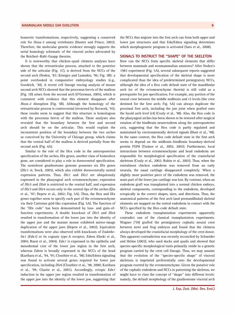
Figure 6. Interpretations of mammalian tympanic membrane evolution proposed by comparative morphologists. (A, B) Medial views of the
primary jaw joint with middle ear cavities and some of the pterygoid muscle derivatives in chicken and mouse embryos. The chicken,
m. pterygoideus (mptd and mptv), is located in the ventral region of the Eustachian tube (A). If the tympanic membrane is homologous
between mammals and nonmammalian amniotes, m. tensor tympani, homologous with m. pterygoideus, should be located ventral to the
Eustachian tube after the tube moved to the ventral position (predicted by Goodrich, ’14, ’30; arrow with a dotted line). However, in the
mouse embryo, m. tensor tympani is located dorsal to the Eustachian tube (B), suggesting that the tympanic membrane is not homologous
between mammals and nonmammalian amniotes. (C) Diagrams showing the ‘‘recess mandibularis theory’’ put forward by Westoll (’43, ’44,
’45). Three suggested stages of mammalian middle ear evolution from an advanced theriodont (a group of therapsids) viewed from the lateral
side. The functional part of the mammalian tympanic membrane, pars tensa, is considered to be a novel structure that arose by contact
between a novel diverticulum (the recess mandibularis; rec. m.) formed in the ventral position of the original middle ear cavity and the outer
skin. Pars flaccida is believed to be a vestigial reptilian tympanic membrane. (D) Lateral view of the head of the reconstructed Thrinaxodon
based on Westoll’s theory (Allin and Hopson, ’92). (E) The evolutionary transitions from fish to mammals (left to right) in the course of chorda
tympani according to Goodrich’s theory (’14, ’30). The anterior is to the right. Although chorda tympani is generally thought to be a branch of
the posttrematic ramus of the facial nerve (the hyoid ramus; hy), it looks deceptively like a branch of the pretrematic ramus of the facial
nerve (the palatine ramus; pa) in mammals. Goodrich assumed that the mammalian middle ear cavity does not swell laterally but ventrally,
and that it pushes chorda tympani to the anterior, which is why the mammalian chorda tympani looks deceptively like a branch of the
pretrematic ramus.
MAMMALIAN MIDDLE EAR EVOLUTION 11
J. Exp. Zool. (Mol. Dev. Evol.)
common ancestor (Lombard and Bolt, ’79; Laurin, ’98; Clack,
2002a,b; Muller and Tsuji, 2007). Theories in which it was
assumed that the mammalian middle ear evolved from the
reptilian state of the middle ear, as typified by Westoll, do not
seem to be borne out by the paleontological evidence.
PERSPECTIVES—THE TYMPANIC MEMBRANEAND SECONDARY JAW JOINTAlthough Westoll’s theory (’43, ’44, ’45) has been superseded, the
origin of the mammalian tympanic membrane remains to be
explained and seems to be the most important remaining issue in
mammalian middle ear evolution. In this context, elucidation of
the connections and release of connections among the first and
second arch elements is important. The incus is in contact with
the stapes in mammals, which represents hyostylic connectivity
(Fig. 1D, F). By contrast, the quadrate and columella auris are
separate from each other in nonmammalian amniotes, which
illustrates the release of the ancestral hyostylic connectivity (Fig.
1C, E). In mammals, the middle ear has taken over the primitive
hyostylic connectivity that can be observed in sharks and its
skeletal complex has been released from the original task of the
jaw, which was probably facilitated by the establishment of the
secondary jaw joint between the squamosal and dentary (Fig. 2).
Thus, it is very likely that mammalian ancestors experienced
changes in the shark-like developmental program that resulted in
a shift of the skeletal elements and the development of a sound-
transmitting apparatus from the first and second pharyngeal
arches. Nonmammalian amniotes evolved the middle ear as a
result of a different set of changes in the shark-like develop-
mental program that resulted in a shift of a different set of
skeletal elements.
Analyses of paleontological data suggest that these morpho-
logical changes are closely related to the anatomical position at
which the tympanic membrane arose. In nonmammalian
amniotes, the hyomandibular was released from the quadrate
and the tympanic membrane evolved at a region intermediate
between the two elements. Changes in mammalian ancestors
seem to have been more complicated. The reflected lamina of the
angular (Fig. 2A) was once thought to be the attachment for the
pterygoid muscles (e.g., Romer and Price, ’40; Barghusen, ’68).
However, Allin (’75) pointed out that the skeletal configuration of
many therapsids does not favor this concept and claimed that the
lamina served as an attachment site for the tympanic membrane
(see also Sushkin, ’27; Allin, ’86; Allin and Hopson, ’92; Clack
and Allin, 2004), consistent with comprehensive analyses of the
tetrapod ear (Lombard and Bolt, ’79). Based on these assumptions,
it can be hypothesized that airborne sound was transmitted into
the inner ear through the angular–articular–quadrate–hyoman-
dibular route in the early stage of mammalian evolution (possibly
in the early therapsids, such as Biarmosuchia; Allin and
Hopson, ’92) and that selective forces directed toward more
efficient auditory function decreased the size of these elements.
For this reason, the establishment of the tympanic membrane in
the angular position would have been a prerequisite for
morphological changes in the skeletal elements of the mamma-
lian middle ear (Clack and Allin, 2004).
From the above discussion, the most important question to be
solved seems to be how the tympanic membrane formed in the
lower jaw domain in the mammalian ancestor. To address this
issue, we have to focus on evolutionary changes in pharyngeal
arch developmental programs by comparative studies at a
molecular genetic level. To this end, it is first necessary to
identify comparable developmental stages in mammals and
nonmammalians. Our preliminary observations suggest that the
skeletal elements of the primary jaw joint start to form at
different positions of first arches in the mouse and chick
embryos, indicating that similar stages might be identified in
embryos in which prechondrogenic condensations in pharyngeal
arches have not been observed (Takechi and Kuratani, unpub-
lished data). Based on the identification of these stages, it should
be possible to describe in developmental and molecular terms the
critical developmental events that result in the mammalian
tympanic membrane in the lower jaw. Given that the ventral
swelling of the middle ear cavity, predicted by Westoll and
Goodrich, has not been verified in mammalian development, we
should determine which developing tissues (e.g., the prechon-
drogenic condensations, middle ear cavity, and external auditory
meatus) shift along the anteroposterior, dorsoventral, or medio-
lateral axes in mammals and nonmammalians. In this regard,
developmental signals involved in the ‘‘shape’’ of the skeleton,
namely, ectomesenchymal specification, endodermal instruction,
and autonomous roles for skeletal shaping in NCCs, should be
considered to elucidate the shift that took place in mammalian
evolution. We also note that recent genetic analyses in the mouse
have provided information about genes central to middle ear
formation (reviewed by Mallo, ’98, 2001, 2003; Fekete, ’99). Of
these, Goosecoid mutant mice showed deficiencies of the malleus
manubrium, processus brevis and the ectotympanic and tympa-
nic membrane in the middle ear region (Rivera-Perez et al., ’95;
Yamada et al., ’95; Kuratani et al., ’99). The double knockout of
Msx1 and Msx2 resulted in the absence of the malleus
manubrium and the processus brevis, and incomplete tympanic
membrane development (Zhang et al., 2003). These results
strongly suggest that some of the NCC-derived lower jaw
elements and the tympanic membrane exhibit an interdependent
relationship in the mammalian developmental program. It is
important to understand how the ectomesenchyme interacts
with the epithelial structure in nonmammalian middle ear
development.
The establishment of the secondary jaw joint is also an
important issue in mammalian middle ear evolution and its
evolution seems to be related to the Otx2 expression pattern
(reviewed by Kuratani et al., ’97). Otx2 is expressed in NCCs
derived rostral to the boundary between the midbrain and the
TAKECHI AND KURATANI12
J. Exp. Zool. (Mol. Dev. Evol.)
hindbrain, and some of the Otx2-positive NCCs flow into the first
arch (Fig. 5A). In the Otx2 heterozygous knockout mouse, only
the dentary exhibited a graded series of deficiency (from almost
normal-to-absent), whereas the postdentary elements were
normal (Matsuo et al., ’95). This phenotype seems to be
complementary to that of the Hoxa-2 mutant mouse in respect
of the first arch skeleton. In the Hoxa-2 mutant, only the
postdentary component of the first arch skeleton is duplicated
(Fig. 5B), whereas in the Otx2 heterozygous mutant, only the
dentary is lost. It has been reported that NCCs in the first arch
originating from the rhombencephalic and mesencephalic
regions do not seem to intermingle but are spatially dissociated
from each other within the first arch (Le Lievre, ’74; Osumi-
Yamashita et al., ’94, ’96; Imai et al., ’96; Kontges and Lumsden,
’96). Thus, the first arch ectomesenchyme seems to be a
composite structure, consisting at least of the Hox-default
proximal and the distal Otx2-dependent part. Separation of the
compartments seems to have facilitated independent evolution in
the mammalian lineage, as the size of the postdentary elements
was reduced and that of the dentary was increased. Detailed
comparisons of developmental changes in Otx2-positive NCCs
between mammals and nonmammalian amniotes should resolve
the issue of the establishment of the secondary jaw joint.
Mammalian middle ear evolution has attracted the attention
of morphologists and anatomists for many years. This issue is a
good example of the rigidity and flexibility of the vertebrate
pharyngeal developmental system. We believe that a compre-
hensive description of mammalian middle ear evolution can be
developed using extant animals and the perspectives outlined in
this review.
ABBREVIATIONS
ang angular bone
art articular bone
ch ceratohyal
col columella auris
cond condyle process
ct chorda tympani
d dentary bone
EAM external auditory meatus
eh epihyal
ecol extracolumella
ect ectotympanic bone
Et Eustachian tube
gon goniale
hm hyomandibular
hy hyoid ramus of the facial nerve
in incus
inc intercalary
iph infrapharyngohyal
lh laterohyal
ma malleus
Mc Meckel’s cartilage
MEC middle ear cavity
mm malleus manubrium
mptd m.pterygoideus dorsalis
mptv m. pterygoideus ventralis
mtt m. tensor tympani
mty mammalian tympanic membrane
n notchode
oc otic capsule
PA pharyngeal arch
pa palatine ramus of the facial nerve
pb processus brevis
pq palatoquadrate bone
pt pterygoid
q quadrate bone
qj quadratojugal bone
rap retroarticular process
Rc Reichert’s cartilage
rec.m. recess mandibularis
rl reflected lamina
rty reptilian tympanic membrane
sa surangular bone
sh stylohyal
sm stapedial muscle
sp spiracle
sph suprapharyngohyal
sq squamosal bone
st stapes
sty styloid process
ty tympanic membrane
VII facial nerve
ACKNOWLEDGMENTSWe thank Yuko Hirofuji for preparation of the figures.
LITERATURE CITEDAllin EF. 1975. Evolution of the mammalian middle ear. J Morphol
147:403–437.
Allin EF. 1986. The auditory apparatus of advanced mammal-like
reptiles and early mammals. In: Hotton III N, MacLean PD, Roth JJ,
Roth EC, editors. The ecology and biology of mammal-like reptiles.
Washington: Smithsonian Institution Press. p 283–294.
Allin EF, Hopson JA. 1992. Evolution of the auditory system in
synapsida (‘‘mammal-like reptiles’’ and primitive mammals) as
seen in the fossil record. In: Webster DB, Fay RR, Popper AN,
editors. The evolutionary biology of hearing. New York: Springer.
p 587–614.
Anson BJ, Hanson JR, Richany SF. 1960. Early embryology of the
auditory ossicles and associated structures in relation to certain
anomalies observed clinically. Ann Otol 69:427–447.
MAMMALIAN MIDDLE EAR EVOLUTION 13
J. Exp. Zool. (Mol. Dev. Evol.)
Appel TA. 1987. The Cuvier-Geoffroy debate: French biology in the
decades before Darwin. New York: Oxford University Press.
Barghusen HR. 1968. The lower jaw of cynodonts (Reptilia,
Therapsida) and the evolutionary origin of mammal-like adductor
jaw musculature. Postilla 116:1–49.
Barghusen HR. 1986. On the evolutionary origin of the therian
tensor veli palatini and tensor tympani muscles. In: Hotton III N,
MacLean PD, Roth JJ, Roth EC, editors. The ecology and biology of
mammalian-like reptiles. Washington and London: Smithsonian
Institution Press. p 253–262.
Brazeau MD, Ahlberg PE. 2006. Tetrapod-like middle ear architecture
in a Devonian fish. Nature 439:318–321.
Broom R. 1911. On the structure of the skull in cynodont reptiles. Proc
Zool Soc Lond 1911:893–925.
Burdach KF. 1837. Die physiologie als erfahrungswissenschaft. Ed 1.
Leipzig: L Voss. 2p.
Charite J, McFadden DG, Merlo G, Levi G, Clouthier DE, Yanagisawa M,
Richardson JA, Olson EN. 2001. Role of Dlx6 in regulation of an
endothelin-1-dependent, dHAND branchial arch enhancer. Genes
Dev 15:3039–3049.
Clack JA. 1989. Discovery of the earliest-known tetrapod stapes.
Nature 342:425–427.
Clack JA. 1993. Homologies in the fossil record: the middle ear as a
test case. Acta Biotheor 41:391–409.
Clack JA. 2002a. Gaining ground. Bloomington and Indianapolis:
Indiana University Press.
Clack JA. 2002b. Patterns and processes in the early evolution of the
tetrapod ear. J Neurobiol 53:251–264.
Clack JA, Allin EF. 2004. The evolution of single- and multiple-ossicle
ears in fishes and tetrapods. In: Manley GA, Popper AN, Fay RR,
editors. Evolution of the vertebrate auditory system. New York:
Springer. p 128–163.
Clack JA, Ahlberg PE, Finney SM, Dominguez Alonso P, Robinson J,
Ketcham RA. 2003. A uniquely specialized ear in a very early
tetrapod. Nature 425:65–69.
Clouthier DE, Hosoda K, Richardson JA, Williams SC, Yanagisawa H,
Kuwaki T, Kumada M, Hammer RE, Yanagisawa M. 1998. Cranial
and cardiac neural crest defects in endothelin-A receptor-
deficient mice. Development 125:813–824.
Clouthier DE, Williams SC, Yanagisawa H, Wieduwilt M, Richardson JA,
Yanagisawa M. 2000. Signaling pathways crucial for craniofacial
development revealed by endothelin-A receptor-deficient mice.
Dev Biol 217:10–24.
Couly G, Grapin-Botton A, Coltey P, Le Douarin NM. 1996.
The regeneration of the cephalic neural crest, a problem
revisited: the regenerating cells originate from the contralateral or
from the anterior and posterior neural fold. Development
122:3393–3407.
Couly G, Grapin-Botton A, Coltey P, Ruhin B, Le Douarin NM. 1998.
Determination of the identity of the derivatives of the cephalic
neural crest: incompatibility between Hox gene expression and
lower jaw development. Development 125:3445–3459.
Couly G, Creuzet S, Bennaceur S, Vincent C, Le Douarin NM. 2002.
Interactions between hox-negative cephalic neural crest cells and
the foregut endoderm in patterning the facial skeleton in the
vertebrate head. Development 129:1061–1073.
Crompton AW. 1963. On the lower jaw of Diarthrognathus and the
origin of the mammalian lower jaw. Proc Zool Soc Lond
140:697–753.
Crompton AW. 1972. The evolution of the jaw articulation of
cynodonts. In: Joysey KA, Kemp TS, editors. Studies in vertebrate
evolution. Edinburgh: Oliver & Boyd. p 231–251.
Crompton AW, Jenkins FA. 1979. Origin of mammals. In: Lillegraven JA,
Kielan-Jaworowska Z, Clemens WA, editors. Mesozoic mammals:
the first two-thirds of mammalian history. Berkeley: University of
California Press. p 59–73.
Crompton AW, Luo ZX. 1993. Relationships of the Liassic mammals
Sinoconodon, Morganucodon oehleri, and Dinnetherium. In:
Szalay FS, Novacek MJ, McKenna MC, editors. Mammal phylogeny.
New York: Springer. p 30–44.
Crompton AW, Parker P. 1978. Evolution of the mammalian
masticatory apparatus. Am Sci 66:192–201.
Crompton AW, Sun AL. 1985. Cranial structure and relationship of the
Liassic mammal Sinoconodon. Zool J Linn Soc 85:99–119.
de Beer GR. 1937. The development of the vertebrate skull. London:
Oxford University Press.
Depew MJ, Lufkin T, Rubenstein JL. 2002. Specification of jaw
subdivisions by Dlx genes. Science 298:381–385.
Fekete DM. 1999. Development of the vertebrate ear: insights from
knockouts and mutants. Trends Neurosci 22:263–269.
Filan SL. 1991. Development of the middle ear region in Monodelphis
domestica (Marsupialia, Didelphidae): marsupial solutions to an
early birth. J Zool Lond 225:577–588.
Frasch M, Chen X, Lufkin T. 1995. Evolutionary-conserved enhancers
direct region-specific expression of the murine Hoxa-1 and
Hoxa-2 loci in both mice and Drosophila. Development 121:957–974.
Fuchs H. 1905. Bemerkungen uber die herkunft und entwicklungs-
geschichte der Gehohrknochelchen bei kaninchen-embryonen.
Arch Anat Ent Ges 1905:1–178.
Fuchs H. 1931. Uber das Os articulare mandibulae bipartitum einer
echse (Physignathus lesueurii). Morphol Jb 67:318–370.
Gaupp E. 1911a. Beitrage zur kenntnis des unterkiefers der wirbeltiere.
I. Der processus anterior (Folli) des hammers der Sauger und das
goniale der nichtsauger. Anat Anz 39:97–135.
Gaupp E. 1911b. Beitrage zur kenntnis des unterkiefers der wirbeltiere.
II. Die zusammensezung des unterkiefers der quadrupeden. Anat
Anz 39:433–473.
Gaupp E. 1912. Die Reichertsche Theorie. Arch Anat Physiol
Suppl 1–416.
Gendron-Maguire M, Mallo M, Zhang M, Gridley T. 1993. Hoxa-2
mutant mice exhibit homeotic transformation of skeletal elements
derived from cranial neural crest. Cell 75:1317–1331.
Geoffroy Saint-Hilaire E. 1818. Philosophie anatomique (tome
premiere). Paris: J.B. Bailliere.
TAKECHI AND KURATANI14
J. Exp. Zool. (Mol. Dev. Evol.)
Goodrich ES. 1909. Vertebrata craniata (first fascicle: cyclostomes and
fishes). In: Lankester R, editor. A treatise on zoology 1. London:
Adam and Charles Black.
Goodrich ES. 1914. The chorda tympani and middle ear in reptiles,
birds, and mammals. Q J Microsc Sci 61:137–160.
Goodrich ES. 1930. Studies on the structure and development of
vertebrates. London: Macmillan.
Grammatopoulos GA, Bell E, Toole L, Lumsden A, Tucker AS. 2000.
Homeotic transformation of branchial arch identity after Hoxa2
overexpression. Development 127:5355–5365.
Gregory WK. 1913. Critique of recent work on the morphology of the
vertebrate skull, especially in relation to the origin of mammals.
J Morphol 24:1–42.
Gregory WK. 1951. Evolution emerging. New York: Macmillan.
Hall BK. 1998. Evolutionary developmental biology. London: Chapman
& Hall.
Hanson JR, Anson BJ. 1962. Branchial sources of the auditory ossicles
in man. Part II. Observations of embryonic stages from 7 to 28 mm
(CR length). Arch Otolaryngol 76:200–215.
Hopson JA. 1966. The origin of the mammalian middle ear. Am Zool
6:437–450.
Hunt P, Whiting J, Muchamore I, Marshall H, Krumlauf R. 1991.
Homeobox genes and models for patterning the hindbrain and
branchial arches. Development 1:187–196.
Hunt P, Clarke JD, Buxton P, Ferretti P, Thorogood P. 1998. Stability
and plasticity of neural crest patterning and branchial arch Hox
code after extensive cephalic crest rotation. Dev Biol 198:82–104.
Hunter MP, Prince VE. 2002. Zebrafish hox paralogue group 2 genes
function redundantly as selector genes to pattern the second
pharyngeal arch. Dev Biol 247:367–389.
Huschke EH. 1824. Beitrage zur Physiologie und Naturgeschichte. Bd.
1: Uber die Sinne. Weimar.
Huxley TH. 1876. Contributions to morphology. Ichthyopsida. No. 1.
On Ceratodus forsteri, with observation on the classification of
fishes. Proc Zool Soc Lond 1876:24–59.
Imai H, Osumi-Yamashita N, Ninomiya Y, Eto K. 1996. Contribution of
early-emigrating midbrain crest cells to the dental mesenchyme of
mandibular molar teeth in rat embryos. Dev Biol 176:151–165.
Jarvik E. 1980. The middle ear. In: Basic structure and evolution of
vertebrates, vol. 2. New York: Academic Press. p 158–175.
Ji Q, Luo ZX, Zhang X, Yuan CX, Xu L. 2009. Evolutionary development
of the middle ear in Mesozoic therian mammals. Science
326:278–281.
Kontges G, Lumsden A. 1996. Rhombencephalic neural crest
segmentation is preserved throughout craniofacial ontogeny.
Development 122:3229–3242.
Kermack KA, Mussett F, Rigney HW. 1973. The lower jaw of
Morganucodon. Zool J Linn Soc 53:87–175.
Krumlauf R. 1993. Hox genes and pattern formation in the branchial
region of the vertebrate head. Trends Genet 9:106–112.
Kuratani S. 1997. Spatial distribution of postotic crest cells defines
the head/trunk interface of the vertebrate body: embryological
interpretation of peripheral nerve morphology and evolution of the
vertebrate head. Anat Embryol 195:1–13.
Kuratani S. 2005. Craniofacial development and the evolution of the
vertebrates: the old problems on a new background. Zool Sci 22:1–19.
Kuratani SC, Wall NA. 1992. Expression of Hox 2.1 protein in
restricted populations of neural crest cells and pharyngeal
ectoderm. Dev Dyn 195:15–28.
Kuratani S, Tanaka S, Ishikawa Y, Zukeran C. 1988. Early development
of the facial nerve in the chick embryo with special reference to
the development of the chorda tympani. Am J Anat 182:169–182.
Kuratani S, Matsuo I, Aizawa S. 1997. Developmental patterning and
evolution of the mammalian viscerocranium: genetic insights into
comparative morphology. Dev Dyn 209:139–155.
Kuratani S, Satokata I, Blum M, Komatsu Y, Haraguchi R, Nakamura S,
Suzuki K, Kosai K, Maas R, Yamada G. 1999. Middle ear defects
associated with the double knock out mutation of murine
Goosecoid and Msx1 genes. Cell Mol Biol 45:589–599.
Kuratani S, Nobusada Y, Horigome N, Shigetani Y. 2001. Embryology
of the lamprey and evolution of the vertebrate jaw: insights from
molecular and developmental perspectives. Philos Trans R Soc
Lond B Biol Sci 356:1615–1632.
Kurihara Y, Kurihara H, Suzuki H, Kodama T, Maemura K, Nagai R,
Oda H, Kuwaki T, Cao WH, Kamada N. 1994. Elevated blood
pressure and craniofacial abnormalities in mice deficient in
endothelin-1. Nature 368:703–710.
Kurihara Y, Kurihara H, Oda H, Maemura K, Nagai R, Ishikawa T,
Yazaki Y. 1995. Aortic arch malformations and ventricular septal
defect in mice deficient in endothelin-1. J Clin Invest 96:293–300.
Kurihara H, Kurihara Y, Nagai R, Yazaki Y. 1999. Endothelin and neural
crest development. Cell Mol Biol 45:639–651.
Laurin M. 1998. The importance of global parsimony and historical
bias in understanding tetrapod evolution. Part I. Systematics,
middle ear evolution and jaw suspension. Ann Sci Nat 1:1–42.
Le Douarin NM. 1982. The neural crest. Cambridge: Cambridge
University Press.
Le Lievre CS. 1974. Role des cellules mesectodermiques issues des
cretes neurales cephaliques dans la formation des arcs branchiaux
et du skelette visceral. J Embryol 31:453–577.
Lombard RE, Bolt JR. 1979. Evolution of the tetrapod ear: an analysis
and reinterpretation. Biol J Linn Soc 11:19–76.
Luo ZX, Crompton AW, Sun AL. 2001. A new mammaliaform from the
early Jurassic and evolution of mammalian characteristics. Science
292:1535–1540.
Luo ZX, Chen P, Li G, Chen M. 2007. A new eutriconodont mammal
and evolutionary development in early mammals. Nature
446:288–293.
Maier W. 1987a. Der processus angularis bei Monodelphis domestica
(Didelphidae; Marsupialia) und seine beziehungen zum mittelohr:
eine ontogenetische und evolutionsmorphologische untersuchung.
Gegenbaurs Morphologisches Jahrbuch, Leipzig 133:123–161.
Maier W. 1987b. The ontogenetic development of the orbitotemporal
region in the skull of Monodelphis domestica (Didelphidae,
MAMMALIAN MIDDLE EAR EVOLUTION 15
J. Exp. Zool. (Mol. Dev. Evol.)
Marsupialia), and the problem of the mammalian alisphenoid. In:
Kuhn HJ, Zeller U, editors. Mammalia depicta, morphogenesis of
the mammalian skull. Hamburg: Paul Parey Verlag. p 71–90.
Mallo M. 1998. Embryological and genetic aspects of middle ear
development. Int J Dev Biol 42:11–22.
Mallo M. 2001. Formation of the middle ear: recent progress on the
developmental and molecular mechanisms. Dev Biol 231:410–419.
Mallo M. 2003. Formation of the outer and middle ear, molecular
mechanisms. Curr Top Dev Biol 57:85–113.
Mark M, Rijli FM, Chambon P. 1995. Alteration of hox gene expression
in the branchial region of the head causes homeotic transforma-
tions, hindbrain segmentation defects and atavistic changes.
Semin Dev Biol 6:275–284.
Matsuo I, Kuratani S, Kimura C, Takeda N, Aizawa S. 1995. Mouse
Otx2 functions in the formation and patterning of rostral head.
Genes Dev 9:2646–2658.
McGinnis W, Krumlauf R. 1992. Homeobox genes and axial
patterning. Cell 68:283–302.
Meckel JF. 1820. Handbuch der menschlichen Anatomie, vol. 4. Halle
und Berlin: In den Buchhandlungen des Hallischen Waisenhauses.
Morrison A, Chaudhuri C, Ariza-McNaughton L, Muchamore I,
Kuroiwa A, Krumlauf R. 1995. Comparative analysis of chicken
Hoxb-4 regulation in transgenic mice. Mech Dev 53:47–59.
Muller J, Tsuji LA. 2007. Impedance-matching hearing in Paleozoic
reptiles: evidence of advanced sensory perception at an early
stage of amniote evolution. PLoS ONE 2:e889.
Noden DM. 1983. The role of the neural crest in patterning of avian
cranial skeletal, connective, and muscle tissues. Dev Biol
96:144–165.
Noden DM. 1988. Interactions and fates of avian craniofacial
mesenchyme. Development 103:121–140.
Novacek MJ. 1993. Patterns of diversity in the mammalian skull. In:
Hanken J, Hall B, editors. The skull, Vol. 2. Chicago: University of
Chicago Press. p 438–545.
O’gorman S. 2005. Second branchial arch lineages of the middle ear
of wild-type and Hoxa2 mutant mice. Dev Dyn 234:124–131.
Olson EC. 1944. Origin of mammals based upon cranial morphology of
the therapsid suborders. Geol S Am S 55:1–136.
Osumi-Yamashita N, Ninomiya Y, Doi H, Eto K. 1994. The contribution
of both forebrain and midbrain crest cells to the mesenchyme in
the frontonasal mass of mouse embryos. Dev Biol 164:409–419.
Osumi-Yamashita N, Ninomiya Y, Doi H, Eto K. 1996. Rhombomere
formation and hindbrain crest cell migration from prorhombo-
meric origins in mouse embryos. Dev Growth Differ 38:107–118.
Otto HD. 1984. An error in the Reichert–Gaupp theory. A contribution
to onto- and phylogenesis of the temporomandibular joint and ear
ossicles in mammals. Anat Anz 155:223–238.
Owen. 1845. Description of certain fossil crania discovered by A. G.
Bain, Esq. in the sandstone rocks at the southeastern extremity of
Africa, referable to different species of an extinct genus of Reptilia
(Dicynodon) and indicative of a new tribe of Sauria. Trans Geol Soc
Lond 2:59–84.
Ozeki H, Kurihara Y, Tonami K, Watatani S, Kurihara H. 2004.
Endothelin-1 regulates the dorsoventral branchial arch patterning
in mice. Mech Dev 121:387–395.
Palmer RW. 1913. Note on the lower jaw and ear ossicles of a foetal
Perameles. Anat Anz 43:510–515.
Parrington FR. 1955. On the cranial anatomy of some gorgonopsids
and the synapsid middle ear. Proc Zool Soc Lond 125:1–40.
Parrington FR. 1979. The evolution of the mammalian middle and
outer ears: a personal review. Biol Rev 54:369–387.
Pasqualetti M, Ori M, Nardi I, Rijli FM. 2000. Ectopic Hoxa2 induction
after neural crest migration results in homeosis of jaw elements in
Xenopus. Development 127:5367–5378.
Presley R. 1984. Lizards, mammals and the primitive tetrapod
tympanic membrane. Symp Zool Soc Lond 52:127–152.
Presley R. 1993. Development and the phylogenetic Features of the
middle ear region. In: Szalay FS, Novacek MJ, McKenna MC,
editors. Mammal phylogeny. Mesozoic differentiation. multi-
tuberculates, monotremes, early therians and marsupials.
New York: Springer. p 21–29.
Presley R, Steel FLD. 1976. On the homology of the alisphenoid. J Anat
123:441–459.
Prince V, Lumsden A. 1994. Hoxa-2 expression in normal and
transposed rhombomeres: independent regulation in the neural
tube and neural crest. Development 120:911–923.
Qiu M, Bulfone A, Ghattas I, Meneses JJ, Christensen L, Sharpe PT,
Presley R, Pedersen RA, Rubenstein JL. 1997. Role of the Dlx
homeobox genes in proximodistal patterning of the branchial
arches: mutations of Dlx-1, Dlx-2, and Dlx-1 and -2 alter
morphogenesis of proximal skeletal and soft tissue structures
derived from the first and second arches. Dev Biol 185:165–184.
Rabl C. 1887. Uber das Gebiet des Nervus facialis. Anat Anz 2:219–227.
Rathke MHR. 1832. Anatomisch-phyisiologische Untersuchungen
uber den Kiemenapparat und das Zungenbein. Riga and Dorpat.
Valentin, Handbuch der Entwickelungsgeschichte des Menschen,
1835.
Reichert KB. 1837. Uber die visceralbogen der wirbelthiere im
allgemeinen und deren metamorphosen bei den Vogeln und
Saugethieren. Arch Anat Physiol Wiss Med 1837:120–220.
Rijli FM, Mark M, Lakkaraju S, Dierich A, Dolle P, Chambon P. 1993.
A homeotic transformation is generated in the rostral branchial
region of the head by disruption of Hoxa-2, which acts as a
selector gene. Cell 75:1333–1349.
Rivera-Perez JA, Mallo M, Gendron-Maguire M, Gridley T,
Behringer RR. 1995. Goosecoid is not an essential component of
the mouse gastrula organizer but is required for craniofacial and
rib development. Development 121:3005–3012.
Romer AS. 1966. Vertebrate paleontology. Chicago: University of
Chicago Press.
Romer AS, Price LI. 1940. Review of the pelycosauria. Geol S Am S
28:1–538.
Rowe T. 1988. Definition, diagnosis and origin of Mammalia. J Vertebr
Paleontol 8:241–264.
TAKECHI AND KURATANI16
J. Exp. Zool. (Mol. Dev. Evol.)
Rowe T. 1996a. Brain heterochrony and origin of the mammalian
middle ear. In: Ghiselin M, Pinna G, editors. New perspectives on
the history of Life. San Francisco: California Academy of Sciences,
Memoir 20. p 71–95.
Rowe T. 1996b. Coevolution of the mammalian middle ear and
neocortex. Science 273:651–654.
Ruest LB, Xiang X, Lim KC, Levi G, Clouthier DE. 2004. Endothelin-A
receptor-dependent and -independent signaling pathways
in establishing mandibular identity. Development 131:4413–4423.
Ruhin B, Creuzet S, Vincent C, Benouaiche L, Le Douarin NM, Couly G.
2003. Patterning of the hyoid cartilage depends upon signals
arising from the ventral foregut endoderm. Dev Dyn 228:239–246.
Sato T, Kurihara Y, Asai R, Kawamura Y, Tonami K, Uchijima Y,
Heude E, Ekker M, Levi G, Kurihara H. 2008. An endothelin-1
switch specifies maxillomandibular identity. Proc Natl Acad Sci
USA 105:18806–18811.
Schneider RA, Helms JA. 2003. The cellular and molecular origins of
beak morphology. Science 299:565–568.
Shute CC. 1956. The evolution of the mammalian eardrum and
tympanic cavity. J Anat 90:261–281.
Smith KK, Schneider RA. 1998. Have gene knockouts caused evolu-
tionary reversals in the mammalian first arch? Bioessays 20:245–255.
Stock DW. 2005. The Dlx gene complement of the leopard shark,
Triakis semifasciata, resembles that of mammals: implications for
genomic and morphological evolution of jawed vertebrates.
Genetics 169:807–817.
Strickland EM, Hanson JR, Anson BJ. 1962. Branchial sources of the
auditory ossicles in man. Part I. Literature. Arch Otolaryngol
76:100–122.
Sushkin PP. 1927. On the modifications of the mandibular and hyoid
arches and their relations to the brain-case in the early Tetrapoda.
Palaont Z 8:263–321.
Takio Y, Pasqualetti M, Kuraku S, Hirano S, Rijli FM, Kuratani S. 2004.
Evolutionary biology: lamprey hox genes and the evolution of
jaws. Nature 429:1 p following 262.
Trainor PA, Ariza-McNaughton L, Krumlauf R. 2002. Role of the
isthmus and FGFs in resolving the paradox of neural crest
plasticity and prepatterning. Science 295:1288–1291.
Trainor PA, Melton KR, Manzanares M. 2003. Origins and plasticity of
neural crest cells and their roles in jaw and craniofacial evolution.
Int J Dev Biol 47:541–553.
Wagner G. 1959. Untersuchungen an Bombinator-Triton-Chimaeren.
Roux Arch. Ent mech Org 151:36–158.
Wang Y, Hu Y, Meng J, Li C. 2001. An ossified Meckel’s cartilage in
two cretaceous mammals and origin of the mammalian middle
ear. Science 294:357–361.
Watson DMS. 1951. Paleontology and modern biology. New Haven:
Yale University Press.
Watson DMS. 1953. The evolution of the mammalian ear. Evolution
7:159–177.
Westoll TS. 1943. The hyomandibular of Eusthenopteron and the
tetrapod middle ear. Proc Roy Soc B 131:393–414.
Westoll TS. 1944. New light on the mammalian ear ossicles. Nature
154:770–771.
Westoll TS. 1945. The mammalian middle ear. Nature 155:114–115.
Williams PL. 1995. Gray’s anatomy: the anatomical basis of medicine
and surgery. London: Churchill Livingstone.
Yamada G, Mansouri A, Torres M, Stuart ET, Blum M, Schultz M, De
Robertis EM, Gruss P. 1995. Targeted mutation of the murine
Goosecoid gene results in craniofacial defects and neonatal death.
Development 121:2917–2922.
Zeller U. 1993. Ontogenetic evidence for cranial homologies in
monotremes and therians, with special reference to Ornithor-
hynchus. In: Szalay FS, Novacek MJ, McKenna MC, editors.
Mammal phylogeny. Mesozoic differentiation, multituberculates,
monotremes, early therians and marsupials. New York: Springer.
p 95–107.
Zhang Z, Zhang X, Avniel WA, Song Y, Jones SM, Jones TA, Fermin C,
Chen Y. 2003. Malleal processus brevis is dispensable for normal
hearing in mice. Dev Dyn 227:69–77.
MAMMALIAN MIDDLE EAR EVOLUTION 17
J. Exp. Zool. (Mol. Dev. Evol.)