history of cardiac catheterization - ijn collegeijncollege.edu.my/pdf/bcl-history cath and...
TRANSCRIPT
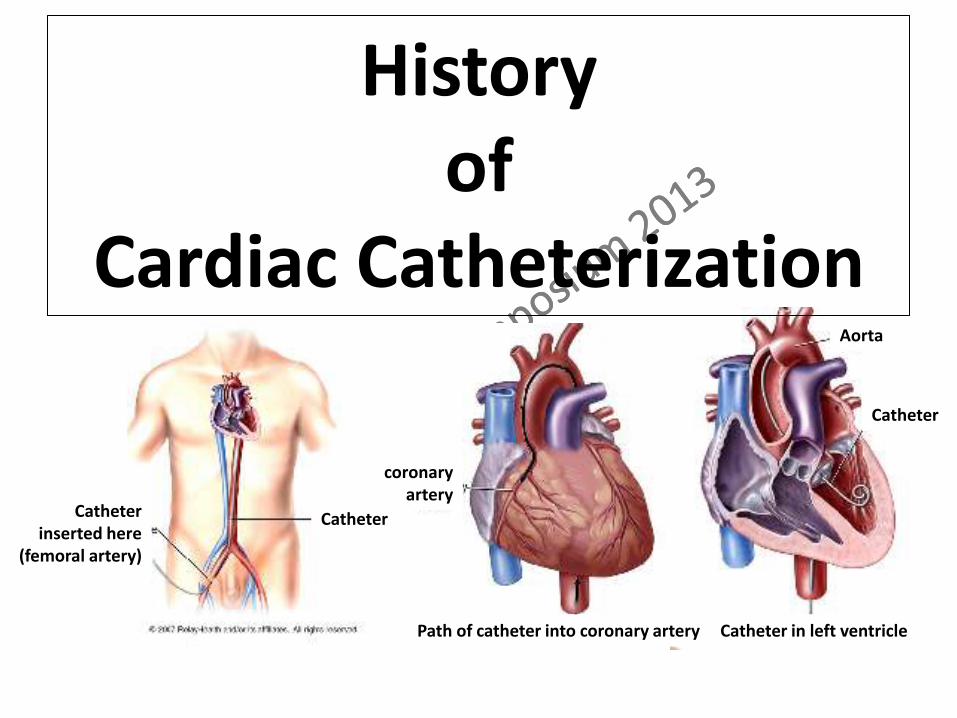
History of
Cardiac Catheterization
Catheter in left ventricle Path of catheter into coronary artery
Aorta
Catheter
Catheter
coronary artery
Catheter inserted here
(femoral artery)
Catheterization =
Putting a hollow tube into a lumen

➣ 3000 B.C. – Egyptions performed bladder catheterizations using metal pipes
➣ 400 B.C. – Catheters fashioned from hollow reeds and pipes were used in cadavers to study the function of cardiac valves
➣ 1711 – Hales conducted the first cardiac catheterization of a horse using brass pipes, a glass tube and the trachea of a goose
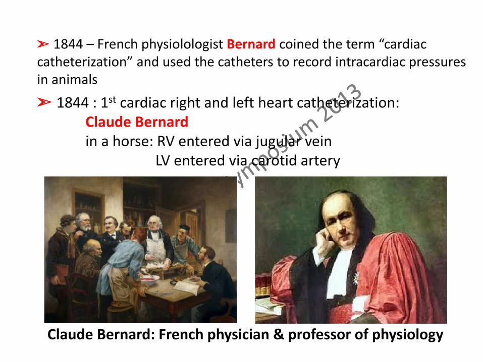
➣ 1844 : 1st cardiac right and left heart catheterization: Claude Bernard in a horse: RV entered via jugular vein LV entered via carotid artery
Claude Bernard: French physician & professor of physiology
➣ 1844 – French physiolologist Bernard coined the term “cardiac catheterization” and used the catheters to record intracardiac pressures in animals

➣ Followed by period of investigations of CV physiology in animals:
pressure manometry
Fick cardiac output
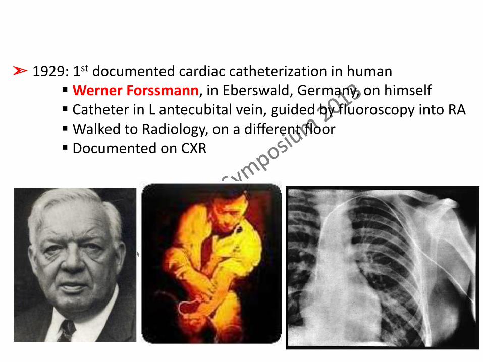
➣ 1929: 1st documented cardiac catheterization in human Werner Forssmann, in Eberswald, Germany, on himself Catheter in L antecubital vein, guided by fluoroscopy into RA Walked to Radiology, on a different floor Documented on CXR
➣ 1941 – Cournand and Richards employed the cardiac catheter
as a diagnostic tool for the first time, utilising catheter techniques
to measure cardiac output
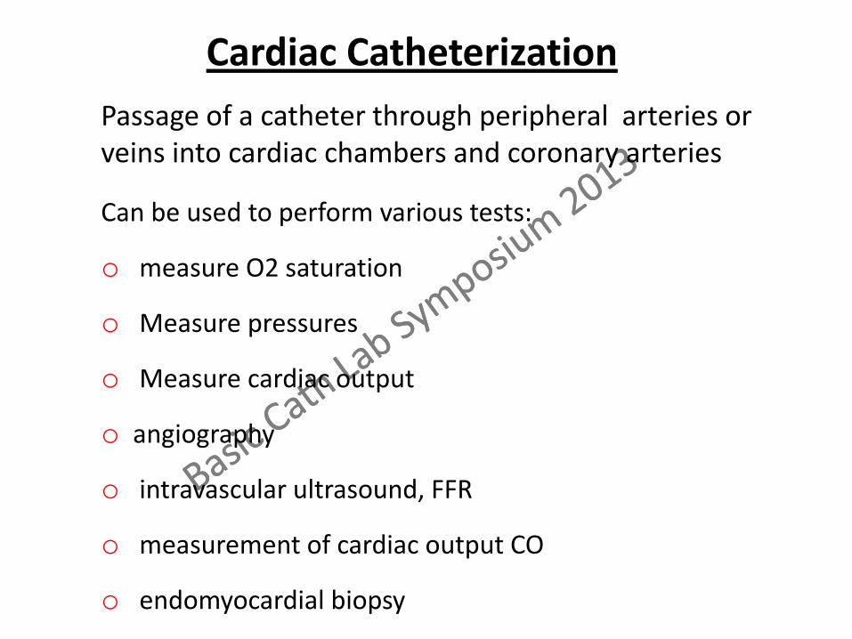
Cardiac Catheterization
Passage of a catheter through peripheral arteries or veins into cardiac chambers and coronary arteries
Can be used to perform various tests:
o measure O2 saturation
o Measure pressures
o Measure cardiac output
o angiography
o intravascular ultrasound, FFR
o measurement of cardiac output CO
o endomyocardial biopsy

Cournand catheter: ➣ A right heart catheter w end hole, no side holes ➣ Suitable for wedge pressure measurement
Andre Cournand: 1895 – 1988 French physician and physiologist Moved to USA and became an American citizen in 1941
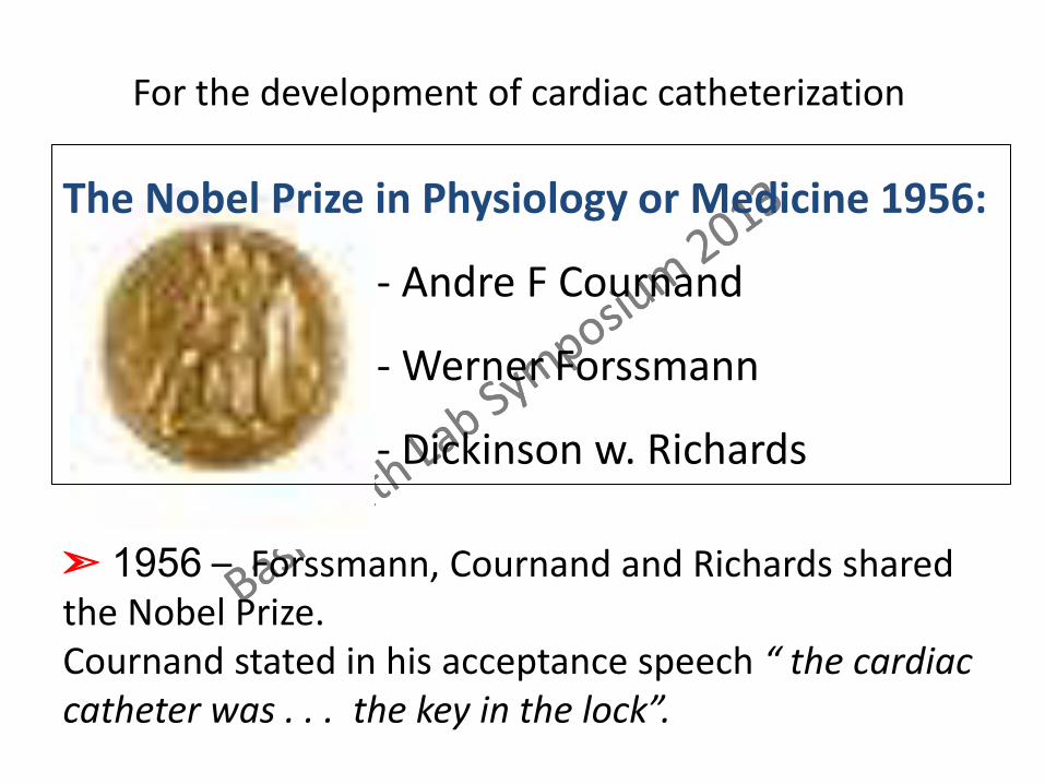
The Nobel Prize in Physiology or Medicine 1956:
- Andre F Cournand
- Werner Forssmann
- Dickinson w. Richards
➣ 1956 – Forssmann, Cournand and Richards shared the Nobel Prize. Cournand stated in his acceptance speech “ the cardiac catheter was . . . the key in the lock”.
For the development of cardiac catheterization
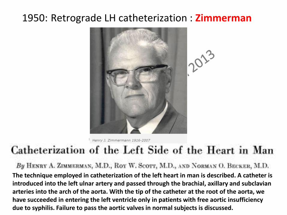
1950: Retrograde LH catheterization : Zimmerman
The technique employed in catheterization of the left heart in man is described. A catheter is introduced into the left ulnar artery and passed through the brachial, axillary and subclavian arteries into the arch of the aorta. With the tip of the catheter at the root of the aorta, we have succeeded in entering the left ventricle only in patients with free aortic insufficiency due to syphilis. Failure to pass the aortic valves in normal subjects is discussed.
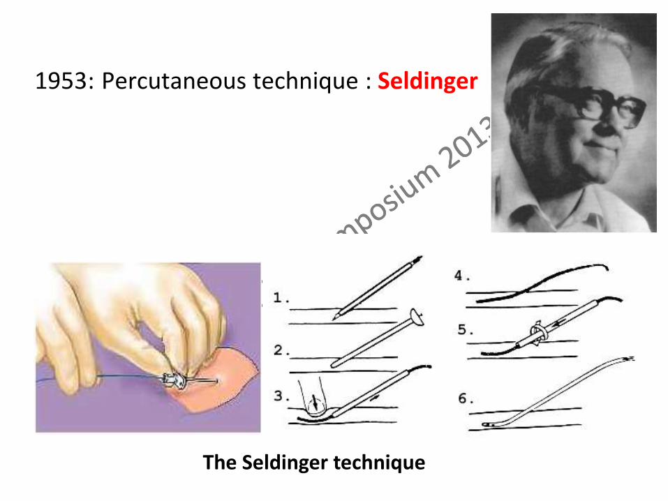
The Seldinger technique
1953: Percutaneous technique : Seldinger
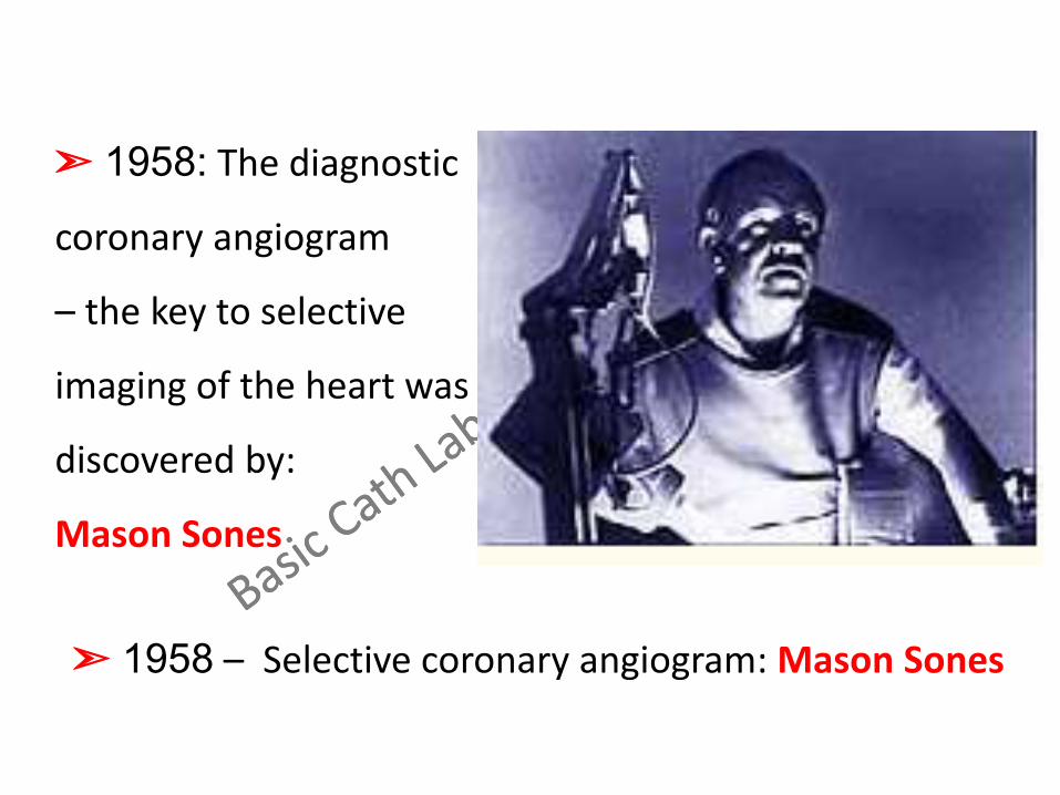
➣ 1958: The diagnostic
coronary angiogram
– the key to selective
imaging of the heart was
discovered by:
Mason Sones
➣ 1958 – Selective coronary angiogram: Mason Sones

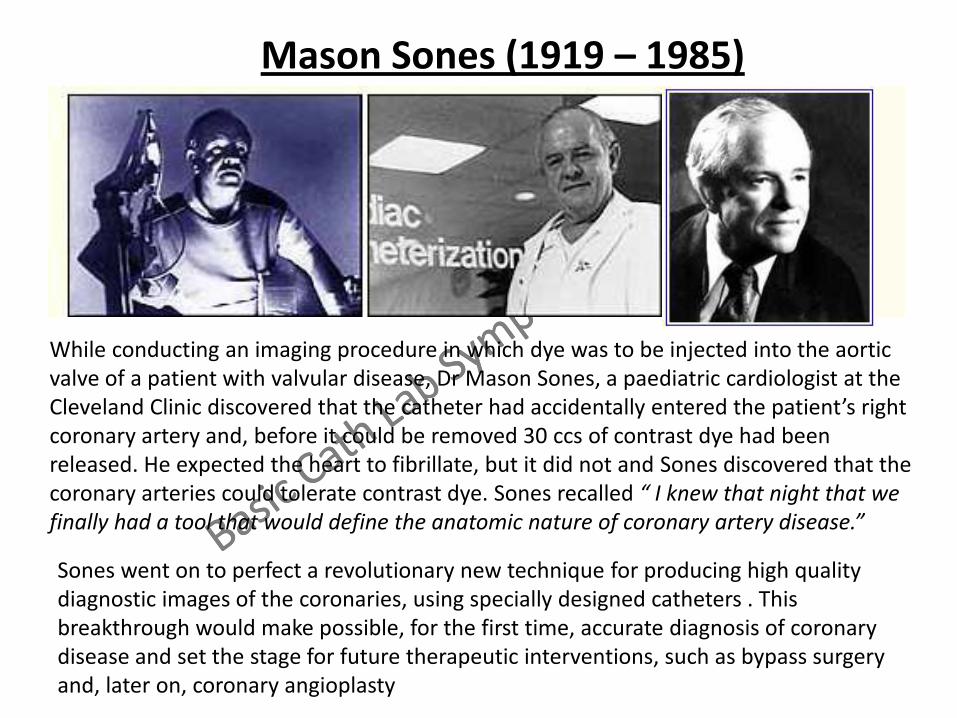
Mason Sones (1919 – 1985)
While conducting an imaging procedure in which dye was to be injected into the aortic valve of a patient with valvular disease, Dr Mason Sones, a paediatric cardiologist at the Cleveland Clinic discovered that the catheter had accidentally entered the patient’s right coronary artery and, before it could be removed 30 ccs of contrast dye had been released. He expected the heart to fibrillate, but it did not and Sones discovered that the coronary arteries could tolerate contrast dye. Sones recalled “ I knew that night that we finally had a tool that would define the anatomic nature of coronary artery disease.”
Sones went on to perfect a revolutionary new technique for producing high quality diagnostic images of the coronaries, using specially designed catheters . This breakthrough would make possible, for the first time, accurate diagnosis of coronary disease and set the stage for future therapeutic interventions, such as bypass surgery and, later on, coronary angioplasty

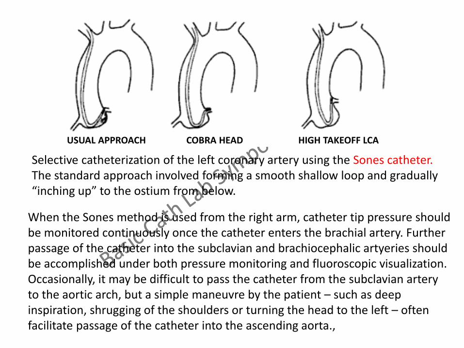
Selective catheterization of the left coronary artery using the Sones catheter. The standard approach involved forming a smooth shallow loop and gradually “inching up” to the ostium from below.
USUAL APPROACH COBRA HEAD HIGH TAKEOFF LCA
Selective catheterization of the left coronary artery using the Sones catheter. The standard approach involved forming a smooth shallow loop and gradually “inching up” to the ostium from below.
When the Sones method is used from the right arm, catheter tip pressure should be monitored continuously once the catheter enters the brachial artery. Further passage of the catheter into the subclavian and brachiocephalic artyeries should be accomplished under both pressure monitoring and fluoroscopic visualization. Occasionally, it may be difficult to pass the catheter from the subclavian artery to the aortic arch, but a simple maneuvre by the patient – such as deep inspiration, shrugging of the shoulders or turning the head to the left – often facilitate passage of the catheter into the ascending aorta.,
USUAL APPROACH COBRA HEAD HIGH TAKEOFF LCA
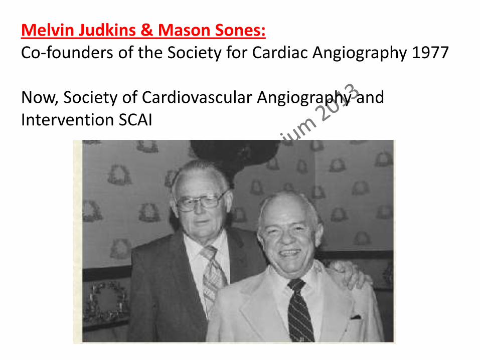
Melvin Judkins & Mason Sones: Co-founders of the Society for Cardiac Angiography 1977 Now, Society of Cardiovascular Angiography and Intervention SCAI

Melvin Judkins & Mason Sones: Co-founders of the Society for Cardiac Angiography 1977 Now, Society of Cardiovascular Angiography and Intervention SCAI
In 1977, the Cardiac Catheterization and Angiography Study Group met to discuss the establishment of a professional society. The first meeting of the society was held in 1978, with Sones and Judkins as Co-Chairs. Sones became the first president of the society and Judkins the second president. The society was open only to individuals who had become experts in the broad field of catheterization and angiography.
➣ 1959 – Transeptal catheterization:
Ross and Cope

1964: Transluminal Angioplasty, the concept of remodelling of the
artery, was introduced by Dr Charles Dotter
Charles Dotter: Considered father of Interventional Radiology. Chair of Diagnostic Radiology, Uni of Oregon Med School
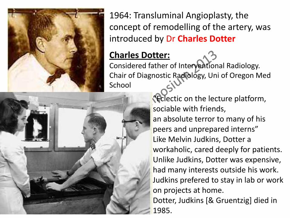
“Eclectic on the lecture platform, sociable with friends, an absolute terror to many of his peers and unprepared interns” Like Melvin Judkins, Dotter a workaholic, cared deeply for patients. Unlike Judkins, Dotter was expensive, had many interests outside his work. Judkins prefered to stay in lab or work on projects at home. Dotter, Judkins [& Gruentzig] died in 1985.
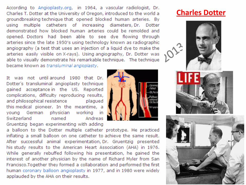
1964: Transluminal Angioplasty, the concept of remodelling of the artery, was introduced by Dr Charles Dotter
Charles Dotter: Considered father of Interventional Radiology. Chair of Diagnostic Radiology, Uni of Oregon Med School
Charles Dotter
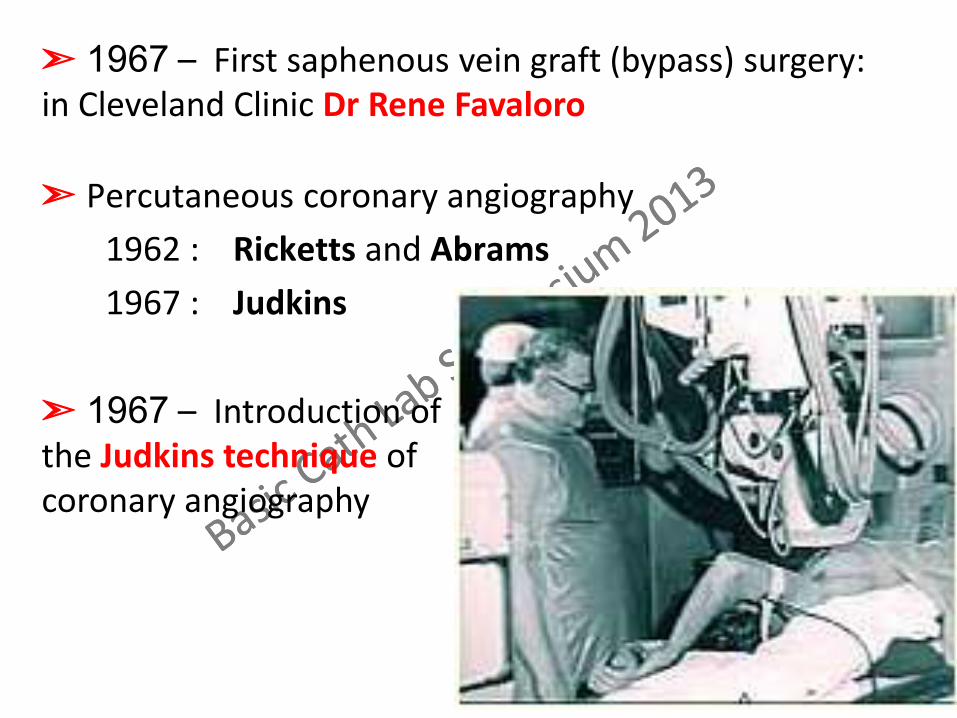
➣ Percutaneous coronary angiography
1962 : Ricketts and Abrams
1967 : Judkins
➣ Percutaneous coronary angiography
1962 : Ricketts and Abrams
1967 : Judkins
➣ 1967 – First saphenous vein graft (bypass) surgery: in Cleveland Clinic Dr Rene Favaloro
➣ 1967 – Introduction of the Judkins technique of coronary angiography

With his left & right coronary catheters & pigtail catheters He initially formed catheters by bending them to conform to the pt’s anatomy. Later, these catheters were commercially preformed by Cordis.
Melvin Judkins: Grew up in Los Angeles. Premedical in Loma Linda Uni College of Arts & Science, Riverside, CA. Obtained BS in 1945
Melvin Judkins: 1922 – 1985
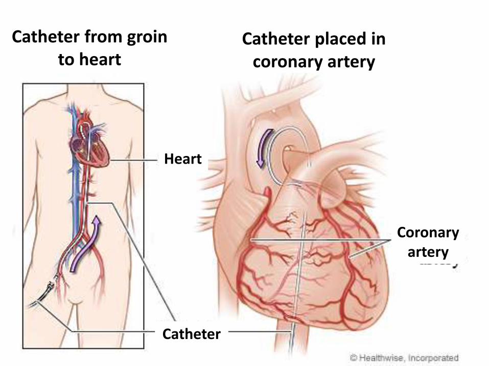
Catheter from groin to heart
Catheter placed in coronary artery
Heart
Catheter
Coronary artery
➣ 1970 :
balloon-tipped, flow guided catheter Swan and Ganz
Further advancements:
➣ Better radiographic imaging
➣ Less toxic contrast
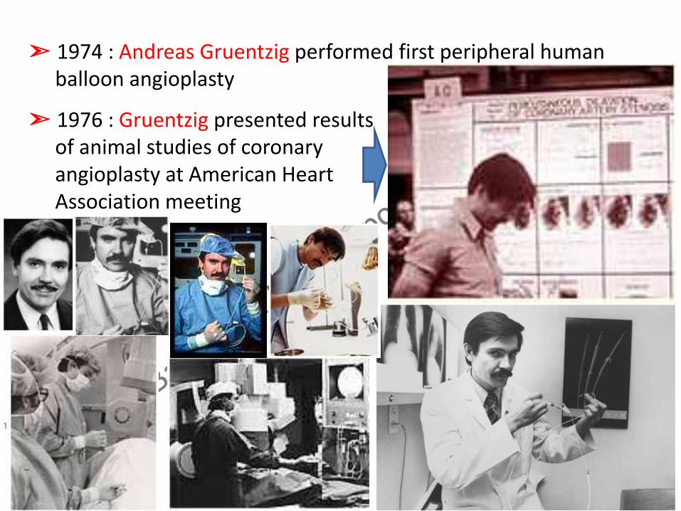
➣ 1974 : Andreas Gruentzig performed first peripheral human balloon angioplasty
➣ 1976 : Gruentzig presented results of animal studies of coronary angioplasty at American Heart Association meeting
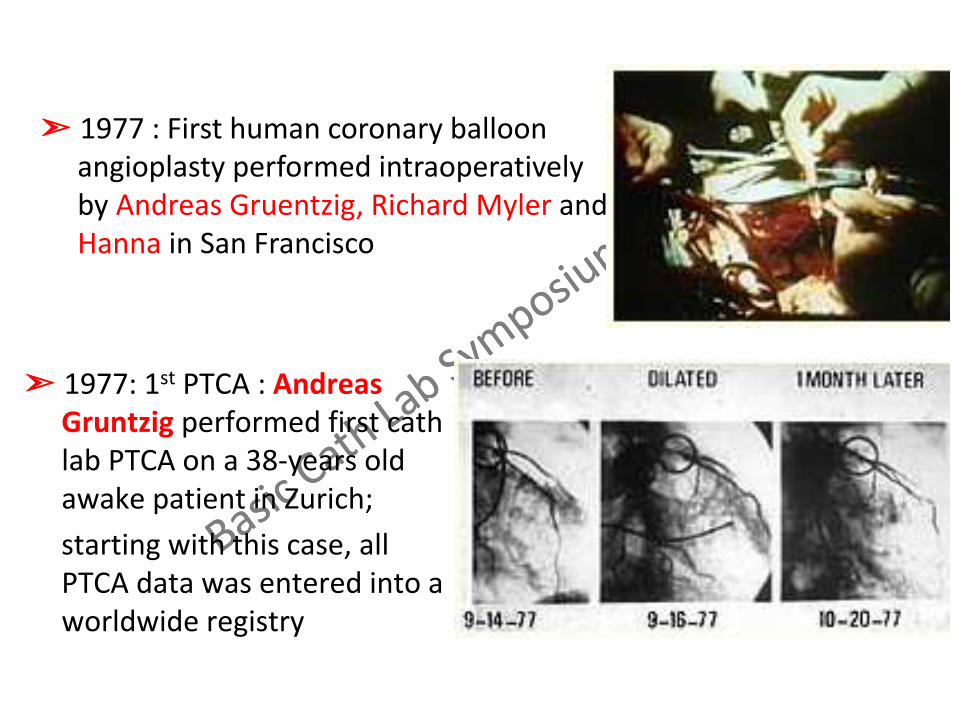
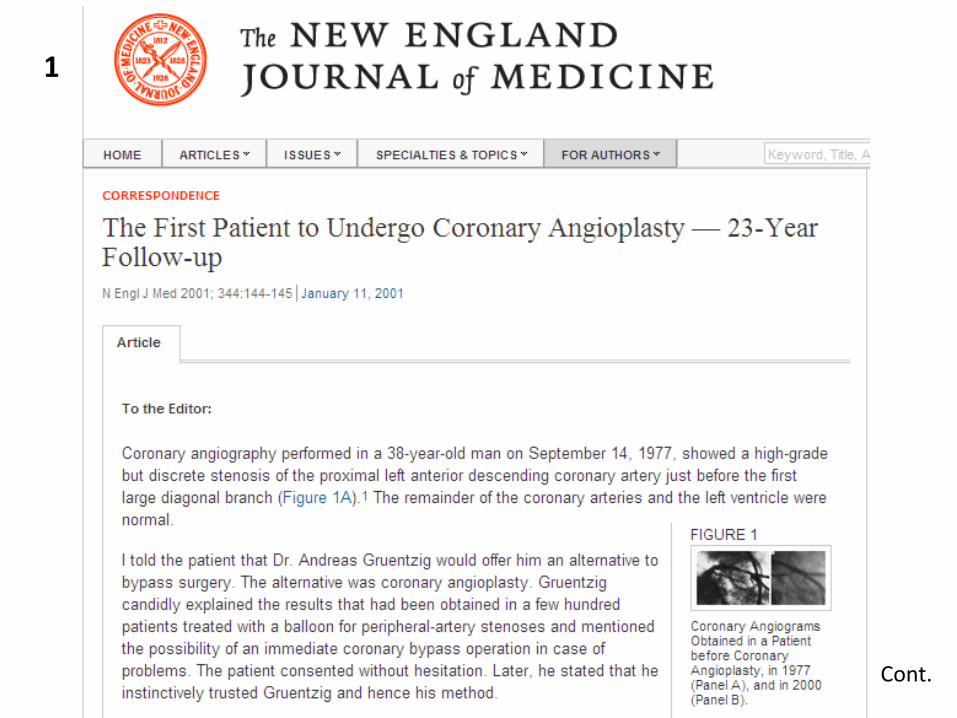
➣ 1977: 1st PTCA : Andreas Gruntzig performed first cath lab PTCA on a 38-years old awake patient in Zurich;
starting with this case, all PTCA data was entered into a worldwide registry
➣ 1977 : First human coronary balloon angioplasty performed intraoperatively by Andreas Gruentzig, Richard Myler and Hanna in San Francisco
1
Cont.
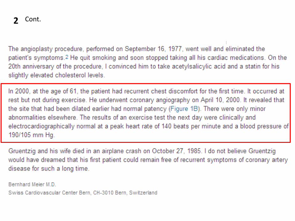
2 Cont.

Gruntzig, Myler, Stertzer
➣ 1978 : First PTCA cases performed in America by Myler in San Francisco and Stertzer in New York;
Gruentzig conducted first demonstration course in Zurich, Switzerland, attended by 28 pioneering physicians, International Dilatation Society was established.
➣ 1980: Gruentzig conducted the last of 5 demonstration courses in Zurich with Sones, Judkins & Dotter in attendance. He then moved to Atlanta, GA where he became the Director of Interventional Cardiology @ Emory University; National Heart, Lung & Blood Institute began support of the existing PTCA registry; 1st 1000 angioplasties were performed worldwide; guiding catheters were introduced.

Balloon Angioplasty


➣ 1985 – A year of loss in the history of interventional medicine: Dotter, Sones, Judkins and Gruentzig all passed away within 9 months of each other
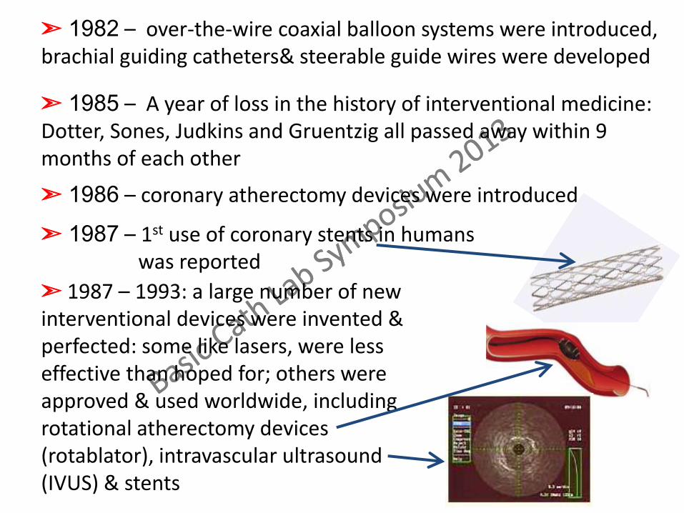
➣ 1982 – over-the-wire coaxial balloon systems were introduced, brachial guiding catheters& steerable guide wires were developed
➣ 1985 – A year of loss in the history of interventional medicine: Dotter, Sones, Judkins and Gruentzig all passed away within 9 months of each other
➣ 1986 – coronary atherectomy devices were introduced
➣ 1987 – 1st use of coronary stents in humans was reported
➣ 1987 – 1993: a large number of new interventional devices were invented & perfected: some like lasers, were less effective than hoped for; others were approved & used worldwide, including rotational atherectomy devices (rotablator), intravascular ultrasound (IVUS) & stents
➣ 1993 – 1991: stents became common place,
they eliminated many complications
➣ 1997: over 1 million angiplasties would be performed
worldwide, making angioplasty the most common medical
intervention in the world
➣ 1997: almost 2 million angiplasties would be performed
worldwide, with an estimated increase of 8% annually
➣ 2002: 25th anniversary of the 1st angioplasty performed in
awake patient
➣ 2007: 30th anniversary of the 1st angioplasty performed in
awake patient
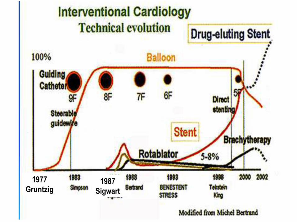
1977
Gruntzig 1987
Sigwart
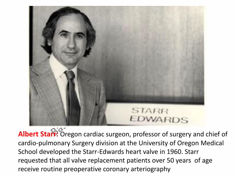
Albert Starr: Oregon cardiac surgeon, professor of surgery and chief of cardio-pulmonary Surgery division at the University of Oregon Medical School developed the Starr-Edwards heart valve in 1960. Starr requested that all valve replacement patients over 50 years of age receive routine preoperative coronary arteriography