history and physical - career steptraining.careerstep.com/pdf/016256_pmcb.pdfhistory and physical...
TRANSCRIPT

HISTORY AND PHYSICAL
HISTORY OF THE PRESENT ILLNESS: The patient is a 24-year-old white female who is gravida 2, para 2,
AB 0 who yesterday afternoon at about 4 o’clock began to have pain in the right upper quadrant and right lower
chest area. It went through to the back. She had pain with respirations, etc.
She called me. It sounded as if she might have the serious problem of a pulmonary embolus or possibly even
collapsed lung or pneumothorax. I was just leaving the office and figured that she ought to be seen at the
emergency room and have full follow up here, where she could have blood gases, etc. if it were necessary.
She was down here at the emergency room and had considerable workup including an ultrasound of the
gallbladder and upper abdomen, which was negative. However, she did have a HIDA scan, which showed a
19% ejection fraction, which is considered normal. She had considerable pain with this, all of which seemed to
agree with a tentative diagnosis by the emergency room doctor Dr. ___ [NAME] of being an acute gallbladder.
Late last night I was called, and he felt that she ought to be admitted, and I agreed. She had been in the hospital
overnight and had had some relief with this but still is hurting. A characteristic of the pain is that it hurts in the
right upper quadrant. It goes through to the back. Changing positions makes it worse. It is bad when she tries
to lie flat on her back.
When she is elevated a little bit she is somewhat better. Trying to lie on her side makes it worse. Sitting up and
leaning forward helps a little bit. She has pain with coughing or sneezing. She has had a little nausea but no
vomiting or diarrhea. There have been no previous episodes of anything like this.
Last night before this episode began she had had sort of a casserole meal with tator tots, meat, and cream of
chicken soup, etc., and bread and butter, and maybe that had something to do with this.
PAST MEDICAL HISTORY: She has had two children with no difficulties. She has had a tonsillectomy and
adenoidectomy at age eight.
FAMILY HISTORY: Her father had diabetes. There was no cancer in the family. Her grandmother had heart
trouble. Two or three relatives have had gallbladder trouble.
REVIEW OF SYSTEMS: Neurologic: She has some migraine headaches. I saw her in the office a few weeks
ago with this, and medication seems to be helping some. HEENT: She has complaints of dry mouth and throat
since she has been in the hospital. Respiratory: Deep breathing hurts the abdomen. Gastrointestinal: She is
commonly constipated, and otherwise she has no difficulties. Genitourinary: Genitourinary system is pretty
much negative. She takes birth control pills and rarely has cramps.
Otherwise system review is negative except she is allergic to Monistat vaginal cream and has intolerance to
aspirin.
PHYSICAL EXAMINATION: Vital Signs: Please see nurse’s notes. HEENT: The HEENT examination is
negative. Neck: The neck is negative. The thyroid is not palpable. The trachea is midline. Chest: The lung
fields are generally clear. There is no tenderness to palpate the chest wall. Heart: There is no murmur, thrill, or
rub. Breasts: There is no mass or tenderness. Axillae/Groins: Axillae and groins are negative. Back: The back
is symmetrical. The costovertebral angle is negative. The low back is negative. Abdomen: Liver, kidneys, and
spleen are not palpable. No masses are palpable, but there is exquisite tenderness over the gallbladder area, even

tenderness to light percussion over the gallbladder itself and very positive Murphy sign. Bowel sounds are
present but slightly hypoactive. There is no particular tenderness in the lower abdominal area. I did not do
pelvic or rectal examination at this time. Extremities: The extremities are within normal limits.
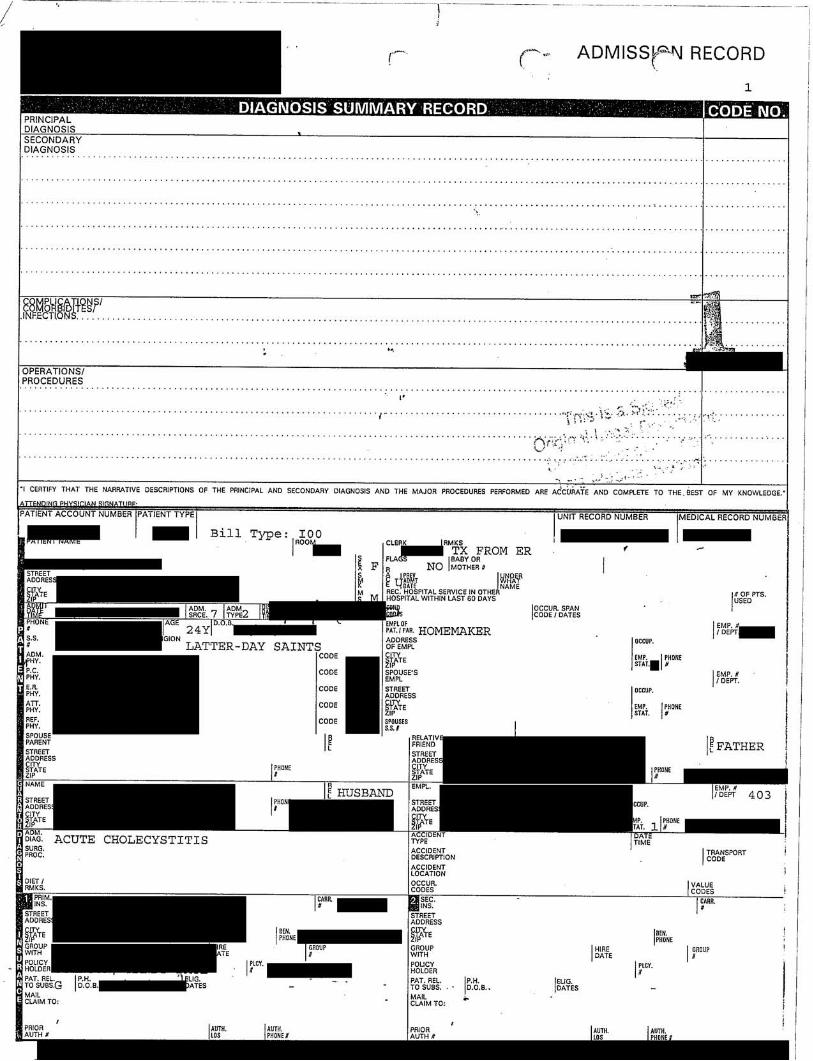
IMPRESSION: Acute cholecystitis.
DISPOSITION: I will seek surgical consultation regarding this because it seems not to be getting any better. I
think it is likely she is going to need to have surgery.

CONSULTATION
CHIEF COMPLAINT: Abdominal pain.
HISTORY OF PRESENT ILLNESS: This is a 24-year-old female who presents with the onset last evening of
fairly intense right upper quadrant abdominal pain, with radiation to the back, which has persisted all night and
in to today. She has had a lot of nausea, but no actual vomiting. She has not had a bowel movement for 48
hours, and she has had no history of diarrhea, constipation, hematemesis, hematochezia, or melena. The pain is
worse when she moves around. She had also been complaining of right lower quadrant abdominal pain. She
has undergone a fairly extensive workup, including laboratory work, which was essentially within normal
limits. She has had a CT scan, which showed a lot of stool in the right colon, but no evidence of acute
appendicitis, and possibly a small right ovarian cyst. An ultrasound was normal, without stones, and a HIDA
scan has been done and showed a 19% ejection fraction, and she had reproduction of her pain and intensification
of her pain when the medication was given.
PAST MEDICAL HISTORY: Allergies: Monistat. Current Medications: None. Surgeries: Tonsillectomy.
Illnesses: No serious illnesses.
FAMILY HISTORY: The patient has several close family members with gallbladder disease. Otherwise, the
patient’s family history is unremarkable.
SOCIAL HISTORY: The patient is married, has two children. She denies the use of alcohol and tobacco.
PHYSICAL EXAMINATION: General: The patient is a fairly thin, alert, oriented, and cooperative female,
who appears to be in mild to moderate distress. Head: The head is atraumatic and normocephalic. Eyes:
Extraocular movements are intact. The pupils are equal, round, and reactive. Ears: The ears are clear bilaterally.
Nose: The nares are patent without discharge. Mouth: The mouth is without lesions, erythema, or exudate.
Neck: The neck is supple without thryomegaly or adenopathy. Lungs: The lungs are clear to auscultation and
percussion. Heart: The heart reveals a regular rate and rhythm, without murmurs, rubs or gallops. Abdomen:
The abdomen is soft. She is tender all along the right side, with a little bit of increased tenderness in the right
lower quadrant at McBurney’s point, with tenderness in the right subcostal area, with positive Murphy’s sign.
No definite organomegaly, masses, or hernias are appreciated. No actual peritoneal signs are identified.
Extremities: The extremities reveal no cyanosis, clubbing, or edema. Neurologic: The neurologic examination
is grossly intact.
IMPRESSION: Right upper quadrant abdominal pain, chronic cholecystitis, with biliary dyskinesia.
RECOMMENDATION: The plan is to give the patient an enema to see if we can resolve her right lower
quadrant or right upper quadrant abdominal pain. If her right upper quadrant abdominal pain persists, then I
would recommend that we proceed with a laparoscopic cholecystectomy with cholangiograms. I have explained
the operation to the patient, along with the risks, and the possibility of an open laparotomy. The patient wishes
to proceed with surgery as outlined above, if the enema does not seem to solve her problem.

EMERGENCY CENTER VISIT
HISTORY OF THE PRESENT PROBLEM: The patient is a 24-year-old white female who was feeling well
today. While she was sitting she developed the sudden onset of right-sided chest pain that was worse with deep
breathing. She has had no cough. She had a migraine two weeks ago and was given pain medications, but has
not used the pain medications for days and has felt well. She had had no pain in her legs recently. No swelling.
She has been on no trips. She has never had a deep venous thrombosis or thrombophlebitis. She has had no
fever, chills, or sweats. No shortness of breath when she is breathing quietly. The pain is worse when she
moves, and when she breathes deeply.
PAST MEDICAL HISTORY: Illnesses: None of significance except childhood illnesses. Operations:
Tonsillectomy and adenoidectomy. Hospitalizations: She has had two children vaginal deliveries.
SOCIAL HISTORY: The patient does not use alcohol, drugs, or tobacco. She is a housewife married with two
children.
FAMILY HISTORY: The patient’s father has diabetes mellitus, otherwise the family history is clear with no
history of gallbladder disease.
PHYSICAL EXAMINATION: On physical examination the blood pressure was 140/99, respirations 18, heart
rate 88, and her temperature was 99.2. Her O2 saturation on room air was 98%. On physical examination she
was alert, still having quite a bit of pain in what appeared to be the right upper quadrant or the bottom of the
right chest. The tympanic membranes were clear. The mouth and throat were clear. She had no cervical
lymphadenopathy. Her lungs were clear to auscultation and percussion. She had no posterior or flank
tenderness to percussion. Her heart was regular without murmurs, gallops, or rubs. Her abdomen was soft.
Bowel sounds were active. She was very tender in the right upper quadrant. I could feel no mass present. She
has no hepatosplenomegaly. She had no tenderness over the calves. Her Homans was negative bilaterally.
RADIOLOGICAL STUDIES: The chest x-ray was clear.
LABORATORY STUDIES: Laboratory work so far has been normal.
FINAL DIAGNOSIS: Dr. ___ [NAME] will do the final disposition on the case.
FINAL IMPRESSIONS: Diagnosis of probable pleurodynia.
ASSESSMENT: I think we need to make sure the patient does not have gallbladder disease. She is very tender
in the right upper quadrant. We also need to be sure she does not have a pulmonary embolism, but that would
be remote based on the history and the physical examination. Dr. ___ [NAME] will do the final disposition on
the case.

EMERGENCY CENTER VISIT
CHIEF COMPLAINT: Right upper quadrant and right sided chest pain.
HISTORY OF THE PRESENTING COMPLAINT: The patient is a 24-year-old female who had a sudden
onset of very intense right upper quadrant abdominal pain and right-sided chest pain all the way to her shoulder.
She has never had this pain before. She has undergone a full evaluation by Dr. ___ [NAME]; please refer to his
dictation for the full history and physical. Dr. ___ [NAME] has requested that I follow up this patient after he
has requested an ultrasound of her gallbladder. As mentioned, the patient was doing well prior to the onset of
this discomfort. She did not receive any trauma. It did not seem associated with any foods. She was sitting
down watching TV when it started. She denies any history of deep venous thrombosis risk factors except she is
on birth control and that is it.
PAST MEDICAL HISTORY: Otherwise very healthy. Her last menstrual period about one week ago was
slightly light at that time.
MEDICATIONS: Birth Control.
ALLERGIES: MONISTAT.
FAMILY HISTORY: Her father has a history of diabetes.
PHYSICAL EXAMINATION: The patient is a 24-year-old female who is open and conversant, definitely
uncomfortable even though she has received 30 mg of Toradol IV as requested by Dr. ___ [NAME].
Nevertheless, she is cooperative. She is alert and oriented times three. She is normocephalic, atraumatic. Her
oropharynx is slightly dry but otherwise pink and intact. Pupils are equal, round and reactive to light.
Extraocular movements are intact. Sclerae white. The neck is supple and nontender with no lymphadenopathy
and no jugular venous distention. Heart regular rate and rhythm with a clear S1 and S2 without murmur, rub or
gallop. The lungs are clear to auscultation at the bases with good lung excursion. No retractions are seen. Her
chest wall is nontender to palpation. Her abdomen at this time is still soft but she does have a localized right
upper quadrant abdominal pain with a significant Murphy’s sign. However, there are no peritoneal signs. No
appreciable involuntary guarding. She does have slight increased pain on the right side even though if I push on
the left but is not a prominent peritoneal sign. No appreciable masses are noted. No hepatosplenomegaly. She
does have costovertebral angle tenderness on the right side as well. Her extremities she moves with 5/5
strength. She has 2+ pulses. No cyanosis or edema.
EMERGENCY DEPARTMENT COURSE: Review of the laboratory data demonstrates really no acute
pathology including an hCG serum being negative, amylase at 41, electrolytes are completely unremarkable
including a bicarbonate at 23. Liver function tests are also unremarkable. Her urinalysis is completely negative.
Her white count is 8,300 with a normal differential. The ultrasound is reported as “very normal.” Therefore, I
have sent the patient back for a HIDA scan because of the persisting pain and still persistent concerns about the
probability of a gallbladder being the source of her complaints. Chest x-ray was completed, a two view which
demonstrates no evidence of any pneumothorax. I have again questioned the patient about possibilities of deep
venous thrombosis risk factors, which she has none except for the birth control pills. Her legs are unremarkable
including no tenderness for swelling. The patient was sent for the HIDA scan which demonstrates an ejection
fraction of 19%, which is very normal. I have talked with Dr. ___ [NAME] regarding this patient who is her
primary care physician. He agrees to have her admitted through the night. My concern about sending her home
is that she is still very uncomfortable even despite receiving morphine sulfate IV and I doubt she will do well

through the night. Dr. ___ [NAME] agrees and she will be sent upstairs.
DIAGNOSIS: Acute cholecystitis.
DISPOSITION: Admitted to the service of Dr. ___ [NAME].
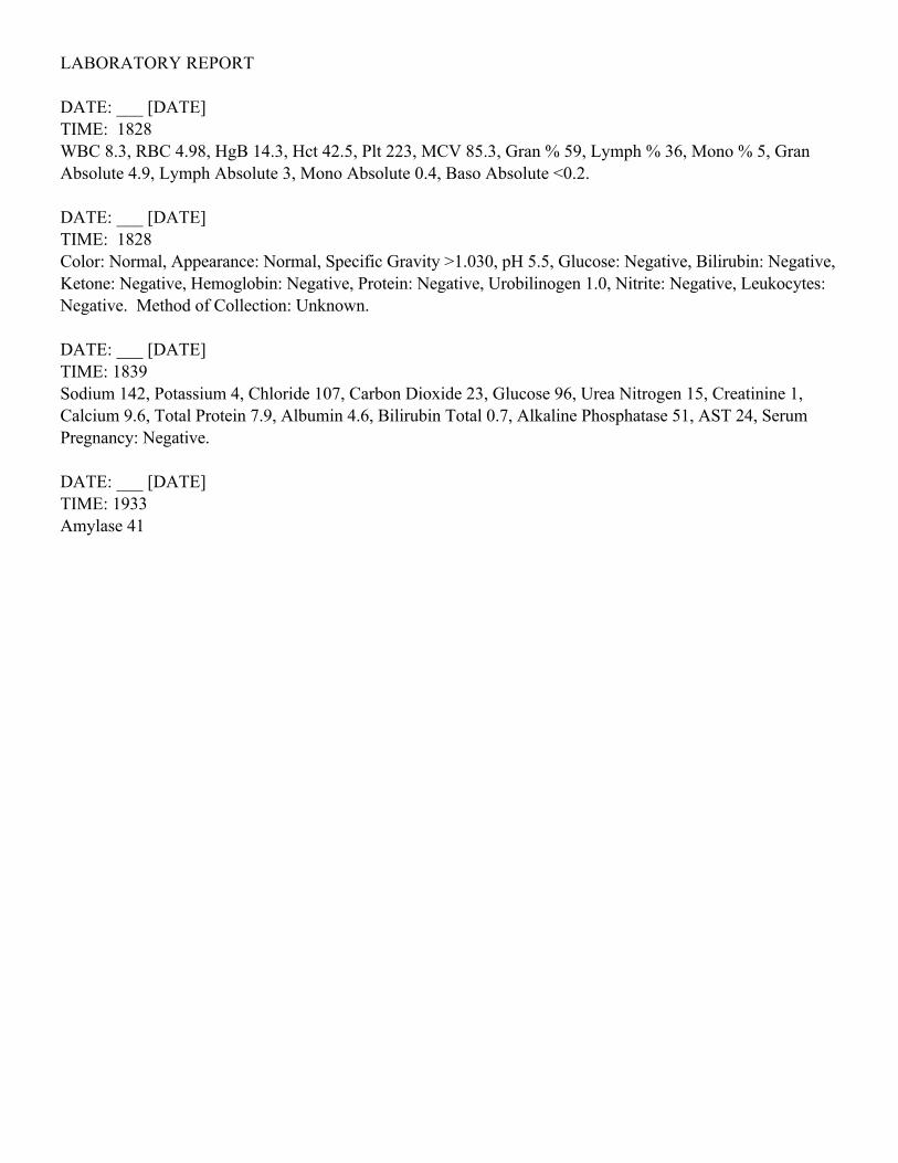
LABORATORY REPORT
DATE: ___ [DATE]
TIME: 1828
WBC 8.3, RBC 4.98, HgB 14.3, Hct 42.5, Plt 223, MCV 85.3, Gran % 59, Lymph % 36, Mono % 5, Gran
Absolute 4.9, Lymph Absolute 3, Mono Absolute 0.4, Baso Absolute <0.2.
DATE: ___ [DATE]
TIME: 1828
Color: Normal, Appearance: Normal, Specific Gravity >1.030, pH 5.5, Glucose: Negative, Bilirubin: Negative,
Ketone: Negative, Hemoglobin: Negative, Protein: Negative, Urobilinogen 1.0, Nitrite: Negative, Leukocytes:
Negative. Method of Collection: Unknown.
DATE: ___ [DATE]
TIME: 1839
Sodium 142, Potassium 4, Chloride 107, Carbon Dioxide 23, Glucose 96, Urea Nitrogen 15, Creatinine 1,
Calcium 9.6, Total Protein 7.9, Albumin 4.6, Bilirubin Total 0.7, Alkaline Phosphatase 51, AST 24, Serum
Pregnancy: Negative.
DATE: ___ [DATE]
TIME: 1933
Amylase 41

RADIOLOGY REPORT
PA AND LATERAL CHEST DATED ___ [DATE]
The heart and pulmonary vessels are normal and the lungs are clear. The bones and soft tissues are
unremarkable.
IMPRESSION: Normal chest.

RADIOLOGY REPORT
UPPER ABDOMINAL SONOGRAM: Multiple images were obtained of the gallbladder. No stones are seen
within it, although the patient is quite tender over the gallbladder. The liver, spleen, kidneys, and pancreas are
otherwise normal.
IMPRESSION: The patient is quite tender over the gallbladder, but no anatomic abnormality is seen in the
upper abdomen.

RADIOLOGY REPORT
NUCLEAR HIDA SCAN: The patient is given 2.17 millicuries of Technetium 99m-Choletec followed by
scanning in the right upper quadrant. There is rapid visualization of the extrahepatic biliary system including
temperature gallbladder. The patient was then given 1.32 micrograms of Cholecystokinin. A gallbladder
ejection fraction of 19% is calculated which is abnormally low and suggests the possibility of biliary dyskinesia.
IMPRESSION: Abnormal nuclear HIDA scan.

RADIOLOGY REPORT
CT SCAN OF THE ABDOMEN AND PELVIS DATED ___ [DATE]
COMMENT: Axial computed tomography was performed through the abdomen and pelvis without
intravenous enhancement.
The liver, spleen, pancreas, and kidneys are unremarkable. The bowel and mesentary are unremarkable. The
region of the appendix is without evidence of mass effect or edema. The appendix per se is not discretely
identified. There is some high-density contrast material in the lower rectal sigmoid perhaps due to ingestion of
calcium or bismis containing material such as Pepto-Bismol or Maalox.
IMPRESSION: Negative CT scan of the abdomen and pelvis.

RADIOLOGY REPORT
Operative cholangiogram shows the common duct to be of normal caliber. There is good spill into the
duodenum. No obstruction or other abnormality is identified.
IMPRESSION: Normal operative cholangiogram.

PATHOLOGY CONSULTATION
FINAL DIAGNOSIS: Gallbladder resection: Chronic cholecystitis and cholesterolosis.
CLINICAL DATA: Preop: “Acute cholecystitis.” Tissue submitted: “Gallbladder.”
GROSS DESCRIPTION: “Gallbladder”: The serosa of this previously opened 8.0 x 2.8 x 0.5 cm gallbladder
is green and bile stained. The cystic duct has a diameter of 0.1 cm at the surgical end. No calculi accompany the
gallbladder nor are calculi found within the gallbladder. The bladder mucosa is green-black-brown velvety and
covered with some yellow-green reticulation. The bladder wall measures up to 0.3 cm in greatest thickness.
The cystic duct and bladder wall sections are submitted in one cassette.
MICROSCOPIC DESCRIPTION: Significant acute inflammation is not seen.

OPERATIVE REPORT
PREOPERATIVE DIAGNOSIS: Chronic cholecystitis, biliary dyskinesia.
POSTOPERATIVE DIAGNOSIS: Chronic cholecystitis, biliary dyskinesia, cholesterolosis.
OPERATION PERFORMED: Laparoscopic cholecystectomy with cholangiogram.
ANESTHESIA: General.
FINDINGS AT OPERATION: This patient had a gallbladder that had some adhesions to the surfaceand a little bit of edema. There was good visualization of both ovaries and uterus, appendix, noother pathology could be identified.
DETAILS OF PROCEDURE: The patient was taken to the operating room and placed underadequate general anesthesia. The abdomen was prepped with Betadine and sterile drapes werearranged. An infraumbilical stab incision was made and the Veress needle was inserted into theperitoneal cavity and pneumoperitoneum was obtained. A 10-11 trocar and sleeve were introducedand the camera was passed into the peritoneal cavity. Right upper quadrant 5 mm trocars andsleeves times two were placed, and a 10-11 trocar and sleeve was placed in the epigastrium. Thegallbladder was grasped by the fundus and infundibulum. Blunt dissection of the lower part of thegallbladder was undertaken until the cystic duct was isolated. A clip was placed on the ductadjacent to the gallbladder and then a small incision was made in the duct and the catheter wasinserted and the x-rays via fluoroscopy were obtained using low osmolar contrast. After this thecatheter was removed and the duct was doubly hemoclipped and divided. The artery was identified,proximally and distally hemoclipped and divided, anterior posterior branch was clipped and divided.The gallbladder was dissected away from the gallbladder fossa using electrocautery dissectionbrought up through the epigastric port without difficulty. The subhepatic and subdiaphragmaticspaces were irrigated and aspirated of excess fluid and inspected for any bleeding. Then #0 Vicrylwith the Endoclose was used to place a stitch to close the fascia at the umbilicus and epigastrictrocar sites. The sleeves were removed under direct visualization and the pneumoperitoneum wasreleased. After the #0 Vicryl was tied, the subcutaneous tissue was closed using 2-0 Vicryl and theskill of all of the incisions was closed using subcuticular #4-0 Vicryl. Steri-Strips were applied andthe patient was returned to the recovery room in satisfactory condition. There were nointraoperative complications. Final sponge and needle counts were all correct.