histone h2a.z is essential for estrogen receptor signaling
TRANSCRIPT

Histone H2A.Z is essential for estrogenreceptor signaling
Nicolas Gevry,1 Sara Hardy,2 Pierre-Etienne Jacques,2 Liette Laflamme,1 Amy Svotelis,1
Francxois Robert,2,3 and Luc Gaudreau1,4
1Departement de biologie, Faculte des sciences, Universite de Sherbrooke, Sherbrooke, Quebec J1K 2R1, Canada; 2Laboratoire dechromatine et expression du genome, Institut de recherches cliniques de Montreal, Montreal, Quebec H2W 1R7, Canada;3Universite de Montreal, Montreal, Quebec H3C 3J7, Canada
Incorporation of H2A.Z into the chromatin of inactive promoters has been shown to poise genes for theirexpression. Here we provide strong evidence that H2A.Z is incorporated into the promoter regions of estrogenreceptor (ERa) target genes only upon gene induction, and that, in a cyclic pattern. Moreover, members of thehuman H2A.Z-depositing complex, p400, also follow the same gene recruitment kinetics as H2A.Z. Importantly,cellular depletion of H2A.Z or p400 leads to a severe defect in estrogen signaling, including loss of estrogen-specific cell proliferation. We find that incorporation of H2A.Z within TFF1 promoter chromatin allowsnucleosomes to adopt preferential positions along the DNA translational axis. Finally, we provide evidence thatH2A.Z is essential to allow estrogen-responsive enhancer function. Taken together, our results provide strongmechanistic insight into how H2A.Z regulates ERa-mediated gene expression and provide a novel link betweenH2A.Z–p400 and ERa-dependent gene regulation and enhancer function.
[Keywords: Chromatin; ER; FoxA1; H2A.Z; p400]
Supplemental material is available at http://www.genesdev.org.
Received February 2, 2009; revised version accepted May 18, 2009.
The integration of physiological and environmental cuesinto complex transcriptional responses is orchestrated bythe coordination of numerous regulatory events andmechanisms that control both the repression and activa-tion of genes. Such signals can be conveyed, for example,by nuclear steroid hormone receptors, such as the estro-gen receptors (ERs). Estrogens are a class of steroid hor-mones that play central roles in female development andreproductive functions (Couse and Korach 1999; Nilssonet al. 2001). In addition, estrogens are directly involved inhormone-dependant diseases, such as breast cancers(Deroo and Korach 2006). The biological functions ofestrogens are mediated through ERa and ERb, whichoperate as ligand-dependent transcription factors thatwork in conjunction with coregulator proteins to regulategene expression and activate transcription of target genesby two distinct, but not mutually exclusive, transcriptionactivation functions (Green and Carroll 2007). Morespecifically, there is an N-terminal ligand-independentactivation function (AF-1) and a C-terminal ligand-dependant activation function (AF-2) located within theligand-binding domain. AF-1 and AF-2 function in a syn-ergistic manner and are required for full transcriptional
activation by ER in most cell types (Tora et al. 1989; Berryet al. 1990; Pham et al. 1992; Tzukerman et al. 1994;Benecke et al. 2000). Little is known about the activationmechanism of AF-1, but several transcriptional coactiva-tors for AF-2 have been found over the past few years. Forexample, ligand-bound AF-2 has been shown to interactwith the LXXLL motif of proteins belonging to the im-portant p160 family of coactivators (Onate et al. 1995;Anzick et al. 1997; Brzozowski et al. 1997; Hong et al.1997). Furthermore, this domain has been linked to therecruitment of the general transcriptional machinery andATP chromatin remodeling complexes such as Swi/Snf(Ichinose et al. 1997; DiRenzo et al. 2000).
Chromatin immunoprecipitation (ChIP) assays haverevealed that ERa can direct ordered and cyclical recruit-ment of cofactors, such as histones modifying enzymes(CBP/p300, Tip60, CARM-1), the Swi/Snf ATP-dependantchromatin remodeling complex, and general transcrip-tion factors (GTF; RNA polymerase II [RNAPII], TBP, andTFIIB), to the TFF1 locus (Shang et al. 2000; Metivier et al.2003). Receptor cycling induces subtle changes in nucle-osome positioning, which in turn plays a key role in therepression and activation of the TFF1 gene (Metivier et al.2003). These changes are characterized by an undefinedtranslational nucleosomal position on the inactive geneto a preferred (stable) nucleosomal position on the ac-tive TFF1 proximal promoter. Moreover, transcriptional
4Corresponding author.E-MAIL [email protected]; FAX (819) 821-8049.Article published online ahead of print. Article and publication date areonline at http://www.genesdev.org/cgi/doi/10.1101/gad.1787109.
1522 GENES & DEVELOPMENT 23:1522–1533 � 2009 by Cold Spring Harbor Laboratory Press ISSN 0890-9369/09; www.genesdev.org
Cold Spring Harbor Laboratory Press on March 15, 2018 - Published by genesdev.cshlp.orgDownloaded from
regulation of TFF1 and an important majority of ERa-regulated genes have been found dependent on thebinding of the transcription factor FoxA1 (Carroll et al.2005). Of note, this factor is thought to contribute to therecruitment of ERa and gene regulation due to its abilityto remodel chromatin. However, it is unclear whetherFoxA1 possesses intrinsic remodeling activity or recruitsa protein with chromatin-modifying properties. FoxA1also retains cell type-specific functions, which rely pri-marily on its differential recruitment to chromatin, pre-dominantly at distant enhancers rather than proximalpromoters (Lupien et al. 2008).
Due to the high level of DNA compaction establishedwithin chromatin, it is generally assumed that this con-densed state is an obstacle to all metabolic transactionsinvolving DNA, including ligand-dependent transcrip-tional regulation by the ER (Mellor 2005). Given thatchromatin often has repressive effects on transcription,the ability of nucleosomes to be disrupted or displacedrepresents a critical step in gene regulation. One mech-anism that generates a specialized chromatin environ-ment is the incorporation of histone variants into specificnucleosomes. H2A.Z is one such histone variant, and ithas been implicated principally in the regulation of geneexpression. Much of what we know regarding the func-tion of H2A.Z stems from studies performed in the yeastSaccharomyces cerevisiae, where it was shown to regu-late genes both positively (Santisteban et al. 2000; Adamet al. 2001; Larochelle and Gaudreau 2003) and negatively(Dhillon and Kamakaka 2000). In addition, genome-widelocalization studies have shown that H2A.Z is preferen-tially localized within a few nucleosomes of the initi-ator region of a large proportion of inactive yeast genes(Guillemette et al. 2005; Li et al. 2005; Raisner et al. 2005;Zhang et al. 2005). In mammalian cells, H2A.Z loci havebeen found predominantly at sites occupied by RNAPIIalong with enhancer regions (Barski et al. 2007). Further-more, our laboratory has shown that yeast H2A.Z is essen-tial for the appropriate positioning of a nucleosome span-ning the initiator region of the GAL1 gene (Guillemetteet al. 2005). Moreover, we observed that promoters thatare enriched in H2A.Z have defined nucleosome loca-tions compared with promoters that are not significantlyenriched in H2A.Z, thereby arguing that H2A.Z may reg-ulate gene expression by allowing nucleosomes to adoptpreferred positions within promoter regions.
One seemingly unique feature of H2A.Z is that it can beincorporated within chromatin by an ATP-dependentchromatin remodeling mechanism, which exchangesH2A–H2B dimers for H2A.Z–H2B. In yeast, H2A.Z hasbeen shown to be incorporated by the Swr1 complex,which shares essential subunits with the NuA4 histoneacetyltransferase complex (Krogan et al. 2003; Kobor et al.2004; Mizuguchi et al. 2004). In mammals, there are twoorthologs of Swr1 that have the ability to exchange H2A–H2B for H2A.Z–H2B within chromatin. SRCAP is thefirst complex to show an ability to catalyze the incorpo-ration of H2A.Z into nucleosomes in vitro (Ruhl et al.2006), and a recent report has demonstrated that SRCAPcan be recruited to inactive and active promoters (Wong
et al. 2007). Moreover, depletion of SRCAP in vivo affectsthe loading of H2A.Z within chromatin (Wong et al.2007). In spite of being isolated as a CREB-binding proteinpartner (Johnston et al. 1999), little is known about howSRCAP is recruited to specific promoters. The secondcomplex that has been shown to be able to incorporateH2A.Z into chromatin is p400 (Gevry et al. 2007). Ourlaboratory showed that H2A.Z, via p400, suppresses theactivation of the p21 gene by p53 and senescence re-sponses. Furthermore, the presence of sequence-specifictranscription factors, such as p53 and Myc, dictates thepositioning of H2A.Z-containing nucleosomes withinthese promoters, thus suggesting that DNA-binding reg-ulatory proteins may participate in targeting H2A.Zwithin specific chromatin loci (Gevry et al. 2007).
Here we show that both H2A.Z and p400 are essentialregulators of ERa-dependent gene activation and cellproliferation. We also demonstrate that p400–H2A.Z isactively recruited to ERa target genes in a cyclic fashionwith a period of ;60 min. We further show that in-corporation of H2A.Z within the promoter region of TFF1allows nucleosomes to adopt preferential positions, a con-dition that is permissive to the recruitment of the generaltranscriptional machinery. Interestingly, ChIP–chip as-says performed on human chromosome 17 illustrate thatH2A.Z is also actively recruited to the proximal promoterof several genes upon treatment of cells with estradiol,thereby expanding the generality of our findings at TFF1.Finally, H2A.Z associates to ERa-responsive enhancersand is required for the association of the pioneer factorFoxA1. Collectively, our data provide strong evidence fora role of H2A.Z in ERa function.
Results
H2A.Z and p400 are required for estrogen-mediatedgene induction and cell proliferation
In an effort to determine whether H2A.Z and p400 canregulate ERa target gene expression upon estradiol sig-naling, we used shRNA constructs that selectively de-plete cellular levels of H2A.Z or p400 (Supplemental Fig.S1; Rangasamy et al. 2004; Chan et al. 2005; Gevry et al.2007) and assayed whether the knockdown of H2A.Z orp400 affected ERa-mediated gene expression upon estra-diol treatment of MCF7 cells. As shown in Figure 1A,cellular depletion of H2A.Z or p400 was found to signif-icantly reduce the estradiol-dependent induction of TFF1,CTSD, GREB1, and PR to a similar extent, but not toaffect the GAPDH housekeeping gene. Given that in-duction of ERa target genes was severely affected byH2A.Z or p400 knockdown, we next wanted to testwhether this condition would affect estrogen-dependentcell proliferation. Knockdown of either H2A.Z or p400had no significant effect on the proliferation of MCF7cells grown in the absence of estrogen (Fig. 1B). However,cellular depletion of both H2A.Z and p400 dramaticallyreduced estrogen-dependent proliferation of MCF7 cellsto levels comparable with those observed in the absenceof any estrogen (Fig. 1C). Taken together, these results
H2A.Z regulates estrogen signaling
GENES & DEVELOPMENT 1523
Cold Spring Harbor Laboratory Press on March 15, 2018 - Published by genesdev.cshlp.orgDownloaded from

show that both H2A.Z and p400 are required for estrogen-dependent cell proliferation by acting as positive factorsthat modulate ERa-dependent transcription.
H2A.Z and members of the p400 complexare transiently recruited to an ERa-dependentpromoter upon estrogen signaling
Since H2A.Z has been shown to be associated with sev-eral promoters in both yeast (Guillemette et al. 2005; Liet al. 2005; Raisner et al. 2005; Zhang et al. 2005) andhuman cells (Farris et al. 2005; Barski et al. 2007; Gevryet al. 2007; Jin and Felsenfeld 2007; Wong et al. 2007), andas H2A.Z is an essential positive regulator of a subset ofERa target genes, we wanted to verify whether the his-tone variant could also associate with ERa target genes.To address this issue, we performed ChIP experiments atthe TFF1 gene using antibodies directed against H2A.Z(Gevry et al. 2007). Figure 2A depicts the TFF1 promoterregions surveyed by ChIP. Figure 2B first shows that inthe absence of estradiol, H2A.Z is not significantly asso-ciated with the TFF1 promoter. On the other hand, uponaddition of estradiol, robust recruitment of H2A.Z is
observed at the promoter region, also occupied by ERa
during the activation process (see Fig. 2C). Note that ERa
association with the TFF1 promoter covers a region of;400 base pairs (bp), which is expected given the presenceof ER predictive binding elements (either a full ERE ortwo half ERE sites) as well as an AP-1 predictive site thatis known to recruit ERa (Barkhem et al. 2002). Given thatERa appears to orchestrate recruitment cycles of thetranscriptional machinery, as well as chromatin remod-eling activities, at certain responsive promoters includingthe TFF1 gene, we wanted to determine whether H2A.Zcould also associate with the TFF1 promoter in a cyclicfashion. To test this, we carried out ChIP experiments atthe TFF1 promoter region using amplicons correspondingto regions C and D of Figure 2A. As shown in Figure 2D,
Figure 1. H2A.Z regulates estradiol-dependent gene transcrip-tion and cell growth. (A) Expression levels of TFF1, CTSD,GREB1, PR, and GAPDH in MCF7 cells depleted or not ofH2A.Z and p400 using specific shRNA constructs, and in theabsence (�E2) or presence (+E2) of estradiol. mRNA expressionlevels were determined by qPCR and normalized against ex-pression levels of the 36B4 ribosomal gene. (B,C) Cell pro-liferation assays were performed using MCF7 cells depleted forp400 and H2A.Z. Ligand-independent (B) and ligand-dependent(C) proliferation were both monitored at different time points asindicated.
Figure 2. H2A.Z colocalizes with ERa at the TFF1 promoter.(A) Diagram of the TFF1 promoter region and its regulatoryelements (ERE, AP1, and TATA box), phased nucleosomes, andsegments (columns A–H) used for qPCR. (B,C) ChIP analysis ofH2A.Z enrichment (percent input of H2A.Z/H3 to account fornucleosome density) and ERa at the TFF1 promoter in MCF7upon estradiol (E2) treatment. (D–I) ChIP assay showing thekinetics of H2A.Z (D), ERa (E), p400 (F), Tip60 (G), Brd8 (H), andRNAPII (I) occupancy during TFF1 promoter activation after E2treatment in MCF7cells. (J) Kinetics of recruitment of H2A.Zand ERa at the TFF1 promoter after E2 treatment. The primersused in these experiments correspond to the regions C and D ofthe TFF1 promoter.
Gevry et al.
1524 GENES & DEVELOPMENT
Cold Spring Harbor Laboratory Press on March 15, 2018 - Published by genesdev.cshlp.orgDownloaded from

maximal H2A.Z incorporation at the TFF1 promoter wasachieved after 30 min of estradiol treatment. In addition,the association of H2A.Z with TFF1 was found to di-minish after 30 min of estradiol treatment, with H2A.Zenrichment nearly reduced to uninduced levels after60 min. This binding profile of H2A.Z is very similar tothat observed with ERa (Fig. 2E), p400 (Fig. 2F), and twoother members of the p400 complex, Tip60 and Brd8 (Fig.2, G,H, respectively). We also performed a ChIP experi-ment to investigate RNAPII binding to TFF1 and foundthat RNAPII has a similar association profile to what isobserved with ERa, but with a slight delay in its ability tocycle, as previously described (see Fig. 2I; Metivier et al.2003). We next sought to determine whether H2A.Zincorporation into TFF1 promoter chromatin could en-gage in more than one cycling event upon estradiol treat-ment. To compare H2A.Z association with the TFF1 pro-moter with that of ERa, we thus repeated the time-courseexperiment taking samples for ChIP every 15 min for aduration of up to 165 min. A graphical representation ofH2A.Z and ERa binding to TFF1 for two ERa completebinding cycles is shown in Figure 2J. While H2A.Z couldengage in at least two binding cycles, a lag was present inthe second cycle when compared with ERa. It remains tobe determined whether this lag is biologically significant.Taken together, our results show that histone H2A.Z andmembers of the p400 complex are actively recruited toTFF1 in a cyclic fashion that resembles patterns pre-viously documented for other ERa-recruited factors(Shang et al. 2000; Metivier et al. 2003). This associationpattern of p400–H2A.Z with TFF1 does not appear to beunique as similar results could also be obtained at an-other promoter (see Supplemental Fig. S2).
H2A.Z is incorporated into TFF1 promoter chromatinindependently of the ligand-dependent activatingfunction of ERa
In order to gain mechanistic insight into how H2A.Z isactively recruited to the TFF1 promoter chromatin, weexamined the recruitment of p400 and incorporation ofH2A.Z into chromatin at the TFF1 promoter followingtreatment with tamoxifen, an antagonist of AF-2, but notAF-1, activity (Brzozowski et al. 1997; Shiau et al. 1998;Celik et al. 2007). Tamoxifen is a selective ER modulator(SERM) used to treat hormone-responsive breast cancerthat acts by competing with estradiol as a ligand for ERa
and by inducing an AF-2 conformational change thatblocks the interaction of ERa with coactivators includingSRC-1, GRIP1, and CBP/p300 (Shang et al. 2000). As wasobserved with estradiol, tamoxifen treatment of MCF7cells induced ERa binding at the TFF1 promoter (Fig. 3B).As expected, the tamoxifen–ERa complex did not recruitRNAPII to the promoter (Fig. 3C). In contrast, recruit-ment of p400 and incorporation of H2A.Z at the TFF1promoter was detected following tamoxifen treatment(Fig. 3D,E). These results suggest that recruitment of p400and incorporation of H2A.Z are dependent on AF-1, butindependent of the ligand-binding domain. To gain fur-ther evidence that these events are AF-1-dependent, we
next determined whether p400 and ERa could physicallyinteract. Protein–protein interaction experiments using theglutathione S-transferase (GST) system, as well as immu-noprecipitation assays, revealed that p400 could indeedinteract with the AF-1 domain of ERa, as well as withits DNA-binding domain (DBD) in a ligand-independentmanner (Supplemental Figs. S3, S4). Reciprocally, ERa
was found to interact with the C-terminal region of p400(amino acids 2033–2062), eliminating the possibility of aninteraction with the two potential LXXLL motifs presentin p400 (Supplemental Fig. S3). The fact that ERa inter-acts with p400 and is able to perform this task indepen-dently of AF-2 suggests strongly that both p400 andH2A.Z are required to allow recruitment of other chro-matin remodelers as well as the transcriptional machin-ery to the TFF1 promoter. To test this, we performedChIP experiments in cells depleted for either p400 orH2A.Z using antibodies directed against ERa, RNAPII,the p300 histone acetyltransferase, TBP, and Brg1 (cata-lytic subunit of Swi/Snf). Our data, shown in Figure 3F,indicate that ERa can be efficiently recruited to the TFF1promoter, even upon acute depletion of p400 or H2A.Z. Incontrast, p400–H2A.Z appears to be essential to allowefficient recruitment of RNAPII, p300, TBP, and Swi/Snfto TFF1 upon estradiol treatment. Taken together, ourresults suggest that ERa recruits the p400 complex
Figure 3. AF-2-independent incorporation of H2A.Z into chro-matin. (A) Diagram of the TFF1 promoter region with theamplicon used for qPCR. (B–E) ChIP analysis of ERa (B), RNAPII(C), H2A.Z enrichment (D), and p400 (E) recruitment before andafter treatment with E2 or tamoxifen (TAM) for 30 min in MCF7cells. (F) ChIP assay showing the effect of H2A.Z and p400depletion on the recruitment of ERa, RNAPII, p300, TBP, andBrg1 at the TFF1 promoter after E2 activation.
H2A.Z regulates estrogen signaling
GENES & DEVELOPMENT 1525
Cold Spring Harbor Laboratory Press on March 15, 2018 - Published by genesdev.cshlp.orgDownloaded from

independently of its hormone-dependent functions, whichthen participates in H2A.Z incorporation within specificnucleosomes at TFF1, an essential mechanism that al-lows chromatin remodelers and the transcriptional ma-chinery to be recruited to the gene.
Replacement of H2A with H2A.Z allowsnucleosomes to adopt preferential positionswithin the TFF1 promoter
An earlier publication by Metivier et al. (2003) reportedthe striking discovery that nucleosomes within the TFF1promoter (NucE and NucT) (see Fig. 4A) adopt ratherrandom positions along the translational axis of thepromoter, a consequence that may well prevent appro-priate recruitment of chromatin remodelers and thetranscriptional machinery to the gene. However, activat-ing ERa by estradiol treatment of cells allowed bothNucE and NucT to adopt preferential sites within thepromoter, thereby allowing ERa to interact and recruit itspartners to the promoter. It is noteworthy to consider,however, that NucE and NucT stabilization upon TFF1induction does not involve gross nucleosome eviction orremodeling that could be observed by conventionalnucleosome mapping assays such as indirect end-labeling(Sewack and Hansen 1997). For this reason, Metivier et al.
(2003) developed a highly sensitive and high-resolutionassay that enables visualization and quantification ofsubtle nucleosome movement that occurs along theDNA translational axis. Since we showed that p400–H2A.Z is essential to the recruitment of remodelers–transcriptional machinery, we hypothesized that incor-poration of H2A.Z into TFF1 promoter chromatin wouldbe the triggering element that would allow NucE–NucTstabilization along the translational axis of the promoter.To test this, we analyzed NucE and NucT positioning atthe TFF1 promoter in the presence or absence of p400–H2A.Z. We thus used a similar strategy as described byMetivier et al. (2003) by performing quantitative PCR(qPCR) analyses on mononucleosomal DNA preparedfrom untreated and estradiol-treated a-amanitin synchro-nized cells. In considering the high sensitivity of thismethod, synchronization of cells using a-amanitin isessential in order to clear the TFF1 promoter of theresidual general transcription factors that are associatedwith DNA and that are responsible for the basal expres-sion of the TFF1 gene (Metivier et al. 2003). This isexpected because it is nearly impossible to completelyeliminate estrogen levels within our culture medium.The general idea of the nucleosome stability assay is ifPCR products can be detected within our defined nucle-osome boundaries, then there has to be a certain degree ofmovement along the translational axis. Conversely, if noPCR product is detected, it is indicative of low nucleo-some movement. The results, shown in Figure 4B, arerepresented as a relative nucleosome stability index,where a high score is assigned for nucleosomes that showa preferred translational position, thus corresponding toweak PCR amplifications. Conversely, a low score is attri-buted to an unstable translational nucleosomal positioncorresponding to a high PCR amplification. Figure 4Aprovides a graphical representation of NucE and NucTwith the corresponding amplicons used for this assay. Thepositions of NucE and NucT vary during gene activationfrom an indefinite translational position on the inactivegene to a well-positioned nucleosome on the active gene(as revealed with the amplicons A, C, D, and F) following30 min of estradiol treatment (see Fig. 4B, wherein amp-licons B and E were used as an internal control for eachMNase-digested sample). In order to determine the par-ticipation of H2A.Z and p400 in the stabilization of thesenucleosomes, we depleted H2A.Z and p400 by shRNAand analyzed the effect on the positional status of bothnucleosomes. Interestingly, depletion of both H2A.Z andp400 prevented the stabilization of NucE and NucTfollowing gene induction by estradiol. These results couldbe taken to suggest that H2A.Z incorporation into TFF1promoter chromatin actually stabilizes NucE and NucT.Alternatively, it could also be a consequence of activelyrecruiting other chromatin remodelers that act throughAF-2. To verify this possibility we again used tamoxifenin order to separate the recruitment of ERa and incorpo-ration of H2A.Z from the actual gene activation process,as translational stabilization of nucleosomes could bea consequence of remodeling events that occur duringactivation. As previously demonstrated with estradiol,
Figure 4. Incorporation of H2A.Z defines nucleosome positionat the TFF1 promoter. (A) Schematic representation of the TFF1
promoter with phased nucleosomes and amplicons used forqPCR. (B) Analysis of NucE and NucT positions on the TFF1
promoter using qPCR amplification of mononucleosome-sizedDNA. Mononucleosomes were prepared from MCF7 cells de-pleted for H2A.Z or p400 and treated with vehicle, E2 or TAMfor 30 min. (C) Table summarizing nucleosomal position withinthe TFF1 promoter following different treatments as indicated.
Gevry et al.
1526 GENES & DEVELOPMENT
Cold Spring Harbor Laboratory Press on March 15, 2018 - Published by genesdev.cshlp.orgDownloaded from

tamoxifen induces the stability of both nucleosomes,suggesting that the function of AF-2 in the recruitmentof coactivators is not implicated in this preferentialnucleosome positioning phenomenon. We further usedthe RNAi approach to determine the effect of p400 andH2A.Z on this translational stability in tamoxifen-treatedcells. As demonstrated with estradiol, H2A.Z and p400knockdown prevented the stabilization of NucE andNucT during gene-inducing conditions. Figure 4C showsa summary of the results obtained in Figure 4B. Takentogether, these results indicate that incorporation ofH2A.Z within TFF1, orchestrated by ERa, organizes thepositions of the proximal nucleosomes to permit therecruitment of other factors, thus allowing transcrip-tional activation to occur.
H2A.Z is present at both the distal and proximalpromoter elements of ERa-regulated genes
The data presented above clearly demonstrate a role ofH2A.Z in the regulation of TFF1 and a small subset ofERa target genes. In order to test the contribution ofH2A.Z to ERa signaling at other genes, we performedChIP–chip experiments with ERa, RNAPII, and H2A.Z inMCF7 cells in the presence or absence of estradiol (seeFig. 5). The ChIP samples were hybridized on a tilingmicroarray covering the nonrepetitive parts of chromo-
some 17 with an average of 4 probes per kilobase. Weidentified 389 genomic regions associated with ERa inthe presence of estradiol in these experiments (at P <10�5). Interestingly, statistically enriched H2A.Z-boundsites overlap with ;16% of these ERa-bound regions,representing a highly significant overlap (P < 10�80)(Supplemental Fig. S7A). This suggests that the varianthistone affects ERa function at many other genes inaddition to TFF1 (see also Supplemental Fig. S5). Figure5A shows the average ERa (blue) and H2A.Z (red) signalsover the fraction of these distal regions that are associatedwith an estradiol-activated gene. A certain level of ERa
can be detected at the majority of these regions even inthe absence of estradiol, although the presence of thehormone greatly stimulates ERa binding (Fig. 5A, cf.dashed blue line and solid blue line). Interestingly,H2A.Z accumulates at these regions, in agreement withwhat was observed at the TFF1 and CTSD promoters.However, the level of H2A.Z tends to remain constant atthose regions regardless of the presence of estradiol. Atthe proximal promoters associated with these regions(these are presumably the promoters regulated by theseERa-targeted sites), the level of H2A.Z increases in thepresence of estradiol in a manner that correlates withRNAPII occupancy (Fig. 5B). Taken together, these datasuggest that H2A.Z occupies distal regulatory regionsalso occupied by ERa, even prior to estradiol signalingand ERa binding, but is recruited to proximal promotersupon gene activation. The recruitment of H2A.Z to prox-imal promoters upon gene activation is also observed atother estradiol-regulated genes not associated with ERa-bound regions (see Supplemental Fig. S6B,C), suggestingthat the recruitment of H2A.Z to proximal promoters isnot unique to ERa but may be a general scheme for geneactivation in mammalian cells.
Some of the genes associated with ERa-bound sites aredown-regulated upon estradiol treatment, which is con-sistent with the known role of ERa as a repressor at somegenes (Zubairy and Oesterreich 2005; Kininis et al. 2007).Interestingly, while the level of RNAPII decreases uponestradiol treatment at the promoter of these genes, thelevel of H2A.Z remains constant both at the promotersand the ERa-bound sites (Fig. 5C,D). This suggests thatwhile H2A.Z recruitment at promoters correlates withgene activation, H2A.Z eviction is not a general schemein gene repression.
H2A.Z is required for FoxA1 associationto the enhancers of ERa-regulated genes
FoxA1 plays a functional role in defining ERa binding tochromatin and subsequent transcriptional activation oftarget genes (Carroll et al. 2005; Eeckhoute et al. 2006;Lupien et al. 2008). To gain further insight to theimplication of H2A.Z in the regulation of ERa-dependentgenes, we investigated the presence and the function ofH2A.Z at enhancers. We focused on the ChIP–chip data atERa-bound sites located at >4 kb from any transcriptionalstart site (TSS) to avoid promoter contamination signal(hereafter referred to as ‘‘enhancers’’), and grouped them
Figure 5. Chromosome-wide localization of H2A.Z, RNAPII,and ERa in MCF7 cells. (A) The average ERa (blue) and H2A.Z(red) occupancy, as determined by ChIP–chip, at 231 genomicregions selected based on (1) the binding of ERa in the presenceof E2 (P < 10�5) and (2) the fact that they are associated withgenes ‘‘induced’’ by E2 (based on RNAPII occupancy). Theclosest gene within the same chromatin domain defined byCTCF binding was used here to assign genes to ERa-boundregions. The data are shown for cells treated with E2 (solid lines)or not (dashed lines). (B) The average RNAPII (green) and H2A.Z(red) occupancy at the 144 genes associated with the 231 ERa-bound regions analyzed in A. (C) Same as A but for 93 regionsassociated with genes that are ‘‘repressed’’ by E2. (D) Same as B
but for the 63 genes associated with the 93 regions analyzed in C.
H2A.Z regulates estrogen signaling
GENES & DEVELOPMENT 1527
Cold Spring Harbor Laboratory Press on March 15, 2018 - Published by genesdev.cshlp.orgDownloaded from

based on the presence or the absence of FoxA1 or EREpredicted binding sites. As mentioned above, H2A.Z oc-cupies ERa enhancers even in the absence of estradiol.However, the addition of estradiol induces H2A.Z de-pletion at enhancers where the binding of ERa is poten-tially indirect (namely, at enhancers containing a FoxA1predictive binding site or enhancers without ERE pre-diction) (see green curves in Fig. 6A,B), whereas it has noeffect on H2A.Z levels at enhancers where the binding ofERa is potentially direct (see red curves in Fig. 6A,B;Supplemental Fig. S8). Admittedly, H2A.Z depletion atthose enhancers is not dramatic but is neverthelesssignificant. One could imagine that the potential cross-
linking of increasing H2A.Z at promoters via a loopingeffect could counterbalance the depletion of H2A.Z atenhancers. This is also supported by the increase ofRNAPII in the presence of estradiol at the enhancers(data not shown). Since the binding of FoxA1 hasbeen shown to decrease following estrogen stimulation(Carroll et al. 2005), this suggests an interplay betweenH2A.Z and FoxA1. To further characterize the relation-ship between H2A.Z and FoxA1, we performed a detailedanalysis of the TFF1 distal enhancer. This enhancer islocated ;11 kb upstream of the TSS and contains a FoxA1predicted binding site. Using ChIP assays, we analyzedthe binding of H2A.Z and p400 at this FoxA1-binding site,at the proximal promoter, and at a control region locatedbetween the proximal and the enhancer regions. Asshown in Figure 2, ERa is recruited to the proximal pro-moter upon estrogen treatment as well as to the enhancerregion of TFF1, but to a lower level (Fig. 6C). On the otherhand, FoxA1 is prebound to the TFF1 enhancer region inthe absence of estrogen and the binding decreased follow-ing estrogen treatment (Fig. 6D). Interestingly, the in-corporation of H2A.Z at the TFF1 gene follows thebinding of FoxA1 at the enhancer and ERa at the proximalpromoter (Fig. 6E). p400 shows the same distributionpattern of recruitment as ERa at proximal promoter andenhancer regions. However, when compared with thecontrol region, the enhancer still shows a significant levelof p400 before estrogen treatment (Fig. 6F). We thus nextinvestigated if this level of p400 is sufficient to incorpo-rate H2A.Z at the FoxA1-bound site of TFF1. We com-bined shRNA-mediated depletion of p400 with ChIPs ofH2A.Z in MCF7 cells treated or not with estrogen. Wealso used an shRNA against H2A.Z as a control. Asexpected, acute depletion of p400 reduces the level ofH2A.Z incorporation at the promoter comparable withthe knockdown observed by the depletion of H2A.Z (Fig.6H). Moreover, p400 depletion also reduces the incorpo-ration of H2A.Z at the enhancer before estrogen treat-ment. This result suggests that p400 is also responsiblefor the incorporation of H2A.Z around the FoxA1-boundsite at the enhancer region of the TFF1 gene.
In order to further study the implication of H2A.Z inthe control of the enhancer region, we investigated byChIP assays the binding of FoxA1 at the TFF1 enhancerregion in H2A.Z- and p400-depleted MCF7 cells. Surpris-ingly, depletion of H2A.Z and p400 considerably decreasethe binding of FoxA1 at the TFF1 enhancer comparedwith the shRNA control (Fig. 6H). The levels observedafter H2A.Z and p400 depletion is comparable with thelevel of FoxA1 after estrogen treatment. Since we showedthat H2A.Z is important for FoxA1 association with theTFF1 distal enhancer, and since Carroll et al. (2005) haveshown that FoxA1 is important for ERa to associate withthe TFF1 distal enhancer and proximal promoter regions,it would imply that H2A.Z would also be important forERa to associate with the TFF1 proximal promoter upongene induction. However, our results shown in Figure 3Fclearly show that H2A.Z depletion does not affect ERa
recruitment to the TFF1 proximal promoter. Thus, inorder to attempt to solve this discrepancy, we performed
Figure 6. H2A.Z defines FoxA1 recruitment at enhancers ofERa-regulated genes. (A,B) Mapping of H2A.Z as in Figure 5A,except that genomic regions bound by ERa in the presence of E2(P < 10�4) at >4 kb of any TSS were grouped by the presence/absence of a predictive binding site of FoxA1 (A: 205 and 199enhancers, respectively, for the green and red curves), or theabsence/presence of a ERE prediction site (B: 227 and 177 en-hancers, respectively, for the red and green curves). The pre-dictions for each region are available in Supplemental Table S4.(C–F) ChIP analysis showing the localization of ERa (C), FoxA1(D), H2A.Z (E), and p400 (F) at the proximal promoter region andenhancer of the TFF1 gene. The control region corresponds toa region located between the proximal promoter and the en-hancer. (G) H2A.Z enrichment at the enhancer and proximalpromoter region of TFF1 after H2A.Z and p400 knockdowntreated or not with E2. (H) ChIP experiments showing the effectof H2A.Z and p400 depletion on the binding of FoxA1 at theenhancer region.
Gevry et al.
1528 GENES & DEVELOPMENT
Cold Spring Harbor Laboratory Press on March 15, 2018 - Published by genesdev.cshlp.orgDownloaded from
an shRNA-mediated knockdown of FoxA1 in MCF7 cellsto test whether it would affect ERa recruitment at theTFF1 proximal promoter. The results of SupplementalFigure S8 show that, as expected, depletion of FoxA1 doesindeed affect recruitment of ERa to the TFF1 distalenhancer, but it does not affect ERa binding to the prox-imal promoter region. Moreover, we are unable to detectFoxA1 binding in the surrounding region where ERa
binds the TFF1 proximal promoter (Fig. 6D; Supplemen-tal Fig. S8A). These results may suggest that H2A.Z isonly required for ERa association with DNA when FoxA1action is required. Taken together, these results suggestthat H2A.Z defines or helps the recruitment of FoxA1 atenhancers of ERa-regulated genes, thus contributing tothe activation of those genes.
Discussion
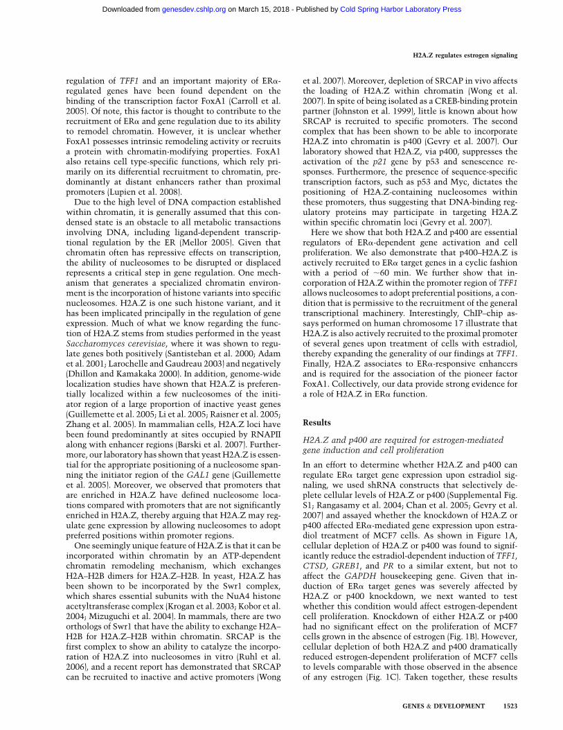
Incorporation of histone H2A.Z into promoter chromatinhas been shown to prepare genes for expression uponappropriate physiological signals (Guillemette et al. 2005;Updike and Mango 2006; Gevry et al. 2007; March-Diazet al. 2008). In all of these cases, H2A.Z association withtarget promoter regions occurs when genes are not active,and upon activation, the variant histone appears to beremodeled, as visualized by the loss of its associationwith the promoter region. Here we describe a case inwhich H2A.Z incorporation into ERa target proximalpromoter chromatin only occurs at the onset of geneinduction by estrogen. Moreover, H2A.Z also associateswith estrogen-responsive enhancers, but in contrast towhat we observe at proximal promoters, H2A.Z is asso-ciated with these enhancer regions prior to estrogensignaling, and this association is reduced upon hormonetreatment of cells. These findings allow for the dissectionof mechanisms by which H2A.Z can facilitate genetranscription: first by allowing translationally unstablenucleosomes to adopt preferential positions within a pro-moter region, and second by allowing FoxA1 to be re-cruited to target enhancers. In both cases, this is essentialto allow the transcriptional machinery and other chro-matin remodelers to be recruited to target genes as well asperhaps permit long-range communication between dis-tal regulatory elements and proximal promoter regions(see Fig. 7).
Regulation of estrogen signaling by ERa is a complexprocess that requires the function of a plethora of coac-tivators and chromatin remodelers (Metivier et al. 2003;Perissi and Rosenfeld 2005). Adding to this innate com-plexity is the fact that several ERa target genes appear toexhibit expression cycles that last ;60 min. The apogeesof these transcription cycles are timed with the concertedrecruitment of positive factors, including ERa and theRNAPII machinery. Conversely, the onset of the non-productive cycles correlates with the recruitment of neg-ative transcription cofactors as well as the ubiquitinationof ERa and several other positive factors (Metivier et al.2003; Reid et al. 2003). The fact that H2A.Z can alsoengage in these cycles, at least at the TFF1 and CTSDtarget genes, suggests that it must first be incorporated
at the promoter in an ERa-dependent fashion, and mustthen also be transiently remodeled by a yet-unidentifiedmechanism, involving possibly histone ubiquitination anddegradation or the action of other chromatin-modifyingactivities. We also entertain the possibility that the p400complex could also be involved in remodeling H2A.Z-containing nucleosomes. The use of tamoxifen in ourexperiments, taken together with the protein–proteininteraction assays, has allowed us to establish the follow-ing order of assembly of factors at the TFF1 proximalpromoter as well as propose a mechanism of action forp400–H2A.Z in this scenario (see Fig. 7). First, the ERa
activator recruits p400 by virtue of interactions withAF-1 (and possibly the DBD). The p400 complex thenincorporates H2A.Z into promoter chromatin, whichallows nucleosomes (NucE and NucT) to adopt preferredpositions along the DNA translational axis. These pre-ferred positions, of NucT in particular, would render theTATA element accessible for TBP to bind, as well aspermit the recruitment of the transcriptional machinery.This notion is also reinforced by our finding that bothH2A.Z and p400 are required for p300, Brg1 (Swi/Snf),TBP, and RNAPII to be efficiently recruited to the TFF1promoter upon estradiol treatment of MCF-7 cells. Inaddition, our proposed model of action is also in accor-dance with previous reports that show that when TFF1 isinactive and free of RNAPII, both NucE and NucT slidealong the translational axis of the promoter, whereasupon activation of the gene, both nucleosomes are forcedto engage their preferred positions (Metivier et al. 2003;Reid et al. 2003). We emphasize that our experiments donot address the issue of whether or not nucleosomescontaining H2A.Z are more or less stable than nucleo-somes bearing canonical H2A. Rather, our studies showthat nucleosomes bearing H2A.Z tend to force nucleo-some positioning at defined sites on DNA. In fact, we alsoobserved a similar scenario in yeast in that promotersenriched in H2A.Z tend to have nucleosomes that arebetter positioned around the TSS as compared with gene
Figure 7. Summarizing model of the role of p400 and H2A.Zin the regulation of the nucleosomal organization and ERa-dependant target gene activation.
H2A.Z regulates estrogen signaling
GENES & DEVELOPMENT 1529
Cold Spring Harbor Laboratory Press on March 15, 2018 - Published by genesdev.cshlp.orgDownloaded from

promoters that are depleted of H2A.Z (Guillemette et al.2005).
The mechanistic role of H2A.Z at estrogen-responsivedistal enhancer regions still remains somewhat elusive.However, since FoxA1 is essential for the full activity ofERa target gene expression, presumably by allowing theenhancer region to loop and thus interact with the basalpromoter region (Carroll et al. 2005), we surmise thatsince H2A.Z is required for FoxA1 recruitment toenhancers, it must also be essential for long-range inter-actions of these distal DNA elements. Our results are alsoin line with a report by the Hager laboratory (John et al.2008), which showed that H2A.Z localized to DNAse I-hypersensitive sites—which are presumably enhancerregions—and that upon GR recruitment to those regions,H2A.Z binding decreased from these hypersensitive sites.
Our study also demonstrates that p400 is an importantregulator of ERa signaling. We have shown in previouswork that p400 can directly incorporate H2A.Z–H2Bdimers into a regular nucleosome in vitro, and does soin an ATP-dependent fashion (Gevry et al. 2007). Thatsame study also demonstrated that p400 was importantfor the incorporation of H2A.Z at several p53 target genes,but that the other Swr1 ortholog SRCAP did not appear tobe as essential. Thus, we surmise that, as in the case ofp53 target genes, p400 would also be directly involved inincorporating H2A.Z into chromatin at ERa target genes.In support of this notion, we observe that acute depletionof p400 severely impedes ERa-mediated gene activation.On the other hand, our results do not exclude the pos-sibility that SRCAP might also participate in the regula-tion of ERa signaling, a possibility that still remains to betested.
Our data also support a link between H2A.Z–p400 andERa-dependent breast tumor proliferation. A few lines ofevidence argue for such a possibility: First, both H2A.Zand p400 are essential components of estradiol-dependentbreast cancer cell growth. Second, H2A.Z and ERa levelshave been shown to correlate in several breast cancer sam-ples tested (A Svotelis, N Gevry, and L Gaudreau, in prep.).Furthermore, a recent report has also associated H2A.Zexpression with metastasis and decreased breast cancersurvival (Hua et al. 2008). Thus, breast tumors whosegrowth depends on ERa would also require p400–H2A.Z.In general, patients with ERa-positive breast cancersreceive a positive prognosis due to the efficiency of anti-estrogenic treatments (Normanno et al. 2005; Rigginset al. 2007). However, a serious problem arises when cer-tain tumors become resistant to the anti-estrogen treat-ment, leading to a decrease in the possibility of recovery(Normanno et al. 2005; Riggins et al. 2007). This re-sistance phenomenon may be attributed to the activity ofAF-1, which in turn has been suggested to be turned on byMAP kinase signaling pathways (Riggins et al. 2007).Since the p400–H2A.Z interaction with ERa occursthrough AF-1 and the DBD, it will be interesting todetermine whether AF-1 activity, under circumstancesin which AF-2 is inactivated by anti-estrogens, alsorequires the action of p400–H2A.Z. Should this be true,it would then be foreseeable that the p400–H2A.Z path-
way would represent an attractive therapeutic target forboth anti-estrogen-responsive and nonresponsive ERa-positive breast cancers.
Materials and methods
Cell culture, transfection, and retroviral infection
MCF7 and MDA-MB-231 cell lines were maintained in DMEM(Invitrogen) containing 10% fetal bovine serum (FBS) and anti-biotics. 17b-Estradiol (Sigma) and tamoxifen were used at con-centrations of 100 nM and 1 mM, respectively. shRNAs directedagainst H2A.Z and p400 were transfected into MCF7 usingFugene6 or by retroviral infections as described previously (Chanet al. 2005; Gevry et al. 2007).
ChIP assays
MCF7 cells were hormone-deprived for at least 3 d and thentreated with 100 nM 17b-estradiol for the indicated time. ChIPassays were performed essentially as described previously (Gevryet al. 2007) with a panel of specific polyclonal antibodies gen-erated in-house or from commercial sources, as well as preim-mune, and no antibody controls. Samples were sonicated togenerate DNA fragments <500 bp. qPCR was performed usinga set of primers relevant to the promoter regions of the TFF1 andCTSD genes. The primers used in qPCR are listed in Supplemen-tal Table S1. Results are shown as percent input except in caseswith H2A.Z, where results are represented as ‘‘enrichment’’ andwhere H3 binding was also considered to account for nucleosomedensity. Thus, in this case, H2A.Z percent input was divided toH3 percent input.
Micrococcal nuclease digestion and nucleosome mapping
Chromatin was isolated as described by Kim et al. (2004) fromformaldehyde-cross-linked MCF7 cells synchronized witha-amanitin and treated with 17b-estradiol for 30 min. Chromatinsamples were subjected to DNA digestion with increasingamounts (0, 5, 10, and 15 U) of micrococcal nuclease (MNase).The reactions were stopped by addition of EDTA and EGTA(50 mM final concentration). Cross-linking was reversed over-night at 65°C. DNA samples were extracted after RNAse A andproteinase K digestion using PCR Qiaquick spin columns (Qia-gen). Mononucleosome-sized DNA was analyzed following theqPCR method derived from Metivier et al. (2003). Relative nucle-osome stability index was calculated as log2(1/CT) where CT =
(CT mononucleosome size of specific DNA amplification) � (CTInput). The primers used are listed in Supplemental Table S3.
ChIP–chip
Details are available in the Supplemental Material.
Statistical methods for ChIP–chip and ChIP expression
analyses
The data were normalized and analyzed as previously (Rufiangeet al. 2007) using the limma’s loess function (Yang et al. 2002) inBioConductor (from the ArrayPipe Analysis Pipeline) (Hokampet al. 2004), and replicates were combined using a weightedaverage method as described previously (Ren et al. 2000). All theexperiments were carried out in duplicate. The combined datasets are available in Supplemental Material. To interpolatebetween probes, a standard Gaussian filter (SD = 200 bp) was
Gevry et al.
1530 GENES & DEVELOPMENT
Cold Spring Harbor Laboratory Press on March 15, 2018 - Published by genesdev.cshlp.orgDownloaded from

applied twice to the data as described previously (Guillemetteet al. 2005). This will be referred to as the ‘‘smoothed data.’’
Effect of estradiol on genes of chromosome 17
In order to evaluate the impact of estradiol on RNAPII occu-pancy, we first calculated from the smoothed data, for each of the2004 unique TSS using UCSC known genes hg18, the meanlog2(RNAPII IP/Input) ratio in a window of 1 kb centered on theTSS. Genes were then sorted by their difference on mean RNAPIIoccupancy in the presence or absence of estradiol.
Mapping of H2A.Z, ERa, and RNAPII on ERa-bound regions
and along genes (Figs. 5, 6A,B; Supplemental Figs. S6, S8)
To automatically identify ERa-bound regions, the algorithmdeveloped in the Young laboratory (Boyer et al. 2005) was appliedto the ERa data set in the presence of estradiol with a P-valuecutoff of P < 10�5. These 389 regions were then associated withthe proximal TSS using the CTCF-bound regions of Ren labora-tory (Kim et al. 2007) as boundary regions (65 regions were notassociated with any gene). Regions and their associated genewere then separated based on the RNAPII occupancy differencein presence of estradiol (as described above). The nonsmootheddata were mapped on the middle of the ERa-bound regions (Fig.5B,D) or on the 59 and 39 boundaries of the associated genes (Fig.5C,E) into 50-bp windows, and a sliding window of 300 bp wasapplied to the average ratios. Independently of the ERa-boundregions, genes were binned into five groups according to theirRNAPII occupancy difference described above (each group con-tains, respectively, 153, 658, 506, 560, and 127 genes). The samemapping procedure was applied on the differential data set (with/without estradiol) of RNAPII and H2A.Z occupancy (Supplemen-tal Fig. S6B,C), and on distal ERa-bound regions identified at P <
10�4 and grouped by the presence/absence of a predictive bindingsite for FoxA1 and ERE (Fig. 6A,B; Supplemental Fig. S7). Bindingsite predictions were obtained in regions of at least 600 bpcentered at the middle point of identified ERa-bound regionswith the ‘‘FoxA1 shared’’ matrix from the Brown laboratory(Lupien et al. 2008) and the half-ERE matrix from TransFacversion 9.2 (Matys et al. 2006) using a PWM fast-matchingapproach developed in the Blanchette laboratory (JS Galan andM Blanchette, in prep.).
Antibodies and shRNA
The ERa polyclonal (HC20), ERa monoclonal (F10), FoxA1polyclonal (H-120), and RNAPII antibodies (N20) were purchasedfrom Santa Cruz Biotechnologies. Polyclonal antibodies for H2A(07-146), H2A.Z (07-594), and Tip60 (07-038) were from UpstateBiotechnologies. Histone H3 (ab 1791) and H2A.Z (ab 4174) werepurchased from Abcam. Brd8 (Ab-2) and p400 (A300-541A) poly-clonal antibodies were from Bethyl Laboratories. The H2A.Zand p400 antibodies used for ChIP assays were raised against anN-terminal H2A.Z peptide (CSLIGKKGQQKT) and C-terminalp400 peptide (MRVPAVRLKTPTKPPCQ). The pSuper-retro-puroshRNA-H2A.Z and p400 were described previously (Gevry et al.2007).
Acknowledgments
We thank Josette-Renee Landry for critical comments on themanuscript and Benoıt Leblanc for the artwork included inFigure 7. We are also grateful to Dr. David Livingston for thegift of the p400 antibody and Mathieu Lupien and Myles Brownfor sending the FoxA1 matrices; and to Javier Sanchez Galan and
Mathieu Blanchette for providing access to an unpublishedbioinformatics tool. This work was supported with funds fromthe Canadian Cancer Society awarded to L.G. and F.R., and withfunds from the CIHR awarded to L.G. L.G. holds a Canada re-search chair on mechanisms of gene transcription, F.R. holdsa New Investigator Award from the CIHR, N.G. was the recipi-ent of a post-doctoral fellowship from NSERC, and P.E.J. and S.H.are recipients of fellowships from the IRCM Training Programin Cancer Research supported by the CIHR.
References
Adam M, Robert F, Larochelle M, Gaudreau L. 2001. H2A.Z isrequired for global chromatin integrity and for recruitment ofRNA polymerase II under specific conditions. Mol Cell Biol
21: 6270–6279.Anzick SL, Kononen J, Walker RL, Azorsa DO, Tanner MM,
Guan XY, Sauter G, Kallioniemi OP, Trent JM, Meltzer PS.1997. AIB1, a steroid receptor coactivator amplified in breastand ovarian cancer. Science 277: 965–968.
Barkhem T, Haldosen LA, Gustafsson JA, Nilsson S. 2002.Transcriptional synergism on the pS2 gene promoter be-tween a p160 coactivator and estrogen receptor-a dependson the coactivator subtype, the type of estrogen responseelement, and the promoter context. Mol Endocrinol 16:2571–2581.
Barski A, Cuddapah S, Cui K, Roh TY, Schones DE, Wang Z, WeiG, Chepelev I, Zhao K. 2007. High-resolution profiling ofhistone methylations in the human genome. Cell 129: 823–837.
Benecke A, Chambon P, Gronemeyer H. 2000. Synergy betweenestrogen receptor a activation functions AF1 and AF2 medi-ated by transcription intermediary factor TIF2. EMBO Rep 1:151–157.
Berry M, Metzger D, Chambon P. 1990. Role of the twoactivating domains of the oestrogen receptor in the cell-typeand promoter-context dependent agonistic activity of theanti-oestrogen 4-hydroxytamoxifen. EMBO J 9: 2811–2818.
Boyer LA, Lee TI, Cole MF, Johnstone SE, Levine SS, Zucker JP,Guenther MG, Kumar RM, Murray HL, Jenner RG, et al.2005. Core transcriptional regulatory circuitry in humanembryonic stem cells. Cell 122: 947–956.
Brzozowski AM, Pike AC, Dauter Z, Hubbard RE, Bonn T,Engstrom O, Ohman L, Greene GL, Gustafsson JA, CarlquistM. 1997. Molecular basis of agonism and antagonism in theoestrogen receptor. Nature 389: 753–758.
Carroll JS, Liu XS, Brodsky AS, Li W, Meyer CA, Szary AJ,Eeckhoute J, Shao W, Hestermann EV, Geistlinger TR, et al.2005. Chromosome-wide mapping of estrogen receptor bind-ing reveals long-range regulation requiring the forkheadprotein FoxA1. Cell 122: 33–43.
Celik L, Lund JD, Schiott B. 2007. Conformational dynamics ofthe estrogen receptor a: Molecular dynamics simulations ofthe influence of binding site structure on protein dynamics.Biochemistry 46: 1743–1758.
Chan HM, Narita M, Lowe SW, Livingston DM. 2005. The p400E1A-associated protein is a novel component of the p53 /p21 senescence pathway. Genes & Dev 19: 196–201.
Couse JF, Korach KS. 1999. Estrogen receptor null mice: Whathave we learned and where will they lead us? Endocr Rev 20:358–417.
Deroo BJ, Korach KS. 2006. Estrogen receptors and humandisease. J Clin Invest 116: 561–570.
Dhillon N, Kamakaka RT. 2000. A histone variant, Htz1p, anda Sir1p-like protein, Esc2p, mediate silencing at HMR. MolCell 6: 769–780.
H2A.Z regulates estrogen signaling
GENES & DEVELOPMENT 1531
Cold Spring Harbor Laboratory Press on March 15, 2018 - Published by genesdev.cshlp.orgDownloaded from

DiRenzo J, Shang Y, Phelan M, Sif S, Myers M, Kingston R,Brown M. 2000. BRG-1 is recruited to estrogen-responsivepromoters and cooperates with factors involved in histoneacetylation. Mol Cell Biol 20: 7541–7549.
Eeckhoute J, Carroll JS, Geistlinger TR, Torres-Arzayus MI,Brown M. 2006. A cell-type-specific transcriptional networkrequired for estrogen regulation of cyclin D1 and cell cycleprogression in breast cancer. Genes & Dev 20: 2513–2526.
Farris SD, Rubio ED, Moon JJ, Gombert WM, Nelson BH,Krumm A. 2005. Transcription-induced chromatin remodel-ing at the c-myc gene involves the local exchange of histoneH2A.Z. J Biol Chem 280: 25298–25303.
Gevry N, Chan HM, Laflamme L, Livingston DM, Gaudreau L.2007. p21 transcription is regulated by differential localiza-tion of histone H2A.Z. Genes & Dev 21: 1869–1881.
Green KA, Carroll JS. 2007. Oestrogen-receptor-mediated tran-scription and the influence of co-factors and chromatin state.Nat Rev Cancer 7: 713–722.
Guillemette B, Bataille AR, Gevry N, Adam M, Blanchette M,Robert F, Gaudreau L. 2005. Variant histone H2A.Z isglobally localized to the promoters of inactive yeast genesand regulates nucleosome positioning. PLoS Biol 3: e384. doi:10.1371/journal.pbio.0030384.
Hokamp, K., Roche, F.M., Acab, M., Rousseau, M.E., Kuo, B.,Goode, D., Aeschliman, D., Bryan, J., Babiuk, L.A., Hancock,R.E. et al. 2004. ArrayPipe: A flexible processing pipeline formicroarray data. Nucleic Acids Res 32: W457–W459. doi:10.1093/nar/gkh446.
Hong H, Kohli K, Garabedian MJ, Stallcup MR. 1997. GRIP1,a transcriptional coactivator for the AF-2 transactivationdomain of steroid, thyroid, retinoid, and vitamin D receptors.Mol Cell Biol 17: 2735–2744.
Hua S, Kallen CB, Dhar R, Baquero MT, Mason CE, Russell BA,Shah PK, Liu J, Khramtsov A, Tretiakova MS, et al. 2008.Genomic analysis of estrogen cascade reveals histone variantH2A.Z associated with breast cancer progression. Mol Syst
Biol 4: 188. doi: 10.1038/msb.2008.25.Ichinose H, Garnier JM, Chambon P, Losson R. 1997. Ligand-
dependent interaction between the estrogen receptor and thehuman homologues of SWI2/SNF2. Gene 188: 95–100.
Jin C, Felsenfeld G. 2007. Nucleosome stability mediated byhistone variants H3.3 and H2A.Z. Genes & Dev 21: 1519–1529.
John S, Sabo PJ, Johnson TA, Sung MH, Biddie SC, Lightman SL,Voss TC, Davis SR, Meltzer PS, Stamatoyannopoulos JA,et al. 2008. Interaction of the glucocorticoid receptor withthe chromatin landscape. Mol Cell 29: 611–624.
Johnston H, Kneer J, Chackalaparampil I, Yaciuk P, Chrivia J.1999. Identification of a novel SNF2/SWI2 protein familymember, SRCAP, which interacts with CREB-binding pro-tein. J Biol Chem 274: 16370–16376.
Kim MY, Mauro S, Gevry N, Lis JT, Kraus WL. 2004. NAD+-dependent modulation of chromatin structure and transcrip-tion by nucleosome binding properties of PARP-1. Cell 119:803–814.
Kim TH, Abdullaev ZK, Smith AD, Ching KA, Loukinov DI,Green RD, Zhang MQ, Lobanenkov VV, Ren B. 2007.Analysis of the vertebrate insulator protein CTCF-bindingsites in the human genome. Cell 128: 1231–1245.
Kininis M, Chen BS, Diehl AG, Isaacs GD, Zhang T, Siepel AC,Clark AG, Kraus WL. 2007. Genomic analyses of transcrip-tion factor binding, histone acetylation, and gene expressionreveal mechanistically distinct classes of estrogen-regulatedpromoters. Mol Cell Biol 27: 5090–5104.
Kobor MS, Venkatasubrahmanyam S, Meneghini MD, Gin JW,Jennings JL, Link AJ, Madhani HD, Rine J. 2004. A protein
complex containing the conserved Swi2/Snf2-related ATPaseSwr1p deposits histone variant H2A.Z into euchromatin.PLoS Biol 2: E131. doi: 10.1371/journal.pbio.0020131.
Krogan NJ, Keogh MC, Datta N, Sawa C, Ryan OW, Ding H,Haw RA, Pootoolal J, Tong A, Canadien V, et al. 2003. A Snf2family ATPase complex required for recruitment of thehistone H2A variant Htz1. Mol Cell 12: 1565–1576.
Larochelle M, Gaudreau L. 2003. H2A.Z has a function remi-niscent of an activator required for preferential binding tointergenic DNA. EMBO J 22: 4512–4522.
Li B, Pattenden SG, Lee D, Gutierrez J, Chen J, Seidel C, GertonJ, Workman JL. 2005. Preferential occupancy of histonevariant H2AZ at inactive promoters influences local histonemodifications and chromatin remodeling. Proc Natl Acad Sci
102: 18385–18390.Lupien M, Eeckhoute J, Meyer CA, Wang Q, Zhang Y, Li W,
Carroll JS, Liu XS, Brown M. 2008. FoxA1 translates epige-netic signatures into enhancer-driven lineage-specific tran-scription. Cell 132: 958–970.
March-Diaz R, Garcia-Dominguez M, Lozano-Juste J, Leon J,Florencio FJ, Reyes JC. 2008. Histone H2A.Z and homo-logues of components of the SWR1 complex are required tocontrol immunity in Arabidopsis. Plant J 53: 475–487.
Matys V, Kel-Margoulis OV, Fricke E, Liebich I, Land S, Barre-Dirrie A, Reuter I, Chekmenev D, Krull M, Hornischer K,et al. 2006. TRANSFAC and its module TRANSCompel:Transcriptional gene regulation in eukaryotes. Nucleic AcidsRes 34: D108–D110. doi: 10.1093/nar/gkj143.
Mellor J. 2005. The dynamics of chromatin remodeling atpromoters. Mol Cell 19: 147–157.
Metivier R, Penot G, Hubner MR, Reid G, Brand H, Kos M,Gannon F. 2003. Estrogen receptor-a directs ordered, cyclical,and combinatorial recruitment of cofactors on a naturaltarget promoter. Cell 115: 751–763.
Mizuguchi G, Shen X, Landry J, Wu WH, Sen S, Wu C. 2004.ATP-driven exchange of histone H2AZ variant catalyzedby SWR1 chromatin remodeling complex. Science 303: 343–348.
Nilsson S, Makela S, Treuter E, Tujague M, Thomsen J,Andersson G, Enmark E, Pettersson K, Warner M, GustafssonJA. 2001. Mechanisms of estrogen action. Physiol Rev 81:1535–1565.
Normanno N, Di Maio M, De Maio E, De Luca A, de Matteis A,Giordano A, Perrone F. 2005. Mechanisms of endocrineresistance and novel therapeutic strategies in breast cancer.Endocr Relat Cancer 12: 721–747.
Onate SA, Tsai SY, Tsai MJ, O’Malley BW. 1995. Sequence andcharacterization of a coactivator for the steroid hormonereceptor superfamily. Science 270: 1354–1357.
Perissi V, Rosenfeld MG. 2005. Controlling nuclear receptors:The circular logic of cofactor cycles. Nat Rev Mol Cell Biol 6:542–554.
Pham TA, Hwung YP, Santiso-Mere D, McDonnell DP,O’Malley BW. 1992. Ligand-dependent and -independentfunction of the transactivation regions of the human estro-gen receptor in yeast. Mol Endocrinol 6: 1043–1050.
Raisner RM, Hartley PD, Meneghini MD, Bao MZ, Liu CL,Schreiber SL, Rando OJ, Madhani HD. 2005. Histone variantH2A.Z marks the 59 ends of both active and inactive genes ineuchromatin. Cell 123: 233–248.
Rangasamy D, Greaves I, Tremethick DJ. 2004. RNA interfer-ence demonstrates a novel role for H2A.Z in chromosomesegregation. Nat Struct Mol Biol 11: 650–655.
Reid G, Hubner MR, Metivier R, Brand H, Denger S, Manu D,Beaudouin J, Ellenberg J, Gannon F. 2003. Cyclic, protea-some-mediated turnover of unliganded and liganded ERa on
Gevry et al.
1532 GENES & DEVELOPMENT
Cold Spring Harbor Laboratory Press on March 15, 2018 - Published by genesdev.cshlp.orgDownloaded from

responsive promoters is an integral feature of estrogen sig-naling. Mol Cell 11: 695–707.
Ren B, Robert F, Wyrick JJ, Aparicio O, Jennings EG, Simon I,Zeitlinger J, Schreiber J, Hannett N, Kanin E, et al. 2000.Genome-wide location and function of DNA binding pro-teins. Science 290: 2306–2309.
Riggins RB, Schrecengost RS, Guerrero MS, Bouton AH. 2007.Pathways to tamoxifen resistance. Cancer Lett 256: 1–24.
Rufiange A, Jacques PE, Bhat W, Robert F, Nourani A. 2007.Genome-wide replication-independent histone H3 exchangeoccurs predominantly at promoters and implicates H3 K56acetylation and Asf1. Mol Cell 27: 393–405.
Ruhl DD, Jin J, Cai Y, Swanson S, Florens L, Washburn MP,Conaway RC, Conaway JW, Chrivia JC. 2006. Purification ofa human SRCAP complex that remodels chromatin byincorporating the histone variant H2A.Z into nucleosomes.Biochemistry 45: 5671–5677.
Santisteban MS, Kalashnikova T, Smith MM. 2000. HistoneH2A.Z regulats transcription and is partially redundant withnucleosome remodeling complexes. Cell 103: 411–422.
Sewack GF, Hansen U. 1997. Nucleosome positioning andtranscription-associated chromatin alterations on the humanestrogen-responsive pS2 promoter. J Biol Chem 272: 31118–31129.
Shang Y, Hu X, DiRenzo J, Lazar MA, Brown M. 2000. Cofactordynamics and sufficiency in estrogen receptor-regulatedtranscription. Cell 103: 843–852.
Shiau AK, Barstad D, Loria PM, Cheng L, Kushner PJ, Agard DA,Greene GL. 1998. The structural basis of estrogen receptor/coactivator recognition and the antagonism of this interac-tion by tamoxifen. Cell 95: 927–937.
Tora L, White J, Brou C, Tasset D, Webster N, Scheer E,Chambon P. 1989. The human estrogen receptor has twoindependent nonacidic transcriptional activation functions.Cell 59: 477–487.
Tzukerman MT, Esty A, Santiso-Mere D, Danielian P, ParkerMG, Stein RB, Pike JW, McDonnell DP. 1994. Humanestrogen receptor transactivational capacity is determinedby both cellular and promoter context and mediated by twofunctionally distinct intramolecular regions. Mol Endocrinol
8: 21–30.Updike DL, Mango SE. 2006. Temporal regulation of foregut
development by HTZ-1/H2A.Z and PHA-4/FoxA. PLoS Genet
2: e161. doi: 10.1371/journal.pgen.0020161.Wong MM, Cox LK, Chrivia JC. 2007. The chromatin remodel-
ing protein, SRCAP, is critical for deposition of the histonevariant H2A.Z at promoters. J Biol Chem 282: 26132–26139.
Yang YH, Dudoit S, Luu P, Lin DM, Peng V, Ngai J, Speed TP.2002. Normalization for cDNA microarray data: A robustcomposite method addressing single and multiple slidesystematic variation. Nucleic Acids Res 30: e15.
Zhang H, Roberts DN, Cairns BR. 2005. Genome-wide dynam-ics of Htz1, a histone H2A variant that poises repressed/basalpromoters for activation through histone loss. Cell 123: 219–231.
Zubairy S, Oesterreich S. 2005. Estrogen-repressed genes–keymediators of estrogen action? Breast Cancer Res 7: 163–164.
H2A.Z regulates estrogen signaling
GENES & DEVELOPMENT 1533
Cold Spring Harbor Laboratory Press on March 15, 2018 - Published by genesdev.cshlp.orgDownloaded from
10.1101/gad.1787109Access the most recent version at doi: originally published online June 10, 200923:2009, Genes Dev.
Nicolas Gévry, Sara Hardy, Pierre-Étienne Jacques, et al. Histone H2A.Z is essential for estrogen receptor signaling
Material
Supplemental
http://genesdev.cshlp.org/content/suppl/2009/06/11/gad.1787109.DC1
References
http://genesdev.cshlp.org/content/23/13/1522.full.html#ref-list-1
This article cites 63 articles, 21 of which can be accessed free at:
License
ServiceEmail Alerting
click here.right corner of the article or
Receive free email alerts when new articles cite this article - sign up in the box at the top
Copyright © 2009 by Cold Spring Harbor Laboratory Press
Cold Spring Harbor Laboratory Press on March 15, 2018 - Published by genesdev.cshlp.orgDownloaded from