histological, histochemical and morphometric changes...
TRANSCRIPT

Sci. Med. J . Cai. Med. Synd., Vo1.2, No.4, Oct.1990 8 7 - --
HISTOLOGICAL, HISTOCHEMICAL AND MORPHOMETRIC CHANGES OF THE
CONTRALATERAL ADRENAL GLAND AFTER UNILATERAL
ADRENALECTOMY
DR : SOAD EL - SAYED DR : SAMIR N, IBRAHIM DR : MOIIAMED K, TAWFIK DR : EL - ARIAN Y : EKLADIOUS
" FROM HISTOLOGY AND ANATOMY DEPARTMENTS, FACULTY OF MEDICINE, AIN- SHAMS UNIVERSTY"
ABSTRACT
Three weeks after unilateral adrenalectomy , the adrenocortical thickness was increased, the lipid content of the cortical zones was markedly decreased, the alkaline phosphatase reaction of the outer part of the zona fasciulata was decreased, the acid phosphatase activi- ty showed an increased reaction in zona glomerulosa and outer part of the zona fasciculata and the succinic dehydrogenase activity was in- creased in the zona fasciculata and zona reticularis.
Eight weeks after the operation, the hypertrophied zones regained their normal thickness, the fasciculata cells with their remarkable ac- cumulation of fat droplets reappeared. The plzosphatase activity (acid and alkaline) regained its normal intensity seen in the control sec- tions, and the reaction of succinic dehydrogenase enzyme was similar to that of the control.
In the cotralateral adrenal medulla the pattern of distribution of adrenaline and nor adrenaline secreting cells was not affected by the operation.

8 8 Soad El-Sayed, et al.
INTRODUCTION
The changes in the contralateral are going to extend the work to see adrenal gland after unilateral adren- the changes that occur after 3 alectomy were extensively studied weeks from the operation in order by a number of investigators in the to know more details about the acute, subacute and chronic cases prolonged effect of unilateral up to 7 days after the operation adrenalectomy on the contralateral (Omoto, 1969). In this study we adrenal.
MATERIAL and METHODS
Thirty adult male albino rats were used in this study.
They were classified into two groups of animals ; a control group and an experimental group to which unilateral adrenalectomy was per- formed. The animals were sacrified 3 and 8 weeks after operation and the glands were dissected. Some of the adrenal glands from both groups were used for pwaplast em- bedding and serial sections (4-6 urn thick) were stained with haematox- ylin and eosin and amoniacal silver nitrate intensification of the chro- maffin reaction for the demonstra- tion of adrenaline and nor adrena- line secreting cells (Culling, 1974).
Fresh cryo-cut sections were ob- tained from both the control and the experimental group and used for
demonstration of the following :
a) Lipid by Sudan 111 stain.
b) Alkaline phosphatase by using the calcium phosphate method of Gomori.
c) Acid phosphatase by using the azo dye coupling method (Bancroft, 1975)
d) Nachla's technique for succinic dehydrogenase.
The thichness of the different cortical zones was measured in both experimental and control sec- tions stained with haematoxylin and eosin. The results were calculated and statistically analysed and the fi- nal results were obtained by the computer.

HISTOLOGICAL, HISTOCHEMICAL AND MORI'HOMETRIC CHANGES 8 9
RESULTS
(A) Histological results :
(I) CONTROL GROUP :
The gland was covered by a rela- tively thick connective tissue cap- sule. Each gland consisted of an outer cortex and an inner medulla. The suprarenal cortex consisted of three concentric zones. Immediately beneath the capsule was the zona glomerulosa ; this was a thin layer composed of clusters of columnar cells with deeply stained nuclei and the cytoplasm containing some lipid droplets. A subglomerular sudano- phobic zone was seen. Next was the zona fasciculata, which was a thick layer composed of radially ar- ranged columns of cells.
The cells were polyhydral with basophilic cytoplasm rich in lipoid droplets (Figs. 1 & 2). The inner-
most layer was the zona reticularis, which consisted of an anastomos- ing network of cells adjacent to the medulla. Their cytoplasm was acid- ophilic of few lipoid droplets.
The suprarenal medulla was composed of groups of epithelial cells supported by a delicate con- nective tissue and separated by blood sinusoids. A few parasympa- thetic ganglion cells were also present.
(11) E X P E R I M E N T A L GROUP :
Three weeks after unilateral adrenalectomy the adrenocortical thikness was increased (Table 1). No significant changes could be observed in the thickness of the zona glomerulosa. The most promi- nent alteration encountered was in

9 0 Soad El-Saged, et al.
Table. ( 1 ) Adrenocortical thickness in control and experimental groups
( valus are the means in urn S.E. )
Control 3w 8w
Cortex + - - + - + 18.83 32.45 21.31
Table. (2) The thickness of adrenocortical zones in control and experimental groups
( valus are the means in urn S.E. )
CONTROL 3W 8W
zona glomer. 49.3 + 1.89 53. 1 ~t 4.88 51.23 + 3.21
zona fascic. 305. 11 + 19.31 629. 49 It 7.21° 314. 23 + 17.32
zona reticu. 183. 14 + 9.36 381.56 + 28.32O 104.61 + 11.25

HISTOLOGICAL, HISTOCHEMICAL AND MORPHOMETRIC CHANGES Y 1
the zona fasciulata. The depth of the zona fasculata. was double that of the control (Table 2), with gener- al compactness of their cells due to decrease of lipid (Fig. 3) Also the zona reticularis showed an increase in thickness and the lipoid vacioles were reduced when compared with the control.
In general the lipid content of the cortical zone was markedly de- creased. The sudanophobic zone disappeared (Fig. 4).
Eight weeks after unilateral adrenalectomy, the hypertrophied zones regained their normal thick- ness and their lipid content and be- came non-sigficantly different from that of the control.
(B) Histochemical results :
(T) CONTROL CROUP :
In control group the capsule of the adrenal gland showed negative alkaline phosphatase reaction. The zona glomerulosa always gave a stronger alkaline phosphatase reac- tion than that of the fasciculata and reticularis. In the glomerulosa, the strong alkaline phosphatase activity was noticed in the endothelial cells lining the blood capillaries.
The parenchymatous cells
showed a weak to moderate reac- tion. In zona fasciculata and reticu- laris a moderate reaction was ob- served in the blood capillaries, while a weak reaction was noticed in the parenchymatous cells (Fig. 5) The reaction of the nuclei varied, some showed weak reaction and others had a moderate reaction.
In the control group a negative acid phosphatase reaction was seen in the capsule. A moderate reaction was observed in the zona glomeru- losa and the outer part of the zona fxxiculata. The inner part of the zona fasciculata and zona reticularis showed a faint acid phosphatase re- action (Fig. 6)
In control group the capsule of the adrenal gland did not show suc- cinic dehydrogenase activity. The zona glomerulosa revealed a mod- erate succinic dehydrogenase reac- tion.
A strong reaction was observed in the zona fasciculata and reticular- is (Fig. 7).
(11) EXPERIMNTAL GROUP
Three weeks after unilateral adrenalectorny the alkline phospha- tase reaction was decreased in the outer part of the zona fasciculata (Fig. 8). The acid phosphatase re-

9 2 Soad El-Sayed, et al.
action of the zona fasciculata (particulary its outer part) and zona glomerulosa was increased (Fig. 9). The succinic dehydroge- nase activity was markedly in- creased in the inner part of the zona fasciculata and zona reticularis (Fig. 10).
Eight weeks after the operation the phosphatase activity (acid and alkaline) regained their normal pat- tern of distribution seen in the con- trol group. The reaction of the suc- cinic dehydrogenase was similar to that of the normal section.
- -
(111) ADRENAL MEDULLA :
In the control group with the use of arnrnoniacal silver nitrate method for staining the adrenal medulla, the noradrenaline cells were stained dark brown while the adrenaline cells took a yellow colour. The nor- adrenaline cells were found in groups scattered between the lighter adrenaline cells (Fig. 1 1).
At 3 weeks after the operation no evident changes could be ob- served in the pattern of adrenaline and noradrenaline (Fig. 12).
DISCUSSION The trophic effect of the unilater-
al adrenalectomy on the cortical thickness could be attributed to stress or increased ACTH secre- tion. Miller (1953), demonstrated an increase in cortical rhickness follwing ACTH adnlinistration.
Following stress or ACTH se- cretion the lipid content of the corti- cal zones decreased and as stress or hormonal level diminished the lipid content was restored and the corti- cal cells changed from compact to clear. Clik and Ochs (1955), ob- served loss of cholestrol in the ad- renal cortex after stimulation by ACTH or stress condition. Since
choslestrol is the main precursor of the steroid hormone biosynthesis it can be assumed that lipid depletion from adrenal cortexmay indicate an increased functional activity. How- ever Fortier et al. (1950), stated that the loss of cholestrol from the adrenal may be unaccompanied by loss of other lipids.
Three weeks after the operation the alkaline phosphatase reaction was decreased in the outer part of the zona fasciculata. Since the alka- line phosphatase was mainly dis- tributed in the endothelium of the blood capillaries, it might play a role in the transport of secretion

HISTOLOGICAL, HISTOCHEMICAL AND MORPHOMETRTC CHANGES y 3
from the cells to the blood. According to the result of Clik and Ochs (1955), the cholestrol ester disappeared completely from the outer part of the fascicular zone follwing stimulation by ACTH or stress condition. From this data we could attributed the loss of the alka- line phosphatase activity from the outer part of the zona fasciculata to loss of cholestrol ester and sub- sequently decreased secretory ac- tivi ty.
In the present study we noticed an increase in acid phosphatase re- action 3 weeks after the operation in the zona glon~erulosa and zona Pas- ciculata. Mietkiewski et al. (1970) attributed this enzymatic activity to the increase in secretory activity of the cells. The acid phosphatase is considered as a component of the content of lysosmes, so we could sugest that the lysosomal system could play a role in the synthesis or the release of cortical hormones.
In the present study, unilateral adrenalectomy increased the succin- ic dehydrogenase reaction in the zona fasciculat and zona reticularis. This activity represent the mito- chondria content and subsequently
the secretory activity of the cells. This data was confirmed by the data obtained by kadioglu and Har- rison (1971) on their electron mi- croscopic study of the adrenal gland after unilateral adrenalecto- my. They observed marked en- largement and increase in number of the mitochondria.
The data of the histochemical studies including decrease of the lipid of the inner zones, and in- creased acid phosphatase as well as succinic dehydrogenase activity of these zones indicated increased sec- retory activtiy of the adrenal cortex 3 weeks after the operation. This increased activity can be explained by increased secetion of ACTH as a compansatory mechanism to the unilateral adrenalectomy.
In the contralateral adrenal me- dulla 3 weeks after the operation no evident changes could be observed in the adrenaline and noradrenaline secreting cells. However degranu- lation of these cells in addition to the small granule chromaffin cells was noticed to occur in rat exposed to stress for 8-22 hours by kobaya- shi and Serizawa (1979).

9 4 Soad El-Sayed, et al.
REFERENCES
Bancroft,J.D. (1975) : Histo- chemical techniques. 2 n d - ed . Butterworths, eds. London and Boston.
Click, D. and Ochs. J.M. (1955) : Study in histochem- istry. Quantitative histologi- cal distribution of cholestrol in adrenal glands of the cow, rat and monkey and effects of stress condition, ACTH, cortisone and desoxy corti- Costerone. Endocrinology. 56: 285 - 298.
Culling, C.F.A. (1974) : Hand book of histopathological and histochemical techniques. 3 r d ed. Butterworths, ed. London.
Kadioglu, L). and Harrison, G. ( 1971) : The functional rela- tionships of mithochondria in the rat adrenal cortex.
J. Anat. 110 (2) : 283 - 296.
Kobayashi,~. and Serizawa, Y . (1979): Stress induced degr- nulation accompanied by ves-
icle formation in the adrenal chromaff in cells of the mouse. Arch. Histol. Jap. 42: 375 - 388.
Mietkiewski, K.L. , Malendow- ic , Z . and Lukaszyk,A. (1970) : A histochemical and karyometric study on the ef- fect of stilboestrol and tes- tosterone on adrenal cortex of adult male rat. Acta. Histochern. 38: I63 - 180.
Miller, R.A. (1953) : Relation of mitochondria to secretory activity in fascicular zone of the rat's adrenal Am. J . Anat. 92: 329 - 359.
Omoto, T. (1969) : Histological changes of the contralateral adrenal cortex in acute and subacute phases of unilateral adrenalectotny in rats, with special reference to the mode of compansatory hypertro- phy. Endocrinol. Japon. 16 (3) : 315 - 329.

HISTOLOGICAL, HISTOCHI3WCAL AND MORPHOMISTRIC CHASGI'5 y 5
Fig. I I &,I I ( 1 1 I I K , I ~ I L , I I ~ L I L O I ~ C ~ 01. a control animal showing that the cclls of the zona glomcrulosa and Pascicula- ta arc clcar and vaculakd.
(H. & E. X 100)
Fig. 2: l'a1.1 ol LIK adrcnal cortcx of a control animal showing that the zona Pasciculata was heavily loaded with l i - poid droplets. Notice Lhc subglomcrular sudano- phobic zone.
(Sudan 111 X 100)
Fig. 3 : Part of lhc adrenal concx of an cxpcrimcntal animal 3 wccks aftcr adrcnalccto- my. Thc cclls of zona f lo- mcrulosa and fascicu ata showcd gcncral compact- ncss duc to dcpltion of lip- id.
(H. & E. X 100)
Fig. 4 : Pa11 01' L ~ C adrenal cortcx of an cxpcrimcntal animal 3 wccks aftcr adrcnalcctorny showing that thc lipid con- tent of the conical zoncs was markcdly dccrcascd Thc sudanophobic zone disappcarcd.
(Sudan 111 X 100)

9 6 Soad El-Saved. e t n i
Fig. 6 : Part of the adrenal cortex of a control animal showing a moder- ate acid phosphatase activity in zona glomerulosa and outer part of zona fasciculata. Notice the intence acid phosphatase reaction in the medulla.
(Azo dye coupling method X 100)

HISTOLOGXCAI,, HISTOCI-IEMICAL AND MORPAOMETRIC CHANGES y 7
Fig. 7 : Part 0 1 11ic ; I J ICII ;~~ WI-~CX of ;i COI~II-01 animal stained for succin- ic dehydrogenase activity. Notice the strong reaction in the zona fasciculata and reticularis. The zona glomerulosa showed a less intense reaction.
(Nachla's technique X 250)
Fig. 8 : Pan of the adrenal cortex of an experimental aninla13 weeks af- ter adrenalectomy showing diminished alkaline phosphatase re- action in the outer part of the zona fasciculata.
(Grnori method X 100)

9 X Soad El-Sayed, et al.
I
Fig. 9 : Part of the adrenal cortex of a control animal showing a marked increased acid phosphatase reaction in the zona glomerulosa and outer part of the zona fasciculata.
(Azo dye coupling method X 100)
Fig. 10 : Part of the adrenal cortex of an experimental animal 3 weeks after adrenalectomy showing a marked increase in succinic de- heydrogenase reaction of the inner part of zona fasciculata and reticularis.
(Nachla's technique X 250)
HISTOLOGICAL, I-IISTOCHEMICAL ASD MORPHOMETRIC CHASGES 9 $'
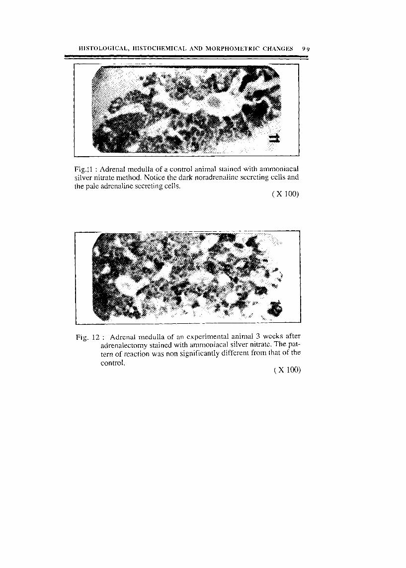
Fig.:l : Adrenal medulla of a control animal stained with ammoniacal silver nitrate method. Notice the dark noradrenaline secreting cells and the pale adrenaline secreting cells.
Fig. 12 : Adrenal medulla of an experimental aninla1 3 weeks after adrenalectomy stained with ammoniacal silver nitrate. The pat- tern of reaction was non significantly different from that of the control.
