histochemical studies on exudates of heterodera...
TRANSCRIPT

Histochemical studies on exudates of Hetemdera schachtii (Nematoda : Heteroderidae) males
Jens AUMANN and Urs WYSS Institut für Phytopathologie, Universitat Kiel, Hemzann-Rodewald-Strasse 9, 2300 Kiel 1, West Gennany.
SUMMARY
Histochemical tests on exudates, produced by males of the beet cyst nematode, Heterodera schachtii, showed that dyes more or less specific to proteins bound to amphidial, spicule and excretory pore exudates. For lectin binding experiments the method of exudate production was modified. Under these conditions excretory pore exudates were no longer produced, but observations of lectin binding showed that those from Canavalia ensifomzis (Con A), Triticunz vulgare (WGA) and Helix ponzatia (HPA) bound specifically to the surface of amphidial and spicule exudates. The exudates of the main chemoreceptors thus consist most likely of protein backbones to which oligosaccharide chains are coupled superficially. Their possible function is discussed.
RESUME
gtudes histochimiques sur les exsudats des nzâles de Heterodera schachtii (Nematoda : Heteroderidae)
Des tests histochimiques sur les exsudats des mâles du nématode à kyste de la betterave (Heterodera schachtii) ont montré que des colorants plus ou moins spécifiques des protéines peuvent se fiier sur les exsudats des amphides, des spicules et du pore excréteur. Lors d'expériences sur les liaisons avec des lectines, la méthode de production des exsudats a été modifiée. De ce fait, les exsudats du pore excréteur n'ont plus été produits; mais les observations faites sur les liaisons avec les lectines ont montré que celles provenant de Canavalia ensifornzis (Con A), Triticum vulgare (WGA) et Helix pornatia (HPA) se fiient spécifiquement à la surface des exsudats des amphides et des spicules. Les exsudats des principaux chemorécepteurs consistent donc essentiellement en un squelette principal Drotéiaue à la surface duauel sont fiiées des chaînes d'oligosaccharides. Les fonctions possibles de ce dispositir sont discutées. -
Nematode chemoreceptors are composed of nerve dendrites located in a cuticle-lined cavity in the head (amphids and inner labial sensilla) and tail region (phasmids) or in the spicules (spicule receptors, e.g. Wright, 1980; Coomans & De Grisse, 1981). The dendrites of these chemoreceptors (not yet proven for the spicules) are surrounded by viscous exudates pro- duced in associated gland cells. In nematodes, at least in Caenorhabditis elegans, the excretory canal is also filled with an exudate synthesized in a gland ce11 (Nelson & Riddle, 1984). Little is yet known about the chemical composition and function of these exudates. Forrest and Robertson (1986) found specific binding sites of WGA on the amphidial exudate of second stage juveniles (J2) of Globodera rostochiensis and Forrest (1985) showed that lectin binding sites on this exudate can be reduced by the protease pronase E. In another electron micro- scopic study McClure and Stynes (1 988) found UEA 1 binding sites on the amphidial exudate of Meloidogyne incognita J2. Aumann and Wyss (1987) detected specific binding sites of Con A, WGA, PNA, HPA and LFA in the region of the amphidial apertures, of Con A, WGA, PNA and HPA on the spicule tips, and of HPA in the region of the excretory pore opening of Heterodera schachtii males. As it was not possible to localize these
Revue Nénzatol. 12 (3) : 309-315 (1989)
binding sites definitely on the exudates, the method of Premachandran e t al. (1988) was used and modified for inducing exudate production. Lectin binding exper- iments were then performed with the fluorochrome- conjugated lectins Con A, WGA, PNA, HPA and LFA from differexit specificity groups according to Goldstein (1981).
Materials and methods
NEMATODES Males of Heterodera schachtii were obtained from
aseptic root cultures of Raphanus sativus var. oleifornzis cv. " Pegletta " (resistant cultivar) grown in the dark in a nutrient agar medium (Dropkin & Boone, 1966); for further details, see Wyss and Zunke (1986).
CHEMICALS
Organic solvents : Ethanol was purchased from a local dealer, toluene from Fluka (89681) and n-butyl acetate from Merck (9652). Dyes : Coomassie Brilliant BIue R-250 was from Serva (Serva Blue R, 35051) and aniline blue from Aldrich (methyl blue, 86,102-2). The cover-
309

slip sealer Glyceel (Hooper, 1986) was a gift from Dr. N. v. Mende, Department of Biological Sciences, University of Missouri. Lectins : The tetramethylrho- damine isothiocyanate (TRITC) labelled lectins Con A (L-3636) and WGA (L-5266) and the fluoresceine iso- thiocyanate (FITC) labelled PNA (L-7381) and HPA (L-1511) were from Sigma. TRITC-LFA (R-5101) was from E. Y. Carbohydrutes : a-methylmannoside (u-ManMe, M-6882) and N-acetylgalactosamine (Gal- NAc, A-2795) were purchased from Sigma, and D- galactose (D-Gal, 4058) and N-acetylneuraminic acid (NeuSAc, 24800) from Merck. The oligomers of N- acetylglucosamine (GlcNAc) were kindly provided by Dr. J. M. S. Forrest from the Scottish Crop Research Institute.
INDUCTION OF EXUDATE PRODUCTION FOR LECTIN BIN- DING EXPERIMENTS
Freshly emerged aseptic males were washed twice for 10 min each in 0.1 mol-1- ' phosphate buffered saline, pH 6.8, with 1 mmol.1- CaC1, and MnCl, (PBS). Then they were transferred into the well of a glass microscope slide, filled with 80 pl of a solution of 0.05 O/O Coomassie Brilliant Blue R-250 in distilled water. The slides were placed on the lid of a Petri dish (90 mm diam.) within a second dish (145 mm diam.). The lid was surrounded by a mixture of ethanol, toluene and n-butyl acetate (1:2:2). The nematodes were kept in the saturated atmosphere for 20-26 h during which time they produc- ed amphidial and spicule exudates that became visible as blue stained strands emanating from the apertures. Nematodes that produced visible exudates were remov- ed from the staining solution with a needle and imme- diately washed twice in PBS (for Con A, WGA, PNA and HPA binding experiments) or in 0.1 mol. 1- tris buffered saline, pH 8.0, with 10 mmol .1- CaC1, (TBS, for LFA binding experiments). The exudate strands broke off during handling. Al1 procedures were perfor- med at room temperature.
LECTIN BINDING
The nematodes were transferred from the washing buffer into a drop (30 pl) of lectin solution (100 pg-ml-') in PBS (Con A, WGA, PNA and HPA) or TBS (LFA) on a glass microscope slide and incubated for 19-24 h in a moisture chamber at room temperature. During incubation in lectin solutions the males con- tinued to produce exudates, but to a lesser extent than during the organic solvent treatment. They became visible as drop-like emanations from the amphidial apertures and spicule tips.
CONTROLS OF SPECIFICITY OF LECTIN BINDING 1
Control experiments were performed by incubating the lectins (50 pg . ml- ') first at 370 (see Aumann and
3 10
Table 1
Sugar specificities of the lectins and inhibitory sugars tested in control experiments
Lectin Sugar specificity" Inhibito y sugars tested
Con A Man-a-1, 2-Man-a-1, 2-Man > a-ManMe Man-a-1 , 2-Man > a- Man > a-Glc > aGlcNAc
3- P-GlcNAc > NeuSAc GlcNAc
P-Ga1
GalNAc > GalNac a-GlcNAc 3- a-Ga1
raminic acid
WGA GlcNAc (0-1, 4-GlcNAc)' - 2 oligomers of
PNA D-Gal-P-1, 3-GalNAc > a- and D-Ga1
HPA GalNAc-a-1, 3-GalNAc > a- GalNAc
LFA a-NeuSAc > a-N-glycolylneu- Neu5Ac
* According to Goldstein and Poretz (1986).
Wyss, 1987) for 90 min with the appropriate inhibitory carbohydrates (Tab. 1). The following solutions were tested : TRITC-Con A with 200 mmol .1- a-methyl- mannoside, TRITC-WGA with 15 mg. ml- ' N-acetyl- glucosamine oligomers, FITC-PNA with 200, 300, 400 and 500 mmol-1- D-galactose, FITC-HPA with 200 mmol .1- N-acetylgalactosamine and TRITC-LFA with 200, 300 and 400 mmol .1- N-acetylneuraminic acid. The nematodes were then incubated with these solutions as described under Lectin binding.
BINDING OF HISTOCHEMICAL DYES
Aseptic freshly emerged males were washed twice for 10 min each in PBS and transferred into a drop (40 pl) of 0.05 O/O Coomassie Brilliant Blue R-250 or aniline blue in distilled water on a glass microscope slide. The dye solution was surrounded by a circle of Glyceel (see Premachandran et al., 1988) onto which a coverslip was placed. The slides were then incubated for 20-23 h at room temperature before light rnicroscopic examination. The dyes were used to visualize the nematode exudates produced during incubation.
MICROSCOFY
The dye-stained nematodes were examined under a Reichert- Jung Polyvar photomicroscope with Nomarski interference microscopy at 100 to 1 O00 fold ma@& cation. Binding of fluorochrome-conjugated lectins was visualized with an epifluorescence system at 400 to 1 O00 fold magnification. TRITC-conjugated lectins were examined with a green filter IG2 (exciter filter BP 520-560 nm, splitting filter DS 580 nm and barrier filter LP 590 nm) and FITC-conjugated lectins with a blue filter IBl (BI? 450-495 nm, DS 510 nm and LP 520 nm).
Revue Nérnatol. 12 (3) : 309-315 (1989)

Exudates of Heterodera schachtii
The fluorescence of FITC-lectins appeared green, whereas the autofluorescence was yellowish. Secondary fluorescence of TRITC-conjugated lectins was visible as a strong red colour and the autofluorescence appeared as a pale red. Photomicrographs were taken on 50 ASA Kodak daylight colour films (lectins and dyes) or on Agfa PAN 100 black-white films (Nomarski interference microscopy). The lectin and dye binding experiments were repeated at least three times, each with ten nema- todes, whereas the specificity controls of lectin binding were performed with at least ten males per treatment.
Results
Table 2 shows the binding of different lectins to the amphidial and spicule exudates and to the excretory pore opening of Heterodera schachtii males, together with the results of the specificity controls of lectin binding. Al1 lectins tested bound to the amphidial and spicule exu- dates. In specimens where the excretory pore was visible only PNA bound to the region of the opening. Apart from LFA that bound only to 25 O/O of the nematodes tested, nearly al1 specimens were labelled at the amphi-
Table 2
Binding of lectins (capital) to exudates of Heterodera schachtii males and controls of binding specificity
Treatment n Labelling
amphidial spicule excreto y exudates exudates pore opening
TRITC-Con A 30 30 17 O TRITC-Con A + 200 mmol .1- a-ManMe 27 2 O O
TRITC-WGA 30 25 30 O TRITC-WGA + 15 mg. ml- ' GlcNAc-oligomers 16 O O O
FITC-PNA 30 30 28 21 FITC-PNA + ZOO mmol - 1- ' D-Ga1 20 17 9 9
mmol 3 1- ' D-Ga1 10 10 6 9
m o l . 1- D-Ga1 10 8 2 3
mmol ~ 1 - I D-Ga1 10 10 3 4
FITC-PNA + 300
FITC-PNA + 400
FITC-PNA 4- 500
FITC-HPA 30 30 25 1 FITC-HPA + 200 mmol .1- ' GalNAc 10 O O O
TRITC-LFA 60 15 5 O
mmol ~ 1 - I Neu5Ac 10 3 O O
mmol .1- ' Neu5Ac 20 15 2 O
mmol .1- Neu5Ac 20 9 4 O
TRITC-LFA + 200
TRITC-LFA 4- 300
TRITC-LFA + 400
dia1 exudates. The majority of the nematodes, with the of PNA and LFA, visualized as the intensity of flu- exception of LFA, were also labelled at the spicule orescence, was reduced in the presence of increasing exudates. The binding of Con A, HPA and WGA was concentrations of D-galactose and N-acetylneuraminic carbohydrate-specific, because the lectin binding was acid respectively (data not shown). completely prevented with the usual concentration of The exudate production of nematodes decreased the appropriate inhibitory sugars (Tab. 1). The binding markedly in the presence of inhibitory carbohydrates
Revue Nénzatol. 12 (3) : 309-315 (1989) 311

J. Aumann di: U. Wyss
Fig. 1. Binding of fluorochrome-conjugated lectins to exudates of Heterodera schachtii males. A, B : Binding of TRITC-Con A to amphidial exudates (A : Nomarski interference microscopy; B : fluorescence microscopy). C, D : Binding of TRITC-WGA to spicule exudates (C : Nomarski interference microscopy; D : fluorescence microscopy). E : Binding of FITC-PNA to spicule exudates; F : Binding of FITC-HPA to amphidial exudates; G, H : Binding of TRITC-LFA to amphidial exudates (G : Nomarski interference microscopy; H : fluorescence microscopy). Bars = 10 Pm.
and also when the males were treated with LFA. Figure 1, A-H shows some selected typical binding patterns of FITC- and TRITC-conjugated lectins at the amphidial and spicule exudates of H. schachtii males.
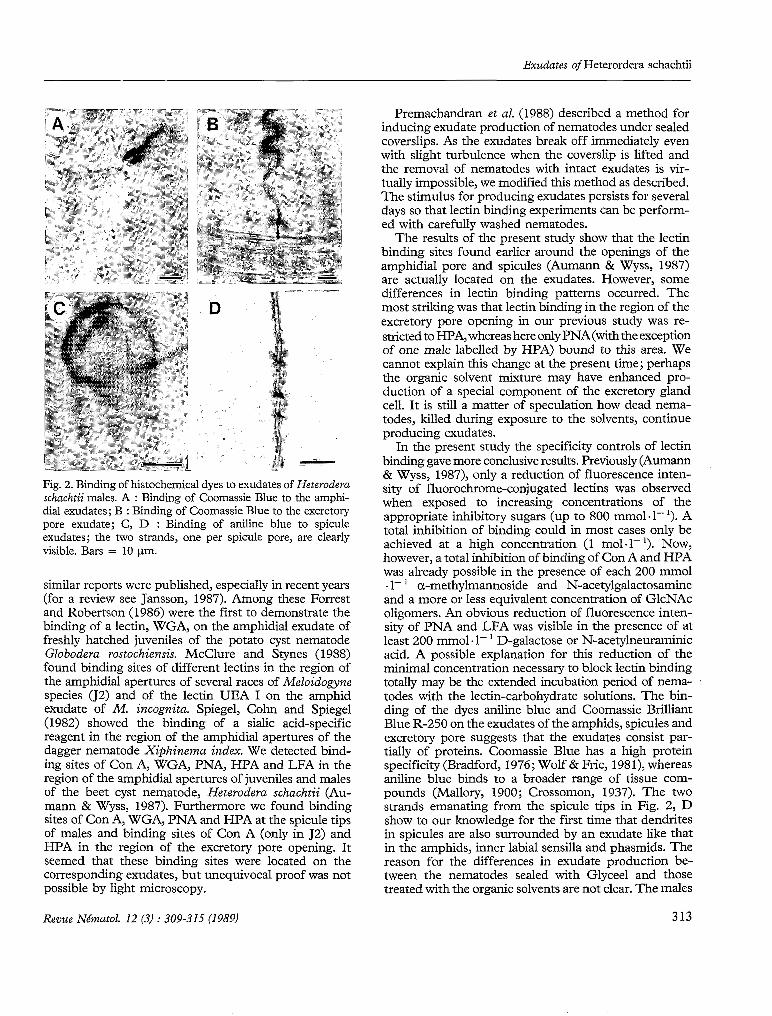
Table 3 presents the binding of the dyes aniline blue and Coomassie Brilliant Blue R-250 to exudates of H. schachtii males under the influence of Glyceel solvents. In contrast to the lectin binding experiments, where the nematodes produced only amphidial and spicule exu- dates, the production of excretory pore exudates was
now also induced. Fig. 2 shows binding to the amphidial exudates (Fig. 2, A), excretory pore exudate (Fig. 2, B) and spicule exudates (Fig. 2, C & D). Most of the nematodes examined produced abundant exudates and al1 exudates were stained with the dyes.
Discussion
Following the first report of a lectin binding site on a nematode (Nordbring-Hertz & Mattiasson, 1979),
Table 3
Binding of histochemical dyes to exudates of Heterodera schachtii males
DYe n Exudate staining
Amphids Spicules Excretoy pore
Aniline blue Coomassie Brilliant
Blue R-250
32 30 32 28
46 43 46 42
312 Revue Nématol. 12 (3) : 309-315 (1989)
Exudates of Heterordera schachtii
Fig. 2. Binding of histochemical dyes to exudates of Heteroderu schuchtii males. A : Binding of Coomassie Blue to the amphi- dia1 exudates; B : Binding of Coomassie Blue to the excretory pore exudate; C, D : Binding of aniline blue to spicule exudates; the two strands, one per spicule pore, are clearly visible. Bars = 10 Pm.
similar reports were published, especially in recent years (for a review see Jansson, 1987). Among these Forrest and Robertson (1986) were the first to demonstrate the binding of a lectin, WGA, on the amphidial exudate of freshly hatched juveniles of the potato cyst nematode Globodera rostochiensis. McClure and Stynes (1988) found binding sites of different lectins in the region of the amphidial apertures of several races of Meloidogyne species (J2) and of the lectin UEA 1 on the amphid exudate of M . imognita. Spiegel, Cohn and Spiegel (1982) showed the binding of a sialic acid-specific reagent in the region of the amphidial apertures of the dagger nematode Xiphinema index. We detected bind- ing sites of Con A, WGA, PNA, HPA and LFA in the region of the amphidial apertures of juveniles and males of the beet cyst nematode, Heterodera schachtii (Au- mann & Wyss, 1987). Furthermore we found binding sites of Con A, WGA, PNA and HPA at the spicule tips of males and binding sites of Con A (only in J2) and HPA in the region of the excretory pore opening. It seemed that these binding sites were located on the corresponding exudates, but unequivocal proof was not possible by light microscopy.
Premachandran et al. (1988) described a method for inducing exudate production of nematodes under sealed coverslips. As the exudates break off immediately even with slight turbulence when the coverslip is lifted and the removal of nematodes with intact exudates is vir- tually impossible, we modified this method as described. The stimulus for producing exudates persists for several days so that lectin binding experiments can be perform- ed with carefully washed nematodes.
The results of the present study show that the lectin binding sites found earlier around the openings of the amphidial pore and spicules (Aumann & Wyss, 1987) are actually located on the exudates. However, some differences in lectin binding patterns occurred. The most striking was that lectin binding in the region of the excretory pore opening in Our previous study was re- stricted to HPA, whereas here only PNA (withthe exception of one male labelled by HPA) bound to this area. We cannot explain this change at the present time; perhaps the organic solvent mixture may have enhanced pro- duction of a special component of the excretory gland cell. It is still a matter of speculation how dead nema- todes, killed during exposure to the solvents, continue producing exudates.
In the present study the specificity controls of lectin binding gave more conclusive results. Previously (Aumann & Wyss, 1987), only a reduction of fluorescence inten- sity of fluorochrome-conjugated lectins was observed when exposed to increasing concentrations of the appropriate inhibitory sugars (up to 800 mmol. 1- '). A total inhibition of binding could in most cases only be achieved at a high concentration (1 mol.1- '). Now, however, a total inhibition of binding of Con A and HPA was already possible in the presence of each 200 mmol al- ' a-methylmannoside and N-acetylgalactosamine and a more or less equivalent concentration of GlcNAc oligomers. An obvious reduction of fluorescence inten- sity of PNA and LFA was visible in the presence of at least 200 mm01 .1- ' D-galactose or N-acetylneuraminic acid. A possible explanation for this reduction of the minimal concentration necessary to block lectin binding totally may be the extended incubation period of nema- todes with the lectin-carbohydrate solutions. The bin- ding of the dyes aniline blue and Coomassie Brilliant Blue R-250 on the exudates of the amphids, spicules and excretory pore suggests that the exudates consist par- tially of proteins. Coomassie Blue has a high protein specificity (Bradford, 1976; Wolf & Fric, 1981), whereas aniline blue binds to a broader range of tissue com- pounds (Mallory, 1900; Crossomon, 1937). The two strands emanating from the spicule tips in Fig. 2, D show to Our knowledge for the first time that dendrites in spicules are also surrounded by an exudate like that in the amphids, inner labial sensilla and phasmids. The reason for the differences in exudate production be- tween the nematodes sealed with Glyceel and those treated with the organic solvents are not clear. The males
Revue Nématol. 12 (3) : 309-315 (1989) 313

J. Aumann & U. Wyss
sealed with Glyceel produced excretory pore exudates, whereas those treated with the organic solvents failed to do so. Possibly, the Glyceel compound methanol may play a role in inducing the production of excretory pore exudates.
The results with the lectin and dye binding sites on the exudates indicate, that these are composed of glyco- proteins. Possibly, they consist of a protein backbone to which oligosaccharide chains are coupled superficially. The partial protein and carbohydrate nature of the amphidial exudate of 52 of potato cyst nematodes was shown by Forrest (1985). We observed a reduction of lectin binding sites in the regions of exudate contact after H. schachtii males had been treated with chymo- trypsin, trypsin and pronase E (unpubl.). Veech, Starr and Nordgren (1987) analyzed the amino acid compo- sition of the stylet exudate of M. incognita females and found a relatively high level of serine. This indicates that not the N-glycosidic linkeage between an N-acetylhexo- samine and asparagine, but rather the O-glycosidic linkeage of N-acetylgalatosamine and serine or threo- nine prevails in proteincarbohydrate bonds (Kornfeld & Kornfeld, 1985). The carbohydrate composition of amphidial and spicule exudates, as tested by lecpn binding patterns, is apparently identical, but it differs markedly from that of the excretory pore exudate. As lectins are ,t specific for one sugar only, sophisticated experiments (e.g. destruction of the sugars with specific enzymes) are necessary xr an exact characterization of the carbohydrates on the exudate surface.
Any discussion of the function of these exudates is still speculative. Wright (1980) described parallels in the chemoreceptor morphology between arthropods and nematodes, and assumed that the exudates may protect the dendrites of the chemosensory nerves. The pores of insect chemosensilla in direct contact with the environ- menr also contain partially viscous exudates (Zacharuk, 1980) which, according to Altner (1977), may serve both for stimulus transmission to the dendrite membranes and as a protectant against dehydration. In nematodes the exudates surrounding chemoreceptive dendrites probably function primarily as protectants against microbial metabolites to which, in the soil, they are continuously exposed. At the moment we stillhesitate to assign them a major function in chemoreception as preliminary biotests of sugar blockings with lectins did not affect the orientation of H. schachtii males to the female pheromone. Out of the five lectins that bound to the exudates in the present study, only HPA showed a slight effect (unpublished results). The function of the excretory pore exudate, like the excretory system itself, is completely unknown. Nelson and Riddle (1984), Who ablated single cells in the excretory system in Caenor- habditis elegans by laser 9crobeam concluded that one function may be osmoregulation. So the pore exudate may act as a carrier of osmotically significant substances.
3 14
Bird, Bonig and Bacic (1988) hypothesized that one function of the excretory system in secementean nema- todes may be the secretion of the glycocalyx covering the cuticle.
ACKNOWLEDGEMENTS
We wish to thank J. M. S . Forrest and W. M. Robertson from the Scottish Crop Research Institute for their critical comments on the manuscript.
REFERENCES
ALTNER, H. (1977). Insektensensillen : Bau- und Funktions- prinzipien. Verh. Dtsch. zool. Ges., 7: 139-153.
AUMANN, J. & WYSS, U. (1988). Lectin binding sites on mobile stages of Heterodera schachtii Schmidt (Nematoda : Hetero- deridae). Nematologica, 33 (1987) : 410-418.
BIRD, A. F., BONIG, 1. & BACIC, A. (1988). A role for the (( excretory )) system in secementean nematodes. J. Nenza-
BRADFORD, M. M. (1976). A rapid and sensitive method for the quantitation of microgram quantities of protein utilizing the principle of protein-dye binding. Analyt. Biochem., 72 :
COOMANS, A. & DE GRISSE, A. (1981). Sensory structures. In : Zuckennan, B. M. & Rohde, R. A. (Eds). Plant parasitic nematodes, Vol. 3. New York, Academic Press : 127-174.
CROSSOMON, G. (1937). A modification of Mallory’s connec- tive tissue stain with a discussion of the principles involved. Anat. Rec., 69 : 33-38.
DROPKIN, V. H. & BOONE, W. R. (1966). Analysis of host- parasite relationships of root-knot nematodes by single-lama inoculations of excised tomato roots. Nematologica, 12 :
FORREST, J. M. S . (1985). The occurrence of glycoproteins on juvenile heads of potato cyst nematodes (PCN). Scott. Crop Res. Inst., Ann. Rep. 1985 : 107-108.
FORREST, J. M. S . & ROBERTSON, W. M. (1986). Characteriz- ation and localization of saccharides on the head region of four populations of the potato cyst nematode Globodera rostochiensis and G. pallida. J . Nematol., 18 : 23-26.
GOLDSTEIN, 1. J. (1981). Plant derived lectins. Rec. Adv. Phytochem., 15 : 25-35.
GOLDSTEIN, 1. J. & PORETZ, R. D. (1986). Isolation, physico- chemical characterization, and carbohydrate-binding speci- ficity of lectins. In : Liener. 1. E., Sharon, N. & Goldstein, 1. J. (Eds). T h e lectins. Properties, functions, and applications in biology and medicine. Orlando, Ha., Academic Press :
HOOPER, D. J. (1986). Handling, fixing, staining and mounting nematodes. In : Southey, J. F. (Ed.). Laborato y nzethods for work with plant and soil nematodes. London, Her Majesty’s Stationery Office : 59-80.
JANSSON, H. B. (1987). Receptors and recognition in nema- todes. In : Veech, J. A. & Dickson, D. W. (Eds). Vistas on
Revue Neinatol. 12 (3) : 309-315 (1989)
tol., 20 : 493-496.
248-254.
225-236.
33-247.

Exudates of Heterordera schachtii
nematology. De Leon Springs, Ha., E. O. Painter Print. Co. :
KORNFELD, R. & KORNFELD, S . (1985). Assembly of aspara- gine-linked oligosaccharides. Ann. Rev. Biochem., 54 :
MALLORY, F. B. (1900). A contribution to staining methods. J. exp. Med., 5 : 15-20.
MCCLURE, M. A. & STYNES, B. A. (1988). Lectin binding sites on the amphidial exudates of Meloidogyne. J . Nenzatol., 20 :
NELSON, F. K. & RIDDLE, D. L. (1984). Functional study of the Caenorhabditis eleguns secretory-excretory system using laser microsurgery. J. exp. Zool., 231 : 45-56.
NORDBRING-HERTZ, B. & MATTASSON, B. (1979). Action of a nematode-trapping fungus shows lectin-mediated host- microorganism interaction. Nature, 281 : 477-479.
PREMACHANDRAN, D., VON MENDE, N., HUSSEY, R. S . & MCCLURE, M. A. (1988). A method for staining nematode secretions and structures. J. Nematol., 20 : 70-78.
153-158.
631-664.
321-326.
Accepté pour publication le 26 octobre 1988.
SPIEGEL, Y., COHN, E. & SPIEGEL, S . (1982). Characterization of sialyl and galactosyl residues on the body Wall of different plant parasitic nematodes. J. Nematol., 14 : 33-39.
VEECH, J. A., STARR, J. L. & NORDGREN, R. M. (1987). Production and partial characterization of stylet exudates from adult females of Meloidogyne incognita. J. Nematol.,
WOLF, G. & FRIC, F. (1981). A rapid staining method for Eysiphe graminisj sp. hordeiin and on whole barley leaves with a protein-specific dye. Phytopathology, 71 : 596-598.
WRIGHT, K. A. (1980). Nematode sense organs. In : Zucker- man, B. M. (Ed.). Nematodes as biological models, Vol. 2. New York, Academic Press : 237-295.
19 : 463-468.
WYSS, U. & ZUNKE, U. (1986). In vitro feeding of root parasitic nematodes. In : Zuckerman, B. M., Mai, W. F. & Harrison, M. B. (Eds). Plant nematology laborato y manual. Amherst, Mass., Univ. Mass. Agric. Exp. Stat. : 91-96.
ZACHARUK, R. Y. (1980). Ultrastructure and function of insect chemosensilla. Ann. Rev. Entonlol., 25 : 27-47.
3 15