high-resolution structure of the histidine-containing …jb.asm.org/content/186/17/5906.full.pdf ·...
TRANSCRIPT

JOURNAL OF BACTERIOLOGY, Sept. 2004, p. 5906–5918 Vol. 186, No. 170021-9193/04/$08.00�0 DOI: 10.1128/JB.186.17.5906–5918.2004Copyright © 2004, American Society for Microbiology. All Rights Reserved.
High-Resolution Structure of the Histidine-Containing PhosphocarrierProtein (HPr) from Staphylococcus aureus and Characterization of
Its Interaction with the Bifunctional HPr Kinase/PhosphorylaseTill Maurer,1† Sebastian Meier,1‡ Norman Kachel,1 Claudia Elisabeth Munte,1§ Sonja Hasenbein,2
Brigitte Koch,2 Wolfgang Hengstenberg,2 and Hans Robert Kalbitzer1*Institut fur Biophysik und Physikalische Biochemie, Universitat Regensburg, Regensburg,1 and
Fakultat fur Biologie, Ruhr-Universitat Bochum, Bochum,2 Germany
Received 5 January 2004/Accepted 17 May 2004
A high-resolution structure of the histidine-containing phosphocarrier protein (HPr) from Staphylococcusaureus was obtained by heteronuclear multidimensional nuclear magnetic resonance (NMR) spectroscopy onthe basis of 1,766 structural restraints. Twenty-three hydrogen bonds in HPr could be directly detected by polar-ization transfer from the amide nitrogen to the carbonyl carbon involved in the hydrogen bond. Differential linebroadening was used to characterize the interaction of HPr with the HPr kinase/phosphorylase (HPrK/P) ofStaphylococcus xylosus, which is responsible for phosphorylation-dephosphorylation of the hydroxyl group of theregulatory serine residue at position 46. The dissociation constant Kd was determined to be 0.10 � 0.02 mMat 303 K from the NMR data, assuming independent binding. The data are consistent with a stoichiometry of1 HPr molecule per HPrK/P monomer in solution. Using transversal relaxation optimized spectroscopy-heteronuclear single quantum correlation, we mapped the interaction site of the two proteins in the 330-kDacomplex. As expected, it covers the region around Ser46 and the small helix b following this residue. In addi-tion, HPrK/P also binds to the second phosphorylation site of HPr at position 15. This interaction may beessential for the recognition of the phosphorylation state of His15 and the phosphorylation-dependent regula-tion of the kinase/phosphorylase activity. In accordance with this observation, the recently published X-raystructure of the HPr/HPrK core protein complex from Lactobacillus casei shows interactions with the two phos-phorylation sites. However, the NMR data also suggest differences for the full-length protein from S. xylosus:there are no indications for an interaction with the residues preceding the regulatory Ser46 residue (Thr41 toLys45) in the protein of S. xylosus. In contrast, it seems to interact with the C-terminal helix of HPr in solution,an interaction which is not observed for the complex of HPr with the core of HPrK/P of L. casei in crystals.
The histidine-containing phosphocarrier protein (HPr) playsa central role in the uptake of carbohydrates by the phospho-enolpyruvate-dependent phosphotransferase system (PTS) andin the regulation of carbohydrate metabolism in bacteria (for areview, see reference 54). In the transport system, it is part ofa phosphate shuttle, which transfers a phosphate group fromphosphoenolpyruvate to the carbohydrate transported throughthe cell membrane. As a second function, HPr is involved ingene regulation of the PTS carbon catabolite repression sys-tem. In that system, it acts as an activator of gene repression.Both of these mechanistically very different processes are con-trolled by HPr through phosphorylation-dephosphorylation re-actions. In the phosphate shuttle, the amino acid that partici-pates in phosphorylation-dephosphorylation reactions in HPris a histidine residue at position 15. It is phosphorylated byenzyme I (EI) at N�1 and transfers this group to Nε2 of a his-tidine residue of the enzyme IIA domain of the enzyme II (EII)
complex. In most gram-positive and some pathogenic gram-negative bacteria, the second phosphorylation site in HPr isSer46, which can be phosphorylated by the ATP-dependent HPrkinase/phosphorylase (HPrK/P), the product of the hprK gene(7, 8, 13, 34, 50, 48). Phosphoserine-HPr functions in a regula-tory fashion, down regulating catabolic activity by its interac-tion with catabolite control protein A (CcpA) (25, 52). Simul-taneously, the phosphorylation of Ser46 inhibits phosphocarrieractivity by perturbing the interaction with phosphorylated EI(3, 48). Furthermore, in some bacteria P-Ser-HPr seems to beinvolved in additional regulatory mechanisms, called inducerexpulsion and inducer exclusion (9, 54, 61, 62).
HPr proteins from different microorganisms have beenstructurally characterized by X-ray crystallography and nuclearmagnetic resonance (NMR) spectroscopy (10, 21, 23, 24, 29,32, 41, 46, 53, 60). Although they differ largely in primarystructure, their general folding structure is well conserved. Itconsists of a four-stranded antiparallel �-pleated sheet and three�-helices arranged in a ������� open �-sandwich topology.
The X-ray structure of the catalytic domain of HPrK/P(amino acids 128 to 319) from Lactobacillus casei (11) and thestructures of the full-length HPrK proteins from Staphylococ-cus xylosus (40) and Mycoplasma pneumoniae (1) have beensolved, and data for the complex of HPrK from L. casei with itssubstrate HPr from Bacillus subtilis are available (12). Thecatalytic mechanism of the bifunctional protein kinase and its
* Corresponding author. Mailing address: Universitat Regensburg,Institut fur Biophysik und Physikalische Biochemie, Universitatsstr.31, D-93040 Regensburg, Germany. Phone: 49 941 943 2594. Fax: 49941 943 2479. E-mail: [email protected].
† Present address: Department of Lead Discovery, Boehringer In-gelheim Pharma GmbH & Co. KG, D-88397 Biberach, Germany.
‡ Present address: Biozentrum der Universitat Basel, Basel, Swit-zerland.
§ Present address: Instituto de Física de Sao Carlos, Universidadede Sao Paulo, 13560-970 Sao Carlos SP, Brazil.
5906
on June 14, 2018 by guesthttp://jb.asm
.org/D
ownloaded from

precise interaction with its substrate protein were explained onthe basis of the complex data (37, 42).
With the capability to interact with numerous other proteins,HPr is an ideal system for the study of protein-protein inter-actions. These complexes are far larger than the 40-kDa sizewhich up to now was considered the limit for studies usingNMR. With the transversal relaxation optimized spectroscopy(TROSY) technique first described by Pervushin et al. (44)making possible the investigation of proteins and complexeswith molecular masses of far more than 40 kDa, the interactionof HPr with HPrK/P, with the focus on HPr, is a system that isnow suited for NMR investigation.
For HPr from Staphylococcus aureus, only a low-resolutionNMR structure, which was solved exclusively with homonuclearmethods under a low magnetic field, has been published (29).In this paper, we present a high-resolution structure of HPrfrom S. aureus as the basis for a study of its interaction withHPrK/P from S. xylosus. HPr from S. aureus is closely relatedto HPr from S. xylosus (with five amino acid differences) and isthus a suitable binding partner for HPrK/P from S. xylosus.
MATERIALS AND METHODS
Protein expression and purification. Unlabeled wild-type HPr from S. aureus(molecular mass, 9.496 kDa) was expressed and purified as described previously(27). For the preparation of uniformly 13C- and 15N-enriched HPr, the plasmidpT7-5 ptsH, coding for HPr from S. aureus, was transformed into Escherichia coliBL21(DE3), which was initially grown in TBY medium (1% casein hydrolysate,0.5% yeast extract, 0.5% NaCl). For protein expression, cells were inoculatedinto 200 ml of M9 minimal medium containing uniformly 13C-enriched (99%)glucose-1-hydrate (25 g/liter), MgSO4 � 7H2O (0.26 g/liter), CaCl2 (0.02 g/liter),Na2HPO4 � 2H2O (7.2 g/liter), KH2PO4 (3.1 g/liter), NaCl (0.52 g/liter), 15NH4Cl(99% isotope enrichment, 1.25 g/liter), thiamine (10 g/liter), and 50 mg ofampicillin/liter and were grown overnight at 310 K. This culture was diluted 1:10in 2 liters of minimal medium and grown until the cells reached an A578 of 0.8.Protein expression was induced by the addition of 0.3 mM isopropyl-�-D-thio-galactopyranoside (IPTG) to the medium. The cells were harvested by centrif-ugation after 3 h. From 2 liters of culture, 3.5 g of cells (wet weight) could beobtained. The enriched protein was purified by the same procedure as that usedfor unlabeled HPr. The yield of isotope-enriched HPr was 12 mg/g of cells.
HPrK/P from S. xylosus was prepared as described previously (40). The mo-lecular mass of a monomer of the most probably hexameric protein is 35,324 Da.It was shown earlier that freeze-drying does not influence the activity of HPrK/P,and therefore freeze-dried protein was used for NMR spectroscopy (as for X-raycrystallography).
Sample preparation. For homonuclear measurements, 3 mM unlabeled HPrprotein was dissolved in 500 �l of 99.75% D2O or 90% H2O–10% D2O at pH 7.0.For heteronuclear experiments, 3 mM uniformly 13C- and 15N-labeled HPrprotein was dissolved in 500 �l of 99.75% D2O or 90% H2O–10% D2O. The pHwas adjusted to 7.0 by the addition of appropriate quantities of 0.4% perdeu-terated KOH to the sample. For the study of the interaction of HPr with HPrK/P,0.5 mM uniformly 13C- and 15N-enriched HPr from S. aureus was dissolved in 500�l of 99.75% D2O or 90% H2O–10% D2O. Unlabeled freeze-dried HPrK/P fromS. xylosus was added in well-defined quantities to the sample. 4,4-Dimethyl-4-silapentane-sulfonic acid (DSS) (0.1 mM) was added as an internal reference.
NMR spectroscopy. 1H NMR spectra were recorded on Bruker DMX-600 and-800 spectrometers operating at proton resonance frequencies of 600 and 800MHz, respectively. All two-dimensional (2D) homonuclear spectra were col-lected in the phase-sensitive mode by use of the time proportional phase incre-mentation method (39), with 8,192 data points in the direct dimension and 512data points in the indirect dimension. Nuclear Overhauser enhancement spec-troscopy (NOESY) spectra (22) were recorded with a mixing time of 100 ms, andtotal correlation spectroscopy (TOCSY) spectra (4) were recorded with a spin-lock time of 60 ms, using the DIPSI-2 (49) sequence for isotropic mixing. All 3Dspectra were acquired with 1,024 or 2,048 data points in the direct protondimension; 128 data points in the 13C dimension, using constant time evolutionand States-time proportional phase incrementation acquisition; and 64 datapoints in the 15N dimension, using echo-antiecho type selection (51). Forwardlinear prediction in the indirect dimensions resulted in a spectral resolution of 5
Hz/data point for 1H, 23 Hz/data point for 13C, and 36 Hz/data point for 15N. Thedata were referenced indirectly by using the 1H chemical shift of the methylgroup in DSS and multiplying this value by 0.25144953 for 13C and 0.101329118for 15N (59). All data were recorded at 303 K.
Assignment strategy. The complete assignment of the backbone of HPr fromS. aureus was accomplished by using the standard 3D NMR experiments HNCA(17), HN(CO)CA (16), CBCANH (17), CBCA(CO)NH (18), and HNCO (16)on 13C- and 15N-labeled protein. The side chain atoms were assigned by usingHCCH-TOCSY (31) and 2D 13C-heteronuclear single quantum correlation(HSQC) (55) experiments. Aromatic chemical shifts were assigned by using datafrom 1H-,13C-HSQC and HCAN (57) experiments. An exhaustive analysis led tothe identification of the chemical shifts of all but approximately 5% of the NMRactive nuclei present in HPr.
Structure calculations. Utilizing resonance assignments, we extracted distanceinformation from both 2D NOESY and 13C- and 15N-edited NOESY spectra(30). NOESY cross peaks were integrated by using the integration routine of theAURELIA software (43) and were calibrated to the geminal H�-H� cross peaksof glycine residues set to 0.179 nm in the homonuclear 2D NOESY spectrum andthe 3D 13C-edited NOESY spectrum. Calibration of the 15N-separated NOESYspectra was accomplished by calibration of the HN-H� cross peaks of atom pairswhose mutual distances were previously calculated from the 2D NOESY data.The calculated distances were corrected by factors implemented in DYANAsoftware to take into account pseudo-atom effects arising from nonstereospecificassignments (19). All non-pseudo-atom constraints were converted into upperand lower bounds by assuming an error of 30%. 3JHNH� coupling constants weredetermined from cross-peak-to-diagonal-peak ratios in the HNHA (35) spec-trum and were corrected for relaxation time effects (56). The generation ofadditional � and � restraints from C� chemical shifts was done as implementedby DYANA (38). Hydrogen bonds were identified by a 2D version of HNCO asproposed by Cordier and Grzesiek (5) and were transformed to additionaldistance restraints. The obtained distance and angle restraints were used as inputfor the high-temperature angular simulated annealing and dynamics protocol asimplemented in DYANA. The protocol consists of the generation of 440 randomtorsion angle structures by use of a random number seed. Each starting structurewas subjected to restrained simulated annealing, with a total of 4,000 steps. Ofthese, 800 were high-temperature steps followed by 3,200 steps with slow cooling.The temperature is defined in DYANA as the units of the target function perdegree of freedom. Finally, 1,000 steps of conjugate gradient minimization wereperformed. An ensemble of 16 structures was selected with regard to minimalpenalty functions, correct backbone dihedral angles in the Ramachandran plot,and low NOE violations. The quality of the structures was checked with theprogram PROCHECK (36). The NMR structures were deposited in the proteindatabase under the accession number 1KA5.
Random coil chemical shifts. Random coil chemical shifts for the side chainnitrogens of Arg were determined in a solution of 3 mg of the tetrapeptideAc-Gly-Gly-Arg-Ala dissolved in 500 �l of 95% H2O–5%D2O at 283 K and pH7.0. The nitrogen N�1,2 resonances could only be detected after lowering the pHto 3.9 but are most probably also true for pH 7.0 since the corresponding protonshifts were not influenced by the pH shift from 7.0 to 3.9. 15N chemical shiftswere obtained by natural abundance HSQC spectroscopy without isotope en-richment of the peptide. The arginine chemical shifts were 7.25 and 84.61 ppmfor Hε and Nε, respectively. The H�1,2 and N�1,2 resonances were found at 6.64,70.23, 6.92, and 71.93 ppm.
NMR binding studies. Differential line-broadening experiments were per-formed by the titration of 13C- and 15N-labeled HPr from S. aureus with HPrK/Pfrom S. xylosus and by the recording for each titration step of a 1D homonuclearspectrum, a 2D 1H-,15N-HSQC TROSY spectrum, and a 2D version of the 3DHNCO-TROSY experiment. For these experiments, suitable quantities offreeze-dried unlabeled HPrK/P were added to the sample, thus leading to only asmall dilution of the sample. In addition, an HPrK/P sample from S. xylosus inD2O–50 mM Tris-HCl, pH 7.5, was titrated with unlabeled HPr. 1D 1H spectrawere recorded at 303 K at HPr-to-HPrK/P ratios of 0, 0.25, 0.5, 0.75, 1, 1.25, 1.5,1.75, 2, 2.5, 3, 3.5, and 4. After normalization of the intensities of the spectra tothe intensity of the Tris peak, the intensity of the single HPr methyl peak ofLeu81 at 0.18 ppm was used to determine the concentration of free HPr. Theabsolute signal intensity was calibrated with well-resolved methyl peaks ofHPrK/P with known concentrations in the initial spectrum in the absence of HPr.
The peak volume changes observed in the HSQC spectra when HPrK/P wasadded to HPr were fitted, assuming independent binding to N independentbinding sites of molecule B (HPrK/P), with an individual dissociation constant,Kd. If PA and PB are the probabilities that molecules A and B are not complexedand PAB is the probability that molecule A (HPr) is bound to molecule B, thenPA is given by the equation
VOL. 186, 2004 STRUCTURE OF HPr FROM S. AUREUS 5907
on June 14, 2018 by guesthttp://jb.asm
.org/D
ownloaded from

PA � 1 �1
2cAcA � NcB � Kd � �cA � NcB � Kd�
2 � 4NcAcb� (1)
with cA and cB representing the total concentrations of A and B in the solution.Different limiting cases must be considered concerning the polarization transfertime as well as the evolution and detection time. The exchange correlation time�e may be much smaller or much larger than time 2� in the n polarization transferperiods, which means that signal losses due to T2 relaxation may be eitheraveraged or not averaged during this period. With T2A and T2AB representing thetransversal relaxation time for molecule A in the free state and the bound state,respectively, and with J representing the relevant coupling constant for insensi-tive nuclei enhanced by polarization transfer (INEPT), the magnetization valueMA is given in the first order by the equation
MA � C APA sin2�J��ne2�
T2A�n (2)
If T2A � aT2AB, MAB is given by the equation
MAB � C ABPAB sin2�J��ne2�aT2A�n (3)
C A and C AB are constants which are not necessarily equal and correct forfactors such as saturation and hydrogen exchange. When no averaging occursduring the mixing time (�e �� �) and detection time (�e �� 1/��A,AB) with thefrequency separation of the resonances in the free and bound states, the cross-peak volume VA of A is given by the equation
VA � CAPA sin2�J��ne2�
T2A�n (4)
When averaging occurs during time � but not during the evolution time, t1, andthe acquisition time, t2 (�e �� � and �e �� 1/��A,AB), VA is given by the equation
VA � CAPA sin2�J��n e2�
T2A�PA � 1 PA�a��n (5)
When averaging occurs during all periods (�e �� � and �e �� 1/��A,AB), theobserved cross-peak volume V is given by the equation
V � CA sin2�J��n e2�
T2A�PA � 1 PA�a��n (6)
The situation becomes more complicated when intermediate exchange correla-tion times cause additional line broadening, which further reduces the INEPTefficiency. In a first approximation, the concentration-dependent effective trans-verse relaxation time would be a factor in equations 4 to 6.
Equations 4 to 6 can be simplified somewhat if the peak volumes are normal-ized to the volume V0 in the absence of the ligand (PA � 1). One then obtains thefollowing equation for �e �� � and �e �� 1/��A,AB:
VA
V0� PA (7)
For �e �� � and �e �� 1/��A,AB, the equation is as follows:
VA
V0� PA e
2�nT2A
�PA � 1 PA�a 1� (8)
For �e �� � and �e �� 1/��A,AB, the equation is as follows:
VA
V0� e
2�nT2A
�PA � 1 PA�a 1� (9)
RESULTS
Resonance assignments and high-resolution structure ofHPr from S. aureus. With the strategy described in Materialsand Methods, all backbone nitrogens except the nitrogen ofthe N-terminal amino group and the imino group of Pro18, allC� resonances, and all carbonyl carbons except that of Arg17and the C-terminal amino acid could be sequentially assigned.For the backbone proton resonances, a similar result was ob-tained: only the N-terminal amino protons could not be ob-served since they exchange too quickly with the bulk water.The H� resonances were completely assigned. From the sidechain carbon resonances, 92% were assigned only a few reso-nances, as the C� resonances of aromatic residues could not bedetected by the experiments used for this study. The reso-nances of the nonexchangeable protons of the side chains wereall observable. Interestingly, the hydroxyl proton of Ser31 wasdirectly observable in TOCSY spectra at 5.60 ppm. Arg17 is aconserved amino acid in the active center of HPr. The Nε andN�1,2 resonances could also be detected, at 84.77 and 71.92ppm, respectively. These resonances can be critical indicatorsof protein-protein interactions involving the active center ofHPr.
The analysis of the H�, C�, C�, and C chemical shifts led toa reliable prediction of the secondary structure elements (Fig.1). It predicted �-strands from amino acids 2 to 9 (strand A),33 to 37 (strand B), 40 to 43 (strand C), and 60 to 66 (strand
FIG. 1. Secondary structure elements predicted from chemical shifts by the program CSI-PLOT (58) on the basis of H�, C�, C�, and C chemical shifts.
5908 MAURER ET AL. J. BACTERIOL.
on June 14, 2018 by guesthttp://jb.asm
.org/D
ownloaded from

D). Analogously, �-helices were predicted from amino acids 16to 27 (helix a), 48 to 51 (helix b), and 70 to 83 (helix c) from thesecondary structure-dependent chemical shifts.
The 3D structure of HPr protein was determined by a sim-ulated annealing approach in torsional angle space (19) asdescribed in Materials and Methods. In total, 1,562 NOEs, 78J couplings, 23 hydrogen bond distance restraints derived froman H(N)CO experiment, and 178 �,� restraints obtained fromTALOS (6) were used for the structure calculation. The cal-culated structures were well-defined, with an overall root meansquare deviation (RMSD) of 0.016 nm for the backbone atomsand 0.054 nm for all heavy atoms (Table 1). The experimentalrestraints were well fulfilled and the DYANA target functionswere in the expected range. Figure 2a shows the superpositionof the backbone of the 16 lowest energy structures. The ob-tained structures are obviously rather well-defined. The 3Darrangement of the secondary structure elements in HPr fromS. aureus is depicted in Fig. 2c together with the hydrogenbonds determined directly by 2D HNCO. HPr from S. aureusconsists of a four-stranded antiparallel �-pleated sheet(strands A, B, C, and D) and three helices located on one sideof the sheet (helices a, b, and c). This arrangement has beenfound in all HPr structures solved so far. The lengths of thecanonical secondary elements in HPr vary somewhat with themicroorganism and are also dependent on the actual methodused for their determination. Identification of the secondarystructure elements in HPr from S. aureus was achieved by usingan algorithm described by Kabsch and Sander (26) as imple-mented in the program MOLMOL (33). Applied to the 16lowest energy structures, it recognized essentially the samesecondary structure elements predicted from the chemical shiftanalysis, but the lengths and positions differed sometimes: he-lix a is two residues longer and helix c is one residue longer atits C terminus than predicted and �-strand A is two residuesshorter at its C terminus than predicted from chemical shifts.
The backbone � and � angles of the lowest energy structuresobtained are all located in the energetically allowed region ofthe Ramachandran plot, as expected for well-resolved struc-tures. Seventy-seven percent of all angles are located in themost favored range and only one residue is located in the leastfavored but allowed region of the plot. Deviations from idealRamachandran plot geometry in the active site around residueHis15 have been observed before for HPr from E. coli (53) andmight arise from conformational averaging on the microsec-ond-millisecond timescale.
Usually, the hydroxyl protons of serine or threonine residuesare exchanged too quickly with the solvent to be observable inCOSY-like spectra. As described earlier (28), for the HPrprotein of S. aureus there is an interesting exception: the crosspeaks between the H� proton of the hydroxyl group and the H�
and H� protons of Ser31 can be observed directly in theTOCSY spectra at 303 K. This is a typical spectroscopic featureof HPr proteins which has been described for HPr from Staph-ylococcus carnosus (14) as well as HPr from E. coli (20), whichboth contain a conserved serine residue at this position. Thehydroxyl proton resonances of Ser31 could be observed at 5.58ppm in HPr from S. carnosus and at 5.77 ppm in HPr from E.coli, very close to the resonance position of 5.56 ppm obtainedfor HPr from S. aureus. In the NOESY spectra, strong contactsbetween the H� of Ser31 and the amide resonances of Asp69(0.23 nm) and Glu70 (0.27 nm) were observed. Especiallystrong NOEs to the backbone amide protons of Asp69 andGlu70 were also observed earlier for HPr from S. carnosus,although for this study no unique, strong hydrogen bond tothese amides was obtained. However, for the new structures ofHPr from S. aureus, the program MOLMOL detected a clearhydrogen bond to the amide of Asp69. Structurally, this hy-drogen bond fixes the position of L2 to the loop L6 and the Nterminus of helix c.
The most important part of the structure is the active centeraround His15 which is transiently phosphorylated during phos-photransfer between EI and EIIA. The imidazole ring caps theN terminus of helix a (Fig. 2). Figures 2d and e show theposition of the imidazole ring together with the NOEs definingthis position. The phosphoryl group acceptor N�1 of His15 isdirected to the solvent and is freely accessible for water (or thephosphohistidyl residue of EI during the phosphotransfer re-action). All exchangeable protons of the conserved Arg17 inthe active center could be observed in the 1H,15N HSQC spec-tra of isotope-enriched protein. Whereas the observation ofthe Hε resonances of arginine residues is usually possible inproteins, the H� resonances are usually not observable at ahigh temperature (303 K). We found cross peaks for the ε-NHgroup at 7.68 and 84.77 ppm. The proton chemical shift valuediffers significantly from the random coil value of 7.25 ppm;however, the nitrogen shift is close to the random coil value of84.61 ppm. For the protons and nitrogens of the guanidinogroup, cross peaks at 6.89 and 71.92 ppm were observed, whichwere again close to the random coil values of 6.92 and 71.93ppm found in random coil peptides. However, the second setof H�1,2 and N�1,2 resonances found in random coil peptides at6.46 and 70.23 ppm could not be identified in HPr. The nitro-gen resonances may be degenerated by a fast motional aver-aging which is supported by the chemical shifts not far awayfrom the random coil values (2; also data from this study). The
TABLE 1. Structural statistics
Constraint Value or no. ofoccurrences
NOEs.............................................................................................. 1,562Intraresidual (i, i)...................................................................... 673Sequential (i, i�1) .................................................................... 255Intermediate range
(i, i�2).................................................................................... 68(i, i�3).................................................................................... 116(i, i�4).................................................................................... 41
Long range (i, j with j � i�4) ................................................. 409Dihedral angle restraints from J couplings
� angles from 3JHN-H�.............................................................. 78H bonds from H(N)CO main chain........................................... 23�/� restraints from C� chemical shifts ....................................... 103Structural statistics for the 16 lowest energy structures
(from 440 calculated structures) .........................................NOE violations of �0.05 nm .................................................. 43Maximum NOE violation (nm)............................................... 0.062Violation of dihedral angle restraints of �2.5° .................... 0Maximum violation of dihedral angle restraints (°) ............. 1.9Minimal target function (structure 1) .................................... 24.4Maximal target function (structure 16).................................. 26.5RMSDs relative to the mean structure
Amino acids 1 to 88 (backbone heavy atomsa) (nm) ......0.0156 � 0.008Amino acids 1 to 88 (all nonhydrogen atoms) (nm) .......0.0535 � 0.013
a NH, C�, and C atoms of the backbone.
VOL. 186, 2004 STRUCTURE OF HPr FROM S. AUREUS 5909
on June 14, 2018 by guesthttp://jb.asm
.org/D
ownloaded from
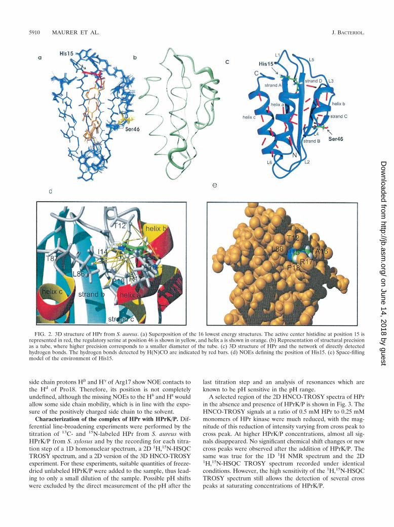
side chain protons H� and H� of Arg17 show NOE contacts tothe Hd of Pro18. Therefore, its position is not completelyundefined, although the missing NOEs to the H� and Hε wouldallow some side chain mobility, which is in line with the expo-sure of the positively charged side chain to the solvent.
Characterization of the complex of HPr with HPrK/P. Dif-ferential line-broadening experiments were performed by thetitration of 13C- and 15N-labeled HPr from S. aureus withHPrK/P from S. xylosus and by the recording for each titra-tion step of a 1D homonuclear spectrum, a 2D 1H,15N-HSQCTROSY spectrum, and a 2D version of the 3D HNCO-TROSYexperiment. For these experiments, suitable quantities of freeze-dried unlabeled HPrK/P were added to the sample, thus lead-ing to only a small dilution of the sample. Possible pH shiftswere excluded by the direct measurement of the pH after the
last titration step and an analysis of resonances which areknown to be pH sensitive in the pH range.
A selected region of the 2D HNCO-TROSY spectra of HPrin the absence and presence of HPrK/P is shown in Fig. 3. TheHNCO-TROSY signals at a ratio of 0.5 mM HPr to 0.25 mMmonomers of HPr kinase were much reduced, with the mag-nitude of this reduction of intensity varying from cross peak tocross peak. At higher HPrK/P concentrations, almost all sig-nals disappeared. No significant chemical shift changes or newcross peaks were observed after the addition of HPrK/P. Thesame was true for the 1D 1H NMR spectrum and the 2D1H,15N-HSQC TROSY spectrum recorded under identicalconditions. However, the high sensitivity of the 1H,15N-HSQCTROSY spectrum still allows the detection of several crosspeaks at saturating concentrations of HPrK/P.
FIG. 2. 3D structure of HPr from S. aureus. (a) Superposition of the 16 lowest energy structures. The active center histidine at position 15 isrepresented in red, the regulatory serine at position 46 is shown in yellow, and helix a is shown in orange. (b) Representation of structural precisionas a tube, where higher precision corresponds to a smaller diameter of the tube. (c) 3D structure of HPr and the network of directly detectedhydrogen bonds. The hydrogen bonds detected by H(N)CO are indicated by red bars. (d) NOEs defining the position of His15. (e) Space-fillingmodel of the environment of His15.
5910 MAURER ET AL. J. BACTERIOL.
on June 14, 2018 by guesthttp://jb.asm
.org/D
ownloaded from

Using the previously assigned chemical shift data for freeHPr, we could identify the lines from HPr in the presence ofHPrK/P. In Fig. 4, the volume changes of the HPr cross peaksat half saturation with HPrK/P are depicted as a function of theHPr sequence. A few cross peaks in the 15N TROSY spectrashow a significantly higher reduction in intensity (larger thanthe mean value ��Vi� plus the standard deviation s). Thecorresponding cross peaks are summarized in Table 2. Asexpected, most of them are located close to Ser46 in the reg-ulatory phosphorylation site. However, some of the signals aresequentially and structurally close to the second phosphoryla-
tion site at His15. A similar picture was obtained from theshifts in the H(N)CO spectrum (Table 2), in which some of thesignals were also significantly influenced by the presence ofHPrK/P.
For a more quantitative evaluation of the data, the dissoci-ation constant Kd of HPr from HPrK/P and the number ofbinding sites N of HPrK/P were determined, assuming as a firstapproximation an independent binding of HPr to the kinase(see Materials and Methods). The interpretation of homo-nuclear 1D spectra is much more direct than that of 2D HSQCspectra, for which possible variations in the polarization trans-
FIG. 3. Binding of HPrK/P to HPr detected in a 2D TROSY-H(N)CO experiment. An overlay plot of 2D TROSY-H(N)CO spectra of HPrin the presence or absence of HPr-K/P recorded at an 800-MHz proton frequency at 303 K is shown. Only a part of the spectrum is shown. 1Hand 13C cross peaks of free HPr and those of HPr in the presence of HPrK/P are shown. The residues that are strongly broadened in the presenceof HPr-K/P are denoted by arrows. The sample initially contained 320 �l of 0.5 mM uniformly 13C- and 15N-enriched HPr from S. aureus in 90%H2O–10% D2O, pH 7.0 (gray cross peaks). Appropriate amounts of freeze-dried HPrK/P were added to obtain a solution containing 0.25 mM(monomer concentration) HPrK/P (black cross peaks).
VOL. 186, 2004 STRUCTURE OF HPr FROM S. AUREUS 5911
on June 14, 2018 by guesthttp://jb.asm
.org/D
ownloaded from

fer must be taken into account. As an example, the resonancesof the methionine methyl groups are shown in Fig. 5 at differ-ent HPrK/P concentrations. Generally, no line shifts were ob-served, but two different cases could be observed; either theline width did not change significantly but the intensity de-creased with the addition of HPrK/P or the line was broadenedextremely with increasing HPrK/P concentrations. The firstcase would be typical for slow exchange conditions for inter-acting groups, and the second case would be typical for non-
interacting residues. Only residues of the first group are usefulfor the calculation of Kd and N since they allow measurementsof the free concentration of HPr in the presence of HPrK/P. Aseries of NMR spectra with unlabeled proteins with well-de-fined concentrations were created, the well-resolved 1D signalfrom free HPr (the methyl group of Leu81) was selected (Fig.6), and its intensity changes upon the addition of HPrK/P werefitted to equation 1. From the NMR data, the dissociationconstant Kd and the number of binding sites N per HPrK/P
FIG. 4. Volume changes of amide and carbonyl cross peaks after addition of HPrK/P. The same set of samples was used as that described forFig. 3. The graphs show the relative volume decrease �Vi {�Vi � [Vi(HPrK/P � 0)] [Vi (HPrK/P � 0.25 mM)]/Vi(HPrK/P � 0)} of the1H,15N-HSQC TROSY (A) and 2D TROSY-H(N)CO (B) cross peaks at half saturation of HPr with HPrK/P. Data were recorded at an 800-MHzproton frequency and 303 K. The mean values ��Vi � (broken lines) and the range defined by the mean value � the standard deviation s aredepicted as black boxes.
5912 MAURER ET AL. J. BACTERIOL.
on June 14, 2018 by guesthttp://jb.asm
.org/D
ownloaded from

TABLE 2. Intermolecular contacts in the HPr-HPrK/P complexa
HPrresidueb
Interaction visible in X-ray structure Interaction visible by NMRe
Atomc HPrK/P residued Atomc HNCO 15N HSQC 1H NMR
Thr12(Ser) O Ile301 sc � �Gly13 H� Ile301 sc � �Ile14 � �His15 H� Leu297 sc � �
H� Ile301 scsc Leu297 scsc Ile301 sc
Ala16 HN Leu297 sc � �sc Asn294(Glu) sc
Arg17 � �Ala19 �Gln24 �Tyr37 sc Asn308 sc �Lys40 sc Thr132(Arg) H�
sc Thr132(Arg) C sc Thr132(Arg) Osc Thr132(Arg) scsc Ser134 scsc Asn176(Arg) sc
Lys41(Thr) O Ser134 scVal42 sc Asn176(Arg) scAsn43 sc His136 scLys45 C Ser153 sc
O Ser153 scsc Ser153 sc
Ser46 sc His136 sc �sc Asp175 sc
Ile47 sc Glu200 sc �Met48 HN Asp175 sc �
O Ile195(Leu) scsc Asp175 scsc Ile195(Leu) scsc Glu200 sc
Gly49 HN Asp175 scH� Asn176(Arg) sc
Val50 �Met51 O Leu297 sc � � �
sc Phe293 scsc Leu297 sc
Ser52 C� Leu194(Ile) sc �H� Leu194(Ile) scO Asn176(Arg) scO Leu194(Ile) scO Glu300(Leu) scsc Asn176(Arg) scsc Leu194(Ile) scsc Ile195(Leu) sc
Leu53 H� Asn176(Arg) sc �Gly54 C� Ile301 sc �
H� Leu297 scH� Glu300(Leu) scH� Ile301 scH� Asn304 scC Ile301 scC Asn304 scO Ile301 H�
O Ile301 scO Arg303(His) scO Asn304 sc
Val55(Ile) N Ile301 sc �C Ile301 scO Ile301 sc
Gly56(Ala) C� Ile301 sc �H�1 Asn304 scH�1 Ile301 scsc Leu297 H�
sc Ile301 O
Continued on following page
VOL. 186, 2004 STRUCTURE OF HPr FROM S. AUREUS 5913
on June 14, 2018 by guesthttp://jb.asm
.org/D
ownloaded from
monomer were determined to be 0.10 � 0.02 mM and 1.02 �0.05, respectively, at 303 K.
In the 1D spectra, the ring resonances of His15 and Tyr37,one of the H� resonances of Leu81, and the Hε resonance ofMet48 show a behavior that is typical for slow exchange, andthey are thus candidates for protein-protein interactions. Theresonances of Met1 and Met51 are superposed, but at least oneof the lines again shows a dependence on the HPrK/P concen-tration, which is typical for slow exchange. From the 3D struc-ture, Met51 is a reasonable candidate for interaction withHPrK/P.
With the above parameters, the concentration dependenceof the cross-peak volumes in the 1H,15N-HSQC spectra can bepredicted on the basis of equations 7 to 9, describing differentinteraction models. The factor a, defining the ratio of trans-verse relaxation times in free and bound HPr, was fixed to 28.3,that is, to a value calculated from the molecular masses of freeHPr and the hexameric HPrK/P decorated with six HPr mol-ecules. It is clear that this is only an approximation since a mustbe concentration dependent because a distribution of HPr-HPrK/P complexes with different numbers of HPrs (and hencedifferent masses) must exist in solution. In addition, exchangebroadening is not taken into account. Simulations show thatthe value of a has a negligible influence on the fit of the datafor a rather wide range of values.
The volume changes of the peaks in the 1H,15N-HSQCTROSY spectra were fitted as a function of the HPrK/P con-centration with the three different models (equations 7 to 9).Residues which would show a large chemical shift change in-duced by the binding of HPrK/P (i.e., described by slow ex-change conditions) should be best fitted by equation 7 or 8, andresidues not involved in the protein-protein interaction shouldbe best fitted by equation 9. It turned out that equation 7 wasin no case the optimal solution, which means it cannot describethe system sufficiently well. This is reasonable since it wouldrequire a very small off-rate of HPr, which is not consistentwith the rather weak binding of the protein. As an example,Fig. 6 shows the calculations using the three different equa-tions for three residues assumed to be involved in protein-
protein interaction. Clearly, equation 7 gives a wrong predic-tion and equation 8 gives a somewhat better fit of the data thanequation 9, although the differences are not very large. There-fore, the complete information from all spectra recorded[1H,15N-HSQC TROSY, 2D TROSY-H(N)CO, and 1D spec-tra] must be used.
DISCUSSION
Assignments and properties of the refined structure. Theapplication of heteronuclear experiments to 15N- and 13C-labeled HPr protein allowed an almost complete assignmentwith the high reliability of heteronuclear methods. Comparedto the structure of HPr from S. aureus determined earlier solelyby homonuclear methods at 500 MHz (28), the sequentialassignment of some resonances had to be corrected (see theBioMagRes database). However, these corrections do not in-fluence the general fold of the molecule. Compared to thealready published structure, a much larger number of restraintscould be obtained by including the data for the direct identi-fication of hydrogen bonds via the weak J coupling through thehydrogen bonds. The low-resolution structure had been calcu-lated from 6 NOEs per residue; with 17.8 NOEs per residue, asubstantially higher level of precision for the structure could beobtained, as the RMSD value of the main chain dropped from0.087 to 0.016 nm. The R-factor (R-factor R5 according toGronwald et al. [15]) calculated for an 800-MHz spectrum ofHPr in H2O is substantially smaller than that obtained for thelow-resolution structure. The regions with still larger structuralvariabilities encompass the regulatory helix (helix b), the activecenter loop, and the C terminus. Since a similar picture wasalso obtained for the low-resolution structures published ear-lier and also for HPr from other microorganisms, this seems tobe a typical feature of HPrs and probably describes the lack ofa unique, rigid structure in these regions. This is also reason-able since it comprises the sites of the proteins that are mostprobably involved in protein-protein interactions.
The function of the strictly conserved residue Arg17 of HPris largely discussed in the literature, and its position relative to
TABLE 2—Continued
HPrresidueb
Interaction visible in X-ray structure Interaction visible by NMRe
Atomc HPrK/P residued Atomc HNCO 15N HSQC 1H NMR
sc Ile305 scsc Arg303(His) HN
sc Arg303(His) scsc Asn304 scsc Gly305(Glu) sc
Lys57 O Gly305(Glu) scsc Gly305(Glu) sc
Ser78(Glu) �Leu81(Met) �
a The X-ray structure of the complex of unphosphorylated HPr from B. subtilis with HPrK/P (1KKL.pdb) from L: casei (12) was used for the analysis. Hydrogen atomswere added with the program Molmol and intermolecular contacts were identified. Contacts were defined as having an interatomic distance of �0.29 nm.
b Residue types and numbering are for HPr from S. aureus. The corresponding residues in HPr from B. subtilis are given in parentheses, and residues that are strictlyconserved in all HPr proteins are given in bold. Compared to HPr from S. aureus, HPr from S. xylosus has five mutated residues, namely Asn4-Lys, Ser71-Thr,Gln75-Glu, Ser78-Thr, and Val80-Ile.
c sc, side chain atoms.d Residue types and numbering are for HPrK/P from S. xylosus. Note that the alignment of the two sequences leads to a shift of four residues. For residues that are
not conserved, the corresponding amino acid HPrK/P from L. casei is given in parentheses.e �, an interaction was visible.
5914 MAURER ET AL. J. BACTERIOL.
on June 14, 2018 by guesthttp://jb.asm
.org/D
ownloaded from

the active center histidine (His15) varies from structure tostructure. In contrast to the case for the structure of HPr fromS. aureus determined earlier, Arg17 does not seem to be inclose contact with the histidine ring system. This could be dueto differences in pH since the structure presented here wasdetermined at pH 7.0, whereas the older structure (28) wassolved at a pH of 7.8, at which the histidine ring is completelyuncharged. At pH 7.0, the interaction between the partialcharge and the positive charge of the arginine side chain willprobably disfavor any close contact between the two sidechains. This is also in line with the observation that the reso-nances of the guanidino groups are averaged here but wereclearly not equivalent at pH 7.8. In general, the position of the
side chain of Arg17 may be strongly dependent on the func-tional state of HPr; in particular, the phosphorylation of thehistidine ring and the interaction with other proteins duringthe phosphotransfer may require different conformations.Analogous to the arginine finger of the Ras-RasGAP system, itmay facilitate the transfer of the phosphoryl group bound tothe active center histidine of EI to HPr.
Interaction of HPr with HPrK/P. The crystal structures ofthe isolated catalytic domain of HPrK/P from L. casei (11) andof full-length HPrK/P from S. xylosus (40) and M. pneumoniae(1) have been reported previously. A crystal structure of thecomplex of HPr from B. subtilis with the nucleotide bindingdomain (amino acids 128 to 319) of HPrK/P from L. casei was
FIG. 5. Spectral changes observed in 1D NMR spectra of HPr induced by the interaction with HPrK/P. A small part of an 800-MHz 1H NMRspectrum of HPr from S. aureus in the presence and absence of HPrK/P is shown. Note that HPr is 13C and 5N enriched. The doublet resonancesof the methyl protons of the four methionine residues of HPr are labeled with 1, 2, and 3, with 1 corresponding to Met21, 2 corresponding to Met1and Met51, and 3 corresponding to Met48. Identical experimental conditions and samples were used as those described for Fig. 3. The molar ratiosof monomers of HPrK/P to HPr were 0, 0.5, 1.0, and 2.0. The quantity of HPr in the active volume of the probe head was held constant.
VOL. 186, 2004 STRUCTURE OF HPr FROM S. AUREUS 5915
on June 14, 2018 by guesthttp://jb.asm
.org/D
ownloaded from

published recently by Fieulaine et al. (12). The HPr proteinfrom B. subtilis has only 64% identity with HPr from L. casei,which means that some of the features observed in the crystalstructures of HPrK/P from L. casei and HPr from B. subtiliscould be due to the use of a heterologous system. The systemused in the solution NMR studies is much more closely relatedsince the HPr proteins from S. xylosus and S. aureus used hereexhibit a sequence identity of 94%.
In the crystals the nucleotide binding domain of HPrK/Pfrom L. casei forms a hexamer with six HPr proteins bound totwo adjacent subunits of the kinase. The structure shows thatHPr mainly interacts via helix b (with Ser46 located in the loopL4 at its N terminus) and with the preceding �-strand C. Asecond interaction site of HPr with a different subunit of
HPrK/P involves helix b and the N terminus of helix a, whichis capped by the active center histidine His15. The residueswith atoms whose interatomic distances are smaller than 0.29nm are listed in Table 2. For a better comparison with our data,the numbering and residue types for our complex are given(HPr from S. aureus and HPrK/P from S. xylosus). The se-quences of HPr from B. subtilis and HPrK/P from L. casei werereplaced with the corresponding residues in HPr from S. au-reus and HPrK/P from S. xylosus. The residues assumed tointeract with HPrK/P according to our NMR data are depictedin Fig. 7.
Since the kinase phosphorylates HPr at position 46, which islocated at the loop preceding helix b, an interaction withHPrK/P is required. In the X-ray structure of the HPr-HPrK/Pcomplex, all residues in the region between Lys40 and Lys57 ofHPr are in contact with residues of HPrK/P, with the onlyexception being Val50. In the NMR data, the first part of theputative interaction site of HPr (Lys40 to Lys45) shows nosigns of a contact with HPrK/P. The subsequent region (Ser46to Lys57) is clearly involved in the interaction; the only differ-ence is that Gly49 and Lys57 do not show a significant responseupon HPrK/P binding. However, in contrast to the case for theX-ray structure, Val50 shows a response upon HPrK/P binding(Table 2). As shown in Table 2, most of the HN and COchemical shift changes are not caused by direct contacts but aretransmitted via changes of the side chain environment. There-fore, it is not surprising that small differences between theX-ray and NMR data exist. However, the interaction with�-strand C and loop L4 (Fig. 2c) seems not to exist in solution.
The interaction with the region around the active centerHis15 encompasses in the X-ray structure the residues of loopL1 (Ser12 to Ala16); the same residues are identified in HPrfrom S. aureus. In addition, Arg17 and two residues of theN-terminal part of helix a (Ala19 and Gln24) are influenced bythe binding of HPrK/P from S. xylosus. Note that in the X-raystructure of the complex between phosphorylated HPr andHPrK/P, additional contacts were observed for Thr20 andGln24 of HPr.
The ring of Tyr37 is located between �-strand B and helicesb and c and is part of the interaction surface in the X-raystructure, and a probable interaction is also predicted in solu-tion (Table 2). However, the chemical shift changes of Ser78and Leu81 located in the C-terminal part of helix c were notexpected from the X-ray data. Ser78 is replaced by a threonineresidue in HPr from S. xylosus, whereas Leu81 is conserved inthe two HPr proteins. Although the observed chemical shiftchanges could be due to changes induced indirectly by confor-mational changes after binding, they could also represent ad-ditional interaction sites of HPrK/P, maybe involving the N-terminal domain of the molecule, which is not present in thepublished X-ray structure.
Conclusions. The NMR data for HPr in the presence ofHPrK/P from S. xylosus indicate the formation of a complexwith a stoichiometry of one HPr molecule per monomer ofHPrK/P. The 1H NMR data can be interpreted as indicating apolymer in solution whose size would be in agreement with thehexameric structure observed for the crystals of HPrK/P fromL. casei. The data show that the second binding site of HPr forHPrK/P is not a crystal artifact but also exists in solution. Inprinciple, the kinase and phosphorylase activities of HPrK/P
FIG. 6. Binding constant of HPr to HPrK/P and fit of volumechanges induced by HPr-HPrK/P interaction with different models.(Top) HprK/P (0.2 mM) from S. xylosus in D2O in 50 mM Tris-HCl,pH 7.5, was titrated with a 4 mM HPr S. aureus solution in the samebuffer. 1D 1H spectra were recorded at 303 K at different HPr-to-HPrK/P ratios. The concentration of free HPr was determined fromthe intensity of the H� resonance of Leu81. The concentrationc(HPrfree) of free HPr was fitted as function of the total concentrationc(HPrtotal) of HPr (and corrected for changes of the HPrK/P concen-tration) with equation 1, calculated by using c(HPrfree) � c(HPrtotal)� PA. A dissociation constant Kd and a number N of binding sites perHPrK/P monomer were obtained as 0.10 � 0.02 mM and 1.02 � 0.05.(Bottom) The same set of samples was used as that described for Fig.3. The volume dependence of the cross peaks of a few selected residuesin the 1H,15N-HSQC TROSY spectra on the HPrK/P concentrationwas calculated with an N of 1.02 and a Kd of 0.10 mM with eitherequation 7 (broken line), 8 (solid bold line), or 9 (solid thin line). Thedata for Leu53 (�), Gly54 (�), and Val55 (‚) which are most probablyinvolved in the protein-protein interaction are shown. Note that thesignal of Val55 was too weak to be observed at higher concentrations.
5916 MAURER ET AL. J. BACTERIOL.
on June 14, 2018 by guesthttp://jb.asm
.org/D
ownloaded from

should depend on the metabolic situation in the cell, which isalso reflected in the phosphorylation state of His15. An inter-action with this region can be used for the recognition of thephosphorylation state of the active center of HPr and thus canregulate the kinase and phosphorylase activities. The data alsoexplain the fact that HPr lacks the capability to be phosphor-ylated at the His15 residue when in complex with HPrK/P andwhile phosphorylated at the regulatory Ser46 residue (47). Apossible additional interacting site could be the C-terminalpart of helix c in solution, maybe involving the N-terminaldomain of HPrK/P.
ACKNOWLEDGMENTS
We thank J. Scheiber for modeling of the HPr-HPrK/P complex, T.Graf for providing random coil shifts, and C. Cabrele for synthesizingthe corresponding model peptide.
This work was supported by the EU (SPINE QL62-CT-2002-00988)and the Deutsche Forschungsgemeinschaft (SFB 521).
REFERENCES
1. Allen, G. S., K. Steinhauer, W. Hillen, J. Stulke, and R. G. Brennan. 2003.Crystal structure of HPr kinase/phosphatase from Mycoplasma pneumoniae.J. Mol. Biol. 326:1203–1217.
2. Arnold, M. R., W. Kremer, H.-D. Luedemann, and H. R. Kalbitzer. 2002. 1HNMR parameters of common amino acid residues measured in aqueoussolutions of the linear tetrapeptides Gly-Gly-X-Ala at pressures between 0.1and 200 MPa. Biophys. Chem. 96:129–140.
3. Audette, G., R. Engelmann, W. Hengstenberg, J. Deutscher, K. Hayakawa,J. W. Quail, and L. Delbaere. 2000. The 1.9 A resolution structure ofphospho-serine 46 HPr from Enterococcus faecalis. J. Mol. Biol. 303:545–553.
4. Braunschweiler, L., and R. R. Ernst. 1983. Coherence transfer by isotropicmixing: application to proton correlation spectroscopy. J. Magn. Reson.61:306–320.
5. Cordier, F., and S. Grzesiek. 1999. Direct observation of hydrogen bonds inproteins by scalar couplings. J. Am. Chem. Soc. 121:1601–1602.
6. Cornilescu, G., F. Delaglio, and A. Bax. 1999. Protein backbone angle re-straints from searching a database for chemical shift and sequence homol-ogy. J. Biomol. NMR 13:289–302.
7. Deutscher, J., and M. H. Saier. 1983. ATP-dependent protein kinase-cata-lyzed phosphorylation of a seryl residue in HPr, a phosphate carrier proteinof the phosphotransferase system in Streptococcus pyogenes. Proc. Natl.Acad. Sci. USA 80:6790–6794.
8. Deutscher, J., B. Pevec, K. Beyreuther, H. H. Kiltz, and W. Hengstenberg.1986. Streptococcal phosphoenolpyruvate-sugar phosphotransferase system:amino acid sequence and site of ATP-dependent phosphorylation of HPr.Biochemistry 25:6543–6551.
9. Dossonnet, V., V. Monedero, M. Zagorec, A. Galinier, G. Perez-Martinez,and J. Deutscher. 2000. J. Bacteriol. 182:2582–2590.
10. El-Kabbani, O. A. L., E. B. Waygood, and L. T. J. Delbaere. 1987. Tertiarystructure of histidine-containing protein of the phosphoenolpyruvate:sugarphosphotransferase system of Escherichia coli. J. Biol. Chem. 262:12926–12929.
11. Fieulaine, S., S. Morera, S. Poncet, V. Monedero, V. Gueguen-Chaignon, A.Galinier, J. Janin, J. Deutscher, and S. Nessler. 2001. X-ray structure of HPrkinase: a bacterial protein kinase with a P-loop nucleotide-binding domain.EMBO J. 20:3917–3927.
12. Fieulaine, S., S. Morera, S. Poncet, L. Mijakovic, A. Galinier, J. Janin, J.Deutscher, and S. Nessler. 2002. X-ray structure of a bifunctional proteinkinase in complex with its protein substrate HPr. Proc. Natl. Acad. Sci. USA99:13437–13441.
13. Galinier, A., M. Kravanja, R. Engelmann, W. Hengstenberg, M.-C. Kilhoffer,J. Deutscher, and J. Haiech. 1998. New protein kinase and protein phos-phatase families mediate signal transduction in bacterial catabolite repres-sion. Proc. Natl. Acad. Sci. USA 95:1823–1828.
14. Gorler, A., W. Hengstenberg, M. Kravanja, W. Beneicke, T. Maurer, andH. R. Kalbitzer. 1999. Solution structure of histidine containing phospho-carrier protein (HPr) from Staphylococcus carnosus. Appl. Magn. Reson.17:465–480.
15. Gronwald, W., R. Kirchhofer, A. Gorler, W. Kremer, B. Ganslmeier, K.-P.Neidig, and H. R. Kalbitzer. 2000. RFAC, a program for automated NMR-R-factor estimation. J. Biomol. NMR 17:137–151.
16. Grzesiek, S., and A. Bax. 1992. Improved 3D triple-resonance NMR tech-niques applied to a 31-kDa protein. J. Magn. Reson. 96:432–440.
17. Grzesiek, S., and A. Bax. 1992. An efficient experiment for sequential back-bone assignment of medium-sized isotopically enriched proteins. J. Magn.Reson. 99:201–207.
18. Grzesiek, S., and A. Bax. 1993. Amino acid type determination in the se-quential assignment procedure of uniformly 13C/15N enriched proteins.J. Biomol. NMR 3:185–204.
19. Guntert, P., C. Mumenthaler, and K. Wuthrich. 1997. Torsion angle dynam-ics for NMR structure calculation with the new program DYANA. J. Mol.Biol. 273:283–298.
FIG. 7. Interaction site of HPr with HPrK/P. (Left) Topological model of HPr. (Right) Same view as in the left panel of a surface model ofHPr in which all residues which show signs of being involved in the interaction with HPrK/P are depicted in red. Residues were assumed to bepotential interaction partners when one of the markers summarized in Table 2 was applied.
VOL. 186, 2004 STRUCTURE OF HPr FROM S. AUREUS 5917
on June 14, 2018 by guesthttp://jb.asm
.org/D
ownloaded from

20. Hammen, P. K., E. B. Waygood, and R. E. Klevit. 1991. Reexamination of thesecondary and tertiary structure of histidine-containing protein from Esch-erichia coli by homonuclear and heteronuclear NMR-spectroscopy. Bio-chemistry 30:11842–11850.
21. Herzberg, O., P. Reddy, S. Sutrina, M. H. Saier, J. Reizer, and G. Kapadia.1992. Structure of the histidine-containing phosphocarrier protein HPr fromBacillus subtilis at 2.0 A resolution. Proc. Natl. Acad. Sci. USA 89:2499–2503.
22. Jeener, J., B. H. Meier, P. Bachmann, and R. R. Ernst. 1979. Investigationof exchange processes by two-dimensional NMR spectroscopy. J. Chem.Phys. 71:4546–4553.
23. Jia, Z., J. W. Quail, E. B. Waygood, and L. T. J. Delbaere. 1993. The 2.0-Aresolution structure of Escherichia coli histidine-containing phosphocarrierprotein HPr. A redetermination. J. Biol. Chem. 268:22490–22501.
24. Jia, Z., M. Vandonselaar, W. Hengstenberg, J. W. Quail, and L. T. J.Delbaere. 1994. The 1.6 A structure of the histidine-containing phosphocar-rier protein HPr from Streptococcus faecalis. J. Mol. Biol. 236:1341–1355.
25. Jones, B. E., V. Dossonnet, E. Kuester, W. Hillen, J. Deutscher, N. Schnell,and R. E. Klevit. 1997. Binding of the catabolite repressor protein CcpA toits DNA target is regulated by phosphorylation of its corepressor HPr.J. Biol. Chem. 272:26530–26535.
26. Kabsch, W., and C. Sander. 1983. Dictionary of protein secondary structure:pattern recognition of hydrogen-bonded and geometrical features. Biopoly-mers 22:2577–2637.
27. Kalbitzer, H. R., W. Hengstenberg, P. Rosch, P. Muss, P. Bernsmann, R.Engelmann, M. Dorschug, and J. Deutscher. 1982. HPr proteins of differentmicroorganisms studied by hydrogen-1 high resolution nuclear magneticresonance: similarities of structures and mechanisms. Biochemistry 21:2879–2885.
28. Kalbitzer, H. R., K.-P. Neidig, and W. Hengstenberg. 1991. Two-dimensional1H-NMR studies on HPr protein from Staphylococcus aureus: completesequential assignments and secondary structure. Biochemistry 30:11186–11192.
29. Kalbitzer, H. R., and W. Hengstenberg. 1993. The solution structure of thehistidine-containing protein (HPr) from Staphylococcus aureus as deter-mined by two-dimensional 1H-NMR spectroscopy. Eur. J. Biochem. 216:205–214.
30. Kay, L. E., P. Keifer, and T. Saarinen. 1992. Pure absorption gradientenhanced heteronuclear single quantum correlation spectroscopy with im-proved sensitivity. J. Am. Chem. Soc. 114:10663–10665.
31. Kay, L. E., G. Y. Xu, A. U. Singer, D. R. Muhandiram, and J. D. Forman-Kay. 1993. A gradient-enhanced Hcch Tocsy experiment for recording side-chain H-1 and C-13 correlations in H2O samples of proteins. J. Magn.Reson. B. 101:333–337.
32. Klevit, R. E., and E. B. Waygood. 1986. Two-dimensional 1H NMR studies ofhistidine-containing protein from Escherichia coli. Secondary and tertiarystructure as determined by NMR. Biochemistry 25:7774–7781.
33. Koradi, R., M. Billeter, and K. Wuthrich. 1996. MOLMOL: a program fordisplay and analysis of macromolecular structures. J. Mol. Graphics 14:51–55.
34. Kravanja, M., R. Engelmann, V. Dossonnet, M. Bluggel, H. E. Meyer, R.Frank, A. Galinier, J. Deutscher, N. Schnell, and W. Hengstenberg. 1999.The hprK gene of Enterococcus faecalis encodes a novel bifunctional en-zyme: the HPr kinase/phosphatase. Mol. Microbiol. 31:59–66.
35. Kuboniwa, H., S. Grzesiek, F. Delaglio, and A. Bax. 1994. Measurement ofHN-H alpha J couplings in calcium-free calmodulin using new 2D and 3Dwater-flip-back methods. J. Biomol. NMR 4:871–878.
36. Laskowski, R. A., M. W. MacArthur, D. S. Moss, and J. M. Thornton. 1993.PROCHECK: a program to check the stereochemical quality of proteinstructures. J. Appl. Cryst. 26:283–291.
37. Lavergne, J.-P., J.-M. Jault, and A. Galinier. 2002. Insights into the func-tioning of Bacillus subtilis HPr kinase/phosphatase: affinity for its proteinsubstrates and role of cations and phosphate. Biochemistry 41:6218–6225.
38. Luginbuhl, P., T. Szyperski, and K. Wuthrich. 1995. Statistical basis for theuse of 13C chemical shifts in protein structure determination. J. Magn.Reson. B 109:229–233.
39. Marion, D., and K. Wuthrich. 1983. Application of phase-sensitive two-dimensional correlated spectroscopy (COSY) for measurements of 1H-1Hspin-spin coupling constants in proteins. Biochem. Biophys. Res. Commun.113:967–974.
40. Marquez, J. A., S. Hasenbein, B. Koch, S. Fieulaine, S. Nessler, R. B.Russell, W. Hengstenberg, and K. Scheffzek. 2002. Structure of the fulllength HPr kinase/phosphatase from Staphylococcus xylosus at 1.95 A reso-lution: mimicking the product/substrate of the phosphotransfer reactions.Proc. Natl. Acad. Sci. USA 99:3458–3463.
41. Maurer, T., R. Doker, A. Gorler, W. Hengstenberg, and H. R. Kalbitzer.2001. Three-dimensional structure of the histidine containing phosphocar-
rier protein (HPr) from Enterococcus faecalis in solution. Eur. J. Biochem.268:635–644.
42. Mijakovic, I., S. Poncet, A. Galinier, V. Monedero, S. Fieulaine, J. Janin, S.Nessler, J. A. Marquez, K. Scheffzek, S. Hasenbein, W. Hengstenberg, and J.Deutscher. 2002. Pyrophosphate-producing protein dephosphorylation byHPr kinase/phosphorylase: a relic of early life? Proc. Natl. Acad. Sci. USA99:13442–13447.
43. Neidig, K. P., M. Geyer, A. Gorler, C. Antz, R. Saffrich, W. Beneicke, andH. R. Kalbitzer. 1995. AURELIA, a program for computer-aided analysis ofmultidimensional spectra. J. Biomol. NMR 6:255–270.
44. Pervushin, K., R. Riek, G. Wider, and K. Wuthrich. 1997. Attenuated T2relaxation by mutual cancellation of dipole-dipole coupling and chemicalshift anisotropy indicates an avenue to NMR structures of very large bio-logical macromolecules. Proc. Natl. Acad. Sci. USA 94:12366–12371.
45. Postma, P. W., J. W. Lengeler, and G. R. Jacobson. 1993. Phosphoenolpyru-vate-carbohydrate phosphotransferase systems of bacteria. Microbiol. Rev.57:543–549.
46. Pullen, K., P. Rajagopal, B. R. Branchini, M. E. Huffine, J. Reizer, M. H.Saier, J. M. Scholtz, and R. E. Klevit. 1995. Phosphorylation of serine-46 inHPr, a key regulatory protein in bacteria, results in stabilization of its solu-tion structure. Protein Sci. 4:2478–2486.
47. Reizer, J., M. J. Novotny, W. Hengstenberg, and M. H. Saier, Jr. 1984.Properties of ATP-dependent protein kinase from Streptococcus pyogenesthat phosphorylates a seryl residue in HPr, a phosphocarrier protein of thephosphotransferase system. J. Bacteriol. 160:333–340.
48. Reizer, J., C. Hoischen, F. Titgemeyer, C. Rivolta, R. Rabus, J. Stulke, D.Karamata, M. H. Saier, Jr., and W. Hillen. 1998. A novel protein kinase thatcontrols carbon catabolite repression in bacteria. Mol. Microbiol. 27:1157–1169.
49. Rucker, S. P., and A. J. Shaka. 1989. Broadband homonuclear cross polar-ization in 2D N.M.R. using DIPSI-2. Mol. Phys. 68:509–517.
50. Russell, R. B., J. A. Marquez, W. Hengstenberg, and K. Scheffzek. 2002.Evolutionary relationship between the bacterial HPr kinase and the ubiqui-tous PEP-carboxykinase: expanding the P-loop nucleotidyl transferase su-perfamily. FEBS Lett. 517:1–6.
51. Schleucher, J., M. Schwendinger, M. Sattler, P. Schmidt, O. Schledletzky,S. J. Glaser, O. W. Sorensen, and C. Griesinger. 1994. A general enhance-ment scheme in heteronuclear multidimensional NMR employing pulsedfield gradients. J. Biomol. NMR 4:301–306.
52. Stulke, J., and W. Hillen. 1999. Carbon catabolite repression in bacteria.Curr. Opin. Microbiol. 2:195–201.
53. Van Nuland, N. A. J., R. Boelens, R. M. Scheek, and G. T. Robillard. 1995.High-resolution structure of the phosphorylated form of the histidine-con-taining phosphocarrier protein HPr from Escherichia coli determined byrestrained molecular dynamics from NMR-NOE data. J. Mol. Biol. 246:180–193.
54. Viana, R., V. Monedero, V. Dossonnet, C. Vadeboncoeur, G. Perez-Martinez,and J. Deutscher. 2000. Enzyme I and HPr from Lactobacillus casei: theirrole in sugar transport, carbon catabolite repression and inducer exclusion.Mol. Microbiol. 36:570–584.
55. Vuister, G. W., and A. Bax. 1992. Resolution enhancement and spectralediting of uniformly C-13-enriched proteins by homonuclear broad-bandC-13 decoupling. J. Magn. Reson. 98:428–435.
56. Vuister, G. W., and A. Bax. 1993. Quantitative J correlation—a new ap-proach for measuring homonuclear 3-bond J(H(N)H(alpha) coupling-con-stants in N-15-enriched proteins. J. Am. Chem. Soc. 115:7772–7777.
57. Wang, A. C., S. Grzesiek, R. R. Tschudin, P. J. Lodi, and A. Bax. 1995.Sequential backbone assignment of isotopically enriched proteins in D2O bydeuterium-decoupled Ha(CA)N and Ha(CACO)N. J. Biomol. NMR 5:376–382.
58. Wishart, D. S., and B. D. Sykes. 1994. The C-13 chemical-shift index—asimple method for the identification of protein secondary structure usingC-13 chemical-shift data. J. Biomol. NMR 4:171–180.
59. Wishart, D. S., C. G. Bigam, J. Yao, F. Abildgaard, H. J. Dyson, E. Oldfield,J. L. Markley, and B. D. Sykes. 1995. 1H, 13C and 15N chemical-shift refer-encing in biomolecular NMR. J. Biomol. NMR 6:135–140.
60. Wittekind, M., P. Rajagopal, B. R. Branchini, J. Reizer, M. H. Saier, andR. E. Klevit. 1992. Solution structure of the phosphocarrier protein HPrfrom Bacillus subtilis by two-dimensional NMR spectroscopy. Protein Sci.1:1363–1376.
61. Ye, J. J., J. Reizer, X. Cui, and M. H. Saier, Jr. 1994. Inhibition of thephosphoenolpyruvate:lactose phosphotransferase system and activation of acytoplasmic sugar-phosphate phosphatase in Lactococcus lactis by ATP-de-pendent metabolite-activated phosphorylation of serine-46 in the phospho-carrier protein HPr. J. Biol. Chem. 269:11837–11844.
62. Ye, J. J., and M. H. Saier, Jr. 1995. Allosteric regulation of the glucose:H�
symporter of Lactobacillus brevis: cooperative binding of glucose andHPr(ser-P). J. Bacteriol. 177:1900–1902.
5918 MAURER ET AL. J. BACTERIOL.
on June 14, 2018 by guesthttp://jb.asm
.org/D
ownloaded from