heterotropic ossification dr k.raghuveer adiga. hetrotropic ossification was first described in 1692...
TRANSCRIPT
Heterotropic Ossification
Dr K.Raghuveer Adiga
• Hetrotropic ossification was first described in 1692 by Patin in children with myositis ossificans progressiva
• Clearer description was provided by Reidel in 1883 Dejerine and Ceillier in 1918
During Ist World war, HO was predominantlyobserved in soldiers who had become paraplegicdue to gunshot wounds.

Based on hypothetical etiopathogentic mechanism several terms used to denote this condition
Ectopic ossification Myositis ossification Neurogenic ossifying fibromyopathy Paraosteoarthropathy Periarticular ossification
• Myositis ossificans refers to a condition in which ectopic bone is formed within muscle and other soft tissues
• Robert(1968)reported HO in a patient with cerebral injury. Elbow was the involved joint.

Clinically: Fever, swelling, erythema Decreased joint movt seen in early HO which may mimic; Cellulitis Osteomyelitis Thrombophlebitis Osteosarcoma Osteochondroma
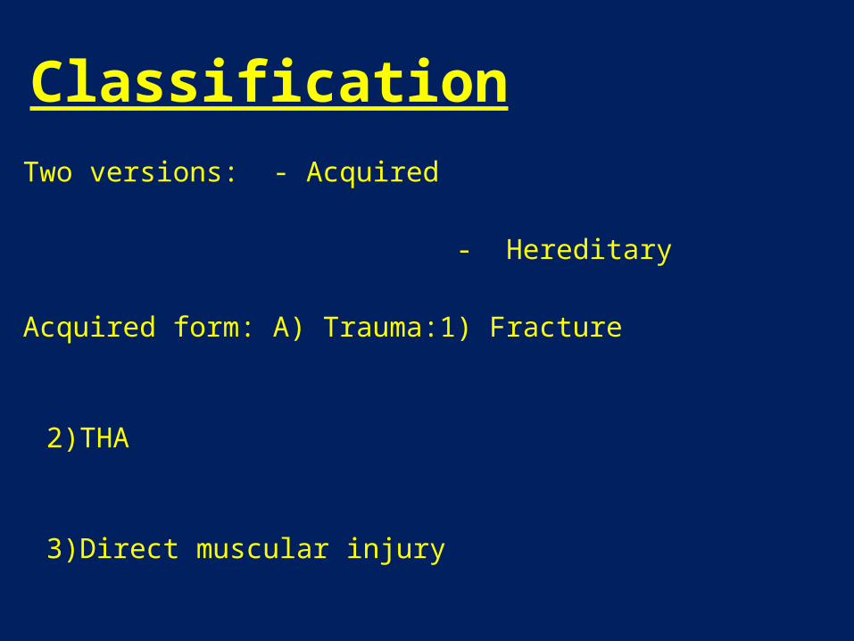
ClassificationTwo versions: - Acquired
- Hereditary
Acquired form: A) Trauma:1) Fracture
2)THA
3)Direct muscular injury
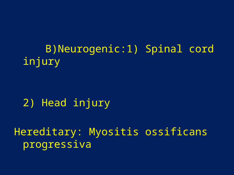
B)Neurogenic:1) Spinal cord injury
2) Head injury
Hereditary: Myositis ossificans progressiva
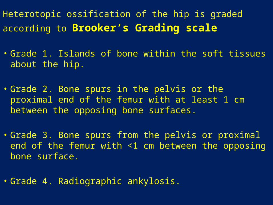
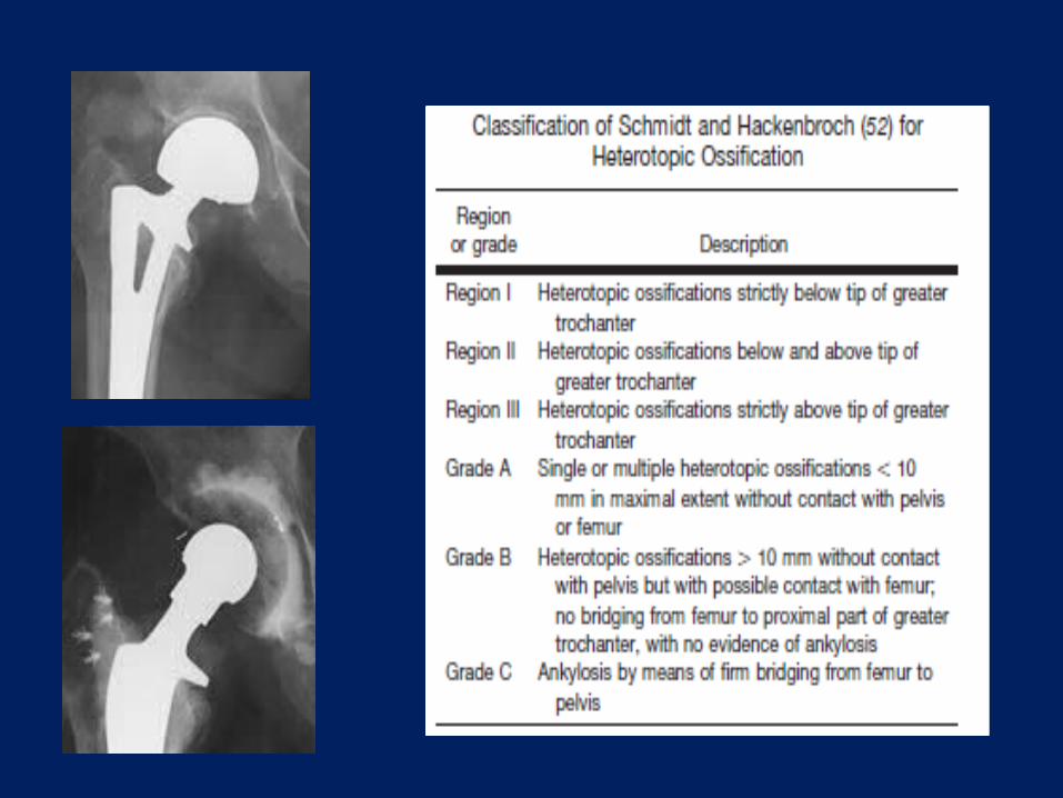
Heterotopic ossification of the hip is graded
according to Brooker’s Grading scale
• Grade 1. Islands of bone within the soft tissues about the hip.
• Grade 2. Bone spurs in the pelvis or the proximal end of the femur with at least 1 cm between the opposing bone surfaces.
• Grade 3. Bone spurs from the pelvis or proximal end of the femur with <1 cm between the opposing bone surface.
• Grade 4. Radiographic ankylosis.
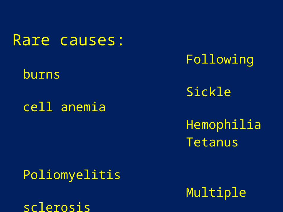
Rare causes: Following burns Sickle cell anemia Hemophilia Tetanus Poliomyelitis Multiple sclerosis Toxic epidermal necrolysis
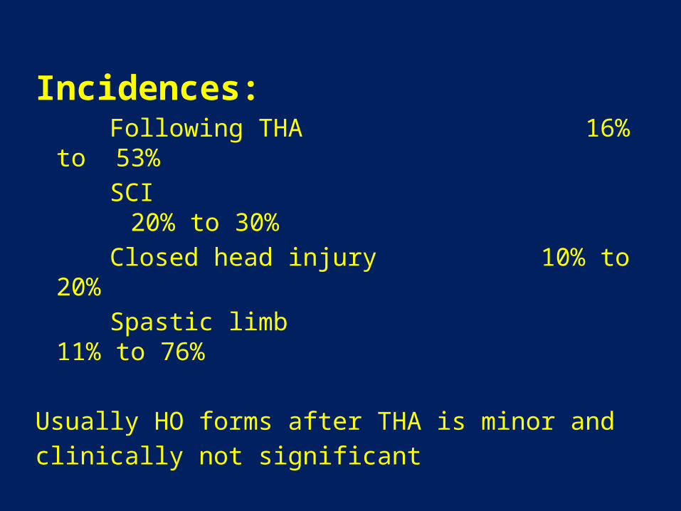
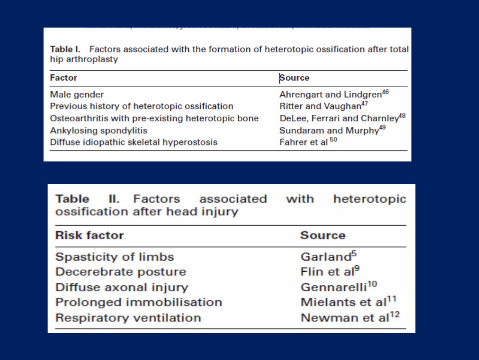
Incidences: Following THA 16% to 53% SCI 20% to 30% Closed head injury 10% to 20% Spastic limb 11% to 76% Usually HO forms after THA is minor andclinically not significant
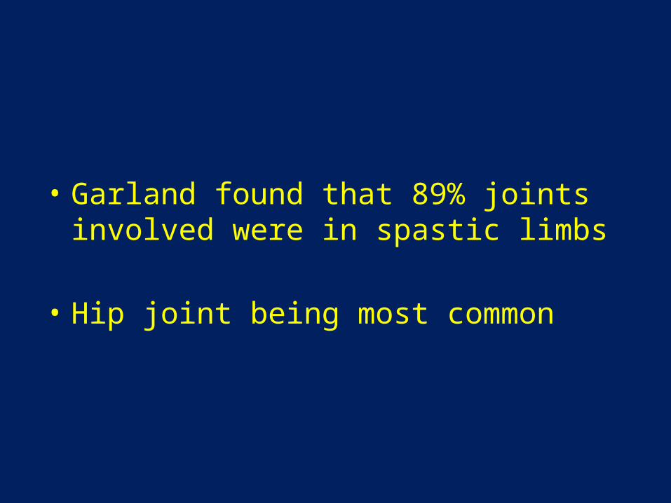
• Garland found that 89% joints involved were in spastic limbs
• Hip joint being most common
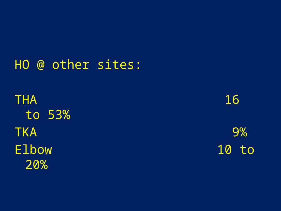
HO @ other sites:
THA 16 to 53%TKA 9%Elbow 10 to 20%
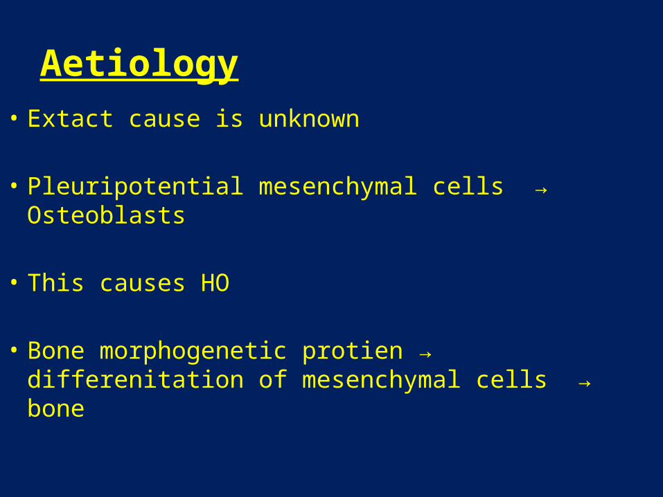
Aetiology• Extact cause is unknown
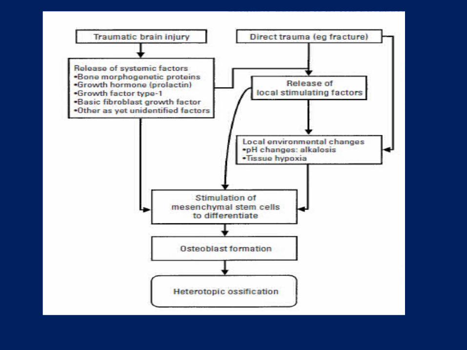
• Pleuripotential mesenchymal cells → Osteoblasts
• This causes HO
• Bone morphogenetic protien → differenitation of mesenchymal cells → bone
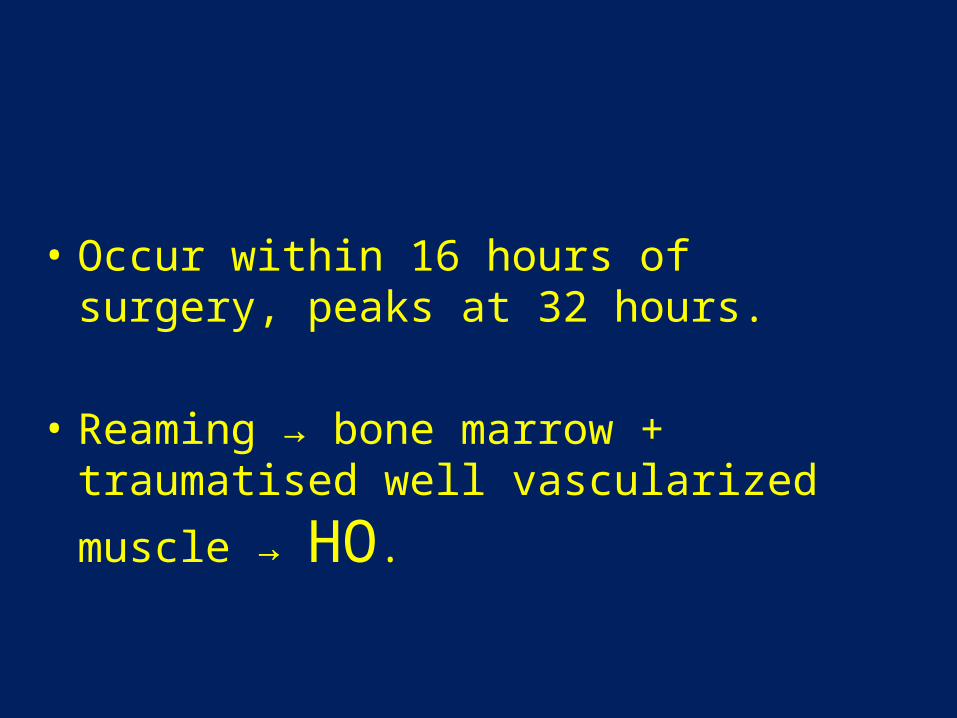
• Occur within 16 hours of surgery, peaks at 32 hours.
• Reaming → bone marrow + traumatised well
vascularized muscle → HO.
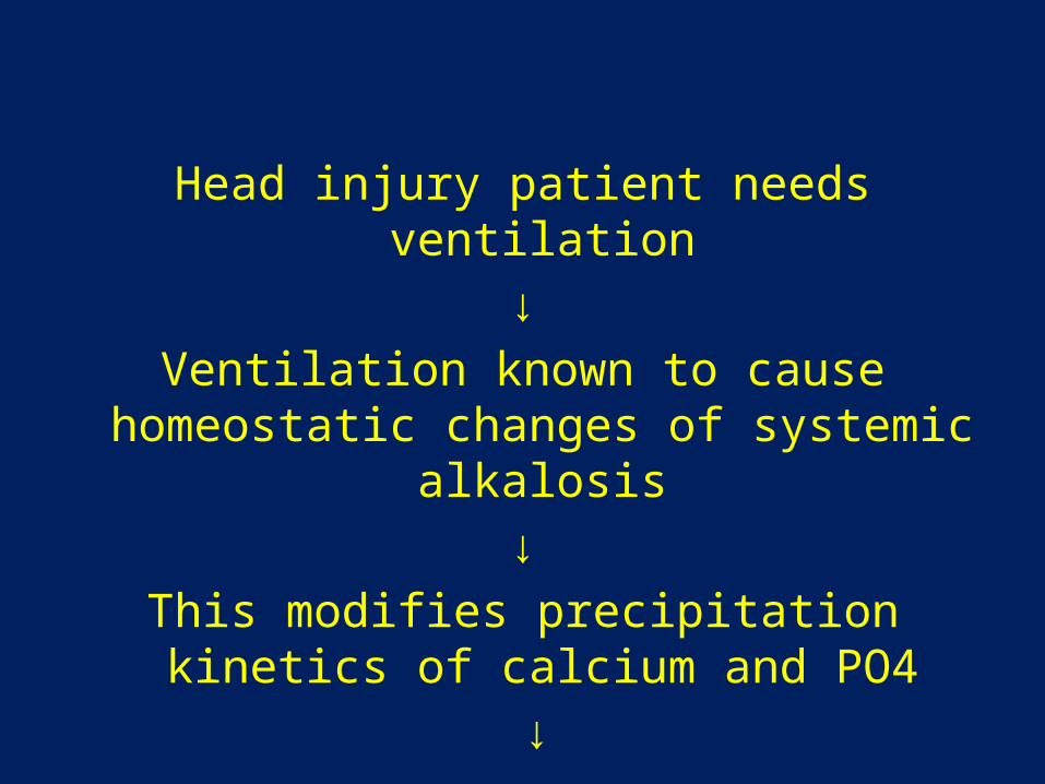
Head injury patient needs ventilation↓
Ventilation known to cause homeostatic changes of systemic alkalosis
↓This modifies precipitation kinetics of calcium
and PO4 ↓
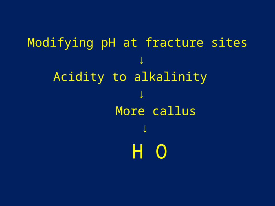
Modifying pH at fracture sites ↓
Acidity to alkalinity ↓
More callus ↓
H O
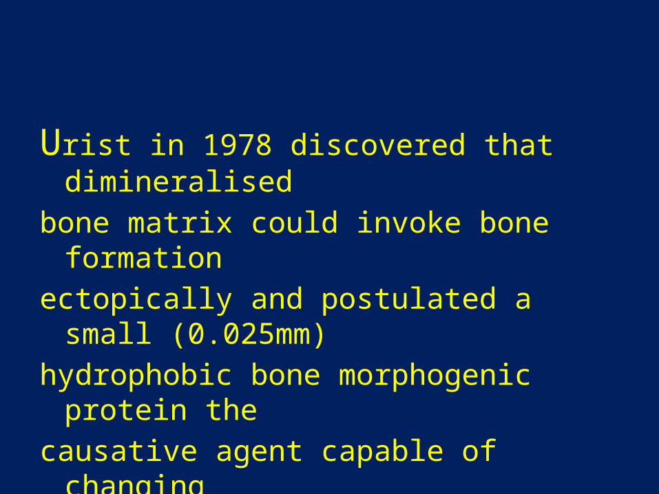
Urist in 1978 discovered that dimineralisedbone matrix could invoke bone formation ectopically and postulated a small (0.025mm)hydrophobic bone morphogenic protein the causative agent capable of changing mesenchymal cells in muscle from fibrous tissueto bone.
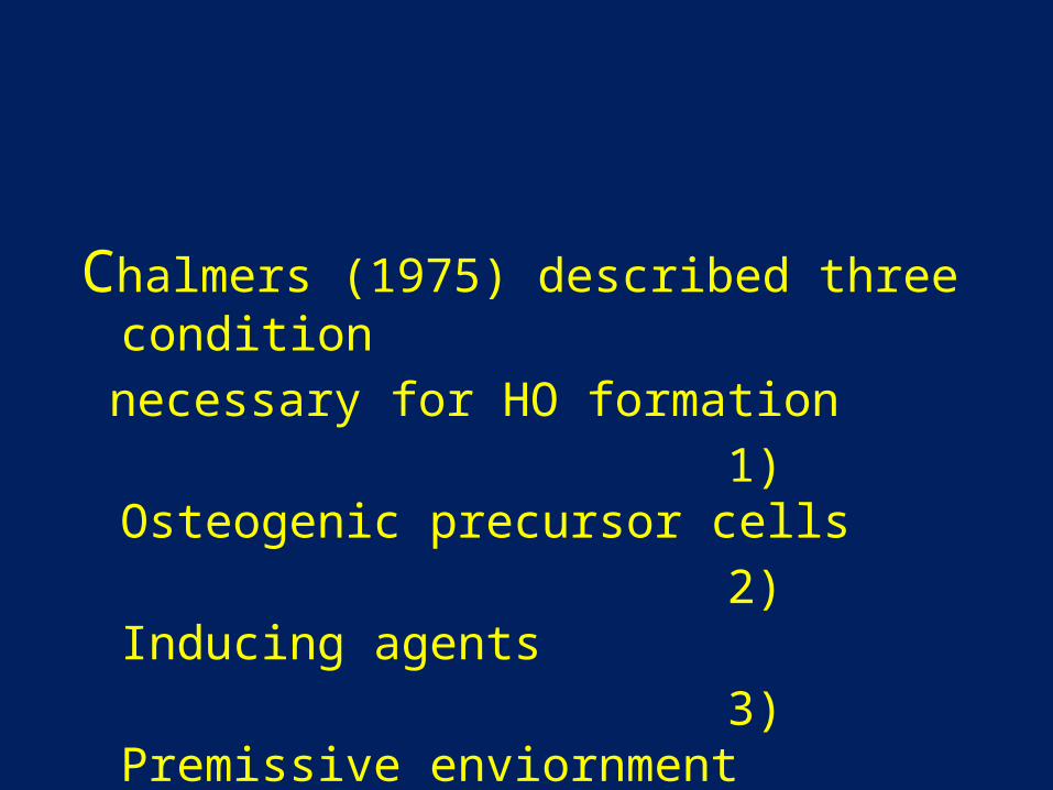
Chalmers (1975) described three condition necessary for HO formation 1) Osteogenic precursor cells 2) Inducing agents 3) Premissive enviornment
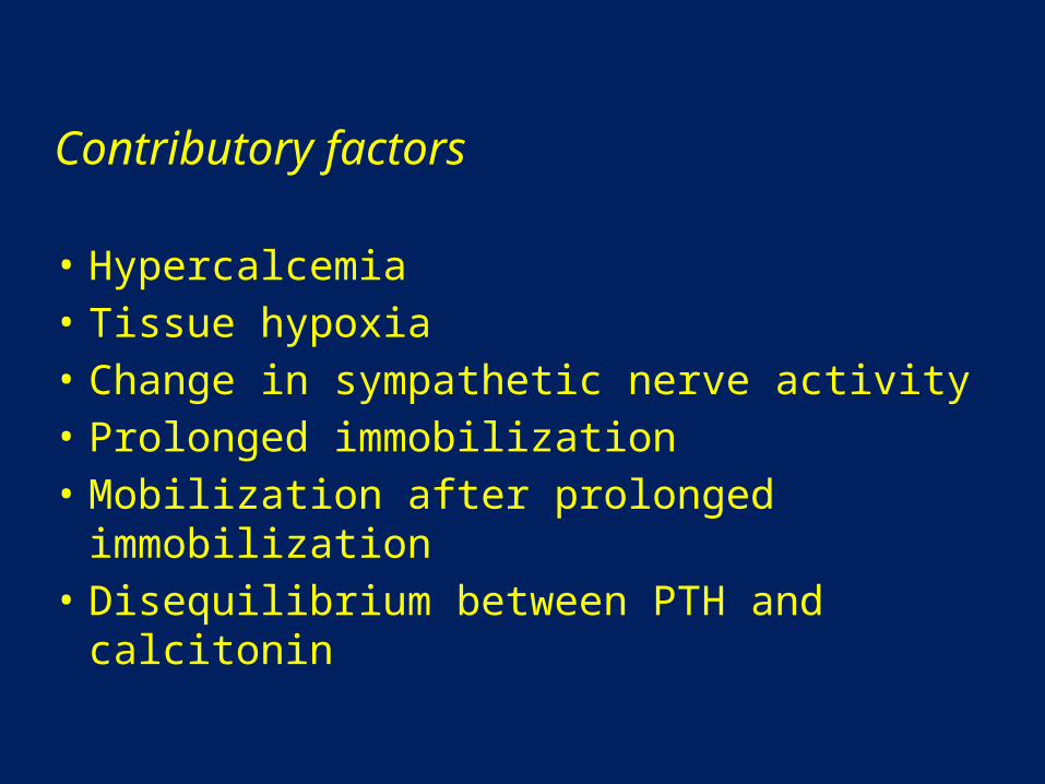
Contributory factors
• Hypercalcemia• Tissue hypoxia• Change in sympathetic nerve activity• Prolonged immobilization• Mobilization after prolonged immobilization• Disequilibrium between PTH and calcitonin
Biochemical changes:
Alk PO4 levels - ↑ 3 ½ times at 4 weeks post injury
PGE2 excretion in 24 hour urine-early indicator of HO
Histology:
Myositis ossificans and HO are fundamentally different.
Important steps in the ossification process is fibroblastic metaplasia.
Histological studies clearly demonstrates a zone of fibroblastic proliferation, followed by chondroblasts which eventually transformed into osteoblasts with blood vessels and haversian canals.
Diagnosis and Investigations• Alk PO4 ase levels
• Three phase bone scintigraphy
- Diagnostic and therapeutic follow up
-very sensitive.
- Usually positive after 2-4 weeks.
- Serial bone scan helps to monitor of metabolic activity.
• Radiography, MRI and CT scan
• Ultrasonography - < one week after THA
Treatment
1) Physiotherapy:
-Assisted range of movement exercise with gentle stretch and terminal resistance training.
-Joint movement not beyond pain free range
2) Medical management:
NSAIDs Indomethacin 25mg tds for 6 weeks
COX2 inhibitors:Meloxicam 7.5mg/15mg per day
Bisphosphonates
retards the ossification
when drug is stopped, osteoid gets mineralized
Etidronate-300mg/day/IV for 3 days, then 20mg/kg/day for 6 months
.
3) Radiation Therapy
Extact mechanism is not known.
Supposed to interfere with the differentiation of pleuripotent mesenchymal cells into osteoblasts.
Coventry and Scanlon(1970)-offered preventive radiation therapy for the first time at Mayo clinic before performing THA.
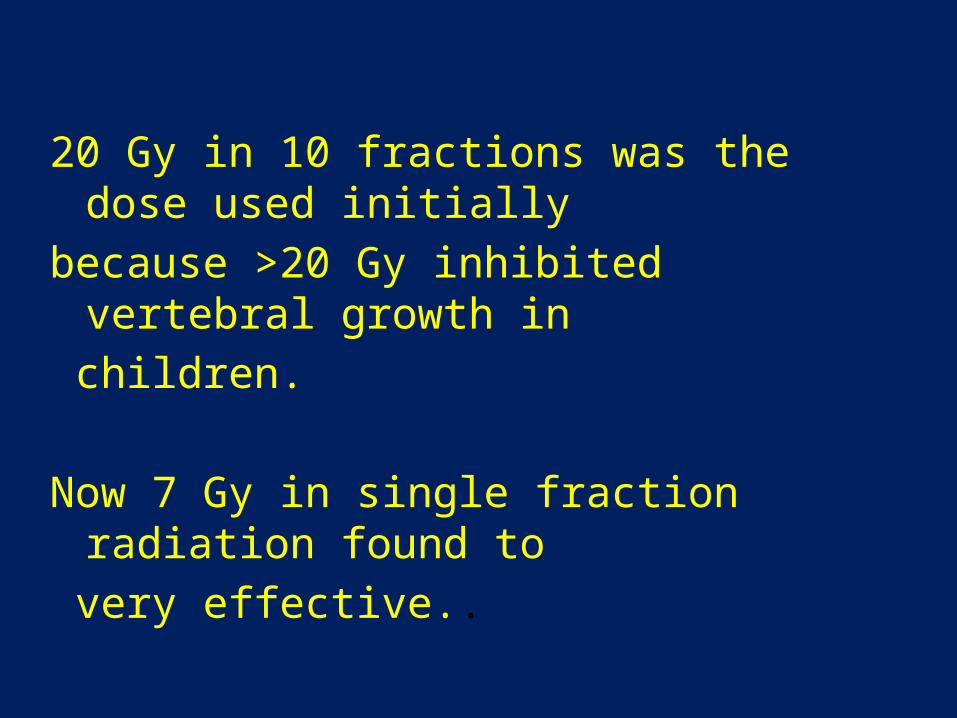
20 Gy in 10 fractions was the dose used initiallybecause >20 Gy inhibited vertebral growth in children.
Now 7 Gy in single fraction radiation found to very effective..
Timing
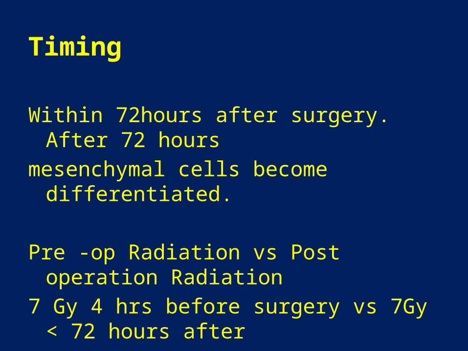
Within 72hours after surgery. After 72 hours mesenchymal cells become differentiated.
Pre -op Radiation vs Post operation Radiation7 Gy 4 hrs before surgery vs 7Gy < 72 hours after surgery showed no difference in outcome.
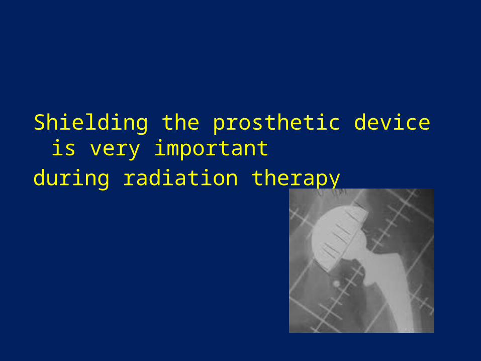
Shielding the prosthetic device is very importantduring radiation therapy
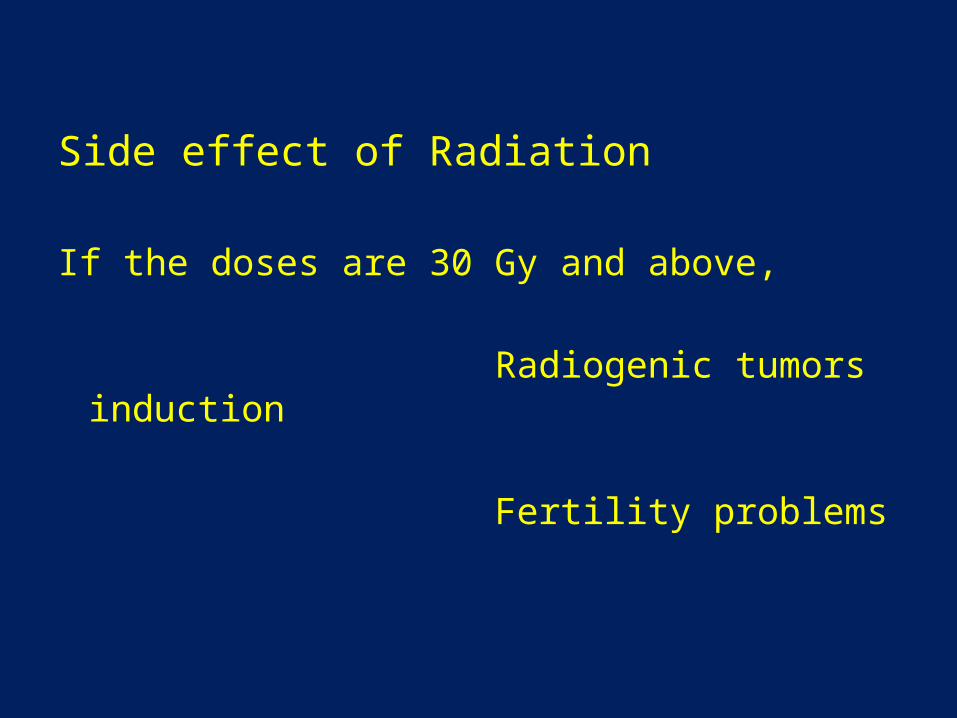
Side effect of Radiation
If the doses are 30 Gy and above,
Radiogenic tumors induction
Fertility problems
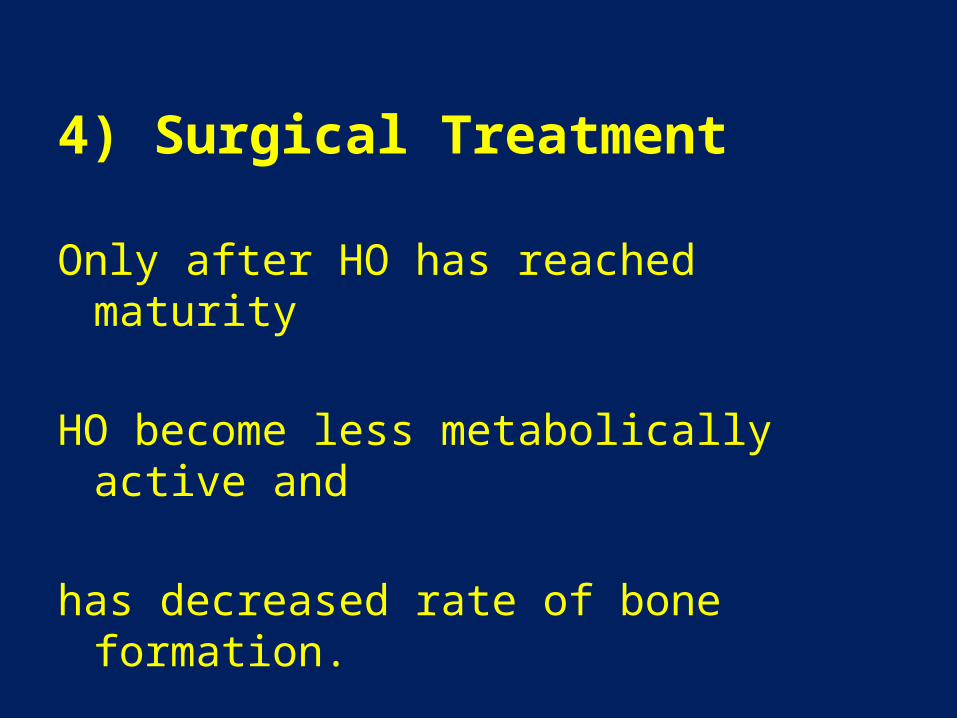
4) Surgical Treatment
Only after HO has reached maturity
HO become less metabolically active and
has decreased rate of bone formation.
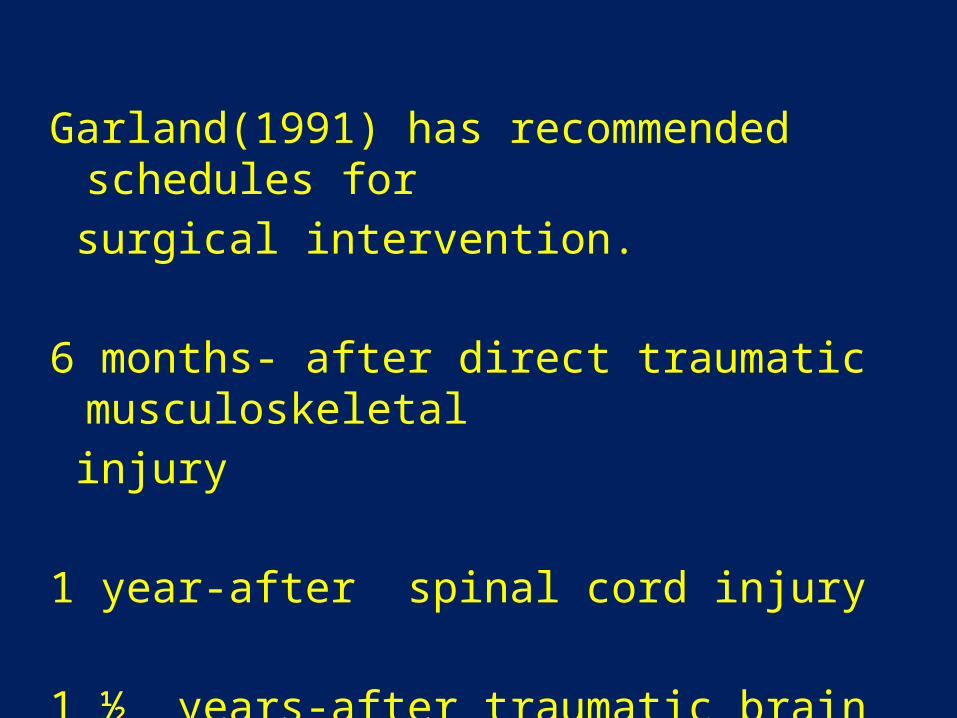
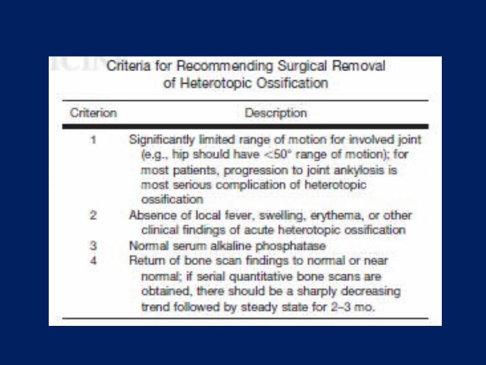
Garland(1991) has recommended schedules for surgical intervention.
6 months- after direct traumatic musculoskeletal injury
1 year-after spinal cord injury
1 ½ years-after traumatic brain injury
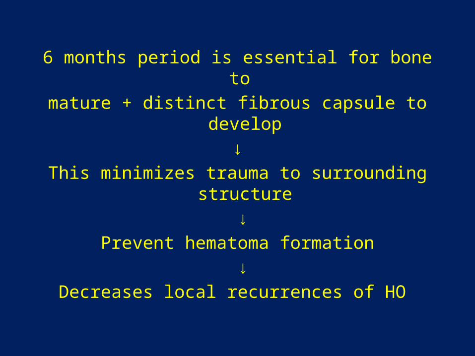
6 months period is essential for bone to mature + distinct fibrous capsule to develop
↓This minimizes trauma to surrounding structure
↓Prevent hematoma formation
↓Decreases local recurrences of HO
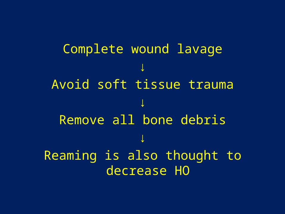
Complete wound lavage↓
Avoid soft tissue trauma↓
Remove all bone debris↓
Reaming is also thought to decrease HO
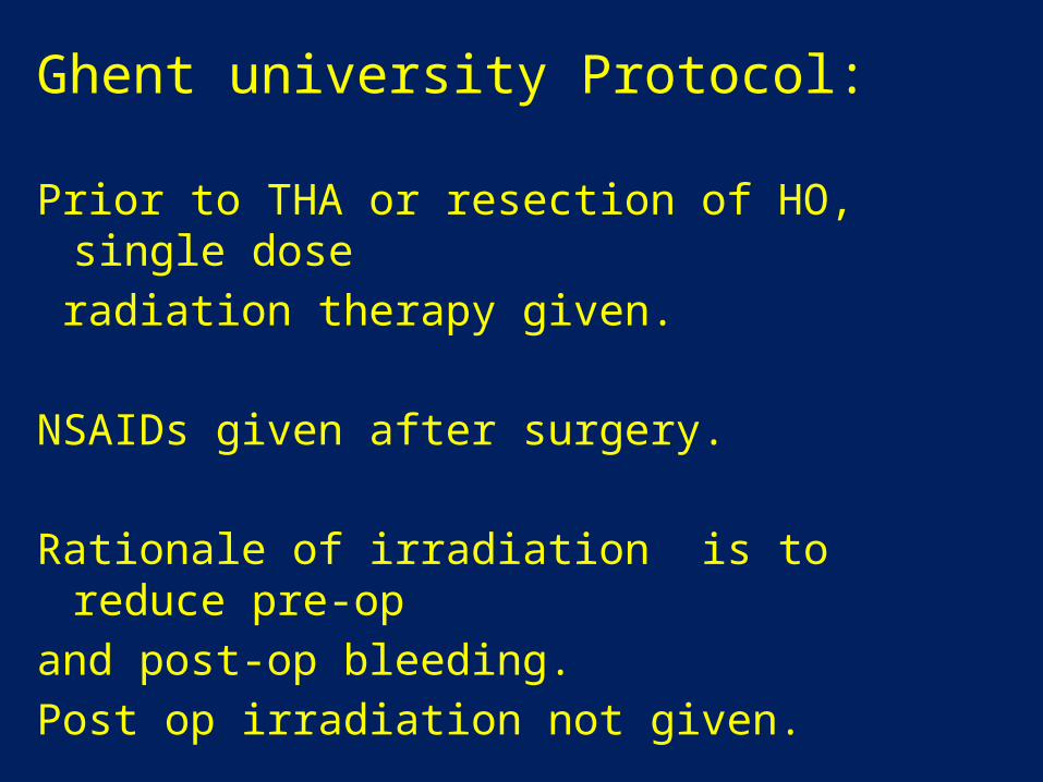
Ghent university Protocol:
Prior to THA or resection of HO, single dose radiation therapy given.
NSAIDs given after surgery.
Rationale of irradiation is to reduce pre-op and post-op bleeding.Post op irradiation not given.
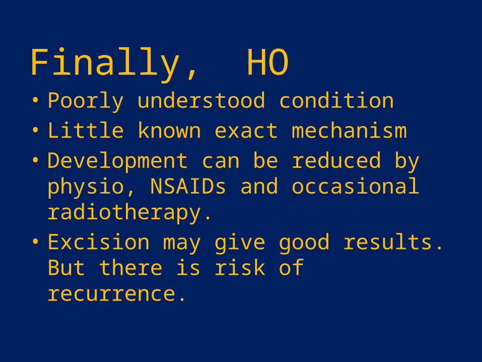
Finally, HO • Poorly understood condition• Little known exact mechanism • Development can be reduced by physio,
NSAIDs and occasional radiotherapy.• Excision may give good results. But there is
risk of recurrence.