heterogeneity of collagens in rabbit cornea: type vi...
TRANSCRIPT

Investigative Ophthalmology & Visual Science, Vol. 29, No. 5, May 1988Copyright © Association for Research in Vision and Ophthalmology
Heterogeneity of Collagens in Rabbit Cornea:Type VI Collagen
Charles Cinrron* and Bor-Shyue Hongf
Normal adult rabbit corneas were digested with 5% pepsin and their collagens extracted with aceticacid. Collagen extracts were fractionated by differential salt precipitation. The 2.5 M NaCl fractionwas then redissolved with tris buffer and precipitated with sodium acetate. The precipitate contained ahigh-molecular-weight disulfide-bonded aggregate which, upon reduction with mercaptoethanol, wasconverted into three distinct polypeptides having molecular weights between 45 and 66 Kd. Thesephysical characteristics, together with the susceptibility of these polypeptides to collagenase and theiramino acid composition, identified the high molecular weight aggregate as type VI collagen. Corneasfrom neonate rabbits and adult corneas containing 2-week-old scars were organ cultured in the pres-ence of [NC) glycine to incorporate radiolabel into collagen. Tissues were digested with 0.02% pepsinand their collagens extracted with formic acid. The total radioactivity of the extracts and tissueresidues was determined before the collagens were separated by SDS-polyacrylamide slab gel electro-phoresis. Radioactive collagen polypeptides bands were then stained with Coomassie blue, processedfor fluorography, and analyzed by densitometry. The results show that: (1) type VI collagen issynthesized by neonate corneas and healing adult corneas; (2) it is not readily solubilized from eithercorneal tissue by 0.02% pepsin digestion and formic acid extraction; and (3) the proportion of type VIcollagen deposited in scar tissue is markedly lower than that found in neonate corneas. Invest Ophthal-mol Vis Sci 29:760-766, 1988
The predominant banded collagen fibrils of mam-malian corneal stroma are composed of type I colla-gen.1"4 Other than a single report, there is no evidenceindicating the presence of type II collagen in mam-malian corneas.5 Although there is as yet some con-troversy as to the distribution and quantity of type IIIcollagen within the cornea, it appears to be associatedwith neonate and healing adult tissues.346"10 Type IVcollagen seems to be restricted to the basement mem-brane of the corneal epithelium and the Descemet'smembrane of normal corneas."12 Type V collagenconstitutes about 10% of the normal stromal colla-gen.1314 The relative quantity of type V collagenwithin the stroma of fetal and healing cornea is mark-edly different from the adult, suggesting that this col-lagen may play a role in the morphogenesis of thecorneal stroma. Although we do not know the ultra-structural distribution of type V collagen in mamma-
From the *Eye Research Institute, Boston, Massachusetts, andfDepartment of Ophthalmology and Visual Sciences, Texas TechUniversity Health Sciences Center, Lubbock, Texas.
Supported by grant EY-01199 from the National Institutes ofHealth, Bethesda, Maryland, and a grant from Research to PreventBlindness.
Submitted for publication: April 14, 1987; accepted December14, 1987.
Reprint requests: Charles Cintron, PhD, Eye Research Institute,20 Staniford Street, Boston, MA 02114.
lian corneas, recent reports have indicated that chickstromal collagen fibrils are copolymers of type I andV collagens.15 Type VI collagen is also located in themammalian stroma and because of its unusual struc-ture, it may be associated with fine filaments betweenthe banded collagen fibrils.16"19 Anchoring fibrils as-sociated with the basement membrane of the cornealepithelium and extending into the stroma have beenidentified with type VII collagen.20-21 Finally, typeVIII collagen, originally designated "endothelial col-lagen,"22 has been described as a prominent collagenin bovine and rabbit Descemet's membranes.2324 Be-cause each corneal collagen has a unique structure,distinctive chemical properties, a specific ultrastruc-tural distribution within the tissue, and different andvarying abundance during morphogenesis, thesemacromolecules must play an important role in nor-mal corneal development and healing.
Our studies have shown that developing neonatecorneas and corneal scar tissue from adult corneassynthesize and deposit mostly types I and V colla-gens.13 The limitations in our analyses were partiallydue to the paucity of tissue and the low specific activ-ity of radiolabeled collagen synthesized after in vivolabeling of the corneal proteins. To increase the spe-cific activity, we developed an organ culture methodthat maintains the whole cornea deturgescent and ul-trastructurally normal during incubation of the tis-
760
Downloaded From: http://iovs.arvojournals.org/pdfaccess.ashx?url=/data/journals/iovs/933142/ on 06/15/2018

No. 5 TYPE VI COLLAGEN IN RADBIT CORNEA / Cinrron ond Hong 761
sues with radiolabeled precursors.25 This we believealso maintains a pattern of macromolecular synthesissimilar to that in vivo.
In the present study, we show that normal adultrabbit corneas contain type VI collagen. Moreover,rabbit neonate corneas and scar tissue from adultcorneas deposit types I, V and VI collagen and syn-thesize these collagens during organ culture. Semi-quantitative analyses of the collagens deposited andsynthesized in these growing corneal tissues indicatethat the relative quantity and solubility of collagensdiffer among different types of collagens as well asbetween tissues. These differences have significanceto our understanding of scar formation.
Material and Methods
Type VI Collagen
Adult rabbit corneas, freed of Descemefs mem-brane, endothelium and epithelium were homoge-nized in 0.1 M acetic acid with a blender at high speedfor 1 min followed by limited pepsin digestion at 4°Cfor 24 hr with a 5% pepsin/tissue wet weight ratio in0.1 M acetic acid. The digest was centrifuged at60,000 rpm for 10 min and the supernate directlyfractionated by differential salt precipitation at 0.7M, 1.2 M and 2.5 M NaCl concentration in acid. The2.5 M precipitate fraction was redissolved in 0.05 Mtris-HCl buffer, pH 7.5, containing 0.1 mMphenylmethylsulfonyl fluoride, 1 mM N-ethyl malei-mide, 1 mM benzamidine, 25 mM ethylene diaminetetra-acetic acid, and 0.5 M NaCl at 4°C for 2 hr. Thesolution was centrifuged and the supernate dialyzedagainst 0.05 M sodium acetate, pH 4.8, to precipitatethe proteins. These precipitated proteins could notpenetrate an SDS-8% polyacrylamide slab gel duringelectrophoresis.26 Disulfide reduction with 5% 2-mercaptoethanol dissociated the large protein aggre-gate, tentatively called high-molecular-weight disul-fide-bonded aggregate (HMWDA), into small, Coo-massie blue-stained proteins, which penetrated thepolyacrylamide gel.
The HMWDA was hydrolyzed with 6 N HC1 undernitrogen at 105°C for 24 hr to determine the aminoacid composition with a Beckman amino acid ana-lyzer, model 121 (Fullerton, CA).
Susceptibility of HMWDA to bacterial collagenasewas determined by incubating 100 yug of theHMWDA with 50 /zg of Closlridium hislolyticumcollagenase, protease free (Cal Biochem, San Diego,CA), in 200 1̂ of 0.05 M Tris-HCl buffer, pH 7.5,containing 10 mM CaCl2 at 37°C for 2 hr. Controlscontained HMWDA in appropriate buffer and incu-bated at the same temperature for the same amountof time. The hydrolysates and controls were then re-
duced with mercaptoethanol and electrophoresed inan SDS-8% polyacrylamide slab gel.
Corneal Organ Culture, Collagen Extraction,and Analysis
Rabbits weighing about 2.5 kg were anesthetizedwith intravenous injections of sodium pentobarbitaland topical application of proparacaine drops to eacheye before wounding the corneas. A 2 mm diameter,full-thickness wound was made in each eye of sixrabbits and allowed to heal for 2 weeks.27 Thewounded rabbits and three newborn rabbits, approxi-mately 3 days old, were killed with an overdose ofsodium pentobarbital. The whole cornea of each eye,with an attached 1-2 mm scleral rim, was removedand placed in ice-cold culture medium. One scarredcornea was not used due to complications duringhealing. Our culture medium contained Hank's basalmedium. Coon's concentrate, newborn calf serumand the antibiotics, penicillin and streptomycin.28
The medium was modified by the addition of 2%(w/v) chondroitin sulfate (Sigma, St. Louis, MO).Corneas were washed with 4 ml of fresh mediumbefore incubation in Linbro tissue culture multi-wellplates with a well capacity of 7.5 ml (Flow Labs,McLean, VA). Enough fresh medium was used tocover the explant with the corneal endolhelium up.The medium contained 75 nC\ of [I4C] glycine per ml(New England Nuclear, Boston, MA) and the tissueswere incubated at 37°C in a 5% CO2/air mixture for18 hr. These procedures have been shown to main-tain the rabbit cornea deturgescent and ultrastructur-ally normal for 48 hr.25
After incubation, explants were washed in freshnonradioactive medium, and the scleral rims re-moved from the corneas. The total nconate corneaand only the scar tissue of the adult cornea wereweighed, frozen in liquid nitrogen and pulverized.Powdered corneas were digested with 0.02% (w/w)pepsin/tissue weight in 0.5 M formic acid at 4°C for18 hr. Digests were centrifuged for 15 min at 12,000rpm and the supernate decanted. The tissue residueswere again treated with pepsin and centrifuged, andthe supernate eventually pooled with the first extrac-tion. Collagens solubilized after the first and secondpepsin treatments were termed first and second pep-sin-soluble fractions, respectively. Similarly, the un-digested residues after each enzyme treatment werereferred to as the first and second pepsin-insolublefractions. The first pepsin-soluble fractions, contain-ing collagens, were precipitated with 1.2 M NaCl(pepsin-soluble, 1.2 M NaCl precipitate fraction).The second pepsin-soluble fraction was too scant toprecipitate with salt. Precipitated collagens were re-
Downloaded From: http://iovs.arvojournals.org/pdfaccess.ashx?url=/data/journals/iovs/933142/ on 06/15/2018

762 INVESTIGATIVE OPHTHALMOLOGY & VISUAL SCIENCE / May 1988 Vol. 29
Results
1 2 3 4 5 Type VI Collagen
SC3
«» <200Kd
^ «116 Kd
m « 93Kd
• « 66Kd
«45Kd
^ <31Kd
Fig. 1. Reduced and unreduced SDS-8% polyacrylamide slab gelelectrophoresis of rabbit cornea type VI collagen stained with Coo-massie blue. Pepsin-digested, acetic acid-extracted collagens fromadult rabbit corneas were subjected to salt fractionation to obtain a2.5 M NaCI-precipitated material which was then dissolved andreprecipitated at pH 4.8. The precipitate was called high-molecu-lar-weight disulfide-bonded aggregate (HMWDA). Lane 1,HMWDA proteins remain in the stacking gel. Lane 2, HMWDAproteins, reduced with 2-mercaptoethanol, migrate into the gel.Three bands are identified as SCI, SC2 and SC3. Lane 3, type Vcollagen. Lane 4, type I collagen. Lane 5, molecular weight markers(myosin, 200 Kd; B-galactosidasc, 1 16 Kd; phosphorylase b, 93 Kd;bovine serum albumin, 66 Kd; ovalbumin, 45 Kd; carbonic anhy-drase, 31 Kd).
dissolved in 0.5 M formic acid, dialyzed against thesolvent and lyophilized. Collagen extracts and insolu-ble tissues were reduced with 5% 2-mercaptoethanoland run on a 6% SDS-polyacrylamide slab gel. Pro-teins were stained with Coomassie blue and the rela-tive quantity of collagen protein bands was measuredby densitometry. Gels were then treated withEn3hance (New England Nuclear, Boston, MA) andexposed to X-ray film for fluorography. Developedfilm was also subjected to densitometry to determinethe relative quantity of [14C] incorporation into col-lagens. All procedures adhered to the ARVO Resolu-tion on the Use of Animals in Research.
The HMWDA obtained in the 2.5 M precipitatefraction from pepsin-solubilized proteins of adultrabbit cornea were electrophoresed on an SDS-poly-acrylamide gel with and without mercaptoethanol re-duction (Fig. 1). Other than the intense Coomassieblue-stained material in the stacking gel, containingunreduced protein, no proteins were detected in therunning gel when mercaptoethanol was not present.Upon reduction, three distinct bands, SCI, SC2 andSC3, migrated between 45 and 66 Kd. The predomi-nant corneal collagens, types I and V, migrated muchmore slowly than the short-chain polypeptides.
Susceptibility of these polypeptides to bacterialcollagenase indicated the presence of collagen-likeproteins in the HMWDA (Fig. 2). Moreover, the
<200Kd
<116Kd«93Kd
<66Kd
«45Kd
«31 Kd
i4Kd
Fig. 2. SDS-8% polyacrylamide slab gel electrophoresis of mer-captoethanol-reduced bacterial collagenase-digested rabbit corneatype VI collagen stained with Coomassie blue. Lane I, bacterialcollagenase. Lane 2, HMWDA (see Fig. I) after digestion withbacterial collagenase. Lane 3, Control HMWDA incubated withcollagenase buffer. Lane 4, Molecular weight markers, as in Figure1, with addition of lysosome, 14 Kd.
Downloaded From: http://iovs.arvojournals.org/pdfaccess.ashx?url=/data/journals/iovs/933142/ on 06/15/2018
No. 5 TYPE VI COLLAGEN IN RABBIT CORNEA / Cinrron and Hong 760
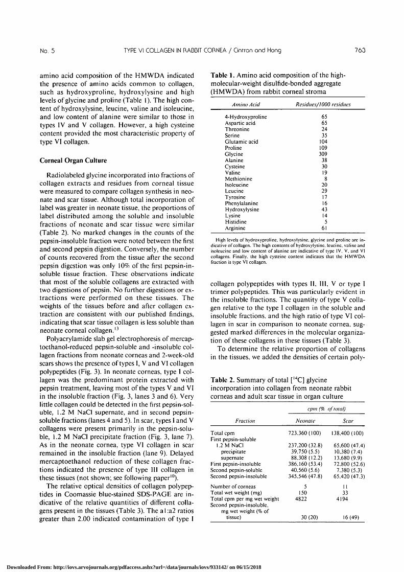
amino acid composition of the HMWDA indicatedthe presence of amino acids common to collagen,such as hydroxyproline, hydroxylysine and highlevels of glycine and proline (Table 1). The high con-tent of hydroxylysine, leucine, valine and isoleucine,and low content of alanine were similar to those intypes IV and V collagen. However, a high cysteinecontent provided the most characteristic property oftype VI collagen.
Corneal Organ Culture
Radiolabeled glycine incorporated into fractions ofcollagen extracts and residues from corneal tissuewere measured to compare collagen synthesis in neo-nate and scar tissue. Although total incorporation oflabel was greater in neonate tissue, the proportions oflabel distributed among the soluble and insolublefractions of neonate and scar tissue were similar(Table 2). No marked changes in the counts of thepepsin-insoluble fraction were noted between the firstand second pepsin digestion. Conversely, the numberof counts recovered from the tissue after the secondpepsin digestion was only 10% of the first pepsin-in-soluble tissue fraction. These observations indicatethat most of the soluble collagens are extracted withtwo digestions of pepsin. No further digestions or ex-tractions were performed on these tissues. Theweights of the tissues before and after collagen ex-traction are consistent with our published findings,indicating that scar tissue collagen is less soluble thanneonate corneal collagen.13
Polyacrylamide slab gel electrophoresis of mercap-toethanol-reduced pepsin-soluble and -insoluble col-lagen fractions from neonate corneas and 2-week-oldscars shows the presence of types I, V and VI collagenpolypeptides (Fig. 3). In neonate corneas, type I col-lagen was the predominant protein extracted withpepsin treatment, leaving most of the types V and VIin the insoluble fraction (Fig. 3, lanes 3 and 6). Verylittle collagen could be detected in the first pepsin-sol-uble, 1.2 M NaCl supernate, and in second pepsin-soluble fractions (lanes 4 and 5). In scar, types I and Vcollagens were present primarily in the pepsin-solu-ble, 1.2 M NaCl precipitate fraction (Fig. 3, lane 7).As in the neonate cornea, type VI collagen in scarremained in the insoluble fraction (lane 9). Delayedmercaptoethanol reduction of these collagen frac-tions indicated the presence of type III collagen inthese tissues (not shown; see following paper10).
The relative optical densities of collagen polypep-tides in Coomassie blue-stained SDS-PAGE are in-dicative of the relative quantities of different colla-gens present in the tissues (Table 3). The al:a2 ratiosgreater than 2.00 indicated contamination of type I
Table 1. Amino acid composition of the high-molecular-weight disulfide-bonded aggregate(HMWDA) from rabbit corneal stroma
Amino Acid Residues/1000 residues
4-HydroxyprolineAspartic acidThreonineSerineGlutamic acidProlineGlycineAlanineCysteineValineMethionineIsoleucineLeucineTyrosinePhenylalanineHydroxylysineLysineHistidineArginine
65652435
104109309
3830198
2029171643145
61
High levels of hydroxyproline, hydroxylysine. glycine and proline are in-dicative of collagen. The high contents of hydroxylysine. leucine. valine andisoleucine and low content of alanine are indicative of type IV. V. and VIcollagens. Finally, the high cysteine content indicates that the HMWDAfraction is type VI collagen.
collagen polypeptides with types II, III, V or type Itrimer polypeptides. This was particularly evident inthe insoluble fractions. The quantity of type V colla-gen relative to the type I collagen in the soluble andinsoluble fractions, and the high ratio of type VI col-lagen in scar in comparison to neonate cornea, sug-gested marked differences in the molecular organiza-tion of these collagens in these tissues (Table 3).
To determine the relative proportion of collagensin the tissues, we added the densities of certain poly-
Table 2. Summary of total [14C] glycineincorporation into collagen from neonate rabbitcorneas and adult scar tissue in organ culture
cpm (% of total)
Fraction Neonate Scar
Total cpmFirst pepsin-soluble
1.2 M NaClprecipitatesupernate
First pepsin-insolubleSecond pepsin-solubleSecond pepsin-insoluble
Number of corneasTotal wet weight (mg)Total cpm per mg wet weightSecond pepsin-insoluble,
mg wet weight (% oftissue)
723.360(100) 138.400(100)
237.200(32.8)39.750(5.5)88.308(12.2)
386.160(53.4)40.560(5.6)
345.546(47.8)
5150
4822
30 (20)
65.600 (47.4)10.380(7.4)13.680(9.9)72.800 (52.6)7.380(5.3)
65.420(47.3)
1133
4194
16(49)
Downloaded From: http://iovs.arvojournals.org/pdfaccess.ashx?url=/data/journals/iovs/933142/ on 06/15/2018

764 INVESTIGATIVE OPHTHALMOLOGY 6 VISUAL SCIENCE / Moy 1988 Vol. 29
1 2 3 4 5 6 7 8 9
• * • •
•
Fig. 3. Type VI collagen from 3-day-old rabbit neonate corneasand 2-wcek-old scars in adult corneas. Pepsin-digested, formicacid-extracted collagens were precipitated with 1.2 M NaCl, re-duced with mcrcaptoethanol, and subjected to SDS-6% polyacryl-amide slab gel electrophoresis. Proteins were stained with Coomas-sie blue. Lane I, type V collagen standard. Lane 2, type VI collagenstandard. Lanes 3-6, neonate fractions. Lane 3, first pepsin-solu-ble, 1.2 M NaCl precipitated fraction. Lane 4, first pepsin-soluble,1.2 M NaCl supernate fraction. Lane 5, second pepsin-solublefraction. Lane 6, pepsin-insoluble fraction. Lanes 7-9, scar frac-tions. Lane 7, first pepsin-soluble, 1.2 M NaCI-precipitated frac-tion. Lane 8, second pepsin-soluble fraction. Lane 9, pepsin-insolu-ble fraction. Dotted lines denote positions of SCI, SC2 and SC3.These proteins are present only in the pepsin-insoluble fractionsfrom neonate and scar tissue. The low-molecular-weight doubleband in lane 5 is pepsin, a-e: Migration of: a. aJV], b. a2[I], c. SCI,d. SC2, and e. SC3.
peptide bands of each collagen type from each frac-tion. For type I collagen we added a2[l], and 1/4 Beta(contributions by Gamma bands were insignificant).For Type V collagen, al[V] polypeptide was mea-sured. For type VI, we added SCI, 2, and 3 polypep-tides. The results indicate that the relative amount oftype VI collagen to type I collagen in the insolublefraction from scar tissue is three times greater thanthat in neonate corneas. However, the ratio of type VIto total type I collagen in scar and neonate tissues is0.131 and 0.372 respectively.
The relative densities of silver deposits on autora-diographs of SDS PAGE slabs containing radiola-beled collagen polypeptides are suggestive of the rela-tive rates of collagen synthesis, the processing of thevarious collagens, or both in neonate and scar tissue(Table 4). As in the Coomassie blue-stained gel, thehigh ratio of a I :a2 in scar tissue indicated synthesis ofother types of collagen such as types II, III and Itrimer, or contamination with a2(V) polypeptide.The ratio of al(V):a2(I) pepsin-soluble collagen poly-peptides from scar is markedly different from that inneonate corneas. In contrast to deposit ion nomarked difference was found in the incorporation oflabel into type VI collagen in the insoluble collagenfractions (Tables 3, 4). However, the ratio of the auto-radiographic band densities of type VI polypeptidesto the sum of the band densities of a2(I) from solubleand insoluble fractions of scar and neonate tissues are0.376 and 0.606, respectively.
DiscussionOur studies confirm previous observations show-
ing that rabbit corneal collagen is readily solubilizedfrom normal tissue after pepsin digestion in compari-son to scar tissue.1329 Moreover, the results are con-sistent with the presence of marked amounts of type Itrimer in scar tissue.29 That our corneal organ culturemethod maintains the pattern of collagen synthesispreviously shown by in vivo labeling is evidenced bythe higher proportion of type V collagen synthesizedin scar tissue than in normal neonate corneas regard-less of labeling method.13 Radiolabeling of collagenduring organ culture has made it possible to see evi-dence of type VI and, as we show in the followingpaper,10 type III collagen deposition in corneal tis-sues, which has not previously been detected by the invivo labeling method.
Our results show that the incorporation of [I4C]glycine into collagen fractions from neonate corneas
Table 3. Semiquantitative analysis of collagenpresent in neonate cornea and corneal scar*
Tissues andfractions
Corneal scarPepsin-soluble}:Pepsin-insoluble
NeonatePepsin-solublePepsin-insoluble
Collagen polypeptide ratios
a 1(0
a2(0
2.543.35
2.033.62
al(V)
a2(I)
0.701.00
0.253.60
K/f
a2(i)
6.35
1.69
* Based on the relative band densities in Coomassie blue-stained SDS-PAGE of collagen polypeptides from 3-day neonate corneas and 2-week-oldcorneal scar tissue. Type I collagen polypeptides are calculated from a 1(1),a2(I). and 1/4/3(1).
t The sum of SCI, SC2 and SC3 polypeptides of type VI collagen.$ Pepsin-soluble, NaCl-precipitable collagen.
Downloaded From: http://iovs.arvojournals.org/pdfaccess.ashx?url=/data/journals/iovs/933142/ on 06/15/2018

No. 5 TYPE VI COLLAGEN IN RADDIT CORNEA / Cinrron ond Hong 765
and scar tissue is about the same when expressed ascpm per mg wet weight of tissue. The tissues in theseexperiments, however, contain large quantities ofwater and extracellular matrix. A more meaningfulway of expressing the data is to relate them to thenumber of cells. The DNA values from similar tissuesin previous studies in our laboratory were 50 and 19ixg DNA for the neonate and scar tissues, respec-tively.2730 Our calculations show that the incorpora-tion of label per ng DNA into collagen fractions fromscar tissue is about half of that from neonate corneas,suggesting that the rate of collagen synthesis is mark-edly lower in the scar.
Previous studies from our laboratory have indi-cated many morphological and biochemical similar-ities between healing adult cornea and the normaldevelopment of fetal and neonate cornea.2730 If weconsider a 2-week-old corneal scar as temporallyequivalent to the first 14 days of fetal corneal devel-opment (27 days of gestation), scar tissue has depos-ited a greater amount of collagen per cell than has the27-day fetal cornea.30 It is not until the first week ofbirth that the neonate cornea accumulates collagen ata rate comparable to the 2-week-old scar. These neo-nate corneal cells are morphologically well differen-tiated into keratocytes, albeit they contain enormousquantities of rough endoplasmic reticulum. Thus, thedeposition of collagen in healing wounds is high dur-ing early stages of healing, whereas normal develop-ing cornea accumulates collagen at a high rate onlyafter cell morphological differentiation is almostcomplete. Since the present data suggest that scar tis-sue synthesizes collagen at a lower rate than that ofthe neonate cornea, assuming the specific activity ofthe precursor pools were the same and incorporationswere constant during incubation, the similarity in ac-cumulation of collagen in these tissues must be due todifferences in the turnover of this macromolecule.Because the neonate cornea continuously remodelsits extracellular matrix to accommodate growth, thecollagen turnover is very high.31-32 The scar tissue, onthe other hand, synthesizes collagen at a slower rateand remodels the scar gradually,13'3334 indicating itscollagen turnover is also gradual. Although our hy-pothesis is consistent with the results of this and pre-vious studies, future studies are needed to test colla-gen turnover in these tissues more directly.
Type VI collagen has been extracted from numer-ous tissues and subjected to several analyses.1635"38
Among the prominent features of this collagen are itsabundance of interchain disulfide cross-links, whichmay be responsible for its resistance to bacterial col-lagenase36-39; its unique polymeric structure withinthe extracellular matrix, identifying it with microfi-brillar structures171837; its unusual carbohydratecomposition of mannose and fucose residues39; and
Table 4. Semiquantitative analysis of collagensynthesized in organ culture of neonatecornea and corneal scar*
Collagen polypeptide ratio
Tissues andfractions
Corneal scarPepsin-soluble^:Pepsin-insoluble
NeonatePepsin-solublePepsin-insoluble
al(I)
a2(l)
2.522.70
2.141.98
al(V)
a2(I)
0.761.03
0.251.18
Wa2(I)
—1.43
—1.35
* Based on the relative band densities in SDS-PAGE fluorographs of col-lagen polypeptides from 3-day neonate corneas and 2-week-old corneal scartissue. Type I collagen polypeptides are calculated from a 1(1) and a2(l) only.
t The sum of SCI, SC2 and SC3 polypeptides of type VI collagen.% Pepsin-soluble, NaCI-precipitable collagen.
its large terminal nonhelical portions of the mole-cules in the completed structure, constituting over50% of the molecular weight.40 Although structuraldetails of type VI collagen are still not clear, chemical,ultrastructural and analytical ultracentrifugationstudies have suggested that the monomer of type VIcollagen, 110-145 Kd, is a 105-nm-long triple helixterminated by globular domains on each end. Mono-mers associate to form dimers with a lateral 30 nmstagger and align themselves in an antiparallel fash-ion. The outer segments of two parallel dimers arecovalently linked to form tetramers. Finally, tetra-mers are assembled end-to-end with overlap betweenouter segments to form a microfibrillar struc-ture.373841
Although indirect immunofluorescence has dem-onstrated the presence of type VI collagen in manytissues and cell cultures,l9-36-41-42 cartilage, basementmembranes, elastin and cross-striated collagen fibrilsdo not appear to contain this collagen.16 The presentresults adds to the list of tissues containing type VIcollagen and suggest that this collagen is deposited ata slower rate in corneal scars than in normal neonatetissue.
Current concepts of corneal transparency requirethat small distances between the collagen fibrils bemaintained.43 If indeed type VI occupies the spacesbetween collagen fibrils,171844 is it possible that theopacity of the scar is partially maintained by thespace-filling type VI collagen? Clearly, further studiesare required to determine the ultrastructural distri-bution of type VI collagen within scar tissue.
Key words: cornea, collagen, wound healing, development,type VI collagen
References1. Schmut O, Reich ME, and Zirm M: Der Nachweis verschie-
dener Kollagentypen im Rinderauge. Graefes Arch Clin ExpOphthalmol 196:71, 1975.
Downloaded From: http://iovs.arvojournals.org/pdfaccess.ashx?url=/data/journals/iovs/933142/ on 06/15/2018

766 INVESTIGATIVE OPHTHALMOLOGY G VISUAL SCIENCE / Moy 1988 Vol. 29
2. Freeman IL: Collagen polymorphism in mature rabbit cornea.Invest Ophthalmol Vis Sci 17:171. 1978.
3. Davison PF, Hong B-S, and Cannon DJ: Quantitative analysisof the collagens in the bovine cornea. Exp Eye Res 29:97, 1979.
4. Newsome DA, Gross J, and Hassell JR: Human cornealstroma contains three distinct collagens. Invest OphthalmolVis Sci 22:376. 1982.
5. Hamisch J-P, Buchen R, Sinha PK. and Barrach HJ: Ultra-structural identification of type I and II collagen in the corneaof the mouse by means of enzyme labeled antibodies. GraefesArch Gin Exp Ophthalmol 208:9. 1978.
6. Schmut O: The identification of type III collagen in calf andbovine cornea and sclera. Exp Eye Res 25:505, 1977.
7. Praus R, Brettschneider I, and Adam M: Heterogeneity of thebovine corneal collagen. Exp Eye Res 29:469, 1979.
8. Welsh C. Gay S. Rhodes RK. Pfister R. and Miller EJ: Colla-gen heterogeneity in normal rabbit cornea: I. Isolation andbiochemical characterization of the genetically distinct colla-gens. Biochim Biophys Acta 625:78. 1980.
9. Ben-Zvi A, Rodriques MM, Krachmer JH, and Fujikawa LS:Immunohistochemical characterization of extracellular matrixin the developing human cornea. Curr Eye Res 5:105. 1986.
10. Cintron C, Hong B-S. Covington HI, and Macarak J: Hetero-geneity of collagen in rabbit cornea: Type HI collagen. InvestOphthalmol Vis Sci 29:000, 1988.
11. Kefalides NA and Denduchis B: Structural components ofepithelial and endothelial basement membranes. Biochemistry8:4613, 1971.
12. Tsuchiya S, Tanaka M. Konomi H, and Hayashi T: Distribu-tion of specific collagen types and fibronectin in normal andkeratoconus corneas. Jpn J Ophthalmol 30:14. 1986.
13. Cintron C, Hong B-S, and Kublin CL: Quantitative analysis ofcollagen from normal developing corneas and corneal scars.Curr Eye Res 1:1. 1981.
14. Lee RE and Davison PF: The collagens of the developing bo-vine cornea. Exp Eye Res 39:639, 1984.
15. Birk DE, Fitch JM, and Linsenmayer TF: Organization ofcollagen types I and V in the embryonic chicken cornea. InvestOphthalmol Vis Sci 27:1470, 1986.
16. Von der Mark H, Aumailley M, Wick G, Fleischmajer R, andTimpl R: Immunochemistry, genuine size and tissue localiza-tion of collagen VI. Eur J Biochem 142:493, 1984.
17. Bruns RR: Beaded filaments and long-spacing fibrils: Relationto type VI collagen. J Ultrastruct Res 89:136, 1984.
18. Bruns RR, Press W, Engvall E, Timpl R, and Gross J: Type VIcollagen in extracellular, 100-nm periodic filaments and fibrils:Identification by immunoelectron microscopy. J Cell Biol103:393, 1986.
19. Zimmerman DR, Trueb B, Winterhalter KH, Witmer R, andFischer RW: Type VI collagen is a major component of thehuman cornea. FEBS Lett 197:55, 1986.
20. Sakai LY, Keene DR, Morris NP, and Burgeson RE: Type VIIcollagen is a major structural component of anchoring fibrils. JCell Biol 103:1577, 1986.
21. Gipson IK, Spurr-Michaud SJ, and Tisdale AS: Anchoringfibrils form a complex network in human and rabbit cornea.Invest Ophthalmol Vis Sci 28:212, 1987.
22. Sage H and Bornstein P: A unique, pepsin-sensitive collagensynthesized by aortic endothelial cells in culture. Biochemistry19:5747, 1980.
23. Benya PD and Padilla SR: Isolation and characterization oftype VIII collagen synthesized by cultured rabbit corneal endo-thelial cells. J Biol Chem 261:4160, 1986.
24. Kapoor R, Bornstein P, and Sage H: Type VIII collagen from
bovine Descemet's membrane: Structural characterization of atriple-helical domain. Biochemistry 25:3930, 1986.
25. Cintron C, Covington H, Damle S, and Gregory J: Mainte-nance of rabbit corneas in organ culture. ARVO Abstracts.Invest Ophthalmol Vis Sci 26 (Suppl):178, 1985.
26. Laemmli UK: Cleavage of structural proteins during the as-sembly of the head of bacteriophage T4. Nature (Lond)227:680. 1970.
27. Cintron C and Kublin CL: Regeneration of corneal tissue. DevBiol 61:346, 1977.
28. Nakazawa K, Hassell JR, Hascall VC, and Newsome KA: Het-erogeneity of proteoglycans in monkey corneal stroma. ArchBiochem Biophys 222:105, 1983.
29. Freeman IL, Shahinian L, and Brown SI: Presence of collagentype I trimer in avascular corneal scars. J Cell Biol 79:155a,1978.
30. Cintron C, Covington H, and Kublin CL: Morphogenesis ofrabbit corneal stroma. Invest Ophthalmol Vis Sci 24:543,1983.
31. Lee RE and Davison PF: Collagen composition and turnoverin ocular tissues of the rabbit. Exp Eye Res 323:739, 1981.
32. Davison PFandGalbavy EJ: Fluorescent dyes demonstrate theuniform expansion of the growing rabbit cornea. Invest Oph-thalmol Vis Sci 26:1202, 1985.
33. Cintron C, Hassinger LC, Kublin CL, and Cannon DJ: Bio-chemical and ultrastructural changes in collagen during cor-neal wound healing. J Ultrastruct Res 65:13, 1978.
34. Cintron C, Szamier RB, Hassinger LC. and Kublin CL: Scan-ning electron microscopy of rabbit corneal scars. Invest Oph-thalmol Vis Sci 23:50, 1982.
35. Furuto DK, and Miller EJ: Isolation of a unique collagenousfraction from limited pepsin digests of human placental tissue:Characterization of one of the constituent polypeptide chains.J Biol Chem 255:290, 1980.
36. Odermatt E, Risteli J, van Deldcn V. and Timpl R: Structuraldiversity and domain composition of a unique collagenousfragment (intima collagen) obtained from human placenta.Biochem J 211:295, 1983.
37. Furthmayr H, Widemann H, Timpl R, Odcramtt E, and EngelJ: Electron-microscopical approach to a structural model ofintima collagen. Biochem J 21 1:303, 1983.
38. Engvall E, Hessle H, and Klier G: Molecular assembly, secre-tion, and matrix deposition of type VI collagen. J Cell Biol102:703, 1986.
39. Heller-Harrison RA and Carter WG: Pepsin-generated type VIcollagen is a degradation product of GP140. J Biol Chem259:6858, 1984.
40. Aumailley M, von der Mark H, and Timpl R: Size and domainstructure of collagen VI produced by cultured fibroblasts.FEBS Lett 182:499, 1985.
41. Linsenmayer TF, Mentzer A, Irwin MH, Waldrep K, andMayne R: Avian type VI collagen: Monoclonal antibody pro-duction and immunohistochemical identification as a majorconnective tissue component of cornea and skeletal muscle.Exp Cell Res 165:518, 1986.
42. Hessle H and Engvall E: Type VI collagen. Studies on its local-ization, structure, and biosynthctic form with monoclonal an-tibodies. J Biol Chem 259:3955, 1984.
43. Benedek GB: Theory of transparency of the eye. AppliedOptics 10:459, 1971.
44. Linsenmayer TF, Bruns RR, Mentzer A, and Mayne R: TypeVI collagen: Immunohistochemical identification as a filamen-tous component of the extracellular matrix of the developingavian corneal stroma. Dev Biol 1 18:425, 1986.
Downloaded From: http://iovs.arvojournals.org/pdfaccess.ashx?url=/data/journals/iovs/933142/ on 06/15/2018