heterogeneity and dynamics in the assembly of the … and dynamics in the assembly of the heat shock...
TRANSCRIPT

Heterogeneity and dynamics in the assembly of theHeat Shock Protein 90 chaperone complexesIma-obong Ebonga,1, Nina Morgnera,1, Min Zhoua, Marco A. Saraivab, Soumya Daturpallib,Sophie E. Jacksonb,2, and Carol V. Robinsona,2
aDepartment of Chemistry, University of Oxford, South Parks Road, Oxford OX1 3TA, United Kingdom; and bDepartment of Chemistry, University ofCambridge, Lensfield Road, Cambridge CB2 1EW, United Kingdom
Edited by F. Ulrich Hartl, Max Planck Institute of Biochemistry, Martinsried, Germany, and approved September 12, 2011 (received for review April 19, 2011)
The Hsp90 cycle depends on the coordinated activity of a rangeof cochaperones, including Hop, Hsp70 and peptidyl-prolyl iso-merases such as FKBP52. Using mass spectrometry, we investigatethe order of addition of these cochaperones and their effects on thestoichiometry and composition of the resulting Hsp90-containingcomplexes. Our results show that monomeric Hop binds specificallyto the Hsp90 dimer whereas FKBP52 binds to both monomeric anddimeric forms of Hsp90. By preforming Hsp90 complexes with eitherHop, followed by addition of FKBP52, or with FKBP52 and subse-quent addition of Hop, we monitor the formation of a predominantasymmetric ternary complex containing both cochaperones. Thisasymmetric complex is subsequently able to interact with thechaperone Hsp70 to form quaternary complexes containing allfour proteins. Monitoring the population of these complexes duringtheir formation and at equilibrium allows us to model the complexformation and to extract 14 different KD values. This simultaneouscalculation of the KDs from a complex system with the same meth-od, from eight deferent datasets under the same buffer conditionsdelivers a self-consistent set of values. In this case, the KD valuesafford insights into the assembly of ten Hsp90-containing complexesand provide a rationale for the cellular heterogeneity and preva-lence of intermediates in the Hsp90 chaperone cycle.
dissociation constants ∣ interaction network
Hsp90 selectively interacts with and regulates the activation ofmany client proteins involved in cellular signalling pathways
such as kinases and steroid hormone receptors (1–3). The activa-tion of Hsp90 clients relies on interaction with its cochaperoneswhich facilitate different stages of the cycle (4–6). In human cells,Hsp90 cochaperones include Hsp70, Hop, Aha1, Cdc37, p23, andpeptidyl-propyl isomerases (PPIases) such as FKBP51, FKBP52,and CyP40. The exact complexes that are formed between Hsp90and cochaperones depend on the client protein. Cdc37 appears tobe specific towards kinase clients whilst p23 is associated with thebinding and activation of steroid hormone receptors. Despite awealth of research, the exact complexes formed during the cycleare not well established and their stoichiometry remains contro-versial (6, 7).
Hsp90 is a homodimer containing three structural domains.The N-terminal domain contains the ATP-binding site and bindscochaperones including p23. The middle domain is suggested asthe client protein binding region. The C-terminal domain encom-passes the dimerization interface and also contains a C-terminalMEEVD motif which binds tetratricopeptide repeat (TPR)-containing cochaperones (8). The ATPase activity of Hsp90 isessential for its function and ATP hydrolysis is linked with a con-formational rearrangement in which the homodimer changesfrom an open conformation to a closed state in which the twoN-terminal domains interact (5, 6, 9–12). A general model wasproposed for the activation of steroid hormone receptors (SHR)by the Hsp90 chaperone machinery (13, 14) in which the SHRbinds to the Hsp40/Hsp70 chaperones and is then transferred toHsp90 through the action of Hop which binds simultaneouslyto Hsp70 and Hsp90 through its different TPR domains (15).
Subsequent binding of PPIases such as FKBP52 and cochaperonep23 leads to a mature complex in which the client protein is cap-able of being activated by binding of ligands and cofactors (13).
The Hsp90 dimer has two C-terminal MEEVD motifs whichare the primary binding sites for TPR-containing cochaperonessuch as Hop and FKBP52 (16, 17). Hop has been establishedas a crucial component of the Hsp90 cycle forming a link thatfacilitates client protein transfer from Hsp70 to Hsp90 (15, 18).Hop binding is also known to inhibit the ATPase activity of Hsp90by stabilizing the open conformation (6, 19). When bound to theHsp90 dimer however there is conflicting evidence showing Hopbinds as a dimer (6, 7, 20, 21) or a monomer (22). Somemoleculardetails of how the TPR domains in FKBP52 and Hop bind tothe C-terminal MEEVD motifs of Hsp90 are known from crystalstructures of isolated TPR domains in complex with shortMEEVD-containing peptides (17). However, many details oftheir binding in the context of the full-length Hsp90 and cochaper-ones remain poorly understood. In particular, as Hsp90 is a homo-dimer containing two MEEVD motifs, little is known about theternary complexes that might form with the different TPR cocha-perones. There is a general view that Hop/FKBP52 may competefor available binding sites in the Hsp90 dimer (13, 16, 23, 24).The lack of reliable dissociation constants means that it is oftendifficult to rationalize competitive binding and to predict the com-plexes that form in the cellular environment.
Mass spectrometry (MS) is making significant inroads as anadjunct to established structural biology tools by defining thestoichiometry and interactions of protein complexes (25). Parti-cularly significant have been recent applications of nanoflowelectrospray to polydisperse molecular chaperone-substrate com-plexes wherein a snapshot of all complexes present at a given timecan be obtained within a single mass spectrum (26). This MSapproach should therefore enable investigation of the dynamicsof a protein system complicated by the formation of multiplespecies in solution. By correlating the intensity of the MS peaks,assigned to proteins and their complexes, with their concentra-tion in solution we show here that it is possible to calculate KDvalues for coexisting species. In this study, we investigate the het-erogeneity and dynamics of the Hsp90 complexes formed in thepresence of Hop, FKBP52, and Hsp70. Although exemplifiedwith part of the Hsp90 reaction cycle, the approach detailed hereis applicable to other protein systems and is particularly suited
Author contributions: M.A.S., S.E.J., and C.V.R. designed research; I.E., N.M., and M.Z.performed research; M.A.S. and S.D. contributed new reagents/analytic tools; I.E. andN.M. analyzed data; and I.E., N.M., M.Z., S.E.J., and C.V.R. wrote the paper.
The authors declare no conflict of interest.
This article is a PNAS Direct Submission.
Freely available online through the PNAS open access option.1I.E. and N.M. contributed equally to this work.2To whom correspondence may be addressed. E-mail: [email protected] or [email protected].
This article contains supporting information online at www.pnas.org/lookup/suppl/doi:10.1073/pnas.1106261108/-/DCSupplemental.
www.pnas.org/cgi/doi/10.1073/pnas.1106261108 PNAS ∣ November 1, 2011 ∣ vol. 108 ∣ no. 44 ∣ 17939–17944
BIOCH
EMISTR
Y
to assemblies that are complicated by heterogeneity and polydis-persity.
By manipulating the ratio of Hsp90 and Hop, we find that thepredominant complex is the ðHsp90Þ2ðHopÞ1 although up to twomolecules of Hop can bind in excess of Hop. By preforming com-plexes of the type ðHsp90Þ2ðHopÞn¼1 or 2 and adding FKBP52, weobserve how FKBP52 competes with Hop for available Hsp90binding sites to form the asymmetric ternary complex ðHsp90Þ2ðHopÞ1ðFKBP52Þ1. Similarly, this complex can be assembled byaddition of Hop to a preformed ðHsp90Þ2ðFKBP52Þn complex.By monitoring the formation of these complexes in real time, wecalculate KD values for all ten complexes using eight differentdatasets. Additionally, in the presence of Hsp70 we identify com-plexes that are competent to bind Hsp70. Overall by monitoringthese heterogeneous chaperone assemblies we reveal a self-consis-tent set of KD values which together with their established cellularconcentrations, allows us to predict the multiple Hsp90 complexesexisting in a cellular environment.
ResultsHsp90 Binds Hop with a 2∶1 or 2∶2 Stoichiometry. We first recordedmass spectra of all four proteins individually (Hsp90, Hsp70,Hop, and FKBP52) under nondenaturing MS conditions (27).Hsp90 revealed well resolved charge state series correspondingto monomeric and predominantly dimeric Hsp90, in line withprevious MS studies (28, 29) (SI Appendix, Fig. S1A). The MS ofHop revealed a mass corresponding to a monomer and no higheroligomers (SI Appendix, Fig. S1B). For Hop, FKBP52, and Hsp70measured masses corresponded in each case to a monomer andno higher oligomers were detected (SI Appendix, Fig. S1 B–D).All calculated and experimental masses, with the exception ofHsp90, confirmed these proteins are monomeric under the con-ditions used here (SI Appendix, Table S1).
To determine the binding stoichiometry of Hop to Hsp90,we varied the concentration of both components. First the Hopconcentration was kept at 1 μM and the concentration of theHsp90 dimer ðHsp90Þ2 varied from 1 to 5 μM. At equimolarconcentrations of Hop and ðHsp90Þ2, the dominant complexwas ðHsp90Þ2ðHopÞ1 as well as Hsp90 monomer ðHsp90Þ1 andunbound Hsp90 dimer ðHsp90Þ2 (SI Appendix, Fig. S1E). No freeHop was seen, implying that all Hop molecules were bound inthe complex. Higher Hsp90 concentrations triggered no changesin stoichiometry, the ðHsp90Þ2ðHopÞ1 remained the prevalent andonly complex formed. The reverse experiment was also performedusing a constant ðHsp90Þ2 concentration of 1 μM and increasingthe Hop concentration from 1 to 5 μM. We found that with excessHop (2 μM) an additional complex of ðHsp90Þ2ðHopÞ2 formed;concomitantly the concentration of free Hsp90 was depleted (SIAppendix, Fig. S1F). A further increase in Hop concentration re-sulted in the ðHsp90Þ2ðHopÞ2 complex becoming more dominantthan the ðHsp90Þ2ðHopÞ1 complex. No other higher-order com-plexes were observed.
Because the Hsp90 dimer adopts an open conformation in thepresence of ADP and converts to a closed state when ATP isbound (5), we examined how nucleotides affected these complexes.Addition of 100 μM ADP to the Hop and Hsp90 complexesshowed no changes in the stoichiometry or ratios of the complexes.Given that the KDs for ATP and AMPpNp are reported to behigher than for ADP (30, 31), we also examined binding at highernucleotide concentration (300 μM). At this nucleotide concentra-tion and using a KD value of 148 μM (30) and an Hsp90 monomerconcentration of 2 μM we calculate that >65% of the Hsp90 bind-ing sites will be occupied. While the peaks are significantly broad-er, due to multiple nonspecific attachments of nucleotide, thereis no change in the ratio of the complexes formed (SI Appendix,Fig. S2 A–F). We conclude that while both Hsp90 subunits withinthe dimer can bind a Hop monomer, the stoichiometry and ratiosof the complexes formed is nucleotide independent.
Hsp90 Can Bind Simultaneously to FKBP52 and Hop. We establishedthe interactions of ðHsp90Þ2 with FKBP52, by incubating equimo-lar concentrations of ðHsp90Þ2 and FKBP52. The spectra recordedrevealed peaks that corresponded to the ðHsp90Þ2ðFKBP52Þ1 andðHsp90Þ2ðFKBP52Þ2 complexes (SI Appendix, Fig. S3A). We alsoobserved minor peaks for both FKBP52 and Hsp90 monomers andan unexpected complex of ðHsp90Þ1ðFKBP52Þ1. It is noteworthythat the analogous complex ðHsp90Þ1ðHopÞ1 was not observedfor Hop binding to Hsp90.
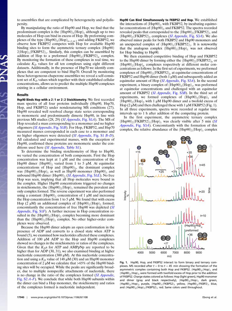
We then examined competitive binding of Hop and FKBP52to the Hsp90 dimer by forming either the ðHsp90Þ2ðFKBP52Þn orðHsp90Þ2ðHopÞn complexes respectively at different molar con-centrations as follows: In the first set of experiments, we preformedcomplexes of ðHsp90Þ2ðFKBP52Þn at equimolar concentrations ofFKBP52 andHsp90 dimer (both 1 μM) and subsequently added anequimolar amount of Hop (SI Appendix, Fig. S3A). In the secondexperiment, a binary complex of ðHsp90Þ2ðHopÞ1, was preformedat equimolar concentrations and challenged with an equimolaramount of FKBP52 (SI Appendix, Fig. S3B). In the third set ofexperiments, we formed complexes of ðHsp90Þ2ðHopÞ1 andðHsp90Þ2ðHopÞ2 with 1 μM Hsp90 dimer and a twofold excess ofHop (2 μM) and then challenged these with 1 μMFKBP52 (Fig. 1).In all three experiments, spectra were recorded at regular timeintervals up to 1 h after addition of the competing protein.
In the first experiment, the asymmetric ternary complexðHsp90Þ2ðFKBP52Þ1ðHopÞ1 was clearly visible after 5 min (SIAppendix, Fig. S3A). Concomitantly with the formation of thiscomplex, the relative abundance of the ðHsp90Þ2ðHopÞ1 complex
Fig. 1. Hsp90, Hop, and FKBP52 interact to form binary and ternary com-plexes. MS recorded after t ¼ 0 to t ¼ 45 min showing the formation of theasymmetric complex containing both Hop and FKBP52. ðHsp90Þ2ðHopÞ1 andðHsp90Þ2ðHopÞ2 were formed with twofold excess of Hop prior to the additionof FKBP52. Charge states colored as follows: Hop (light green), Hsp90monomerand dimer (gray and black respectively), ðHsp90Þ2ðHopÞ1 dark green,ðHsp90Þ2ðHopÞ2 purple, ðHsp90Þ1ðFKBP52Þ1 yellow, ðHsp90Þ2ðFKBP2Þ1 blue,and ðHsp90Þ2ðHopÞ1ðFKBP52Þ1 red. Same colors used throughout.
17940 ∣ www.pnas.org/cgi/doi/10.1073/pnas.1106261108 Ebong et al.

was also dominant compared to the ðHsp90Þ2ðFKBP52Þ1. In thesecond experiment, addition of equimolar FKBP52 to ðHsp90Þ2ðHopÞn complexes preformed at equimolar concentrations re-vealed peaks corresponding ðHsp90Þ2ðFKBP52Þn and the asym-metric ternary complex (SI Appendix, Fig. S3B). Again we observedthe ðHsp90Þ1ðFKBP52Þ1 complex. If we compare the abundanceof the Hsp90 dimer with only one binding partner, we find theðHsp90Þ2ðHopÞ1 is more prevalent than ðHsp90Þ2ðFKBP52Þ1complex throughout the time course of both experiments. In thethird experiment, addition of FKBP52 to complexes containingðHsp90Þ2ðHopÞ1 and ðHsp90Þ2ðHopÞ2 similarly leads to formationof the asymmetric ternary complex, as well as the same complexesobserved in the first two experiments. In this case, the ðHsp90Þ2ðHopÞ2 complex persists throughout the time course showingHop was not displaced, but rather FKBP52 binds on to the alreadyformed ðHsp90Þ2ðHopÞ1 complex (Fig. 1, SI Appendix, Fig. S4A).To test stability of these complexes formed in the third experiment,we also recorded spectra after 3 h (SI Appendix, Fig. S4B). Thesame complexes prevailed allowing us to conclude that the asym-metric complex forms readily from a variety of solution conditionsand is stable for at least 3 h. As a control, we also tested for aninteraction between FKBP52 and Hop alone. No binding wasobserved validating this control experiment and our observationof specific complexes for Hsp90 with Hop or Hsp90 with FKBP52.
To compare the proportion of complexes formed as a functionof the total protein components including individual proteins andcomplexes, we employed a deconvolution algorithm (Massign)(32, 33). An example is shown for a spectrum recorded 15 minafter ðHsp90Þ2ðHopÞn complexes were challenged with an equi-molar ratio of FKBP52 (Fig. 2A). After 15 min the asymmetricternary ðHsp90Þ2ðFKBP52Þ1ðHopÞ1 and ðHsp90Þ2ðFKBP52Þ1complexes represent 19% and 16% of the total population,respectively. Similarly, we calculated the relative abundance ofall other components and plotted their populations as a functionof time (Fig. 2B). We find the asymmetric ternary complex formsrapidly after addition of FKBP52. We also find that the homo-dimeric ðHsp90Þ2 becomes depleted over time implying that moreof the Hsp90 dimer is forming complexes with the TPR-contain-ing cochaperones. The concentration of the ðHsp90Þ2ðHopÞ1complex is also depleted over time while the ðHsp90Þ2ðHopÞ2 re-mains constant throughout implying that Hop is not displaced.The heterodimer complex ðHsp90Þ1ðFKBP52Þ1 forms while Hopis unable to form an ðHsp90Þ1ðHopÞ1 complex in the same fash-ion. Our data shows both TPR-containing proteins readily bindto an available site on the Hsp90 dimer, despite prior binding ofanother, to form the asymmetric ternary complex.
Hsp90-Hop Complexes Promote Subsequent Binding of Hsp70. Thepresence of multiple coexisting complexes prompts investigationof complexes that are competent to bind Hsp70. Initially, we in-vestigated binary interactions between Hsp70 and Hop or Hsp70and Hsp90. At equimolar or higher concentrations of Hop, nointeraction was seen between Hsp70 and Hop in the absence ofnucleotides. The presence of 100 μM ADP however inducedformation of a weak 1∶1 complex of the Hsp70∶Hop heterodimer(SI Appendix, Fig. S5A). Subsequently, we also examined whetherthere was any direct interaction between Hsp90 and Hsp70 but nobinding was observed (SI Appendix, Fig. S5B). In contrast, onaddition of Hsp90 to Hsp70 in the presence of equimolar amountof Hop, we observed a species with a measured mass correspond-ing to an intermediate complex containing ðHsp90Þ2ðHopÞ1ðHsp70Þ1 (Fig. 3A). Also present was a predominant complex ofðHsp90Þ2ðHopÞ1 andminor charge state series for Hsp70, ðHsp90Þ1,and ðHsp90Þ2. Other minor complexes containing ðHsp90Þ2ðHopÞ2ðHsp70Þ1 and ðHsp90Þ2ðHopÞ2ðHsp70Þ2 were also formed(SI Appendix, Fig. S6). As observed in the binary experiments,the presence of ADP revealed no changes in the ratios of thecomplexes formed (SI Appendix, Fig. S5 C and D). These data
show that Hsp70 binding to Hop is facilitated by formation of theðHsp90Þ2ðHopÞn complexes because Hop alone showed only aweak interaction with Hsp70 in the presence of ADP and wasunable to bind directly to Hsp70 in the absence of nucleotide.
To examine if FKBP52 could bind to the intermediate com-plex, we first formed complexes of ðHsp90Þ2ðHopÞnðHsp70Þn andsubsequently added FKBP52 (Fig. 3B). Results showed unambi-guously an additional complex that corresponds to ðFKBP52Þ1ðHsp90Þ2ðHopÞ1ðHsp70Þ1 (SI Appendix, Fig. S7). The observationof Hsp70-containing complexes of ðHsp90Þ2ðHopÞ2ðHsp70Þ1,ðHsp90Þ2ðHopÞ2ðHsp70Þ2, and ðFKBP52Þ1ðHsp90Þ2ðHopÞ1ðHsp70Þ1 allows us to conclude that the binding of Hsp70 isdirectly linked to the number of Hop subunits bound to ðHsp90Þ2.
Fig. 2. Monitoring the formation of Hsp90 complexes in real-time (A) MSrecorded 15 min after addition of an equimolar ratio of FKBP52 toðHsp90Þ2ðHopÞn (black) is compared with a simulated spectrum (blue) formedby summing the deconvoluted spectra of all components (SI Appendix,Fig. S3). The composition, relative intensity (%), and mass of each componentare given. (B) Kinetic model established from the relative intensity of allcomponents plotted against time (SI Appendix). The simulated kinetics(lines) are shown together with the experimental data (symbols) from 0 to40 min. The data with dashed lines is t ¼ 15 min.
Ebong et al. PNAS ∣ November 1, 2011 ∣ vol. 108 ∣ no. 44 ∣ 17941
BIOCH
EMISTR
Y

A Kinetic Model for Hsp90 Complexes. A model was constructed toshow all feasible reaction pathways (SI Appendix, Fig. S8) andhighlight all possible complexes observed in our experiments(Fig. 4). Starting with the Hsp90 dimer at micromolar concentra-tions (green oval) a population of monomeric Hsp90 is formedwhich is able to interact directly with FKBP52 but not Hsp70 orHop. Dimeric Hsp90 is able to bind either FKBP52 or Hop to formbinary complexes of ðHsp90Þ2ðFKBP52Þ1 or ðHsp90Þ2ðHopÞ1.Having formed binary complexes containing the Hsp90 dimerwith either FKBP52 or Hop both complexes can bind anothercopy of the same cochaperone to form ðHsp90Þ2ðFKBP52Þ2 orðHsp90Þ2ðHopÞ2 or bind the other cochaperone to form ðHsp90Þ2ðFKBP52Þ1ðHopÞ1. The latter is favored statistically given equalprotein concentrations of Hop and FKBP52. Our data shows amaximum of two copies of Hop or FKBP52 can bind per Hsp90dimer. Interestingly, FKBP52 cannot bind complexes containingtwo Hop monomers and Hsp70 cannot bind to any complex thatdoes not contain Hop and Hsp90.
To establish which complexes likely form under cellular condi-tions, we determined KD values using the intensity ratios of the10 different species and monitored their population as a functionof time (Fig. 2). We first established that the intensities of thepeaks assigned to each Hsp90-containing complex are relatedto their concentrations in solution using an internal standard overappropriate concentration ranges (SI Appendix, Fig. S9). Weassume that association and dissociation events follow second-and first-order kinetics respectively (SI Appendix). KD values arecalculated by satisfying equations relating to the concentration ofall components at every time point. We performed eight differentsets of experiments including different concentrations, differentorder of addition as well as replicate experiments (SI Appendix,Table S2). This data yields a series of KD values that are reprodu-cible within error between the different experimental conditionsas well as replicate experiments (Fig. 4). All KD values that deter-mine complex formations are in the range ∼10–250 nM.
These KD values reveal several interesting features. The bind-ing of the first Hop to a binding site on the free ðHsp90Þ2 dimer toform ðHsp90Þ2ðHopÞ1 has a KD of 15 nM, whilst binding of thesecond Hop to form ðHsp90Þ2ðHopÞ2 has a KD of 55 nM. Bindingof FKBP52 to the ðHsp90Þ2 to form either ðHsp90Þ2ðFKBP52Þ1or ðHsp90Þ2ðFKBP52Þ2 is slightly less favorable than for Hopwith corresponding KD values of 145 nM and 120 nM, respectively.
Comparing the value obtained for Hop binding to ðHsp90Þ2ðFKBP52Þ1 (KD ¼ 15 nM), with that obtained for Hop bindingto free ðHsp90Þ2 (KD ¼ 15 nM) we deduce that prior bindingof FKBP52 has no effect on subsequent binding of Hop to Hsp90.Likewise, adding FKBP52 to ðHsp90Þ2ðHopÞ1 or by addition ofFKBP52 directly to ðHsp90Þ2, yields similar KD values of 140 nMand 145 nM respectively. This data not only shows that the subse-quent binding of a TPR-containing cochaperone is independent ofprior binding but also confirms the reproducibility of our approach.The KD values obtained for Hsp70 binding to the three Hsp90∕Hop complexes yields low nanomolar values affirming our methodand implying a favorable binding event for Hsp70 (KD ¼ 20 nM)following the binding of Hop to Hsp90.
DiscussionThe functionality of the Hsp90 machinery is known to depend ona number of chaperones and cochaperones to drive the cycle. Theprecise order of binding and the stoichiometries of the variouscomplexes formed however have remained controversial. UsingMS we have explored all possible complexes that can be formedby four proteins known to have critical roles in the Hsp90 chaper-one cycle: Hsp90, Hsp70, Hop, and FKBP52. Initially, we foundthat Hsp90 was predominantly dimeric and FKBP52 monomeric,both were anticipated. Recently there has been some controversyover the oligomeric state of Hop reported as dimeric (6, 20, 34)ormonomeric (22, 35) andHsp70 can form high oligomeric speciesunder some conditions (36). We have shown that under the con-ditions used here both proteins are monomeric.
To characterize the complexes formed between the compo-nents of the Hsp90 machinery we incubated different ratios ofthe four proteins, varying both their concentration and order ofaddition. The results enabled us to construct a network of pos-sible complexes (Fig. 4). Despite the fact that all proteins are atmicromolar concentration in solution, Hsp90 exists in monomericand dimeric forms. Each Hsp90 monomer possesses a C-terminalMEEVD motif for TPR protein binding. We found that somecomplexes, which one might expect to form in theory, such as theðHsp90Þ1ðHopÞ1 complex in practice did not form. For the 10 dif-ferent species of Hsp90 and cochaperones complexes observed,we determined their binding stoichiometry as well as the likelyassembly pathway. Starting with Hsp90 dimers (green oval Fig. 4),up to two Hop monomers can bind to form ðHsp90Þ2ðHopÞ1 andðHsp90Þ2ðHopÞ2 complexes (Fig. 1 A and B). The absence of theðHsp90Þ1ðHopÞ1 complex was in contrast to the results obtainedfor FKBP52, wherein the complex ðHsp90Þ1ðFKBP52Þ1 wasobserved. Our observation implies that dimerization of Hsp90 iscritical for Hop but not for FKBP52 binding, consistent with the15
´Å resolution EM structure of the Hsp90-Hop complex in whicheach Hop monomer interacts with both subunits of ðHsp90Þ2 (37).
The binding affinities of FKBP52 and Hop for the two bindingsites in the Hsp90 dimer, as well as additional binding of Hsp70,are all in the nanomolar range. However, there is a wide varia-bility in the KD values reported for binary interactions betweenthese proteins, both for human and yeast proteins (5, 6, 38)(SI Appendix, Table S3 and Fig. S10). This variability could arisefrom possible differences between the protein homologues, thevaried solution conditions, and methods employed in their mea-surement. An apparent advantage of our method is that it gives aconsistent set of KD values for a system of complexes, using thesame method and solution conditions. Our data allows a directcomparison of the values determined for the different complexesformed in the cycle. Overall, our KD values are somewhat lowerthan those measured in an earlier study for C-terminal peptidesof Hsp90 binding to isolated TPR domains (39) but are howeververy similar to some published values, where available, for thefull-length human proteins (7, 40) (see SI Appendix, Table S3and Fig. S10). Our KD values of 15 nM and 55 nM for the bindingof the first and second Hop molecule are in accord with the
BA
Fig. 3. Hsp70 binds to ðHsp90Þ2 and Hop to form an intermediate complex.(A) MS of an intermediate complex ðHsp90Þ2ðHopÞ1ðHsp70Þ1 (pink) formedafter addition of Hsp70 following Hop binding to the ðHsp90Þ2. B) Additionof FKBP52 to the intermediate complex to form ðHsp90Þ2ðHopÞ1ðFKBP52Þ1ðHsp70Þ1 (olive green), (SI Appendix, Figs. S6 and S7 for additional quaternarycomplexes).
17942 ∣ www.pnas.org/cgi/doi/10.1073/pnas.1106261108 Ebong et al.

proposal that binding of the first Hop results in a subtle decreasein affinity of the Hsp90 dimer for the second Hop (37).
The KD values for the binding of FKBP52 and Hop to eithersite in the ðHsp90Þ2 are similar. We speculate that the slight pre-ference for Hop over FKBP52 binding may be due to the addi-tional interactions Hop makes with the C-terminal and middledomain of Hsp90, as previously established using ITC (20) andmore recently revealed by the Hsp90-Hop EM structure (37).Despite its slightly less favorable KD, FKBP52 is however able toform an asymmetric complex with Hop and Hsp90 (Fig. 1B). Thisfinding is in accord with the recent observation that the ternarycomplex is preferentially formed (22). The asymmetric complexobserved here forms regardless of the order of addition of eitherFKBP52 or Hop and is statistically favored and stable for at least3 h under these conditions.
It has been proposed that Hop binds directly to Hsp70 withADP present (7, 41). However, other studies have shown thatHop can bind to Hsp70 in the absence of nucleotide (42). Ourresults show evidence of a weak binding between Hsp70 andHop but only when ADP is present (KD ¼ 6 μM). Under the con-ditions used here, no binding was observed for Hsp70 and Hopin the absence of nucleotide. Favorable binding was howeverobserved after prior binding of Hop to the Hsp90 dimer (KD ¼20 nM). Our observation suggests a mechanism in which Hopand/or Hsp90 undergo a conformational change upon bindingwhich renders the complex competent to bind Hsp70, and agreeswith the recent EM structure where Hop binds a distinct con-formation of Hsp90 which lies somewhere between the open con-formation, observed for the apo state, and the N-terminallydimerized closed state (37). We find that the number of Hsp70molecules that bind to the various Hsp90 assemblies is directlyrelated to the number of Hop molecules within the complex.We also observe a complex with all four components ðFKBP52Þ1ðHsp90Þ2ðHopÞ1ðHsp70Þ1 proposed previously (22) and discov-ered experimentally here. Interestingly, our KD values show thatbinding of Hsp70 is not affected by prior binding of FKBP52,Hop, or Hop and Hsp70 to the other binding site. Consideringthe cellular concentrations recently reported as 10 μM for Hsp70,5 μM ðHsp90Þ2, and 3 μM for Hop (43), even though two Hopmonomers can bind to an Hsp90 dimer the ðHsp90Þ2ðHopÞ1 com-plex is likely the physiologically relevant species. It has also been
shown recently that a single Hop molecule is sufficient to com-pletely inhibit the ATPase activity of Hsp90 (22). The ðHsp90Þ2ðHopÞ1 complex in turn can bind to Hsp70 to form the inter-mediate complex, that binds FKBP52 to form the functionalchaperone complex (Fig. 4).
Given the ratios of ðHsp90Þ2 to Hop and FKBP52 (10∶1 and15∶1 respectively) in the cell (44), it is perhaps surprising that theasymmetric complex is able to form by addition of an FKBP52molecule. However, estimated concentrations do not take intoaccount elevated protein levels in response to the stress environ-ment, or the effects of crowding within the cell (45). The asym-metric complex is, however, statistically more likely to form thanthe ðHsp90Þ2ðHopÞ2 complex if concentrations of FKBP52 andHop are comparable. Our results do show however that no addi-tional FKBP52 molecule can bind to the ðHsp90Þ2ðHopÞ2 com-plex (Fig. 1), or those containing two Hop molecules with one ortwo Hsp70 molecules (SI Appendix, Fig. S7). Because FKBP52is always present as a final component of the mature receptorcomplex (14), our data implies that binding of two Hop moleculesrenders the complex incompetent of further development into afunctional chaperone complex. Our data is in contrast to earlierstudies in which ðHsp90Þ2 binding to dimeric Hop was consideredto be a prerequisite for chaperone function (4, 6, 7) but is con-sistent with the proposal that the asymmetric nature of Hsp90-Hop complexes are important in the Hsp90 cycle (22, 37).
The simultaneous binding of Hsp70 and Hsp90 to Hop hasbeen proposed to bring the two proteins together into an activecomplex in which client proteins are transferred from Hsp70to Hsp90 to complete their folding and maturation (15). Giventhe cellular concentration of Hsp70, which is higher than bothðHsp90Þ2 and Hop, the favorable KD for Hsp70 (20 nM) bindingwould ensure that the ðHsp90Þ2ðHopÞ1 complexes once formedcan proceed to productive and competent folding complexes bybinding FKBP52 to form ðHsp90Þ2ðHopÞ1ðFKBP52Þ1ðHsp70Þ1.By contrast the complexes of ðHsp90Þ2ðHopÞ2ðHsp70Þ1 andðHsp90Þ2ðHopÞ2ðHsp70Þ2, if formed, are incapable of subsequentFKBP52 binding and are therefore unlikely to play any furtherrole in the chaperone cycle.
In summary, by incubating Hsp90 with different cochaperones,and exploring all possible reaction products, we have determinedthe dominant complexes formed during the Hsp90 cycle. We have
Fig. 4. An interaction networkfor Hsp90, Hsp70, Hop, andFKBP52 KDS for Hop, ðHsp90Þ2and FKBP52 determined frommultiple spectra from eight differ-ent experimental datasets, includ-ing replicate experiments (greenbackground). Favorable KDs arerepresented with thicker arrows.Addition of Hsp70 to the ternaryðHsp90Þ2ðHopÞnðFKBP52Þ1 com-plexes leads to the formation ofquaternary complexes with favor-able KD values (blue background).Complexes likely to form, giventhe cellular concentrations of thefour protein components, arecircled (dotted line). KD values andassociated errors are shown withvalues deduced form the samedataset represented by the samesymbols.
Ebong et al. PNAS ∣ November 1, 2011 ∣ vol. 108 ∣ no. 44 ∣ 17943
BIOCH
EMISTR
Y
also identified some key binding events that likely lead to theassembly of productive folding machinery in the Hsp90 chaper-one cycle. The similarity of the KD values for the ten speciesstudied here ensure that in the cellular environment the chaper-one pathway will be populated with complexes that are bothheterogeneous and dynamic and which can likely be finely tunedto respond to the stress conditions experienced. More generally,our results highlight the distinct advantages of MS in being ableto determine simultaneously, and in real time, the compositionand relative abundance of each complex in a highly heteroge-neous system from many different datasets. This approach shouldhave broad application to other systems as it not only gives in-sights into the interactions, but also leads to a self-consistent setof KD values as shown here for the different complexes formed inthe Hsp90 chaperone cycle.
MethodsProtein Purification. Hsp90 and cochaperones were human, expressed inEscherichia coli and purified as previously described (20, 29). Prior to MSanalysis, 20 μL of the protein solutions were buffer exchanged into 50 mMammonium acetate (AmAc), pH 7.5 at 4 °C using micro Bio-Spin® columns(Bio-Rad Laboratories).
Binding Studies. Proteins were combined to give a range of concentrationsfrom 1 to 5 μM in 50 mM AmAc pH 7.5 and incubated for 30 min on ice.For Hsp90 and Hop binary complexes were formed in 50 mM AmAc pH 7.5.As a control all complexes were formed in a standard buffer 50 mM Tris HClpH 7.5. MS showed no difference in the ratios of the complexes formed.Competition experiments were performed by preforming binary complexesof Hsp90 with Hop or FKBP52 in 50 mM AmAc and adding FKBP52 or Hopimmediately prior to MS. For nucleotide studies, ADP, AMPpNp, and ATPwere prepared at 1 mM in 50 mM AmAc, pH 7.5 and added to give a finalsolution concentration of 100 μM and 300 μM nucleotide with an equivalentof MgðOAcÞ2.
MS. Spectra were obtained on a QToF II MS (Waters) modified for studyingnoncovalent interactions (27, 46). 2.5 μL aliquots were introduced into theMSfrom a gold-plated capillary needle (Harvard Apparatus). Capillary voltageswere 1.5–1.8 kV, cone voltages from 80–130 V, and a collision voltages werefrom 50–150 V. Spectra were calibrated externally using caesium iodide.
ACKNOWLEDGMENTS. We thank funding from the European Union Prospectsgrant number (HEALTH-F4-2008-201648) (to N.M.) and Engineering andPhysical Sciences Research Council for funding (to I.E.) Faculty of scienceand technology (FCT), Portugal for funding (to M.A.S.) and British PetroleumCentenary Murray Edwards College Fund and Cambridge CommonwealthTrust for funding (to S.D.). C.V.R. is a Royal Society Professor.
1. Pratt WB, Dittmar KD (1998) Studies with purified chaperones advance the under-standing of the mechanism of glucocorticoid receptor-hsp90 heterocomplex assembly.Trends Endocrin Met 9:244–252.
2. Richter K, Buchner J (2001) Hsp90: chaperoning signal transduction. J Cell Physiol188:281–290.
3. Picard D (2002) Heat-shock protein 90, a chaperone for folding and regulation. CellMol Life Sci 59:1640–1648.
4. Siligardi G, et al. (2002) Regulation of Hsp90 ATPase activity by the co-chaperoneCdc37p/p50(cdc97). J Biol Chem 277:20151–20159.
5. Siligardi G, et al. (2004) Co-chaperone regulation of conformational switching in theHsp90 ATPase cycle. J Biol Chem 279:51989–51998.
6. Prodromou C, et al. (1999) Regulation of Hsp90 ATPase activity by tetratricopeptiderepeat (TPR)-domain co-chaperones. EMBO J 18:754–762.
7. Hernandez MP, Sullivan WP, Toft DO (2002) The assembly and intermolecularproperties of the hsp70-Hop-hsp90 molecular chaperone complex. J Biol Chem 277:38294–38304.
8. Pearl LH, Prodromou C (2006) Structure and mechanism of the Hsp90 molecularchaperone machinery. Annu Rev Biochem 75:271–294.
9. Obermann WMJ, Sondermann H, Russo AA, Pavletich NP, Hartl FU (1998) In vivo func-tion of Hsp90 is dependent on ATP binding and ATP hydrolysis. J Cell Biol 143:901–910.
10. Grenert JP, Johnson BD, Toft DO (1999) The importance of ATP binding and hydrolysisby hsp90 in formation and function of protein heterocomplexes. J Biol Chem 274:17525–17533.
11. Krukenberg KA, Street TO, Lavery LA, Agard DA (2011) Conformational dynamics ofthe molecular chaperone Hsp90. Q Rev Biophys 1:1–27.
12. Southworth DR, Agard DA (2008) Species-dependent ensembles of conserved confor-mational states define the Hsp90 chaperone ATPase cycle. Mol Cell 32:631–640.
13. Owens-Grillo JK, et al. (1996) A model of protein targeting mediated by immunophi-lins and other proteins that bind to hsp90 via tetratricopeptide repeat domains. J BiolChem 271:13468–13475.
14. Pratt WB, Toft DO (1997) Steroid receptor interactions with heat shock protein andimmunophilin chaperones. Endocr Rev 18:306–360.
15. Chen SY, Smith DF (1998) Hop as an adaptor in the heat shock protein 70 (Hsp70) andHsp90 chaperone machinery. J Biol Chem 273:35194–35200.
16. D’Andrea LD, Regan L (2003) TPR proteins: the versatile helix. Trends Biochem Sci28:655–662.
17. Scheufler C, et al. (2000) Structure of TPR domain-peptide complexes: critical elementsin the assembly of the Hsp70-Hsp90 multichaperone machine. Cell 101:199–210.
18. Morishima Y, et al. (2000) The Hsp organizer protein Hop enhances the rate of butis not essential for glucocorticoid receptor folding by the multiprotein Hsp90-basedchaperone system. J Biol Chem 275:6894–6900.
19. Richter K, Muschler P, Hainzl O, Reinstein J, Buchner J (2003) Sti1 is a non-competitiveinhibitor of the Hsp90 ATPase—binding prevents the N-terminal dimerization reactionduring the ATPase cycle. J Biol Chem 278:10328–10333.
20. Onuoha SC, Coulstock ET, Grossmann JG, Jackson SE (2008) Structural studies on theco-chaperone Hop and its complexes with Hsp90. J Mol Biol 379:732–744.
21. Hildenbrand ZL, et al. (2011) Hsp90 can accommodate the simultaneous binding of theFKBP52 and Hop proteins. Oncotarget 2:45–58.
22. Li J, Richter K, Buchner J (2011) Mixed Hsp90—cochaperone complexes are importantfor the progression of the reaction cycle. Nat Struct Mol Biol 18:61–66.
23. Young JC, Obermann WMJ, Hartl FU (1998) Specific binding of tetratricopeptiderepeat proteins to the C-terminal 12-kDa domain of hsp90. J Biol Chem 273:18007–18010.
24. Radanyi C, Chambraud B, Baulieu EE (1994) The ability of the immunophilin FKBP59-HBI to interact with the 90-kDa heat shock protein is encoded by its tetratricopeptiderepeat domain. Proc Natl Acad Sci USA 91:11197–11201.
25. Sharon M, Robinson CV (2007) The role of mass spectrometry in structure elucidationof dynamic protein complexes. Annu Rev Biochem 76:167–193.
26. Stengel F, et al. (2010) Quaternary dynamics and plasticity underlie small heat shockprotein chaperone function. Proc Natl Acad Sci USA 107:2007–2012.
27. Hernandez H, Robinson CV (2007) Determining the stoichiometry and interactions ofmacromolecular assemblies from mass spectrometry. Nat Protoc 2:715–726.
28. Karagoz GE, et al. (2011) N-terminal domain of human Hsp90 triggers binding to thecochaperone p23. Proc Natl Acad Sci USA 108:580–585.
29. McLaughlin SH, Smith HW, Jackson SE (2002) Stimulation of the weak ATPase activityof human Hsp90 by a client protein. J Mol Biol 315:787–798.
30. McLaughlin SH, Ventouras LA, Lobbezoo B, Jackson SE (2004) Independent ATPase ac-tivity of Hsp90 subunits creates a flexible assembly platform. J Mol Biol 344:813–826.
31. Prodromou C, et al. (1997) Identification and structural characterization of the ATP/ADP-binding site in the Hsp90 molecular chaperone. Cell 90:65–75.
32. Hernandez H, et al. (2009) Isoforms of U1-70k control subunit dynamics in the humanspliceosomal U1 snRNP. PLoS One 4:e7202.
33. Lane LA, et al. (2011) Mass spectrometry reveals stable modules in holo and apo RNApolymerases I and III. Structure 19:90–100.
34. Flom G, Behal RH, Rosen L, Cole DG, Johnson JL (2007) Definition of the minimal frag-ments of Sti1 required for dimerization, interaction with Hsp70 and Hsp90 and in vivofunctions. Biochem J 404:159–167.
35. Yi F, Doudevski I, Regan L (2010) HOP is a monomer: investigation of the oligomericstate of the co-chaperone Hop. Protein Sci 19:19–25.
36. Benaroudj N, Batelier G, Triniolles F, Ladjimi MM (1995) Self-association of the mole-cular chaperone HSC70. Biochemistry 34:15282–15290.
37. Southworth DR, Agard DA (2011) Client-loading conformation of the Hsp90molecularchaperone revealed in the Cryo-EM structure of the human Hsp90∶Hop complex.Mol Cell 42:771–781.
38. Wandinger SK, Richter K, Buchner J (2008) The Hsp90 chaperone machinery. J BiolChem 283:18473–18477.
39. Cortajarena AL, Regan L (2006) Ligand binding by TPR domains. Protein Sci 15:1193–1198.
40. Pirkl F, Buchner J (2001) Functional analysis of the Hsp90-associated human peptidylprolyl Cis/Trans isomerases FKBP51, FKBP52 and cyp40. J Mol Biol 308:795–806.
41. Johnson BD, Schumacher RJ, Ross ED, Toft DO (1998) Hop modulates hsp70∕hsp90interactions in protein folding. J Biol Chem 273:3679–3686.
42. Wegele H, Haslbeck M, Reinstein J, Buchner J (2003) Sti1 is a novel activator of the Ssaproteins. J Biol Chem 278:25970–25976.
43. Kundrat L, Regan L (2010) Balance between folding and degradation for Hsp90-dependent client proteins: a key role for CHIP. Biochemistry 49:7428–7438.
44. Ghaemmaghami S, et al. (2003) Global analysis of protein expression in yeast. Nature425:737–741.
45. Ellis RJ, Minton AP (2006) Protein aggregation in crowded environments. Biol Chem387:485–497.
46. Sobott F, Hernández H, McCammon MG, Tito MA, Robinson CV (2002) A tandemmass spectrometer for improved transmission and analysis of large macromolecularassemblies. Anal Chem 74:1402–1407.
17944 ∣ www.pnas.org/cgi/doi/10.1073/pnas.1106261108 Ebong et al.