hereditary systemic immunoglobulin light-chain amyloidosis
TRANSCRIPT

Hereditary systemic immunoglobulin light-chain amyloidosis
Merrill D. Benson,1,2 Juris J. Liepnieks,1 Barbara Kluve-Beckerman1
1Indiana University School of Medicine, Department of Pathology and Laboratory Medicine,
2RLR Veterans Affairs Medical Center, Indianapolis, Indiana, USA
Running Title: Hereditary AL amyloidosis
Keywords: amyloid, amyloidosis, Ig constant region gene
Key Points:
• Protein and DNA analyses reveal that mutation in the immunoglobulin kappa light-chain
constant region gene may cause hereditary amyloidosis.
• Sequencing of Ig LC constant region genes is indicated for patients with AL amyloidosis
who have no evidence of a plasma cell dyscrasia.
Copyright © 2015 American Society of Hematology
_________________________________________________________________________________
This is the author's manuscript of the article published in final edited form as:
Benson, M. D., Liepnieks, J. J., & Kluve-Beckerman, B. (2015). Hereditary systemic immunoglobulin
light-chain amyloidosis. Blood, 125(21), 3281-3286. https://doi.org/10.1182/blood-2014-12-618108

2
Abstract
Several members of a family died with renal failure due to systemic amyloidosis. Extensive
studies to detect previously documented gene mutations associated with amyloidosis failed to
identify a causative factor. In search for the genetic basis for this syndrome amyloid fibrils were
isolated from renal tissue of a member of the kindred who died while on renal dialysis. Amino
acid sequencing of isolated amyloid protein identified sequences compatible with the constant
region of the immunoglobulin kappa light-chain. Isolation and characterization of kappa light-
chain protein from serum of an affected member of the kindred revealed mutation in the constant
region of kappa light-chain with cysteine replacing serine at amino acid residue 131. This
mutation (Ser131Cys) was confirmed by DNA analysis which identified a single base change of
cytosine to guanine at the second position of codon 131 of the kappa light-chain gene
(TCT131TGT). DNA analysis of members of the extended family revealed transmission of the
Ser131Cys mutation and association with systemic amyloidosis. This form of AL amyloidosis
which is a hereditary type of amyloidosis and not the result of a monoclonal plasma cell
dyscrasia may be misdiagnosed and lead to inappropriate chemotherapy.

3
Introduction
Immunoglobulin light-chain amyloidosis (AL) is the most frequent form of systemic
amyloidosis.1 It is a sporadic disease due to plasma cell dyscrasia with resulting amyloid fibril
formation from monoclonal immunoglobulin light-chain either kappa or lambda. There are,
however, a number of forms of systemic amyloidosis which are caused by mutations in other
plasma proteins and are expressed with autosomal dominant genetics.2 These inherited forms of
amyloidosis may show predilection for specific organ involvement such as the heart, peripheral
nervous system, or kidney, but are all systemic in nature and very often clinically mimic AL
amyloidosis. A number of the inherited forms of systemic amyloidosis cause renal
manifestations and, therefore, may be mistaken for AL amyloidosis which often presents with
major renal dysfunction.3
While plasma cell dyscrasia may be more prevalent in certain families, AL amyloidosis
has rarely been reported in first degree relatives and no definite inheritance pattern has been
identified. Here we report a systemic form of immunoglobulin light-chain amyloidosis which is
inherited as an autosomal dominant trait and is associated with a mutation in the constant region
of the kappa immunoglobulin light-chain.

4
Methods
The family
Six members (4 men and 2 women) of a family of eight died with renal failure between 53 and
74 years of age (Figure 1). Five had received renal dialysis. Four (II-3, II-8, II-10, II-12) had the
diagnosis of amyloidosis made by renal biopsy. Their mother (I-2) had died at age 58 with renal
failure.
In the next generation, one man (III-2) with biopsy proven renal amyloidosis died at age
74 after several years treatment with renal dialysis, and two cousins (III-12 and III-15) have
biopsy proven amyloidosis, one on renal biopsy (III-12) and one on gastric mucosal biopsy (III-
15, index case).
Review of medical histories revealed that one man (II-3) who died of renal failure also
had amyloid deposition on laryngeal and lung biopsies verifying that he had a systemic form of
amyloidosis. Medical records indicated that immunohistological analysis of kidney biopsies for
four individuals showed positive staining for kappa light-chain, and laser dissected mass
spectrometry analysis of amyloid identified on a gastric biopsy of a member of the third
generation (III-15) identified kappa light-chain constant region peptides.
Amyloid protein characterization
Amyloid fibrils were isolated from formalin-fixed, paraffin-embedded renal tissue of one
member of the family who died while on dialysis.4 Tissue was solubilized in 8 M guanidine-HCl
under reducing conditions, and solubilized protein subjected to amino acid sequencing on an
Applied Biosystems Procise 491 cLC protein sequencer using the manufacturer’s standard cycles

5
and methods. Isolated protein was also subjected to tryptic digestion and resultant peptides
separated by reverse phase HPLC and characterized by amino acid sequencing.5
Immunoglobulin light-chain isolation and characterization
Serum (10 ml) from a member of the kindred with biopsy proven amyloidosis was made 33%
saturated with ammonium sulfate at room temperature. The precipitated fraction recovered by
centrifugation was dissolved in PBS and chromatographed on a Sepharose CL6B column
equilibrated and eluted with PBS. The major peak, which on SDS-PAGE analysis in the
presence of 2-mercaptoethanol contained 25 kDa and 50 kDa bands (immunoglobulin light-chain
and heavy-chain), was dialyzed against distilled water and lyophilized. Dried protein was
solubilized in 8 M guanidine hydrochloride, 0.5 M Tris pH 8.2 containing 10 mg
dithiothreitol/ml, alkylated with iodoacetic acid, and centrifuged. Chromatography of the
supernatant on Sepharose CL6B equilibrated and eluted with 4 M guanidine hydrochloride, 25
mM Tris pH 8.2 yielded two major peaks corresponding to Ig heavy and Ig light-chains.
Fractions containing Ig light-chains were combined, exhaustively dialyzed against distilled water
and lyophilized. After tryptic digestion, peptides were separated by HPLC on a Beckman
Ultrasphere ODS column and isolated peptides subjected to amino acid sequencing on an
Applied Biosystems Procise 491 cLC protein sequencer using the manufacturer’s standard cycles
and methods.
Histochemistry
Tissue biopsy sections (4 micron) were stained with haematoxylin and eosin. Sections were also
stained with alkaline Congo red. Immunohistochemistry of biopsy tissues was performed by the
indirect immunoperoxidase technique using rabbit anti-kappa and anti-lambda polyclonal

6
antisera and immunoperoxidase labeled goat anti-rabbit IgG antibody. Sections were developed
with diaminobenzidine.
DNA analysis
DNA was isolated from EDTA-treated blood and formalin-fixed, paraffin-embedded tissue.6
PCR amplification of the constant region of the kappa light-chain gene used primers 5’
ACCATCCTGTTTGCTTCT 3’ and 5’CTCTGTGACACTCTCCTG 3’ with standard cycles 1
min 94°, 1 min 56°, 1 min 72° for 35 cycles. Sequencing was performed by a Beckman CEQ
System.

7
Results
Review of medical records for several members of a family who died with renal failure
documented that kidney biopsies demonstrated amyloid deposition in glomerular basement
membrane and blood vessel walls. In several cases positive staining for kappa light-chain was
described but evaluation for monoclonal gammopathy was negative (Table 1). In the present
study amyloid deposition in a renal biopsy of subject III-12 was identified by routine histology,
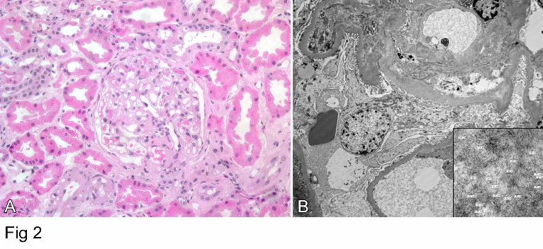
and electron microscopy demonstrated 7-10 micron fibrils consistent with amyloid (Figure 2).
Immunohistochemistry for kappa light-chain performed on cholecystectomy tissue from the
index case (III-15) gave positive staining of blood vessel walls which coincided with Congo red
staining (Figure 3).7
Amino acid sequence analysis of tryptic peptides of amyloid fibril protein isolated from
post-mortem formalin-fixed paraffin-embedded kidney tissue gave sequences consistent with
immunoglobulin kappa light-chain constant region, residues 109-120, 127-142, 150-163, 170-
179, and 191-205 (Figure 4). All numbering is according to the Kabat numbering system.8
Residue 131 which is normally a serine and residue 134 which is normally cysteine could not be
positively identified.
Immunoglobulin light-chain proteins were isolated from serum of an affected family
member and subjected to amino acid sequencing which identified the entire kappa constant
region except for amino acid residues 143-145, 172-183, and 208-214 (Figure 4). Of note was
identification of separate peptides containing either serine or cysteine at position 131. Position
134 had only the normal cysteine which is involved in the intramolecular disulfide bridge to the
cysteine at position 194.

8
Nucleotide sequencing of the constant region of the kappa light-chain gene in the subject
described above (III-15) whose kappa light-chain serum protein had both serine and cysteine at
position 131 showed guanine as well as the normal cytosine in the second position of codon 131
(nucleotide 403). The cytosine to guanine transversion gives a sequence encoding cysteine
(TGT) in place of serine (TCT) consistent with the presence of cysteine 131 in the kappa light-
chain protein (Figure 5).
Sequencing of DNA isolated from post mortem tissue of the subject whose amyloid fibril
protein had been characterized also showed heterozygosity for the Ser131Cys change in the
kappa light-chain gene. In addition, two children of subject II-7 who died with amyloidosis were
positive for the Ser131Cys mutation indicating that their affected parent (II-7) had been an
obligate carrier of the Ser131Cys mutation.

9
Discussion
All forms of hereditary systemic amyloidosis identified so far are inherited in an autosomal
dominant pattern which is consistent with the structural nature of mutant proteins that form
amyloid fibrils.2 Mutation in only one allele is sufficient for production of protein that can
follow the amyloid fibril forming pathway.
In the usual type of Ig light-chain amyloidosis (AL) no hereditary pattern has been
identified and the disease is considered “sporadic”. The only definite requirement for amyloid
formation appears to be a plasma cell dyscrasia that produces excess monoclonal amyloid fibril
precursor protein. In addition, it has been proposed that certain amino acid residues in the
variable region of the light-chain predispose to fibril formation with certain light-chain structures
(e.g. lambda-6) having more fibril forming potential.9,10 While structure of the variable region
dictates fibril formation, it is common to find some portions of the light-chain constant region,
either kappa or lambda, in the final fibril product.
The immunoglobulin kappa light-chain amyloidosis described here is different. The
disease is obviously hereditary and segregates with a Ser131Cys mutation in the constant region
of the kappa light-chain gene. No significant amount of peptide sequence consistent with any
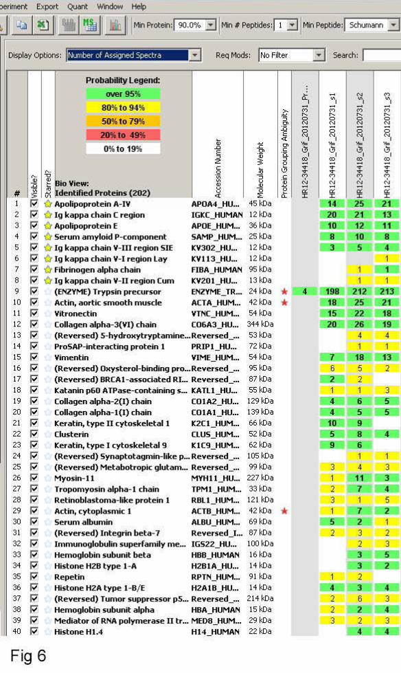
known kappa variable region was identified in isolated fibrils. In one case subjected to mass
spectrometry analysis, minor amounts of kappa variable peptide sequences were noted but
included kappa–I, –II, and –III, and would not support presence of a monoclonal
immunoglobulin (Figure 6).
Essentially these are two different diseases, one the result of a monoclonal plasma cell
dyscrasia in which the specific variable region of the light-chain is predisposed to amyloid fibril
10
formation when produced in excess. The other is the result of a mutation in the light–chain
constant region gene which, for an individual heterozygous for the mutation, predictably would
be present in 50% of serum IgG kappa antibodies. Excess production of the mutated constant
region peptide is not likely to be a determining factor in amyloid formation, although stimulation
of antibody production by repeated immunizations or response to infectious diseases could
possibly be a factor in initiation and progression of amyloid formation. More likely age-related
metabolic factors in the catabolism of serum proteins, as with other forms of hereditary
amyloidosis, dictate the late-onset of amyloid formation from a protein that has been present
from birth. Review of the published tertiary structure of the kappa light-chain constant region
obtained by X-ray diffraction indicates that the cysteine at position 131 may cause marked
change in protein folding but it is not likely to interfere with the normal intramolecular disulfide
bridge from 134Cys to 194Cys (Figure 7).11
There are two reports of cases in which mutation in the kappa constant gene was
associated with systemic amyloidosis.12,13 Both had a kappa light-chain mutation (Ser177Asn)
identified in amyloid fibril isolates and both were from patients with plasma cell dyscrasias. In
the case reported by Solomon, et al.,12 nucleotide sequencing of DNA from family members of
the patient with light-chain amyloidosis was consistent with the Ser177Asn kappa constant
region change being the result of a germ-line rather than a somatic mutation. A similar
conclusion was proposed by the studies of Wally, et al.,13 with the suggestion that the Ser177Asn
did not cause but contributed to the generation of amyloid formation. In both of the reported
cases12,13 rapid progression of clinical disease was noted. While it has been proposed that
Ser177Asn is a kappa light-chain polymorphism as are Ala153Val and Val191Leu, additional
studies are needed to support this hypothesis. Since there are no reports identifying the kappa

11
light-chain constant region Ser131Cys mutation in published studies, it is unlikely to meet the
designation of a genetic polymorphism.
There have also been reports of AL amyloidosis in first degree relatives. Miliani, et al.,
reported three siblings with AL amyloidosis.14 Gertz, et al., reported AL amyloidosis in three
families.15 Enquist, et al., reported a father and son with AL amyloidosis.16 In all of these cases
an underlying plasma cell dyscrasia was identified. Extensive searches for plasma cell dyscrasia
in several of the patients in the present report of kappa light-chain amyloidosis were negative.
This is the first report of AL amyloidosis due to an identified mutation in the constant
region of an immunoglobulin light-chain gene in the absence of a monoclonal plasma cell
dyscrasia. In the present family the high incidence of affected individuals makes the hereditary
nature of the disease obvious and this probably has protected them from inappropriate treatment
with chemotherapy designed to treat plasma cell dyscrasia. One can only speculate as to whether
other mutations in the constant region of light-chain genes, kappa or lambda, might give
systemic amyloidosis without the presence of a plasma cell dyscrasia. If no obvious family
history of amyloidosis is present, a misdiagnosis and inappropriate treatment with chemotherapy
might result. It is also possible that amyloidosis could result from a somatic mutation in the
constant region of the light-chain gene. In such a case there would be no family history of
amyloidosis. In the small, but significant, number of patients with AL amyloidosis who do not
have a detectible plasma cell dyscrasia by present testing methods nucleotide sequencing of the
Ig light-chain constant region genes should be considered as part of the diagnostic evaluation.

12
Acknowledgements
The studies described herein were supported in part by the Indiana University Foundation
Accounts of the Machado Family Amyloid Research Fund, the Aldo Family Amyloid Research
Fund, and the Jacobson Family Amyloid Research Fund.
13
Authorship
Author Contribution: M.D.B. performed clinical evaluations, designed experiments and
analyzed data; J.J.L. isolated proteins and performed amino acid sequencing; B.K.-B. performed
immunohistochemistry and supervised DNA analyses.
Non-Author Contributions: The authors thank Dr. Thomas D. Hurley for analysis of X-ray
structure data and Dr. Jill R. Murrell for DNA sequencing. Dr. M. Carney Taylor provided
clinical data on members of the kindred. Dr. Reinhold Linke provided expert histochemistry
assistance. Members of the kappa light-chain (C-kappa) family provided historical medical
records and participated in the genetic studies.
Conflict-of interest disclosure: The authors declare no competing financial interests.
Correspondence: Merrill D. Benson, M.D., Indiana University School of Medicine,
Department of Pathology and Laboratory Medicine, 635 Barnhill Drive, MS-128, Indianapolis,
Indiana 46202-5126 USA; e-mail: [email protected]
14
References
1. Gertz MA. Immunoglobulin light-chain amyloidosis: 2013 update on diagnosis, prognosis,
and treatment. Am J Hematol 2013;88(5):416-425.
2. Benson MD. The Hereditary Amyloidoses. In: Picken MM, Dogan A, Herrera GA, eds.,
Amyloid and Related Disorders [Surgical Pathology and Clinical Correlations], Current
Clinical Pathology Series Editor, Giordano A. Humana Press, part of Springer Science+
Business Media; Part I: Introduction/General – Chapter 4, 2012:53-67.
3. Picken MM. Amyloidosis – where are we now and where are we heading? Arch Pathol Lab
Med 2010;134(4):545-551.
4. Yazaki M, Liepnieks JJ, Callaghan J, Connolly CE, Benson MD. Chemical characterization
of a lambda I amyloid protein isolated from formalin-fixed and paraffin-embedded tissue
sections. Amyloid 2004;11(1):50-55.
5. Liepnieks JJ, Wilson DL, Benson MD. Biochemical characterization of vitreous and cardiac
amyloid in Ile84Ser transthyretin amyloidosis. Amyloid 2006;13(3):170-177.
6. Nichols WC, Gregg RE, Brewer HB, Benson MD. A mutation in apolipoprotein A-I in the
Iowa type of familial amyloidotic polyneuropathy. Genomics 1990;8(2):318-323.
7. Linke RP. On typing amyloidosis using immunohistochemistry. Detailed illustrations,
review and a note of mass spectrometry. Prog Histochem Cytochem 2012;47(2):61-132.
8. Kabat EA, Wu TT, Perry HM, Gottesman KS, Foeller C. Sequences of Proteins of
Immunological Interest. (US Department of Health and Human Services, Public Health
Service, National Institutes of Health, Bethesda, MD), 5th Edition, 1991:647-660.
9. Stevens FJ. Four structural risk factors identify most fibril-forming kappa light chains.
Amyloid 2000;7(3):200-211.
15
10. Dwulet FE, Strako K, Benson MD. Amino acid sequence of a lambda VI primary (AL)
amyloid protein (WLT). Scand J Immunol 1985;22(6):653-660.
11. Roussel A, Spinelli S, DeÂret S, Navaza J, Aucouturier P, Cambillau C. The structure of an
entire noncovalent immunoglobulin kappa light-chain dimer (Bence-Jones protein) reveals a
weak and unusual constant domains association. Eur J Biochem 1999;260(1):192-199.
12. Solomon A, Weiss DT, Murphy CL, Hrncic R, Wall JS, Schell M. Light chain-associated
amyloid deposits comprised of a novel κ constant domain. Proc Natl Acad Sci 1998;95(16):
9547-9551.
13. Wally J, Kica G, Zhang Y, et al. Identification of a novel substitution in the constant region
of a gene coding for an amyloidogenic kappa1 light chain. Biochem Biophys Acta 1999;
1454(1):49-56.
14. Miliani A, Bergesio F, Salvadori M, et al. Familial AL-amyloidosis in three Italian siblings.
Haematologica 1996;81(2):105-109
15. Gertz MA, Garton JP, Kyle RA. Primary amyloidosis (AL) in families. Am J Hematol
1986;22(2):193-198.
16. Enqvist S, Mellqvist U-H, Mölne J, et al. A Father and his son with systemic AL
amyloidosis. Haematologica 2009;94(3):437-439.

16
Table 1. Clinical features of C kappa amyloidosis.
SUBJECT CLINICAL FEATURES
AGE AT
DEATH
TISSUE-POSITIVE
AMYLOID
BONE
MARROW
IMMUNOGLOBULIN
LIGHT-CHAIN Kappa131Cys
I-2 Renal Failure 58
II-1 Renal Failure – age 52 53
II-3 Renal Failure – age 60
Dialysis – age 64 66
Kidney, Larynx, Lung,
Bone marrow Hypocellular
II-7 Renal Failure
Dialysis 73
II-8 Renal Failure
Dialysis 54 Kidney
II-10 Renal Failure
Dialysis 62
Post-mortem Kidney,
Heart, Pancreas,
Parathyroid
Hypocellular +
II-12 Renal Failure – age 60+
Dialysis
III-2 Renal Failure – age 66
Dialysis – age 70 74 Kidney
III-8 Alive – 73 years –
III-9 Proteinuria – age 64
(Stage 3 renal disease)
Increased kappa
and lambda +
III-10 Asymptomatic – age 37 +
III-12 Proteinuria – age 66 Kidney Normal +
III-15 Cholecystitis – age 53 Stomach,
Gall bladder Normal Normal +
IV-1 –IV-2 –IV-3 IV-4 Asymptomatic – age 40 Normal +
IV-5 Asymptomatic – age 39 Normal +
IV-6 –

17
Figure Legends
1. Pedigree of family showing members with biopsy proven amyloidosis (�) and presumed
affected (◩) showed typical autosomal dominant inheritance. The index case is indicated by arrow.
2. Kidney biopsy from subject III-12: (A) Haematoxylin and eosin stained section showingdeposits in glomerular basement membrane (original X 100). (B) Electron micrograph showing amyloid deposits with 7-10 nm fibrils (insert) consistent with amyloid.
3. Serial microscopic sections of gallbladder wall from index patient (III-15): (A) Stained withCongo red and viewed by fluorescence microscopy. (B) Stained by indirect immunohistochemistry with polyclonal anti-Ig kappa light-chain antiserum showing specific staining of amyloid deposits (original X 100).
4. Amino acid sequences of tryptic peptides of (A) amyloid fibril protein isolated from post-mortem kidney of patient II-10, and (B) plasma kappa light-chain from a member of the next generation (III-15) with biopsy proven amyloidosis. The normal sequence of the constant region of kappa light-chains is shown. Residue numbering is by Kabat et al.8 The lines indicate the sequences obtained by Edman degradation of HPLC purified tryptic peptides. The parentheses at the ends of the amyloid fibril protein peptides (A) denote residues not completely verified due to decreasingly low Edman degradation yields. The X at residues 131 and 134 denote no amino acid was identified at these positions. The dots at the ends of some plasma light-chain peptides (B) indicate that the peptide continued but was not analyzed further.
5. Nucleotide sequence of PCR product for kappa light-chain DNA showing heterozygosity atcDNA position 403 with both cytosine and guanine giving coding sequence for cysteine (TGT) and serine (TCT) at position 131.
6. Report of LCMS analysis of gastrointestinal biopsy amyloid from index case (III-15).
7. Ribbon diagram of kappa light-chain molecular structure to show position of the Ser131residue in relation to the Cys134 which is part of the normal intramolecular disulfide bridge. Modified from A. Roussel, et al., Eur J Biochem 1999;260(1):192-199.11

For personal use only.on March 8, 2017. by guest www.bloodjournal.orgFrom
For personal use only.on March 8, 2017. by guest www.bloodjournal.orgFrom

For personal use only.on March 8, 2017. by guest www.bloodjournal.orgFrom

For personal use only.on March 8, 2017. by guest www.bloodjournal.orgFrom

For personal use only.on March 8, 2017. by guest www.bloodjournal.orgFrom
For personal use only.on March 8, 2017. by guest www.bloodjournal.orgFrom

For personal use only.on March 8, 2017. by guest www.bloodjournal.orgFrom

doi:10.1182/blood-2014-12-618108Prepublished online April 9, 2015;
Merrill D. Benson, Juris J. Liepnieks and Barbara Kluve-Beckerman
Hereditary systemic immunoglobulin light-chain amyloidosis
http://www.bloodjournal.org/site/misc/rights.xhtml#repub_requestsInformation about reproducing this article in parts or in its entirety may be found online at:
http://www.bloodjournal.org/site/misc/rights.xhtml#reprintsInformation about ordering reprints may be found online at:
http://www.bloodjournal.org/site/subscriptions/index.xhtmlInformation about subscriptions and ASH membership may be found online at:
digital object identifier (DOIs) and date of initial publication. indexed by PubMed from initial publication. Citations to Advance online articles must include final publication). Advance online articles are citable and establish publication priority; they areappeared in the paper journal (edited, typeset versions may be posted when available prior to Advance online articles have been peer reviewed and accepted for publication but have not yet
Copyright 2011 by The American Society of Hematology; all rights reserved.Hematology, 2021 L St, NW, Suite 900, Washington DC 20036.Blood (print ISSN 0006-4971, online ISSN 1528-0020), is published weekly by the American Society of