hepatitis b virus genotypes and mutations affecting hbeag ... · hepatitis b virus genotypes and...
TRANSCRIPT

Med & Health 2007; 2(1): 1-25 REVIEW ARTICLE
Hepatitis B Virus Genotypes and Mutations Affecting HBeAg Production: A Malaysian Perspective Chee Kent Lim1 and So Har Ton2
1 Faculty of Biotechnology, Malaysia University of Science and Technology, Petaling Jaya
47301, Malaysia 2 School of Arts and Sciences, Monash University Malaysia, Petaling Jaya 46150,
Malaysia ABSTRAK Jangkitan daripada virus hepatitis B (HBV) merupakan satu isu kesihatan besar sedunia. HBV merupakan suatu faktor terhadap peningkatan risiko penyakit hepar seperti sirosis dan juga dalam perkembangan penyakit kanser hepatosellular. Masalah ini terutamanya tersebar luas di rantau Asia-Pasifik, termasuk Malaysia. Semasa jangkitan, e antigen hepatitis B (HBeAg) adalah dihasilkan di dalam perumah. Antigen ini adalah satu penanda serologi yang penting di dalam diagnosis kesakitan kronik hepatitis B. Perubahan (seroconversion) kepada anti-bodi (anti-HBe) merupakan satu faktor terhadap kemajuan prognosis pengyakit. Tetapi terdapat beberapa mutasi seperti dwi mutasi ke atas promoter teras (core promoter double mutations) (A1762G1764→T1762A1764), varian kodon 15 (C1858/ T1858) dan mutasi pada kodon henti ke atas pra-teras (pre-core stop codon mutation) (TGG→TAG) yang boleh memberi kesan kepada pengeluaran HBeAg. Ini boleh mengakibatkan implikasi terhadap diagnosis dan klinikal penyakit tersebut. Selain itu, HBV juga boleh dibahagikan kepada lapan genotip (A ke H), subjenis genotip dan rekombinan juga ditemui. Kajian telah menunjukkan bahawa genotip berbezaan dari segi hubungan dengan mutasi-mutasi tersebut dan juga dengan prognosis penyakit hepatitis B.
Kata Kunci: Virus hepatitis B, genotip, dwi mutasi pada promoter teras, varian kodon 15,
mutasi henti pada pra-teras ABSTRACTS Infection by hepatitis B virus (HBV) is a major global health-care problem. HBV is an accepted factor in the elevated risks for liver disease such as cirrhosis and development of hepatocellular carcinoma. This problem is particularly prevalent in the Asia-Pacific region which includes Malaysia. During infection, the hepatitis B e antigen (HBeAg) is produced in the hosts. This antigen is an important serological marker for diagnosing chronic hepatitis B. Seroconversion to anti-body (anti-HBe) corresponds to the improvement of disease prognosis. However, certain mutations such as the core promoter dual mutations (A1762G1764→T1762A1764), the codon 15 variants (C1858/ T1858) and the precore stop codon mutations (TGG→TAG) can affect the HBeAg expression. This has diagnostic and clinical implications. Besides that, the HBV can be grouped into eight genotypes (A to H).
Address for correspondence and reprint request: Dr. Ton So Har, School of Arts and Sciences, Monash University Malaysia, Petaling Jaya 46150, Malaysia. Email: [email protected]
1

Med & Health 2007; 2(1): 1-25 Lim C.K & Ton S.H Moreover, genotypic subtypes and recombinants have been observed as well. Studies have observed that these can differ in their affiliations with the mutations above as well as with disease prognosis. Keywords: Hepatitis B virus (HBV), genotypes, core promoter double mutations, codon 15
variants, pre-core stop codon mutation Hepatitis B virus (HBV) In 1965, Blumberg and co-workers discovered a unique antigen among the sera of Australian aborigines (Blumberg et al., 1965). They referred to this as the “Australian antigen”. Subsequent electron microscopy study by Dane et al. (1970) on the sera of antigen-positive patients revealed vast numbers of spherical and filamentous particles of 22 nm in diameter and also larger particles of 42 nm in diameter with central nucleocapsids and outer enveloping coats. With the discovery of the 42 nm particles, the term “Dane particles” was used. It was shown later that the Dane particles were in fact complete viral particles and were the causative agent for the liver hepatitis. Subsequently, the Dane particles were changed to a name which we are familiar with today, the hepatitis B virus (HBV). The 22 nm particles turn out to be the excess non-infectious surface protein complexes of the virus without the nucleocapsid cores. Molecular background of hepatitis B virus (HBV) Hepatitis B virus (HBV) is a small enveloped virus which is 42 nm in diameter. Its partially double-stranded DNA genome of about 3.2 kb is enclosed by an icosahedral structure consisting of 180 or 240 HBV core proteins (HBcAg) (Bowyer and Sim, 2000). Enveloping this structure is a coat of lipid bilayer membrane studded with the HBV viral surface proteins (HBsAg). The genome of HBV is unique in that it has overlapping reading frames. Thus it
can encode about 50 % more polypeptides than would be expected from the size of its genome (Ganem and Varmus, 1987). The compact genome contains four reading frames which are the precore/core reading frame, preS/ S reading frame, polymerase reading frame and the X reading frame (Figure 1) (Kidd-Ljunggren et al., 2002). The compactness of the HBV genome is such that no non-translational region exists in it and the regulatory sequences reside inside the reading frames themselves (Torre and Naoumov, 1998). The precore/core reading frame overlaps with the polymerase and X reading frames. In this reading frame, two start codons are situated, corresponding to the precore and core domains of the reading frame. The translation from first start codon across both the domains will produce the HBeAg. Figure 1: Schematic diagram depicting the genome of HBV. The negative (-ve) strand DNA is partially complemented by its positive (+ve) strand DNA. The HBV genomes encode for four reading frames which are the polymerase (P), preS/ S reading frame (preS1/ -S2/ S), precore/ core reading frame (preCore/ Core) and the X reading frame (X). Note the overlapping nature of the reading frames.
2
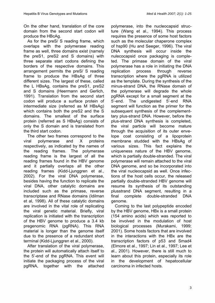
Hepatitis B Virus Genotypes and Mutations Med & Health 2007; 2(1): 1-25 On the other hand, translation of the core domain from the second start codon will produce the HBcAg. As for the preS/ S reading frame, which overlaps with the polymerase reading frame as well, three domains exist (namely the preS1, preS2 and S domains) with three separate start codons defining the borders of the respective domains. This arrangement permits the preS/ S reading frame to produce the HBsAg of three different sizes. The largest of these, called the L HBsAg, contains the preS1, preS2 and S domains (Heermann and Gerlich, 1991). Translation from the second start codon will produce a surface protein of intermediate size (referred as M HBsAg) which contains both the preS2 and the S domains. The smallest of the surface protein (referred as S HBsAg) consists of only the S domain and is translated from the third start codon. The other two frames correspond to the viral polymerase and X proteins respectively, as indicated by the names of the reading frames. The polymerase reading frame is the largest of all the reading frames found in the HBV genome and it partially overlaps all the other reading frames (Kidd-Ljunggren et al., 2002). For the viral DNA polymerase, besides having the function to replicate the viral DNA, other catalytic domains are included such as the primase, reverse transcriptase and RNase domains (Idilman et al, 1998). All of these catalytic domains are involved in the vital role of replicating the viral genetic material. Briefly, viral replication is initiated with the transcription of the HBV genome to produce a 3.4 kb pregenomic RNA (pgRNA). This RNA material is longer than the genome itself due to the presence of a redundant short terminal (Kidd-Ljunggren et al., 2000). After translation of the viral polymerase, the protein will automatically attach itself to the 5’-end of the pgRNA. This event will initiate the packaging process of the viral pgRNA, together with the attached
polymerase, into the nucleocapsid struc-ture (Wang et al., 1994). This process requires the presence of some host factors such as the molecular chaperone complex of hsp90 (Hu and Seeger, 1996). The viral DNA synthesis will occur inside the nulecocapsid once packaging is comple-ted. The primase domain of the viral polymerase has a role in initiating the DNA replication process through reverse transcription where the pgRNA is utilized as the template. During the synthesis of the minus-strand DNA, the RNase domain of the polymerase will degrade the whole pgRNA except for a small segment at the 5’-end. The undigested 5’-end RNA segment will function as the primer for the subsequent synthesis of the complement-tary plus-strand DNA. However, before the plus-strand DNA synthesis is completed, the viral particle will become mature through the acquisition of its outer enve-lope coat consisting of a lipoprotein membrane studded with the HBsAg of various sizes. This fact explains the uniqueness nature of the HBV genome, which is partially double-stranded. The viral polymerase will remain attached to the viral DNA genome, and so be incorporated into the viral nucleocapsid as well. Once infec-tions of the host cells occur, the released partially double-stranded HBV genome will resume its synthesis of its outstanding plusstrand DNA segment, resulting in a final complete double-stranded DNA genome. Coming to the last polypeptide encoded by the HBV genome, HBx is a small protein (154 amino acids) which was reported to be involved in the modulation of host biological processes (Murakami, 1999; 2001). Some hosts factors that are involved in the interactions with the HBx are the transcription factors of p53 and Smad4 (Elmore et al., 1997; Lin et al., 1997; Lee et al., 2001). However, there is still much to learn about this protein, especially its role in the development of hepatocellular carcinoma in infected hosts.
3

Med & Health 2007; 2(1): 1-25 Lim C.K & Ton S.H Epidemiology and pathogenesis of HBV HBV is one of the most common viral pathogen affecting the world population. Approximately 350 million people around the world are infected with the virus (Purcell, 1993). It is estimated that 50 million new cases are observed annually with 5 to 10 % of them being adults while the remaining consists of children (Mayerat et al., 1999). Infections by HBV can result in acute and chronic hepatitis diseases. The figure of deaths attributed to HBV infection is estimated at 1 million people per year (Thomas and Jacyna, 1993). Although all body secretions and excretions have been found to contain HBsAg, only blood, semen, menstrual and vaginal fluids have been shown to be infectious (Hollinger and Liang, 2001). When infected, the occurrence of liver hepatitis is not caused directly by the HBV per se but due to the host own immune response. When HBV infection occurred, the host immune response is stimulated where the presence of HBV-specific CD8+ cells can be detected in infected subjects. As HBV does not directly cause the destruction of the hepatocyte it invades, the infected hepatocyte acts as a reservoir for further infection of other similar host cells. This permits the HBV-infected hepatocytes to continuously shed the viral particles into the blood stream, thus ensuring that all the hepatocytes in a particular individual are infected (Seeger and Mason, 2000). Infected hepatocytes will display the immunodominant T cell epitopes presented by HLA class I mole-cules on their surfaces (Chisari and Ferrari, 1995). This acts as the targets for the CD8+ cells to the infected hepatocytes, thus resulting in the destruction of the infected hepatocytes. Infections by HBV can lead to either acute or chronic hepatitis. Acute infection usually runs within a period of between 1 to 6 months, which includes the incubation period of between 8 to 20 weeks (Webster and Bertoletti, 2002). The clinical symp-
toms that developed following acute infection are fever, severe malaise, loss of appetite and development of jaundice. Most individuals with acute hepatitis B will recover with clearance of viral antigens and HBV DNA in their sera (Yuen and Lai, 2001). This is possible when the host immune system is strong enough to overcome the viral infection. It is believed that resolution of the acute hepatitis B occurs when adequate productions of B and broad-based CD8+ T cells producing protective antibodies targeting the epitopes of the viral proteins are achieved (Bertoletti et al., 1991; Nayersina et al., 1993). Besides that, suppression of the viral infection is also performed by cytokines (especially a and g interferons) and tumor necrosis factor (TNF-a) which play a part in destruction of the viral replicative inter-mediates such as viral proteins, the replicating DNA and RNA (Tsui et al., 1995). Individuals who recovered from acute hepatitis B will be conferred with life-long immunity against the virus. However, for those who are suffering from acute hepatitis B, there is a 1 % chance that the conditions for these individuals will dete-riorate and develop into fulminant hepatitis (Lee, 1993). Fulminant hepatitis is cha-racterized by liver failure which is very often fatal. The failure of the liver functions is due to sudden massive immune-mediated lysis of the infected hepatocytes without giving enough time for the liver to recuperate (Gove and Hughes, 1991; Leifeld et al., 2002). A recent study speculated that vigorous replication of HBV might be a factor involved in the development of fulminant hepatitis (Inoue et al., 2006). The other form of liver disease that HBV infection can lead to is chronic hepatitis B. Chronic hepatitis B is characterized by high HBV DNA levels, high risk of trans-missions, elevated liver enzymes such as alanine aminotransferase (ALT) in the sera and high risk of developing liver cirrhosis and liver cancer, hepatocellular carcinoma (HCC) (Lok and McMahon, 2001). The
4

Hepatitis B Virus Genotypes and Mutations Med & Health 2007; 2(1): 1-25 activity of viral replication in patients with chronic hepatitis B is relatively high where it is estimated that 1 x 10¹¹ HBV particles are released into the blood circulation everyday (Nowak et al., 1996). Chronic hepatitis B could occur when the rate of infected liver cells death is balanced by HBV infections of new liver cells due to the host immune response being not vigorous enough to clear the infection. The responses of cytotoxic T cells and HLA class H restricted T cells against the viral nucleocapsid antigens are weak in chronic patients (Chisari and Ferrari, 1995). One of the contributing factors of chronic hepatitis B is the long life-span of hepatocytes with an average half-life of 6 to 12 months. The stable hostvirus interactions also play a role in the persistence of chronic hepatitis B. The weak host immune response usually leads to scarring, disruption of blood flow and obstruction of bile drainage of the liver organ, thus interfering with the vital liver functions. Given the major role plays by host immune system on the development of chronic hepatitis B, this liver disease is most often incurred by individuals with weak immune system. It has been estimated that about 90 % of newborns to mothers positive for HBeAg will develop chronic hepatitis B (Beasley et al., 1983). Besides that, about 25 to 30 % of infants and children under the age of 5 years old will develop chronic hepatitis in their life through contacts with persons infected with HBV, especially their mothers (Beasley et al., 1982; McMahon et al., 1985). New-borns and young infants are especially vulnerable because of the immaturity of their immune systems. Besides infants and children, immunosuppressed individuals such as those infected with HIV-1 or on maintenance haemodialysis have a greater risk of developing chronic hepatitis B than healthy persons when exposed to HBV (Bodsworth et al., 1989; Horvath and Raffanti, 1994). There is a great chance that chronic hepatitis B patients will eventually die from
liver injuries such as cirrhosis or HCC, which is estimated to be about 25 to 40 %. Out of these, the fatal rates are 50 % for male and 15 % for female (Lau et al., 1997). It has been estimated that chronic hepatitis B patients have a 200 to 300 fold of increased risk of developing HCC than the general population (Maynard, 1990). In some individuals infected by HBV, no discernable symptoms for either acute or chronic hepatitis B can be observed. These individuals are grouped as the “asymp-tomatic carriers”. The sera from these individuals are positive for HBsAg but have normal or minimal raised liver enzymes such as ALT (Torbenson and Thomas, 2002). They can be diagnosed to be negative for HBeAg and positive for anti-HBe with undetectable or low HBV DNA level (Lok and McMahon, 2001). Long-term follow-up studies showed that asymptomatic carriers have lower risk of developing liver diseases like cirrhosis or HCC and they are less likely to infect other individuals (Lindh et al., 2000; Hsu et al., 2002; Martinot-Peignoux et al., 2002). However, about 20 to 30 % of them can reacquire hepatitis B disease where elevated biochemical indicators, high HBV DNA level and sero-reversion from anti-HBe back to HBeAg can be observed (de Franchis et al., 1993; Bellentani et al., 2002; Hsu et al., 2002). Repeated reactiva-tions of hepatitis B or a sustained one can result in liver injury but frequently symp-toms may be absent. Prevalence of HBV infection in Malaysia HBV infections are endemic in Asia, South Pacific, sub-Saharan Africa, Middle East and the prevalence are particularly high in some indigenous populations such as the Aborigines and Native American Indians in South and Central Americas (Maynard, 1990; McMahon, 1997; Mast et al., 1999). It is estimated that 75 to 80 % of the HBV infection occur in the Western Pacific and in the South East Asia regions (Gust, 1996; Merican et al., 2000). The high endemicity
5
Med & Health 2007; 2(1): 1-25 Lim C.K & Ton S.H in these regions is thought to be due to mother-to-child perinatal transmission (Simmonds, 2001). In Malaysia, one of the earlier studies on HBV was performed by Ton et al. (1979). In the study, it was found that HBsAg and anti-HBc in blood donors from the Kuala Lumpur hospital was 5.5 % and 50.1 % respectively. When comparison of the HBsAg-positivities between the three major races in the Malaysian population (Malay, Chinese and Indians) was made, no significant difference was found between them. A study on Malaysian intravenous drug users by Mangalam et al. (1986) observed that the rate of HBV infections among the drug abusers were similar to the general population. Thus, this could indicate that intravenous drug injections might not be a major route of transmission for HBV in Malaysia. As in the general case for the Asia-Pacific region, the main routes of HBV transmission among the Malaysian popu-lation could be due to mother-to-child peri-natal transmission as well. In Malaysia, the prevalence of HBV infection incidence has been significantly reduced since the introduction of a nationwide vaccination program in 1989 (Ng et al., 2005). The program, called the Expanded Program of Immunization, invol-ved three doses of vaccination for the newborns. The first dose was given to a newborn soon after birth followed by the second dose 1 month later and 5 months later with the third dose. A particular per-son is considered to be protected against HBV when an anti-HBs titer of at least 10 mIU/ mL is detected in his or her serum (Ng et al., 2005). Prior to the implement-tation of the vaccination program, a study performed by Ton and Noriah (1987) on 653 healthy Malaysian individuals of both sexes revealed that 29.6 % of them were positive for the anti-HBs. This is comparable to a report on 3807 unvac-cinated Malaysian school children before the program was started where 27.4 % was found to be positive for the anti-HBs (Ng et
al., 2005). After the implementation of the program, a statistical analysis on 97786 school children vaccinated from 1989 to 1996 showed that 59.3 % of them were positive for the anti-HBs and had humoral immunity against HBV (Ng et al., 2005). The successful result of this vaccination program saw a reduction from 2.7 % to 0.4 % of HBsAg carriers among school children during the study period. However, despite the implementation of the vaccination program, a significant proportion of the Malaysian population is still afflicted with HBV. Merican et al. (2000) reported that about 75.3 % of the chronic hepatitis diseases suffered by patients is caused by HBV. A study on 17048 healthy Malaysian volunteers (mean age, 34 years) found that 893 were positive for HBsAg (Merican, 1999). Taking this study as a representative figure of the current HBV infection in the Malaysian population, it is estimated that about 5.24 % are struck down with chronic hepatitis B. When stratified according to the ethnic composition of the Malaysian population, the Chinese incurred the highest proportion of HBV infections, followed by the Malays and Indians (Yap, 1994; Merican et al., 2000). When compared with the study done by Ton et al. in 1979, it can be observed that the current level of the Ma-laysian population positive for HBsAg is comparable to that found in a sample population of more than 20 years ago. However, it seems that the demography of infection between the different races has changed since the study was performed in 1979. The infections by HBV in Malaysia have created a major healthcare problem as acute and chronic hepatitis B patients require substantial medical attention. In a study to determine the causes of jaundiced cases surveyed among patients admitted into the hospitals in Kelantan and Terengganu in 1996, Saat et al. (1999) found that 1.4 % of the cases could be traced to infection by HBV. Due to their frequent close proximity with HBV carriers,
6

Hepatitis B Virus Genotypes and Mutations Med & Health 2007; 2(1): 1-25 health care workers are at an increased risk to exposure to HBV. A study among the health care workers in Negeri Sembilan in 1989 observed that a substantial number of the health care workers were positive for the serological markers for HBV (Tan et al., 1992). The study involved 2500 health-care workers of which about 25 % were found to be positive for the serological markers. Further stratification revealed that 2.1 % had HBsAg while 22.8 % were positive for anti-HBs. However, for the latter cases, it was not known whether the positivity for anti-HBs was due to history of HBV infection or to vaccination. Given this situation, it is imperative that steps should be taken to vaccinate the health-care workers in order to offer them protection against HBV. In lieu of this, the Universal Precautions Policy guidelines were adopted for health care workers. To find out if the policy was properly implemented, a survey on the protective status against HBV was performed on 625 subjects comprising health-care workers and under-graduate students undergoing clinical training in two hospitals in Kuala Lumpur in 2001 (Hesham et al., 2005). It was ob-served that only 58.4 % of the subjects had taken a complete HBV vaccination course while 82.2 % of the subjects had only been vaccinated with a single dose. HBV infections could also increase the burden faced by the current health-care system. HBV-contaminated blood could cause in-fections among patients seeking treatment for other types of illnesses. For example, in a report by Menon and Aiyar (1997), it was mentioned that about 20% of the 55 paediatric oncology patients who received blood products at Hospital Universiti Sains Malaysia in Kota Baru were positive for HBsAg. Besides that, the treatment costs for HBV infections are expensive. The current regime of anti-viral treatments administered for patients infected with HBV in Malaysia are the a-interferon, lamivudine and thymosin a-1 (Mohamed et al., 2004). Besides it was observed that about 80 % of
the HCC cases observed in the country were caused by HBV (Yap, 1994). HBeAg / anti-HBe productions and the associated mutations HBeAg is a viral protein that does not form a part of the structure of the viral particles but shares about 90 % of the amino acid sequence with the HBcAg. This is because the HBeAg is translated from the first start codon of the precore/ core reading frame, resulting in larger precursor protein of HBeAg with an extra N-terminal domain of 29 amino acid residues. The first 19 amino acid residues of the extra N-terminal functions as a signal peptide to guide the precursor HBeAg to the host endoplasmic reticulum compartment to be processed. Processing of this immature protein in the endoplasmic reticulum will result in the cleavage of the 19 residues from the precursor HBeAg. Subsequently, the C-terminal domain will be cleaved off as well at the arginine-rich region, thus resulting in a shortened mature form of HBeAg which is then secreted out from the host cells (Lau and Wright, 1993). Some of the mature HBeAg is localized on the host cell membrane while the rest is secreted into the blood stream. In comparison, the HBcAg does not have the N-terminal domain and retains its C-terminal domain. Despite the high similarity between these two viral proteins, their tertiary structural conformations are quite different. This is due to the presence of an intramolecular disulfide bond in the HBeAg (Nassal and Rieger, 1993; Wasenauer et al., 1993). HBeAg is utilized as a serological marker for the infectivity of HBV, in particular individuals, in clinical diagnosis (Magnius and Espmark, 1972; Shikata et al., 1977). HBeAg is usually detected in the serum when HBsAg begins to appear. The presence of HBeAg in the serum correlates with the presence of viral activity in the liver. In the diagnosis of acute hepatitis, HBeAg does not play an important role as
7

Med & Health 2007; 2(1): 1-25 Lim C.K & Ton S.H a diagnostic marker because the antigens are cleared within weeks after the acute disease has resolved. However, in chronic hepatitis B, this antigen is important as an indicative marker as HBeAg can remain detectable for a sustained period of time, usually in terms of months or years (Merican et al., 2000). The function of HBeAg with respect to the virus is not fully understood yet. In an in vitro study, the absence of HBeAg does not seem to impede replicative ability of the virus (Tong et al., 1991). It is speculated that HBeAg might play a role in tricking the immune defense system of the infected host. It has been proposed that the expres-sion of HBeAg during perinatal infection can induce immune tolerance of the HBV by the host and that the HBeAg acts as a buffer to attack by anti-HBc on infected hepatocytes. This reasoning is based on the high sequence similarity between HBeAg and HBcAg (Tong et al., 2005). Nevertheless, HBeAg positivity in HBV-infected patients has been found to be one of the risk factor for the development of liver cirrhosis (Liaw et al., 1988; Realdi et al., 1994; Yu et al., 1997). In cirrhotic patients, the presence of HBeAg is one of the risk factors in the occurrence of liver decomposition (Fattovich et al., 1995). It was reported that patients with decom-pensated cirrhosis who were negative for HBeAg have a better 5 year survival rate than those who were positive for the antigens (de Jongh et al., 1992). A reduce-tion in the risk of developing hepatic decompensation and survival improvement can be achieved after the clearance of HBeAg, either spontaneously or after undergoing anti-viral treatments (de Jongh et al., 1992). With the disappearance of HBeAg from the serum, seroconversion to anti-HBe can occur. The presence of anti-HBe in the early phase of acute hepatitis is an indication of the resolution of the acute infection. On the other hand, in chronic hepatitis B, the disappearance of HBeAg and appearance of anti-HBe is associated
with improved biochemical and histological standing of the liver disease. Although anti-HBe positivity in HBV-infected patients signifies clinical im-provement, in some cases liver damage may still persist with active HBV DNA replication (Hadziyannis, 1995). Even after seroconversion to anti-HBe, reactivation of viral replication can still occur where seroreversion back to HBeAg positive status can happen. Besides that, mutations in the HBV genome affecting HBeAg productions can also result in the emergence of HBeAg-negative mutants. Two of the most pro-minent mutations in these HBeAg-negative mutants are the A1762G1764→ T1762A1764 core promoter mutations and the precore stop codon mutation. The A1762G1764→T1762A1764 dual muta-tions occur in the core promoter region that regulates the transcription of both the precore-mRNA, which is the template for HBeAg and the pgRNA (Figure 2) (Yuh et al., 1992; Takahashi et al., 1995). The precore stop codon mutant is characterized by a G to A nonsense nucleotide mutation at nucleotide 1896 of codon 28 of the HBeAg (TGG→TAG) which can result in the truncating of the translation of HBeAg into a short peptide of 28 amino acids (Figure 3) (Brunetto et al., 1989; Carman et al., 1989; Tong et al., 1990). Besides these two mutations, the base variants at nucleotide 1858 in codon 15 can affect HBeAg production as well. The common variants exist at the aforemen-tioned site are the C1858/ T1858 variants. These variants are not directly involved in the truncation of the HBeAg production per se as conversion from one variant to the other will result in a silent mutation. However, they affect the HBeAg production through their pairing to nucleotide 1896 of the codon 28 in the secondary loop structure of the pgRNA (Figure 3). This loop is a hair-pin structure which functions as an encapsidation signal. This structure is essential for recruitment of the HBV polymerase which in turn is essential for
8

Hepatitis B Virus Genotypes and Mutations Med & Health 2007; 2(1): 1-25 Figure 2 : The dual mutations of A1762G1764→T1762A1764 at the core promoter region preceding the preCore/ Core gene region. Figure 3 : The encapsidation signal structures of the pgRNA and the bases pairing between the C1858/ T1858 variants at nucleotide positions 1858 and 1896. (a) Although pairing of uracil (nt 1858) and guanine (nt 1896) are not compatible, this pairing is permitted due to the wobble pairing nature between the bases, thus conserving the essential stem loop structure of the encapsidation signal. (b) The compatible pairing between cytosine (nt 1858) and guanine (nt 1896) will permit for the conservation of the encapsidation signal structure. (c) Pairing between uracil (nt 1858) and adenine (nt 1896) is compatible. This compatibility will allow for the stable mutation of G to A at nt 1896, thus preserving the precore stop codon mutation (TGG→TAG). (d) The incompatible paring between cytosine (nt 1858) with adenine (nt 1896) will result in the disruption of the encapsidation signal structure.
9

Med & Health 2007; 2(1): 1-25 Lim C.K & Ton S.H
(2002) had also performed sequencing analyses on the HBeAg-negative samples which were not infected by the mutants. The sequencing analyses revealed that 5 of them had the A1762G1764→ T1762A1764 dual mutations while another sample had mutation at the start codon. On top of that, they found a significant association between T1858 variants with the precore stop codon mutations, which concurs with many other studies.
the synthesis of the HBV genome and viral replication. Disruption of this structure is detrimental to the HBV replication. In the hair-pin secondary structure of the encapsidation signal, the nucleotide at 1858 is intimately paired to nucleotide 1896 through base pair bonding. Thus, the fate of precore stop codon mutation (TGG→TAG) is dependent upon the nucleotide at 1858. When cytosine is the nucleotide in place at nucleotide 1858 (hence referred as the C1858 variant), stable formation of the precore stop codon mutant will be prevented (Figure 3). This is due to the reason that incompatible pairing of adenine at 1896 with cytosine at nucleotide 1858 will confer a weak binding force to the encapsidation signal structure.
In another study conducted in Malaysia, it was observed that among the sera samples from 44 HBV-infected patients, the prevalence of the core promoter mutations and precore stop codon muta-tions were determined to be 56.8 % and 9.1 % respectively (Ton et al., 2002). Intriguingly, in their study, it was observed that the number of patients with HBeAg-positive status was higher than those with anti-HBe-positive status when those in-fected with the core promoter mutants were taken into account. In comparison to a study on Brazilian HBV carriers with anti-HBe positive status, only about 24 % of them were found to be infected with the precore stop codon mutations (Castro et al., 2001). They reasoned that this was due to the large majority of the subjects being infected by the C1858 variants which, as discussed above, would preclude the stop codon mutation.
This will cause a disruption to the all important structure and so prohibiting the replication of the HBV mutant. On the other hand, if the nucleotide at 1858 is a thymine (hence referred as the T1858 variant), the formation of the precore stop codon mutant is conducive as the thymine can stably pairs with adenine at nucleotide 1896. Besides that, TGG wild-type can exist as well because the thymine at 1858 can wobble pair with guanine at nucleotide 1896 without disrupting the essential encapsidation signal structure (Lok et al., 1994). In Malaysia, an early study performed by Ton et al. (1996) on 43 HBsAg Malaysian carriers showed that infections by the C1858 variants were quite substantial. In the study, 4 of the 43 samples (9.3 %) were found to be of precore stop codon mutants. Of interest was that two of the mutants were found to co-exist with the TGG wild-type. In another study, Yap et al. (2002) detected 13 precore stop codon mutants out of 33 HBeAg-positive and 32 mutants out of 48 HBeAg-negative Malaysians sampled. In comparison to a study outside the country, Okamoto et al. (1994) observed a staggering high frequency of about 90 % of the mutants in patients positive to anti-HBe. Yap et al.
However, in a later study on HBV-infected Malaysian patients, only low numbers of the above mutants were observed (Lim et al., 2006). In the study, only 11 out of 77 (14.3 %) HBV-infected patients examined were found to be infected with the core promoter mutants while 7 (9.1 %) were co-infected by both wild-type and the mutants. Interestingly, no significant correlation was observed between the core promoter mutations and the HBeAg/ anti-HBe status in this study. This contradicts with a few other studies in other part of the world where such correlations were observed (Hussain et al., 2003; Okamoto et al., 1994). Despite this
10

Hepatitis B Virus Genotypes and Mutations Med & Health 2007; 2(1): 1-25 anomaly, many other studies did not observe such correlations (França et al., 2004; Lindh et al., 1999, Orito et al., 2001, Takahashi et al., 1995). One possible explanation for this is that the occurrences of T1762A1764 mutations only decrease the productive capability of HBeAg of the particular HBV strain but not totally abolishing it. It was shown by Parekh et al. (2003) that T1762A1764 mutations could only reduce the HBeAg production by about 20 %. Of interest is the observations of sera which were positive for both HBeAg and anti-HBe in two of the sera samples in the Malaysian study (Lim et al., 2006). This was also encountered in an earlier study by Ton et al. (1995) on sera taken from some Malaysians infected with HBV. The status of positivities for both HBeAg and anti-HBe might signify the ongoing process of sero-conversion whereby the HBeAg serological marker was not completely eliminated from the blood circulation yet. Also, in the study by Lim et al. (2006), no precore stop codon mutations were observed among the HBV samples examined. The study also showed that of the 46 samples that were successfully assayed, 33 of them were infected by the T1858 variants while 13 were infected by the C1858 variants. This was in concordance to an earlier study by Ton et al. (2001) where the occurrences of the T1858 variants significantly predominate over the C1858 variants (82.1 % vs. 14.8 %). Due to the lack of any precore stop codon mutant in the study by Lim et al. (2006), no correlation study between T1858/ C1858 variants of codon 15 with nucleotide 1896 of codon 28 was performed. However, co-infections of the precore stop codon mutants and the wild-type strains could still be observed among the Malaysian HBV carriers (Ton et al., 2001). The contrasting results observed between the various studies conducted in Malaysia could indicate that the distribution of the HBV mutants among the Malaysian
population is not permanent but varies in time as well as in locations. Other than the core promoter mutations and the precore stop codon mutations, there are other less well-known mutations which could reduce the production rate of HBeAg. It was shown by Parekh et al. (2003) that, in combination with the T1762A1764 mutations, mutations at nucleotides 1753 and 1766 could result in a decrease in HBeAg production by up to 80 %. Besides that, some mutations occurring downstream of the precore start codon can decrease HBeAg translation too (Ahn et al., 2003). In addition to these, mutations such as at the precore start codon or TAA stop codon mutation at codon 2 could also abolish the production of HBeAg (Lindh et al., 1996). These mutations could explain for the presence of HBV in anti-HBe-positive patients where both the core promoter and the precore stop codon mutations were not detected. Association of mutations affecting HBeAg productions with liver disease prognosis Although the mutations mentioned previously are associated specifically with HBeAg productions, many studies found that they could be related to the prognosis of liver diseases as well. The A1762G1764→T1762A1764 dual muta-tions were frequently observed in patients with chronic hepatitis but were rarely found in asymptomatic carriers (Okamoto et al., 1994; Kim et al., 1999). These mutations were also associated with increased inflammation and fibrosis in both HBeAg-positive and HBeAg-negative HBV infected chronic hepatitis patients (Lindh et al., 1999). The mutations had also been found to be associated with fulminant hepatitis in a few studies (Sato et al., 1995; Friedt et al., 1999). These mutations do not affect the transcription of HBV pgRNA from which the viral core peptides are translated (Okamoto et al., 1994). With the occur-rences of these mutations, the equilibrium
11

Med & Health 2007; 2(1): 1-25 Lim C.K & Ton S.H balance between HBeAg in the blood circulation and the core peptides presented by the class 1 HLA molecules on the sur-face of infected hepatocytes will be eliminated. As HBeAg has been shown to have suppressive effect on T helper cells, its down-regulation may explain the asso-ciated clinical prognosis observed. The core peptide has been determined to contain most of the primary sequence of HBeAg. Thus, it is possible that circulating HBeAg may prevent cytotoxic T lympho-cytes from recognizing infected hepatocytes which display the core peptides on their surface by their HLA molecules. This is significant because cytotoxic T lymphocytes may be directed to the infected hepatocytes that display the core-derived peptides and so causing injury to them (Mondelli et al., 1982; Bertoletti et al., 1991). The decreased synthesis of HBeAg could free some of the cytotoxic T lymphocytes from their suppressive effect and so a greater number of the lymphocytes may be directed toward the infected hepatocytes thus resulting in greater liver injury (Takahashi et al., 1995). One study has shown that HBcAg is able to activate a more vigorous immune response than HBeAg in vivo, thus causing more severe liver damage (Nishizono et al., 1997). As mentioned, the disruption of HBeAg production could result in the withdrawal of hepatocyte display of HBeAg-derived cyto-toxic CD8+ T cell epitopes but viral replication will not be affected (Carman et al., 1993). In a study by Yuasa et al. (2000), it was found that most Vietnamese patients with fulminant hepatitis carried the precore stop mutation while none was found in patients with acute non-fulminant hepatitis. This is in concordance to an earlier study where similar observation was made (Omata et al., 1991). Transmission of these HBV mutants may lead to development of fulminant hepatitis in the newly-infected individuals (Yotsumoto et al., 1992; Liang et al., 1994). Besides, mutation in codon 28 could render the HBV
strain with enhanced encapsidation advan-tage that makes them more infectious and causes hepatitis when they do so (Scaglioni et al., 1997). Lai et al. (1994) reported that mutations at codon 28 were associated with most anti-HBe seroconversion. Generally, the occurrences of mutations at the precore stop codon before seroconversion appear to shorten the time of onset of sero-conversion (Brunetto et al., 1991). It was found to be correlated with persistence viral detection in the blood circulation and increased ALT levels for at least 6 months after seroconversion (Brunetto et al., 1991). It was also found to be correlated with elevated frequency of cirrhosis development in patients (Brunetto et al., 1991). These show that the timing of the emergence of the mutation could have clinical implications on affected patients. Besides, Marusawa et al. (2000) found that the predominance of TAG mutation after seroconversion correlated with increased liver injury. The precore stop codon mutations were also associated with severe recurrent HBV infection after liver transplantation (Angus et al., 1995). This mutation has also been associated with increased susceptibility to coinfection by hepatitis D virus (HDV) (Raimondo et al., 1991). Hepatitis D virus is a satellite virus of HBV. This virus can only replicate in the hepatocytes that are infected by HBV. This is because HDV requires the proteins that are expressed by HBV in order to form its own nucleocapsid and has its own RNA genome packaged. Lindh et al. (1996) observed that carriers infected with the C1858 variants showed more liver damage than carriers infected with the T1858 variants. They suggested that carriers with the C1858 variants might remain longer in the HBeAg-positive phase or maintain some HBeAg production capabilities even after seroconversion to anti-HBe. They hypothesized that the HBeAg is a target for the necroinflam-matory process, thus causing more severe liver injury. However, patients infected with
12

Hepatitis B Virus Genotypes and Mutations Med & Health 2007; 2(1): 1-25 the C1858 variants responded better to interferon therapy than those infected with the T1858 variants (Zhang et al., 1996). In Malaysia, Ton et al. (2001) observed that many of the asymptomatic carriers and patients with HCC were infected with this T1858 variant than in those suffering from chronic hepatitis. Further, many studies found that simultaneous occurrences of the precore stop codon mutations and the core promoter mutations would result in more severe cases of liver injury (Brunetto et al., 1991; 1993; Laskus et al., 1998; Inoue et al., 1998; Honda et al., 1999). Although a lot of studies point toward the association of the above mutations with increased risks of liver injuries, many other studies did not find such correlations. Castro et al. (2001) demonstrated that the core promoter mutations were not linked to increased liver injuries. In the case of the C1858/ T1858 variants, no significant clinical differences were observed between patients infected by these two variants (Sakurai et al., 2004). Similarly, the issue of increased risk of liver injury associated with infection by HBV strains with simultaneous precore stop codon mutations and the core promoter mutations is not resolved yet as many other studies refute this claim (Okamoto et al., 1990; Hsu et al., 1995; Lindh et al., 1996; Pollicino et al., 1995).
sequences of the S gene by Norder et al. (1993). Genotype G was discovered by Stuyver et al. (2000) from HBV carriers in France and blood donors in the United States of America. They reported that this genotype has a minimum divergence of 11.7 % (genotype G versus E) and a maximum divergence of 15.3 % (genotype G versus genotype H) when compared with other genotypes over the whole genomes. This genotype is unique in that it has a nucleotide insertion of 36 bp, situated after the fifth nucleotide after the core translation initiation point (nucleotide 1901) (Stuyer et al., 2000). The latest addition to the family is the genotype H which was observed from HBV-infected patients in Central America (Arauz-Ruiz et al., 2002). Geographic distribution of HBV genotypes The prevalence of the HBV genotypes can be localized into different geographic areas. Genotype A has been found to be predominant in Northern Europe and North America while genotype D is predominantly found in the Mediterranean region (Magnius and Norder, 1995). In Asia, genotype A can be found in high frequency in India, Indonesia and the Philippines (Huy and Abe, 2004). Genotypes B and C are found predominantly in the Far East such as China, Japan and the South-East Asia region (Magnius and Norder, 1995). There is a tendency for genotype C to have higher prevalence in Northern mainland China and in Japan than genotype B (Orito et al., 2001). Genotype E is found mainly in parts of East, Central and West Africa. As for genotype F, it is found mainly in South and Central Americas as well as in some Pacific islands such as the Polynesia. So far, genotype G has been observed in the United States of America, Germany and France and could be found in Mexico as well (Stuyer et al., 2000; Vieth et al., 2002; Sanchez et al., 2002). As mentioned previously, genotype H seems to be
HBV Genotypes The genotype system of HBV was first proposed by Okamoto et al. (1988) with an initial of 4 genotypic groups (i.e. A to D) classified, being distinguished based on an inter-genotypic divergence of 8.5 to 10.0 % between 18 complete HBV genomes and with a divergence of 1.1 to 2.7 % between isolates within the same genotype. The genotype classification was later extended to 6 genotypic groups (genotypes A to F), with addition of genotypes E and F, through phylogenetic analysis on 122
13

Med & Health 2007; 2(1): 1-25 Lim C.K & Ton S.H localized to the Central America region. In the Malaysian context, a small study performed by Ong et al. (2005) through sequencing on the pre-S and S regions of the HBV genome extracted from sera of HBV infected Malaysian carriers revealed that the genotypes B and C were the most prevalent strains. This was confirmed in later study on a larger Malaysian population sample where the prevalence of genotype B was to be highest (56.9 %) followed by genotype C (31.4 %) (Lim et al., 2006). Co-infections by several genotypes Besides single genotypic infections, simultaneous infections by two different HBV genotypic strains are possible. These events can occur in countries where endemicity of several genotypic strains of HBV is prevalent. One of the first evidence of co-infections by two different genotypes was reported by Kao et al. (2001). In their large study, the frequency of co-infections by genotypes B and C was found to be 0.8 %. Since then, other studies had revealed that co-infections could be quite common, with a rate of co-infections of between 4.4 to 17.5 % (Ding et al., 2003; Kato et al., 2003, Osiowy and Giles, 2003; Chen et al., 2004; Olinger et al., 2006). In the local setting, a study on Malaysian HBV carriers revealed that 7.8 % of the sample population studied was coinfected by genotypes B and C (Lim et al., 2006). Even more intriguing is the report of triple simultaneous infections by three different genotypic strains (Chen et al., 2004). The report stated that in a group of intravenous drug users infected by HBV, 0.9 % of them had triple infections by the genotypic strains of A, B and C. HBV genotypic subtypes The main genotypes can be further divided into subtypes. These had been determined in a few studies based on phylogenetic analysis (Norder et al., 1994; Bowyer et al.,
1997; Bowyer and Sim, 2000; Mbayed et al., 2001). It was found that genotype C could be further subdivided into subtypes with genotype C adrq serotype being more common in the Pacific islands than in South-East Asia (Norder et al., 1993). Norder et al. (1994) showed that Cq- subtype of genotype C in the Pacific region also differed from subtypes found in the Asia region. A study by Lindh et al. (1997) using the PCR-RFLP on the S region showed that within genotype C, RFLP pattern C1 was present in all Northeast Asian carriers while South-East Asian carriers had another RFLP pattern, designated as C2. Furthermore, a later study has found that the genotype C can be subdivided into the subtypes Ce and Cs (Chan et al., 2005). A study performed on the urban population of South Africa by Bowyer et al. (1997) showed that there exists two subtypes for genotype A, designated as subtypes A and A’. Mbayed et al. (2001) found the existence of four clusters of subtypes within genotype F which were associated with different geographical locations. Esteban et al. (1999) theorized that HBV genotypic subtypes were likely to evolve in different ethnic backgrounds over the centuries of HBV infections. In a study by Sugauchi et al. (2003), two distinct subtypes of genotype B were found, namely Bj and Ba. The main difference between these two subtypes is that the genome of Ba subtype has undergone recombination with the genotype C over the regions of the precore and core gene while in Bj, no such recombination occurred. In Japan, the Bj subtype is the dominant strain while Ba is found mainly in other Asian countries such as China, Hong Kong, Taiwan, Thailand and Vietnam. As more and more studies are conducted in this area, it is expected that more genotypic subtypes will be discovered. The consensus for distinguishing between genotypic subtypes now stands at 4% (Kramvis and Kew, 2005). A total of at least
14

Hepatitis B Virus Genotypes and Mutations Med & Health 2007; 2(1): 1-25
authors suggested that these could be due to recombinatorial events between the genomes of different genotypes (Hannoun et al., 2000; Owiredu et al., 2001). It is postulated that this phenomenon occurs in an environment when co-infections by several genotypes exists. HBV has a high replicative capability where up to 1013 viral particles can be produced everyday as well as high mutation rate due to the absence of proof-reading capability on the part of the viral polymerase (Nowak et al., 1996; Brechtbuehl et al., 2001). This leads to a proposal by Schaefer (2007) which states that the existence of such hybrids is a result of rapid adaptation of the HBV to new host immunological selections encountered in different human populations. Another more sophisticated theory on the occurrence of such hybrids is the theory of HBV genome mosaicism (Bowyer and Sim, 2000). This theory suggests that the existence of HBV genotypes are due to the modular construction of the HBV genome from different small segments of HBV genomes of various genotypes (Bowyer and Sim, 2000; Simmonds and Midgley, 2005). It is supported by a discovery of the activation or repression of the preCore-pregenomic promoter (enhancer II) by the transcription factor COUP-TF which is genotype-dependent (Fischer et al., 2006).
24 genotypic subtypes have been identified so far (Schaefer, 2007). It remains to be established whether the HBV strains of genotypes B and C infecting Malaysians are of the subtypes Ba or Bj in the case for genotype B and of subtypes Ce or Cs for genotype C or it could be some other undiscovered subtype alto-gether. Recombinant hybrids of different genotypes In certain situations, recombinations between HBV genomes of different genotypic strains were reported. As discussed earlier, one of this is the genotypic subtype Ba, which is a result of recombination between HBV genomes of genotypes B and C (Sugauchi et al., 2003). From the current literature, the majority of the recombinant hybrids observed are of genotypes B/ C and A/ D (Simmonds and Midgley, 2005; Yang et al., 2006). Other recombination hybrids that have been discovered are A/ B/ C, A/ C, A/ E, A/ G, C/ D, C/ F and C/ G (Schaefer, 2007). In some regions, the recombinant hybrids are the predominant HBV strain infecting the local population. In Tibet for example, it was observed that the recombinant hybrid of genotypes C/ D was the most prevalent strain detected (Cui et al., 2002; Wang et al, 2005; Zeng et al., 2005). Whether such hybrids exist among the Malaysian local HBV strains remain to be seen.
The associations of HBV genotypes with clinical prognosis From the various studies, it was
observed that there is a tendency for the recombinatorial breakpoints to cluster at certain regions of the HBV genome. Some of these recombination breakpoints which commonly occur are at the preS1/ 2 region (nt 3150-100), the 3’-end region of the Core gene (nucleotide 2330-2450) as well as at the 3’-end region of the Surface gene (nt 650-830) (Simmonds and Midgley, 2005; Yang et al., 2006).
Studies have shown that HBV genotypes correlated to different degree of severity of liver diseases. Kao et al. (2000a) observed that HBV genotype C associated more with severe liver disease while genotype B was associated with the development of HCC in young HBsAg-positive carriers. A further study revealed that patients with genotype C infections had slightly higher sera HBV DNA levels and significantly higher ALT levels than patients infected with genotype B (Kao et al., 2000b). Besides individuals
However, the mechanism in which these events occur is still not clear. Some
15

Med & Health 2007; 2(1): 1-25 Lim C.K & Ton S.H infected with genotype B would undergo HBeAg seroconversion to anti-HBe at a younger age (Chu and Lok, 2002; Kao et al., 2002; Sumi et al., 2003). On the other hand, genotype A was found to be present in higher frequency in chronic hepatitis patients than in acute patients (Mayerat et al., 1999). In a study performed by Kobayashi et al. (2002a), they found that chronic hepatitis tend to occur more frequently in patients infected by genotype A than by genotypes B or C. Cases had also been reported where acute infections by HBV genotype A evolved more frequently into chronic infection (Kikuchi et al., 1998; Niitsuma et al., 1999). These findings could lead to the reasoning that in Western countries, where genotype A infections are prevalent, there is a high rate of chronic infection after HBV infection during adulthood (Sherlock et al., 1987; Heijtink et al., 1999; Lindh et al., 2000). Furthermore, Kobayashi et al. (2002b) showed that patients infected with HBV genotype A had higher HBeAg levels than those infected by genotypes B or C. In the Indian sub-continent, genotype D was more associated with severe forms of liver diseases than genotype A (Thakur et al., 2002) and that patients below the age of 40 years old had higher incidence of getting HCC when infected by genotype D than by genotype A. Mayerat et al. (1999) suggested that HBV genotype A has lower antigenicity than genotype D, thus it is less capable of inducing the immune clearance phase and so lead to a higher incidence of chronic hepatitis. In the Mediterranean region, infection by genotype D was associated with the prevalence of anti-HBe-positive chronic hepatitis (Chu et al., 2003). However, it is important to note that this issue is not firmly established yet. This is because some other studies did not find such correlations between the genotypes and the prognosis of the liver diseases (McMahon et al., 2001; Sumi et al., 2003). In Malaysia, it is not known whether such
correlation exists as no study has been undertaken to look into this question. Correlations of HBV genotypes with mutations affecting HBeAg production and HBe/ anti-HBe status Over the years of research on HBV genotypes, promoter mutation (A1762G1764→T1762A1764), precore stop codon mutations (TGG→TAG) and codon 15 variants (C1858/ T1858), various reports had been compiled on the correlations between these variables as well as the correlations between the genotypes with HBeAg/ anti-HBe status. The core promoter mutations (A1762G1764→T1762A1764) had been observed to be more common in patients with HBV genotype C infections than genotype B (Lindh et al., 1999; Kao et al., 2000b; Orito et al., 2001; Fang et al., 2002; Sakurai et al., 2004). However, in some studies, the correlations between HBV genotypes with core promoter mutations were not observed (Franca et al., 2004; Kidd-Ljunggren et al., 2004). This type of correlation was not observed in the Malaysian study undertaken by Lim et al. (2006) as well. HBV genotype A had been associated with C1858 variants (codon 15) (Li et al., 1993). Due to this, the cytosine (C) at nucleotide 1858 will not permit base-pairing to occur with adenine base (A) of nucleotide 1896 in genotype A. Thus, HBV genotype A has constraint in undergoing mutation from G to A at nucleotide 1896 to produce HBV strain with precore stop codon mutations, hence HBeAg-negative phenotype. This could partially explain for the high rate of chronic HBV infections in adult patients who are infected with this genotype, as observed in many European countries. This is also true in the case of genotype F (Kramvis and Kew, 1998). This is supported by the finding that patients infected with genotype A had higher HBeAg levels than those infected by
16

Hepatitis B Virus Genotypes and Mutations Med & Health 2007; 2(1): 1-25 genotypes B and C, as discussed above (Kobayashi et al., 2002b). Genotypes B, C, D and E had been associated with the T1858 variants (codon 15). However infrequently, the C1858 variants could still be observed among patients infected with genotypes C and D (Kramvis and Kew, 1998). This situation was observed in Malaysia where some C1858 variants were observed in some genotype C infected samples (Lim et al., 2006). Despite these, some HBV strains of genotype A could still undergo the TAG mutation (codon 28). This is possible when the strain develops the C to T mutation at nucleotide 1858, thus preventing the destabilization of the pregenomic RNA stem loop structure which is essential for viral replication (Li et al., 1993; Blackberg and Kidd-Ljunggren, 2000). Due to the tendency of genotypes B, C, D and E to be associated with the T1858 variants (codon 15), they are predisposed to TAG mutations (codon 28). Thus genotypes B, C, D and E were found to be associated more often with the TAG mutations (codon 28) than genotype A (Huy et al., 2004). However, some other studies did not observe such correlations. Orito et al. (2001) observed no significant difference between the predispositions to the precore stop codon mutation development between genotypes B and C. This phenomenon was not observed as well in a later study by Kidd-Ljunggren et al. (2004) on samples from various geographic origins. The suggested correlation between genotype B with precore stop codon mutations seems to contradict to a study by Lim et al. (2006) on Malaysian samples infected with HBV. Even though large numbers of individuals infected with HBV genotypes B were found in their study, no precore stop codon mutations were detected among them. This could imply that other factors might play a role here, such as the geographic origin of the HBV strains. However, this does not seem to be the case as other studies conducted on HBV-infected Malaysian samples did observe some precore stop
codon mutants even though their genotypes were unknown (Ton et al., 1996; 2002; Yap et al., 2002). Other than the associations of genotypes with the mutations, some studies had also observed the inequality in the distributions of HBeAg/ anti-HBe statuses between patients infected by different HBV genotypes. It was reported that patients infected by genotype B were more prone to be negative for HBeAg than in patients infected by genotypes C (Lindh et al., 1999; Sakurai et al., 2004). This was supported by a study on HBV-infected patients in Thailand (Sugauchi et al., 2002a). The reason behind these could be explained by the fact that patients infected by HBV genotype C experienced a longer delay in seroconversion to anti-HBe status (Lindh et al., 1999). Thus the consequence of this is that they will remain in the HBeAg positive status for a longer period of time. But this scenario was not observed among the Malaysian carriers infected with HBV genotypes B and C where no difference in the distributions of the HBeAg/anti-HBe statuses was observed (Lim et al., 2006). The collection timing of the HBV infected sera from the patients could be important with regards to the determination of HBeAg/ anti-HBe statuses of the patients as older patients tend to have seroconverted to anti-HBe. Associations of genotypic subtypes with other variables It was observed by Sugauchi et al. (2003) that the Bj and Ba subtypes correlated differently with the core promoter mutations. They found that the Ba subtype was more associated with the core promoter mutations than with the Bj subtype (33 % vs. 15 %) while the precore stop codon mutations occurred more frequently in the Bj subtype (50 % vs. 18 %). Thus, the implication of these findings is that the correlation studies between genotypes and other factors such as disease prognosis or mutations associated
17

Med & Health 2007; 2(1): 1-25 Lim C.K & Ton S.H with HBeAg production is not that straight forward anymore. This is because when correlation studies between genotypes and other variables were performed, the proportions of the subtypes of one particular genotype could significantly influence the final outcome of the results. The final correlation results might be dependent upon the varying degree of proportions of the subtypes since it was shown that the association between the subtypes and other variables could be different. Besides, a few studies had found that genotypic subtypes are significantly correlated with the prognosis of the liver disease as well. Studies in Japan revealed that the subtype Ba was more associated with severe liver disease as well as had higher tendency to develop HCC than in patients infected with the subtype Bj (Sugauchi et al., 2002b; 2003; Orito et al., 2005). In Hawaii, it was observed that patients of Asian ethnicity (excluding Japanese) which were exclusively infected with the Ba subtype had higher rates of HBeAg positivity and core promoter mutations than the Bj subtype that was predominantly found to infect the Japanese (Orito et al. 2001; Sakurai et al., 2004). Another example of clinical difference resulted from different subtypes infections is shown in a study by Hayashi et al. (2007) in Japan where the genotypic subtypes of Ba and Cs were more frequently found in patients suffering from acute hepatitis B than in patients with chronic hepatitis B. The varying proportions of the genotypic subtypes within the samples of studies could be the reason why contradictory results were reported when correlation studies between genotypes and other variables were performed. This includes a study carried out on a Malaysian sample population where no difference was ob-served between genotypes B and C and the core promoter mutations. This is in contrast to what has been observed elsewhere (Lim et al., 2006).
SUMMARY After many years of research on HBV, it is accepted that HBV is one of the causative agents for the development of acute and chronic hepatitis. Besides, infection by HBV is a risk factor for HCC. As the prevalence of HBV infection among the Malaysian population is relatively high, these infected individuals run the risk of developing the above conditions. Moreover a number of HBV variants exist which have diagnostic and clinical implications (Raimondo et al., 1991; Yotsumoto et al., 1992; Okamoto et al., 1994; Sato et al., 1995; Lindh et al., 1996; Kao et al., 2000a). Mutations in the HBV genome can affect the expression of HBeAg. Examples of these are the core promoter dual mutations (A1762G1764→T1762A1764), the codon 15 variants (C1858/ T1858) and the precore stop codon mutations (TGG→ TAG). The HBV can be categorized into eight genotypes (A to H) as well as genotypic subtypes and recombinants. These could differ in their affiliations with the mutations and with disease prognosis. Although vaccination program has been introduced into Malaysia, a substantial proportion of the Malaysian population is still afflicted with HBV infections though the prevalence has decreased. The number of studies undertaken in Malaysia on this virus is still considerably low. Therefore it is important that more studies on HBV in Malaysia are conducted so as to better understand the epidemiology, morpho-genesis and pathogenesis behavior of the virus in relation to the Malaysian po-pulation. One particular area of HBV research that is still lacking is the study on the genotypic subtypes which is getting more important as it has been shown to be possibly associated with other health risks. REFERENCES Ahn S, Kramvis A, Kawai S, Spangenberg H, Li J,
Kimbi G, Kew M, Wands J, Tong S. Sequence variation upstream of precore translation initiation codon reduces hepatitis B virus e
18

Hepatitis B Virus Genotypes and Mutations Med & Health 2007; 2(1): 1-25
antigen production. Gastroenterology 2003; 125:1370-8.
Angus PW, Locarnini SA, McCaughan GW, Jones RM, McMillan JS, Bowden DS Hepatitis B virus precore mutant infection is associated with severe recurrent disease after liver transplantation. Hepatology 1995; 21: 14-8.
Arauz-Ruiz P, Norder H, Robertson BH, Magnius LO. Genotype H: a new Amerindian genotype of hepatitis B virus revealed in Central America. J Gen Virol 2002; 83: 2059- 73.
Beasley RP, Hwang LY, Lee GC, Lan CC, Roan CH, Huang FY, Chen CL. Prevention of perinatally transmitted hepatitis B virus infections with hepatitis B immune globulin and hepatitis B vaccine. Lancet 1983; 2(8359):1099-102.
Beasley RP, Hwang LY, Lin CC, Leu ML, Stevens CE, Szmuness W, Chen KP. Incidence of hepatitis B virus infections in preschool children in Taiwan. J Infect Dis 1982; 146: 198-204.
Bellentani S, Dal Molin G, Miglioli L, Crocè L, Masutti F, Castiglione A, Campello C, Tiribelli C. Natural history of HBV infection: a 9 years follow-up of the Dionysos cohort. J Hepatol 2002; 36 Suppl 1: 228.
Bertoletti A, Ferrari C, Fiaccadori F, Penna A, Margolskee R, Schlicht HJ, Fowler P, Guilhot S, Chisari FV. HLA class I-restricted human cytotoxic T cells recognize endogenously synthesized hepatitis B virus nucleocapsid antigen. Proc Natl Acad Sci USA 1991; 88: 10445-9.
Blackberg J, Kidd-Ljunggren K. Genotypic differences in the hepatitis B virus core promoter and precore sequences during seroconversion from HBeAg to anti-HBe. J Med Virol 2000; 60: 107-12.
Blumberg BS, Alter HJ, Visnich S. A "new" antigen in leukemia sera. JAMA 1965; 191: 541-6.
Bodsworth N, Donovan B, Nightingale BN. The effect of concurrent human immunodeficiency virus infection on chronic hepatitis B: a study of 150 homosexual men. J Infect Dis 1989; 160: 577-82.
Bowyer SM, Sim JG. Relationships within and between genotypes of hepatitis B virus at points across the genome: footprints of recombination in certain isolates. J Gen Virol 2000; 81: 379-92.
Bowyer SM, van Staden L, Kew MC, Sim JG. A unique segment of the hepatitis B virus group A genotype identified in isolates from South Africa. J Gen Virol 1997; 78: 1719-29.
Brechtbuehl K, Whalley SA, Dusheiko GM, Saunders NA. A rapid real-time quantitative polymerase chain reaction for hepatitis B virus. J Virol Methods 2001; 93: 105-13.
Brunetto MR, Giarin M, Saracco G, Oliveri F, Calvo P, Capra G, Randone A, Abate ML, Manzini P, Capalbo M, Piantino P, Verme G, Bonino F: Hepatitis B virus unable to secrete e antigen and
response to interferon in chronic hepatitis B. Gastroenterology 1993, 105: 845-50.
Brunetto MR, Giarin MM, Oliveri F, Chiaberge E, Baldi M, Alfarano A, Serra A, Saracco G, Verme G, Will H, Bonino F: Wild-type and e antigen-minus hepatitis B viruses and course of chronic hepatitis. Proc Natl Acad Sci USA 1991, 88: 4186-90.
Brunetto MR, Stemler M, Schodel F, Will H, Ottoberlli A, Rizzetto M, and Bonino F. Identification of HBV variants which cannot produce precore derived HBeAg and may be responsible for severe hepatitis. Ital J Gastroenterol 1989; 21: 151-4.
Carman W, Jacyna M, Hadziyannis S, Karayiannis P, McGarvey M, Makris A, Thomas H. Mutation preventing formation of the hepatitis B e antigen in patients with chronic hepatitis B infection. Lancet 1989; 2: 588-91.
Carman W, Thomas H, Domingo E. Viral genetic variation: hepatitis B virus as a clinical example. Lancet 1993; 341: 349-53.
Castro LD, Niel C, Gomes SA Low frequency of mutations in the core promoter and precore regions of hepatitis B virus in anti-HBe positive Brazilian carriers. BMC Microbiology 2001; 1: 10.
Chan HL, Tsui SK, Tse CH, Ng EY, Au TC, Yuen L, Bartholomeusz A, Leung KS, Lee KH, Locarnini S, Sung JJ. Epidemiological and virological characteristics of 2 subgroups of hepatitis B virus genotype C. J Infect Dis 2005; 191: 2022-32.
Chen BF, Kao JH, Liu CJ, Chen DS, Chen PJ. Genotypic dominance and novel recombinations in HBV genotype B and C coinfected intravenous drug users. J Med Virol 2004; 73: 13-22.
Chisari FV, Ferrari C. Hepatitis B virus immunopathology. Springer Semin Immunopathol 1995; 17: 261-81.
Chu CJ, Keeffe EB, Han SH, Perrillo RP, Min AD, Soldevila-Pico C, Carey W, Brown RS, Luketic VA, Terrault N, Lok AS; U.S. HBV Epidemiology Study Group. Prevalence of HBV precore/core promoter variants in the United States. Hepatology 2003; 38: 619-28.
Chu CJ, Lok ASF. Clinical significance of hepatitis B virus genotypes. Hepatology 2002; 35: 1274-6.
Cui C, Shi J, Hui L, Xi H, Zhuoma, Quni, Tsedan, Hu G. The dominant hepatitis B virus genotype identified in Tibet is a C/D hybrid. J Gen Virol 2002; 83: 2773-7.
Dane DS, Cameron CH, Briggs M. Virus-like particles in serum of patients with Australia-antigen-associated hepatitis. Lancet 1970; 1: 695-8.
de Franchis R, Meucci G, Vecchi M, Tatarella M, Colombo M, Del Ninno E, Rumi MG, Donato MF, Ronchi G. The natural history of asymptomatic hepatitis B surface antigen carriers. Ann Intern Med 1993; 118: 191-4.
de Jongh FE, Janssen HL, de Man RA, Hop WC,
19

Med & Health 2007; 2(1): 1-25 Lim C.K & Ton S.H
Schalm SW, van Blankenstein M. Survival and prognostic indicators in hepatitis B surface antigen-positive cirrhosis of the liver. Gastroenterology 1992; 103: 1630-5.
Ding X, Gu H, Zhong ZH, Zilong X, Tran HT, Iwaki Y, Li TC, Sata T, Abe K. Molecular epidemiology of hepatitis viruses and genotypic distribution of hepatitis B and C viruses in Harbin, China. Jpn J Infect Dis 2003; 56: 19-22.
Elmore LW, Hancock AR, Chang SF, Wang XW, Chang S, Callahan CP, Geller DA, Will H, Harris CC. Hepatitis B virus X protein and p53 tumor suppressor interactions in the modulation of apoptosis. Proc Natl Acad Sci USA 1997; 94: 14707-12.
Esteban JI, Martell M, Carman WF, Gomez J. The impact of rapid evolution of the hepatitis viruses. In: Domingo E, Webster RG, Holland JJ, eds: Origin and evolution of viruses. San Diego: Academic Press, 1999: 345-376.
Fang ZL, Yang J, Ge X, Zhuang H, Gong J, Li R, Ling R, Harrison TJ. Core promoter mutations (A1762T and G1764A) and viral genotype in chronic hepatitis B and hepatocellular carcinoma in Guangxi, China. J Med Virol 2002; 68: 33-40.
Fattovich G, McIntyre G, Thursz M, Colman K, Giuliano G, Alberti A, Thomas HC, Carman WF. Hepatitis B virus precore/ core variation and interferon therapy. Hepatology 1995; 22: 1355-62.
Fischer SF, Schmidt K, Fiedler N, Glebe D, Schuttler C, Sun J, Gerlich WH, Repp R, Schaefer S. Genotype-dependent activation or repression of HBV enhancer II by transcription factor COUP-TF1. World J Gastroenterol 2006; 12: 6054-8.
França PH, González JE, Munné MS, Brandão LH, Gouvea VS, Sablon E, Vanderborght BO. Strong association between genotype F and hepatitis B Virus (HBV) e antigennegative variants among HBV-infected argentinean blood donors. J Clin Microbiol 2004; 42: 5015-21.
Friedt M, Gerner P, Lausch E, Trubel H, Zabel B, Wirth S. Mutations in the basic core promotor and the precore region of hepatitis B virus and their selection in children with fulminant and chronic hepatitis B. Hepatology 1999; 29: 1252-8.
Ganem D, Varmus HE. The molecular biology of the hepatitis B viruses. Annu Rev Biochem 1987; 56: 651-93.
Gove CD, Hughes RD. Liver regeneration in relationship to acute liver failure. Gut 1991; 32 Suppl: S92-S96.
Gust ID. Epidemiology of hepatitis B infection in the Western Pacific and South East Asia. Gut 1996; 38 Suppl 2: S18-S23.
Hadziyannis SJ. Antigen HBe positive chronic hepatitis B: from clinical recognition to pathogenesis and treatment. Viral Hepatitis Review 1995; 1: 7-36.
Hannoun C, Norder H, Lindh M. An aberrant genotype revealed in recombinant hepatitis B virus strains from Vietnam. J Gen Virol 2000; 81: 2267-72.
Hayashi K, Katano Y, Takeda Y, Honda T, Ishigami M, Itoh A, Hirooka Y, Nakano I, Yano M, Goto H, Yoshioka K, Toyoda H, Kumada T. Comparison of hepatitis B virus subgenotypes in patients with acute and chronic hepatitis B and absence of lamivudineresistant strains in acute hepatitis B in Japan. J Med Virol 2007; 79: 366-73.
Heermann KH, Gerlich WH. Surface proteins of hepatitis B viruses. In: McLachlan A, ed: Molecular Biology of the Hepatitis B Virus. Boca Raton, FL: CRC Press, 1991: 109-143.
Heijtink RA, Paulij W, van Roosmalen M, Hellings JA, Niesters HG, Schalm SW, Osterhaus AD. Characteristics of the early phase of chronicity in acute hepatitis B infection. J Med Virol 1999; 57: 331-6.
Hesham R, Zamberi S, Tajunisah ME, Ariza A, Ilina I. Hepatitis B immunisation status among health care workers in two Kuala Lumpur hospitals. Med J Malaysia 2005; 60: 407-10.
Hollinger FB, Liang TJ. Hepatitis B Virus. In: Knipe DM, Howley PM, eds: Fields Virology, 4th ed. Philadelphia: Lippincott, Williams and Wilkins, 2001: 2971-3036.
Honda A, Yokosuka O, Ehata T, Tagawa M, Imazeki F, Saisho H: Detection of mutations in the enhancer 2/ core promoter region of hepatitis B virus in patients with chronic hepatitis B virus infection: comparison with mutations in precore and core regions in relation to clinical status. J Med Virol 1999; 57: 337-44.
Horvath J, Raffanti SP. Clinical aspects of the interactions between human immunodeficiency virus and the hepatotropic viruses. Clin Infect Dis 1994; 18: 339-47.
Hsu HY, Chang MH, Lee CY, Hsieh KH, Ni YH, Chen PJ, Chen DS. Precore mutant of hepatitis B virus in childhood fulminant hepatitis B: an infrequent association. J Infect Dis 1995; 171: 776-81.
Hsu YS, Chien RN, Yeh CT, Sheen IS, Chiou HY, Chu CM, Liaw YF. Long-term outcome after spontaneous HBeAg seroconversion in patients with chronic hepatitis B. Hepatology 2002; 35: 1522-7.
Hu J, Seeger C. Hsp90 is required for the activity of a hepatitis B virus reverse transcriptase. Proc Natl Acad Sci USA 1996; 93: 1060-4.
Hussain M, Chu CJ, Sablon E, Lok AS. Rapid and sensitive assays for determination of hepatitis B virus (HBV) genotypes and detection of HBV precore and core promoter variants. J Clin Microbiol 2003; 41: 3699-705.
Huy TT, Abe K. Molecular epidemiology of hepatitis B and C virus infections in Asia. Pediatr Int 2004; 46: 223–30.
Huy TT, Ushijima H, Quang VX, Ngoc TT, Hayashi S, Sata T, Abe K. Characteristics of core promoter
20

Hepatitis B Virus Genotypes and Mutations Med & Health 2007; 2(1): 1-25
and precore stop codon mutants of hepatitis B virus in Vietnam. J Med Virol 2004; 74: 228-36.
Idilman R, De Maria N, Colantoni A, Van Thiel DH. Pathogenesis of hepatitis B and Cinduced hepatocellular carcinoma. J Viral Hepat 1998; 5: 285-99.
Inoue K, Ogawa O, Yamada M, Watanabe T, Okamoto H, Yoshiba M. Possible association of vigorous hepatitis B virus replication with the development of fulminant hepatitis. J Gastroenterol 2006; 41:383-7.
Inoue K, Yoshiba M, Sekiyama K, Okamoto H, Mayumi M: Clinical and molecular virological differences between fulminant hepatic failures following acute and chronic infection with hepatitis B virus J Med Virol 1998, 55: 35-41.
Kao JH, Chen PJ, Lai MY, Chen DS. Acute exacerbations of chronic hepatitis B are rarely associated with superinfection of hepatitis B virus. Hepatology 2001; 34: 817-23.
Kao JH, Chen PJ, Lai MY, Chen DS. Genotypes and clinical phenotypes of hepatitis B virus in patients with chronic hepatitis B virus infection. J Clin Microbiol 2002; 40: 1207-9.
Kao JH, Chen PJ, Lai MY, Chen DS. Hepatitis B genotypes correlate with clinical outcomes in patients with chronic hepatitis B. Gastroenterology 2000a; 118: 554-9.
Kao JH, Wu NH, Chen PJ, Lai MY, Chen DS. Hepatitis B genotypes and the response to interferon therapy. J Hepatol 2000b; 33: 998-1002.
Kato H, Orito E, Sugauchi F, Ueda R, Koshizaka T, Yanaka S, Gish RG, Kurbanov F, Ruzibakiev R, Kramvis A, Kew MC, Ahmad N, Khan M, Usuda S, Miyakawa Y, Mizokami M. Frequent coinfection with hepatitis B virus strains of distinct genotypes detected by hybridization with type-specific probes immobilized on a solid-phase support. J Virol Methods 2003; 110: 29-35
Kidd-Ljunggren K, Miyakawa Y, Kidd AH. Genetic variability in hepatitis B viruses. J Gen Virol 2002; 83: 1267-80.
Kidd-Ljunggren K, Myhre E, Blackberg J. Clinical and serological variation between patients infected with different Hepatitis B virus genotypes. J Clin Microbiol 2004; 42: 5837-41.
Kidd-Ljunggren K, Zuker M, Hofacker IL, Kidd AH. The hepatitis B virus pregenome: prediction of RNA structure and implications for the emergence of deletions. Intervirology 2000; 43: 154-64.
Kikuchi K, Mirakawa H, Abe K, Kitazawa E, Fujikawa H, Kawaguchi N, Nagai K, Kako M. A case of acute hepatitis B transmitted from her husband with HBV carrier state after primary infection of HBV-genotype A. Kanzo 1998; 13: 533-8.
Kim YS, Kim SI, Hwang SG, Kim JO, Cho JY, Lee JS, Lee MS, Hwang SD, Shim CS. Diversity of core promoter mutations in immune clearance phase
of chronic HBV infection. Eur J Gastroenterol Hepatol 1999; 11: 821-5.
Kobayashi M, Arase Y, Ikeda K, Tsubota A, Suzuki Y, Saitoh S, Kobayashi M, Suzuki F, Akuta N, Someya T, Matsuda M, Sato J, Takagi K, Miyakawa Y, Kumada H. Viral genotypes and response to interferon in patients with acute prolonged hepatitis B virus infection of adulthood in Japan. J Med Virol 2002a; 68: 522-8.
Kobayashi M, Arase Y, Ikeda K, Tsubota A, Suzuki Y, Saitoh S, Kobayashi M, Suzuki F, Akuta N, Someya T, Matsuda M, Sato J, Kumada H. Clinical characteristics of patients infected with hepatitis B virus genotypes A, B, and C. J Gastroenterol 2002b; 37: 35-9.
Kramvis A, Kew MC. Relationship of genotypes of hepatitis B virus to mutations, disease progression and response to antiviral therapy. J Viral Hepat 2005; 12: 456-64.
Kramvis A, Kew MC. Structure and function of the encapsidation signal of hepadnaviridae. J Viral Hepat 1998; 5: 357-67.
Lai ME, Solinas A, Mazzoleni AP, Deplano A, Farci P, Lisci V, Porru A, Tocco A, Balestrieri A. The role of pre-core hepatitis B virus mutants on the long-term outcome of chronic hepatitis B virus hepatitis. A longitudinal study. J Hepatol 1994; 20: 773-81.
Laskus T, Radkowski M, Nowicki M, Wang LF, Vargas H, Rakela J. Association between hepatitis B virus core promoter rearrangements and hepatocellular carcinoma. Biochem Biophys Res Commun 1998; 244: 812-4.
Lau GK, Lai CL, Wu PC. The natural history of chronic hepatitis B infection. Hong Kong Med J 1997; 3: 283-8.
Lau JY, Wright TL. Molecular virology and pathogenesis of hepatitis B. Lancet 1993; 342: 1335-40.
Lee WM. Acute liver failure. N Engl J Med 1993; 329: 1862.
Lee DK, Park SH, Yi Y, Choi SG, Lee C, Parks WT, Cho H, de Caestecker MP, Shaul Y, Roberts AB, Kim SJ. The hepatitis B virus encoded oncoprotein pX amplifies TGF-beta family signaling through direct interaction with Smad4: potential mechanism of hepatitis B virus-induced liver fibrosis. Genes Dev 2001; 15: 455–66.
Leifeld L, Cheng S, Ramakers J, Dumoulin FL, Trautwein C, Sauerbruch T, Spengler U. Imbalanced intrahepatic expression of interleukin 12, interferon gamma, and interleukin 10 in fulminant hepatitis B. Hepatology 2002; 36: 1001-8.
Li JS, Tong SP, Wen YM, Vitvitski L, Zhang Q, Trepo C. Hepatitis B virus genotype A rarely circulates as an HBe-minus mutant: possible contribution of a single nucleotide in the precore region. J Virol 1993 Sep; 67: 5402-10.
Liang TJ, Hasegawa K, Munoz SJ, Shapiro CN, Yoffe
21

Med & Health 2007; 2(1): 1-25 Lim C.K & Ton S.H
B, McMahon BJ, Feng C, Bei H, Alter MJ, Dienstag JL. Hepatitis B virus precore mutation and fulminant hepatitis in the United States. A polymerase chain reaction-based assay for the detection of specific mutation. J Clin Invest 1994; 93: 550-5.
Liaw YF, Tai DI, Chu CM, Chen TJ. The development of cirrhosis in patients with chronic type B hepatitis: a prospective study. Hepatology 1988; 8: 493-6.
Lim CK, Tan JTM, Khoo JBS, Ravichandran A, Low HM, Chan YC, Ton SH. Correlations of HBV genotypes, mutations affecting HBeAg expression and HBeAg/ anti-HBe status in HBV carriers. Int J Med Sci 2006; 3: 14-20.
Lin Y, Nomura T, Yamashita T, Dorjsuren D, Tang H, Murakami S. The transactivation and p53-interaction functions of HBx are mutually interfering but distinct. Cancer Res 1997; 57: 37–42.
Lindh M, Andersson AS, Gusdal A. Genotypes, nt 1858 variants, and geographic origin of hepatitis B virus-large-scale analysis using a new genotyping method. J Infect Dis 1997; 175: 1285-93.
Lindh M, Hannoun C, Dhillon AP, Norkrans G, Horal P. Core promoter mutations and genotypes in relation to viral replication and liver damage in East Asian hepatitis B virus carriers. J Infect Dis 1999; 179: 775-82.
Lindh M, Horal P, Dhillon AP, Furuta Y, Norkrans G. Hepatitis B virus carriers without precore mutations in hepatitis B e antigen negative stage show more severe liver damage. Hepatology 1996; 24: 494-501.
Lindh M, Horal P, Norkrans G. Acute hepatitis B in Western Sweden-genotypes and transmission routes. Infection 2000; 28: 161-3.
Lok AS, Akarca U, Greene S. Mutations in the pre-core region of hepatitis B virus serve to enhance the stability of the secondary structure of the pre-genome encapsidation signal. Proc Natl Acad Sci USA 1994; 91: 4077-81.
Lok ASF, McMahon BJ. Chronic hepatitis B. Hepatology 2001; 34: 1225-41.
Magnius LO, Espmark JA. New specificities in Australia antigen positive sera distinct from the Le Bouvier determinants. J Immunol 1972; 109: 1017-21.
Magnius LO, Norder H. Subtypes, genotypes and molecular epidemiology of the hepatitis B virus as reflected by sequence variability of the S-gene. Intervirology 1995; 38: 24-34.
Mangalam S, Tan DS, Vijayamalar B, Collett D, Fang R. Markers of hepatitis B virus infection in asymptomatic drug abusers in Malaysia. Southeast Asian J Trop Med Public Health 1986; 17: 209-13.
Martinot-Peignoux M, Boyer N, Colombat M, Akremi R, Pham BN, Ollivier S, Castelnau C, Valla D,
Degott C, Marcellin P. Serum hepatitis B virus DNA levels and liver histology in inactive HBsAg carriers. J Hepatol 2002; 36: 543-6.
Marusawa H, Uemoto S, Hijikata M, Ueda Y, Tanaka K, Shimotohno K, Chiba T. Latent hepatitis B virus infection in healthy individuals with antibodies to hepatitis B core antigen. Hepatology 2000; 31: 488-95.
Mast EE, Alter MJ, Margolis HS. Strategies to prevent and control hepatitis B and C virus infections: a global perspective. Vaccine 1999; 17: 1730-3.
Mayerat C, Mantegani A, Frei PC. Does hepatitis B virus (HBV) genotype influence the clinical outcome of HBV infection? J Viral Hepat 1999; 6: 299-304.
Maynard JE. Hepatitis B: global importance and need for control. Vaccine 1990; 8 Suppl: S18-20; discussion S21-3.
Mbayed VA, Barbini L, Lopez JL, Campos RH. Phylogenetic analysis of the hepatitis B virus (HBV) genotype F including Argentine isolates. Arch Virol 2001; 146: 1803-10.
McMahon BJ, Alward WL, Hall DB, Heyward WL, Bender TR, Francis DP, Maynard JE. Acute hepatitis B virus infection: relation of age to the clinical expression of disease and subsequent development of the carrier state. J Infect Dis 1985; 151: 599-603.
McMahon BJ, Holck P, Bulkow L, Snowball M. Serologic and clinical outcomes of 1536 Alaska natives chronically infected with hepatitis B virus. Ann Internal Med 2001; 135: 759-68.
McMahon BJ. Hepatocellular carcinoma and viral hepatitis. In: Wilson RA, ed: Viral Hepatitis. New York: Marcel Dekker, 1997: 315-330.
Menon BS, Aiyar S. The prevalence of hepatitis B surface antigen and anti-HCV antibody in paediatric oncology patients in Hospital Universiti Sains Malaysia. Med J Malaysia 1997; 52: 331-4.
Merican I, Guan R, Amarapuka D, Alexander MJ, Chutaputti A, Chien RN, Hasnian SS, Leung N, Lesmana L, Phiet PH, Sjalfoellah Noer HM, Sollano J, Sun HS, Xu DZ. Chronic hepatitis B virus infection in Asian countries. J Gastroenterol Hepatol 2000; 15:1356–61.
Merican I. Epidemiology of hepatitis B and C in Malaysia. J Hepat Biliary Pancr Surg 1999; 6: 4.
Mohamed R, Desmond P, Suh DJ, Amarapurkar D, Gane E, Guangbi Y, Hou JL, Jafri W, Lai CL, Lee CH, Lee SD, Lim SG, Guan R, Phiet PH, Piratvisuth T, Sollano J, Wu JC. Practical difficulties in the management of hepatitis B in the Asia-Pacific region. J Gastroenterol Hepatol 2004; 19: 958-69.
Mondelli M, Vergani GM, Alberti A, Vergani D, Portmann B, Eddleston AL, Williams R. Specificity of T lymphocyte cytotoxicity to autologous hepatocytes in chronic hepatitis B virus infection: evidence that T cells are directed against HBV core antigen expressed on
22

Hepatitis B Virus Genotypes and Mutations Med & Health 2007; 2(1): 1-25
hepatocytes. J Immunol 1982; 129: 2773-8. Murakami S. Hepatitis B Virus X protein: a
multifunctional regulatory protein. J Gastroenterol 2001; 36: 651–60.
Murakami S. Hepatitis B Virus X protein: structure, function biology. Intervirology 1999; 42: 82-99.
Nassal M, Rieger A. An intramolecular disulfide bridge between Cys-7 and Cys61 determines the structure of the secretory core gene product (e antigen) of hepatitis B virus. J Virol 1993; 67: 4307-15.
Nayersina R, Fowler P, Guilhot S, Missale G, Cerny A, Schlicht HJ, Vitiello A, Chesnut R, Person JL, Redeker AG, Chisari FV. HLA A2 restricted cytotoxic T lymphocyte responses to multiple hepatitis B surface antigen epitopes during hepatitis B virus infection. J Immunol 1993; 150: 4659-71.
Ng KP, Saw TL, Baki A, Rozainah K, Pang KW, Ramanathan M. Impact of the Expanded Program of Immunization against hepatitis B infection in school children in Malaysia. Med Microbiol Immunol (Berl) 2005; 194: 163-8.
Niitsuma H, Ishi M, Ojima T, Kikuchi K, Suzuki C, Kobayashi T, Igarashi T, Mano T, Ueno Y, Kobayashi K, Shimosegaya T, Toyota T. An adult patient with acute hepatitis B virus (genotype A) infection who became a chronic carrier. Kanzo 1999; 10: 346-9.
Nishizono A, Kohno K, Takita-Sonoda Y, Hiraga M, Terao H, Fujioka T, Nasu M, Mifune K. Sequential analyses of the mutations in the core upstream and precore regions of hepatitis B virus genome in anti-HBe positive-carriers developing acute exacerbation. J Med Virol 1997; 53: 266-72.
Norder H, Courouce AM, Magnius LO. Complete genomes, phylogenetic relatedness, and structural proteins of six strains of the hepatitis B virus, four of which represent two new genotypes. Virology 1994; 198: 489-503.
Norder H, Hammas B, Lee SD, Bile K, Courouce AM, Mushahwar IK, Magnius LO. Genetic relatedness of hepatitis B viral strains of diverse geographical origin and natural variations in the primary structure of the surface antigen. J Gen Virol 1993; 74: 1341-8.
Nowak MA, Bonhoeffer S, Hill AM, Boehme R, Thomas HC, McDade H. Viral dynamics in hepatitis B virus infection. Proc Natl Acad Sci USA 1996; 93: 4398-402
Okamoto H, Tsuda F, Akahane Y, Sugai Y, Yoshiba M, Moriyama K, Tanaka T, Miyakawa Y, Mayumi M. Hepatitis B virus with mutations in the core promoter for an e antigen-negative phenotype in carriers with antibody to e antigen. J Virol 1994; 68: 8102-10.
Okamoto H, Tsuda F, Sakugawa H, Sastrosoewignjo RI, Imai M, Miyakawa Y, Mayumi M. Typing hepatitis B virus by homology in nucleotide
sequence: comparison of surface antigen subtypes. J Gen Virol 1988; 69: 2575-83.
Okamoto H, Yotsumoto S, Akahane Y, Yamanaka T, Miyazaki Y, Sugai Y, Tsuda F, Tanaka T, Miyakawa Y, Mayumi M: Hepatitis B viruses with precore region defects prevail in persistently infected hosts along with seroconversion to the antibody against e antigen. J Virol 1990; 64: 1298-303.
Olinger CM, Venard V, Njayou M, Oyefolu AO, Maiga I, Kemp AJ, Omilabu SA, le Faou A, Muller CP. Phylogenetic analysis of the precore/core gene of hepatitis B virus genotypes E and A in West Africa: new subtypes, mixed infections and recombinations. J Gen Virol 2006; 87: 1163-73.
Omata M, Ehata T, Yokosuka O, Hosoda K, Ohto M. Mutations in the precore region of hepatitis B virus DNA in patients with fulminant and severe hepatitis. N Engl J Med 1991; 324: 1699–704.
Ong HT, Duraisamy G, Kee Peng N, Wen Siang T, Seow HF. Genotyping of hepatitis B virus in Malaysia based on the nucleotide sequence of preS and S genes. Microbes Infect. 2005; 7: 494-500.
Orito E, Mizokami M, Sakugawa H, Michitaka K, Ishikawa K, Ichida T, Okanoue T, Yotsuyanagi H, Iino S. A case-control study for clinical and molecular biological differences between hepatitis B viruses of genotypes B and C. Japan HBV Genotype Research Group. Hepatology 2001; 33: 218-23.
Orito E, Sugauchi F, Tanaka Y, Ichida T, Sata M, Tanaka E, Okanoue T, Sakugawa H, Watanabe H, Miyakawa H, Nishiguchi S, Kumada H, Ueda R, Mizokami M. Differences of hepatocellular carcinoma patients with hepatitis B virus genotypes of Ba, Bj or C in Japan. Intervirology 2005; 48: 239–45.
Osiowy C, Giles E. Evaluation of the INNO-LiPA HBV genotyping assay for determination of hepatitis B virus genotype. J Clin Microbiol 2003; 41: 5473-7.
Owiredu WK, Kramvis A, Kew MC. Hepatitis B virus DNA in serum of healthy black African adults positive for hepatitis B surface antibody alone: possible association with recombination between genotypes A and D. J Med Virol 2001; 64: 441-54.
Parekh S, Zoulim F, Ahn SH, Tsai A, Li J, Kawai S, Khan N, Trepo C, Wands J, Tong S. Genome replication, virion secretion, and e antigen expression of naturally occurring hepatitis B virus core promoter mutants. J Virol 2003; 77: 6601-12.
Pollicino T, Campo S, Raimondo G: PreS and core gene heterogeneity in hepatitis B virus (HBV) genomes isolated from patients with long-lasting HBV chronic infection Virology 1995; 208: 672-7.
Purcell RH. The discovery of hepatitis viruses. Gastroenterology 1993; 104: 955–63.
23

Med & Health 2007; 2(1): 1-25 Lim C.K & Ton S.H Raimondo G, Rodino G, Brancatelli S, Sardo MA,
Campo S, Smedile V, Villari D, Pernice M, Longo G, Squadrito G. HBe antibody unrelated to 'e minus' hepatitis B virus variant infection in patients with chronic type D hepatitis. J Hepatol 1991; 13 Suppl 4: S87-9.
Realdi G, Fattovich G, Hadziyannis S, Schalm SW, Almasio P, Sanchez-Tapias J, Christensen E, Giustina G, Noventa F. Survival and prognostic factors in 366 patients with compensated cirrhosis type B: a multicenter study. The Investigators of the European Concerted Action on Viral Hepatitis (EUROHEP). J Hepatol 1994; 21: 656-66.
Saat Z, Sinniah M, Kin TL, Baharuddin R, Krishnasamy M. A four year review of acute viral hepatitis cases in the east coast of peninsular Malaysia (1994-1997). Southeast Asian J Trop Med Public Health. 1999; 30: 106-9.
Sakurai M, Sugauchi F, Tsai N, Suzuki S, Hasegawa I, Fujiwara K, Orito E, Ueda R, Mizokami M. Genotype and phylogenetic characterization of hepatitis B virus among multi-ethnic cohort in Hawaii. World J Gastroenterol 2004; 10: 2218-22.
Sanchez LV, Maldonado M, Bastidas-Ramirez BE, Norder H, Panduro A. Genotypes and S-gene variability of Mexican hepatitis B virus strains. J Med Virol 2002; 68: 24-32.
Sato S, Suzuki K, Akahane Y, Akamatsu K, Akiyama K, Yunomura K, Tsuda F, Tanaka T, Okamoto H, Miyakawa Y, Mayumi M. Hepatitis B virus strains with mutations in the core promoter in patients with fulminant hepatitis. Ann Intern Med 1995; 122: 241-8.
Scaglioni PP, Melegari M, Wands JR. Biologic properties of hepatitis B viral genomes with mutations in the precore promoter and precore open reading frame. Virology 1997; 233: 374-81.
Schaefer S. Hepatitis B virus taxonomy and hepatitis B virus genotypes World J Gastroenterol 2007; 13: 14-21.
Seeger C, Mason WS. Hepatitis B virus biology. Microbiol Mol Biol Rev 2000; 64: 51-68.
Sherlock S. The natural history of hepatitis B. Postgrad Med J 1987; 63 Suppl 2: 7-11.
Shikata T, Karasawa T, Abe K, Uzawa T, Suzuki H, Oda T, Imai M, Mayumi M, Moritsugu Y. Hepatitis B e antigen and infectivity of hepatitis B virus. J Infect Dis 1977; 136: 571-6.
Simmonds P, Midgley S. Recombination in the genesis and evolution of hepatitis B virus genotypes. J Virol 2005; 79: 15467-76.
Simmonds P. The origin and evolution of hepatitis viruses in humans. J Gen Virol 2001; 82: 693-712.
Stuyver L, De Gendt S, Van Geyt C, Zoulim F, Fried M, Schinazi RF, Rossau R. A new genotype of hepatitis B virus: complete genome and
phylogenetic relatedness. J Gen Virol 2000; 81: 67-74.
Sugauchi F, Chutaputti A, Orito E, Kato H, Suzuki S, Ueda R, Mizokami M. Hepatitis B virus genotypes and clinical manifestation among hepatitis B carriers in Thailand. J Gastroenterol Hepatol 2002a; 17: 671-6.
Sugauchi F, Orito E, Ichida T, Kato H, Sakugawa H, Kakumu S, Ishida T, Chutaputti A, Lai CL, Ueda R, Miyakawa Y, Mizokami M. Hepatitis B virus of genotype B with or without recombination with genotype C over the precore region plus the core gene. J Virol 2002b; 76: 5985–92.
Sugauchi F, Orito E, Ichida T, Kato H, Sakugawa H, Kakumu S, Ishida T, Chutaputti A, Lai CL, Gish RG, Ueda R, Miyakawa Y, Mizokami M. Epidemiologic and virologic characteristics of hepatitis B virus genotype B having the recombination with genotype C. Gastroenterology 2003; 124: 925-32.
Sumi H, Yokosuka O, Seki N, Arai M, Imazeki F, Kurihara T, Kanda T, Fukai K, Kato M, Saisho H. Influence of hepatitis B virus genotypes on the progression of chronic hepatitis B liver disease. Hepatology 2003; 37: 19-26.
Takahashi K, Aoyama K, Ohno N, Iwata K, Akahane Y, Baba K, Yoshizawa H, Mishiro S. The precore/ core promoter mutant (T1762A1764) of hepatitis B virus: clinical significance and an easy method for detection. J Gen Virol 1995; 76: 3159-64.
Tan TC, Vadivale M, Ong CN. Prevalence of hepatitis B surface antigen and antibody among health care employees in Negri Sembilan, Malaysia, 1989. Asia Pac J Public Health 1992; 6: 134-9.
Thakur V, Guptan RC, Kazim SN, Malhotra V, Sarin SK. Profile, spectrum and significance of HBV genotypes in chronic liver disease patients in the Indian subcontinent. J Gastroenterol Hepatol 2002; 17: 165-70.
Thomas HC, Jacyna MR. Hepatitis B virus: pathogenesis and treatment of chronic infection. In: Zuckerman AJ, Thomas HC, eds: Viral hepatitis: scientific basis and clinical management. Edinburgh: Churchill Livingstone, 1993: 185–207.
Ton SH, Iskandar K, Mazlam Z , Thanaletchimy N. Prevalence of nucleotide 1858 variants of hepatitis B virus in HBsAg-positive Malaysians. Int Med Res J 2001; 5: 15-21.
Ton SH, Iskandar K, Mazlam Z, Thanaletchimy N. Core promoter/ precore mutations in hepatitis B virus DNA from Malaysian carriers. Mal J Biochem Mol Biol 2002; 7: 24-9.
Ton SH, Iskandar K, Noriah R, Thanaletchimy N. Nucleotide sequence of precore region of hepatitis B virus DNA in HBsAg-positive carriers in Malaysia. Scand J Infect Dis 1996; 28: 543-8.
Ton SH, Lopez CG, Hasnah H. Prevalence of anti-HBc in Malaysian male blood donors and its
24

Hepatitis B Virus Genotypes and Mutations Med & Health 2007; 2(1): 1-25
correlation with DNA polymerase activity. Southeast Asian J Trop Med Public Health 1979; 10: 1-6.
Ton SH, Noriah R. Correlation between anti-HBs, sex and ethnic groups in Malaysia. JMHLTM 1987; 11: 25-6.
Ton SH, Yeoh KS, Lim WC, Noriah R, Cheong SK, Thanaletchimy N. Infectiousness of HBsAg carriers in Malaysia. J Trop Med Hyg 1995; 98: 277-80.
Tong S, Diot C, Gripon P, Vitvitski L, Trepo C, and Guguen-Guillouzo C. In vitro replication competence of a cloned hepatitis B virus variant with a nonsense mutation in the distal pre-C region. Virology 1991; 181: 733-7.
Tong S, Kim KH, Chante C, Wands J, Li J. Hepatitis B Virus e Antigen Variants. Int J Med Sci 2005; 2: 2-7.
Tong S, Li J, Vitvitski L, and Trepo C. Active hepatitis B virus replication in the presence of anti-HBe is associated with viral variants containing an inactive pre-C region. Virology 1990; 176: 596-603.
Torbenson M, Thomas DL. Occult hepatitis B. Lancet Infect Dis 2002; 2: 479-86.
Torre F, Naoumov NV. Clinical implications of mutations in the hepatitis B virus genome. Eur J Clin Invest 1998; 28: 604-14.
Tsui LV, Guidotti LG, Ishikawa T, Chisari FV. Posttranscriptional clearance of hepatitis B virus RNA by cytotoxic T lymphocyte-activated hepatocytes. Proc Natl Acad Sci USA 1995; 92: 12398-402.
Vieth S, Manegold C, Drosten C, Nippraschk T, Gunther S. Sequence and phylogenetic analysis of hepatitis B virus genotype G isolated in Germany. Virus Genes 2002; 24: 153-6.
Wang GH, Zoulim F, Leber EH, Kitson J, Seeger C. Role of RNA in enzymatic activity of the reverse transcriptase of hepatitis B viruses. J Virol 1994; 68: 8437-42.
Wang Z, Liu Z, Zeng G, Wen S, Qi Y, Ma S, Naoumov NV, Hou J. A new intertype recombinant between genotypes C and D of hepatitis B virus identified in China. J Gen Virol 2005; 86: 985-90.
Wasenauer G, Kock J, Schlicht H. Relevance of cysteine residues for biosynthesis and antigenicity of human hepatitis B virus e protein. J Virol 1993; 67: 1315-21.
Webster GJ, Bertoletti A. Control or persistence of hepatitis B virus: the critical role of initial host-virus interactions. Immunol Cell Biol 2002; 80: 101-5.
Yang J, Xing K, Deng R, Wang J, Wang X. Identification of Hepatitis B virus putative intergenotype recombinants by using fragment typing. J Gen Virol 2006; 87: 2203-15.
Yap SF, Wong PW, Chen YC, Rosmawati M. The frequency of pre-core gene mutations in chronic hepatitis B infection: a study of Malaysian subjects. Southeast Asian J Trop Med Public Health 2002; 33: 102-9.
Yap SF. Chronic hepatitis B infection in Malaysians. Malays J Pathol 1994; 16: 3-6.
Yotsumoto S, Kojima M, Shoji I, Yamamoto K, Okamoto H, Mishiro S. Fulminant hepatitis related to transmission of hepatitis B variants with precore mutations between spouses. Hepatology 1992; 16: 31-5.
Yu MW, Hsu FC, Sheen IS, Chu CM, Lin DY, Chen CJ, Liaw YF. Prospective study of hepatocellular carcinoma and liver cirrhosis in asymptomatic chronic hepatitis B virus carriers. Am J Epidemiol 1997; 145: 1039-47.
Yuasa R, Takahashi K, Dien BV, Binh NH, Morishita T, Sato K, Yamamoto N, Isomura S, Yoshioka K, Ishikawa T, Mishiro S, Kakumu S. Properties of hepatitis B virus genome recovered from Vietnamese patients with fulminant hepatitis in comparison with those of acute hepatitis. J Med Virol 2000; 61: 23-8.
Yuen MF, Lai CL. Treatment of chronic hepatitis B. Lancet Infect Dis 2001; 1: 232-41.
Yuh CH, Chang YL, Ting LP. Transcriptional regulation of precore and pregenomic RNAs of hepatitis B virus. J Virol 1992; 66: 4073-84.
Zeng G, Wang Z, Wen S, Jiang J, Wang L, Cheng J, Tan D, Xiao F, Ma S, Li W, Luo K, Naoumov NV, Hou J. Geographic distribution, virologic and clinical characteristics of hepatitis B virus genotypes in China. J Viral Hepat 2005; 12: 609-17.
Zhang X, Zoulim F, Habersetzer F, Xiong S, Trepo C. Analysis of hepatitis B virus genotypes and pre-core region variability during interferon treatment of HBe antigen negative chronic hepatitis B. J Med Virol 1996; 48: 8-16.
25