hemostasis. normal hemostasis is a consequence of tightly regulated processes that maintain blood in...
TRANSCRIPT

Hemostasis

Normal hemostasis is a consequence of tightly regulated processes that maintain blood in a fluid state in normal vessels, yet also permit the rapid formation of a hemostatic clot at the site of a vascular injury.
Thrombosis involves blood clot formation within intact vessels. Both hemostasis and thrombosis involve three components: the vascular wall, platelets and the coagulation cascade.
Hemostasis and Thrombosis

Elements of the Hemostatic process
• Endothelium
• Anti-thrombosis
• Pro-thrombosis
• Platelets
• Platelet-endothelial cell interaction
• Coagulation cascade

After initial injury there is a brief period of arteriolar vasoconstriction mediated by reflex neurogenic mechanisms and augmented by the local secretion of factors such as endothelin (a potent endothelium-derived vasoconstrictor)
The effect is transient, however, and bleeding would resume if not for activation of the platelet and coagulation systems.

http://www.as.miami.edu/chemistry/2086/chapter_21/NEW-Chap21_class_part1_files/image002.jpg

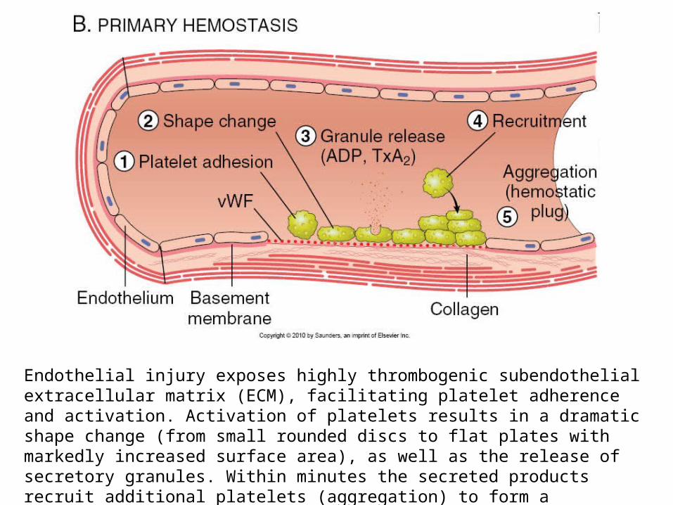
Endothelial injury exposes highly thrombogenic subendothelial extracellular matrix (ECM), facilitating platelet adherence and activation. Activation of platelets results in a dramatic shape change (from small rounded discs to flat plates with markedly increased surface area), as well as the release of secretory granules. Within minutes the secreted products recruit additional platelets (aggregation) to form a hemostatic plug; this process is referred to as primary hemostasis.

http://medcell.med.yale.edu/histology/blood_bone_marrow_lab/images/platelets_em.jpg
http://www.ouhsc.edu/platelets/Platelet%20Pics/Platelets3.jpg

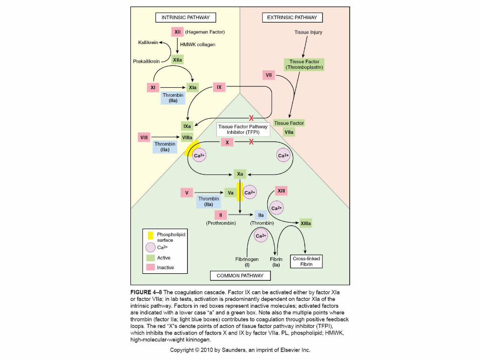
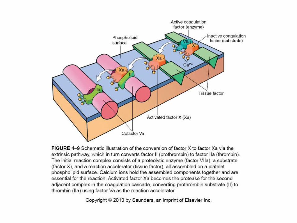
Tissue factor is also exposed at the site of injury. Also known as factor III and thromboplastin, tissue factor is a membrane-bound procoagulant glycoprotein synthesized by endothelial cells. It acts in conjunction with factor VII (see below) as the major in vivo initiator of the coagulation cascade, eventually culminating in thrombin generation. Thrombin cleaves circulating fibrinogen into insoluble fibrin, creating a fibrin meshwork, and also induces additional platelet recruitment and activation. This sequence, secondary hemostasis, consolidates the initial platelet plug.

Polymerized fibrin and platelet aggregates form a solid, permanent plug to prevent any further hemorrhage. At this stage, counter-regulatory mechanisms (e.g., tissue plasminogen activator, t-PA) are set into motion to limit the hemostatic plug to the site of injury

Endothelial cells are key players in the regulation of homeostasis, as the balance between the anti- and prothrombotic activities of endothelium determines whether thrombus formation, propagation, or dissolution occurs
Normally, endothelial cells exhibit antiplatelet, anticoagulant, and fibrinolytic properties.
After injury or activation they acquirenumerous procoagulant activities
Endothelium

Under normal circumstances endothelial cells actively prevent thrombosis by producing factors that block platelet adhesion and aggregation, inhibit coagulation, and lyse clots.
Antithrombotic Properties

Intact endothelium prevents platelets (and plasma coagulation factors) from engaging the highly thrombogenic subendothelial ECM.
Non-activated platelets do not adhere to endothelial cells, and even if platelets are activated, prostacyclin (PGI2) and nitric oxide produced by the endothelial cells impede platelet adhesion.
PGI2 and NO are potent vasodilators and inhibitors of platelet aggregation; their synthesis by the endothelium is stimulated by several factors produced during coagulation (e.g., thrombin and cytokines). Endothelial cells also elaborate adenosine diphosphatase, which degrades adenosine diphosphate (ADP) and further inhibits platelet aggregation (see below).
Antiplatelet Effects


Mediated by endothelial membrane-associated heparin-like molecules, thrombomodulin, and tissue factor pathway inhibitor.
The heparin-like molecules act indirectly; they are cofactors that greatly enhance the inactivation of thrombin and several other coagulation factors by the plasma protein antithrombin III.
Thrombomodulin binds to thrombin and converts it from a procoagulant into an anticoagulant via its ability to activate protein C, which inhibits clotting by inactivating factors Va and VIIIa
Endothelium also produces protein S, a co-factor for protein C, and tissue factor pathway inhibitor (TFPI), a cell surface protein that directly inhibits tissue factor–factor VIIa and factor Xa activities.
Anticoagulant effects

Endothelial cells synthesize tissue-type plasminogen activator (t-PA), a protease that cleaves plasminogen to form plasmin; plasmin, in turn, cleaves fibrin to degrade thrombi.
Fibrinolytic Effects

Date of download: 2/26/2013
Copyright © American College of Chest Physicians. All rights reserved.
Thrombin and Fibrinolysis*
CHEST. 2003;124(3_suppl):33S-39S.
The balance between the formation and degradation of FN. The coagulation cascade ultimately generates thrombin, which catalyzes the conversion of fibrinogen to the fibrin clot. The fibrinolytic cascade generates plasmin, which catalyzes solubilization of the FN. The thrombin-thrombomodulin complex promotes down-regulation of thrombin formation by generating activated protein C (APC). It also suppresses fibrinolysis by forming TAFIa. The two cascades are thereby linked through the thrombin, thrombomodulin, and TAFI pathway. PC = protein C.
Figure Legend:
http://upload.wikimedia.org/wikipedia/commons/0/0e/Fibrinolysis.png

While normal endothelial cells limit clotting, trauma and inflammation of endothelial cells induce a prothrombotic state that alters the activities of platelets, coagulation proteins, and the fibrinolytic system.
Prothrombotic Properties

Endothelial injury allows platelets to contact the underlying extracellular matrix; subsequent adhesion occurs through interactions with von Willebrand factor (vWF), which is a product of normal endothelial cells and an essential cofactor for platelet binding to matrix elements.
Platelet Effects
Procoagulant effects. In response to cytokines (e.g., tumor necrosis factor [TNF] or interleukin-1 [IL-1]) or bacterial endotoxin, endothelial cells synthesize tissue factor, the major activator of the extrinsic clotting cascade. In addition, activated endothelial cells augment the catalytic function of activated coagulation factors IXa and Xa.
Antifibrinolytic effects. Endothelial cells secrete inhibitors of plasminogen activator (PAIs), which limit fibrinolysis and tend to favor thrombosis.

Platelets are disc-shaped, anucleate cell fragments that are shed from megakaryocytes in the bone marrow into the blood stream.
They play a critical role in normal hemostasis by forming the hemostatic plug that initially seals vascular defects, and by providing a surface that recruits and concentrates activated coagulation factors.
Their function depends on several glycoprotein receptors, a contractile cytoskeleton, and two types of cytoplasmic granules. α-Granules have the adhesion molecule P-selectin on their membranes and contain fibrinogen, fibronectin, factors V and VIII, platelet factor 4 (a heparin-binding chemokine), platelet-derived growth factor (PDGF), and transforming growth factor-β (TGF-β). Dense (or δ) granules contain ADP and ATP, ionized calcium, histamine, serotonin, and epinephrine.
Platelets

http://path.upmc.edu/cases/case37/images/micro8.jpg
Megakaryocyte in Bone Marrow
http://www.med-ed.virginia.edu/courses/path/innes/images/nhjpeg/nh%20megakaryocyte%20x50a.jpeg
http://upload.wikimedia.org/wikipedia/commons/7/7f/Platelets_by_budding_off_from_megakaryocytes.jpg
http://www.hematology.org/assets/0/71/73/78/135/137/238/8c1482dc-1d4c-4ea5-b199-aca3f01eac83.jpg?n=8006

Platelet structure
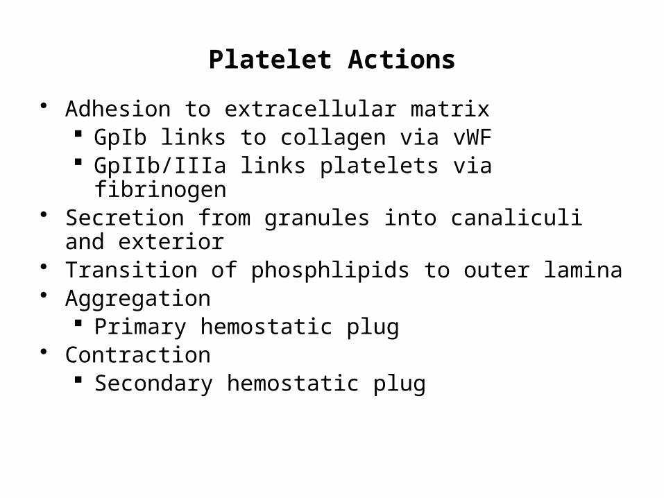
Platelet Actions
• Adhesion to extracellular matrix GpIb links to collagen via vWF GpIIb/IIIa links platelets via fibrinogen
• Secretion from granules into canaliculi and exterior• Transition of phosphlipids to outer lamina• Aggregation
Primary hemostatic plug• Contraction
Secondary hemostatic plug

Platelet aggregation

After vascular injury, platelets encounter ECM constituents such as collagen and the adhesive glycoprotein vWF. On contact with these proteins, platelets undergo:(1) adhesion and shape change, (2) secretion (release reaction)(3) aggregation

Platelet adhesion to ECM is mediated largely via interactions with vWF, which acts as a bridge between platelet surface receptors (e.g., glycoprotein Ib [GpIb]) and exposed collagen.
Although platelets can also adhere to other components of the ECM (e.g., fibronectin), vWF-GpIb associations are necessary to overcome the high shear forces of flowing blood.

Secretion (release reaction) of both granule types occurs soon after adhesion. Various agonists can bind platelet surface receptors and initiate an intracellular protein phosphorylation cascade ultimately leading to degranulation.
Release of the contents of dense-bodies is especially important, since calcium is required in the coagulation cascade, and ADP is a potent activator of platelet aggregation. ADP also begets additional ADP release, amplifying the aggregation process.
Platelet activation leads to the appearance of negatively charged phospholipids (particularly phosphatidylserine) on their surfaces. These phospholipids bind calcium and serve as critical nucleation sites for the assembly of complexes containing the various coagulation factors.
http://eurheartjsupp.oxfordjournals.org/content/10/suppl_D/D30/F1.large.jpg
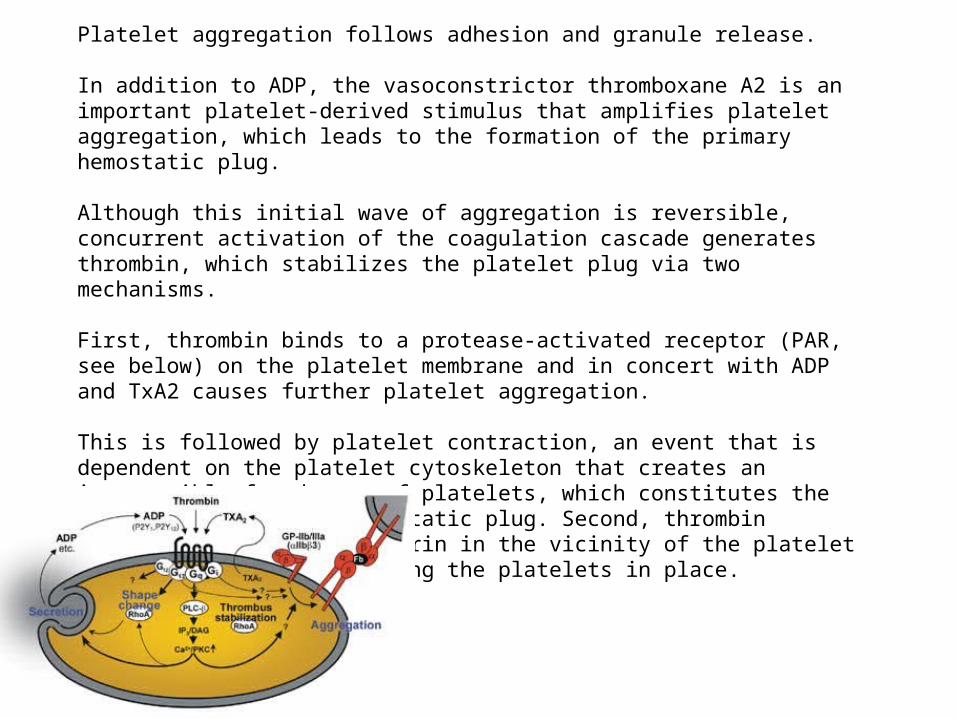
Platelet aggregation follows adhesion and granule release.
In addition to ADP, the vasoconstrictor thromboxane A2 is an important platelet-derived stimulus that amplifies platelet aggregation, which leads to the formation of the primary hemostatic plug.
Although this initial wave of aggregation is reversible, concurrent activation of the coagulation cascade generates thrombin, which stabilizes the platelet plug via two mechanisms.
First, thrombin binds to a protease-activated receptor (PAR, see below) on the platelet membrane and in concert with ADP and TxA2 causes further platelet aggregation.
This is followed by platelet contraction, an event that is dependent on the platelet cytoskeleton that creates an irreversibly fused mass of platelets, which constitutes the definitive secondary hemostatic plug. Second, thrombin converts fibrinogen to fibrin in the vicinity of the platelet plug, functionally cementing the platelets in place.
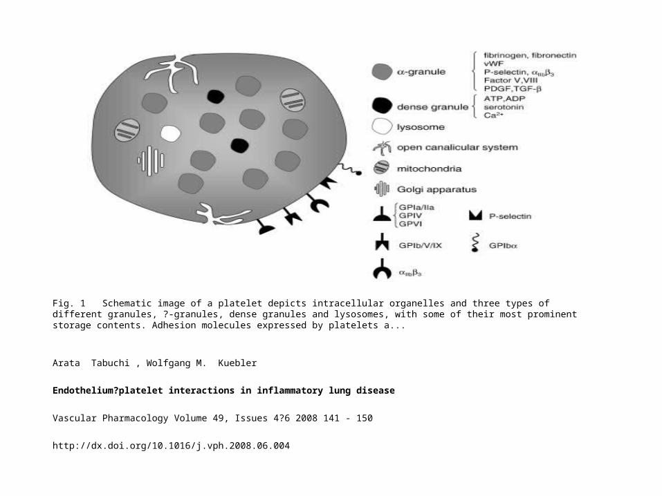
Fig. 1 Schematic image of a platelet depicts intracellular organelles and three types of different granules, ?-granules, dense granules and lysosomes, with some of their most prominent storage contents. Adhesion molecules expressed by platelets a...
Arata Tabuchi , Wolfgang M. Kuebler
Endothelium?platelet interactions in inflammatory lung disease
Vascular Pharmacology Volume 49, Issues 4?6 2008 141 - 150
http://dx.doi.org/10.1016/j.vph.2008.06.004


The endothelial cell-derived prostaglandin PGI2(prostacyclin) inhibits platelet aggregation and is a potent vasodilator; conversely, the platelet-derived prostaglandin TxA2 activates platelet aggregation and is a vasoconstrictor.
Effects mediated by PGI2 and TxA2 are balanced to effectively modulate platelet and vascular wall function: at baseline, platelet aggregation is prevented, whereas endothelial injury promotes hemostatic plug formation.
Platelet-Endothelial Cell Interactions

Fig. 2 Platelet adhesion to the injured vessel wall. At sites of vascular injury and endothelial denudation, platelets adhere to the subendothelial matrix. Platelet surface receptors GPIa/IIa, GPIV and GPVI interact directly with collagen, GPIb/V...
Arata Tabuchi , Wolfgang M. Kuebler
Endothelium?platelet interactions in inflammatory lung disease
Vascular Pharmacology Volume 49, Issues 4?6 2008 141 - 150
http://dx.doi.org/10.1016/j.vph.2008.06.004
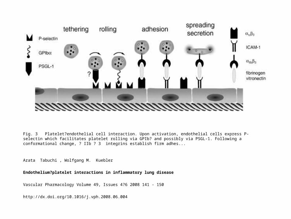
Fig. 3 Platelet?endothelial cell interaction. Upon activation, endothelial cells express P-selectin which facilitates platelet rolling via GPIb? and possibly via PSGL-1. Following a conformational change, ? IIb ? 3 integrins establish firm adhes...
Arata Tabuchi , Wolfgang M. Kuebler
Endothelium?platelet interactions in inflammatory lung disease
Vascular Pharmacology Volume 49, Issues 4?6 2008 141 - 150
http://dx.doi.org/10.1016/j.vph.2008.06.004
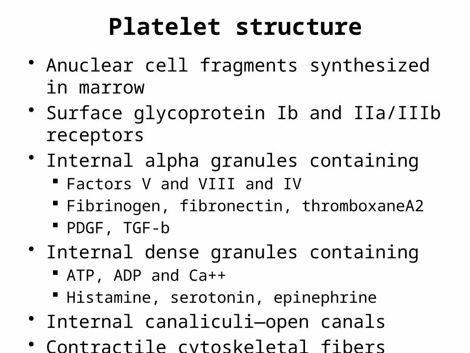
Platelet structure
• Anuclear cell fragments synthesized in marrow• Surface glycoprotein Ib and IIa/IIIb receptors• Internal alpha granules containing
Factors V and VIII and IV Fibrinogen, fibronectin, thromboxaneA2 PDGF, TGF-b
• Internal dense granules containing ATP, ADP and Ca++ Histamine, serotonin, epinephrine
• Internal canaliculi—open canals• Contractile cytoskeletal fibers


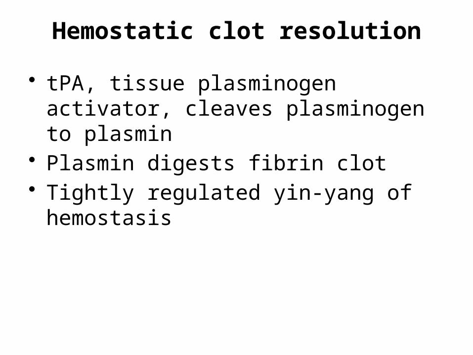
Hemostatic clot resolution
• tPA, tissue plasminogen activator, cleaves plasminogen to plasmin
• Plasmin digests fibrin clot• Tightly regulated yin-yang of hemostasis
Primary hemostatic clot formation• Platelets are activated by contact with
Extra Cellular Matrix• Circulating von Willebrand Factor tethers
platelet glycoprotein receptors to ECM collagen
• Thrombin is released to cleave fibrinogen creating fibrin nets that capture more platelets as well as RBCs and WBCs
• Platelets contract with microtubular contractile proteins, consolidating plug