pathophysiological mechanisms in antiphospholipid syndrome€¦ · pathophysiological mechanisms in...
TRANSCRIPT

157
Review
ISSN 1758-427210.2217/IJR.11.9 © 2011 Future Medicine Ltd Int. J. Clin. Rheumatol. (2011) 6(2), 157–171
Pathophysiological mechanisms in antiphospholipid syndrome
Antiphospholipid syndrome (APS) is a systemic autoimmune disease characterized by recurrent thrombosis and fetal loss in the presence of per-sistently positive antiphospholipid (aPL) antibod-ies (Abs) including lupus anticoagulant (LAC), IgG/IgM anticardiolipin (aCL) Abs and anti-b2-glycoprotein I (b
2GPI) Abs [1–3]. aPL Abs are a
heterogenous group of autoantibodies that react to phospholipids (PLs), PL-binding proteins and PL–protein complexes. aPL Abs mainly target the antigen b
2GPI and along with Abs acting against
prothrombin (PT) account for more than 90% of the Ab-binding activity in APS patients [4–6]. While many aPL Abs exhibit specificity for a sin-gle antigen, purified aPL Abs from some patients bind multiple proteins involved in coagulation, suggesting a single aPL Ab clone can induce multiple changes in coagulation and cell activity resulting in thrombosis or fetal loss [7].
Presence of aPL Abs per se does not guaran-tee a patient will develop APS as only 8.1% of patients with aPL Abs without a history of clini-cal thrombosis developed thrombosis during a 5-year follow-up period, suggesting that a patient needs an additional insult to develop the clinical disease [8]. Supporting this ‘two-hit’ hypothesis, comorbidities that have been identified as signifi-cant risk factors for thrombosis in APS include hypertension, presence of an autoimmune dis-ease, hypercholesterolemia, presence of anti-dsDNA Abs or medium-to-high titer aCL Abs [8]. Risk factors particularly for arterial thrombo-sis include hypertension, hyperhomocysteinemia
and use of hormone-replacement therapy or oral contraceptives [9]. By contrast, venous thrombo-sis was associated with presence of hypertriglyc-eridemia, presence of a hereditary thrombophilia or aCL IgG more than 40 IU [9].
Antiphospholipid syndrome causes significant morbidity with positive LAC and/or aPL Abs conferring an increased risk of thrombosis with an odds ratio ranging from 3.1 to 9.4 [10,11]. The risk of recurrent thrombosis in APS over 5 years is 16.6% despite the use of anticoagulants and/or aspirin. APS is also associated with a 5-year mor-tality of 5.3% with most deaths occurring within the first year of diagnosis, with the leading causes of death being bacterial infections, myocardial infarction, stroke and cerebral hemorrhage [12]. Catastrophic APS – multiple simultaneous arte-rial or venous thromboses in the presence of aPL Abs – is a much-feared, albeit rare complication occurring in 0.9% of patients with APS. Despite aggressive treatment, mortality rates still range between 44 and 55.6% [12,13]. These data under-score the need for development of more effec-tive therapies that target the pathologic processes involved in APS without the toxicities associated with chronic anticoagulation.
Structure & binding of b2GPIStudies into the structure of b
2GPI have revealed
that the individual domains are important for interaction with aPL Abs as well as cell-sur-face molecules leading to the pathologic fea-tures of APS. Anti-b
2GPI Abs associated with
Antiphospholipid syndrome is a systemic autoimmune disease associated with thrombosis and recurrent fetal loss in the setting of detectable antiphospholipid (aPL) antibodies. The major antigenic target has been identified as b2-glycoprotein I (b2GPI), which mediates binding of aPL antibodies to target cells including endothelial cells, monocytes, platelets and trophoblasts, leading to prothrombotic and proinflammatory changes that ultimately result in thrombosis and fetal loss. This article summarizes recent insights into the role of b2GPI in normal hemostasis, interactions between aPL antibodies, b2GPI and cell-surface molecules, molecular prothrombotic and proinflammatory changes induced by aPL antibodies and pathogenic changes leading to fetal loss in antiphospholipid syndrome. New directions in therapy using these insights are examined.
KEYWORDS: annexin n anti-b2-glycoprotein I antibody n antiphospholipid antibodies n antiphospholipid syndrome n endothelial cell activation n pathogenesis n platelet activation n pregnancy loss n thrombosis n treatment
Brock E Harper1, Rohan Willis1 & Silvia S Pierangeli†1
1Department of Internal Medicine, Division of Rheumatology, University of Texas Medical Branch, Galveston, TX, USA †Author for correspondence: [email protected]

Int. J. Clin. Rheumatol. (2011) 6(2)158 future science group
Review Harper, Willis & Pierangeli Pathophysiological mechanisms in antiphospholipid syndrome Review
thrombosis have been shown to bind readily to domain I of recombinant b
2GPI but interact
only with plasma b2GPI attached to an anionic
surface, implying that a conformational change is necessary for b
2GPI binding [14–16]. This was
recently confirmed and expanded upon by Agar et al. who demonstrated that b
2GPI has a
native circular structure in plasma maintained by interaction of domains V and I. Exposure to an anionic surface leads to the opening of the structure into a fish-hook shape via interac-tion between the anionic surface and domain V. This conformational change facilitates binding of anti-b
2GPI to b
2GPI by exposing an epitope
in domain I that is hidden in the circular con-formation [17]. The light chain fragments of anti-b
2GPI Abs carry the antigen specificity,
establishing that the interaction is with b2GPI
itself and not immunoglobulin in general [18]. Polymorphisms of b
2GPI are also found in APS
with the replacement of leucine by valine at position 247 being found more frequently in APS patients in certain populations [19–22]. This polymorphism, particularly in the homozygous state, has been shown to be associated with the production of anti-b
2GPI Abs, which may be
due to the presence of valine at this site, caus-ing an increase in the antigenicity of b
2GPI [20].
Providing clinical evidence of the importance of domain I binding in APS, de Laat et al. demon-strated that presence of domain I-specific anti-b
2GPI Abs was associated with increased risk of
thrombosis, while there was no increased risk from anti-b
2GPI Abs targeting other domains
of b2GPI [15,23].
Recent studies have established that domain V is the primary site of interaction between b
2GPI and the various cell-surface molecules
with which it interacts. Rahgozar et al. demon-strated that b
2GPI binds to thrombin and this
binding prevents the inactivation of thrombin by heparin/heparin cofactor II [24,25]. Binding of anti-b
2GPI Abs potentiates this interaction,
leading to increased thrombin activity [24,25]. b
2GPI also binds to ApoER2 ,́ a splice variant of
ApoE receptor 2 (ApoER2) found on platelets, and this interaction is dependent upon domain V [26,27]. Binding to ApoER2´ by dimerized b
2GPI induces platelet activation and platelet
aggregation. This effect was reproduced with anti-b
2GPI Abs but not normal plasma b
2GPI,
indicating that anti-b2GPI Abs exert some of
their effects by dimerization of b2GPI and
interaction of dimerized b2GPI with cell-sur-
face receptors [28]. In a recent study, Romay-Penabad et al. showed that ApoER2 mediates
aPL-mediated pathogenic effects in vivo, in experiments that utilized ApoER2´-deficient mice and specific inhibitors [29]. b
2-glycoprotein I interacts with annexins,
a family of phospholipid-binding proteins of which annexin (Ann) A2 and Ann A5 have been implicated in the pathogenesis of APS. Ann A2 acts as an endothelial cell-surface receptor for plasminogen and tissue plas-minogen activator (tPA), and also mediates the binding of b
2GPI/anti-b
2GPI complexes
to endothelial cells and monocytes, leading to cell activation and expression of a proco-agulant phenotype [30–33]. Furthermore, aPL Ab-induced increases in endothelial cell adhe-sion molecules, ICAM and E-selectin have been shown to be abrogated by treatment with anti-Ann A2 Abs [34]. Also, knockout of Ann A2 in mice significantly blunted thrombus for-mation and diminished vascular tissue factor (TF) and VCAM expression induced by aPL Ab exposure [34]. Similarly, Zhou et al. have found that increased expression of ANX2, the gene coding for Ann A2, leads to increased anti-b
2GPI Ab-induced TF expression while
silencing of ANX2 partially blocks this increase in TF expression in HEK 293 T cells [31]. All this evidence highlights the essential role that Ann A2 plays as a cell-surface receptor for aPL in the induction of a procoagulant state in APS patients. However, it is important to note that although cell-surface Ann A2 lacks an intra-cellular tail and is unable to induce intracel-lular signal transduction by itself, it can act as a binding partner for intracellular surface molecules in lipid rafts [34,35]. This means that Ann A2 most likely utilizes a coreceptor for intracellular signal transduction and subse-quent cell activation as a result of aPL action. The effects of anti-b
2GPI Ab binding appear
to be dependent upon Ann A2 crosslinking, suggesting anti-b
2GPI Abs may exert their
effects on cell signaling by inducing interaction between Ann A2-associated intracellular sign-aling molecules [36]. There is evidence for Toll-like receptors (TLRs), particularly TLR4 and TLR2, functioning as coreceptors for Ann A2 in aPL-mediated cell activation, both of these TLRs being present on endothelial cells and monocytes [37,38]. Abs directed against Ann A2 itself have also been detected in 14.8–40.4% of patients with APS and are associated with in vitro prothrombotic changes including increased expression of TF and inhibition of tPA-mediated plasmin activation suggesting an anti-b
2GPI Ab-independent role in APS [30,39].
Review Harper, Willis & Pierangeli
www.futuremedicine.com 159future science group
Pathophysiological mechanisms in antiphospholipid syndrome Review
Annexin A5 is another member of the annexin family and is expressed by placental villous syncytiotrophoblasts and vascular endothelial cells. It exerts anticoagulant activity by binding to anionic PL blocking the activation of factor (F)IX and FX by TF and FVIIa and the activa-tion of PT by FXa and FVa [40]. This interaction with anionic PL has made Ann A5 a promising possible target for aPL Abs. The anticoagulant effect of Ann A5 and aPL Ab-induced effects on Ann A5 can be monitored by evaluation of the Ann A5 anticoagulant ratio, a ratio of coagula-tion time in the presence of Ann A5 compared with the absence of Ann A5. Sera from patients with APS, particularly those with Abs target-ing domain I of b
2GPI, were found to decrease
Ann A5 binding and decrease the Ann A5 anti-coagulant ratio, indicating a prothrombotic state compared with control sera from healthy individuals and patients with syphilis [41–44]. This decrease in binding appears to be due to augmented b
2GPI binding to anionic PL by aPL
Abs and thus occupying the binding sites for Ann A5, preventing the anticoagulant effect of Ann A5 [44,45].
Thrombogenic mechanisms in APS�n Effects of aPL Abs on monocytes,
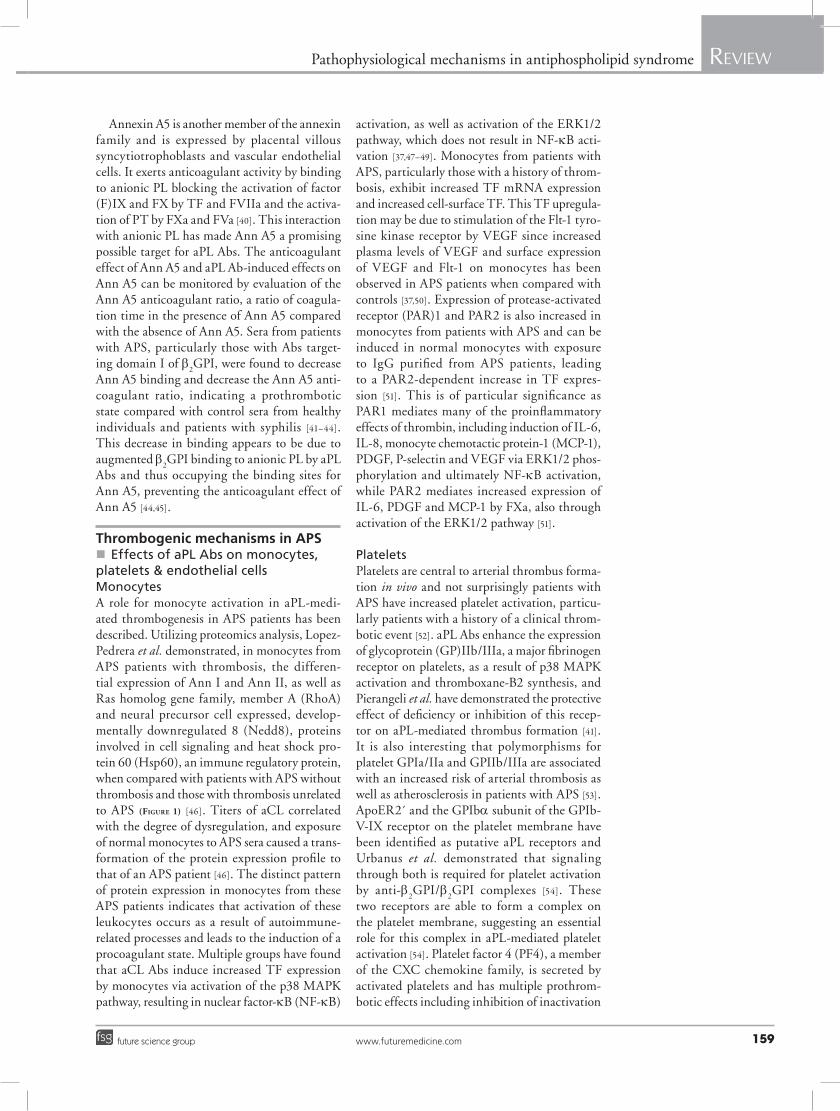
platelets & endothelial cells MonocytesA role for monocyte activation in aPL-medi-ated thrombogenesis in APS patients has been described. Utilizing proteomics analysis, Lopez-Pedrera et al. demonstrated, in monocytes from APS patients with thrombosis, the differen-tial expression of Ann I and Ann II, as well as Ras homolog gene family, member A (RhoA) and neural precursor cell expressed, develop-mentally downregulated 8 (Nedd8), proteins involved in cell signaling and heat shock pro-tein 60 (Hsp60), an immune regulatory protein, when compared with patients with APS without thrombosis and those with thrombosis unrelated to APS (Figure 1) [46]. Titers of aCL correlated with the degree of dysregulation, and exposure of normal monocytes to APS sera caused a trans-formation of the protein expression profile to that of an APS patient [46]. The distinct pattern of protein expression in monocytes from these APS patients indicates that activation of these leukocytes occurs as a result of autoimmune-related processes and leads to the induction of a procoagulant state. Multiple groups have found that aCL Abs induce increased TF expression by monocytes via activation of the p38 MAPK pathway, resulting in nuclear factor-kB (NF-kB)
activation, as well as activation of the ERK1/2 pathway, which does not result in NF-kB acti-vation [37,47–49]. Monocytes from patients with APS, particularly those with a history of throm-bosis, exhibit increased TF mRNA expression and increased cell-surface TF. This TF upregula-tion may be due to stimulation of the Flt-1 tyro-sine kinase receptor by VEGF since increased plasma levels of VEGF and surface expression of VEGF and Flt-1 on monocytes has been observed in APS patients when compared with controls [37,50]. Expression of protease-activated receptor (PAR)1 and PAR2 is also increased in monocytes from patients with APS and can be induced in normal monocytes with exposure to IgG purified from APS patients, leading to a PAR2-dependent increase in TF expres-sion [51]. This is of particular significance as PAR1 mediates many of the proinflammatory effects of thrombin, including induction of IL-6, IL-8, monocyte chemotactic protein-1 (MCP-1), PDGF, P-selectin and VEGF via ERK1/2 phos-phorylation and ultimately NF-kB activation, while PAR2 mediates increased expression of IL-6, PDGF and MCP-1 by FXa, also through activation of the ERK1/2 pathway [51].
PlateletsPlatelets are central to arterial thrombus forma-tion in vivo and not surprisingly patients with APS have increased platelet activation, particu-larly patients with a history of a clinical throm-botic event [52]. aPL Abs enhance the expression of glycoprotein (GP)IIb/IIIa, a major fibrinogen receptor on platelets, as a result of p38 MAPK activation and thromboxane-B2 synthesis, and Pierangeli et al. have demonstrated the protective effect of deficiency or inhibition of this recep-tor on aPL-mediated thrombus formation [41]. It is also interesting that polymorphisms for platelet GPIa/IIa and GPIIb/IIIa are associated with an increased risk of arterial thrombosis as well as atherosclerosis in patients with APS [53]. ApoER2´ and the GPIba subunit of the GPIb-V-IX receptor on the platelet membrane have been identified as putative aPL receptors and Urbanus et al. demonstrated that signaling through both is required for platelet activation by anti-b
2GPI/b
2GPI complexes [54]. These
two receptors are able to form a complex on the platelet membrane, suggesting an essential role for this complex in aPL-mediated platelet activation [54]. Platelet factor 4 (PF4), a member of the CXC chemokine family, is secreted by activated platelets and has multiple prothrom-botic effects including inhibition of inactivation
Int. J. Clin. Rheumatol. (2011) 6(2)160 future science group
Review Harper, Willis & Pierangeli Pathophysiological mechanisms in antiphospholipid syndrome Review
of thrombin by anti-thrombin, potentiation of platelet aggregation and accelerating cleavage of activated protein C (aPC). It is also an anti-genic target in APS. Abs to PF4 are implicated in heparin-induced thrombocytopenia but have also been detected in patients with APS without a history of heparin-induced thrombocytopenia or recent exposure to heparin. Levels of anti-PF4 Abs also correlate with levels of IgM aPL and IgM anti-b
2GPI Abs, but have activity indepen-
dent of both [55,56]. These data suggest another
potentially important mechanism in APS, but correlation with clinical features remains to be established. Recently, Sikara et al. demonstrated that PF4 may also facilitate the dimerization of b
2GPI and subsequent binding to anti-b
2GPI
Abs and platelet cell-surface receptors includ-ing activation of the p38 MAPK pathway and NF-kB production [57]. This presents not only an attractive mechanistic model for platelet activation in APS but also for activation of endothelial cells and monocytes since PF4 is
TLR4
Proinflammatorystate
Platelet activation
Platelet
Endothelium
MAC
Thrombosis
Complementactivation
Polymorphonuclear leukocyte
TNF-α IL-8
IL-6
TF
TF TF
C5aC5a
aPLβ2GPI
TXB2
ApoER2´
ApoER2´
GPIb-V-IX
NF-κBp38 MAPK
GPIIb/IIIa
Ann A2
Endothelialinjury
VEGF
VEGFVEGF
C5aR
IL-1
NF-κB
NF-κB
p38 MAPK
p38 MAPK
C5aR
Monocyte
Flt-1
VCAM-1 E-selectin ICAM-1
NF-κBp38 MAPK
Int. J. Clin. Rheumatol. © Future Science Group (2011)
Figure 1. Cell activation in antiphospholipid syndrome. aPL, including anti-b2GPI–b
2GPI complexes, can activate platelets,
endothelial cells and monocytes. Endothelial cell activation leads to the release of proinflammatory cytokines and increased leukocyte adhesion, making possible the activation of the polymorphonuclear leukocyte. Monocyte activation leads to the release of proinflammatory cytokines as well including TF, which can potentiate coagulation factor activation and ultimately fibrin production. Platelet activation leads to the release of thromboxane-B2, which potentiates the increased expression of GPIIb/IIIa, a major fibrinogen receptor. The net effect is the induction of a procoagulant state ultimately leading to thrombosis, occurring especially on the background of complement activation and endothelial injury due to infection and trauma. b
2GPI: b
2-glycoprotein I; Ann: Annexin; aPL: Antiphospholipid; MAC: Membrane attack complex; NF-kB: Nuclear factor kB;
TF: Tissue factor.

Review Harper, Willis & Pierangeli
www.futuremedicine.com 161future science group
Pathophysiological mechanisms in antiphospholipid syndrome Review
also expressed in these and other immune cells, albeit at lower concentrations than in platelets. PF4, like b
2GPI, occurs in abundance in plasma
and has many immunomodulatory effects. It is possible that PF4/b
2GPI complexes may play
a coordinating role in the activation of many immune-related cells in APS [58]. Furthermore, the fact that PF4 is released in abundance by dendritic cells following trauma may be one of the factors underlying the association of severe trauma with catastrophic APS [59]. However, fur-ther study is needed to fully characterize the role that PF4 plays in the pathophysiology of APS.
Endothelial cells Endothelial cells play a key role in thrombosis by expression of integrins and TF upon activation and have also been implicated in APS. Patients with APS exhibit evidence of increased endothe-lial cell activity with impairment of endothe-lium-dependent vasodilatation and increased expression of von Willebrand factor (vWF), tPA, placental growth factor and soluble ICAM-1 [36,52,60–65]. Anti-b
2GPI Abs induce a prothrom-
botic milieu in endothelial cells via increased production of FVIIa, PT-fragments I and II, and decreased levels of FXIIa and active uroki-nase-type plasminogen activator [66]. Microarray studies of endothelial cells have shown induction of apoptosis-related genes, BCL-2A1, TRAF1, CARD15 and BIRC3, multiple adhesion mol-ecules including E-selectin, ICAM and VCAM, coagulation factors, cytokine/chemokine recep-tors and cytokines including IL-8, IL-6, IL-18 receptor, IL-1b and the TNF receptor superfamily [67]. This suggests that the procoagulant pheno-type induced in endothelial cells in response to aPL Abs is characterized by increased apoptosis, leuko cyte adhesion and release of proinflam-matory cytokines. Similar to the cell signaling changes observed in monocytes, treatment of endothelial cells with aPL Abs induces activa-tion of the p38 MAPK pathway and endothe-lial cell activation with increased expression of TF, VCAM, ICAM, P-selectin and E-selectin [48,61,68–70]. Mutations in the endothelial cell P-selectin ligand have also been identified as a risk factor for thrombosis in APS [71].
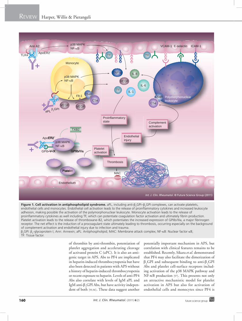
�n Effects of aPL Abs on humoral regulators of thrombosis Regulators of coagulationProtein C (PC) is activated by thrombomod-ulin-bound thrombin and acts as a key regulator of thrombosis by inactivating FVa and FVIIa and activating PAR1, resulting in inhibition
of apoptosis (Figure 2). Modulation of aPC has been found in APS patients who have increased resistance to aPC, resulting in greater thrombin generation over time. The presence of in vitro aPC resistance is associated with the presence of LAC and a clinical history of thrombosis [72]. IgM Abs against aPC have been isolated from patients with APS and confer an increased risk of venous thromboembolism [73]. PC is released by membrane-bound endothelial PC receptor (EPCR) but soluble EPCR binds aPC, providing a counter-regulator mechanism. Anti-EPCR Abs have also been identified in APS sera and pres-ence of these Abs is associated with a significant increase in risk of venous thrombosis and fetal loss, suggesting interference with this regulatory mechanism [74,75].
Antiphospholipid Abs have also been shown to interfere with the function of anti-thrombin III, another important regulator of coagulation, resulting in reduced inactivation of several coag-ulation factors including FXa and FIXa [76,77]. Anti-b
2GPI Abs also induce increased thrombin
generation by interference with TF pathway inhibitor-dependent inhibition of TF-induced thrombin generation [78]. b
2GPI also exerts anti-
thrombotic effects by interaction with activated vWF. b
2GPI binds to vWF leading to a decrease in
platelet binding by vWF and inhibition of plate-let activation. Anti-b
2GPI Abs exert some of their
prothrombotic effects by interference with the binding of b
2GPI to activated vWF, resulting in
an increased concentration of circulating activated vWF and enhanced platelet aggregation [79].
Coagulation factorsDysregulation of thrombosis in APS also occurs due to interference with multiple coagulation fac-tors by aPL Abs. Anti-FXa Abs are identified in 13.2% of patients with APS and result in a pro-thrombotic state by interference with inactivation of FXa by anti-thrombin [76]. Mutation of FXIIIa with substitution of valine for leucine at posi-tion 34 appears to be protective with decreased risk of thrombosis in patients with aPL Abs [80]. Antiprothrombin (aPT) Abs have also been sug-gested as an antigenic target in APS. PT is the precursor form of thrombin, an integral protein in perpetuation and modulation of the throm-botic response. aPT Abs exhibit an anticoagulant effect in vitro but are associated with thrombosis and recurrent miscarriages in vivo and induce increased thrombin and fibrin generation and increased TF and E-selectin expression by endothelial cells [81–83]. These contradictory find-ings may be due to modulation of these effects by
Int. J. Clin. Rheumatol. (2011) 6(2)162 future science group
Review Harper, Willis & Pierangeli Pathophysiological mechanisms in antiphospholipid syndrome Review
the local concentration of other coagulation fac-tors, particularly FVa and FXa with low levels of FVa and high levels of FXa, being associated with increased thrombin generation [81]. However, it is important to note that there is currently no consensus regarding the role of aPT Abs in APS, at least partly due to a lack of standardization of laboratory assays leading to difficulty integrating results from groups using different assays.
Fibrinolytic systemAntiphospholipid syndrome patients also demon strate defects in fibrinolysis in addition to the decreased expression of tPA by endothe-lial cells described above [66]. b
2GPI regulates
hemostasis by modulation of fibrinolysis as well. Plasma b
2GPI binds to tPA, increasing
its catalytic activity and inducing an increase in plasminogen resulting in an augmentation of fibrino lysis. Treatment with anti-b
2GPI Abs
blocks the stimulation of tPA-mediated plas-minogen activation [84]. Antiplasminogen Abs are detectable in 25–46.9% of patients with APS and are associated with clinical throm-botic events. Patients with these autoantibodies
had impaired fibrinolysis by inhibition of tPA-dependent plasminogen activation [84]. Fibrinolysis may also be impaired due to interac-tion with thrombin-activatable fibrinolysis inhib-itor (TAFI), a proenzyme that upon activation by thrombin–thrombo modulin and multiple pro-teases potently inhibits fibrinolysis. While TAFI levels are elevated in patients with APS, actual activity of TAFI was lower in patients with APS, particularly in those with LAC [85]. In addition, aCL Abs inhibited the formation of TAFI [85]. However, modulation of TAFI activity was not correlated with clinical thrombosis and therefore its role in the pathogenesis of APS is still unclear.
�n Induction of proinflammatory changes by aPL AbsIn addition to activation of the canonical coagula-tion pathways, APS is marked by several proin-flammatory changes. Family studies have sug-gested involvement of TLRs including TLR4 and inflammatory signaling involving IL-1b, TNF-a, IL-6 and TGF-b. Recent genetic and protein expression studies have uncovered associations with single-nucleotide polymorphisms in genes
TF
Fibrinolysis
Plasmin
tPA
Plasminogen
FDP
Fibrin
Fibrinogen
ThrombinProthrombin
aPC
X
Xa VaIXa
VIIa
VIIIa
XIXIa
XIIa
IX
ATIII
Activatedplatelets
β2GPI
PAI-1
Thrombosis
Thrombus
Figure 2. Dysregulation of anticoagulant and fibrinolytic systems in antiphospholipid syndrome. aPC, an important anticoagulant, functions by inactivating activated forms of factors V (Va) and VIII (VIIIa). Antibodies acting against aPC inhibit its function. The function of anti-thrombin III, another major regulator of coagulation factors, is also inhibited by antibodies isolated from antiphospholipid syndrome patients. Ultimately this results in increased thrombin generation and subsequent fibrin formation. Dysregulation of the fibrinolytic system can also occur in antiphospholipid syndrome due to antibodies that inhibit tPA-mediated conversion of plasminogen to plasmin and those that directly inhibit plasmin function. aPC: Activated protein C; tPA: Tissue plasminogen activator.

Review Harper, Willis & Pierangeli
www.futuremedicine.com 163future science group
Pathophysiological mechanisms in antiphospholipid syndrome Review
coding for the cell signaling molecules STAT4 and BLK [86–88]. Patients with APS have been found to have elevated levels of TNF-a, which correlate with the presence of LAC and presence of both aCL and anti-b
2GPI Abs [89]. In vitro
studies have demonstrated that anti-b2GPI Abs
induce TNF-a production by monoctyes [90]. In addition to endothelial cell and monocyte acti-vation, and increased TF expression described above, aPL Ab-induced activation of endothelial cell p38 MAPK also leads to increased expression of the proinflammatory cytokines TNF-a, IL-1b, TGF-b, macrophage inflammatory protein 3 and MCP-1 in monocytes and increased expression of IL-6 and IL-8 in endothelial cells [49,68].
There has been much interest over the past sev-eral years in TLRs in APS. Prothrombotic effects of sera from patients with APS were decreased in mice resistant to lipopolysaccharide, a potent activator of TLR4, implicating involvement of TLR4 signaling in APS [91]. This finding was supported by further studies demon strating that b
2GPI associates with lipid rafts and colocalizes
with TLR2 and TLR4. b2GPI interacts with
TLR2 as well as TLR4, leading to activation of cell signaling cascades including ERK and p38 MAPK, resulting in IRAK phosphorylation and activation of NF-kB [90,92]. The interaction with TLR2 is potentiated by dimerized b
2GPI, sug-
gesting the need for interactions with other cell-surface molecules such as Ann A2 to induce sig-naling [92]. In fibroblasts, this TLR2 activation leads to increased expression of IL-6, ICAM-1 and MCP-1 [38]. aPL Abs are also able to induce a inflammatory milieu via other TLRs. TLR8 activation in monocytes and TLR 7 activation in dendritic cells by aPL Abs results in an increase in the proinflammatory cytokine IL-1b [93].
T cells play a key role in the regulation of the adaptive immune response and have also been implicated in the pathogenesis of APS. Patients with APS have increased circulating CD4+ T cells, decreased naive T cells (Th0) and cytotoxic CD8+ T-cell populations com-pared with patients with stable coronary artery disease with increased IFN-g, IL-1b, IL-4 and IL-6 and decreased IL-8 and IL-10 pro-duction [63]. These findings suggest APS pro-motes an enhanced Th2 response promoting a humoral immune response and impairment of counter-regulatory cytokine production. T cells in peripheral blood cell cultures become acti-vated when exposed to b
2GPI/anti-b
2GPI Ab
complexes and also in the presence of oxidized low-density lipoprotein (oxLDL) and activated platelets [94]. This activation is dependent on
the presence of macrophages and the FcgRI immuno globulin receptor suggesting T-cell acti-vation is induced by presentation of PL antigens by macrophages [94].
Complement activation has also been impli-cated in APS. One study of a Japanese cohort of patients with primary APS found the majority had hypocomplementemia, which was associated with increased levels of TNF-a and the pres-ence of LAC and anti-PT Abs [95]. Complement fixation occurs on the platelets of patients with APS and correlates with the presence of IgG aPL Abs, IgG anti-b
2GPI Abs, platelet activation and
also history of arterial thrombosis [96]. In vitro exposure to aPL Abs produced complement fixa-tion on normal platelets and increased platelet activity correlated with complement fixation [96]. Additionally, patients with APS have been found to have lower levels of soluble CD21, the cell surface receptor for multiple components of activated complement protein 3 (C3). These lower levels of CD21 are independent of the presence of anti-b
2GPI Abs, which supports the
role of other aPL Abs in immune system activa-tion in APS [97]. C5 has also been implicated as blockade of the C5a receptor. The use of specific inhibitors of C5a prevents aPL Ab-induced TF expression in neutrophils and knockout of C5a receptor protects mice from aPL Ab-induced thrombosis [62, 98,99].
Defective apoptosis is thought to be an inte-gral part of the pathogenesis of systemic lupus erythematosus (SLE) and has also been impli-cated in APS [100]. Alessandri et al. demonstrated that aCL Abs bind to CL expressed on the surface of apoptotic endothelial cells in APS patients, including those that do not have SLE [101]. They speculated this may represent defective apoptosis leading to presentation of CL to the immune system and subsequent development of autoantibodies. Some patients with APS also exhibit antiendothelial cell Abs, which decrease the uptake of neighboring apoptotic debris by endothelial cells while healthy endothelial cells will readily uptake apoptotic cells [102]. These antiendothelial cell Abs also opsonized the apop-totic cells, increasing uptake by macrophages. Macrophages that took up these apoptotic cells exhibited increased TF production, resulting in a prothrombotic state.
Nonthrombotic mechanisms in APS�n APS & atherosclerosis
Antiphospholipid syndrome is associated with markers of accelerated atherosclerosis including increased carotid intima medial thickness and

Int. J. Clin. Rheumatol. (2011) 6(2)164 future science group
Review Harper, Willis & Pierangeli Pathophysiological mechanisms in antiphospholipid syndrome Review
impairment of flow-mediated dilatation, a meas-ure of endothelial function [103]. APS sera were also able to suppress high-density lipoprotein-induced nitric oxide release, VCAM-1 expres-sion and superoxide production by endothelial cells as well as inhibiting monocyte adhesion to endothelial cells, indicating decreased endothe-lial cell activation [103]. b
2GPI exerts anti-athero-
sclerotic effects in normal physiology by bind-ing of b
2GPI to oxLDL, forming complexes that
inhibit the uptake of oxLDL by the macrophage scavenger receptor [104]. Anti-b
2GPI Abs can bind
to oxLDL–b2GPI complexes, facilitating uptake
by the macrophage scavenger receptor and induc-ing transformation of macrophages into foam cells, thus promoting atherogenesis [105,106]. Some patients with APS express anti-oxLDL Abs, which can bind to b
2GPI and are also associated
with increased risk of arterial thrombosis [107].
Obstetric manifestations of APSAs described in aPL-mediated thrombosis, several pathogenic mechanisms have been sug-gested to play a role in APS-associated obstet-rical manifestations, which is supported by the heterogeneity of the histological lesions found [108].
Intraplacental thrombosis with maternal–fetal blood exchange impairment was initially sug-gested to be the main pathogenic mechanism. Widespread thrombosis and infarction of pla-centas obtained from women with APS was reported both in first- and second-trimester abortions. However, such a histological find-ing is not specific for APS, also being present in other conditions [108,109].
In favor of the pathogenic role of thrombotic events in aPL-associated pregnancy loss there is evidence from in vitro studies that aPL may induce a procoagulant state at the placental level [110]. A further thrombophilic mechanism mediated by aPL Abs involves the relationship between the autoantibodies and Ann A5. In physiological conditions, a crystal shield of Ann A5 is suggested to cover thrombogenic anionic surfaces and prevent the activation of the coagula-tion cascade by inhibiting the binding of activated FX and PT. In vitro studies demonstrated that aPL/anti-b
2GPI might disrupt the anticoagulant
Ann A5 crystal shield; such an effect was also reproduced on trophoblast and endothelial cell monolayers [43,111,112]. According to the hypoth-esis that a loss of the Ann A5 shield may play a pathogenic role, the same group reported a significantly lower distribution of Ann A5 cov-ering the intervillous surfaces in the placenta of
aPL-positive women [111]. A new mechanistic test that measures the decrease of the anticoagu-lant effect of Ann A5 by aPL Abs in plasma has recently been developed. Studies utilizing this test have shown that the assay correctly identifies aPL-mediated thrombosis and pregnancy losses and it is ‘positive’ in approximately 50% of patients with APS [113]. Pregnancy loss in APS is due to multi-ple causes including mechanisms independent of thrombosis. Trophoblast differentiation and inva-sion is a vital step in early fetal implantation and development, and is dependent on altered expres-sion of the apoptosis regulatory proteins B-cell lymphoma 2 (Bcl-2) and Bcl-2-associated X pro-tein (Bax). This change in expression is impaired by exposure to anti-b
2GPI Abs [114]. Patients with
APS without clinical thrombosis but with fetal loss were found to have lower expression of fibrin-ogen, which has been identified as a risk factor for recurrent spontaneous miscarriages due to impaired trophoblast implantation and vascular rupture, suggesting another potential factor in the fetal loss observed in APS [46]. Anti-b
2GPI Abs
also cause trophoblasts to express a proinflamma-tory cytokine profile characterized by increased IL-8, IL-1b, MCP-1 and growth-regulated onco-gene-a via activation of the TLR4/myeloid differ-entiation primary response protein 88 (MyD88) pathway [115]. Expression of these proinflamma-tory cytokines was associated with an increase in trophoblast apoptosis. Interestingly, heparin attenuated these increases in cytokine produc-tion and increased trophoblast survival, suggest-ing immunomodulatory effects in addition to its well-characterized anticoagulant properties [115]. Mulla et al. also demonstrated that anti-b
2GPI
Abs impaired first-trimester trophoblast migration by downregulating IL-6 expression and STAT3 activation, which are constitutively expressed by first-trimester trophoblasts [116]. This occurs in a TLR4/MyD88-independent manner, suggest-ing stimulation of other intracellular signaling pathways [116].
Additional immune system abnormalities have been associated with recurrent fetal loss in APS. High levels of circulating natural killer cells have been found in APS patients with recurrent miscarriages [117,118]. Natural killer cell fractions of greater than 18% of circulating leukocytes were strongly associated with recur-rent fetal loss [117]. Redecha et al. demonstrated that aPL Abs that activate complement induce TF expression on mouse decidua and led to fetal resorption while the inhibition of comple-ment activation prevented fetal loss. Induction of TF production by complement occurred

Review Harper, Willis & Pierangeli
www.futuremedicine.com 165future science group
Pathophysiological mechanisms in antiphospholipid syndrome Review
indepen dently of membrane attack complex activation in this animal model while depletion of granulo ctyes or selective knockout of TF in myeloid cells prevented aPL-induced fetal loss. This indicates that complement does not directly cause fetal injury but rather leads to fetal loss by recruitment of neutrophils and other cells associated with innate immunity [119]. Further studies by Redecha et al. showed that aPL Abs induce neutrophil recruitment and activation, with increased TF expression and ultimately fetal loss, by activation of PAR2 by TF–FVIIa complexes, supporting the role of neutrophils in obstetric manifestations of APS [120].
Di Simone et al. recently demonstrated that purified aPL IgG from patients with APS impaired human endometrial endothelial cell angiogenesis in vitro, and this was at least partly due to suppression of VEGF and matrix metallo-proteinase 2 expression [121]. Finally, a recent study by Praprotnik et al. found an intriguing association with hyperprolactinemia and recur-rent fetal loss in APS without signs of a prolac-tinoma or major menstrual disturbances [122]. Elevated prolactin levels also seemed to be pro-tective of thrombotic manifestations of APS, implying a nonthrombotic mechanism in fetal loss in APS [122]. Further studies will need to be carried out to clarify this relationship.
In vitro effects of novel therapies & new insights into old medicationsImproved understanding of the interactions between b
2GPI, aPL Abs and cell-surface mol-
ecules such as annexins has led to the develop-ment of a potential therapy by blocking binding of anti-b
2GPI Abs to b
2GPI. Ioannou et al. were
able to demonstrate the use of a recombinant domain I peptide, which effectively inhibits binding of anti-b
2GPI Abs to b
2GPI in vitro. In
a mouse model of thrombosis, the recombinant domain I peptide inhibited aPL Ab-induced thrombus formation and also abrogated aPL Ab-induced upregulation of VCAM-1 expres-sion in endothelium and TF expression in macrophages [123].
Another new treatment for APS is the use of inhibitors of B-cell activating factor (BAFF), a cytokine that promotes B-cell expansion and its receptors BAFF-R, TACI and B-cell maturation antigen (BCMA), which have shown promise in treating SLE [124–126]. A recent study demon-strated that blockade of BAFF-receptor (BAFF-R) and TACI with BAFF-R-Ig or TACI-Ig blocked the expression of VCAM and P-selectin, resulted in fewer activated B and T cells, delayed disease
onset and enhanced survival in a mouse model of APS. There were no changes in the develop-ment of aCL Abs and, interestingly, no preven-tion of thrombocyto penia in this study [127]. A tetramer of domain I peptide that interacts with B cells has also been developed with the goal of inducing tolerance to b
2GPI and is currently in
preclinical trials [128,129]. The use of older medications is also being
evaluated in APS. Hydroxychloroquine has been associated with lower rates of throm-bosis in cohorts of patients with APS as well as in mouse models of APS [87,130,131]. These protective effects may be mediated by inter-action with Ann A5. Hydroxychloroquine treatment induces Ann A5 deposition over anti-b
2GPI Ab–b
2GPI complexes deposited on
the PL bilayer but do not affect the immune complexes per se. Augmented deposition of Ann A5 results in restoration of the Ann A5 anticoagulant ratio, indicating normalization of the hypercoaguable state found in APS [44,45]. Hydroxymethylglutaryl coenzyme A reductase inhibitors, more commonly called statins, have also been investigated in APS with promising results. Simvastatin has decreased neutrophil activation and prevented aPL Ab-induced fetal loss in a mouse model of APS, and fluvastatin mitigates the increased proinflammatory and prothrombotic changes in APS with reduc-tions in expression of ICAM, selectins, VEGF, TF, IL-6 and TNF-a and decreased thrombus formation and platelet aggregation in animal models of APS [60,120,132–134]. These effects may be due to inhibition of NF-kB binding to DNA [60]. These treatments offer the promise of improved outcomes in APS without the toxicities associated with chronic anticoagulation.
Conclusion & future perspectiveWhile recent advances in our understanding of the pathogenesis of APS have revealed many interesting insights, they have also unveiled sev-eral areas of interest to be explored. The discov-ery of binding of b
2GPI to cell-surface molecules
such as Ann A2, which does not itself initiate intracellular signaling but rather is located in lipid rafts in association with intracellular pro-teins and cell-surface receptors capable of initiat-ing cell signaling, reinforces the need for further study of these receptors and intracellular signal-ing molecules and possibly interactions induced by anti-b
2GPI–b
2GPI complexes. Studies
are already ongoing to evaluate interactions between annexins and TLR4 and their effects on intracellular signaling and protein expression.

Int. J. Clin. Rheumatol. (2011) 6(2)166 future science group
Review Harper, Willis & Pierangeli Pathophysiological mechanisms in antiphospholipid syndrome Review
Recognition of the importance of domain I in binding for anti-b
2GPI Abs opens additional
avenues of study in APS. Blockade of binding of anti-b
2GPI to b
2GPI by decoy domain I and
induction of tolerance to b2GPI are concep-
tually very promising therapies for APS but need to be further evaluated in animal studies and clinical trials. Additionally, development of clinical assays for domain I-specific anti-b
2GPI Abs may be able to identify patients
truly at increased risk for thrombosis and who would thus potentially benefit from pri-mary prophylaxis against a thromboembolic event while sparing those without increased risk from the long-term complications of chronic anticoagulation.
Insights into the intracellular signaling involved in APS also opens the possibility of treat-ments targeting cell signaling pathways, particu-larly the p38 MAPK pathway and NF-kB, with early evidence of abrogation of the prothrombotic state induced by APS [50,135]. Inhibition of ERK signaling also has in vitro data supporting it as a possible therapeutic target with abrogation of anti-b
2GPI-induced TNF-a and TF expression
by monocytes [90]. Supporting the feasibility of modulating cell signaling in the treatment of autoimmune diseases, the use of signaling inhibi-tors has shown promising results in Phase II trials in rheumatoid arthritis [136–138]. Further studies in animal models of APS and clinical trials of cell signaling inhibitors should be carried out to
Executive summary
Structure & binding of b2-glycoprotein I�� b
2-glycoprotein I�(b
2GPI) is the primary antigenic target in antiphospholipid syndrome (APS) and acts in normal physiology by binding
with exposed anionic phospholipids and associated proteins to produce overall procoagulant effects. � Interaction with exposed anionic phospholipids induces a conformational change, unfurling the circular conformation found in
plasma to a fish-hook shape, thereby allowing binding to proteins involved in hemostasis by domain V and interaction with anti-b2GPI
antibodies (Abs) by domain I. � Dimerization of b
2GPI by anti-b
2GPI Abs leads to close association and interaction of cell surface-associated signaling proteins associated
in lipid rafts, particularly in association with annexin (Ann) A2, and induction of intracellular signaling cascades. � Disruption of the Ann A5 crystal shield by anti-b
2GPI–b
2GPI complex interaction with the exposed anionic phospholipid bilayer leads to
prolonged exposure of the phospholipid bilayer and activation of coagulation factors.
Effects of antiphospholipid Abs on monocytes, platelets & endothelial cells � Antiphospholipid (aPL) Abs activate platelets, monocytes and endothelial cells by activation of p38 MAPK and ERK1/2 signaling
pathways, resulting in increased nuclear factor kB activation and expression of prothrombotic proteins. Inhibition of these signaling cascades is a potential therapeutic target.
� aPL Abs activate platelets by interaction with both GPIb-IX-V receptor and ApoER2´. � Platelet factor 4 (PF4) may be involved in APS by increased platelet activation by anti-b
2GPI–b
2GPI–PF4 complexes, resulting in activation
of the p38 MAPK pathway.
Effects of aPL Abs on humoral regulators of thrombosis � aPL Abs induce increased thrombin generation by interfering with inactivation of factor (F)Xa and FIXa by anti-thrombin and inducing
resistance to activated protein C. � Antiplasminogen Abs contribute to thrombosis in APS by inhibition of fibrinolysis.
Induction of proinflammatory changes by aPL Abs � aPL Abs induce expression of proinflammatory cytokines via activation of p38 MAPK in endothelial cells and monocytes. � TLR2 and 4 are activated by anti-b
2GPI–b
2GPI complexes, resulting in activation of p38 MAPK and ERK pathways.
� Complement activation occurs in APS and is associated with neutrophil activation.
APS & atherosclerosis � APS is associated with markers of accelerated atherosclerosis, and atherogenesis may be driven by interaction with oxididzed
low-density lipoprotein.
Obstetric complications in APS � Fetal loss in APS is associated with a prothrombotic state at the level of the placenta, including disruption of the Ann A5 shield. � Fetal loss in APS is also associated with impairments with trophoblast differentiation, migration and induction of proinflammatory
phenotype, resulting in trophoblast apoptosis. � Natural killer cells and neutrophils may be involved in the fetal loss in APS and further studies are necessary.
In vitro effects of novel therapies & new insights into old medications � Promising therapies targeting domain I of b
2GPI by inhibiting binding of anti-b
2GPI Abs to native b
2GPI and induction of B-cell tolerance
to b2GPI have emerged.
� Hydroxychloroquine is associated with improved outcomes in APS. This is at least partially due to restoration of Ann A5 function. � B-cell activating factor blockade is a potential new therapy for prevention of thrombosis in APS but does not prevent the development
of anticardiolipin autoantibodies. � Statin therapy is another potential therapy for APS by modulation of the proinflammatory changes in APS.

Review Harper, Willis & Pierangeli
www.futuremedicine.com 167future science group
Pathophysiological mechanisms in antiphospholipid syndrome Review
determine the efficacy and safety of these medi-cations in the prevention of thrombosis in APS. Well-designed clinical trials, some of which are ongoing, are also needed to evaluate the potential benefit of currently available medications with clearly defined and generally favorable risk pro-files such as statins and hydroxychloroquine for the prevention of thrombosis.
Further studies are needed to clarify the effects of aPL on trophoblast differentiation and func-tion as well as involvement of cells associated with the innate immune system in the pathogenesis of APS. Hopefully, these studies will reveal insights leading to the development of novel therapies resulting in improved obstetric outcomes. The
potential immunomodulatory effects of heparin also warrant further investigation and may provide additional insights into the pathogenesis of APS.
Financial & competing interests disclosureSilvia S Pierangeli is funded by an Arthritis Foundation (Texas chapter), an Arthritis Research Campaign and two National Institutes of Health RO-1 grants. The authors have no other relevant affiliations or financial involvement with any organization or entity with a financial interest in or financial conflict with the subject matter or materials discussed in the manuscript apart from those disclosed.
No writing assistance was utilized in the production of this manuscript.
BibliographyPapers of special note have been highlighted as:n of interestnn of considerable interest
1 Harris E: Syndrome of the black swan. Br. J. Rheumatol. 26(5), 324–326 (1987).
2 Wilson W, Gharavi A, Koike T et al.: International consensus statement on preliminary classification criteria for definite antiphospholipid syndrome. Arthritis Rheum. 42(7), 1309–1311 (1999).
3 Miyakis S, Lockshin M, Atusmi T et al.: International consensus statement on an update of the classification criteria for definite antiphospholipid syndrome (APS). J. Thromb. Haemost. 4, 295–306 (2006).
4 Fleck R, Rapaport S, Rao L: Anti-prothrombin antibodies and the lupus anticoagulant. Blood 72(2), 512–519 (1988).
5 McNeil H, Simpson R, Chesterman C, Krilis S: Antiphospholipid antibodies are directed against a complex antigen that includes a lipid-binding inhibitor of coagulation: b2-glycoprotein I (apolipoprotein H). Proc. Natl Acad. Sci. USA 87, 4120–4124 (1990).
6 Danowski A, Kickler T, Petri M: Anti-b2-glycoprotein I: prevalence, clinical correlations, and importance of persistent positivity in patients with antiphospholipid syndrome and systemic lupus erythematosus. J. Rheumatol. 33(9), 1775–1779 (2006).
7 Lin W-S, Chen P-C, Yang C-D et al.: Some antiphospholipid antibodies recognize conformational epitopes shared by b2-glycoprotein I and the homologous catalytic domains of several serine proteases. Arthritis Rheum. 56(5), 1638–1647 (2007).
8 Ruffatti A, Del Ross T, Ciprian M et al.: Risk factors for a first thrombotic event in antiphospholipid antibody carriers.
A multicentre, retrospective follow-up study. Ann. Rheum. Dis. 68, 397–399 (2009).
9 Danowski A, de Azevedo M, de Souza Papi J, Petri M: Determinants of risk for venous and arterial thrombosis in primary antiphospholipid syndrome and in antiphospholipid syndrome with systemic lupus erythematosus. J. Rheumatol. 36, 1195–1199 (2009).
10 Galli M, Luciani D, Bertolini G, Barbui T: Lupus anticoagulants are stronger risk factors for thrombosis than anticardiolipin antibodies in the antiphospholipid syndrome: a systematic review of the literature. Blood 101(5), 1827–1832 (2003).
11 de Groot P, Lutters B, Derksen R, Lisman T, Maijers J, Rosendaal F: Lupus anticoagulants and the risk of a first episode of deep venous thrombosis. J. Thromb. Haemost. 3, 1993–1997 (2005).
12 Cervera R, Khamashta M, Shoenfeld Y et al.: Morbidity and mortality in the antiphospholipid syndrome during a 5-year period: a multicentre prospective study of 1000 patients. Ann. Rheum. Dis. 68, 1428–1432 (2009).
13 Bucciarelli S, Espinosa G, Cervera R et al.: Mortality in the catastrophic antiphospholipid syndrome: causes of death and prognostic factors in a series of 250 patients. Arthritis Rheum. 54(8), 2568–2576 (2006).
14 de Laat B, Derksen R, Lummel M, Pennings M, de Groot P: Pathogenic anti-b2-glycoprotein I antibodies recognize domain I of the b2-glycoprotein I only after a conformational change. Blood 107, 1916–1924 (2006).
15 de Laat B, Derksen R, Urbanus R, de Groot P: IgG antibodies that recognize epitope Gly40-Arg43 in domain I of
b2-glycoprotein I cause LAC, and their presence correlates strongly with thrombosis. Blood 105, 1540–1545 (2005).
16 Ioannou Y, Pericleous C, Giles I, Latchman D, Isenberg D, Rahman A: Binding of antiphospholipid antibodies to discontinuous epitopes on domain I of human b2-glycoprotein I. Arthritis Rheum. 56(1), 280–290 (2007).
17 Agar C, van Os G, Morgelin M et al.: b2-glycoprotein I can exist in 2 conformations: implications for our understanding of the antiphospholipid syndrome. Blood 116, 1336–1343 (2010).
nn� Confirms the important role of conformational changes of b
2-glycoprotein I
(b2GPI) in the pathogenesis of
antiphospholipid syndrome (APS) and prompts interpretation of previous and future studies with consideration of what conformation of either plasma-purified or recombinant b
2GPI was used.
18 Kumar S, Nagl S, Kalsi J et al.: b-2-glycoprotein specificity of human anti-phospholipid antibody resides on the light chain: a novel mechanism for acquisition of cross-reactivity by an autoantibody. Mol. Immunol. 42, 39–48 (2004).
19 Atsumi T, Tsutsumi A, Amengual O et al.: Correlation between b2-glycoprotein I valine/leucine 247 polymorphism and anti-b2-glycoprotein I antibodies in patients with primary antiphospholipid syndrome. Rheumatology 38(8), 721–723 (1999).
20 Yasuda S, Atusmi T, Matsuura E et al.: Significance of valine/leucine 247 polymorphism of b2-glycoprotein I in antiphospholipid syndrome: increased reactivity of anti-b2-glycoprotein I autoantibodies to the valine 247 b2-glycoprotein I variant. Arthritis Rheum. 52(1), 212–218 (2005).

Int. J. Clin. Rheumatol. (2011) 6(2)168 future science group
Review Harper, Willis & Pierangeli Pathophysiological mechanisms in antiphospholipid syndrome Review
21 Hirose N, Williams R, Alberts A et al.: A role for the polymorphism at position 247 of the b2-glycoprotein I gene in the generation of anti-b2-glycoprotein I antibodies in the antiphospholipid syndrome. Arthritis Rheum. 42(8), 1655–1661 (1999).
22 Pernambuco-Climaco J, Brochado M, Frietas M, Roselino A, Louzado-Junior P: Val/Leu247 polymorphism of b2-glycoprotein I in Brazilian patients with antiphospholipid syndrome – a genetic risk factor? Ann. NY Acad. Sci. 1173, 509–514 (2009).
23 de Laat B, Pengo V, Pabinger I et al.: The association between circulating antibodies against domain I of b2-glycoprotein I and thrombosis: an international multicenter study. J. Thromb. Haemost. 7, 1767–1773 (2009).
24 Rahgozar S, Yang Q, Giannakopoulos B, Yan X, Miyakis S, Krilis S: b2-glycoprotein I binds thrombin via exosite I and exosite II: anti-b2-glycoprotein I antibodies potentiate the inhibitory effect of b2-glycoprotein I on thrombin-mediated factor XIa generation. Arthritis Rheum. 56(2), 605–613 (2007).
25 Rahgozar S, Giannakopoulos B, Yan X et al.: b2-glycoprotein I protects thrombin from inhibition by heparin cofactor II: potentiation of this effect in the presence of anti-b2-glycoprotein I autoantibodies. Arthritis Rheum. 58, 1146–1155 (2008).
26 van Lummel M, Pennings M, Derksen R et al.: The binding site in b2-glycoprotein I for ApoER2´ on platelets is located in domain V. J. Biol. Chem. 280(44), 36729–36736 (2005).
27 Pennings M, Derksen R, Urbanus R, Tekelenburg W, Hemrika W, de Groot P: Platelets express three different splice variants of ApoER2 that are all involved in signaling. J. Thromb. Haemost. 5, 1538–1544 (2007).
28 Lutters B, Derksen R, Tekelenburg W, Lenting P, Arnout J, de Groot P: Dimers of b2-glycoprotein I increase platelet deposition to collagen via interaction with phospholipids and the apolipoprotein E receptor 2 .́ J. Biol. Chem. 278(36), 33831–33838 (2003).
29 Romay-Penabad Z, Aguilar-Valenzuela R, Urbanus R et al.: Apolipoprotein E receptor 2´ is involved in the thrombotic complications in a murine model of the antiphospholipid syndrome. Blood 117(4), 1408–1414 (2011).
n� This study demonstrated the importance of the interaction of ApoER2´ and b
2GPI in an
in vivo model of APS.
30 Salle V, Maziere J, Smail A et al.: Anti-annexin II antibodies in systemic autoimmune disease and antiphospholipid syndrome. J. Clin. Immunol. 28, 291–297 (2008).
31 Zhou H, Wang H, Li N et al.: Annexin A2 mediates anti-b
2GPI/b2GPI-induced tissue
factor expression on monocytes. Int. J. Mol. Med. (24), 557–562 (2009).
32 Cesarman G, Guevara C, Hajjar K: An endothelial cell receptor for plasminogen/tissue plasminogen activator (t-PA). J. Biol. Chem. 269(33), 21198–21203 (1994).
33 Ma K, Simantov R, Zhang J-C, Silverstein R, Hajjar K, McCrae K: High affinity binding of b2-glycoprotein I to human endothelial cells is mediated by annexin II. J. Biol. Chem. 275(20), 15541–15548 (2000).
34 Romay-Penabad Z, Montiel-Manzano M, Shilagard T et al.: Annexin A2 is involved in antiphospholipid antibody-mediated pathogenic effects in vitro and in vivo. Blood 114, 3074–3083 (2009).
nn� Demonstrated that the prothrombotic effects of anti-b
2GPI antibodies (Abs) are
mediated in part by binding to annexin (Ann) A2 both in vitro and in a murine model of APS. The discussion provides a clear overview of the suspected role of Ann A2 in APS.
35 He K, Deora A, Xiong H et al.: Endothelial cell annexin A2 regulates polyubiquitination and degradation of its binding partner S100A10/p11. J. Biol. Chem. 283(28), 19192–19200 (2008).
36 Zhang J, McCrae K: Annexin A2 mediates endothelial cell activation by antiphospholipid/anti-{b}-2 glycoprotein I antibodies. Blood 105, 1964–1969 (2005).
37 Lambrianides A, Carroll C, Pierangeli S et al.: Effects of polyclonal IgG derived from patients with different clinical types of the antiphospholipid syndrome on monocyte signalling pathways. J. Immunol. 184, 6622–6628 (2010).
38 Satta N, Dunoyer-Geindre S, Reber G et al.: The role of TLR2 in the inflammatory activation of mouse fibroblasts by human antiphospholipid antibodies. Blood 109, 1507–1514 (2007).
39 Cesarman-Maus G, Rios-Luna N, Deora A et al.: Autoantibodies against the fibrinolytic receptor, annexin 2, in antiphospholipid syndrome. Blood 107, 4375–4382 (2006).
40 Frank M, Sodin-Semri S, Irman S, Bozic B, Rozman B: b2-glycoprotein I and annexin A5 phospholipid interactions: artificial and cell membranes. Autoimmun. Rev. 9, 5–10 (2009).
41 Pierangeli S, Vega-Ostertag M, Harris E: Intracellular signaling triggerd by antiphospholipid antibodies in platelets and endothelial cells: a pathway to targeted therapies. Thromb. Res. 114, 467–476 (2004).
42 Wu X, Pierangeli S, Rand J: Resistance to annexin A5 binding and anticoagulant activity in plasmas from patients with the antiphospholipid syndrome but not with syphilis. J. Thromb. Haemost. 4, 271–273 (2006).
43 de Laat B, Wu X, van Lummel M, Derksen R, de Groot P, Rand J: Correlation between antiphospholipid antibodies that recognize domain I of b2-glycoprotein I and a reduction in the anticoagulant activity of annexin A5. Blood 109, 1490–1494 (2007).
n� Demonstrates the interference of b2GPI with
Ann A5 with in vitro evidence of induction of a procoagulant state and establishes that this is specifically mediated through domain I of b
2GPI.
44 Rand J, Wu X, Quinn A et al.: Hydroxychloroquine protects the annexin A5 anticoagulant shield from disruption by antiphospholipid antibodies: evidence for a novel effect for an old anitmalarial drug. Blood 115, 2292–2299 (2010).
nn� Showed both the disruption of the Ann A5 shield by anti-b
2GPI Abs and
restoration of this barrier with hydroxychloroquine, providing a mechanistic rationale for the use of hydroxychloroquine in APS.
45 Rand J, Wu X, Quinn A, Chen P, Hathcock J, Taatjes D: Hydroxychloroquine directly reduces the binding of antiphospholipid antibody-b2-glycoprotein I complexes to phospholipid bilayers. Blood 112, 1687–1695 (2008).
46 Lopez-Pedrera C, Cuadrado M, Hernandez V et al.: Proteomic analysis in monocytes of antiphospholipid syndrome patients: deregulation of proteins related to the development of thrombosis. Arthritis Rheum. 58(9), 2835–2844 (2006).
47 Lopez-Pedrera C, Buendia P, Cuadrado M et al.: Antiphospholipid antibodies from patients with the antiphospholipid syndrome induce monocyte tissue factor expression through the simultaneous activation of NF-kB/Rel proteins via the p38 mitogen-activated protein kinase pathway, and of the MEK-1/ERK pathway. Arthritis Rheum. 54(1), 301–311 (2006).
n� Established the involvement of cell signaling pathways resulting in induction of a prothrombotic state in monocytes.
48 Vega-Ostertag M, Ferrara D, Romay-Penabad Z et al.: Role of p38 mitogen-activated protein kinase in antiphospholipid antibody-mediated thrombosis and endothelial cell activation. J. Thromb. Haemost. 5, 1828–1834 (2007).

Review Harper, Willis & Pierangeli
www.futuremedicine.com 169future science group
Pathophysiological mechanisms in antiphospholipid syndrome Review
169www.futuremedicine.com
49 Bohgaki M, Atsumi T, Yamashita Y et al.: The p38 mitogen-activated protein kinase (MAPK) pathway mediates induction of the tissue factor gene in monocytes stimulated with human monoclonal anti-b
2glycoprotein I antibodies.
Int. Immunol. 16(1), 1633–1641 (2004).
50 Cuadrado M, Buendia P, Velasco F et al.: Vascular endothelial growth factor expression in monocytes from patients with primary antiphospholipid syndrome. J. Thromb. Haemost. 4, 2461–2469 (2006).
51 Lopez-Pedrera C, Aguirre M, Buendia P et al.: Differential expression of protease-activated receptors in monocytes from patients with primary antiphospholipid syndrome. Arthritis Rheum. 62, 869–877 (2010).
52 Jy W, Tiede M, Bidot C et al.: Platelet activation rather than endothelial injury identifies risk of thrombosis in subjects positive for antiphospholipid antibodies. Thromb. Res. 121, 319–325 (2007).
53 Jimenez S, Tassies D, Espinosa G et al.: Double heterozygosity polymorphisms for platelet glycoproteins Ia/IIa and IIb/IIIa increases arterial thrombosis and arteriosclerosis in patients with antiphospholipid syndrome or with systemic lupus erythematosus. Ann. Rheum. Dis. 67, 835–840 (2008).
54 Urbanus R, Pennings M, Derksen R, de Groot P: Platelet activation by dimeric b2-glycoprotein I requires signaling via both glycoprotein Iba and apolipoprotein E receptor 2 .́ J. Thromb. Haemost. 6, 1405–1412 (2008).
55 Alpert D, Mandl L, Erkan D, Yin W, Peerschke W, Salmon J: Anti-heparin platelet factor 4 antibodies in systemic lupus erythaematosus are associated with IgM antiphospholipid antibodies and the antiphospholipid syndrome. Ann. Rheum. Dis. 67, 395–401 (2008).
56 Martin-Toutain I, Piette J, Diemert M, Faucher C, Jobic L, Ankri A: High prevalence of antibodies to platelet factor 4 heparin in patients with antiphospholipid syndrome in absence of heparin-induced thrombocytopenia. Lupus 16, 79–83 (2007).
57 Sikara M, Routsias J, Samiotaki M, Panayotou G, Moutsopoulos H, Vlachoyiannopoulos P: b2 glycoprotein I (b
2GPI) binds platelet factor 4 (PF4):
implications for the pathogenesis of antiphospholipid syndrome. Blood 115, 713–723 (2010).
nn� Establishes formation of anti-b
2GPI–b
2GPI–platelet factor 4
complexes as playing a key role in platelet activation and subsequent thrombosis in APS.
58 Slungaard A: Platelet factor 4: a chemokine enigma. Int. J. Biochem. Cell Biol. 37, 1162–1167 (2008).
59 Maier M, Wutzler S, Bauer M, Trendafilov P, Henrich D, Marzi I: Altered gene expression patterns in dendritic cells after severe trauma: implications for systemic inflammation and organ injury. Shock 30, 344–351 (2008).
60 Meroni P, Raschi E, Testoni C et al.: Statins prevent endothelial cell activation induced by antiphospholipid (anti-b2-glycoprotein I) antibodies: effect on the proadhesive and proinflammatory phenotype. Arthritis Rheum. 44(12), 2870–2878 (2001).
61 Pierangeli S, Colden-Stanfield M, Liu X, Barker J, Anderson G, Harris E: Antiphospholipid antibodies from antiphospholipid syndrome patients activate endothelial cells in vitro and in vivo. Circulation 99, 1997–2002 (1999).
62 Romay-Penabad Z, Liu X, Montiel-Manzano G, de Martinez E, Pierangeli S: C5a receptor-deficient mice are protected from thrombophilia and endothelial cell activation induced by some antiphospholipid antibodies. Ann. NY Acad. Sci. 1108, 554–566 (2007).
63 Soltesz P, Der H, Veres K et al.: Immunological features of primary anti-phospholipid syndrome in connection with endothelial dysfunction. Rheumatology 47, 1628–1634 (2008).
64 Cugno M, Borghi M, Lonati L et al.: Patients with antiphospholipid syndrome display endothelial perturbation. J. Autoimmunity 34, 105–110 (2010).
65 Smadja D, Gaussem P, Roncal C, Fischer AM, Emmerich J, Darnige L: Arterial and venous thrombosis is associated with different angiogenic cytokine patterns in patients with antiphospholid syndrome. Lupus 19(7), 837–843 (2010).
66 Lazaro I, Carmona F, Reverter J, Cervera R, Tassies D, Balasch J: Antiphospholipid antibodies may impair factor-XIIa-dependent activation of fibrinolysis in pregnancy: in vitro evidence with human endothelial cells in culture and monoclonal anticardiolipin antibodies. Am. J. Obstet. Gynecol. 201(87), e1–e6 (2009).
67 Hamid C, Norgate K, D’Cruz D et al.: Anti-b
2GPI-antibody-induced endothelial cell
gene expression profiling reveals induction of novel pro-inflammatory genes potentially involved in primary antiphospholipid syndrome. Ann. Rheum. Dis. 66, 1000–1007 (2007).
68 Vega-Ostertag M, Casper K, Swerlick R, Ferrara D, Harris E, Pierangeli S: Involvement of p38 MAPK in the up-
regulation of tissue factor on endothelial cells by antiphospholipid antibodies. Arthritis Rheum. 52(5), 1545–1554 (2005).
69 Espinola R, Liu X, Colden-Stanfield M, Hall J, Harris E, Pierangeli S: E-Selectin mediates pathogenic effects of antiphospholipid antibodies. J. Thromb. Haemost. 1(4), 843–848 (2003).
70 Pierangeli S, Espinola R, Liu X, Harris E: Thrombogenic effects of antiphospholipid syndrome antibodies are mediated by intercellular cell adhesion molecule-1, vascular cell adhesion molecule-1, and P-selectin. Circ. Res. 88, 245–250 (2001).
71 Diz-Kucukkaya R, Inanc M, Afshar-Kharghan V, Zhang Q, Lopez J, Pekcelen Y: P-selectin glycoprotein ligand-1 VNTR polymorphisms and risk of thrombosis in the antiphospholipid syndrome. Ann. Rheum. Dis. 66, 1378–1380 (2007).
72 Liestol S, Sandset P, Mowinckel MC, Wisloff F: Activated protein C resistance determined with a thrombin generation-based test is associated with thrombotic events in patients with lupus anticoagulants. J. Thromb. Haemost. 5, 2204–2210 (2007).
73 Rossetto V, Spiezia L, Franz F et al.: The role of antiphospholipid antibodies toward the protein C/protein S system in venous thromboembolic disease. Am. J. Hematol. 84(9), 594–596 (2009).
74 van Hylckama Vlieg A, Montes R, Rosendaal F, Hermida J: Autoantibodies against endothelial protein C receptor and the risk of a first deep vein thrombosis. J. Thromb. Haemost. 5, 1449–1454 (2007).
75 Hurtado V, Montes R, Gris J et al.: Autoantibodies against EPCR are found in antiphospholipid syndrome and are a risk factor for fetal death. Blood 104, 1369–1374 (2004).
76 Yang Y-H, Hwang K-K, FitzGerald J et al.: Antibodies against the activated coagulation factor X (FXa) in the antiphospholipid syndrome that interfere with the FXa inactivation by antithrombin. J. Immunol. 177(8219), 8225 (2006).
77 Yang Y-H, Chien D, Wu M et al.: Novel autoantibodies against the activated coagulation factor IX (FIXa) in the antiphospholipid syndrome that interpose the FIXa regulation by antithrombin. J. Immunol. 182, 1674–1680 (2009).
78 Lean S, Ellery P, Ivey L et al.: The effects of tissue factor pathway inhibitor and anti-b-2-glycoprotein-I IgG on thrombin generation. Haematologica 91, 1360–1366 (2006).
79 Hulstein J, Lenting P, de Laat B, Derksen R, Fijnheer R, de Groot P: b2-glycoprotein I inhibits von Willebrand factor dependent platelet adhesion and aggregation. Blood 110, 1483–1491 (2007).

Int. J. Clin. Rheumatol. (2011) 6(2)170 future science group
Review Harper, Willis & Pierangeli Pathophysiological mechanisms in antiphospholipid syndrome Review
80 de la Red G, Tassies D, Espinosa G et al.: Factor XIII-A subunit Val34Leu polymorphism is associated with the risk of thrombosis in patients with antiphospholipid antibodies and high fibrinogen levels. Thromb. Haemost. 101, 312–316 (2009).
81 Sakai Y, Atsumi T, Ieko M et al.: The effects of phosphatidylserine-dependent antiprothrombin antibody on thrombin generation. Arthritis Rheum. 60(8), 2457–2467 (2009).
82 Sabatini L, Torricelli M, Scaccia V et al.: Increased plasma concentration of antiprothrombin antibodies in women with recurrent spontaneous abortions. Clin. Chem. 53, 228–232 (2007).
83 Vega-Ostertag M, Liu X, Kwank-Ki H, Chen P, Pierangeli S: A human monoclonal antiprothrombin antibody is thrombogenic in vivo and upregulates expression of tissue factor and E-selectin on endothelial cells. Br. J. Haematol. 135(2), 214–219 (2006).
84 Bu C, Gao L, Xie W et al.: b2-glycoprotein I is a cofactor for tissue plasminogen activator-mediated plasminogen activation. Arthritis Rheum. 60(2), 559–568 (2009).
85 Ieko M, Yoshida M, Naito S et al.: Increase in plasma thrombin-activatable fibrinolysis inhibitor may not contribute to thrombotic tendency in antiphospholipid syndrome because of inhibitory potential of antiphospholipid antibodies toward TAFI activation. Int. J. Haematol. 91, 776–783 (2010).
86 Horita T, Atusmi T, Yoshida N et al.: STAT4 single nucleotide polymorphism, rs7574865 G/T, as a risk for antiphospholipid syndrome. Ann. Rheum. Dis. 68, 1366–1367 (2009).
87 De Angelis V, Scurati S, Raschi E et al.: Pro-inflammatory genotype as a risk factor for aPL-associated thrombosis: report of a family with multiple anti-phospholipid positive members. J. Autoimmunity 32, 60–63 (2009).
88 Yin H, Borghi M, Delgado-Vega A, Tincani A, Meroni P, Alarcon-Riquelme M: Association of STAT4 and BLK, but not BANK1 or IRF5 with primary antiphospholipid syndrome. Arthritis Rheum. 60(8), 2468–2471 (2009).
89 Swadzba J, Iwaniec T, Musial J: Increased level of tumor necrosis factor-a in patients with antiphospholipid syndrome: marker not only of inflammation but also of the prothrombotic state. Rheumatol. Int. 31(3), 307–313 (2009).
90 Sorice M, Longo A, Capozzi A et al.: Anti-b2-glycoprotein I antibodies induce monocyte release of tumor necrosis factor a and tissue factor by signal transduction pathways involving lipid rafts. Arthritis Rheum. 56, 2687–2697 (2007).
nn� Provides support for the theory that interaction of anti-b
2GPI Abs and cell
surface signaling molecules is mediated by Toll-like receptors and associated cell surface molecules, including Ann A2 in lipid rafts, with induction of a prothrombotic and proinflammatory state being dependent on these lipid rafts in monocytes.
91 Pierangeli S, Vega-Ostertag M, Raschi E et al.: Toll-like receptor and antiphospholipid mediated thrombosis: in vivo studies. Ann. Rheum. Dis. 66, 1327–1333 (2007).
92 Alard J, Gaillard F, Daridon C, Shoenfeld Y, Jamin C, Youinou P: TLR2 is one of the endothelial receptors for b2-glycoprotein I. J. Immunol. 185, 1550–1557 (2010).
93 Hurst J, Prinz N, Lorenz M et al.: TLR7 and TLR8 ligands and antiphospholipid antibodies show synergistic effects on the induction of the IL-1b and caspase-1 in monocytes and dendritic cells. Immunobiology 214, 683–691 (2009).
94 Yamaguchi Y, Seta N, Kaburaki J, Kobayashi K, Matsuura E, Matsuura E: Excessive exposure to anionic surfaces maintains autoantibody response to b2-glycoprotein I in patients with antiphospholipid syndrome. Blood 110, 4312–4318 (2007).
95 Oku K, Atsumi T, Bohgaki M et al.: Complement activation in patients with primary antiphospholipid syndrome. Ann. Rheum. Dis. 68, 1030–1035 (2009).
96 Peerschke E, Yin W, Alpert D, Roubey R, Salmon J, Ghebrehiwet B: Serum complement activation on heterologous platelets is associated with arterial thrombosis in patients with systemic lupus erythematosus and antiphospholipid syndrome. Lupus 18, 530–538 (2009).
97 Singh A, Blank M, Shoenfeld Y, Illges H: Antiphospholipid syndrome patients display reduced titers of soluble CD21 in their sera irrespective of circulating anti-b2-glycoprotein-I autoantibodies. Rheumatol. Int. 28, 661–665 (2008).
98 Ritis K, Doumas M, Mastellos D et al.: A novel C5a receptor-tissue factor cross-talk in neutrophils links innate immunity to coagulation pathways. J. Immunol. 177, 4794–4802 (2006).
99 Pierangeli S, Girardi G, Vega-Ostertag M, Liu X, Espinola R, Salmon J: Requirement of activation of complement C3 and C5 for antiphospholipid antibody-mediated thrombophilia. Arthritis Rheum. 52(7), 2120–2124 (2005).
100 Munoz L, Lauber K, Schiller M, Manfredi A, Hermann M: The role of defective clearance of apoptotic cells in systemic autoimmunity. Nat. Rev. Rheumatol. 6(5), 280–289 (2010).
101 Alessandri C, Sorice M, Bombardieri M et al.: Antiphospholipid reactivity against cardiolipin metabolites occurring during endothelial cell apoptosis. Arthritis Res. Ther. 8, R180 (2006).
102 Graham A, Ford I, Morrison R, Barker R, Greaves M, Erwig L-P: Anti-endothelial antibodies interfere in apoptotic cell clearance and promote thrombosis in patients with antiphospholipid syndrome. J. Immunol. 182, 1756–1762 (2009).
103 Charakida M, Besler C, Batuca J et al.: Vascular abnormalities, paraoxonase activity, and dysfunctional HDL in primary antiphospholipid syndrome. JAMA 302(11), 1210–1217 (2009).
104 Lin K, Pan J, Yang D et al.: Evidence for inhibition of low density lipoprotein oxidation and cholesterol accumulation by apolipoprotein H (b2-glycoprotein I). Life Sci. 69(6), 707–719 (2001).
105 Kobayashi K, Tada K, Ueno T et al.: Distinguished effects of antiphospholipid antibodies and anti-oxidized LDL antibodies on oxidized LDL uptake by macrophages. Lupus 16, 929–938 (2007).
106 Kajiwara T, Yasuda T, Matsuura E: Intracellular trafficking of b2-glycoprotein I complexes with lipid vesicles in macrophages: implications on the development of antiphospholipid syndrome. J. Autoimmunity 29, 164–173 (2007).
107 Pengo V, Bison E, Ruffatti A, Iliceto S: Antibodies to oxidized LDL/b2-glycoprotein I in antiphospholipid syndrome patients with venous and arterial thromboembolism. Thromb. Res. 122, 556–559 (2008).
108 Van Horn J, Craven C, Wark K, Branch D, Silver R: Histologic features of placentas and abortion specimens from women with antiphospholipid and antiphospholipid-like syndromes. Placenta 25, 642–648 (2004).
109 Nayar R, Lage J: Placental changes in a first trimester missed abortion in maternal systemic lupus erythematosus with antiphospholipid syndrome: a case report and review of the literature. Hum. Pathol. 27, 201–206 (1996).
110 Peaceman A, Rehnberg K: The effect of immunoglobulin G fractions from patients with lupus anticoagulant on placental prostacyclin and thromboxane production. Am. J. Obstet. Gynecol. 169, 1403–1406 (1993).
111 Rand J, Wu X, Guller S et al.: Reduction of annexin-V (placental anticoagulant protein-I) on placental villi of women with antiphospholipid antibodies and recurrent spontaneous abortion. Am. J. Obstet. Gynecol. 171(6), 1566–1572 (1994).

Review Harper, Willis & Pierangeli
www.futuremedicine.com 171future science group
Pathophysiological mechanisms in antiphospholipid syndrome Review
171www.futuremedicine.com
112 Rand J: Molecular pathogenesis of the antiphospholipid syndrome. Circ. Res. 11, 29–37 (2002).
113 Rand J, Wu X, Quinn A, Taatjes D: The annexin A5-mediated pathogenic mechanism in the antiphospholipid syndrome: role in pregnancy losses and thrombosis. Lupus 19, 460–469 (2010).
114 Di Simone N, Castellani R, Raschi E, Borghi M, Meroni P, Caruso A: Anti-b-2 glycoprotein i antibodies affect Bcl-2 and Bax trophoblast expression without evidence of apoptosis. Ann. NY Acad. Sci. 1069, 364–376 (2006).
115 Mulla M, Brosens J, Chamley L et al.: Antiphospholipid antibodies induce a pro-inflammatory response in first trimester trophoblast via the TLR4/MyD88 pathway. Am. J. Reprod. Immunol. 62, 96–111 (2009).
116 Mulla M, Myrtolli K, Brosens J et al.: antiphospholipid antibodies limit trophoblast migration by reducing IL-6 production and STAT3 activity. Am. J. Reprod. Immunol. 63, 339–348 (2010).
nn� Establishes the possible role of nonthrombotic mechanisms, including induction of a proinflammatory state and impairment of trophoblast function, in fetal loss in APS.
117 King K, Smith S, Chapman M, Sacks G: Detailed analysis of peripheral blood natural killer (NK) cells in women with recurrent miscarriage. Hum. Reprod. 25(1), 52–58 (2010).
118 Perricone C, De Carolis C, Giacomelli R et al.: High levels of NK cells in the peripheral blood of patients affected with anti-phospholipid syndrome and recurrent sponatenous abortion: a potential new hypothesis. Rheumatology 46, 1574–1578 (2007).
119 Redecha P, Tilley R, Tencati M et al.: Tissue factor: a link between C5a and neutrophil activation in antiphospholipid antibody induced fetal injury. Blood 110, 2423–2431 (2007).
120 Redecha P, Franzke C-W, Ruf W, Mackman N, Girardi G: Neutrophil activation by the tissue factor/factor VIIa/PAR2 axis mediates fetal death in a mouse model of antiphospholipid syndrome. J. Clin. Immunol. 118(10), 3453–3454 (2008).
121 Di Simone N, Di Nicuolo F, D’Ippolito S et al.: Antiphospholipid antibodies affect human endometrial angiogenesis. Biol. Reprod. 83, 212–219 (2010).
122 Praprotnik S, Agmon-Levin N, Porat-Katz B et al.: Prolactin’s role in the pathogenesis of the antiphospholipid syndrome. Lupus 19(13), 1515–1519 (2010).
123 Ioannou Y, Romay-Penabad Z, Pericleous C et al.: In vivo inhibition of antiphospholipid antibody-induced pathogenecity utilizing the antigenic target peptide domain of b2-glycoprotein I: proof of concept. J. Thromb. Haemost. 7, 833–842 (2009).
124 Jacobi A, Huand W, Wang T et al.: Effect of long-term belimumab treatment on B cells in systemic lupus erythematosus: extension of a Phase II, double-blind, placebo-controlled, dose-ranging study. Arthritis Rheum. 62(1), 201–210 (2010).
125 Wallace D, Stohl W, Furie R et al.: A Phase II, randomized, double-blind, placebo-controlled, dose-ranging study of belimumab in patients with active systemic lupus erythematosus. Arthritis Rheum. 61(9), 1168–1178 (2009).
126 Dillon S, Harder B, Lewis K et al.: B-lymphocyte stimulator/a proliferation-inducing ligand heterotrimers are elevated in the sera of patients with autoimmune disease and are neutralized by atacicept and B-cell maturation antigen-immunoglobulin. Arthritis Res. Ther. 12(2), R48 (2010).
127 Kahn P, Ramanujam M, Bethunaickan R et al.: Prevention of murine antiphospholipid syndrome by BAFF blockade. Arthritis Rheum. 58(9), 2824–2834 (2008).
128 Jia L, Gu Y, Zeng E, Linnik M, Jones D: Pharmacokinetics of LJP 993, a tetrameric conjugate of domain 1 of b2-glycoprotein I for antiphospholipid syndrome. Lupus 19(2), 130–137 (2010).
129 Jones D, Cockerill K, Gamino C et al.: Synthesis of LJP 993, a multivalent conjugate of the N-terminal domain of b
2GPI and
suppression of an anti-b2GPI immune
response. Bioconjugate Chem. 12(6), 1012–1020 (2001).
130 Petri M: Hydroxychloroquine use in the Baltimore Lupus Cohort: effects on lipid, glucose and thrombosis. Lupus 5(Suppl. 1), S16–S22 (1996).
131 Edwards M, Pierangeli S, Liu X, Barker J, Anderson G, Harris E: Hydroxochloroquine reverses thrombogenic properties of antiphospholipid antibodies in mice. Circulation 96(12), 4380–4384 (1997).
132 Ferrara D, Swerlick R, Casper K et al.: Fluvastatin inhibits up-regulation of tissue factor expression by antiphospholipid antibodies on endothelial cells. J. Thromb. Haemost. 2(9), 1558–1563 (2004).
133 Ferrara D, Liu X, Espinola R et al.: Inhibition of the thrombogenic and inflammatory properties of antiphospholipid antibodies by fluvastatin in an in vivo animal model. Arthritis Rheum. 48(11), 3272–3279 (2003).
134 Jajoria P, Murthy V, Papalardo E, Romay-Penabad Z, Gleason C, Pierangeli S: Statins for the treatment of antiphospholipid syndrome? Ann. NY Acad. Sci. 1173, 736–745 (2009).
135 Montiel-Manzano G, Romay-Penabad Z, de Martinez E et al.: In vivo effects of an inhibitor of nuclear factor-kB on thrombogenic properties of antiphospholipid antibodies. Ann. NY Acad. Sci. 1108, 540–553 (2007).
136 Coombs J, Bloom B, Breedveld F et al.: Improved pain, physical functioning and health status in patients with rheumatoid arthritis treated with CP-690,550, an orally active Janus kinase (JAK) inhibitor: results from a randomized, double-blind placebo controlled trial. Ann. Rheum. Dis. 69(2), 413–416 (2010).
137 Kremer J, Bloom B, Breedveld F et al.: The safety and efficacy of a JAK inhibitor in patients with active rheumatoid arthritis: Results of a double-blind, placebo-controlled Phase IIa trial of three dosage levels of CP-690,550 versus placebo. Arthritis Rheum. 60(7), 1895–1905 (2009).
138 Weinblatt M, Kavanaugh A, Genovese M, Musser T, Grossbard E, Magilavy D: An oral spleen tyrosine kinase (Syk) inhibitor for rheumatoid arthritis. N. Engl. J. Med. 363(14), 1303–1312 (2010).