head and neck cancer guidelines
DESCRIPTION
The guidelines for head and neck cancerTRANSCRIPT

Diagnosis and management of head and neck cancer A national clinical guideline
1 Introduction 1
2 Presentation, screening and risk factors 3
3 Referral and diagnosis 6
4 Histopathology reporting 10
5 Overview of treatment of the primary tumour and neck 12
6 Treatment: radiotherapy as the major treatment modality 17
7 Treatment: surgery as the major treatment modality 22
8 Treatment: chemotherapy in combination with
surgery or radiotherapy 25
9 Treatment: management of locoregional recurrence 28
10 Treatment: palliation of incurable disease 30
11 Laryngeal cancer 32
12 Hypopharyngeal cancer 36
13 Oropharyngeal cancer 39
14 Oral cavity cancer 43
15 Follow up, rehabilitation and patient support 47
16 Information for discussion with patients and carers 53
17 Implementation, resource implications, audit and
further research 63
18 Development of the guideline 65
Abbreviations 68
Annexes 70
References 78
October 2006
90
COPIes OF ALL sIGN GuIDeLINes ARe AvAILAbLe ONLINe AT www.sIGN.AC.uk
90Scottish Intercollegiate Guidelines Network
SIGN

This document is produced from elemental chlorine-free material and is sourced from sustainable forests
keY TO evIDeNCe sTATeMeNTs AND GRADes OF ReCOMMeNDATIONs
LeveLs OF evIDeNCe
1++ High quality meta-analyses, systematic reviews of randomised controlled trials (RCTs), or RCTs with a very low risk of bias
1+ Well conducted meta-analyses, systematic reviews of RCTs, or RCTs with a low risk of bias
1 - Meta-analyses, systematic reviews of RCTs, or RCTs with a high risk of bias
2++ High quality systematic reviews of case control or cohort studies High quality case control or cohort studies with a very low risk of confounding or bias and a high probability that the relationship is causal
2+ Well conducted case control or cohort studies with a low risk of confounding or bias and a moderate probability that the relationship is causal
2 - Case control or cohort studies with a high risk of confounding or bias andasignificantriskthattherelationshipisnotcausal
3 Non-analytic studies, eg case reports, case series
4 Expert opinion
GRADES OF RECOMMENDATION
Note: The grade of recommendation relates to the strength of the evidence on which the recommendation is based. It does not reflect the clinical importance of the recommendation.
A At least one meta-analysis, systematic review of RCTs, or RCT rated as 1++ and directly applicable to the target population; or
A body of evidence consisting principally of studies rated as 1+, directly applicable to the target population, and demonstrating overall consistency of results
b A body of evidence including studies rated as 2++, directly applicable to the target population, and demonstrating overall consistency of results; or
Extrapolated evidence from studies rated as 1++ or 1+
C A body of evidence including studies rated as 2+, directly applicable to the target population and demonstrating overall consistency of results; or
Extrapolated evidence from studies rated as 2++
D Evidence level 3 or 4; or
Extrapolated evidence from studies rated as 2+
GOOD pRACTICE pOINTS
Recommended best practice based on the clinical experience of the guideline development group
Supplementary material available on our website www.sign.ac.uk
Scottish Intercollegiate Guidelines Network
Diagnosis and management of head and neck cancerA national clinical guideline
October 2006

© Scottish Intercollegiate Guidelines NetworkIsbN (10) 1 905813 007
IsbN (13) 978 1 905813 00 1 First published 2006
SIGN consents to the photocopying of this guideline for the purpose of implementation in NHSScotland
scottish Intercollegiate Guidelines Network 28 Thistle street, edinburgh eH2 1eN
www.sign.ac.uk
11
1 INTRODuCTION
1 Introduction
1.1 THe NeeD FOR A GuIDeLINe
Approximately 1,000 patients with new cancers of the head and neck are registered in Scotland each year. The incidence of disease has tended to increase with age and in the UK 85% of cases are in people aged over 50. There is now evidence that the incidence of head and neck cancers is increasing amongst young people of both sexes.1, 2 The disease tends to be a disease of deprivation, with the risk of developing the disease four times greater for men living in the most deprived areas.
Thecurrentoverallfive-yearsurvivalratesvarybytumoursite.3 In general, patients with early disease stand a better chance of cure or increased survival. Many patients with head and neck cancer present at a late stage, and improved survival for patients may be achieved with rapid detection and treatment.
Clear guidelines for management of tumours of all stages arising at all sites are lacking and there is a lack of good quality evidence from randomised controlled trials (RCTs).
Improved awareness and the implementation of a national guideline should improve patient outcomes.
1.2 ReMIT OF THe GuIDeLINe
The guideline follows the patient’s journey of care from prevention and awareness through treatment to follow up and rehabilitation, making generic recommendations which hold for all headandneckcancers.Thetreatmentsectionsfocusspecificallyoncancersofthelarynx,oralcavity, oropharynx and hypopharynx, as these are the tumour sites with the highest incidences. The guideline does not cover tumours of the nasopharynx, sinuses, salivary glands or thyroid.
This guideline will be of interest to all healthcare professionals working with patients with head and neck cancers, including ear, nose and throat specialists, oral and maxillofacial surgeons, plastic surgeons, general surgeons, clinical oncologists, nurses and allied health professionals.
1.3 DeFINITIONs
1.3.1 lARyNGEAl CANCER
laryngeal cancer includes tumours of the:4
supraglottisglottissubglottis.
1.3.2 HypOpHARyNGEAl CANCER
Hypopharyngeal cancer includes tumours of the:4
postcricoid areapyriform sinusposterior pharyngeal wall.
1.3.3 OROpHARyNGEAl CANCER
Oropharyngeal cancer includes tumours of the:4
base of tonguetonsilsoft palate.

2
DIAGNOsIs AND MANAGeMeNT OF HeAD AND NeCk CANCeR
1.3.4 ORAl CAvITy CANCER
Oral cavity cancer includes tumours of the:4
buccal mucosaretromolar trianglealveolushard palateanterior two-thirds of tonguefloorofmouthmucosal surface of the lip.
1.4 TuMOuR sTAGING
For the purposes of the guideline each tumour subsite is divided into “early disease” – equivalent to stages 1 and 2 following the Union Internationale Contre le Cancer (UICC)/TNMClassificationofMalignantTumours–and“locallyadvanceddisease”–UICC/TNMstages 3 and 4. (See Annex 1.)4
1.5 sTATeMeNT OF INTeNT
This guideline is not intended to be construed or to serve as a standard of care. Standards of care are determined on the basis of all clinical data available for an individual case and are subject to changeasscientificknowledgeandtechnologyadvanceandpatternsofcareevolve.Adherencetoguideline recommendations will not ensure a successful outcome in every case, nor should they be construed as including all proper methods of care or excluding other acceptable methods of care aimed at the same results. The ultimate judgement must be made by the appropriate healthcare professional(s) responsible for clinical decisions regarding a particular clinical procedure or treatment plan. This judgement should only be arrived at following discussion of the options with the patient, covering the diagnostic and treatment choices available. It is advised, however, that significantdeparturesfromthenationalguidelineoranylocalguidelinesderivedfromitshouldbe fully documented in the patient’s case notes at the time the relevant decision is taken.
1.6 RevIew AND uPDATING
This guideline was issued in 2006 and will be considered for review in three years. Any updates to the guideline in the interim period will be noted on the SIGN website: www.sign.ac.uk.

3
2+
4
2++
2++
4
1++
2 Presentation, screening and risk factors
2.1 CHANGING ePIDeMIOLOGY
Head and neck cancers are traditionally associated with older men who smoke and consume alcohol. A percentage of patients will not have the traditional risk factors, but the absence of these risk factors does not preclude the diagnosis. Evidence suggests that the incidence in the younger population of both sexes is rising. This coincides with an increase in the incidence of oral cancer.1 Noevidencetoexplainthesechangeswasidentified.
2.2 RIsk FACTORs
Healthcare professionals should be aware of the possible risk factors for head and neck cancer and that patients with a combination of risk factors may be at greater risk.
A detailed case history should be taken for patients with suspected head and neck cancer.
2.2.1 SMOKING AND TObACCO USE
Smoking is a risk factor for all tumour sites covered by this guideline.5-12 leaving a cigarette on the lip is predictive of lip cancer risk irrespective of cumulative tobacco consumption.13
Chewing tobacco is a risk factor for cancer of the oral cavity.14
b The population of scotland should be discouraged from smoking or chewing tobacco.
The Smoking Cessation Guidelines for Scotland: 2004 Update,15 commissioned by NHSScotland and ASH Scotland makes recommendations for the organisation and implementation of clinical interventions to promote smoking cessation in Scotland.
D Healthcare professionals should put people in contact with the appropriate smoking cessation services.
A small cohort study comparing smokers, ex-smokers and non-smokers showed that smoking alters gene expression in bronchial epithelium cells. Two years after discontinuation of smoking all but 13 of the 97 genes reverted to normal expression levels.16
C Patients with precancerous oral lesions who use tobacco should be advised to give up.
2.2.2 AlCOHOl CONSUMpTION
Alcohol consumption strongly increases the risk of developing cancers of the oral cavity, pharynx and larynx.17,18 There is a strong relationship between the quantity of alcohol consumption and thelevelofrisk.Nothresholdwasidentifiedbelowwhichtherewasnoincreasedrisk.17,18
b The population of scotland should be encouraged to limit their alcohol consumption, in line with government recommended guidelines.
Further information is available from SIGN 74, a guideline on the management of harmful drinking and alcohol dependence in primary care.19
D Healthcare professionals should put people in contact with the appropriate alcohol counselling service.
2 PReseNTATION, sCReeNING AND RIsk FACTORs
4
DIAGNOsIs AND MANAGeMeNT OF HeAD AND NeCk CANCeR
2+
2+
2+
2++
2+
2+
1+
2.2.3 COMbINED EFFECTS OF SMOKING AND AlCOHOl CONSUMpTION
The combination of smoking and alcohol consumption increases the risk of developing cancer for all sites covered by this guideline.20
2.2.4 DIETARy FACTORS
poor diet is a risk factor for head and neck cancer. Conversely, people with a good Mediterranean diet have less than half the risk of developing oral/pharyngeal cancer and half the risk of developing laryngeal cancer (results adjusted for smoking and body mass index; bMI).21 The keyprotectiveelementsoftheMediterraneandietinclude:citrusfruit;vegetables,specificallytomatoes(freshandprocessed);oliveoilandfishoils.22-25 An increase in N-3 polyunsaturates by 1 g per week reduces the risk of oral cancer.26
C The population of scotland should be encouraged to increase their intake of fruit and vegetables (specifically tomatoes), olive oil and fish oils.
A high intake of red meat, processed meat and fried food increases the risk of pharyngeal, laryngeal and oral cancer.27-30
C The population of scotland should be encouraged to reduce their intake of red meat, fried food and fat.
people should be given information about healthy eating guidelines such as the NHS Health Scotland healthy eating recommendations (www.healthyliving.gov.uk/ healthyeating) and the World Health Organisation (WHO) backed ‘5 a day’ campaign.
2.2.5 GASTRO-OESOpHAGEAl REFlUx DISEASE
Thereisevidencetosuggestthatthepresenceofgastro-oesophagealrefluxdisease(GORD)isa risk factor for laryngeal and pharyngeal cancer.31
2.2.6 GENETIC FACTORS
There is evidence to suggest a genetic susceptibility to head and neck cancer. At present there are no valid genetic screening tools.32-36
2.2.7 HUMAN pApIllOMAvIRUS
Human papillomavirus (Hpv) 16 sero-positivity is associated with an increased risk of oral/pharyngeal cancer.37,38
2.3 PubLIC AwAReNess
public awareness of head and neck cancer is low.39-43
A randomised controlled trial found that patients attending primary care who had read an informationleafletaboutheadandneckcancerhadincreasedawarenessofriskcomparedtopatientswhohadnotseentheleaflet.Aquestionnaireofawarenessofsignsandsymptomsandrisksoforalcancershowedthatallthosewhoreceivedtheleaflet(smokers,non-smokersand past smokers) reported greater knowledge (p< 0.001) with smokers 16 times more likely to perceive that they were at greater risk.44
b Leaflets about signs, symptoms and risks of head and neck cancer should be available in primary care.
Analysis of the impact of a campaign on public awareness of oral cancer, launched by the West of Scotland Cancer Awareness project (WoSCAp), on the NHS is available (see supplementary material on the SIGN website).

5
2.4 PReseNTING wITH HeAD AND NeCk CANCeR
The most appropriate primary care setting in which to advise patients seeking help for suspected headandneckcancerhasnotbeenidentified.Patientshavedifferentperceptionsoftheabilityofdentists and doctors to diagnose and treat oral lesions. The signs and symptoms and the location ofthelesionsallinfluenceapatient’schoiceofhealthprofessionalforfirstconsultation.45
All healthcare practitioners, including dental and medical practitioners, should be aware of the presenting features of head and neck cancer, and the local referral pathways for suspected cancers.
2.5 sCReeNING FOR HeAD AND NeCk CANCeR
There is no evidence for an effective screening programme for head and neck cancers.46 In particular, toluidine blue dye does not appear to be a cost-effective method of screening for oral cancers in a primary care (dental) setting.47
Dental practitioners should include a full examination of the oral mucosa as part of routine dental check up.
2 PReseNTATION, sCReeNING AND RIsk FACTORs

6
DIAGNOsIs AND MANAGeMeNT OF HeAD AND NeCk CANCeR
4
3
3 Referral and diagnosis
3.1 ReFeRRAL
The Scottish Referral Guidelines for Suspected Cancer recommend urgent referral for patients meeting the following criteria:48
with red or red and white patches of the oral mucosa which persist for more than three weeks at any particular site
ulceration of oral mucosa or oropharynx which persists for more than three weeksoral swellings which persist for more than three weeksunexplained tooth mobility not associated with periodontal diseasepersistent, particularly unilateral, discomfort in the throat for more than four weekspain on swallowing persisting for three weeks that does not resolve with antibioticsdysphagia which persists for more than three weekshoarseness which persists for more than three weeksstridor (requires same day referral)unresolved head or neck mass which persists for more than three weeksunilateral serosanguineous nasal discharge which persists for more than three weeks,
particularly with associated symptomsfacial palsy, weakness or severe facial pain or numbnessorbital massesear pain without evidence of local ear abnormalities.
Early detection and treatment improves the prognosis of oral cancer.49 The longest delay in diagnosis and treatment is time to presentation to specialist services.50 This may result from patients delaying attending a general practitioner (Gp), delayed onward referral or a combination of both.50 The longest delay is from onset of symptoms to the patient presenting to a general or dental practitioner.51
Rapid access and “one stop” clinics may provide fast diagnosis of patients suspected of having head and neck cancer.52,53
D Rapid access or “one stop” clinics should be available for patients who fulfil appropriate referral criteria.
patients should be seen within two weeks of urgent referral.
patients should be seen by an experienced clinician with access to the necessary diagnostic tools.
General or dental practitioners should be aware of symptoms suggestive of head and neck cancer.
3.2 DIAGNOsIs AND sTAGING
Diagnosis and staging of head and neck malignancy will normally include clinical examination byanexperiencedclinician,fibreopticendoscopy,fineneedleaspiration(FNA)/corebiopsyofany neck masses, followed by further examination under anaesthetic with additional biopsies ifneeded.HeadandnecktumoursarestagedbytheUICC:TNMClassificationofMalignantTumours, which describes the anatomical extent of disease based on an assessment of the extent of the primary tumour, the absence or presence and extent of regional lymph node metastasis and the absence or presence of distant metastasis (see Annex 1).4 Patientswith confirmedmalignancy will also undergo radiological staging by computerised tomography (CT) or magnetic resonance imaging (MRI).

7
3
3
3
34
4
4
2+
3.2.1 INvESTIGATING NECK lUMpS
Fine needle aspiration cytology (FNAC) of head and neck masses is an effective, safe diagnostic tool, reliable in the diagnosis of neck masses, relatively easy to perform and with low associated costs.54,55
D Fine needle aspiration cytology should be used in the investigation of head and neck masses.
3.2.2 ENDOSCOpy
Routineoesophagoscopyandbronchoscopyintheabsenceofspecificsymptomsappeartohaveminimumbenefitwithrespecttodetectionofsynchronousprimarytumours.56
Direct pharyngolaryngoscopy and chest x-ray are recommended for patients with squamous cell carcinoma of the head and neck, while oesophagoscopy and bronchoscopy might be reserved for patients with associated symptoms.57
Symptom-directed selective endoscopy appears to be an effective alternative to panendoscopy fortheidentificationofsynchronousprimarytumours.58 When combined with a chest x-ray, symptom-directed endoscopy will detect most second primaries of the upper aerodigestive tract.59
D All patients with head and neck cancer should have direct pharyngolaryngoscopy and chest X-ray with symptom-directed endoscopy where indicated.
Autofluorescentendoscopy,ifperformed,mustbecarriedoutbyanexperiencedoperator,andshould be complementary to microlaryngoscopy and/or white light endoscopy, rather than a replacement for them.60-64
3.2.3 IMAGING THE pRIMARy TUMOUR
CTismoresensitivethanendoscopyormanualexaminationatdefiningtheTstageoftheprimarytumour (size of tumour, relationship to critical deep structures).65 Due to improved detection of superficialtumoursandlackofartefactfromdentalamalgam,MRIismoreaccuratethanCTinstaging oropharyngeal and oral tumours.66 There is no evidence that CT or MRI improves the accuracy of primary staging of T1 laryngeal tumours which are localised to the vocal cord.67 There is evidence that CT or MRI should be performed on all tumours, apart from laryngeal tumoursconfined toonevocalcordwithoutextension into theanteriorcommissure.67 The stageoftheprimarytumouraffectsthelikelihoodoffindingasecondarytumourinthelung.67 In T1a tumours CT or MRI adds little to the staging of the primary tumour.
CT is often better tolerated than MRI.65
D CT or MRI of the primary tumour site should be performed to help define the T stage of the tumour.
D MRI should be used to stage oropharyngeal and oral tumours.
CTisusefulforassessingcorticalboneinvolvement.Fortumoursconfinedtothemucosa,directendoscopy is more accurate than cross-sectional imaging.65 MRI has a higher sensitivity but lowerspecificitythanCTintheassessmentoflaryngealcartilageinvasion.67 MRI is superior to CT in assessing perineural or perivascular extension, or in tumour suspected to involve the skull base, cervical spine or orbit (most suprahyoid tumours).65
D MRI should be used in assessing:laryngeal cartilage invasiontumour involvement of the skull base, orbit, cervical spine or neurovascular
structures (most suprahyoid tumours).
Tumour depth of >4mm on MRI is a strong predictor of locoregional ipsilateral nodal metastases.68
3 ReFeRRAL AND DIAGNOsIs
8
DIAGNOsIs AND MANAGeMeNT OF HeAD AND NeCk CANCeR
2++
4
4
2++
3
2+
2++
2++
For laryngeal tumours, tumour volume of >3.5 cm3 calculated from CT is a strong predictor of recurrence following radiotherapy alone.69
Neitherfluorodeoxyglucosepositronemissiontomography(FDG-PET)norultrasoundhasaspecificroleinthefirstlineinvestigationofprimaryheadandnecktumours,thoughtheymayoccasionallybeofvalueindifficultdiagnosis.65
3.2.4 IMAGING NECK NODES
CT and MRI are of similar accuracy in detecting neck node metastases, and are superior to physical examination.70 CT is marginally more accurate in detecting infrahyoid node metastasis.70 MRI is more accurate than CT in detecting perivisceral nodal involvement.65
D CT or MRI from skull-base to sternoclavicular joints should be performed in all patients at the time of imaging the primary tumour to stage the neck for nodal metastatic disease.
Intheclinicallynodenegativeneck,ultrasoundguidedfineneedleaspiration(USFNA)hasahigherspecificitythanCTfordiagnosinglymphnodemetastases,thoughoverallaccuracyis similar.71 Where CT or MRI show marginally enlarged nodes (short axis diameter 5 mm or more),targetedUSFNAincreasesthespecificity.71 FDG-pET increases the accuracy of diagnosing lymph node metastases.72,73
b where the nodal staging on CT or MRI is equivocal, usFNA and/or FDG-PeT increase the accuracy of nodal staging.
3.2.5 IMAGING FOR DISTANT METASTASES AND SyNCHRONOUS TUMOURS
The incidence of synchronous second malignant tumours in the thorax is 4%.74 Higher rates (15%-33%) of synchronous tumours and pulmonary metastases are seen in patients with more advanced (T3/T4) primary tumours, or where there is level Iv nodal involvement.75,76 The sensitivityandspecificityofCTscanfordetectingsynchronoustumoursorpulmonarymetastaticdisease is 100% and 95% compared to 33% and 97% for chest radiograph.77
NostudieswereidentifiedcomparingCTandMRimaging.
D All patients with head and neck cancer should undergo CT of the thorax.
3.2.6 METASTATIC CERvICAl lyMpH NODES WITH UNKNOWN pRIMARy
FDG-pET is more accurate than CT and MRI in identifying occult primary tumours and in staging distant disease, detecting 24-26% more primaries, and alters the treatment plan in 20% of cases.78-80
pET is highly accurate for picking up unknown primaries.80
C In patients presenting with cervical lymph node metastases, where CT or MRI does not demonstrate an obvious primary tumour, FDG-PeT should be performed as the next investigation of choice.
3.2.7 RESTAGING pATIENTS WITH SUSpECTED RECURRENT DISEASE
FDG-PEThas a higher accuracy (sensitivity 100%, specificity 61-71%) thanCTorMRI indetecting recurrent head and neck cancer.81,82Thespecificityisreducedduetofalsepositiveuptakeininflammatorylesions.Theaccuracyisgreatestwhenimagingisperformedatleastthree months after completion of therapy.82
C In patients presenting with suspected recurrent head and neck cancer, where CT/MRI does not demonstrate a clear cut recurrence, FDG-PeT should be performed as the next investigation of choice.

9
3.2.8 ROlE OF SURvEIllANCE IN DETECTING RECURRENT HEAD AND NECK CANCER
There is no consistent evidence that surveillance with cross-sectional imaging alters outcome following treatment for head and neck cancer.
3 ReFeRRAL AND DIAGNOsIs

10
DIAGNOsIs AND MANAGeMeNT OF HeAD AND NeCk CANCeR
32+
4
2++
3
2+
2++
2+
2++
2+
2++
3
2+
2++
2+
2++
4 Histopathology reporting
The following factors, with the exception of proliferation indices and human papillomavirus infection,83 have a direct impact on patient management.84 They are included in the Royal College of pathologists standards and minimum data set for reporting head and neck cancers (www.rcpath.org).85
pathologists are advised to use the Royal College of pathologists standards and minimum data set as a minimum standard of reporting head and neck cancers.
4.1 PRIMARY TuMOuR
4.1.1 TUMOUR GRADE
There is consistent evidence of the value of tumour grade in determining prognosis: a higher grade equates to a poorer prognosis.86-89
4.1.2 T STAGE
This includes the maximum tumour dimension and the presence or absence of invasion of adjacent structures. Higher T stage correlates with poorer prognosis (see Annex 1).87,90-92
4.1.3 DEpTH OF INvASION
Tumour thickness of greater than 4 mm imparts a worse prognosis.87,90-92
4.1.4 TUMOUR TypE
Certain tumour types behave differently from conventional squamous carcinomas.93 papillary and verrucous carcinomas generally have a better prognosis, whilst basaloid and spindle cell variants behave more aggressively.
4.1.5 pATTERN OF INFIlTRATION
Anon-cohesive, infiltrativepatternofgrowth,asopposedtoacohesivepatternwithbroadstrandsandsheetsoftumour,isrelatedtoapooreroutcome,especiallyinthetongue,floorofmouth and supraglottis.94-96
4.1.6 ExCISION MARGINS
The margin of excision of the invasive tumour and the presence of severe dysplasia at the excision margin predict local recurrence. A distance of less than 1 mm between the invasive tumour and the surgical margin is considered to be a ‘positive margin’.97-100 The use of frozen sections to assess margins has not been shown to alter prognosis.101,102
4.1.7 vASCUlAR AND pERINEURAl INFIlTRATION
Perineuralinfiltrationisasensitivepredictoroflocalrecurrenceandprognosis.99
4.1.8 pRIMARy SITE
Few studies compared directly different sites in the head and neck but supraglottic tumours have a worse prognosis than glottic tumours and hypopharynx fares worse than larynx.83,103-105
4.2 MeTAsTATIC DIseAse
4.2.1 NODAl INvOlvEMENT
Nodal involvement affects prognosis adversely. Higher numbers and more inferior levels of nodes involved are adversely related to prognosis (see Annex 2) as is extracapsular spread (microscopic or macroscopic).68,86,105-111
11
4
2+
2++
2+
4
The presence of microscopic foci of disease and disease detected only by immunochemistry is ofuncertainsignificanceatpresent.112
The reporting of nodal dissections should include a description of the type of dissection (comprehensive, selective or extended) and the levels and structures included in the specimen.
4.3 OTHeR PROGNOsTIC FACTORs
4.3.1 Hpv INFECTION
SixstudieswereidentifiedthataddresstheroleofHPVinheadandneckcancer.Fiveshowedthat for oropharyngeal tumours, Hpv infection was associated with younger age, absence of additional risk factors (such as smoking and alcohol consumption), high proliferation indices, high grade, basaloid subtype, better response to radiotherapy and a better survival.37,113-116
In patients that fall into the above category Hpv subtyping may be appropriate although this is outwith the remit of most pathology departments at present.116
4.3.2 pROlIFERATION INDICES AND OTHER MOlECUlAR MARKERS
Results from studies addressing the value of proliferation indices and other molecular markers in predicting progressive disease are inconsistent, although there is a tendency to support the use of Ki-67 in identifying patients with a higher risk of progression.100,117,118
4.4 ReCOMMeNDeD esseNTIAL DATA ITeMs
4.4.1 pRIMARy SITE
C Histopathology reporting of specimens from the primary site of head and neck cancer should include:
tumour sitetumour grademaximum tumour dimensionmaximum depth of invasionmargin involvement by invasive and/or severe dysplasiapattern of infiltrationperineural involvement
D tumour type
lymphatic/vascular permeation.
4.4.2 METASTATIC DISEASE
C Histopathology reporting of specimens from areas of metastatic disease in patients with head and neck cancer should include:
number of involved nodeslevel of involved nodesextracapsular spread of tumour
type of nodal dissectionsize of largest tumour mass.
4 HIsTOPATHOLOGY RePORTING

12
DIAGNOsIs AND MANAGeMeNT OF HeAD AND NeCk CANCeR
3
3
5 Overview of treatment of the primary tumour and neck Thissectionaddressesthefirstlinetreatmentofheadandneckcancer.Managementofrecurrenttumour is discussed in section 9.
The aim of treatment is to maximise locoregional control and survival with minimal resulting functional damage. The most important functions that must be considered when planning treatment are swallowing, respiration and speech.
Cancers of the head and neck are relatively rare and should be managed by specialists as part of a multidisciplinary team. The team should include:
a radiologista pathologistspecialist head and neck cancer surgeons (ear, nose and throat; maxillofacial and plastic)a clinical oncologista restorative dentista clinical nurse specialista speech and language therapista dietitian.
There is evidence that patients experience greater dental toxicity including tooth loss and periodontalattachmentlossinteethincludedinhigherdoseradiotherapyfields.119,120
patients with head and neck cancer require early nutritional screening to identify those who should be referred to a specialist dietitian, who can assess the patient’s nutritional needs and evaluate how treatment will impact on their nutritional status. Early nutritional intervention, either by gastrostomy tube or by nasogastric (NG) tube feeding, and ongoing nutritional support for patients with head and neck cancer are important issues in terms of treatment outcomes and quality of life (see section 15.2.3).
Treatment plans should be formulated by a multidisciplinary team in consultation with the patient. As part of this process, dental, speech and language and nutritional assessments are essential.
C Patients with head and neck cancer, especially those planned for resection of oral cancers or whose teeth are to be included in a radiotherapy field, should have the opportunity for a pre-treatment assessment by an appropriately experienced dental practitioner.
All head and neck cancer patients should be screened at diagnosis for nutritional status using a validated screening tool appropriate to the patient population.
patients at risk of undernutrition should be managed by an experienced dietitian.
Individual patient characteristics, local expertise and patient preference should guide management of head and neck cancer.
5.1 TReATMeNT OF THe PRIMARY TuMOuR
5.1.1 CHOICE OF DEFINITIvE lOCOREGIONAl TREATMENT
Thereislittlegoodqualityevidencetohelpdefinetheoptimaltreatmentforeachtumoursubsite.The single published RCT comparing survival following surgery and postoperative radiotherapy withdefinitiveradiotherapyandconcurrentchemotherapywasunderpowered.121
A large number of non-randomised single centre case series report the local control, survival and morbidity rates associated with both surgical resection and radiotherapy, but this evidence is not ofsufficientqualitytosupportaclearrecommendationregardingthebestmodalityfortreatingthe primary tumour in each subsite.122-141
13
Surgery may be the treatment of choice if the primary tumour can be excised with an appropriate margin of normal tissue without resulting in major functional compromise.
Giventhelackofgoodqualityevidence,thechoiceofdefinitivelocaltherapymusttakeintoaccount:
likely functional outcome of treatmentresectability of the tumourgeneral medical condition of the patientpatient’s wishes.
Whenever possible, surgery for a primary head and neck cancer should preserve organ function.
Where necessary, surgical resection should be followed by reconstruction using the most appropriate technique.
Non-surgical treatment (radiotherapy with or without chemotherapy) should be offered to patients if survival rates are comparable with surgical resection.
Salvage surgery must be available if an organ preservation approach is to be pursued.
Following surgical resection of the primary tumour, adjuvant postoperative radiotherapy should be considered where indicated.
Non-surgical treatment of the primary tumour is described in detail in sections 6 and 8.
5.2 TReATMeNT OF THe NeCk
5.2.1 lyMpH NODE lEvElS
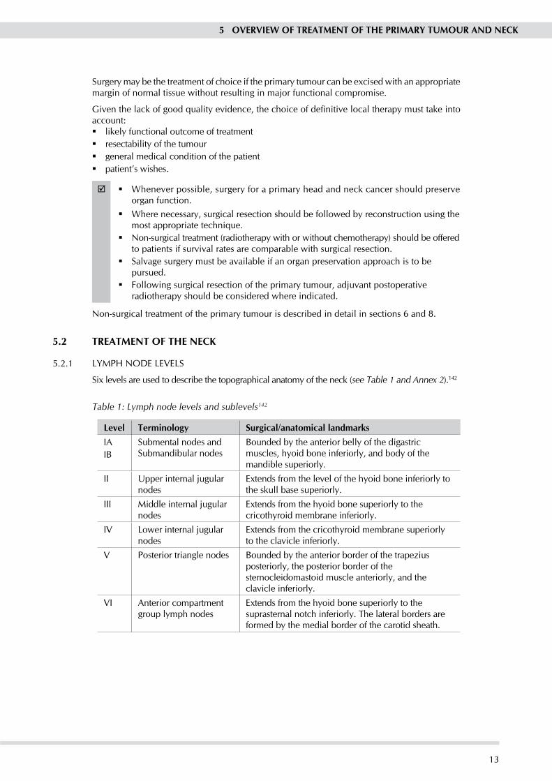
Six levels are used to describe the topographical anatomy of the neck (see Table 1 and Annex 2).142
Table 1: Lymph node levels and sublevels142
Level Terminology surgical/anatomical landmarksIAIb
Submental nodes and Submandibular nodes
bounded by the anterior belly of the digastric muscles, hyoid bone inferiorly, and body of the mandible superiorly.
II Upper internal jugular nodes
Extends from the level of the hyoid bone inferiorly to the skull base superiorly.
III Middle internal jugular nodes
Extends from the hyoid bone superiorly to the cricothyroid membrane inferiorly.
Iv lower internal jugular nodes
Extends from the cricothyroid membrane superiorly to the clavicle inferiorly.
v posterior triangle nodes bounded by the anterior border of the trapezius posteriorly, the posterior border of the sternocleidomastoid muscle anteriorly, and the clavicle inferiorly.
vI Anterior compartment group lymph nodes
Extends from the hyoid bone superiorly to the suprasternal notch inferiorly. The lateral borders are formed by the medial border of the carotid sheath.
5 OveRvIew OF TReATMeNT OF THe PRIMARY TuMOuR AND NeCk
14
DIAGNOsIs AND MANAGeMeNT OF HeAD AND NeCk CANCeR
3
3
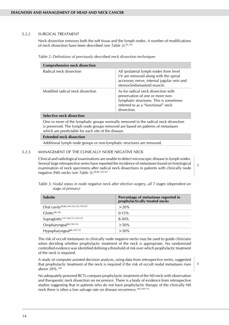
5.2.2 SURGICAl TREATMENT
Neckdissectionremovesboththesofttissueandthelymphnodes.Anumberofmodificationsof neck dissection have been described (see Table 2).85,143
Table 2: Definitions of previously described neck dissection techniques
Comprehensive neck dissectionRadical neck dissection All ipsilateral lymph nodes from level
I-v are removed along with the spinal accessory nerve, internal jugular vein and sternocleidomastoid muscle.
Modifiedradicalneckdissection As for radical neck dissection with preservation of one or more non-lymphatic structures. This is sometimes referred to as a “functional” neck dissection.
selective neck dissectionOne or more of the lymphatic groups normally removed in the radical neck dissection is preserved. The lymph node groups removed are based on patterns of metastases which are predictable for each site of the disease.
extended neck dissectionAdditional lymph node groups or non-lymphatic structures are removed.
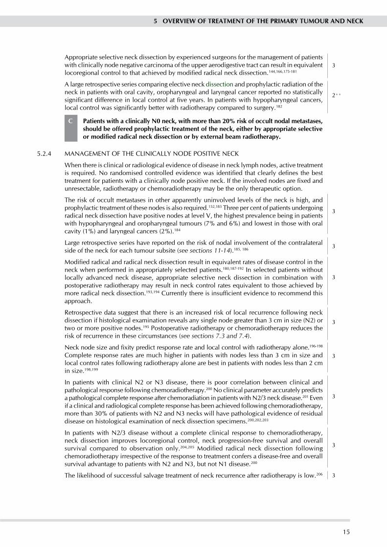
5.2.3 MANAGEMENT OF THE ClINICAlly NODE NEGATIvE NECK
Clinical and radiological examinations are unable to detect microscopic disease in lymph nodes. Several large retrospective series have reported the incidence of metastases found on histological examination of neck specimens after radical neck dissections in patients with clinically node negative (N0) necks (see Table 3).68,86,144-167
Table 3: Nodal status in node negative neck after elective surgery, all T stages (dependent on stage of primary)
subsite Percentage of metastases reported in prophylactically treated necks
Oral cavity68,86,144,145,153,159-167 >20%
Glottic68,146 0-15%
Supraglottic147-149,151,154-157 8-30%
Oropharyngeal68,150-152 >50%
Hypopharyngeal68,150-152 >50%
The risk of occult metastases in clinically node negative necks may be used to guide clinicians when deciding whether prophylactic treatment of the neck is appropriate. No randomised controlledevidencewasidentifieddefiningathresholdofriskoverwhichprophylactictreatmentof the neck is required.
A study of computer assisted decision analysis, using data from retrospective series, suggested that prophylactic treatment of the neck is required if the risk of occult nodal metastases rises above 20%.168
No adequately powered RCTs compare prophylactic treatment of the N0 neck with observation and therapeutic neck dissection on recurrence. There is a body of evidence from retrospective studies suggesting that in patients who do not have prophylactic therapy of the clinically N0 neck there is often a low salvage rate on disease recurrence.166,169-174
15
2++
3
3
3
3
3
3
3
3
3Appropriate selective neck dissection by experienced surgeons for the management of patients with clinically node negative carcinoma of the upper aerodigestive tract can result in equivalent locoregionalcontroltothatachievedbymodifiedradicalneckdissection.144,166,175-181
A large retrospective series comparing elective neck dissection and prophylactic radiation of the neck in patients with oral cavity, oropharyngeal and laryngeal cancer reported no statistically significantdifferenceinlocalcontrolatfiveyears.Inpatientswithhypopharyngealcancers,localcontrolwassignificantlybetterwithradiotherapycomparedtosurgery.182
C Patients with a clinically N0 neck, with more than 20% risk of occult nodal metastases, should be offered prophylactic treatment of the neck, either by appropriate selective or modified radical neck dissection or by external beam radiotherapy.
5.2.4 MANAGEMENT OF THE ClINICAlly NODE pOSITIvE NECK
When there is clinical or radiological evidence of disease in neck lymph nodes, active treatment isrequired.Norandomisedcontrolledevidencewasidentifiedthatclearlydefinesthebesttreatmentforpatientswithaclinicallynodepositiveneck.Iftheinvolvednodesarefixedandunresectable, radiotherapy or chemoradiotherapy may be the only therapeutic option.
The risk of occult metastases in other apparently uninvolved levels of the neck is high, and prophylactic treatment of these nodes is also required.152,183 Three per cent of patients undergoing radical neck dissection have positive nodes at level v, the highest prevalence being in patients with hypopharyngeal and oropharyngeal tumours (7% and 6%) and lowest in those with oral cavity (1%) and laryngeal cancers (2%).184
large retrospective series have reported on the risk of nodal involvement of the contralateral side of the neck for each tumour subsite (see sections 11-14).185, 186
Modifiedradicalandradicalneckdissectionresultinequivalentratesofdiseasecontrolintheneck when performed in appropriately selected patients.180,187-192 In selected patients without locally advanced neck disease, appropriate selective neck dissection in combination with postoperative radiotherapy may result in neck control rates equivalent to those achieved by more radical neck dissection.193,194Currentlythereisinsufficientevidencetorecommendthisapproach.
Retrospective data suggest that there is an increased risk of local recurrence following neck dissection if histological examination reveals any single node greater than 3 cm in size (N2) or two or more positive nodes.195 postoperative radiotherapy or chemoradiotherapy reduces the risk of recurrence in these circumstances (see sections 7.3 and 7.4).
Necknodesizeandfixitypredictresponserateandlocalcontrolwithradiotherapyalone.196-198 Complete response rates are much higher in patients with nodes less than 3 cm in size and local control rates following radiotherapy alone are best in patients with nodes less than 2 cm in size.198,199
In patients with clinical N2 or N3 disease, there is poor correlation between clinical and pathological response following chemoradiotherapy.200 No clinical parameter accurately predicts a pathological complete response after chemoradiation in patients with N2/3 neck disease.201 Even if a clinical and radiological complete response has been achieved following chemoradiotherapy, more than 30% of patients with N2 and N3 necks will have pathological evidence of residual disease on histological examination of neck dissection specimens.200,202,203
In patients with N2/3 disease without a complete clinical response to chemoradiotherapy, neck dissection improves locoregional control, neck progression-free survival and overall survival compared to observation only.204,205Modified radical neck dissection followingchemoradiotherapy irrespective of the response to treatment confers a disease-free and overall survival advantage to patients with N2 and N3, but not N1 disease.200
The likelihood of successful salvage treatment of neck recurrence after radiotherapy is low.206
5 OveRvIew OF TReATMeNT OF THe PRIMARY TuMOuR AND NeCk

16
DIAGNOsIs AND MANAGeMeNT OF HeAD AND NeCk CANCeR
3If the primary tumour is small it is possible to resect advanced nodal disease prior to treating the primary tumourwith definitive radiotherapywhilst delivering postoperative adjuvantradiotherapy to the neck without compromising cancer control.207,208
D Patients with clinically N1 disease should be treated by appropriate neck dissection or radical radiotherapy (with or without chemotherapy).
D In patients with clinically N1 disease and a complete clinical response to radiotherapy, observation rather than further surgical management is recommended.
D Following neck dissection for clinically N1 disease, adjuvant postoperative radiotherapy must be considered for those patients who are at high risk of locoregional recurrence.
D Patients with clinical N2 or N3 disease should be treated either by:comprehensive neck dissection followed by external beam radiotherapy, or radical radiotherapy followed by comprehensive neck dissection.
D In patients where the primary tumour is small and the nodal disease is resectable, neck dissection may be performed before treating both the primary tumour and the neck with radiotherapy (with or without chemotherapy).

17
1++
3
1++
6 Treatment: radiotherapy as the major treatment modality
Radiotherapy uses ionising radiation to treat malignancy. Ionising radiation may be delivered as an external radiation beam targeting the tumour (external beam radiotherapy), or by directly implanting radioactive sources within the tumour (brachytherapy). External beam radiotherapy is usually fractionated which means that the total dose is delivered over time in smaller doses or fractions. The dose of radiation that can be delivered to a tumour is limited by the tolerance of the surrounding normal tissues, which are also unavoidably irradiated during treatment. There are several different systems used for grading radiotherapy side effects (toxicities) caused by irradiation of normal tissues.209-211 In general grade 1 toxicity is the mildest, whilst grade 4 toxicity is very severe.
Radiotherapy can be delivered with curative intent (radical radiotherapy), in order to improve local control following surgery (adjuvant radiotherapy, see section 7.3) or to provide symptomatic relief only (palliative radiotherapy, see section 10.2).
6.1 RADIOTHeRAPY sCHeDuLes
The effect of radiotherapy on the tumour and surrounding normal tissue is dependent on:the total dose administeredthe size of each fractionthe overall time over which the total dose is delivered.
6.2 CONveNTIONAL FRACTIONATION
Conventionalfractionationschedulesdelivertreatmentinsingledailyfractionsof1.8-2Gy,fivedays per week. This results in dose accumulation of approximately 10Gy per week.
6.3 MODIFIeD FRACTIONATION
Modifiedfractionationcanbedividedinto:hypofractionationhyperfractionationaccelerated fractionation.
6.3.1 HypOFRACTIONATION
Hypofractionationisamodifiedfractionationschedulewherethedoseperfractionsubstantiallyexceeds the conventional level of 1.8-2Gy.
Studiesofhypofractionatedradiotherapyhavebeenmainlyconfinedtothetreatmentofpatientswith glottic cancer. In patients with early glottic cancer hypofractionated radiotherapy results in excellent local control with no increase in late normal tissue toxicity (see section 11.1.1).212-214
6.3.2 HypERFRACTIONATION
Hyperfractionationisamodifiedfractionationschedulewherethetotaldoseisdeliveredinanincreased number of fractions, and fraction size is below the conventional level of 1.8-2Gy.
pooled data suggest that hyperfractionated radiotherapy using an increased total radiation dose inpatientswithlocallyadvancedheadandneckcancerresultsinasignificantlyreducedriskofdeathandsignificantlyenhancedlocoregionalcontrolwhencomparedtoconventionallyfractionated treatment.215Randomisedcontrolledtrialdataconfirmsanincreaseinlocoregionalcontrol but no survival advantage with this approach.216Hyperfractionationresultsinsignificantlyincreased grade 3 or 4 acute toxicity, but no increase in late toxicity at 24 months.216
6 TReATMeNT: RADIOTHeRAPY As THe MAJOR TReATMeNT MODALITY6 TReATMeNT: RADIOTHeRAPY As THe MAJOR TReATMeNT MODALITY
18
DIAGNOsIs AND MANAGeMeNT OF HeAD AND NeCk CANCeR
1++
1++
1++
1++
1++
1++
1++
6.3.3 ACCElERATED FRACTIONATION
During accelerated fractionation the rate of dose delivery exceeds 10Gy per week, resulting in a reduction of overall treatment time.
A systematic review comparing both moderately accelerated and very accelerated fractionated radiotherapy with conventional fractionation in patients with head and neck cancer shows significantimprovementinlocoregionalcontrolwithacceleratedradiotherapybutnosignificantdifference in two year overall survival.217
Moderately accelerated fractionated radiotherapy (six fractions per week whilst maintaining the same total dose) in patients with laryngeal, pharyngeal and oral cavity tumours results inbetterlocalcontroloftheprimarytumourandincreaseddiseasespecific,butnotoverallsurvival compared to conventional fractionation. Neither local control of bulky nodal disease,218 locoregional control or survival in patients with T1-3 glottic or supraglottic cancer are improved by this fractionation regimen,219andacutetoxicityissignificantlyincreased.218,219 late skin changes may be more frequent, but there is no evidence that other late toxicities are increased.218,219
72Gy in six weeks using a concomitant boost technique results in a 9% improvement in locoregional control compared to conventional radiotherapy but no difference in survival. Acute but not late toxicity is increased.216
Amore rapidlyaccelerated regimenof72Gy infiveweeks (three fractionsperdayat fourhourlyintervals)improveslocoregionalcontrol,butalsosignificantlyincreasesgrade3and4acute and late effects.220
6.3.4 DECREASED TOTAl DOSE AND vERy ACCElERATED FRACTIONATION
very rapid acceleration of radiotherapy with a decreased total dose, for example, continuous hyperfractionated accelerated radiotherapy (CHART, 54Gy in 36 fractions over 12 days) does not improve or reduce locoregional control or survival in patients with early (excluding T1N0) or locally advanced disease.221,222Thisfractionationschedulesignificantlyincreasesacutetoxicity,althoughtheremaybeasignificantreductioninlatetoxicity,particularlygrade2orworseaffecting the skin and subcutaneous tissue, laryngeal oedema and deep mucosal ulceration, when compared to conventional fractionation.221,222
6.3.5 MODIFIED FRACTIONATION AND CHEMOTHERApy
The addition of concurrent chemotherapy to altered fractionation radiotherapy improves locoregional control, but increases mucosal toxicity, when compared to the same dose of altered fractionation radiotherapy alone.223,224 The long term morbidity of this approach is not clear.
No RCTs were identified comparing survival following conventionally fractionated chemoradiotherapy with that following altered fractionation radiotherapy alone. There is a body of evidence demonstrating a survival advantage when chemotherapy is administered concurrently with radiotherapy and the majority of this relates to conventionally fractionated radiotherapy (see section 8).
A randomised trial comparing hyperfractionated accelerated radiotherapy (total dose 70.6Gy) with concurrentmitomycin and 5FU (5-fluorouracil) and dose-escalated hyperfractionatedaccelerated radiotherapy alone (total dose 77.6Gy) showed significantly better five-yearlocoregional control and overall survival with chemoradiotherapy.225
Theevidencesuggeststhatmodifiedfractionationradiotherapyshouldbereservedforthosepatients undergoing radical radiotherapy who are unable to receive concurrent chemotherapy or cetuximab (see section 8.2).226
A where radiotherapy is the primary treatment modality, moderately accelerated schedules (six fractions/week) or hyperfractionated schedules with increased total dose should be considered for patients with head and neck cancer (except T1-3 glottic or supraglottic) who are unable to receive concurrent chemotherapy or cetuximab.

19
2+
3
3
3
Ifmodifiedfractionationisbeingconsideredtheremustbe:adequate monitoring of acute toxicity suffered by the patient during and after
treatmentaccess to outpatient and inpatient services for treatment of acute toxicity and
nutritional support.
6.4 INTeRRuPTIONs TO PLANNeD RADIOTHeRAPY TReATMeNT sCHeDuLes
prolonging the overall time taken for the delivery of a radical course of radiotherapy due to an unscheduled interruption in treatment affects local control.227,228
C Interrupting and prolonging a course of radical radiotherapy should be avoided.
Guidance on the management of unscheduled interruption to planned radiotherapy schedules can be found in “Guidelines for the Management of the Unscheduled Interruption or prolongation of a Radical Course of Radiotherapy”.229
6.5 bRACHYTHeRAPY
Norandomisedcontrolledevidencewasidentifiedcomparingoutcomefollowingbrachytherapywith outcome following external beam radiotherapy or surgery for patients with head and neck cancer. Evidence supporting the use of brachytherapy comes from large case series from centres experienced in the technique.
Localcontrolratesatfiveyearsof79-97%(T1)and65-87%(T2)havebeenachievedforpatientswithearlycancersoftheoraltongueandfloorofmouthtreatedwithinterstitialbrachytherapyalone.230-238Thefive-yearlocalcontrolrateinoneserieswasequivalenttothatfollowingsurgicalresection in the same centre.236Thefive-yearlocalcontrolrateforpatientsfollowinginterstitialbrachytherapy for T3 oral cavity tumours is 49-70%.232,236,237,239
A dose of 65Gy results in optimal local control.233 Doses in excess of 65Gy result in an increased risk of necrosis and bone complication.239-241
In patients with oropharyngeal tumours a brachytherapy boost of 25-30Gy following external beam radiotherapy (45-50Gy) results in local control of 89% (T1), 86% (T2) and 57% (T3).242,243
There is no clear evidence to determine whether local control in oropharyngeal cancer treated with a brachytherapy boost following external beam radiotherapy is better than with external beam radiotherapy alone.244,245 There is also no robust evidence to determine whether brachytherapy used as a boost following external beam radiotherapy results in reduced morbidity and better quality of life than when the same total dose of radiation is delivered entirely as external beam radiotherapy.246
Adoserateinexcessof0.55Gy/hourandintersourcespacingofmorethan15mmsignificantlyincreases bone and soft tissue necrosis.235,242,243,247
There is no reported role for brachytherapy in the treatment of laryngeal or hypopharyngeal tumours.
D Patients with small accessible (T1/2) tumours of the oral cavity and oropharynx may be treated by interstitial brachytherapy to a dose of 65-70Gy at a dose rate of less than 0.55Gy/hour.
Interstitial brachytherapy for patients with head and neck cancer should be performed by experienced teams in centres with adequate radiation protection facilities.
6.6 INTeNsITY MODuLATeD RADIOTHeRAPY
Intensity modulated radiotherapy (IMRT) is currently under development in UK cancer centres. NorandomisedcontrolledevidencewasidentifiedcomparingoutcomefollowingIMRTwiththat
6 TReATMeNT: RADIOTHeRAPY As THe MAJOR TReATMeNT MODALITY
20
DIAGNOsIs AND MANAGeMeNT OF HeAD AND NeCk CANCeR
1+
3
following conventionally delivered radiotherapy for patients with head and neck cancer. Case serieswereidentifiedwhichdescribetheuseofIMRTtoreduceradiationtoxicity,particularlyxerostomia (see section 6.7.2) and its use in re-irradiation following tumour recurrence (see section 9.2).
6.7 PReveNTION AND MANAGeMeNT OF RADIATION sIDe eFFeCTs
The side effects of radiotherapy are caused by unavoidable irradiation of the normal tissues surrounding the tumour. They can be described as “acute” (those that occur during or immediately after radiotherapy) or “late” (those that occur months or years after treatment has been completed). In patients with head and neck cancer common side effects that are likely to cause patient discomfort are:mucositis(inflammationanddesquamationofthemucosalliningofirradiatedareasofthe
upper aerodigestive tract)xerostomia (dry mouth) caused by irradiation of the salivary glands, particularly the parotid
glands,andconsequentreductioninsalivaryflow.Xerostomiaisoftenpermanentandresults indiscomfort,eatingdifficulties,tastealterationandhighriskoframpantdentalcaries.
Skin included in the irradiated volume may also suffer from acute and late toxicity from radiotherapy.
6.7.1 pREvENTION AND TREATMENT OF RADIATION-INDUCED MUCOSITIS
The use of benzydamine oral rinse reduces the frequency and severity of ulcerative oral lesions and decreases pain in radiation-induced oral mucositis.248-250 The largest of these trials used a regimen of 15 mls four to eight times daily starting before radiotherapy, continuing throughout treatment and for two to three weeks after completion.248 Most patients included in these studies weretreatedwithconventionallyfractionatedradiotherapy,andthebenefitofbenzydamineusedwithchemoradiotherapyormodifiedfractionationregimensislessclear.
A Patients with oral cavity, laryngeal, oropharyngeal or hypopharyngeal tumours who are being treated with radiotherapy should be offered benzydamine oral rinse before, during, and up to three weeks after completion of radiotherapy.
There is no evidence to support any other intervention for prevention or treatment of radiation-induced mucositis.251-265
patients should be advised on how to maintain good oral hygiene during and after radiotherapy.
patients’ mucosa should be inspected regularly during treatment, and analgesia and antimicrobial/antifungal agents to treat infection should be made available.
6.7.2 pREvENTION AND TREATMENT OF RADIATION-INDUCED xEROSTOMIA
Theevidencedoesnotsupportaspecificinterventionforthepreventionofradiation-inducedxerostomia.
Amifostinegivenconcurrentlywithradiotherapyorchemoradiotherapysignificantlyreducestherate of acute and late xerostomia.252 There is no evidence that amifostine affects survival at 24 months or recurrence at 18 months after cancer therapy, or the rate of incomplete response to radiotherapy.251,253 Survival data are only available for 24 months post-treatment. Without longer followup,theprotectiveeffectofamifostineonthetumourisunclear.Vomitingissignificantlyincreased with amifostine compared to control, but hypotension and nausea are not.253
The use of amifostine in the prevention of radiation-induced xerostomia cannot be recommended outsideclinicaltrials.NorandomisedcontrolledevidencewasidentifiedaddressingtheuseofIMRT in the prevention of radiation-induced xerostomia. Observational evidence suggests that decreasing the mean radiation dose to the parotid gland, whether by IMRT or 3-dimensional conformalradiotherapy,resultsinimprovedstimulatedsalivaryflowandqualityoflife(intermsof oral discomfort, eating and speaking) at six months after completion of radiotherapy.266

21
1+
1++
1+
Administration of oral pilocarpine during a course of radiotherapy to an area containing salivary tissueresultedinsignificantlyimprovedsalivaryflowatthreemonthspost-treatmentcomparedto placebo in a single RCT.267 This did not translate into improved quality of life.
Analysis of pooled data suggests that administration of oral pilocarpine (5-10 mg orally three times per day) to patients with xerostomia (and evidence of pre-existing salivary function) following conventionally fractionated radiotherapy results in statistically significant improvements insubjective overall xerostomia and the need for salivary substitutes compared to placebo.268
Norandomisedcontrolleddatawereidentifiedwhichdefinetheoptimumdurationofpilocarpinetherapy.
A Pilocarpine (5-10 mg three times per day) may be offered to improve radiation-induced xerostomia following radiotherapy to patients with evidence of some intact salivary function, providing there are no medical contraindications to its use.
Duration of pilocarpine therapy should be determined by clinical judgement regarding its effectiveness in individual patients.
patients with chronic xerostomia following radiotherapy should be encouraged to maintain good oral hygiene. They should have regular dental assessment with access to a restorative dentist where necessary.
6.7.3 pREvENTION AND TREATMENT OF SKIN COMplICATIONS
Norandomisedcontrolledtrialswereidentifiedwhichexamineskincareduringradiotherapyin head and neck cancer patients. Most studies also include patients undergoing breast or chest wall radiotherapy. There is no evidence to suggest that washing during radiotherapy increases acute radiation skin toxicity.269
prophylactic administration of aloe vera gel, aqueous cream or sucralfate cream does not reduce frequency or severity of acute skin toxicity.270-272 In a single small RCT, Cavilon™ No-sting barrier Film (3M®) reduced the duration of moist desquamation compared to 10% glycerine cream.270
Basedonthisevidenceitisnotpossibletorecommendspecificinterventionsforthepreventionor treatment of radiation skin toxicity.
6 TReATMeNT: RADIOTHeRAPY As THe MAJOR TReATMeNT MODALITY
22
DIAGNOsIs AND MANAGeMeNT OF HeAD AND NeCk CANCeR
3
3
3
7 Treatment: surgery as the major treatment modality
The main aim of surgery is to excise the area of malignancy completely by ensuring, where possible, that a margin of normal tissue surrounding the tumour is also removed and that radical excision is performed with curative intent. Access to the hidden recesses of the head and neck is essential to excise the tumour and perform surgical reconstruction. The open approach uses facial splits and incorporates skeletal osteotomies so that the tumour can be widely exposed. A minimally invasive approach, incorporating the use of endoscopes, is a surgical alternative in areas such as the sinuses and larynx.
In many instances the scalpel has been replaced by newer technology, such as cutting diathermy and the use of lasers, both as a cutting tool and as a method of ablation (vaporisation).
The wide variety of surgical techniques now available for head and neck tumour surgery demands a multidisciplinary approach with surgeons experienced in several techniques.
7.1 ReseCTION
Norandomisedcontrolledevidencewasidentifiedcomparingdifferentresectiontechniquesinthe tumour subsites. Evidence exists mainly in the form of retrospective case series. Resection techniques vary between different tumour subsites, and are discussed in sections 11-14.
The evidence to support positive margins as a predictor for recurrence is inconsistent among head and neck cancer subsites. For squamous carcinoma of the oral cavity,101,273-275 and larynx,276 evidence suggests that the presence of positive margins leads to locoregional recurrence. In oropharyngeal and hypopharyngeal tumours, there is some evidence that margins may be as important as T stage and N stage for predicting recurrence (all p<0.0001 for locoregional relapse).277
Inadequate initial excision biopsy can be managed effectively by re-excision.98 A small case series reported 88.5% of patients with oral cancer had positive margins after biopsy. After re-excision 96% of those treated were alive and disease free.
D If an inadequate initial excision biopsy has been performed or if the tumour has been excised with positive excision margins, re-resection should be considered.
If re-resection is not possible, postoperative radiotherapy should be considered.
The role of postoperative radiotherapy is discussed in section 7.3.
7.2 ReCONsTRuCTION
To completely excise a tumour with an adequate margin of surrounding normal tissue it is often necessary to perform an extensive surgical resection, which may involve the removal of soft tissue,boneorcartilage.Thismayleaveamajorphysicaldeficitthatcannotberepairedbyprimary mucosal closure or skin grafting. Surgical reconstruction aims to repair any physical deficitandrestoreorminimise functionaldeficit thatwouldarise fromthe lossof resectedtissue.
Reconstruction techniques are diverse and vary by anatomical region. No randomised controlled evidencewasidentifiedcomparingtheoutcomesofdifferenttechniques.Theevidenceisfromretrospective case series, mainly relating to intraoral and hypopharyngeal tumours.
Freeflaptransferisasafeandreliabletechniqueforreconstructioninpatientswithheadandneck cancer in general, and particularly for oral cavity and hypopharyngeal cancer.278-285 A retrospectivecaseseriesof400consecutivemicrovascularfreeflapproceduresperformedbyasinglesurgeonoverasevenyearperiodshoweda0.8%incidenceoffreeflapfailure,3%partial necrosis rate and perioperative mortality rate of 1.3%.286

23
3
3
1++
2++
3
3
There is evidence that free jejunal autograft is effective for aiding swallowing, but is poor for speech rehabilitation following surgical resection for hypopharyngeal cancer.287 pectoralis major myocutaneousflapissuitableforelderlyandfrailpatients.288
Surgical reconstruction should be available for patients undergoing extensive surgical resection for head and neck cancer.
Reconstruction should be performed by appropriately trained and experienced surgical teams who should be familiar with a variety of reconstruction techniques.
Choice of reconstruction technique should be made on an individual basis for each patient according to the tumour’s anatomical location, patient’s general condition, and patient’s and surgeon’s preference.
7.3 ADJuvANT RADIOTHeRAPY FOLLOwING suRGeRY
patients who are considered to be at high risk of locoregional recurrence following surgery are often treated with adjuvant radiotherapy to improve local control and survival. No good quality randomised controlled trials examining the role of adjuvant radiotherapy in combination with surgerywereidentified.
Non-randomised studies suggest that adjuvant radiotherapy improves local control, disease-free and overall survival at three years in patients with extracapsular lymph node spread and/orpositivemargins (definedas<1mm)after radicalsurgery for laryngeal,oralcavity,oropharyngeal and hypopharyngeal cancer.289 It also decreases neck recurrence rates especially in patients with high risk pathology.178,195,290-292
When compared to preoperative radiotherapy, postoperative radiotherapy results in better local control, but not overall survival, in patients with surgically resected T2-4, N0-2 oral cavity, oropharyngeal, supraglottic laryngeal and hypopharyngeal cancer.293,294 preoperative and postoperative radiotherapy result in similar rates of surgical and radiotherapy complications.
Theroleofadjuvantpostoperativeradiotherapyhasnotbeenclearlydefinedfromrandomisedcontrolled trials. pathological risk factors that predict local recurrence have been assessed in prospective studies and retrospective case series and indications for adjuvant radiotherapy have been extrapolated from these risk factors. Extracapsular lymph node spread, even when microscopic, is the most important predictor for local recurrence after neck dissection.105,107,295-299 Increased local recurrence rates after surgery are also associated with close or positive surgical margins, increased T stage, an oral cavity primary tumour, any positive node >3 cm, microvascular invasion and perineural invasion.99,107,295,298-302 Recurrence rates in the neck are higher after neck dissection if any nodes are found to be histologically positive. The risk of recurrence increases as the number of histologically positive nodes increases.150,291,296,298,303,304 Since the evidence is fromheterogeneousretrospectivestudies,itisdifficulttodeterminewhetheritisappropriateto offer adjuvant radiotherapy to all patients with any positive neck nodes, or to restrict it to those who have more than one, or even more than two positive nodes.
Locoregionalcontrolsignificantlydecreasesinthepresenceoftwoormorehistologicalindicatorsof poor prognosis.295,296,305
C Postoperative radiotherapy should be considered following surgical resection of oral cavity, oropharyngeal, laryngeal and hypopharyngeal cancers for patients with the following adverse risk features:
oral cavity primary tumouradvanced T stageclose or positive surgical marginsperineural invasionlymphovascular invasionany positive lymph nodes , but especially if more than one node is positivepositive nodes at level Iv or vany node 3 cm or greaterextracapsular lymph node spread.
7 TReATMeNT: suRGeRY As THe MAJOR TReATMeNT MODALITY

24
DIAGNOsIs AND MANAGeMeNT OF HeAD AND NeCk CANCeR
1++
2++
1++
1++
1++
For patients with advanced head and neck cancer, where postoperative radiotherapy is indicated, the optimal dose of conventionally fractionated postoperative radiotherapy is no less than 57.6Gy in 1.8Gy fractions (56.6Gy-2Gy per fraction equivalent) to areas at low risk. At sites of increased risk, especially sites of extracapsular spread, a higher dose of at least 63Gy in 1.8Gy fractions (62Gy-2Gy per fraction equivalent) is required.306
Adoseof54-60Gyin27-30fractions,fivedaysperweekto theprimarysiteandnodesatrisk with boost to 66Gy in 33 fractions in 6.5 weeks to high risk areas has also been used effectively.307,308
Acceleratedfractionationradiotherapyoffersnosignificantimprovementinlocoregionalcontrolor survival compared to conventional fractionation radiotherapy when delivered postoperatively to patients with high risk adverse pathological factors.300,309
The cumulative time of combined therapy (from surgery to completion of adjuvant radiotherapy) significantlyaffectslocoregionalcontrolandsurvivalinhighriskpatients.300,310
A Postoperative radiotherapy should be conventionally fractionated:54-60Gy in 27-30 fractions over 5.5-6 weeks to the primary site and nodes at risk66Gy in 33 fractions over 6.5 weeks to areas of very high risk.
b Overall treatment time from surgery to completion of radiotherapy should be 10-11 weeks or less in the absence of postoperative medical or surgical complications.
In patients with high risk pathological features following surgical resection of oral cavity, oropharyngeal, laryngeal and hypopharyngeal cancers, the addition of concurrent chemotherapy (cisplatin) to postoperative radiotherapy improves local control,307,308 disease-free survival,308,311 andoverallsurvivalatfiveyears.307,311Retrospectivesubgroupanalysisshowsthatthisbenefitis greatest in those patients with extracapsular extension and/or positive surgical margins.312
Acute, but not late, toxicity is significantly increasedwith postoperative chemoradiationcompared to radiotherapy alone.307,308
The addition of cisplatin/5FU chemotherapy prior to postoperative radiotherapy for completely resected stage III/Iv cancer of the oral cavity, oropharynx, larynx or hypopharynx does not confer any advantage in terms of locoregional control or survival.313
Noevidencewas identifiedsupporting theadditionofconcurrentchemotherapy toalteredfractionation radiotherapy in the postoperative setting.
A In patients with extracapsular spread and/or positive surgical margins, who are medically fit, postoperative concurrent chemoradiotherapy with single agent cisplatin and conventionally fractionated radiotherapy should be considered.
Inpatientswhoarenotfitforchemotherapyconventionallyfractionatedradiotherapy alone may be used.
The decision to undertake a course of postoperative radiotherapy or chemoradiotherapy should be made in consultation with the patient and multidisciplinary team.
7.4 CHeMOTHeRAPY IN COMbINATION wITH suRGeRY
There is no evidence to support the use of neoadjuvant or adjuvant chemotherapy in combination with surgery in laryngeal, oral cavity, oropharyngeal or hypopharyngeal cancer (see section 8).

25
1++
1++
1++
8 Treatment: chemotherapy in combination with surgery or radiotherapy
Noevidencewasidentifiedtosupporttheuseofchemotherapyaloneasacurativetreatmentfor squamous carcinoma of head and neck.
In patients with head and neck cancer the administration of chemotherapy in combination with locoregional therapy (surgery or radiotherapy) may be:
neoadjuvant – delivered in the weeks before surgery or radiotherapyadjuvant – delivered following radiotherapy or surgeryconcurrent with radiotherapy – delivered during the course of radiotherapy.
8.1 CHeMOTHeRAPY wITH LOCOReGIONAL THeRAPY
The addition of chemotherapy to locoregional treatment for patients with non-metastatic squamous carcinoma of the head and neck (primarily locally advanced, stage III and Iv disease)significantly improvessurvival,withanabsolutesurvivalbenefitof5%at twoandfiveyears.314,315
Chemotherapyresultsinasmallstatisticallynon-significant(2%)overallsurvivalbenefitatfiveyearswhengivenneoadjuvantlyandnosurvivalbenefitatfiveyearswhengivenadjuvantly.Neoadjuvantchemotherapyusingcisplatin/5FUchemotherapyresultsinasignificantsurvivalbenefitcomparedtolocoregionaltreatmentalone.314,316
When chemotherapy is administered concurrently with radiotherapy in resectable and non-resectablediseasethereisanabsoluteoverallsurvivalbenefitof8%atfiveyears(percentageriskreduction;RR,ofdeath19%comparedtonochemotherapy,p<0.0001),andabenefitofevent-freesurvivalatfiveyearsof8%.Theabsolutesurvivalbenefitatfiveyearsforconcurrentsingle agent cisplatin as opposed to all other drugs is 11%. The reduction in risk of death has been calculated for each subsite (see Table 4).315 The size of benefitwith concurrentchemoradiotherapy isagedependent,with the largestbenefit in thoseaged60or less (see Table 5).Thesurvivalbenefitwithconcurrentchemoradiotherapyisseenwithconventionalfractionation and altered fractionation when radiation is the main modality of treatment, and also in postoperative radiotherapy following surgery (see section 7.3).314,315
Table 4: Risk reduction of death after concurrent chemotherapy and radiotherapy compared to no chemotherapy315
subsite Percentage reduction in risk of deathoropharynx 23%
larynx 22%
oral cavity 17%
hypopharynx 16%
Table 5: Risk reduction of death after concurrent chemotherapy by age314,315
Age Percentage reduction in risk of death60 or less 22-24%
60-70 12%
over 70 3%
8 TReATMeNT: CHeMOTHeRAPY IN COMbINATION wITH suRGeRY OR RADIOTHeRAPY
26
DIAGNOsIs AND MANAGeMeNT OF HeAD AND NeCk CANCeR
1++
1++
1+
1++
1++
platinum monochemotherapy is as effective as polychemotherapy containing platinum when given concurrently with radiotherapy. Non-platinum chemotherapy is less effective.314,315
The survival benefits associatedwith concurrent chemoradiotherapy are at the expenseofincreased acute toxicity (mucosal and haematological)120,224,317-326 and possibly late toxicity, particularly dental problems.120,318 late toxicity reporting in RCTs is frequently absent or is reported after short follow up in a small number of patients. Most acute toxicity and late toxicity data relate to chemoradiation with conventionally fractionated radiotherapy.120,323,327-329
The additionof concurrent chemotherapy tomodified fractionation radiotherapy improveslocoregional control, but increases mucosal toxicity, when compared to the same dose of modifiedfractionationradiotherapyalone.223, 224 There is little evidence describing the long term morbidity of this approach.
When compared to dose-escalated hyperfractionated accelerated radiotherapy alone (total dose 77.6Gy), hyperfractionated accelerated radiotherapy (total dose 70.6Gy) with concurrent mitomycin and5FU showed significantly better five-year locoregional control andoverallsurvival, with increased acute toxicity, but not late toxicity.225
In patients with T2-T4 N0-N2b and N3 stage II-Iv hypopharyngeal cancer, who have a complete response to chemotherapy, the larynx can be preserved without compromising survival using induction chemotherapy (cisplatin/5FU) with radical radiotherapy.315,330
There is no evidence to support the use of neoadjuvant or adjuvant chemotherapy in combination with surgery alone.314,315,331
There is no published RCT validating the routine use of taxanes in combination with locoregional therapy in head and neck cancer. Initial results from a large phase III trial, published only in abstract,reportedasignificantimprovementinprogression-freeandoverallsurvivalfollowingneoadjuvant cisplatin/5FU and docetaxel compared to cisplatin and 5FU prior to radical radiotherapy in patients with unresectable locally advanced head and neck cancer.332 Another largephaseIIItrial,publishedonlyinabstract,reportedasignificantimprovementinoverallsurvival with a risk reduction of 30% (p=0.006) following the addition of docetaxel to cisplatin/5FU induction chemotherapy followed by concurrent carboplatin and irradiation compared to cisplatin/5FU induction chemotherapy followed by concurrent carboplatin and irradiation.333
A In patients with locally advanced non-metastatic squamous carcinoma of the oral cavity, oropharynx, larynx and hypopharynx, who are medically fit for chemotherapy, (especially those aged 70 or under), concurrent chemoradiotherapy should be considered rather than radiotherapy alone if:
organ preservation is being pursuedthe primary tumour is unresectable.
A single agent cisplatin is recommended as the chemotherapeutic agent of choice in concurrent chemoradiotherapy.
A The routine use of neoadjuvant chemotherapy in oral cavity, oropharyngeal and laryngeal cancer is not recommended.
A Neoadjuvant cisplatin/5Fu followed by radical radiotherapy alone may be used in patients with locally advanced resectable hypopharyngeal cancers who have a complete response to chemotherapy.
A The routine use of adjuvant chemotherapy following radiotherapy is not recommended.
A The routine use of neoadjuvant or adjuvant chemotherapy in combination with surgery is not recommended.
A Concurrent chemoradiotherapy should only be administered where there are appropriate facilities for monitoring toxicity, with rapid access to appropriate outpatient and inpatient support for the treatment of acute radiotherapy and chemotherapy toxicity.
27
1++
8.2 CeTuXIMAb IN ADDITION TO RADICAL RADIOTHeRAPY
A multicentre randomised controlled trial involving 424 patients has demonstrated that concurrent administration of cetuximab, a monoclonal antibody against the epidermal growth factor (EGF) receptor, with radical external beam radiotherapy in locoregionally advanced head and neck cancer resulted in an 11% improvement in progression-free survival and a 10% improvement in overall survival compared to external beam radiotherapy alone.226 There was no increase in radiotherapy-related toxicity. patients receiving cetuximab had a 17% incidence of grade 3 or more acneiform rash and a 3% incidence of grade 3 or more infusion-related toxicity. Radiotherapy was either conventionally fractionated, hyperfractionated or accelerated.
No randomised controlled trial has compared chemoradiotherapy with and without concurrent cetuximab administration.
A In patients undergoing radical radiotherapy for locally advanced head and neck cancer, who are medically unfit for concurrent chemoradiotherapy, concurrent administration of cetuximab with radiotherapy should be considered.
8 TReATMeNT: CHeMOTHeRAPY IN COMbINATION wITH suRGeRY OR RADIOTHeRAPY

28
DIAGNOsIs AND MANAGeMeNT OF HeAD AND NeCk CANCeR
3
3
3
3
9 Treatment: management of locoregional recurrence
local recurrence at the site of the primary tumour is the most common cause of treatment failure and disease-related death in patients with head and neck cancer.
Therapeuticoptionsforpatientswithheadandneckcancerwhosefirstlinetreatmenthasfailedinclude:
surgery (salvage)radiotherapy (including re-irradiation)palliative treatment only, including best supportive care, if a further attempt at cure is not
appropriate either due to advanced nature of the tumour, poor general condition of the patient, or at the patient’s request (see section 10).
Decisions regarding the appropriate management of a locoregional recurrence of head and neck cancer should be made on an individual basis taking into account:
the stage of recurrent tumour and its potential resectabilityprevious treatmentlikelytreatmentefficacylikely treatment-related morbidity and functional outcome and consequent effects
on quality of lifethe patient’s general healththe patient’s wishes.
Decisions regarding the management of locoregional recurrence of head and neck cancer should be made by the multidisciplinary team in consultation with the patient following histologicalconfirmationofrecurrenceandfullrestaging(clinicalandradiological).
patients and their relatives/carers should be carefully counselled about the likely outcome of surgical and radiotherapeutic salvage, with respect to survival, risk of treatment-related morbidity and mortality, and likely resulting quality of life.
Early referral to palliative care services for symptom control should be considered.
9.1 sALvAGe suRGeRY AFTeR PRevIOus RADIOTHeRAPY OR suRGeRY
Ameta-analysisofretrospectivecaseseriesreportedtheweightedaverageoffive-yearsurvivalfollowing salvage surgery for recurrent, previously irradiated laryngeal, pharyngeal and oral cavity tumours as 39% in 1,080 patients from 28 different institutions.334Site-specificfive-yearsurvival was 43.4% (oral cavity), 26% (pharynx), and 47.5% (larynx).
Disease-free survival following salvage therapy decreases with increasing stage of recurrence.72,334 There is no correlation between outcome and tumour subsite, time from initial presentation to recurrence, or stage of the original tumour. Disease-free survival following salvage is not influencedbythemodality(surgeryorradiotherapy)usedtotreattheoriginaltumour.334
Following salvage surgery for head and neck cancer, the total complication rate varies from 39-53%.334,335Significantcomplicationshavebeenreportedin18.5-27%ofpatientsundergoingsalvage surgery, with an operative mortality rate of 3.2-5.2%.334,335 An increased rate of postoperative complications is seen with increasing stage of recurrent tumour.335 From the available evidence it is not clear whether there is an increased complication rate following salvage surgery in previously irradiated compared to non-irradiated tissues.334,335
In 109 patients, 50% returned to their baseline preoperative quality of life (functional living index for cancer, FlIC score) after salvage surgery. Quality of life following salvage correlates with the stage but not site of the recurrence.334

29
3
3
3
3
D salvage surgery should be considered in any patient with a resectable locoregional recurrence of oral cavity, oropharyngeal, laryngeal or hypopharyngeal cancer following previous radiotherapy or surgery.
Salvage surgery should only be performed by an experienced surgical team with adequate experience in reconstructive techniques, in centres with appropriate facilities for medical support and rehabilitation.
9.2 RADIOTHeRAPY AND Re-IRRADIATION
If the tumour was previously treated surgically, without the addition of radiotherapy, it may be possible to achieve long term tumour control or cure following a locoregional recurrence with radical external beam radiotherapy (or chemoradiotherapy). This assumes that the recurrent diseasecanbeencompassedinareasonabletreatmentvolume.Noevidencewasidentifiedreporting local control, survival or morbidity rates using this approach.
External beam radiotherapy should be considered as potentially curative salvage treatment for patients with locoregionally recurrent disease after previous surgery, particularly if the recurrence is unresectable, or resection would result in unacceptable loss of function or cosmesis.
If the site of the locoregional recurrence has been previously irradiated, it may be possible toofferre-irradiationasatherapeuticoption.NoRCTswereidentifiedcomparingsurvivalorquality of life following re-irradiation, salvage surgery or palliative chemotherapy in locally recurrent head and neck cancer.
In patients with small, early (T1N0 and T2N0) recurrences or new primaries in previously irradiatedoropharynx, interstitialbrachytherapyalone(60Gy)canresult inafive-year localcontrol rate of 69-80%,336,337withafive-yearoverallsurvivalof30%,mostdeathsbeingdueto causes other than the cancer.336
In patients with unresectable recurrent disease following previous radiotherapy, re-irradiation with potentially curative doses of external beam radiotherapy with or without concurrent chemotherapy has been used in a number of centres on the basis that it offers the only chance of cure under these circumstances. Several small series of highly selected patients reported five-yearsurvivalrangesfrom9-20%338-342 and local control rates of 11-48%.340-343
local control is significantly better if the radiotherapy dose for re-irradiation is >50Gy.340,341,343
Normal tissue toxicity may be considerable. Severe late radiation toxicity is reported in 9-18% of patients.338,342,344 Inone largeseries,41%ofpatientshadcervicalfibrosis,41%mucosalnecrosis and 30% trismus following re-irradiation,339 and an 11% fatal complication rate has been reported.340 Severe acute toxicity is more likely in those older than 80 years, and if the neck rather than the head is being re-irradiated.344Noapparent improvementinefficacyortoxicity was seen with conformal radiotherapy techniques.338
There may be a role for IMRT in improving the therapeutic index during re-irradiation.345
D selected patients who have unresectable locally recurrent disease following previous radiotherapy may be considered for potentially curative re-irradiation.
D Patients with small accessible recurrences in a previously irradiated region may be considered for interstitial brachytherapy in centres with appropriate facilities and expertise.
Re-irradiation should only be performed in centres with adequate expertise, and ideally only in the context of a clinical trial. Centres must be experienced in the recognition and management of acute and late radiation toxicity.
9 TReATMeNT: MANAGeMeNT OF LOCOReGIONAL ReCuRReNCe

30
DIAGNOsIs AND MANAGeMeNT OF HeAD AND NeCk CANCeR
1++
1++
10 Treatment: palliation of incurable disease
Head and neck cancer may be incurable because: the disease is very locoregionally advanced at presentation, rendering it both unresectable
and incurable by radiotherapythe patient’s general medical condition precludes surgical resection or radical radiotherapythepatientissufferingfromalocoregionalrecurrenceafterearlierdefinitivetreatment,which
is not amenable to salvage therapy or re-irradiationthe patient has presented with or developed distant metastases.
patients with incurable head and neck cancer often have multiple physical and psychological problems,whichmaybedifficulttomanage.Theymaybenefitfrominputfromawidevarietyof clinical services. Guidance on palliative care is available from the NHSScotland publication “Clinical Standards: Specialist palliative Care”.346
The care of patients with incurable head and neck cancer should be managed by the palliative care services in conjunction with the multidisciplinary team.
All modalities of therapy should be considered as options for the palliation of head and neck cancer.
Short term toxicity and length of hospital stay should be balanced against likely symptomatic relief.
A documented pathway of care should be discussed and agreed with the patient, relatives, carers and Gp.
10.1 PALLIATIve CHeMOTHeRAPY
Norandomisedcontrolledevidencewasidentifieddemonstratingthatpalliativechemotherapyimproves symptom control, quality of life or survival compared to best supportive care alone. Therearenorandomisedcontrolledcomparisonsofsymptomaticbenefitandqualityoflifeachieved with differing palliative chemotherapy regimens.
In patients with advanced, recurrent or metastatic head and neck cancer, the response rate to chemotherapy ranges from 10-35%.347-351 A trial of high dose cytarabine in combination with cisplatin and 5FU reported a response rate of 57%.352 patients with good performance status have a better response rate to chemotherapy.350
A Patients of adequate performance status should be considered for palliative chemotherapy which may reduce tumour volume.
palliative treatment with single agent cisplatin chemotherapy may result in longer survival than single agent methotrexate, but is more toxic.347 The response rate to palliative chemotherapy may be improved by the combination of chemotherapeutic agents. There is no evidence that combination chemotherapy improves survival compared to treatment with single agents.347-350,352 The increased response rate with combination chemotherapy is at the expense of increased haematological and non-haematological toxicity.348-350 Cisplatin and paclitaxel in combination, using a 3-hour paclitaxel infusion, results in similar toxicity rates to cisplatin/5FU, but no difference in response rate or survival.353 The use of a 24-hour infusion of paclitaxel in combination with cisplatin is excessively haematologically toxic.354
A single agent methotrexate, single agent cisplatin, or cisplatin/5Fu combination should be considered for palliative chemotherapy in patients with head and neck cancer.
A excessive toxicity from intensive chemotherapeutic combination regimens should be avoided.

31
3
10.2 PALLIATIve RADIOTHeRAPY
No randomised controlled evidence demonstrating the effectiveness of palliative radiotherapy inadvancedheadandneckcancerwasidentified.Optimaldoseandschedulingforpalliativeradiotherapyhasnotbeendefinedinclinicaltrials.
Inasinglecaseseriesof505patients,shortcoursepalliativeradiotherapy(20Gyinfivefractionsoverfivedays)provideddurablesymptomreliefin55%ofpatients.355
D Radiotherapy may be considered for palliative treatment in patients with locally advanced incurable head and neck cancer.
10.3 PALLIATIve suRGeRY
The aim of palliative surgery is to debulk tumour mass, reducing symptoms, especially pain, bleeding and breathing problems associated with tumour growth and expansion.
The efficacy of, and indications for, palliative surgery in head and neck cancer have notbeendefined inRCTs. Small retrospective case series and clinical experience suggest thatpalliative surgical or interventional radiology procedures such as tracheotomy, laser debulking, embolisation, percutaneous endoscopic gastrostomy (pEG) tube insertion and nerve block havearoleinthemanagementofspecificproblemssuchasairwayobstruction,debridementof fungating malodourous tumours, haemorrhage, dysphagia and pain.356-361
Appropriate surgical procedures should be considered for palliation of particular symptoms, taking local expertise into consideration.
10 TReATMeNT: PALLIATION OF INCuRAbLe DIseAse
32
DIAGNOsIs AND MANAGeMeNT OF HeAD AND NeCk CANCeR
3
1+
2++
3
3
3
11 Laryngeal cancer
11.1 eARLY LARYNGeAL CANCeR (sTAGe I AND II)
11.1.1 EARly GlOTTIC CANCER
Thereisnogoodqualityrandomisedcontrolledevidencewhichdefinestheoptimaltreatmentfor early glottic cancer.362
There is no evidence that total laryngectomy results in improved survival compared to laryngeal preservation approaches. Good local control may be achieved by external beam radiation or surgical resection (either endoscopic laser excision or partial laryngectomy).122,123,214,363-379
Hypofractionated external beam radiotherapy schedules, using a fraction size greater than 2Gy, result in equivalent or possibly better local control and disease-free survival than longer schedules, with no difference in acute and late toxicity.212-214,380-383
There is no evidence to support the use of concurrent chemoradiation in the management of patients with early glottic cancer.
The incidence of occult metastases in cervical nodes is low.68,146,384
At least one member of the multidisciplinary team should be familiar with the technique of endoscopic resection.
D Patients with early glottic cancer may be treated either by external beam radiotherapy or conservation surgery.
b when external beam radiotherapy is used as the primary treatment modality in patients with early glottic cancer, hypofractionated regimens with fraction size >2Gy (eg 53- 55Gy in 20 fractions over 28 days or 50-52Gy in 16 fractions over 22 days) without concurrent chemotherapy should be used.
patients with early glottic cancer should not receive concurrent chemotherapy with radical radiotherapy treatment.
D surgery for patients with early glottic cancer may be either endoscopic laser excision or partial laryngectomy.
D Prophylactic treatment of the neck nodes is not required for patients with early glottic cancer.
11.1.2 EARly SUpRAGlOTTIC CANCER
NoRCTswereidentifiedcomparingsurgerywithexternalbeamradiotherapyfortreatmentofearly supraglottic cancer. There has been no randomised controlled comparison of the various surgical resection techniques available for early supraglottic cancer.
Comparison of conservative surgical resectionwith radical radiotherapy is difficult as theevidence from case series may be biased in favour of surgery since radiotherapy is often reserved for patients with a poorer prognosis. Radiotherapy and surgery appear to have similar survival outcomes. local control with conservative resection may be better than with radiotherapy if performed in highly selected patient groups by experienced surgeons.135,157,185,377,385-396
There is no evidence to support the use of concurrent chemoradiation in the management of early supraglottic cancer.
In patients with early supraglottic carcinoma, survival rates are similar following supraglottic laryngectomy and endoscopic laser resection.363,397

33
1+
1++
1++
3
3
3
3
1++
Appropriate management of the clinically node negative neck in patients with supraglottic cancer has not been addressed in an RCT. From the evidence it is not possible to determine whether prophylactic treatment of nodes results in a survival advantage over observation and therapeutic intervention as required.
The reported incidence of occult lymph node metastases in supraglottic cancer is high (21-38%).148,149,151,154-157 bilateral metastases are more common if the tumour is not strictly lateralised.155 The incidence of pathologically positive nodes in the contralateral neck in clinically N0 patients has been reported as 26-44%147,154,156 and contralateral neck recurrence rates are 11-21% without prophylactic treatment.157,398,399 Recurrence in the contralateral neck following routine bilateral neck dissection is reduced to 6-9%.157,400,401
Nodes at levels II, III and Iv are most commonly involved in laryngeal cancer.151,152,402 There is some evidence that in supraglottic cancer the incidence of disease at level Iv in patients with clinically N0 neck may be less than 10%.154,155,403 The incidence of occult positive nodes at level I and v is low, especially if other nodal levels are uninvolved.151,154,155,158,184,404-406
AsmallRCTreportednodifferenceinlocoregionalcontrolordisease-specificsurvivalfollowingeitheraselective (lateral)ormodifiedradicalneckdissection inpatientswithclinicallyN0supraglottic and transglottic cancer.407
Radiotherapy is also an effective prophylactic treatment for the clinically N0 neck. Tumour control is equivalent to that reported for surgery182,408 When both sides of the neck are includedintheradiationfield,areductionincontralateralmetastasesto1.5%from11-21%is reported.385 Locoregional control increaseswith increasing radiotherapy field size andcorresponding increased inclusion of the cervical nodes.394,396,409 Treating the primary tumour andadjacentnodesusingmodestfieldsizes(30-50cm2, with a total dose of 50-55Gy in 16 fractions over 21 days), and with close monthly follow up and early surgical intervention for relapse, survival and locoregional control is comparable to prophylactic treatment of the whole neck.410
D Patients with early supraglottic cancer may be treated by either external beam radiotherapy or conservation surgery.
D Radiotherapy for patients with early supraglottic cancer should include prophylactic bilateral treatment of levels II-III lymph nodes in the neck.
D endoscopic laser excision or supraglottic laryngectomy with selective neck dissection to include levels II-III nodes should be considered for patients with early supraglottic cancer.
D Neck dissection should be bilateral if the tumour is not well lateralised.
11.2 LOCALLY ADvANCeD LARYNGeAL CANCeR (sTAGe III AND Iv)
Total laryngectomy is frequently used to treat advanced laryngeal cancer. There is, however, increasing evidence to support alternative organ preservation approaches.
Induction chemotherapy followed by radical external beam radiotherapy allows preservation of the larynx in patients with resectable stage III-Iv laryngeal cancer who respond to chemotherapy. Survival is comparable to those patients undergoing immediate total laryngectomy and postoperative radiotherapy.411 Surgery may be reserved for patients who do not respond to chemotherapy.411
Treatment of resectable disease with concomitant chemoradiation (single agent cisplatin) gives better locoregional control and laryngeal preservation rates with comparable survival rates than induction chemotherapy followed by radiotherapy alone.328
The addition of concurrent chemotherapy to external beam radiotherapy in the treatment of patientswithlaryngealcancerresultsinasignificantsurvivalbenefitcomparedtoexternalbeamradiotherapy alone (22% reduction in the risk of death, see section 8.1).314,315
11 LARYNGeAL CANCeR

34
DIAGNOsIs AND MANAGeMeNT OF HeAD AND NeCk CANCeR
1++
1++
1++
1+
1+
3
1+
3
3
Administration of cetuximab concurrently with radiotherapy in locally advanced laryngeal cancer results in significantly improved locoregional control, progression-free survival andoverall survival compared with radiotherapy alone (see section 8.2).226
No randomised controlled evidencewas identified comparing neoadjuvant chemotherapyfollowed by chemoradiation with chemoradiation alone.
Accelerated radiotherapy or hyperfractionated radiotherapy with increased total dose results in improved locoregional control compared with conventionally fractionated radiotherapy alone (see section 6.3).
Radical radiotherapy alone in locally advanced supraglottic laryngeal cancer results in decreased survival compared with surgery and postoperative radiotherapy alone.412
Organ conservation may be possible in patients with advanced laryngeal cancer who have no cartilage invasion. Evidence to support the organ conservation approach in patients with T4 tumours with cartilage invasion extending into soft tissue is lacking. 328
A Patients with locally advanced resectable laryngeal cancer should be treated by:total laryngectomy with or without postoperative radiotherapyan initial organ preservation strategy reserving surgery for salvage.
The choice of approach will be dependent on the patient’s desire for organ preservation and general performance status.
A Treatment for organ preservation or non-resectable disease should be concurrent chemoradiation with single agent cisplatin.
A In patients medically unsuitable for chemotherapy, concurrent administration of cetuximab with radiotherapy should be considered.
A Radiotherapy should only be used as a single modality when comorbidity precludes the use of concurrent chemotherapy, concurrent cetuximab or surgery.
A where radiotherapy is being used as a single modality without concurrent chemotherapy or cetuximab, a modified fractionation schedule should be considered.
Salvage surgery should be available if an organ preservation approach is being pursued.
patients with T4 tumours extending through cartilage into soft tissue may be best treated by total laryngectomy with postoperative radiotherapy.
Occult nodal metastases may be present in 19-40% of patients with locally advanced laryngeal cancer (both glottic and supraglottic) and clinically N0 neck.146,167,407,413 Nodal metastases may be bilateral in 27% of patients.158 Occult disease is most common at neck nodal levels II, III and Iv.151,152,402 The incidence of nodal metastases at levels I and v is low (7-14%).158,404,405,407
A small RCT reported no significant difference in overall survival or neck recurrence ratefollowingeithermodifiedradicalneckdissectionorlateral(selective)neckdissectioninpatientswith clinically N0 disease.407
prophylactic radiotherapy is an effective treatment for patients with clinically N0 neck. Tumour control is equivalent to that reported in surgical series (see section 5.2.2).182,408 Including both sides of the neck in the target volume results in an incidence of subsequent contralateral metastases of 1.5%.385
In patients with a clinically node positive neck, the incidence of metastases at levels I and v remains low (2-6%). levels II, III and Iv are most commonly involved.151,414 The incidence of contralateral metastases is 37-40%147,158 and has been reported as 100% in patients with N2b disease.185

35
3
3
1++
Nostudiescomparingselectiveneckdissectionwithmodifiedradicalneckdissectioninnodepositivelaryngealcancerwereidentified(see section 5.2.3).
NoRCTswere identified comparing surgerywith radiotherapy (or chemoradiotherapy) fortreatment of patients with laryngeal cancer and clinically node positive neck (see section 5.2.4). Nodal size predicts response to radiotherapy.196,198 In patients with laryngeal cancer it may be possible to treat a single node <3 cm by radiotherapy and to reserve neck dissection for those patientswithout a complete clinical response four to sixweeks after definitiveradiotherapy.199
N2 and N3 disease is better treated by a combination of surgery and chemoradiotherapy, (or radiotherapy in those unable to tolerate chemotherapy) rather than by either modality alone (see sections 5.2.4 and 8.1).
D In patients with clinically N0 disease, nodal levels II-Iv should be treated prophylactically by:
surgery (selective neck dissection)external beam radiotherapy.
If the tumour is not well lateralised both sides of the neck should be treated.
D Patients with a clinically node positive neck should be treated by:modified radical neck dissection, with postoperative chemoradiotherapy or
radiotherapy when indicatedchemoradiotherapy followed by neck dissection when there is clinical evidence of
residual disease following completion of therapy (N1 disease)chemoradiotherapy followed by planned neck dissection (N2 and N3 disease).
The target volume should include neck nodal levels II-Iv.
Radiotherapy delivered postoperatively to the primary site and/or neck in patients at high risk of locoregional recurrence may improve locoregional control178,195,289-292 and survival289,292 (see section 7.3).
The administration of cisplatin chemotherapy concurrently with postoperative irradiation results insignificantlybetterlocoregionalcontrol307,308 and survival307 than with radiotherapy alone particularly in patients with extracapsular spread and/or positive surgical margins.
D Postoperative radiotherapy should be considered for patients with clinical and pathological features that indicate a high risk of recurrence.
A Administration of cisplatin chemotherapy concurrently with postoperative radiotherapy should be considered, particularly in patients with extracapsular spread and/or positive surgical margins.
11 LARYNGeAL CANCeR
36
DIAGNOsIs AND MANAGeMeNT OF HeAD AND NeCk CANCeR
3
3
3
3
1++
1++
3
12 Hypopharyngeal cancer
12.1 eARLY HYPOPHARYNGeAL CANCeR (sTAGe I AND II)
Early hypopharyngeal cancer is uncommon. The majority of patients have locoregionally advanced disease at presentation.415,416
NoRCTswereidentifiedcomparingoutcomesfollowinglaryngopharyngectomy,partialsurgicalprocedures or radiotherapy in early hypopharyngeal cancer.
Conservation surgery with laryngeal preservation is possible with careful case selection and surgical expertise.128,417-419
Localcontrolcanbeachievedbytreatingpatientswithdefinitiveradiotherapyalone.420-422 The addition of concurrent chemotherapy to external beam radiotherapy for treatment of patients withhypopharyngealcancerresultsinasignificantsurvivalbenefit(16%reductionintheriskof death, see section 8.1).314,315
prophylactic treatment of the neck of patients with early hypopharyngeal cancer is necessary due to the high incidence of occult disease in the cervical lymph nodes.68,150-152,158 Occult nodal metastases predominate in nodal levels II, III and Iv and are uncommon in levels I and v.154,158 Contralateral nodal metastases are found in 27-59% of patients who have had elective neck dissections.154,158
Noevidencewasidentifiedcomparingselectiveneckdissectionwithmodifiedradicalneckdissection in patients with hypopharyngeal cancer and clinically N0 neck.
Neck recurrence rates following selective procedures in patients with clinically N0 neck are comparable to those achieved by more extensive neck dissection.150,178,363
local control in the neck is better following prophylactic bilateral radiotherapy of the neck than prophylactic unilateral neck dissection.182
D Patients with early hypopharyngeal cancer may be treated by:radical external beam radiotherapy with concomitant cisplatin chemotherapy and
prophylactic irradiation of neck nodes (levels II-IV bilaterally)conservative surgery and bilateral selective neck dissection (levels II-IV, where local
expertise is available)radiotherapy alone in those patients who are not suitable for either concurrent
chemoradiation or surgery due to comorbidity.
Radiotherapy delivered postoperatively to the primary site and/or neck in patients at high risk of locoregional recurrence may improve locoregional control178,195,289-292 and survival289,292 (see section 7.3).
The administration of cisplatin chemotherapy concurrently with postoperative irradiation results insignificantlybetterlocoregionalcontrol307, 308 and survival307 than with radiotherapy alone, particularly in patients with extracapsular spread and/or positive surgical margins.
D Postoperative radiotherapy should be considered for patients with clinical and pathological features that indicate a high risk of recurrence.
A Administration of cisplatin chemotherapy concurrently with postoperative radiotherapy should be considered, particularly in patients with extracapsular spread and/or positive surgical margins.

37
3
1++
1++
12.2 LOCALLY ADvANCeD HYPOPHARYNGeAL CANCeR (sTAGe III AND Iv)
locally advanced hypopharyngeal cancer may be treated by surgery or an organ preservation approach. No randomised controlled comparisons of surgical resection or reconstruction techniqueswere identified.Choice of resection techniquewill dependon local expertise,tumour size and location within the hypopharynx.
There is some evidence for the role of near-total laryngectomy in highly selected patients with pyriform fossa tumours.423 Reconstruction technique will depend on the tumour, patient and surgeon’s preference and expertise (see section 7.2).
Induction chemotherapy (cisplatin/5FU) with radical external beam radiotherapy allows preservation of the larynx in patients with resectable stage II-Iv hypopharyngeal cancer who have a complete response to chemotherapy.330 Survival is comparable to immediate laryngopharyngectomy and postoperative radiotherapy.330 External beam radiotherapy with concurrent chemotherapy, rather than radiotherapy alone improves the laryngeal preservation rate in patients with resectable disease328andresultsinasurvivalbenefitforthosewithresectableand non-resectable disease.314
Administration of cetuximab concurrently with radiotherapy in locally advanced hypopharyngeal cancer results in significantly improved locoregional control and progression-free survivalcompared with radiotherapy alone (see section 8.2).226
Norandomisedcontrolledevidencewasidentifiedcomparinginductionchemotherapyfollowedby radiotherapy alone with induction chemotherapy followed by concurrent chemoradiotherapy in responders.
Accelerated radiotherapy or hyperfractionated radiotherapy with increased total dose results in improved locoregional control compared with conventionally fractionated radiotherapy alone (see section 6.3).
A Patients with resectable locally advanced hypopharyngeal cancer may be treated by:surgical resectionan organ preservation approach.
Surgical resection is usually laryngopharyngectomy with appropriate reconstruction and should be performed in centres with adequate expertise in the surgical technique and postoperative rehabilitation.
A For patients with resectable locally advanced hypopharyngeal cancer who wish to pursue an organ preservation strategy, external beam radiotherapy with concurrent cisplatin chemotherapy should be considered.
A Neoadjuvant cisplatin/5Fu followed by radical radiotherapy alone may be used in patients who have a complete response to chemotherapy.
D Patients with resectable locally advanced disease should not be treated by radiotherapy alone unless comorbidity precludes both surgery and concurrent chemotherapy.
A Patients with unresectable disease should be treated by external beam radiotherapy with concurrent cisplatin chemotherapy.
A In patients medically unsuitable for chemotherapy, concurrent administration of cetuximab with radiotherapy should be considered.
A where radiotherapy is being used as a single modality without concurrent chemotherapy or cetuximab, a modified fractionation schedule should be considered.
Salvage surgery should be available if an initial organ preservation approach is pursued.
12 HYPOPHARYNGeAL CANCeR

38
DIAGNOsIs AND MANAGeMeNT OF HeAD AND NeCk CANCeR
34
3
3
1++
34
prophylactic treatment of the neck is necessary in patients with clinically N0 neck because of the high incidence of occult nodal disease.68,150-152,158 Occult nodal metastases tend to predominate in levels II, III and Iv and are uncommon in levels I and v.154,158 Occult contralateral nodal metastases are found in 27-59% of patients.154,158 local control in the neck of patients with hypopharyngeal cancer is better following prophylactic radiotherapy than prophylactic unilateral neck dissection.182
In patients with a clinically node positive neck, levels II, III and Iv are most commonly involved. level I is positive in 12.6% of patients and level v in 9.7–23%.151,165 level v is only involved if there are positive nodes at other levels.151,152
In patients with a small primary tumour, it is possible to resect advanced nodal disease prior totreatingtheprimarywithdefinitiveradiotherapy,whilstdeliveringpostoperativeadjuvantradiotherapy to the neck without compromising cancer control.207,208,424
Surgery has not been compared with radiotherapy for treatment of the node positive neck in a randomised controlled trial. Nodal size predicts response to radiotherapy.196 It may be possible to treat patients with a single node <3 cm by radiotherapy or chemoradiotherapy alone.196,198
patients with N2 and N3 disease are better treated by a combination of surgery and chemoradiotherapy (or radiotherapy in those unable to tolerate chemotherapy) rather than by either modality alone (see sections 5.2.4 and 8.1).
Thereisinsufficientevidencetosupporttheuseofselectiveneckdissectioninhypopharyngealcancer with advanced nodal disease.
D Patients with a clinically N0 neck should undergo prophylactic treatment of the neck, either by selective neck dissection or radiotherapy, including nodal levels II-Iv bilaterally.
D Patients with a clinically node positive neck should be treated by:modified radical neck dissection, with postoperative chemoradiotherapy or radiotherapy
when indicatedchemoradiotherapy followed by neck dissection when there is clinical evidence of
residual disease following completion of therapy (N1 disease)chemoradiotherapy followed by planned neck dissection (N2 and N3 disease)
The target volume should include neck nodal levels II-Iv.
D In patients with a small primary tumour, locally advanced nodal disease may be resected prior to treating the primary with definitive radiotherapy (with or without chemotherapy) and the neck with adjuvant radiotherapy (with or without chemotherapy).
Radiotherapy delivered postoperatively to the primary site and/or neck in patients at high risk of locoregional recurrence may improve locoregional control178,195,289-292 and survival289,292 (see section 7.3).
The administration of cisplatin chemotherapy concurrently with postoperative irradiation results insignificantlybetterlocoregionalcontrol307,308 and survival307 than with radiotherapy alone particularly in those patients with extracapsular spread and/or positive surgical margins.
D Postoperative radiotherapy should be considered for patients with clinical and pathological features that indicate a high risk of recurrence.
A Administration of cisplatin chemotherapy concurrently with postoperative radiotherapy should be considered, particularly in patients with extracapsular spread and/or positive surgical margins.

39
3
3
3
3
13 Oropharyngeal cancer Oropharyngeal tumours may arise from the base of tongue, vallecula, tonsil and tonsillar fossa, posterior wall and the inferior surface of the soft palate and uvula. The choice of therapeutic option for patients with cancer of the oropharynx should be determined by the tumour’s site and extent, the patient’s general condition and preference and availability of local expertise. It is important to consider the treatment related morbidity, and likely cosmetic and functional outcome of treatment as well as tumour control when making decisions about treatment.
13.1 eARLY OROPHARYNGeAL CANCeR (sTAGe I AND II)
NoRCTswere identifiedcomparing surgical treatmentwithnon-surgical treatment inearlyoropharyngeal cancer.
Thereisnodifferenceinlocalcontrol,five-yearcausespecificandfive-yearabsolutesurvivalwhen surgery with or without radiotherapy is compared to radiotherapy with or without neck dissection in patients with tonsillar and base of tongue carcinoma.425 The risk of severe and fatal complications is lower in patients treated with primary radiotherapy.425
Noevidencecomparingfunctionaloutcomefollowingsurgeryorradiotherapywasidentified.There is no evidence to support the routine use of concurrent chemotherapy with radiotherapy in early oropharyngeal cancer.
If appropriate expertise is available it may be possible to treat patients with small oropharyngeal tumours with a combination of external beam radiotherapy and interstitial brachytherapy.242,243,426
Although the incidence of occult metastases in the lymph nodes of the neck of patients with oropharyngeal cancer is high (>50%),68,150-152 there is no randomised controlled evidence showing that prophylactic treatment of the neck improves survival. Occult metastases predominate in levels II, III and Iv,150-152,154 although distribution varies with the anatomical site of the tumour within the oropharynx. If the primary is in the base of tongue 17% of patients may have level v nodal involvement, and 55% may have bilateral involved nodes.154 Only 3% of patients with early carcinoma of the tonsil develop contralateral nodal metastases after ipsilateral radiation to the primary tumour and neck.427
NoRCTscomparingselectiveneckdissectiontomodifiedradicalneckdissectioninpatientswithclinicallyN0oropharyngealcancerswereidentified.
Neck recurrence rates following selective procedures in patients with clinically N0 neck compare favourably with those achieved by more extensive neck dissection.150,178,428 Radiotherapy and surgery are equally effective for prophylactic treatment of patients with N0 neck.182,429
Management of early oropharyngeal cancer should be individualised for each patient.Decisions regarding the choice of primary treatment modality should be made in
consultation with the patient and should take into account the anatomical location of the tumour and availability of local expertise.
D Patients with early oropharyngeal cancer may be treated by:primary resection, with reconstruction as appropriate, and neck dissection (selective
neck dissection encompassing nodal levels II-IV, or II-V if base of tongue)external beam radiotherapy encompassing the primary tumour and neck nodes
(levels II-IV, or levels II-V if base of tongue).
D Patients with small accessible tumours may be treated by a combination of external beam radiotherapy and brachytherapy in centres with appropriate expertise.
D In patients with well-lateralised tumours prophylactic treatment of the ipsilateral neck only is required.
D bilateral treatment of the neck is recommended when the incidence of occult disease in the contralateral neck is high (tumour is encroaching on base of tongue or soft palate).
13 OROPHARYNGeAL CANCeR
40
DIAGNOsIs AND MANAGeMeNT OF HeAD AND NeCk CANCeR
3
1++
3
1++
3
3
3
1++
1++
Radiotherapy delivered postoperatively to the primary site and/or neck in patients at high risk of locoregional recurrence may improve locoregional control178,195,289-292 and survival289,292 (see section 7.3).
The administration of cisplatin chemotherapy concurrently with postoperative irradiation results insignificantlybetterlocoregionalcontrol307,308 and survival307 than with radiotherapy alone particularly in those patients with extracapsular spread and/or positive surgical margins.
D Postoperative radiotherapy should be considered for patients with clinical and pathological features that indicate a high risk of recurrence.
A Administration of cisplatin chemotherapy concurrently with postoperative radiotherapy should be considered, particularly in patients with extracapsular spread and/or positive surgical margins.
13.2 LOCALLY ADvANCeD OROPHARYNGeAL CANCeR (sTAGe III AND Iv)
NogoodqualityRCTswere identifiedcomparing radiotherapyorchemoradiotherapywithsurgery and postoperative radiotherapy in patients with locally advanced head and neck cancer.
local control and overall survival are comparable in patients treated with primary radiotherapy followed by neck dissection and those receiving primary surgery followed by postoperative irradiation.425 The risk of severe and fatal complications is lower in patients treated by primary radiotherapy.425
Noevidencewasidentifiedcomparingfunctionaloutcomeinpatientsfollowingeithersurgeryor radiotherapy.
If external beam radiotherapy is used as the primary modality of treatment, concurrent administrationofchemotherapyresultsina23%reductionintheriskofdeathatfiveyearswhen compared with radiotherapy alone.298
Administration of cetuximab concurrently with radiotherapy in advanced oropharyngeal cancer resultsinsignificantlyimprovedlocoregionalcontrol,progression-freesurvivalcomparedwithradiotherapy alone (see section 8.2).226
Accelerated radiotherapy or hyperfractionated radiotherapy with increased total dose results in improved locoregional control compared with conventionally fractionated radiotherapy alone (see section 6.3).
There are no RCTs comparing surgery with radiotherapy (with or without chemotherapy) in the treatment of patients with oropharyngeal cancer and node positive neck. In node positive oropharyngeal cancer, levels II, III and Iv are most commonly involved. level v is positive in 6-11% of patients.151,152,165,184 levels I and v are only involved if there are positive nodes at other levels.151,152
Thereiscurrentlyinsufficientevidencetosupporttheuseofselectiveneckdissectioninpatientswith oropharyngeal cancer and advanced nodal disease.314
In patients with a small primary tumour, it is possible to resect advanced nodal disease prior totreatingtheprimarywithdefinitiveradiotherapywhilstdeliveringpostoperativeadjuvantradiotherapy to the neck without compromising cancer control (see section 5.2.4).207,208,424
Nodal size predicts response to radiotherapy and it may be possible to treat a single node <3 cm with radiotherapy or chemoradiotherapy alone.196,198
patients with N2 and N3 disease are better treated by a combination of surgery and chemoradiotherapy (or radiotherapy in those unable to tolerate chemotherapy) rather than by either modality alone (see sections 5.2.4 and 8.1).

41
3
1++
In patients with N2 or N3 oropharyngeal tumours with clinically detectable residual disease after chemoradiotherapy, there is evidence of improved overall survival if a neck dissection is performed.205 It is unclear from current evidence whether it is safe to omit neck dissection for patients with N2 and N3 disease who have a complete clinical response to chemoradiotherapy. AfterdefinitiveradiotherapyitmaybepossibletodissectnodallevelsII-IVonlyandomitlevelsI and v if there is no clinical or radiological sign of residual disease at these levels.430
The administration of cisplatin chemotherapy concurrently with postoperative irradiation resultsinsignificantlybetterlocoregionalcontrol307,308 and survival307 than with radiotherapy alone particularly in those patients with extracapsular nodal spread and/or positive surgical margins.
The decision regarding the choice of primary treatment modality in advanced oropharyngeal cancer should be made in consultation with the patient and be dependent on local expertise.
In patients where surgical resection is possible, the likelihood of obtaining adequate surgical margins with acceptable morbidity, functional outcome and quality of life must be taken into account.
D Patients with advanced oropharyngeal cancer may be treated by:primary surgery (if a clear surgical margin can be obtained)an organ preservation approach.
13.2.1 pRIMARy SURGERy
Resection of the primary tumour should be followed by reconstruction as necessary.
D Patients treated by primary surgery who have a clinically node positive neck should have a modified radical neck dissection.
Ipsilateral neck dissection may be performed if the tumour is well lateralised.prophylactic treatment of the contralateral neck should be considered, especially
when tumours encroach on the midline.
D Postoperative chemoradiotherapy to the primary site and neck should be considered for patients treated by primary surgery who show high risk pathological features.
A Administration of cisplatin chemotherapy concurrently with postoperative radiotherapy should be considered, particularly in patients with extracapsular spread and/or positive surgical margins.
13 OROPHARYNGeAL CANCeR
42
DIAGNOsIs AND MANAGeMeNT OF HeAD AND NeCk CANCeR
13.2.2 ORGAN pRESERvATION THERApy
A Radiotherapy should be administered with concurrent cisplatin chemotherapy.
D The primary tumour and neck node levels (II-V) should be treated bilaterally.
A In patients medically unsuitable for chemotherapy, concurrent administration of cetuximab with radiotherapy should be considered.
A where radiotherapy is being used as a single modality without concurrent chemotherapy or cetuximab, a modified fractionation schedule should be considered.
D Patients with N1 disease should be treated with chemoradiotherapy followed by neck dissection where there is clinical evidence of residual disease following completion of therapy.
Patients with N2 and N3 nodal disease should be treated with chemoradiotherapy followed by planned neck dissection.
D In patients with a small primary tumour, locally advanced nodal disease may be resected prior to treating the primary with definitive chemoradiotherapy and the neck with adjuvant chemoradiotherapy.
Salvage surgery should be available if an initial organ preservation approach is pursued.

43
3
3
3
3
3
3
1+
14 Oral cavity cancer
Oralcavitytumoursmayarisefromtheanteriortwo-thirdsoftongue(theoraltongue),floorof mouth, buccal mucosa, retromolar trigone, hard palate, or gingiva. Choice of therapeutic option for patients with early cancer of the oral cavity should be determined by the tumour’s site and extent, the patient’s general condition and preference and availability of local expertise. It is important to consider the treatment related morbidity, and likely cosmetic and functional outcome of treatment, as well as tumour control, when making decisions about treatment.
14.1 eARLY ORAL CAvITY CANCeR (sTAGe I AND II)
No RCTs comparing surgery with radiotherapy in patients with early oral cavity tumours were identified.
Small and superficial tumours of the oral cavitymaybe cured either by surgical resectionor radiation.236,431-434No evidencewas identified to support the belief that local control isbetter with surgery rather than radiotherapy in patients with tumours invading bone. The risk of osteoradionecrosis (ORN) following radiotherapy is increased if the tumour involves bone.163,435
Histologically involved soft tissue margins are predictive of local recurrence and decreased survival following surgery.305,431,436-439 Thismay reflect the biologically aggressive nature oftumours likely to have positive margins, rather than margin status itself.99,440 Re-excision of involved surgical margins to achieve histologically clear margins results in good local control.98 If resection of bone is required to achieve histologically clear margins, segmental resection is not always necessary, and rim resection is adequate in selected patients.441-443 postoperative radiotherapy with doses of 60Gy or more results in good local control in patients with close or positive margins (92%), although this is less effective in oral tongue primaries.444
Treatmentofpatientswithearly(T1andT2)cancersoftheoraltongueandfloorofmouthwithaninterstitialbrachytherapyimplantresultsinlocalcontrolratesatfiveyearsofbetween75-97%(T1) and 51-87% (T2).230-234,236-238,445 brachytherapy as a single modality is more effective than in combination with external beam radiotherapy for local control of the primary tumour.239
Thereisinsufficientevidencetosupporttheuseofconcurrentchemoradiotherapyinthetreatmentof early oral cavity carcinoma.
A high rate of occult nodal metastases (20-40%) is reported in patients with oral cavity tumours and a clinically N0 neck. On histological examination of elective neck dissection specimens extracapsular spread is reported in a high number of clinically occult nodes.68,86,144,145,153,159-167 Depthofinvasionoftheprimarylesionintheoraltongueandfloorofmouthmaypredictthelikelihood of occult disease in the lymph nodes in the neck. A threshold depth below which prophylactic neck treatment can safely be omitted cannot be determined.68,446-450
Nodal levels I, II and III are most commonly involved in patients with oral cavity cancer. level v is rarely involved in patients with clinically N0 neck (0-1%).152,154 Tumours arising in the midline andfloorofmouthhaveanincreasedriskofcontralateralnecknodespread.451 Retrospective data suggests that prophylactic treatment of the neck improves regional control.452,453 The salvage rate on disease relapse is poor if the neck is not treated prophylactically.172,174,454,455
In patients with clinically N0 oral cavity tumours, locoregional control and survival are comparable following either radical neckdissectionormodified radical neckdissection.183 locoregional control and survival are comparable for patients with clinically N0 oral cavity primary tumours undergoing eithermodified radical or supraomohyoid (levels I-III) neckdissection performed by experienced surgeons.456
Good local control following supraomohyoid neck dissection in patients with clinically N0 oralcavitytumourshasbeenconfirmedinotherretrospectiveseries.144,161,166,428 local control is improved when radiotherapy is given postoperatively to those patients with positive nodes on pathological assessment.144,161,166,428
14 ORAL CAvITY CANCeR
44
DIAGNOsIs AND MANAGeMeNT OF HeAD AND NeCk CANCeR
3
1++
3
3
Radiotherapy is an effective prophylactic treatment for patients with a clinically N0 neck.182,408,457,458 Tumour control is equivalent to that reported in surgical series.144,161,166,428
Management of early oral cavity tumours should be individualised for each patient.Decisions regarding the choice of primary treatment modality should be made in
consultation with the patient and should take into account the anatomical location of the tumour and availability of local expertise.
In those patients where surgical resection is possible, the likelihood of obtaining adequate surgical margins with acceptable morbidity, functional outcome and quality of life must be considered.
The likely short and long term morbidity resulting from radiotherapy must be considered.
D Patients with early oral cavity cancer may be treated by:surgical resection, where rim rather than segmental resection should be performed,
where possible, in situations where removal of bone is required to achieve clear histological margins
brachytherapy in accessible, well demarcated lesions.
D Re-resection should be considered to achieve clear histological margins if the initial resection has positive surgical margins.
Reconstruction should be performed where necessary following surgical resection to achieve a good functional and cosmetic result.
D The clinically N0 neck (levels I-III) should be treated prophylactically either by external beam radiotherapy or selective neck dissection.
Postoperative radiotherapy should be considered for patients who have positive nodes after pathological assessment.
Radiotherapy delivered postoperatively to selected patients at high risk of locoregional recurrence may improve locoregional control178,195,289-292 and survival289,292 (see section 7.3).
The administration of cisplatin chemotherapy concurrently with postoperative irradiation results insignificantlybetterlocoregionalcontrol307,308 and survival307 than with radiotherapy alone particularly if there is extracapsular spread and/or positive surgical margins.
D Postoperative radiotherapy should be considered for patients with clinical and pathological features that indicate a high risk of recurrence.
A Administration of cisplatin chemotherapy concurrently with postoperative radiotherapy should be considered, particularly in patients with extracapsular spread and/or positive surgical margins.
14.2 ADvANCeD ORAL CAvITY CANCeR (sTAGe III AND Iv)
Norandomisedcontrolledevidencewasidentifiedwhichcomparedoutcomefollowingsurgicalresection with outcome following radiotherapy alone or in combination with chemotherapy.
Recurrence rates following radical radiotherapy alone in locally advanced oral cavity cancer may be higher than in other head and neck sites.459Patientswithadvancedfloorofmouthtumoursmay be best treated by a combination of surgery and radiotherapy rather than radiotherapy or surgery alone.434
Norandomisedcontrolledevidencewasidentifiedtodemonstratethesuperiorityofaparticularresection or reconstruction technique in surgical management of patients with oral cavity tumours. Choice will depend on individual factors relating to the patient and tumour, and the preference of the surgeon and the patient (see section 7.2).
45
1++
1++
3
3
Addition of concurrent chemotherapy to radical radiotherapy for treatment of patients with locally advanced oral cavity cancer results in a 17% reduction in the risk of death.314,315
Administration of cetuximab concurrently with radiotherapy in locally advanced oral cavity cancerresultsinsignificantlyimprovedlocoregionalcontrol,progression-freesurvivalcomparedwith radiotherapy alone (see section 8.2).226
Accelerated radiotherapy or hyperfractionated radiotherapy with increased total dose results in improved locoregional control compared with conventionally fractionated radiotherapy alone (see section 6.3).
Nodal levels I, II and III are most frequently involved in advanced oral cavity cancer. level Iv is involved in 7-17% of patients and level v in 0-6%.152,165 In patients with unilateral oral cavity tumours, there is increased risk of contralateral neck node metastasis with increased T stage, multi-involvement of ipsilateral neck nodes and high grade histology.460 Tumours arising inthemidlineandfloorofmouthhaveanincreasedriskofcontralateralnecknodespread.451
There are no RCTs comparing surgery with radiotherapy (with or without chemotherapy) for the treatment of node positive neck in patients with oral cavity carcinoma.
Nodal size predicts response to radiotherapy193,198 and it may be possible to treat patients with a single node <3 cm by radiotherapy or chemoradiotherapy alone.196,198
N2 and N3 disease is better treated by a combination of surgery and chemoradiotherapy (or radiotherapy in those unable to tolerate chemotherapy) rather than by either modality alone (see sections 5.2.4 and 8.1).
D Patients with resectable disease who are fit for surgery should have surgical resection with reconstruction.
The likelihood of obtaining adequate surgical margins with acceptable morbidity, functional outcome and quality of life must be considered before undertaking surgical resection.
D Patients with node positive disease should be treated by modified radical neck dissection.
elective dissection of the contralateral neck should be considered if the primary tumour is locally advanced, arises from the midline, or if there are multiple ipsilateral nodes involved.
A Radical external beam radiotherapy with concurrent cisplatin chemotherapy should be considered when:
the tumour cannot be adequately resectedthe patient’s general condition precludes surgerythe patient does not wish to undergo surgical resection.
D Nodal levels I-Iv should be irradiated bilaterally.
D Patients with N1 disease who are receiving radiotherapy to the primary tumour should be treated with chemoradiotherapy where there is clinical evidence of residual disease following completion of therapy.
Patients with N2 and N3 nodal disease who are receiving radiotherapy to the primary tumour should be treated with chemoradiotherapy followed by planned neck dissection.
A In patients medically unsuitable for chemotherapy, concurrent administration of cetuximab with radiotherapy should be considered.
A where radiotherapy is being used as a single modality without concurrent chemotherapy or cetuximab, a modified fractionation schedule should be considered.
14 ORAL CAvITY CANCeR

46
DIAGNOsIs AND MANAGeMeNT OF HeAD AND NeCk CANCeR
3
1++
Radiotherapy delivered postoperatively to selected patients at high risk of locoregional recurrence may improve locoregional control178,195,289-292 and survival289,292 (see section 7.3).
The administration of cisplatin chemotherapy concurrently with postoperative irradiation results insignificantlybetterlocoregionalcontrol307,308 and survival307 than with radiotherapy alone in patients with extracapsular spread and/or positive surgical margins.
D Postoperative radiotherapy should be considered for patients with clinical and pathological features that indicate a high risk of recurrence.
A Administration of cisplatin chemotherapy concurrently with postoperative radiotherapy should be considered, particularly in patients with extracapsular spread and/or positive surgical margins.

47
4
2++
1+
2+
1++
2+
15 Follow up, rehabilitation and patient support
15.1 FOLLOw uP
15.1.1 FREQUENCy OF FOllOW Up
Inpatientswithheadandneckcancer,76%ofrecurrencesoccurwithinthefirsttwoyearspost-treatment, and 11% occur in the third year.461 In one study, 61% of patients with recurrence reported symptoms but 39% had no symptoms.461
D Patients should be seen frequently and regularly within the first three years post- treatment.
15.1.2 IMpACT ON QUAlITy OF lIFE
Although patients are most anxious at the earliest part of diagnosis, evidence suggests that the time patients experience their most severe depression is at two to three months post-diagnosis.462 Quality of life scores show improvement over time, unless the patient experiences recurrent disease.463 patients need social and psychological help and palliative care to support them as they deal with these issues and with the discovery that functions such as dry mouth, taste, smell and sexuality may not return to normal.464
patients receiving chemoradiotherapy are more likely than those receiving radiotherapy alone to suffer from post-treatment dental problems and require access to dental expertise.120
patients should be offered multidisciplinary follow up.
15.1.3 INTERvENTIONS
pET scanning is useful for assessing recurrence of tumour (distant and metastatic, see sections 3.2.6 and 3.2.7).Itislessusefulforassessingtheprimarysiteinthefirstthreemonthspost-treatmentduetofalsepositiveresultscausedbylocalinflammation,infectionandORNatthepostoperative site.82,465
C Patients should have access to PeT scanning, if appropriate, when recurrence is suspected.
Two simple assessments during follow up that are useful markers for recurrent disease are patient pain466 and weight loss.467
C Patients’ weight should be monitored at follow up.
C Patients’ complaints of pain should be investigated.
Analysis of pooled data from two RCTs involving 347 patients suggests that in patients with xerostomia following conventionally fractionated radiotherapy, with evidence of pre-existing salivary function, administration of oral pilocarpine (5-10 mg orally three times per day) results instatisticallysignificantimprovementsinsubjectiveoverallxerostomiaandtheneedforsalivarysubstitutes compared to placebo.261 No randomised controlled data are available regarding the optimum duration of pilocarpine therapy.
A Pilocarpine (5-10 mg three times per day) may be offered to improve radiation-induced xerostomia in those patients with evidence of some intact salivary function, providing there are no medical contraindications to its use.
Duration of pilocarpine therapy should be determined by clinical judgement regarding its effectiveness in individual patients.
15 FOLLOw uP, ReHAbILITATION AND PATIeNT suPPORT

48
DIAGNOsIs AND MANAGeMeNT OF HeAD AND NeCk CANCeR
2++
1+
2+
3
34
2+
2++
2++
There is no evidence to support the use of:routine chest x-ray468-470
routine testing of serum markers471,472 supplemental beta carotene.473
b Routine use of chest X-rays or serum markers is not recommended.
A During follow up, routine supplementation with beta carotene is not recommended.
15.2 ReHAbILITATION
15.2.1 ORAl AND DENTAl REHAbIlITATION
The occurrence of oral and dental problems in patients with head and neck cancer is well documented.474 Ninety per cent of patients presenting with head and neck cancer have dental disease (caries, periodontal disease or sepsis), yet dental management is regarded by many patients as a low priority in their treatment.475
Radiotherapy accelerates periodontal disease in high dose areas. Dental extractions in irradiated bone have much higher healing complication rates, and this is exacerbated by adjuvant chemotherapy.119,120
During resection of oral cancers, teeth and their supporting bone are often removed. partially dentate patients who do not wear dentures appear to have a lower quality of life with regards to eating and food enjoyment than patients who wear dentures.476Patientsmayfinddenturesdifficultorimpossibletowearbecauseofdistortedanatomyandtissuelossaftersurgery.
Dental implants into either remaining bone or in free transfer vascularised bone grafts are a well established method of oral/dental rehabilitation. The failure rate of implants is higher in irradiated bone, especially in smokers.477,478
There is little good quality evidence for the most appropriate prosthetic management for patients with oral cancer.479
Oral/dental rehabilitation should be carried out by specialist practitioners with a working knowledge of the principles of radiotherapy and surgery.119 Even in such an environment, attendance is poor with a reported 51% of patients lost to follow up.119
C Patients receiving oral surgery or radiotherapy to the mouth (with or without adjuvant chemotherapy) should have post-treatment dental rehabilitation.
C Patients should access lifelong dental follow up and dental rehabilitation.
C Dental extractions in irradiated jaws should be carried out in hospital by a specialist practitioner.
patients should have access to a consultant restorative dentist.
The most serious complication after radiotherapy/chemotherapy for patients with oropharyngeal cancer (especially the tonsillar/retromolar region) is ORN with an incidence of around 5%.480 The mandibular molar area is the most affected site, often precipitated by dental extraction.480
Recurrent cancer is responsible for 20% of the cases of suspected ORN in patients with head and neck cancer.481 Radiotherapy doses above 60Gy and concomitant chemotherapy greatly increase the risk of ORN.481,482 The management of patients with ORN depends on the severity of the necrosis and may be by local irrigation, antibiotic treatment, local sequestrectomy or wide segmental excision with or without reconstruction.482-484
Good quality evidence for the use of hyperbaric oxygen therapy (HbOT) to prevent or treat ORN or to improve the success of dental implant treatment in irradiated patients is lacking and theefficacyofHBOTintheseareasiscontroversial.

49
1+
1+
1+
2++
2+
2+
2+
2+
AmulticentreRCTshowednobenefitfromHBOTforpatientswithovertORNwithoutsurgicalintervention.485
There is weak evidence from a systematic review for the role of HbOT as an adjunctive treatment to prevent ORN in irradiated jaws after dental extractions.486 Evidence also exists for the use of HbOT as an adjunct to surgery and reconstruction for the management of ORN.484,486
Despite some observational evidence that adjuvant HbOT can reduce implant failures,478 a systematic review found no reliable randomised controlled evidence for or against the clinical effectiveness of HbOT.487
C Hyperbaric oxygen facilities should be available for selected patients.
15.2.2 SpEECH AND lANGUAGE THERApy
The speech and language therapist (SlT) will consider the impact and possible consequences of a communication and/or swallowing disorder in patients with head and neck cancer.488
Dysphagia
Any patient with dysphagia and the inability to take adequate nutrition and hydration by mouth is considered at high nutritional risk. Untreated or poorly managed dysphagia adversely affects quality of life, interferes with cancer treatment and may lead to life threatening conditions, such as aspiration pneumonia.489 patients with head and neck cancer often have multiple risk factors for aspiration pneumonia. predicting the likelihood of aspiration and its prevention are primary goals for SlTs.489Swallowposturemodificationcanmarkedlyreduceaspirationinheadandneck cancer patients.489
SlT involvement is crucial for planning appropriate swallowing rehabilitation.490,491
C Head and neck cancer patients with dysphagia should receive appropriate speech and language therapy to optimise residual swallow function and reduce aspiration risk.
Modifiedbarium swallow (MBS) via videofluoroscopy can be used to link tumour site toaspiration risk prior to commencement of treatment.491 The risk of aspiration prior to the onset of treatment is 14% for patients with oral cancer, 30% for those with oropharyngeal cancer, 67% for those with laryngeal cancer and 80% for those with hypopharyngeal cancer. Aspiration is also common after partial laryngectomy especially if the arytenoid cartilage is included in the resection.490
Fibre optic endoscopic evaluation of swallow (FEES) is a valid tool for identifying dysphagia and planning appropriate dysphagia rehabilitation. FEES is an inexpensive, portable and reliable alternative to MbS.492
C All patients with oral, oropharyngeal, hypopharyngeal and laryngeal cancer should have access to instrumental investigation for dysphagia.
Mbs and Fees are both valid methods for assessing dysphagiathe sLT should consider which is the most appropriate for different patients in
different settings.
patients treated with chemoradiation are at risk of developing aspiration-associated pneumonia. One study showed a third of patients with advanced cancer who were treated with chemoradiation had aspiration pneumonia.493
videofluoroscopy can be used to screen head and neck cancer patients undergoing chemoradiation for risk of pneumonia493 and can be used to aid diagnosis of tumour recurrence orotherstructuraldisorderssuchasfistulaeandstrictures.490
C All patients undergoing chemoradiation should have access to a specialist speech and language therapist before, during and after treatment.
15 FOLLOw uP, ReHAbILITATION AND PATIeNT suPPORT
50
DIAGNOsIs AND MANAGeMeNT OF HeAD AND NeCk CANCeR
2+
2+
2+
Communication
The goal of speech rehabilitation is to maximise the mobility of the remaining oral structures and to regain functional communication.
Speech and language therapy is effective in improving the intelligibility of patients undergoing glossectomy and major resection.494
C where communication problems are likely to occur, patients should be seen by a specialist head and neck speech and language therapist soon after diagnosis and before treatment commences.
In patients who have had a laryngectomy, tracheoesophageal and/or oesophageal speech should be taught by a speech therapist.495 The SlT should start voice restoration by 14 days postoperatively where appropriate.496 Surgical voice restoration offers the best opportunity for achieving good quality voice in the shortest time.494
An electrolarynx may be appropriate for some patients.
C Patients undergoing laryngectomy should have specialist speech and language therapy to restore voice either by a tracheoesophageal voice prosthesis and/or oesophageal speech.
C Patients with communication impairment should have access to a speech and language therapist.
Electrolarynx should be offered where appropriate.
15.2.3 NUTRITIONAl SUppORT
Early nutritional intervention, either by gastrostomy tube or by nasogastric tube feeding, and ongoing nutritional support for patients with head and neck cancer impacts on treatment outcome and quality of life.
All head and neck cancer patients should be screened at diagnosis for nutritional status using a validated screening tool appropriate to the patient population.
An example of an appropriate tool is the malnutrition universal screening tool (MUST),497 which is endorsed by the british Dietetic Association. Further information is available at www.bapen.org.uk.
A retrospective review of preoperative risk assessment for gastrostomy tube placement, reported significantfactorsforpatientsrequiringnutritionalsupport.498
Risk factors included: heavy alcohol useinvolvement of the base of tonguepharyngectomyreconstructionwithpectoralismajorflapradiation therapylarge tumour size moderately or poorly differentiated pathology (higher T stage).
Gastrostomy feeding is safe and effective and gastrostomy tubes must be placed by a trained practitioner.499,500 There is little evidence to support the timing of placement. One study suggested prophylactic placement in patients receiving intensive chemoradiation or radiotherapy.501 PatientsfindpercutaneousendoscopicgastrostomymoreacceptablethananNGtubebutPEGis associated with more persistent dysphagia and an increased need for pharyngeal-oesophageal dilation.502
C After screening, at-risk patients should receive early intervention for nutritional support by an experienced dietitian.

51
2-
2+
1+
C The multidisciplinary team should include healthcare professionals skilled in gastrostomy placement.
patients should be offered information about feeding tube alternatives, including possible complications.
Further information about tube feeding is available from the European Society of parenteral and Enteral Nutrition (ESpEN) www.espen.org.
15.3 PATIeNT suPPORT
There is evidence that patients with head and neck cancer suffer from anxiety, depression, disturbanceofbodyimageanddifficultyinmaintainingqualityoflife.503,504 The complex needs of patients with head and neck cancer require psychological support to address the problems they may encounter. There is some evidence that maximum psychological support should continue for three months post-radiotherapy.503Nostudieshaveaddressedtheclinicalbenefitof psychological support, or who should provide the support.
Head and neck cancer patients should be offered emotional support, which may be provided by clinical nurse specialists and non-clinically trained counsellors.
In some situations it may be appropriate to refer patients to a clinical psychologist.
15.3.1 SUppORT REQUIREMENTS
There is no evidence to indicate what package of social support (hospice care and other outside agencies)wouldbeofmostbenefittopatientswithheadandneckcancer.
One systematic review described using a checklist at clinic check in to screen for at-risk patients pre-diagnosis so that early support could be offered.505 Key risk factors were:
low secondary educationno childrenmale genderhigh alcohol abuseunemployment.
Interviews with people attending a self help group following laryngectomy highlighted problems that they experienced, such as talking on the telephone and getting along with their families. The worst points of the patient journey were seen to be at diagnosis and surgery.506 No studies have addressed similar issues for other groups of head and neck cancer patients.
Onestudyreportedthatpatientssufferingfromdisfigurementwhoattendedasupportgrouphad lower life happiness. Women seemed to have more depressive symptoms but also seem tobenefitmorefromsupport.507
patients should be assessed for support requirements at initial diagnosis by a suitably skilled individual who is aware of the complex needs of the patient group.
patients should be offered information about support groups.
15.3.2 INFORMATION NEEDS
Availabilityofinformationinprimarycarereducesanxiety.Bothmenandwomenfindwritteninformationuseful.Patientinformationleafletsaboutriskfactors,preventionandearlydetectionof oral cancer increased knowledge, decreased anxiety and increased patients’ intention to have an oral cancer screen.508,509
b Leaflets about risk factors, prevention and early detection of head and neck cancer should be available in primary care.
15 FOLLOw uP, ReHAbILITATION AND PATIeNT suPPORT

52
DIAGNOsIs AND MANAGeMeNT OF HeAD AND NeCk CANCeR
3A small study of structured interviews with patients and carers demonstrated the need for individualised information. The study also showed that patients are not able to absorb other information that is given at the same time as their diagnosis.510
patients should be given information about their diagnosis and treatment at separate meetings
Individualised information should be made available.
53
16 Information for discussion with patients and carers
16.1 INTRODuCTION
This section of the guideline is to help patients, who have been diagnosed with head and neck cancer, and their carers understand all the stages of their care. It will focus on diagnosis, investigation, treatment and follow up for head and neck cancer. It can only give a broad view as each patient’s cancer and treatment will be different. Detailed verbal, written and visual informationregardingspecificcancersandtheirtreatmentshouldbereadilyavailabletopatientsfrom the specialist cancer team at all stages of their care.
The following information may be useful in guiding the production of local patient information material.
16.2 wHAT Is HeAD AND NeCk CANCeR?
Head and neck cancer generally means any cancer arising in the mouth or throat but includes cancers of the ear, nose and salivary glands. It does not include cancers arising in the brain or eyes. Diagrams of the head, neck, mouth and throat may be helpful in illustrating where a patient’s cancer is situated (see Annex 3).
16.3 wHO wILL be INvOLveD IN MY CARe?
Members of the team may vary from centre to centre, but the people that you are likely to meet are:
Clinical nurse specialist (CNs) or support nurseTheseseniornurseshaveexpertiseinheadandneckcancerandmayoftenbeyourfirstcontactfor information and queries. He or she may be the main link between you and other members of the team and will monitor your treatment.
Head and neck surgeonSurgeons who treat head and neck cancer may have different specialties. For example, your surgeon may be:
an ear nose and throat (ENT) surgeon and have particular expertise in these areasa maxillofacial surgeon with oral and dental expertise a plastic surgeon with a specialist interest in reconstructive techniques.
OncologistThese doctors specialise in the non-surgical treatments for your cancer. They will decide on the correct combination of radiotherapy and/or chemotherapy and will prescribe the optimum dose.
A speech and language therapist (sLT) will advise you on swallowing and speech techniques and the provision of specialist equipment such as speaking valves.
A dietitian will advise you on nutrition and feeding. Some patients require a feeding tube and the dietitian will help to assess when tube feeding is necessary. Specialist nurses or doctors will insert the feeding tube and give advice on how to use it.
Restorative dentistA dentist who specialises in oral rehabilitation after surgery or radiotherapy to the mouth will assess your teeth before, during and after treatment. you may also be asked to see:
your own dentist, ora hospital dentist, ora dental hygienist.
16 INFORMATION FOR DIsCussION wITH PATIeNTs AND CAReRs
54
DIAGNOsIs AND MANAGeMeNT OF HeAD AND NeCk CANCeR
ward nurses are responsible for your day-to-day care whilst you are in hospital. The radiotherapy nurse/CNs will regularly check how you are getting on during your treatment. They will give you advice on mouth care and help you to cope with the side effects of treatment.
The chemotherapy nurse will deliver your chemotherapy, give you advice and help you to cope with the side effects of treatment.
If you are eligible, you may be invited to participate in a clinical trial and research nurses will give you information and help with this.
A specialist counsellor can offer you emotional support and help following diagnosis and treatment. A clinical psychologist will be able to offer you specialist psychological support.
If you are having radiotherapy you will meet the mould room technicianwhowillfittheheadsupport that you will need to keep your head still during treatment.
A specialist head and neck radiographer will make sure that your radiotherapy is delivered correctly and will work with the oncologist to make any adjustments.
A physiotherapist will assess any postoperative complications and mobility problems you may have. They will help by giving exercises, if you need them, and support and advice to improve your quality of life.
An occupational therapist will offer you advice on how to cope with the effects of your cancer on your day-to-day activity.
There is also a large number of people whom you are less likely to meet, who work “behind the scenes” but have valuable input into your care.
A pathologist,wholooksatyourbiopsiesunderthemicroscope,willconfirmthediagnosisandassess the tumour. your x-rays and scans will be reviewed by a radiologist who has a specialist interest in head and neck cancer.
All new cases of head and neck cancer are discussed at the cancer team meeting where many of the people above will be present. Other healthcare professionals with specialist skills may beinvitedtothesemeetingswhennecessary.Theseteammeetingsrequireasignificantamountof administration and will often have the team secretary in attendance. An audit assistant will also be present to collect data so that the cancer centre performance can be monitored.
Radiographers and physics staff will set up and check the plans and prescription of radiotherapy. The pharmacist will prepare your chemotherapy.

55
OtherspecialistGP Dentist
Referral
Diagnosis
CombinedClinic
Investigations
TreatmentSurgery
ChemoradiotherapyPalliative care
Follow up
Discharge
SpecialistHead & NeckTeam
Support
Advice
Information
16.4 PATHwAY OF CARe
This section describes a general outline of how you should be cared for. Every patient is different andinformationspecifictoyouwillbeavailablefromthespecialistheadandneckteamatallstages of your care.
Figure 1: Outline of the pathway of care for a person with suspected head and neck cancer
16 INFORMATION FOR DIsCussION wITH PATIeNTs AND CAReRs
56
DIAGNOsIs AND MANAGeMeNT OF HeAD AND NeCk CANCeR
16.4.1 WHEN SHOUlD I bE REFERRED?
Oftenthefirststepofyourpatientjourneyisavisittoyourgeneralpractitionerordentistwithsymptoms. These symptoms will vary for cancers at different sites in the head and neck. Symptoms areusuallynon-specificanddonotindicatethatsomeonedefinitelyhascancer.Forexample,hoarseness could be due to a simple infection. your Gp or dentist will refer you to the hospital or dental hospital when a symptom appears to be persistent despite usual treatment.
Ifhospitalinvestigationssuggestthatcancerislikelyorisconfirmed,thenthespecialistheadandneck cancer team will be asked to take over your care. you may have to travel to another hospital where the specialist head and neck cancer team is based and treatments are available.
16.4.2 WHAT TESTS AM I lIKEly TO RECEIvE?
When you go to hospital, the surgeon will:ask you about your symptomsperform an examination of the mouth and throatexamine the neck for swollen lymph glandstake a sample of cells or a small piece of tissue that looks suspicious (biopsy) and send it to
thepathologisttofindoutwhatitisandtocheckforcancer.
A biopsy can be done as an outpatient under local anaesthetic. It can also be done as an inpatient under general anaesthetic. This allows the surgeon to have a good look at the cancer and is known as examination under anaesthetic (EUA). A sample of cells can be taken with a syringe andneedle.Thisisknownasfineneedleaspiration(FNA)andcanbedoneintheoutpatientdepartment.
16.4.3 HOW WIll THE CANCER TEAM KNOW I HAvE CANCER?
The pathologist will determine if your biopsy contains cancer cells. Other investigations will be needed to see the size, exact position and any possible spread of the cancer. These may include x-rays or scans, for example, CT or MRI scans.
Results of biopsies and investigations should be discussed with you at all stages of your care.
16.4.4 WHO WIll DECIDE ON My TREATMENT?
At the combined clinic members of the cancer team will discuss with you their recommendations for the best treatment for you along with alternatives. They will agree a plan with you.
16.4.5 WHAT TREATMENT WIll I RECEIvE?
There are several treatments for cancer such as: surgery to remove some or all of the cancer. For some people laser treatment may be usedradiotherapy, a course of treatment using high energy x-rays or radioactive implantschemotherapy, a course of drug treatment, usually given into a vein but sometimes in tablet form.
These treatments are given alone or in combination. There may be an opportunity to take part in a clinical trial and the cancer team will discuss the implications of this with you.
16.4.6 WHAT WIll HAppEN AFTER I lEAvE HOSpITAl?
Once you have been discharged from hospital you should continue to see the cancer team regularly. you should receive examinations to help the cancer team decide if you need extra help such as social help, psychological help and palliative care. patients are usually discharged fromfollowupatfiveyears.
57
16.5 NOTes FOR DIsCussION wITH PATIeNTs
Whenattendinganappointmentataclinic,patientsmayfinditusefulto:write down all of the questions that they would like answered and bring them to the clinicbring a family member or close friend for supportwrite down the answers to their questions to discuss with family and friends after the visitask for written information at any stage of their treatmentaskfornameandtelephonenumberofthepersonwhoistheirfirstpointofcontact(usually
the CNS)bring any medication they are currently taking.
The following questions were drawn up by the guideline development group and may be of use to healthcare professionals when discussing head and neck cancer with patients, family, friends and carers. The questions are divided into sections to highlight issues that may be appropriate at different stages of the patient’s care. Annex 4 shows an example of an information sheet that patients could use to record useful information.
16.5.1 TREATMENT
Will I be involved fully in treatment decision making?
What has caused this?
How long will my treatment last?
How long will my operation take?
Will I be in pain?
What are the side effects?
Will my hair fall out?
How successful is this treatment?
Will it come back?
What happens if it does come back?
How can I stop it coming back?
Can I have a second opinion?
Are there alternative therapies?
Who do I contact if I’m worried between appointments?
16.5.2 COpING
Will I return to normal?
How will I look?
Will I be able to talk?
Will I be able to eat?
What can I eat?
Can I use my dentures?
Will I need new dentures?
Will I be able to kiss?
Can other people catch this?
Can I still use make-up/hair dye?
Who can I talk to?
Are there other patients that I can talk to?
Wherecanmyfamilyandfriendsfindmoreinformation?
16 INFORMATION FOR DIsCussION wITH PATIeNTs AND CAReRs

58
DIAGNOsIs AND MANAGeMeNT OF HeAD AND NeCk CANCeR
16.5.3 pRACTICAlITIES
How will I get to the hospital?
Can I have relatives/friends at the clinic with me?
Where will my partner/relative/friend stay?
When can my relatives/friends visit?
Can I still go to work?
How much sick leave will I need?
Who will give me my ‘sick line’?
WhatbenefitsamIentitledto?WhodoIask?
Who will look after my children/dependants?
Who will look after my pet?
Can I get help at home?
Will my Gp or dentist be kept informed?
16.5.4 SOCIAl ACTIvITIES
Can I carry on with my usual sports/activities?
Can I still drive?
CanIgoonholiday?CanIfly?
Will I need extra insurance?
16.6 sOuRCes OF FuRTHeR INFORMATION Many cancer care centres and public libraries have access to the internet. While the internet can provide a vast range of information, patients should be advised to act cautiously as they may not have the means of determining the accuracy or reliability of a site. Healthcare professionals should guide patients to appropriate sites and advise patients that any information found on the internet should be discussed with members of their multidisciplinary team.
16.6.1 NATIONAl ORGANISATIONS
Alcoholics AnonymouspO box 1, Stonebow House, Stonebow, york yO1 7NJTel: 01904 644026 • National Helpline: 0845 769 7555www.alcoholics-anonymous.org.uk
Alcoholics Anonymous is a fellowship of men and women who share their experience, strength and hope with each other that they may solve their common problem and help others to recover from alcoholism.
AsH scotland8 Frederick Street, Edinburgh EH2 2HbTel: 0131 225 4725 • Fax. 0131 220 6604 www.ashscotland.org.uk • Email: [email protected]
ASH Scotland is the leading voluntary organisation campaigning for effective tobacco control legislation and providing an expert information service.
ben walton TrustThe ben Walton Trust, The bank House, Main Street, West linton, peeblesshire, EH46 7EEFax: 01968 660514www.benwaltontrust.org • Email: [email protected]
The ben Walton Trust is a charity which offers direct patient support and advice and information on oral cancers. The trust has a particular interest in younger patients.
59
british Dental Health FoundationDental Helpline: 0845 063 1188 (local rate) 9am-5pm Monday to Fridaywww.mouthcancer.org.uk
Offers free expert advice on oral health problems including mouth cancer. provides an informationleaflet“Tellmeaboutmouthcancer”whichhasinformationonthecauses,diagnosisand treatments of mouth cancer.
Cancer in scotlandScottish Executive Health Department, St Andrew’s House, Regent Road, Edinburgh, EH1 3DGTel: 0131 244 2346 • Fax: 0131 244 2989www.show.scot.nhs.uk/sehd/cancerinscotland/ • Email: [email protected]
CancerinScotlandidentifiesthewiderangeofactionsnecessarytoprevent,detectandimprovetreatment and care for people with cancer in Scotland.
Cancer Laryngectomee TrustpO box 618, Halifax, West yorkshire, Hx3 8WxTel: 01422 205522 • Fax: 01422 205522www.cancerlt.org
Offers free help for sufferers of cancer of the larynx, people who have had a laryngectomy and their carers.
Cancer Research uk scotlandFederation House, 222 Queensferry Road, Edinburgh EH4 2bNTel: 0131 343 1344www.cancerresearchuk.org
A free information service for patients with cancer and their families. provides a reading list for head and neck cancer and produces an e-newsletter called Cancer Spotlight for anyone affected by cancer.
CancerbACuP scotlandSuite 2, Third Floor, Cranston House 104/114 Argyle Street, Glasgow G2 8bHTel: 0141 223 7676/0808 800 1234 • Fax: 0141 248 8422www.cancerbacup.org.uk
A free one to one service which provides counselling and emotional support for people with cancer and their families and friends. produces over 50 booklets and a bACUp NEWS three times a year.
Changing Faces33-37 University Street, londonFreephone: 0845 4500 275www.changingfaces.co.uk • Email: [email protected]
Offershelpwithanyconcernsaboutdisfigurementanddisfiguringconditions.
DIPex (Database of individual experiences)www.dipex.org/main.asp
Dipex is a website that reports on a wide variety of personal experiences of health and illness. Peoplecanwatch,listentoorreadinterviews,findreliableinformationontreatmentchoicesandwheretofindsupport.Thesitecoversheartdisease,epilepsy,screeningprogrammesandcancers.
Guise and Dollspatience Ward, 15th Floor, The Tower, Guy’s Hospital, St Thomas Street, london, SE1 9RTTel: 020 7955 2633/4
Support for people who have had treatment for head and neck cancer and their carers. An informal patient to patient scheme operates.
16 INFORMATION FOR DIsCussION wITH PATIeNTs AND CAReRs

60
diagnosis and management of head and neck cancer
HeadstartMaxillofacial Unit, The Queen victoria NHS Trust, Holtye Road, East Grinstead, RH19 3DZTel: 01342 410210
A group for patients with head and neck cancer and their families that provides telephone support and information. The group is facilitated by health professionals including a speech and language therapist, pain control nurse and physiotherapist.
healthylivingHelpline: 0845 2 78 88 78www.healthyliving.gov.uk/
promotes Scotland’s healthy living programme and is designed to help people attain a healthier diet and a more active lifestyle by providing resources, advice and support on healthy eating and physical activity.
Let’s Face It (Head and Neck Cancer Support Group)72 victoria Avenue, Westgate on Sea, Kent, CT8 8bHTel: 01843 833724www.lets-face-it.org.uk
Offers one to one support and befriending, telephone communication and letter writing. provides literature, information and resources for recovery.
Macmillan Cancer Relief (scotland)Osborne House, 1-5 Osborne Terrace, Edinburgh EH1 2DpTel:01313465346•Fax:01313465347www.macmillan.org.uk•Email:[email protected]
TheScottishofficeoftheUKcharity,whichsupportspeoplewithcancerandtheirfamilieswithspecialist information, treatment and care.
Maggie’s Centres scotland � Maggie's Dundee, Tom McDonald Avenue,
Ninewells Hospital, Dundee DD2 1Zv Tel: 01382 632 999 � Maggie’s Edinburgh, The Stables,
Western General Hospital, Crewe Road South, Edinburgh EH4 2xU. Tel:01315373131•Fax:01315373130 � Maggie’s Glasgow, The Gatehouse, WesternInfirmary,10DumbartonRoad,GlasgowG116PA Tel:01413303311•Fax:01413303363 � Maggie’s Highlands,
Raigmore Hospital, Old perth Road, Inverness Iv2 3UJ Tel: 01463 706306www.maggiescentres.org•Email:[email protected]
Maggie’s provides practical, emotional and social support to people with cancer, their family and friends. built alongside NHS cancer hospitals and staffed with professional experts, Maggie’s Centres are warm and welcoming, full of light and open space, with a big kitchen table at their heart.
Marie Curie Cancer Care (scotland)29 Albany Street, Edinburgh, EH1 3QNTel:01314563700•Fax:01314563701www.mariecurie.org.uk
Marie Curie Cancer Care, a comprehensive cancer care charity, provides practical nursing care at home and specialist multidisciplinary care through its ten Marie Curie Centres.
61
Mouth Cancer Foundation (MCF)www.rdoc.org.uk/
TheMCFaimstohelppatients,carersandhealthprofessionalsfindfreeinformationonmouthcancers easily. It provides direct links to the relevant sections of existing cancer sites and includes patient experiences as well as an online support group.
National Association of Laryngectomy Clubs (NALC)Groundfloor,6RickettStreet,Fulham,London,SW61RUTel: 020 7381 993 • Fax: 020 7381 0025
Membership of the clubs consists of patients who, in the company of relatives, friends and interestedprofessionalpeople,meetregularlytotrytheirnewvoices,achievefluencyandgiveand receive encouragement.
Pain Association scotlandwww.painassociation.com
For all cancer patients suffering from pain. Offers the opportunity for patients and their carers to join support groups.
samaritansThe Upper Mill, Kingston Road, Ewell, Surrey, KT17 2AF Tel: 020 8394 8300 • Fax: 020 8394 8301www.samaritans.org.uk • Email: [email protected]: 0845790 90 90Email: [email protected] to: Chris, p.O. box 90 90, Stirling, FK8 2SA
Samaritansisavailable24hoursadaytoprovideconfidentialemotionalsupportforpeoplewhoare experiencing feelings of distress or despair, including those which may lead to suicide.
smokeline and Tobacco unwrappedTel: 0800 848484www.hebs.org/topics/smoking/index.htm
For advice and support on giving up smoking.
Tak Tent Cancer support scotlandFlat 5, 30 Shelley Court, Gartnavel Complex, Glasgow G12 0yNTel: 0141 211 0122 • Fax: 0141 211 3988 www.taktent.org.uk • Email: [email protected]
promotes the care of cancer patients, their families, friends and the staff involved professionally in cancer care by providing practical and emotional support, information, counselling and therapies as required. Network of local support groups throughout Scotland, including a youth project for 16-25 year olds.
16.6.2 lOCAl ORGANISATIONS
Aberdeen Laryngectomy ClubSpeechandLanguageTherapyDepartment,AberdeenRoyalInfirmary,AB92ZBTel: 01224 681818 x 53143
Support group for laryngectomees and other head and neck cancer patients. ENT nursing staff, speech and language therapists and social workers are available to give the group support, information and advice. They have occasional guest speakers.
16 INFORMATION FOR DIsCussION wITH PATIeNTs AND CAReRs

62
DIAGNOsIs AND MANAGeMeNT OF HeAD AND NeCk CANCeR
Cancer Link Aberdeen and North (CLAN)Cancer Support Centre, Clan House, Caroline place Aberdeen Ab25 2THTel: 01224 647 000 • Freephone: 0800 783 7922www.clanhouse.org • Email: [email protected]
provides emotional support and information through a team of volunteers trained in listening skills; ClAN counsellors, with their personal experience of cancer, provide the opportunity to talk with someone who cares and understands.
Lothian Laryngectomy AssociationWard 11, Western General Hospital, Crewe Road, Edinburgh EH4 2xUTel: 0131 537 1256
NosCAN (North of scotland cancer network)www.noscan.scot.nhs.uk • Email: [email protected]
A network of the people in the north of Scotland striving to improve cancer care and improve information to patients, the public and health professionals.
sCAN (south east scotland cancer network)www.scan.scot.nhs.uk • Email: [email protected]
Aims to bring together up to date, relevant and accurate information about local services for people affected by cancer and healthcare professionals in south east Scotland.
scottish Centre of Technology for the Communication ImpairedEmail: [email protected], Westmarc, Southern General Hospital, 1345 Govan Road, Glasgow G51 4TF Tel: 0141 201 2619 • Fax: 0141 201 2618
west of scotland cancer patient information and support The West of Scotland Head and Neck Cancer Service Managed Clinical Network Manager, Ward38,SurgicalBlock,GlasgowRoyalInfirmary,84CastleStreet,Glasgow,G40SFwww.cancerinfosupport.org.uk
A website designed to complement the information available in the West of Scotland Cancer Service Information Folders. High quality, reliable information relevant to those persons directly or indirectly associated with head and neck cancer in the west of Scotland.
wosCAN (west of scotland cancer network)www.show.scot.nhs.uk/woscan/index.htm
Strives to produce a regional service, which provides equitable access to good quality clinical care for all cancer patients regardless of any geographical or socioeconomic factors.
63
17 Implementation, resource implications, audit and further research
17.1 LOCAL IMPLeMeNTATION
Implementation of national clinical guidelines is the responsibility of each NHS board and is an essential part of clinical governance. It is acknowledged that every board cannot implement every guideline immediately on publication, but mechanisms should be in place to ensure that the care provided is reviewed against the guideline recommendations and the reasons for any differences assessed and, where appropriate, addressed. These discussions should involve both clinical staff and management. local arrangements may then be made to implement the national guideline in individual hospitals, units and practices, and to monitor compliance. This maybedonebyavarietyofmeansincludingpatient-specificreminders,continuingeducationand training, and clinical audit.
17.2 ResOuRCe IMPLICATIONs
Groupmembersidentifiedthefollowingrecommendationswhichhaveresourceimplicationsfor NHSScotland.
17.2.1 FROM SECTION 3.2.5
D All patients with head and neck cancer should undergo CT of the thorax.
Around 690 cases of head and neck cancer are diagnosed each year in Scotland.48 This recommendation will increase the number of CT scans required across Scotland. This may have a resulting effect on waiting times for CT for other patients.
17.2.2 FROM SECTION 5
C Patients with head and neck cancer, especially those planned for resection of oral cancers or whose teeth are to be included in a radiotherapy field, should have the opportunity for a pre-treatment assessment by an appropriately experienced dental practitioner.
Access to restorative dentistry varies widely across Scotland. Implementation of this recommendationmayrequirefurthersessionstobemadeavailable.Thismayhavestaffingandtraining implications.
17.2.3 FROM SECTION 8.2 AND 11.2 AND 12.2 AND 13.2.2 AND 14.2
A In patients undergoing radical radiotherapy for locally advanced head and neck cancer, who are medically unfit for concurrent chemoradiotherapy, concurrent administration of cetuximab with radiotherapy should be considered.
A In patients medically unsuitable for chemotherapy, concurrent administration of cetuximab with radiotherapy should be considered.
Recent advice from the Scottish Medicines Consortium approved cetuximab for this indication. SMC analysis of budgetary impact includes an estimate of 180 patients per annum who are intolerant of chemoradiotherapy and consequently receiving radiotherapy. It is estimated that up to 80% of these patients may be prescribed cetuximab, at an estimated annual cost of £780,500 to NHSScotland.
17 IMPLeMeNTATION, ResOuRCe IMPLICATIONs, AuDIT AND FuRTHeR ReseARCH
64
DIAGNOsIs AND MANAGeMeNT OF HeAD AND NeCk CANCeR
17.3 keY POINTs FOR AuDIT
The National Clinical Dataset Development programme (NCDDp) is part of a national eHealth strategy that aims to standardise data items across NHSScotland. National data sets take into account audit and evidence based guidelines, NHS Quality Improvement Scotland (NHS QIS) standards for the basic elements of the patient’s journey and the collection of core cancer registration data items. They also support the measurement of national cancer waiting times targets. TheHead andNeckCancerNationalMinimumCoreData SetDefinitionswere developed by Information Services, NHS National Services Scotland, (ISD Scotland) in collaboration with the regional cancer networks. The dataset was published in March 2005 and is available from:
www.isdscotland.org/isd/files/Head_and_Neck_Definitions_March%202005.pdf
17.4 ReCOMMeNDATIONs FOR ReseARCH
Further research is required to address the numerous areas mentioned within this document wherethereisinsufficientevidencetomakearecommendationortosupportcurrentclinicalpractice.Thefollowingareasareidentifiedasespeciallyimportant:
the aetiological factors responsible for the changing incidence and age distribution of head and neck cancer
tumour biology that may direct new treatment strategiescollaborative clinical trials supported by bodies such as the National Cancer Research
Institute (NCRI)theefficacyofhyperbaricoxygentherapyinthreeareas:preventionandmanagementof
osteoradionecrosis and increasing implant success rates in irradiated bonemethods of improving quality of life for cancer suffererspatients’ support needs, experiences and views.
65
18 Development of the guideline
18.1 INTRODuCTION
SIGN is a collaborative network of clinicians and other healthcare professionals, funded by NHS Quality Improvement Scotland. SIGN guidelines are developed by multidisciplinary groups of practicing clinicians using a standard methodology based on a systematic review of the evidence. Further details about SIGN and the guideline development methodology are contained in “SIGN 50; A Guideline Developer’s Handbook”, available at www.sign.ac.uk
18.2 THe GuIDeLINe DeveLOPMeNT GROuP
Dr Elizabeth Junor (Chair) Consultant Clinical Oncologist, Western General Hospital, Edinburgh Mr Kim Ah-See Consultant Otolaryngologist/Head and Neck Surgeon, Aberdeen Royal Infirmary Dr Emma brown Specialist Registrar in Clinical Oncology, Western General Hospital, Edinburgh Dr David Carroll General Practitioner Facilitator in Palliative Care, Grampian Ms lisa Cohen Project Manager, West of Scotland Cancer Awareness Project, PaisleyDr Don Collie Consultant Neuroradiologist, Western General Hospital, Edinburgh Ms Freda Cunningham Support Care Liaison Officer, St John’s Hospital, Livingston Dr Hamish Greig General Practitioner, Brechin Health Centre, Angus Ms Fiona Haston Head and Neck Clinical Nurse Specialist, Edinburgh Cancer CentreDr Janet Ironside Consultant Clinical Oncologist, Edinburgh Cancer Centre Dr Roberta James Programme Manager, SIGN Dr Charles Kelly Clinical Oncologist, Northern Cancer Centre, Newcastle Mr Jamie lyall Maxillofacial Surgeon, Queen Margaret Hospital, Dunfermline Dr lorna McCaul Consultant Restorative Dentist, Crosshouse Hospital, Kilmarnock Mr Ken MacKenzie Consultant Ear, Nose and Throat Surgeon, Glasgow Royal Infirmary Dr Torquil Macleod Consultant Pathologist, Stirling Royal Infirmary Ms Angela Macleod Charge Nurse, Raigmore Hospital, Inverness Dr Kathryn Mclaren Senior Lecturer in Pathology, Royal Infirmary of Edinburgh Ms paula Morrison Pharmacist, Beatson Oncology Centre, Glasgow Dr Tim palmer Consultant Pathologist, Raigmore Hospital, Inverness Ms Tracey Rapson Statistician, Scottish Cancer Intelligence Unit, Edinburgh Dr Gerry Robertson Consultant Clinical Oncologist, Beatson Oncology Centre, Glasgow Ms Elaine Ross Macmillan Head and Neck Nurse Specialist, Southern General Hospital, Glasgow Ms Emer Scanlon Specialist Speech and Language Therapist, Western General Hospital, Edinburgh Ms Moira Smith Senior Dietitian, St John’s Hospital, Livingston Ms Maria Smith Head and Neck Nurse, Royal Alexandra Hospital, Paisley Mr David Soutar Consultant Plastic Surgeon, Canniesburn Plastic Surgery Unit, Glasgow
18 DeveLOPMeNT OF THe GuIDeLINe

66
DIAGNOsIs AND MANAGeMeNT OF HeAD AND NeCk CANCeR
Mrs Maureen Thomson Superintendent II – Radiographer, Beatson Oncology Centre, Glasgow Mr Michael Walton Patient representative,The Ben Walton Trust, Peebleshire Ms Joanna Welsh Information Officer, SIGN
Themembershipoftheguidelinedevelopmentgroupwasconfirmedfollowingconsultationwith the member organisations of SIGN. All members of the guideline development group made declarations of interest and further details of these are available on request from the SIGN Executive. Guideline development and literature review expertise, support and facilitation were provided by the SIGN Executive.
The guideline development group wishes to extend special thanks to Dr Emma brown for the outstanding contribution that she made to this guideline, in terms of time, effort and attention to detail.
18.3 ACkNOwLeDGeMeNTs
SIGN is grateful to the following former members of the guideline development group.
Ms Jenni brockie Information Officer, SIGN Mr Wesley Finegan Lay representative, Larbert, Stirlingshire Ms Oighrig park Clinical Nurse Specialist, Beatson Oncology Centre, Western Infirmary, Glasgow Dr Nick Grey Consultant Restorative Dentist, The University Dental Hospital of Manchester
18.4 sYsTeMATIC LITeRATuRe RevIew
The evidence base for this guideline was synthesised in accordance with SIGN methodology. A systematic review of the literature was carried out using an explicit search strategy devised by a SIGN InformationOfficer.Databases searched includeMedline, Embase,Cinahl, andthe Cochrane library. The year range covered was 1998-2004, although searches for certain questions went back to 1990. Internet searches were carried out on various websites including the New Zealand Guidelines programme, the Canadian Medical Association, NElH Guidelines Finder, and the US National Guidelines Clearinghouse. The Medline version of the main search strategies can be found on the SIGN website, in the section covering supplementary guideline material.Themainsearchesweresupplementedbymaterialidentifiedbyindividualmembersof the development group. Each of the selected papers was evaluated by two members of the group using standard SIGN methodological checklists before conclusions were considered as evidence.
18.5 CONsuLTATION
18.5.1 NATIONAl OpEN MEETING
A national open meeting is the main consultative phase of SIGN guideline development, at whichtheguidelinedevelopmentgrouppresentsitsdraftrecommendationsforthefirsttime.The national open meeting for this guideline was held on 21st September 2004 and was attended by representatives of all the key specialties relevant to the guideline. The draft guideline was also available on the SIGN website for a limited period at this stage to allow those unable to attend the meeting to contribute to the development of the guideline.
18.5.2 SpECIAlIST REvIEW
This guideline was also reviewed in draft form by the following independent expert referees, who were asked to comment primarily on the comprehensiveness and accuracy of interpretation of the evidence base supporting the recommendations in the guideline. SIGN is very grateful to all of these experts for their contribution to the guideline.
67
Dr Frazer Andrews General Dental Practitioner, Edinburgh Mr David Conway Lecturer in Dental Public Health, Glasgow Dental School Ms Christine Currie Speech and Language Therapist, Southern General Hospital, Glasgow Ms Fiona Dawson Senior Dietitian, Glasgow Royal Infirmary Mr Martin Donachie Consultant in Restorative Dentistry, Aberdeen Royal Infirmary Mr Dave Furniss Physiotherapy Team Leader, Southern General Hospital, Glasgow Mr Stuart Hislop Consultant Maxillofacial Surgeon, Crosshouse Hospital, Kilmarnock Mr vinod Joshi Reconstructive Dental Surgeon, Mouth Cancer Foundation, Wakefield Ms Joanne Mathie Senior Occupational Therapist, Southern General Hospital, Glasgow Ms Carole Anne McAtear Specialist Dietitian, Southern General Hospital, Glasgow Mr James McCaul Specialist Registrar in Maxillofacial Surgery, Southern General Hospital, Glasgow Ms Gill McHattie Nutrition Nurse Specialist, Southern General Hospital, Glasgow Dr Douglas Mclellan Consultant Pathologist, Victoria Infirmary, Glasgow professor Khursheed Moos Consultant Oral and Maxillofacial Surgeon, Glasgow Dental Hospital Mr Stephen Moralee Consultant Ear, Nose and Throat Surgeon, Borders General Hospital, Melrose Dr Andrew Murray Accident and Emergency Consultant, Crosshouse Hospital, Kilmarnock professor Grahame Ogden Professor of Oral and Maxillofacial Surgery, Dundee Dental Hospital and School Mr Nick Renny Consultant Oral and Maxillofacial Surgeon, Aberdeen Royal Hospitals NHS Trust Dr John Ross Senior Lecturer, School of Medicine, Environmental and Occupational Medicine, University of Aberdeen Mr brendan Scott Senior Lecturer/Consultant in Restorative Dentistry, DundeeDr Andrew Wight General Dental Practitioner, Dundee Mr Ian Watson Consultant in Restorative Dentistry, Glasgow Dental Hospital and School
18.5.3 SIGN EDITORIAl GROUp
Asafinalqualitycontrolcheck,theguidelinewasreviewedbyaneditorialgroupcomprisingthe relevant specialty representatives on SIGN Council to ensure that the specialist reviewers’ comments were addressed adequately and that any risk of bias in the guideline development process as a whole has been minimised. The editorial group for this guideline was as follows.
Dr Hugh Gilmour Senior Lecturer in Pathology, Royal Infirmary of Edinburgh Dr Grahame Howard Consultant Radiation Oncologist, Western General Hospital, Edinburgh professor Gordon lowe Chair of SIGN; Co-Editor Mr Chris Oliver Consultant Trauma Orthopaedic Surgeon, Edinburgh Orthopaedic Trauma Unit DrSafiaQureshi SIGN Programme Director; Co-Editor Dr Sara Twaddle Director of SIGN; Co-Editor
18 DeveLOPMeNT OF THe GuIDeLINe

68
DIAGNOsIs AND MANAGeMeNT OF HeAD AND NeCk CANCeR
Abbreviations 5Fu 5-fluorouracil
bMI body mass index
CNs clinical nurse specialist
CT computerised tomography
CHART continuous hyperfractionated accelerated radiotherapy
eGF epidermal growth factor
eNT ear nose and throat
esPeN European Society of parenteral and Enteral Nutrition
euA examination under anaesthetic
Fees fibreopticendoscopicevaluationofswallow
FNA fineneedleaspiration
FNAC fineneedleaspirationcytology
FDG-PeT fluorodeoxyglucosepositronemissiontomography
FLIC functional living index for cancer
GORD gastro-oesophagealrefluxdisease
GP general practitioner
Gy Gray
HbOT hyperbaric oxygen therapy
HPv human papillomavirus
IsD Information Services, NHS National Services Scotland
IMRT intensity modulated radiotherapy
Mbs modifiedbariumswallow
MRI magnetic resonance imaging
MusT malnutrition universal screening tool
NCDDP The National Clinical Dataset Development programme
NCRI National Cancer Research Institute
NG nasogastric
NICe National Institute for Health and Clinical Excellence
NHs QIs NHS Quality Improvement Scotland
N0 node negative
ORN osteoradionecrosis
PeT positron emission tomography
PeG percutaneous endoscopic gastrostomy
RCT randomised controlled trial
RR risk reduction
sIGN Scottish Intercollegiate Guidelines Network
sLT speech and language therapist

69
TNM T: the extent of the primary tumour N: the absence or presence and extent of regional lymph node metastasis M: the absence or presence of distant metastasis
usFNA ultrasoundguidedfineneedleaspiration
uICC Union Internationale Contre le Cancer
wHO World Health Organisation
AbbRevIATIONs

70
DIAGNOsIs AND MANAGeMeNT OF HeAD AND NeCk CANCeR
Annex 1Staging of head and neck cancer
T categories for oral cavity, oropharyngeal and hypopharyngeal cancers from UICC:TNM ClassificationofMalignantTumours4
stageTX primary tumour cannot be assessed
T0 No evidence of primary tumour
Tis Carcinoma in situ
Oral cavityT1 ≤2cm
T2 >2 cm to 4 cm
T3 >4 cm
T4a Through cortical bone, deep/extrinsic muscle of tongue, maxillary sinus, skin
T4b Masticator space, pterygoid plates, skull base, internal carotid artery
OropharynxT1 ≤2cm
T2 >2 cm to 4 cm
T3 >4 cm
T4a larynx, deep/extrinsic muscle of tongue, medial pterygoid, hard palate, mandible
T4b lateral pterygoid muscle, pterygoid plates, lateral nasopharynx, skull base, carotid artery
HypopharynxT1 ≤2cmandlimitedtoonesubsite
T2 >2 cm to 4 cm or more than one subsite
T3 >4cmorwithhemilarynxfixation
T4a Thyroid/cricoid cartilage, hyoid bone, thyroid gland, oesophagus, central compartment soft tissue
T4b prevertebral fasia, carotid artery, mediastinal structures
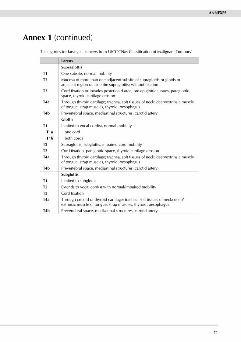
71
Annex 1 (continued)
TcategoriesforlaryngealcancersfromUICC:TNMClassificationofMalignantTumours4
Larynxsupraglottis
T1 One subsite, normal mobility
T2 Mucosa of more than one adjacent subsite of supraglottis or glottis or adjacentregionoutsidethesupraglottis;withoutfixation
T3 Cordfixationorinvadespostcricoidarea,pre-epiglottictissues,paraglotticspace, thyroid cartilage erosion
T4a Through thyroid cartilage; trachea, soft tissues of neck: deep/extrinsic muscle of tongue, strap muscles, thyroid, oesophagus
T4b prevertebral space, mediastinal structures, carotid artery
GlottisT1 limited to vocal cord(s), normal mobility
T1a one cord
T1b both cords
T2 Supraglottis, subglottis, impaired cord mobility
T3 Cordfixation,paraglotticspace,thyroidcartilageerosion
T4a Through thyroid cartilage; trachea, soft tissues of neck: deep/extrinsic muscle of tongue, strap muscles, thyroid, oesophagus
T4b prevertebral space, mediastinal structures, carotid artery
subglottisT1 limited to subglottis
T2 Extends to vocal cord(s) with normal/impaired mobility
T3 Cordfixation
T4a Through cricoid or thyroid cartilage; trachea, soft tissues of neck: deep/extrinsic muscle of tongue, strap muscles, thyroid, oesophagus
T4b prevertebral space, mediastinal structures, carotid artery
ANNeXes

72
DIAGNOsIs AND MANAGeMeNT OF HeAD AND NeCk CANCeR
Annex 1 (continued)
N categories for oral cavity, oropharyngeal, hypopharyngeal and laryngeal and cancers from UICC:TNMClassificationofMalignantTumours4
NX Regional lymph nodes cannot be assessed
N0 No regional lymph nodes metastasis
N1 Ipsilateralsingle≤3cm
N2 a. Ipsilateral single > 3 to 6 cmb.Ipsilateralmultiple≤6cmc.Bilateral,contralateral≤6cm
N3 >6 cm
M categories for oral cavity, oropharyngeal, hypopharyngeal and laryngeal and cancers from UICC:TNMClassificationofMalignantTumours4
MX Distant metastasis cannot be assessed
M0 No distant metastasis
M1 Distant metastasis
Stage grouping for oral cavity, oropharyngeal, hypopharyngeal and laryngeal and cancers from UICC:TNMClassificationofMalignantTumours4
stage 0 Tis N0 M0
stage I T1 N0 M0
stage II T2 N0 M0
stage III T1, T2 N1 M0
T3 N0, N1 M0
stage IvA T1, T2, T3 N2 M0
T4a N0, N1, N2 M0
stage Ivb T4b Any N M0
Any T N3 M0
stage IvC Any T Any N M1
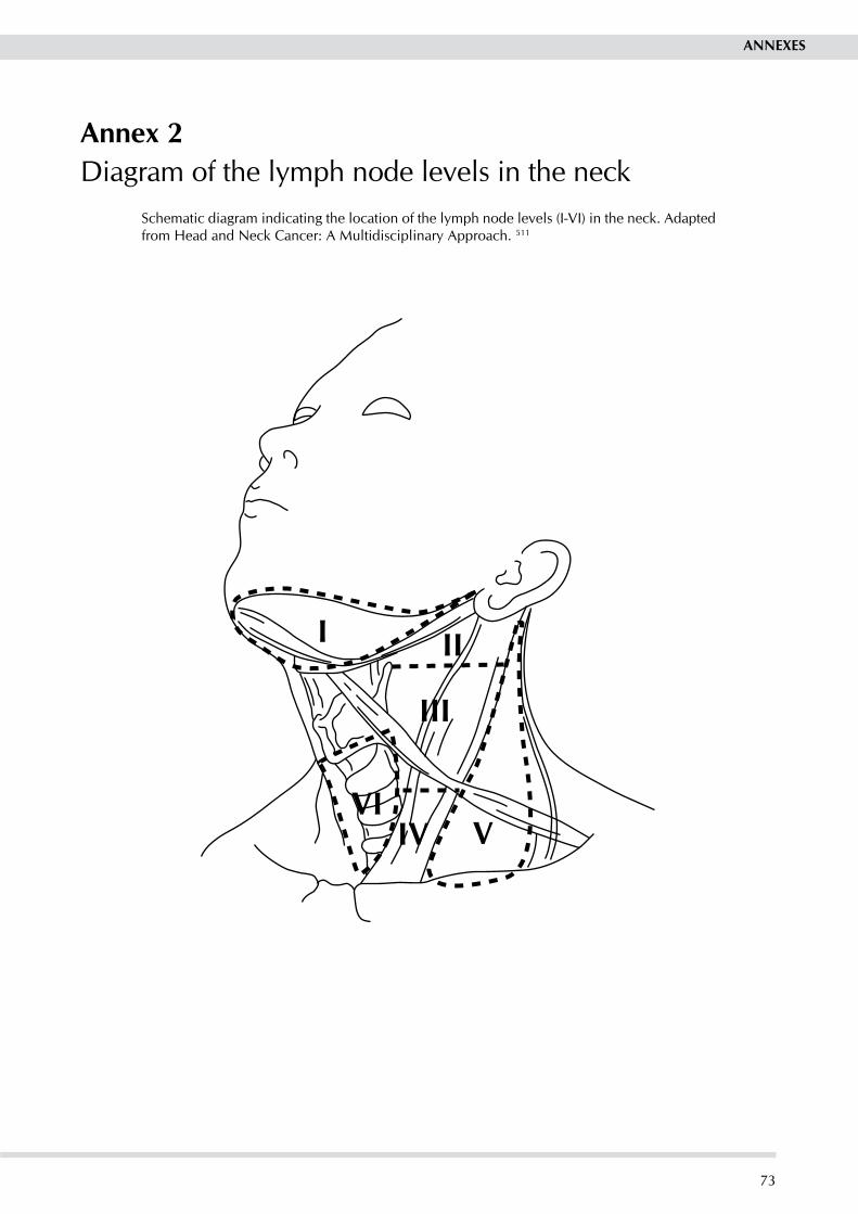
73
I II
III
IV VVI
Annex 2Diagram of the lymph node levels in the neck
Schematic diagram indicating the location of the lymph node levels (I-vI) in the neck. Adapted from Head and Neck Cancer: A Multidisciplinary Approach. 511
ANNeXes

74
DIAGNOsIs AND MANAGeMeNT OF HeAD AND NeCk CANCeR
Oral cavity
Larynx
Oropharynx
Hypopharynx
Annex 3Diagrams of the head and neck
Diagram of the head and neck, which may be helpful in illustrating where a patient’s cancer is situated.

75
Soft palate
Tonsil
Retromolar trigone
Anterior tongue
Alveolus (gum)
Lip
Posterior tongue
Lip
Floor of mouth
Underside of tongue
Annex 3 (continued)Diagram of the mouth and throat, which may be helpful in illustrating where a patient’s cancer is situated.
Diagram of the mouth and underside of the tongue, which may be helpful in illustrating where a patient’s cancer is situated.
ANNeXes
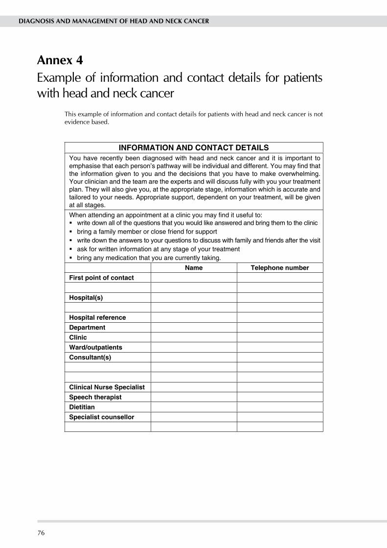
76
DIAGNOsIs AND MANAGeMeNT OF HeAD AND NeCk CANCeR
Annex 4Example of information and contact details for patients with head and neck cancer
This example of information and contact details for patients with head and neck cancer is not evidence based.
INFORMATION AND CONTACT DETAILSYou have recently been diagnosed with head and neck cancer and it is important to emphasise that each person’s pathway will be individual and different. You may find that the information given to you and the decisions that you have to make overwhelming. Your clinician and the team are the experts and will discuss fully with you your treatment plan. They will also give you, at the appropriate stage, information which is accurate and tailored to your needs. Appropriate support, dependent on your treatment, will be given at all stages.
When attending an appointment at a clinic you may find it useful to:write down all of the questions that you would like answered and bring them to the clinicbring a family member or close friend for supportwrite down the answers to your questions to discuss with family and friends after the visitask for written information at any stage of your treatmentbring any medication that you are currently taking.
Name Telephone numberFirst point of contact
Hospital(s)
Hospital referenceDepartmentClinicWard/outpatientsConsultant(s)
Clinical Nurse SpecialistSpeech therapistDietitianSpecialist counsellor

77
Annex 4 (continued)
Diagnosis
Medications
Questions
ANNeXes

78
DIAGNOsIs AND MANAGeMeNT OF HeAD AND NeCk CANCeR
References1 Macfarlane GJ, boyle p, Scully C. Oral cancer in Scotland:
changing incidence and mortality. bMJ. 1992;305:1121-3. 2 Department of Dental Sciences. Screening for oral cancer.
london: Royal College of Surgeons of England; 1994. 3 british Dental Association. Opportunistic oral cancer
screening. bDA occasional paper, April 2000 (issue number 6). [cited 11 August 2006]. Available from url: http://www.bda-dentistry.org.uk/about/docs/mouth_cancer.pdf
4 Sobin LH,WittekindC, editors. TNMClassification ofMalignant Tumours. 6th Edition ed. Hoboken, NJ: Wiley; 2002.
5 Altieri A, bosetti C, Talamini R, Gallus S, Franceschi S, levi F, et al. Cessation of smoking and drinking and the risk of laryngeal cancer. b J Cancer 2002;87(11):1227-9.
6 balaram p, Sridhar H, Rajkumar T, vaccarella S, Herrero R, Nandakumar A, et al. Oral cancer in Southern India: The influence of smoking, drinking, paan-chewing and oralhygiene. Int J Cancer 2002;98(3):440-5.
7 Cheng yJ, Hildesheim A, Hsu MM, Chen IH, brinton lA, levine pH, et al. Cigarette smoking, alcohol consumption and risk of nasopharyngeal carcinoma in Taiwan. Cancer Causes Control 1999;10(3):201-7.
8 De boer MF, van den borne b, pruyn JF, Ryckman RM, volovics l, Knegt pp, et al. psychosocial and physical correlates of survival and recurrence in patients with head and neck carcinoma: results of a 6-year longitudinal study. Cancer 1998;83(12):2567-79.
9 de Graeff A, de leeuw JR, Ros WJ, Hordijk GJ, blijham GH, Winnubst JA. Sociodemographic factors and quality of life as prognostic indicators in head and neck cancer. Eur J Cancer 2001;37(3):332-9.
10 De Stefani E, boffetta p, Oreggia F, Mendilaharsu M, Deneo-pellegrini H. Smoking patterns and cancer of the oral cavity and pharynx: a case-control study in Uruguay. Oral Oncol 1998;34(5):340-6.
11 Franceschi S, levi F, la vecchia C, Conti E, Dal Maso l, barzan l, et al. Comparison of the effect of smoking and alcohol drinking between oral and pharyngeal cancer. Int J Cancer 1999;83(1):1-4.
12 lissowska J, pilarska A, pilarski p, Samolczyk-Wanyura D, piekarczyk J, bardin-Mikolajczak A, et al. Smoking, alcohol, diet, dentition and sexual practices in the epidemiology of oral cancer in poland. Eur J Cancer prev 2003;12(1):25-33.
13 perea-Milla lopez E, Minarro-Del Moral RM, Martinez-Garcia C, Zanetti R, Rosso S, Serrano S, et al. lifestyles, environmental and phenotypic factors associated with lip cancer: A case-control study in southern Spain. b J Cancer 2003;88(11):1702-7.
14 Critchley JA, Unal b. Health effects associated with smokeless tobacco: a systematic review. Thorax 2003;58(5):435-43.
15 NHS Scotland and ASH Scotland. Smoking Cessation Guidelines for Scotland: 2004 update. Edinburgh: NHS Health Scotland; 2004. [cited 11 August 2006]. Available from url: http://www.hebs.scot.nhs.uk/services/pubs/pdf/SmokingCes2004.pdf
16 Spira A, beane J, Shah v, liu G, Schembri F, yang x, et al. Effects of cigarette smoke on the human airway epithelial cell transcriptome. proc Natl Acad Sci USA 2004;101(27):10143-8.
17 bagnardi v, blangiardo M, la vecchia C, Corrao G. Alcohol consumption and the risk of cancer: a meta-analysis. Alcohol Res Health 2001;25(4):263-70.
18 Corrao G, bagnardi v, Zambon A, Arico S. Exploring the dose-response relationship between alcohol consumption and the risk of several alcohol-related conditions: a meta-analysis. Addiction 1999;94(10):1551-73.
19 Scottish Intercollegiate Guideline Network (SIGN). The management of harmful drinking and alcohol dependence in primary care. Edinburgh: SIGN; 2003 (SIGN publication no. 74). [cited 11 August 2006]. Available from url: http://www.sign.ac.uk/pdf/sign74.pdf
20 Talamini R, bosetti C, la vecchia C, Dal Maso l, levi F, bidoli E, et al. Combined effect of tobacco and alcohol on laryngeal cancer risk: a case-control study. Cancer Causes Control 2002;13(10):957-64.
21 bosetti C, la vecchia C, Talamin R, Negri E, levi F, Fryzek J, et al. Energy, macronutrients and laryngeal cancer risk. Ann Oncol 2003;14(6):907-12.
22 Uzcudun AE, Retolaza IR, Fernandez pb, Sanchez Hernandez JJ, Grande AG, Garcia AG, et al. Nutrition and pharyngeal cancer: results from a case-control study in Spain. Head Neck 2002;24(9):830-40.
23 Franceschi S, Favero A, Conti E, Talamini R, volpe R, Negri E, et al. Food groups, oils and butter, and cancer of the oral cavity and pharynx. b J Cancer 1999;80(3-4):614-20.
24 De Stefani E, Oreggia F, boffetta p, Deneo-pellegrini H, Ronco A, Mendilaharsu M. Tomatoes, tomato-rich foods, lycopene and cancer of the upper aerodigestive tract: a case-control in Uruguay. Oral Onco. 2000;36(1):47-53.
25 bosetti C, la vecchia C, Talamini R, Negri E, levi F, Dal Maso l, et al. Food groups and laryngeal cancer risk: a case-control study from Italy and Switzerland. Int J Cancer 2002;100(3):355-60.
26 Tavani A, pelucchi C, parpinel M, Negri E, Franceschi S, levi F, et al. n-3 polyunsaturated fatty acid intake and cancer risk in Italy and Switzerland. Int J Cancer 2003;105(1):113-6.
27 bosetti C, Gallus S, Trichopoulou A, Talamini R, Franceschi S,NegriE,etal.InfluenceoftheMediterraneanDietontheRisk of Cancers of the Upper Aerodigestive Tract. Cancer Epidemiol biomarkers prev 1091;12(10):1091-4.
28 bosetti C, Talamini R, levi F, Negri E, Franceschi S, Airoldi l, et al. Fried foods: a risk factor for laryngeal cancer? b J Cancer 2002;87(11):1230-3.
29 levi F, pasche C, lucchini F, Chatenoud l, Jacobs DR, Jr., LaVecchiaC.Refinedandwholegraincerealsandtheriskof oral, oesophageal and laryngeal cancer. Eur J Clin Nutr 2000;54(6):487-9.
30 Oreggia F, De Stefani E, boffetta p, brennan p, Deneo-pellegrini H, Ronco Al. Meat, fat and risk of laryngeal cancer: a case-control study in Uruguay. Oral Oncol 2001;37(2):141-5.
31 El-Serag Hb, Hepworth EJ, lee p, Sonnenberg A. Gastroesophageal reflux disease is a risk factor for laryngeal and pharyngeal cancer. Am J Gastroenterol 2001;96(7):2013-8.
32 Elahi A, Zheng Z, park J, Eyring K, McCaffrey T, lazarus p. The human OGG1 DNA repair enzyme and its association with orolaryngeal cancer risk. Carcinogenesis 2002;23(7):1229-34.
33 Elahi A, bendaly J, Zheng Z, Muscat JE, Richie Jr Jp, Schantz Sp, et al. Detection of UGT1A10 polymorphisms and their association with orolaryngeal carcinoma risk. Cancer 2003;98(4):872-80.
34 Cheng l, Sturgis EM, Eicher SA, Spitz MR, Wei Q. Expression of nucleotide excision repair genes risk for squamous cell carcinoma of the head and the risk for squamous cell carcinoma of the head and neck. Cancer 2002;94(2):393-7.
35 Zheng y, Shen H, Sturgis EM, Wang lE, Eicher SA, Strom SS, et al. Cyclin D1 polymorphism and and neck: a case-control study. Carcinogenesis 2001;22(8):1195-9.
36 Zheng Z, park Jy, Guillemette C, Schantz Sp, lazarus p. Tobacco carcinogen-detoxifying enzyme UGT1A7 and its association with orolaryngeal cancer risk. J Nat Cancer Inst 2001;93(18):1411-8.
37 Dahlstrom KR, Adler-Storthz K, Etzel CJ, liu Z, Dillon l, El-Naggar AK, et al. Human papillomavirus type 16 infection and squamous cell carcinoma of the head and neck in never-smokers: A matched pair analysis. Clin Cancer Res 2003;9(7):2620-6.
38 Herrero R, Castellsague x, pawlita M, lissowska J, Kee F, balaram p, et al. Human papillomavirus and oral cancer: the International Agency for Research on Cancer multicenter study. J Nat Cancer Inst 2003;95(23):1772-83.
39 Cruz GD, le Geros RZ, Ostroff JS, Hay Jl, Kenigsberg H, Franklin DM. Oral cancer knowledge, risk factors and characteristics of subjects in an oral cancer screening programme. J Am Dent Assoc 2002;133:1064-71.
40 Hay Jl, Ostroff JS, Cruz GD, leGeros RZ, Kenigsberg H, Franklin DM. Oral cancer risk perception among participants in an oral cancer screening program. Cancer Epidemiol biomarkers prev 2002;11:155-8.
41 Horowitz AM, Canto MT, Child Wl. Maryland adults’ perspectives on oral cancer prevention and early detection. J Am Dent Assoc 2002;133:1058-63.

79
ReFeReNCes
42 Warnakulasuriya KA, Harris CK, Scarrott DM, Watt R, Gelbier S, peters TJ, Johnson NW. An alarming lack of public awareness towards oral cancer. br Dent J 1999;187:319-22.
43 vora AR, yeoman CM, Hayter Jp. Alcohol, tobacco and paan use and understanding of oral cancer risk among Asian males in leicester. br Dent J 2000;188:444-51.
44 Humphris GM, Freeman R, Clarke HM. Risk perception of oral cancer in smokers attending primary care: a randomised controlled trial. Oral Oncol 2004;40(9):916-24.
45 de Faria pR, Cardoso Sv, de ANS, Silva SJ, loyola AM. Clinical presentation of patients with oral squamous cell carcinoma whenfirstseenbydentistsorphysiciansinateachinghospitalin brazil. Clin Oral Investig 2003;7(1):46-51.
46 Kujan O, Glenny AM, Duxbury AJ, Thakker N, Sloan p. Screening programmes for the early detection and prevention of oral cancer (Cochrane Review). In: The Cochrane library, Issue 3, 2004. Chichester: John Wiley & Sons ltd.
47 West Midlands Health Technology Assessment Collaboration (WMHTAC). The effectiveness of Toludine blue Dye as an adjunct to oral cancer screening in general dental practice: Report 24. University of birmingham: Department of public Health & Epidemiology; 2000.
48 Scottish Executive Health Department. Scottish Referral Guidelines for Suspected Cancer. Edinburgh: Scottish Executive Health Department; 2002. [cited 11 August 2006]. Available from url: http://www.show.scot.nhs.uk/sehd/mels/hdl2002_45.pdf
49 pitiphat W, Diehl SR, laskaris G, Cartsos v, Douglass CW, Zavras AI. Factors associated with delay in the diagnosis of oral cancer. J Dent Res 2002;81(3):192-7.
50 Jones TM, Hargrove O, lancaster J, Fenton J, Shenoy A, Roland NJ. Waiting times during the management of head and neck tumours. J laryngol Otol 2002;116(4):275-9.
51 Hollows p, McAndrew pG, perini MG. Delays in the referral and treatment of oral squamous cell carcinoma. br Dent J 2000;188(5):262-5.
52 Robinson J, Chew F. Rapid access for patients with head and neck lumps. Nurs Stand 2001;16(2):33-7.
53 Murray A, Stewart CJR, McGarry GW, MacKenzie K. patients with neck lumps: Can they be managed in a ‘one-stop’ clinic setting? Clin Otolaryngol Allied Sci 2000;25(6):471-5.
54 el Hag IA, Chiedozi lC, al Reyees FA, Kollur SM. Fine needle aspiration cytology of head and neck masses. Seven years’ experience in a secondary care hospital. Acta Cytol 2003;47(3):387-92.
55 Ellison E, lapuerta p, Martin SE. Supraclavicular masses: results of a series of 309 cases biopsiedby fineneedleaspiration. Head Neck 1999;21(3):239-46.
56 Shaha AR, Hoover El, Marti J, Krespi y. Is routine triple endoscopy cost-effective in head and neck cancer? Am J Surg 1988;155:750-3.
57 benninger MS, Enrique RR, Nichols RD. Symptom-directed selectrive endoscopy and cost containment for evaluation of head and neck cancers. Head Neck 1993;15:532-6.
58 benninger MS, Shariff A, blazoff K. Symptom-directed selective endoscopy: long-term efficacy. Archives of Otolaryngology – Head Neck Surg 2001;127(7):770-3.
59 de vries N. Multiple primary tumours in oral cancer. Int J Oral Maxillofac Surg 1986;15:85-7.
60 Delank W, Khanavkar b, Nakhosteen JA, Stoll W. A pilot studyofautofluorescentendoscopyfortheinvivodetectionof laryngeal cancer. laryngoscope 2000;110(3 pt 1):368-73.
61 Kulapaditharom b, boonkitticharoen v. performance characteristics of fluorescence endoscope in detectionof head and neck cancers. Ann Otol Rhinol laryngol 2001;110(1):45-52.
62 MalzahnK,DreyerT,GlanzH,ArensC.Autofluorescenceendoscopy in the diagnosis of early laryngeal cancer and its precursor lesions. laryngoscope 2002;112(3):488-93.
63 pak MW, To KF, leung SF, van Hasselt CA. In vivo diagnosis of persistent and recurrent nasopharyngeal carcinoma by contact endoscopy. laryngoscope 2002;112(8 pt 1):1459-66.
64 Declercq A, van den Hauwe l, van Marck E, van de Heyning pH, Spanoghe M, De Schepper AM. patterns of framework invasion in patients with laryngeal cancer: correlation of in vitromagneticresonanceimagingandpathologicalfindings.Acta Otolaryngol 1998;118(6):892-5.
65 Wilson JA, editor. Effective head and neck cancer management: consensus document. london: british Association of Otorhinolaryngologists, Head and Neck Surgeons; 1998.
66 leslie A. Staging of squamous cell carcinoma of the oral cavity and oropharynx:a comparison of MRI and CT in T- and N- staging. J Comput Assist Tomog 1999;23:43-9.
67 Kaanders JH, Hordijk GJ, Dutch Cooperative H, Neck Oncology G. Carcinoma of the larynx: the Dutch national guideline for diagnostics, treatment, supportive care and rehabilitation. Radiother Oncol 2002;63(3):299-307.
68 byers R, El-Naggar A, lee yy, Rao b, Fornage b, Terry NH, et al. Can we detect or predict the presence of occult nodal metastases in patients with squamous carcinoma of the oral tongue? Head Neck 1998;20(2):138-44.
69 pameijer FA, Mancuso AA, Mendenhall WM, parsons JT, Kubilis pS, van den brekel MW, et al. Can pretreatment computed tomography predict local control in T3 squamous cellcarcinomaoftheglotticlarynxtreatedwithdefinitiveradiotherapy? Modern imaging techniques and ultrasound-guided aspiration cytology for the assessment of neck node metastases: a prospective comparative study. Int J Radiat Oncol biol phys 1997;37(5):1011-21.
70 van den brekel MW, Castelijns JA, Stel Hv, Golding Rp, Meyer CJ, Snow Gb. Modern imaging techniques and ultrasound-guided aspiration cytology for the assessment of neck node metastases: a prospective comparative study. Eur Arch Otorhinolaryngol 1993;250(1):11-7.
71 Takes Rp. The value of ultrasound with ultrasound-guided fine-needle aspiration biopsy compared to computed tomography in the detection of regional metastases in the clinically negative neck. Int J Oncol biol phys 1998;40:1027-32.
72 Stokkel Mp, ten broek FW, Hordijk GJ, Koole R, van Rijk pp. preoperative evaluation of patients with primary head and neckcancerusingdual-head18fluorodeoxyglucosepositronemission tomography. Ann Surgery 2000;231(2):229-34.
73 Adams S, baum Rp, Stuckensen T, bitter K, Hor G. prospective comparison of 18F-FDG pET with conventional imaging modalities (CT, MRI, US) in lymph node staging of head and neck cancer. E J Nucl Med 1998;25(9):1255-60.
74 Haughey bH, Gates GA, Arfken Cl, Harvey J. Meta-analysis of second malignant tumors in head and neck cancer: the case for an endoscopic screening protocol. Ann Otol Rhinol laryngol 1992;101(2 pt 1):105-12.
75 de bree R, Deurloo EE, Snow Gb, leemans CR. Screening for distant metastases in patients with head and neck cancer. laryngoscope 2000;110(3 pt 1):397-401.
76 Warner GC, Cox GJ. Evaluation of chest radiography versus chest computed tomography in screening for pulmonary malignancy in advanced head and neck cancer. J Otolaryngol 2003;32(2):107-9.
77 Arunachalam pS, putnam G, Jennings p, Messersmith R, Robson AK. Role of computerized tomography (CT) scan of the chest in patients with newly diagnosed head and neck cancers. Clin Otolaryngol Allied Sci 2002;27(5):409-11.
78 Jungehulsing M, Scheidhauer K, Damm M, pietrzyk U, EckelH, SchichaH, et al. 2[18F]-fluoro-2-deoxy-D-glucose positron emission tomography is a sensitive tool for the detection of occult primary cancer (carcinoma of unknown primary syndrome) with head and neck lymph node manifestation. Otolaryngology Head Neck Surg 2000;123(3):294-301.
79 Johansen J, Eigtved A, buchwald C, Theilgaard SA, Hansen HS.Implicationof18F-fluoro-2-deoxy-D-glucosepositronemission tomography on management of carcinoma of unknown primary in the head and neck: a Danish cohort study. laryngoscope 2002;112(11):2009-14.
80 Regelink G, brouwer J, de bree R, pruim J, van der laan bF, vaalburg W, et al. Detection of unknown primary tumours and distant metastases in patients with cervical metastases: value of FDG-pET versus conventional modalities. E J Nucl Med Mol Imaging 2002;29(8):1024-30.
81 bongers v, Hobbelink MG, van Rijk pp, Hordijk GJ. Cost-effectivenessofdual-head18F-fluorodeoxyglucosePETforthe detection of recurrent laryngeal cancer. Cancer biother Radiopharm 2002;17(3):303-6.
82 lonneux M, lawson G, Ide C, bausart R, Remacle M, pauwels S. positron emission tomography with fluorodeoxyglucose for suspectedhead andneck tumorrecurrence in the symptomatic patient. laryngoscope 2000;110(9):1493-7.

80
DIAGNOsIs AND MANAGeMeNT OF HeAD AND NeCk CANCeR
83 Spector JG, Sessions DG, Haughey bH, Chao KS, Simpson J, El Mofty S, et al. Delayed regional metastases, distant metastases, and second primary malignancies in squamous cell carcinomas of the larynx and hypopharynx. laryngoscope 2001;111(6):1079-87.
84 Ozdek A, Sarac S, Akyol MU, Unal OF, Sungur A. Histopathological predictors of occult lymph node metastases in supraglottic squamous cell carcinomas. Eur Arch Otorhinolaryngol 2000;257(7):389-92.
85 Royal College of pathologists. Standards and Datasets for Reporting Cancers:Datasets for histopathology reports on head and neck carcinomas and salivary neoplasms. 2nd Edition. london: The Royal College of pathologists; 2005. [cited 11 August 2006]. Available from url: http:// www.rcpath.org/resources/pdf/HeadNeckDatasetJun05.pdf
86 Coatesworth Ap, Maclennan K. Squamous cell carcinoma of the upper aerodigestive tract: the prevalence of microscopic extracapsular spread and soft tissue deposits in the clinically N0 neck. Head Neck 2002;24(3):258-61.
87 Carinci F, pelucchi S, Farina A, De Franciscis G, Calearo C. Extension as a prognostic factor in oropharyngeal cancer: largest mucosal dimension compared with number of (sub)sites involved. br J Oral Maxillofac Surg 1998;36(6):440-5.
88 Hirvikoski p, Kumpulainen E, virtaniemi J, pirinen R, Salmi l, Halonen p, et al. Enhanced apoptosis correlates with poor survival in patients with laryngeal cancer but not with cell proliferation, bcl-2 or p53 expression. Eur J Cancer 1999;35(2):231-7.
89 Fortin A, Couture C, Doucet R, Albert M, Allard J, Tetu b. Does histologic grade have a role in the management of head and neck cancers? J Clin Oncol 2001;19(21):4107-16.
90 yamazaki H, Inoue T, Teshima T, Tanaka E, Koizumi M, Kagawa K, et al. Tongue cancer treated with brachytherapy: is thickness of tongue cancer a prognostic factor for regional control? Anticancer Res 1998;18(2b):1261-5.
91 O’brien CJ, lauer CS, Fredricks S, Clifford AR, McNeil Eb, BagiaJS,etal.TumorthicknessinfluencesprognosisofT1and T2 oral cavity cancer - but what thickness? Head Neck 2003;25(11):937-45.
92 Nishimaki T, Kanda T, Nakagawa S, Kosugi S, Tanabe T, Hatakeyama K. Outcomes and prognostic factors after surgical resection of hypopharyngeal and cervical esophageal carcinomas. Int Surg 2002;87(1):38-44.
93 barnes l, Eveson JW, Reichart p, Sidransky D, editors. pathology and Genetics of Head and Neck Tumours. lyon: IARCPress;2005.(WHOClassificationofTumours)
94 Ang KK, berkey bA, Tu x, Zhang HZ, Katz R, Hammond EH, et al. Impact of epidermal growth factor receptor expression on survival and pattern of relapse in patients with advanced head and neck carcinoma. Cancer Res 2002;62(24):7350-6.
95 Spiro RH, Guillamondegui O, Jr., paulino AF, Huvos AG. pattern of invasion and margin assessment in patients with oral tongue cancer. Head Neck 1999;21(5):408-13.
96 Wong RJ, Keel Sb, Glynn RJ, varvares MA. Histological pattern of mandibular invasion by oral squamous cell carcinoma. laryngoscope 2000;110(1):65-72.
97 Weijers M, Snow Gb, bezemer pD, van der Wal JE, van der Waal I. The clinical relevance of epithelial dysplasia in the surgicalmarginsoftongueandfloorofmouthsquamouscell carcinoma: an analysis of 37 patients. J Oral pathol Med 2002;31(1):11-5.
98 bailey JS, blanchaert RH, Jr., Ord RA. Management of oral squamous cell carcinoma treated with inadequate excisional biopsy. J Oral Maxillofac Surg. 2001;59(9):1007-10; discussion 11.
99 McMahon J, O’brien CJ, pathak I, Hamill R, McNeil E, HammersleyN, et al. Influenceof conditionof surgicalmarginsonlocalrecurrenceanddisease-specificsurvivalin oral and oropharyngeal cancer. br J Oral Maxillofac Surg 2003;41(4):224-31.
100 Slootweg pJ, Hordijk GJ, Schade y, van Es RJ, Koole R. Treatment failure and margin status in head and neck cancer. A critical view on the potential value of molecular pathology. Oral Oncol 2002;38(5):500-3.
101 Ribeiro NF, Godden DR, Wilson GE, butterworth DM, Woodwards RT. Do frozen sections help achieve adequate surgical margins in the resection of oral carcinoma? Int J Oral Maxillofac Surg 2003;32(2):152-8.
102 DiNardo lJ, lin J, Karageorge lS, powers CN. Accuracy, utility, and cost of frozen section margins in head and neck cancer surgery. laryngoscope 2000;110(10 pt 1):1773-6.
103 Danic D, Milicic D, prgomet D, leovic D. prognostic factors in carcinoma of the larynx: relevance of DNA ploidy, S-fractions and localization of the tumour. J laryngol Otol 1999;113(6):538-41.
104 volavsek M, bracko M, Gale N. Distribution and prognostic significanceofcellcycleproteinsinsquamouscarcinomaof the larynx, hypopharynx and adjacent epithelial hyperplastic lesions. J laryngol Otol 2003;117(4):286-93.
105 brasilino de Carvalho M. Quantitative analysis of the extent ofextracapsularinvasionanditsprognosticsignificance:aprospective study of 170 cases of carcinoma of the larynx and hypopharynx. Head Neck 1998;20(1):16-21.
106 Tankere F, Camproux A, barry b, Guedon C, Depondt J, Gehanno p. prognostic value of lymph node involvement in oral cancers: a study of 137 cases. laryngoscope 2000;110(12):2061-5.
107 Woolgar JA, Rogers SN, lowe D, brown JS, vaughan ED. Cervical lymph node metastasis in oral cancer: the importance of even microscopic extracapsular spread. Oral Oncol 2003;39(2):130-7.
108 Suoglu y, Erdamar b, Katircioglu OS, Karatay MC, Sunay T. Extracapsular spread in ipsilateral neck and contralateral neck metastases in laryngeal cancer. Ann Otol Rhinol laryngol 2002;111(5 pt 1):447-54.
109 Greenberg JS, Fowler R, Gomez J, Mo v, Roberts D, El Naggar AK, et al. Extent of extracapsular spread: a critical prognosticator in oral tongue cancer. Cancer 2003;97(6):1464-70.
110 Esposito ED, Motta S, Cassiano b, Motta G. Occult lymph node metastases in supraglottic cancers of the larynx. Otolaryngology - Head Neck Surg 2001;124(3):253-7.
111 Enepekides DJ, Sultanem K, Nguyen C, Shenouda G, black MJ, Rochon l. Occult cervical metastases: immunoperoxidase analysis of the pathologically negative neck. Otolaryngol Head Neck Surg 1999;120(5):713-7.
112 Ferlito A, partridge M, brennan J, Hamakawa H. lymph node micrometastasis in head and neck cancer: a review. Acta Otol 2001;121:660–5.
113 lindel K, beer KT, laissue J, Greiner RH, Aebersold DM. Human papillomavirus positive squamous cell carcinoma of the oropharynx: a radiosensitive subgroup of head and neck carcinoma. Cancer 2001;92(4):805-13.
114 buchwald C, lindeberg H, pedersen bl, Franzmann Mb. Human papilloma virus and p53 expression in carcinomas associated with sinonasal papillomas: a Danish Epidemiological study 1980-1998. laryngoscope 2001;111(6):1104-10.
115 Friesland S, Mellin H, Munck-Wikland E, Nilsson A, lindholm J, Dalianis T, et al. Human papilloma virus (Hpv) and p53 immunostaining in advanced tonsillar carcinoma – relation to radiotherapy response and survival. Anticancer Res 2001;21(1b):529-34.
116 Gillison Ml, Koch WM, Capone R, Spafford M, Westra WH, Wu l, et al. Evidence for a causal association between human papillomavirus and a subset of head and neck cancers. J Natl Cancer Inst 2000;92(9):709-20.
117 Takes R, baatenburg De Jong R, Alles M, Meeuwis CA, Marres, HA, et al. Markers for nodal metastasis in head and neck squamous cancer. Arch Otolaryngol Head Neck Surg 2002;128:512–8.
118 Helliwell T. Molecular markers of metastasis in squamous carcinomas. J pathol 2001;194:289–93.
119 Epstein Jb, lunn R, le N, Stevenson-Moore p. periodontal attachment loss in patients after head and neck radiation therapy. Oral Surg Oral Med Oral pathol Oral Radiol Endod 1998;86(6):673-7.
120 Denis F, Garaud p, bardet E, Alfonsi M, Sire C, Germain T, bergerot p, Rhein b, Tortochaux J, Oudinot, p, Calais G. late toxicity results of the GORTEC 94-01 randomized trial comparing radiotherapy with concomitant radiochemotherapy for advanced-stage oropharynx carcinoma: Comparison of lENT/SOMA, RTOG/EORTC, and NCI-CTC scoring systems. Int J Radiat Oncol biol phys 2003;55(1):93-8.
121 Soo KC, Tan EH, Wee J, lim D, Tai bC, Khoo Ml, et al. Surgery and adjuvant radiotherapy vs concurrent chemoradiotherapy in stage III/Iv nonmetastatic squamous cell head and neck cancer: a randomised comparison. br J Cancer 2005;93(3):279-86.

81
ReFeReNCes
122 Crampette l, Garrel R, Gardiner Q, Maurice N, Mondain M,MakeieffM,etal.Modifiedsubtotallaryngectomywithcricohyoidoepiglottopexy--long term results in 81 patients. Head Neck 1999;21(2):95-103.
123 Giovanni A, Guelfucci b, Gras R, yu p, Zanaret M. partial frontolateral laryngectomy with epiglottic reconstruction for management of early-stage glottic carcinoma. laryngoscope 2001;111(4 pt 1):663-8.
124 luukkaa M, Aitasalo K, pulkkinen J, lindholm p, valavaara R, Grenman R. Neodymium yAG contact laser in the treatment of cancer of the mobile tongue. Acta Oto-laryngologica 2002;122(3):318-22.
125 Edwards DM, Johnson NW. Treatment of upper aerodigestive tract cancers in England and its effect on survival. b J Cancer 1999;81(2):323-9.
126 bahadur S, Thakar A, Mohanti bK, lal p. Results of radiotherapy with, or without, salvage surgery versus combined surgery and radiotherapy in advanced carcinoma of the hypopharynx. J laryngol Otol 2002;116(1):29-32.
127 leon x, Quer M, Orus C, Moran J, Recher K. Results of an organ preservation protocol with induction chemotherapy and radiotherapy in patients with locally advanced pyriform sinus carcinoma. Eur Arch Otorhinolaryngol 2002;259(1):32-6.
128 barzan l, Talamini R, politi D, Minatel E, Gobitti C, Franchin G. Squamous cell carcinoma of the hypopharynx treated with surgery and radiotherapy. J laryngol Otol 2002;116(1):24-8.
129 MacKenzie RG, Franssen E, balogh JM, Gilbert RW, birt D, Davidson J. Comparing treatment outcomes of radiotherapy and surgery in locally advanced carcinoma of the larynx: a comparison limited to patients eligible for surgery. Int J Radiat Oncol biol phys 2000;47(1):65-71.
130 porter MJ, McIvor Np, Morton Rp, Hindley AC. Audit in themanagementofT3fixed-cordlaryngealcancer.AmJOtolaryngol 1998;19(6):360-4.
131 Staton J, Robbins KT, Newman l, Samant S, Sebelik M, vieira F. Factors predictive of poor functional outcome after chemoradiation for advanced laryngeal cancer. Otolaryngology – Head Neck Surg 2002;127(1):43-7.
132 Terrell JE, Fisher SG, Wolf GT. long-term quality of life after treatment of laryngeal cancer. The veterans Affairs laryngeal Cancer Study Group. Archives of Otolaryngology – Head Neck Surg 1998;124(9):964-71.
133 bergqvist M, brodin O, pouzon A, linder A, Hesselius p, blomquist E. Radiation treatment of T1-T4 squamous cell carcinoma of the larynx: a retrospective analysis and long-term follow-up of 135 patients. Anticancer Research. 2002;22(2b):1239-42.
134 paisley S, Warde pR, O’Sullivan b, Waldron J, Gullane pJ, payne D, et al. Results of radiotherapy for primary subglottic squamous cell carcinoma. Int J Radiat Oncol biol phys 2002;52(5):1245-50.
135 Orus C, leon x, vega M, Quer M. Initial treatment of the early stages (I, II) of supraglottic squamous cell carcinoma: partial laryngectomy versus radiotherapy. Eur Arch Otorhinolaryngol 2000;257(9):512-6.
136 Medini E, Medini I, lee CK, Gapany M, levitt SH. Curative radiotherapy for stage II-III squamous cell carcinoma of the glottic larynx. Am J Clin Oncol 1998;21(3):302-5.
137 lavertu p, bonafede Jp, Adelstein DJ, Saxton Jp, Strome M, Wanamaker JR, et al. Comparison of surgical complications after organ-preservation therapy in patients with stage III or Iv squamous cell head and neck cancer. Archives of Otolaryngology – Head Neck Surg 1998;124(4):401-6.
138 Mendenhall WM, Amdur RJ, Stringer Sp, villaret Db, Cassisi NJ. Radiation therapy for squamous cell carcinoma of the tonsillar region: a preferred alternative to surgery? J Clin Oncol 2000;18(11):2219-25.
139 Mendenhall WM, Stringer Sp, Amdur RJ, Hinerman RW, Moore-Higgs GJ, Cassisi NJ. Is radiation therapy a preferred alternative to surgery for squamous cell carcinoma of the base of tongue? J Clin Oncol 2000;18(1):35-42.
140 Tschudi D, Stoeckli S, Schmid S. Quality of life after different treatment modalities for carcinoma of the oropharynx. laryngoscope 2003;113(11):1949-54.
141 pfreundner l, Hoppe F, Willner J, preisler v, bratengeier K, Hagen R, et al. Induction chemotherapy with paclitaxel and cisplatin and CT-based 3D radiotherapy in patients with advanced laryngeal and hypopharyngeal carcinomas – a possibility for organ preservation. Radiother Oncol 2003;68(2):163-70.
142 Chao CKS. practical essentials of intensity modulated radiation therapy. 2nd ed. philadelphia: lippincott Williams & Wilkins; 2005.
143 Robbins KT, Clayman G, levine pA, Medina J, Sessions R,ShahaA,et al.Neckdissectionclassificationupdate:revisions proposed by the American Head and Neck Society and the American Academy of Otolaryngology-Head and Neck Surgery. Arch Otolaryngol Head Neck Surg 2002;128(7):751-8.
144 Spiro RH, Morgan GJ, Strong EW, Shah Jp. Supraomohyoid neck dissection. Am J Surg 1996;172(6):650-3.
145 Nieuwenhuis EJ, Castelijns JA, pijpers R, van den brekel MW, brakenhoff RH, van der Waal I, et al. Wait-and-see policy for the N0 neck in early-stage oral and oropharyngeal squamous cell carcinoma using ultrasonography-guided cytology: is therea role for identificationof thesentinelnode? Head Neck. 2002;24(3):282-9.
146 yang Cy, Andersen pE, Everts EC, Cohen JI. Nodal disease in purely glottic carcinoma: is elective neck treatment worthwhile? laryngoscope 1998;108(7):1006-8.
147 Gallo O, Fini-Storchi I, Napolitano l. Treatment of the contralateral negative neck in supraglottic cancer patients with unilateral node metastases (N1-3). Head Neck 2000;22(4):386-92.
148 pinilla M, Gonzalez FM, lopez-Cortijo C, Arellano b, Herrero J, Trinidad A, et al. Management of N0 neck in laryngeal carcinoma. Impact on patient’s survival. J laryngol Otol 2003;117(1):63-6.
149 Tu Gy. Upper neck (level II) dissection for no neck supraglottic carcinoma. laryngoscope 1999;109(3):467-70.
150 byers RM, Clayman Gl, McGill D, Andrews T, Kare Rp, Roberts Db, et al. Selective neck dissections for squamous carcinoma of the upper aerodigestive tract: patterns of regional failure. Head Neck 1999;21(6):499-505.
151 Candela FC, Kothari K, Shah Jp. patterns of cervical node metastases from squamous carcinoma of the oropharynx and hypopharynx. Head Neck 1990;12(3):197-203.
152 Shah Jp, Candela FC, poddar AK. The patterns of cervical lymph node metastases from squamous carcinoma of the oral cavity. Cancer 1990;66(1):109-13.
153 Spiro JD, Spiro RH, Shah Jp, Sessions Rb, Strong EW. Critical assessment of supraomohyoid neck dissection. Am J Surg 1988;156(4):286-9.
154 byers RM, Wolf pF, ballantyne AJ. Rationale for elective modifiedneckdissection.HeadNeckSurg1988;10(3):160-7.
155 Redaelli de Zinis lO, Nicolai p, Tomenzoli D, Ghizzardi D, Trimarchi M, Cappiello J, et al. The distribution of lymph node metastases in supraglottic squamous cell carcinoma: therapeutic implications. Head Neck 2002;24(10):913-20.
156 Hicks Wl, Jr., Kollmorgen DR, Kuriakose MA, Orner J, bakamjian vy, Winston J, et al. patterns of nodal metastasis and surgical management of the neck in supraglottic laryngeal carcinoma. Otolaryngol Head Neck Surg 1999;121(1):57-61.
157 Scola b, Fernandez-vega M, Martinez T, Fernandez-vega S, Ramirez C. Management of cancer of the supraglottis. Otolaryngol Head Neck Surg 2001;124(2):195-8.
158 buckley JG, Maclennan K. Cervical node metastases in laryngeal and hypopharyngeal cancer: a prospective analysis of prevalence and distribution. Head Neck 2000;22(4):380-5.
159 Smith GI, O’brien CJ, Clark J, Shannon KF, Clifford AR, McNeil Eb, et al. Management of the neck in patients with T1 and T2 cancer in the mouth. br J Oral Maxillofac Surg 2004;42(6):494-500.
160 Crean SJ, Hoffman A, potts J, Fardy MJ. Reduction of occult metastatic disease by extension of the supraomohyoid neck dissection to include level Iv. Head Neck 2003;25(9):758-62.
161 Hao Sp, Tsang NM. The role of supraomohyoid neck dissection in patients of oral cavity carcinoma(small star, filled).OralOncol2002;38(3):309-12.
162 O’brien CJ, Traynor SJ, McNeil E, McMahon JD, Chaplin JM. The use of clinical criteria alone in the management of the clinically negative neck among patients with squamous cell carcinoma of the oral cavity and oropharynx. Arch Otolaryngol Head Neck Surg 2000;126(3):360-5.
163 Turner Sl, Slevin NJ, Gupta NK, Swindell R. Radical external beam radiotherapy for 333 squamous carcinomas of the oral cavity – evaluation of late morbidity and a watch policy for the clinically negative neck. Radiother Oncol 1996;41(1):21-9.

82
DIAGNOsIs AND MANAGeMeNT OF HeAD AND NeCk CANCeR
164 Henick DH, Silver CE, Heller KS, Shaha AR, El GH, Wolk Dp. Supraomohyoid neck dissection as a staging procedure for squamous cell carcinomas of the oral cavity and oropharynx. Head Neck 1995;17(2):119-23.
165 Cole I, Hughes l. The relationship of cervical lymph node metastases to primary sites of carcinoma of the upper aerodigestive tract: a pathological study. Aust N Z J Surg 1997;67(12):860-5.
166 McGuirtWF,Jr.,JohnsonJT,MyersEN,RothfieldR,WagnerR. Floor of mouth carcinoma. The management of the clinically negative neck. Arch Otolaryngol Head Neck Surg 1995;121(3):278-82.
167 Kligerman J, lima R, Soares J, prado l, Dias Fl, Freitas E, et al. Supraomohyoid neck dissection in the treatment of T1/T2 squamous cell carcinoma of oral cavity. Am J Surg 1994;168(5):391-4.
168 Weiss MH, Harrison lb, Isaacs RS. Use of decision analysis in planning a management strategy for the stage N0 neck. Arch Otolaryngol Head Neck Surg 1994;120(7):699-702.
169 Fakih AR, Rao R, borges A, patel A. Elective versus therapeutic neck dissection in early carcinoma of the oral tongue. Am J Surg 1989;158(4):309-13.
170 van den brekel MW, Castelijns JA, Snow Gb. Diagnostic evaluation of the neck. Otolaryngol Clin North Am 1998;31(4):601-20.
171 Ho CM, lam KH, Wei WI, lau WF. Treatment of neck nodes in oral cancer. Surg Oncol 1992;1(1):73-8.
172 lydiatt DD, Robbins KT, byers RM, Wolf pF. Treatment of stage I and II oral tongue cancer. Head Neck 1993;15(4):308-12.
173 Grandi C, Mingardo M, Guzzo M, licitra l, podrecca S, Molinari R. Salvage surgery of cervical recurrences after neck dissection or radiotherapy. Head Neck 1993;15(4):292-5.
174 vandenbrouck C, Sancho-Garnier H, Chassagne D, Saravane D, Cachin y, Micheau C. Elective versus therapeutic radical neck dissection in epidermoid carcinoma of the oral cavity: results of a randomized clinical trial. Cancer 1980;46(2):386-90.
175 Johnson JT. Carcinoma of the larynx: selective approach to the management of cervical lymphatics. Ear, Nose, & Throat J 1994;73(5):303-5.
176 Manni JJ, van den Hoogen FJ. Supraomohyoid neck dissection with frozen section biopsy as a staging procedure in the clinically node-negative neck in carcinoma of the oral cavity. Am J Surg 1991;162(4):373-6.
177 leon x, Quer M, Orus C, Sancho FJ, bague S, burgues J. Selective dissection of levels II-III with intraoperative control of the upper and middle jugular nodes: a therapeutic option for the N0 neck. Head Neck 2001;23(6):441-6.
178 ByersRM.Modifiedneckdissection.Astudyof967casesfrom 1970 to 1980. Am J Surg 1985;150(4):414-21.
179 bocca E, pignataro O, Oldini C, Cappa C. Functional neck dissection: an evaluation and review of 843 cases. laryngoscope 1984;94(7):942-5.
180 Molinari R, Cantu G, Chiesa F, Grandi C. Retrospective comparison of conservative and radical neck dissection in laryngeal cancer. Ann Otol Rhinol laryngol 1980;89(6 pt 1):578-81.
181 Spiro RH, Gallo O, Shah Jp. Selective jugular node dissection in patients with squamous carcinoma of the larynx or pharynx. Am J Surg 1993;166(4):399-402.
182 Chow JM, levin bC, Krivit JS, Applebaum El. Radiotherapy or surgery for subclinical cervical node metastases. Archives of Otolaryngology Head Neck Surg 1989;115(8):981-4.
183 bier J. Radical neck dissection versus conservative neck dissection for squamous cell carcinoma of the oral cavity. Recent Results Cancer Res 1994;134:57-62.
184 Davidson bJ, Kulkarny v, Delacure MD, Shah Jp. posterior triangle metastases of squamous cell carcinoma of the upper aerodigestive tract. Am J Surg 1993;166(4):395-8.
185 Gregor RT, Oei SS, Hilgers FJ, Hart AA, balm AJ, Keus Rb. Management of cervical metastases in supraglottic cancer. Ann Otol Rhinol laryngol 1996;105(11):845-50.
186 barkley HT, Jr., Fletcher GH, Jesse RH, lindberg RD. Management of cervical lymph node metastases in squamous cell carcinoma of the tonsillar fossa, base of tongue, supraglottic larynx, and hypopharynx. Am J Surg 1972;124(4):462-7.
187 JesseRH,BallantyneAJ, LarsonD.Radical ormodifiedneck dissection: a therapeutic dilemma. Am J Surg 1978;136(4):516-9.
188 lingeman RE, Helmus C, Stephens R, Ulm J. Neck dissection: radical or conservative. Ann Otol Rhinol laryngol 1977;86(6 pt 1):737-44.
189 brandenburg JH, lee Cy. The eleventh nerve in radical neck surgery. laryngoscope 1981;91(11):1851-9.
190 Andersen pE, Shah Jp, Cambronero E, Spiro RH. The role of comprehensive neck dissection with preservation of the spinal accessory nerve in the clinically positive neck. Am J Surg 1994;168(5):499-502.
191 bocca E, pignataro O. A conservation technique in radical neck dissection. Ann Otol Rhinol laryngol 1967;76(5):975-87.
192 Chu W, Strawitz JG. Results in suprahyoid, modified radical, and standard radical neck dissections for metastatic squamous cell carcinoma: recurrence and survival. Am J Surg 1978;136(4):512-5.
193 Chepeha Db, Hoff pT, Taylor RJ, bradford CR, Teknos TN, Esclamado RM. Selective neck dissection for the treatment of neck metastasis from squamous cell carcinoma of the head and neck. laryngoscope 2002;112(3):434-8.
194 Andersen pE, Warren F, Spiro J, burningham A, Wong R, Wax MK, et al. Results of selective neck dissection in management of the node-positive neck. Arch Otolaryngol Head Neck Surg 2002;128(10):1180-4.
195 vikram b, Strong EW, Shah Jp, Spiro R. Failure in the neck following multimodality treatment for advanced head and neck cancer. Head Neck Surg 1984;6:724-9.
196 varghese C, Sankaranarayanan R, Nair b, Nair MK. predictors of neck node control in radically irradiated squamous cell carcinoma of the oropharynx and laryngopharynx. Head Neck 1993;15(2):105-8.
197 bataini Jp, bernier J, Asselain b, lave C, Jaulerry C, brunin F, et al. primary radiotherapy of squamous cell carcinoma of the oropharynx and pharyngolarynx: tentative multivariate modelling system to predict the radiocurability of neck nodes. Int J Radiat Oncol biol phys 1988;14(4):635-42.
198 Mendenhall WM, Million RR, bova FJ. Analysis of time-dose factors in clinically positive neck nodes treated with irradiation alone in squamous cell carcinoma of the head and neck. Int J Radiat Oncol biol phys 1984;10(5):639-43.
199 Chan AW, Ancukiewicz M, Carballo N, Montgomery W, Wang CC. The role of postradiotherapy neck dissection in supraglottic carcinoma. Int J Radiat Oncol biol phys. 2001;50(2):367-75.
200 brizel DM, prosnitz RG, Hunter S, Fisher SR, Clough Rl, Downey MA, et al. Necessity for adjuvant neck dissection in setting of concurrent chemoradiation for advanced head-and-neck cancer. Int J Radiat Oncol biol phys 2004;58(5):1418-23.
201 McHam SA, Adelstein DJ, Rybicki lA, lavertu p, Esclamado RM, Wood bG, et al. Who merits a neck dissection after definitive chemoradiotherapy forN2-N3 squamous cellhead and neck cancer? Head Neck 2003;25(10):791-8.
202 Frank DK, Hu KS, Culliney bE, persky MS, Nussbaum M, Schantz Sp, et al. planned neck dissection after concomitant radiochemotherapy for advanced head and neck cancer. laryngoscope 2005;115(6):1015-20.
203 Wang SJ, Wang Mb, yip H, Calcaterra TC. Combined radiotherapy with planned neck dissection for small head and neck cancers with advanced cervical metastases. laryngoscope 2000;110(11):1794-7.
204 Argiris A, Stenson KM, brockstein bE, Mittal bb, pelzer H, Kies MS, et al. Neck dissection in the combined-modality therapy of patients with locoregionally advanced head and neck cancer. Head Neck 2004;26(5):447-55.
205 Clayman Gl, Johnson CJ, 2nd, Morrison W, Ginsberg l, lippman SM. The role of neck dissection after chemoradiotherapy for oropharyngeal cancer with advanced nodal disease. Arch Otolaryngol Head Neck Surg 2001;127(2):135-9.
206 Mabanta SR, Mendenhall WM, Stringer Sp, Cassisi NJ. Salvage treatment for neck recurrence after irradiation alone for head and neck squamous cell carcinoma with clinically positive neck nodes. Head Neck 1999;21(7):591-4.
207 byers RM, Clayman Gl, Guillamondequi OM, peters lJ, Goepfert H. Resection of advanced cervical metastasis prior to definitive radiotherapy for primary squamouscarcinomas of the upper aerodigestive tract. Head Neck 1992;14(2):133-8.

83
ReFeReNCes
208 Reddy AN, Eisele DW, Forastiere AA, lee DJ, Westra WH, Califano JA. Neck dissection followed by radiotherapy or chemoradiotherapy for small primary oropharynx carcinoma with cervical metastasis. laryngoscope 2005;115(7):1196-200.
209 Cox J, Stetz J, pajak TF. Toxicity criteria of the radiation therapy oncology group (RTOG) and the European organization for research and treatment of cancer (EORTC). Int J Radiat Oncol biol phys 1995;31(5):1341-6.
210 Rubin p, Constine S, Fajardo lF, phillips Tl, Wasseman TH. RTOG late Effects Working Group. Overview. late Effects of Normal Tissues (lENT) scoring system. Int J Radiat Oncol biol phys 1995;31:1041-2.
211 The National Cancer Institute. Cancer Therapy Evaluation programme. [cited 11 August 2006]. Available from url: http://ctep.info.nih.gov
212 Wiernik G, bates TD, bleehen NM, brindle JM, bullimore J, Fowler JF, et al. Final report of the general clinical results of the british Institute of Radiology fractionation study of 3F/wk versus 5F/wk in radiotherapy of carcinoma of the laryngo-pharynx. br J Radiol 1990;63(747):169-80.
213 Rezvani M, Alcock CJ, Fowler JF, Haybittle Jl, Hopewell JW, Wiernik G. Normal tissue reactions in the british Institute of Radiology Study of 3 fractions per week versus 5 fractions per week in the treatment of carcinoma of the laryngo-pharynx by radiotherapy. br J Radiol 1991;64(768):1122-33.
214 Gowda Rv, Henk JM, Mais Kl, Sykes AJ, Swindell R, Slevin NJ. Three weeks radiotherapy for T1 glottic cancer: the Christie and Royal Marsden Hospital Experience. Radiother Oncol 2003;68(2):105-11.
215 Stuschke M, Thames HD. Hyperfractionated radiotherapy of human tumors: overview of the randomized clinical trials. Int J Radiat Oncol biol phys 1997;37(2):259-67.
216 Fu KK, pajak TF, Trotti A, Jones CU, Spencer SA, phillips Tl, et al. A radiation therapy oncology group (RTOG) phase III randomized study to compare hyperfractionation and two variants of accelerated fractionation to standard fractionation radiotherapy for head and neck squamous cell carcinomas: First report of RTOG 9003. Int J Radiat Oncol biol phys 2000;48(1):7-16.
217 Cancer Care Ontario practice Guidelines Initiative. Accelerated radiotherapy for locally advanced squamous cell carcinoma of the head and neck, revised 2002. 2002. [cited 11 August 2006]. Available from url:http://www.cancercare.on.ca/pdf/full5_6c.pdf
218 Overgaard J, Hansen HS, Specht l, Overgaard M, Grau C, Andersen E, et al. Five compared with six fractions per week of conventional radiotherapy of squamous-cell carcinoma of head and neck: DAHANCA 6&7 randomised controlled trial. lancet 2003;362(9388):933-40.
219 Hliniak A, Gwiazdowska b, Szutkowski Z, Kraszewska E, Kukolowicz p, Jarzabski A, et al. A multicentre randomized/controlled trial of a conventional versus modestly accelerated radiotherapy in the laryngeal cancer: Influenceofa1weekshorteningoveralltime.RadiotherOncol 2002;62(1):1-10.
220 Horiot JC, bontemps p, van den bogaert W, le Fur R, van den Weijngaert D, bolla M, et al. Accelerated fractionation (AF) compared to conventional fractionation (CF) improves loco-regional control in the radiotherapy of advanced head and neck cancers: results of the EORTC 22851 randomized trial. Radiother Oncol 1997;44(2):111-21.
221 Dische S, Saunders M, barrett A, Harvey A, Gibson D, parmar M. A randomised multicentre trial of CHART versus conventional radiotherapy in head and neck cancer. Radiother Oncol 1997;44(2):123-36.
222 poulsen MG, Denham JW, peters lJ, lamb DS, Spry NA, Hindley A, et al. A randomised trial of accelerated and conventional radiotherapy for stage III and Iv squamous carcinoma of the head and neck: A Trans-Tasman Radiation Oncology Group Study. Radiother Oncol 2001;60(2):113-22.
223 brizel DM, Albers ME, Fisher SR, Scher Rl, Richtsmeier WJ, Hars v, et al. Hyperfractionated irradiation with or without concurrent chemotherapy for locally advanced head and neck cancer. N Engl J Med 1998;338(25):1798-804.
224 Staar S, Rudat v, Stuetzer H, Dietz A, volling p, SchroederM,FlentjeM,EckelHE,MuellerRP.Intensifiedhyperfractionated accelerated radiotherapy limits the additionalbenefitofsimultaneouschemotherapy--resultsof a multicentric randomized German trial in advanced head-and-neck cancer Int J Radiat Oncol biol phys. 2001 Aug 1;50(5):1161-71. Erratum in: Int J Radiat Oncol biol phys 2001 Oct 1;51(2):569
225 budach v, Stuschke M, budach W, baumann M, Geismar D, Grabenbauer G, et al. Hyperfractionated accelerated chemoradiationwith concurrent fluorouracil-mitomycinis more effective than dose-escalated hyperfractionated accelerated radiation therapy alone in locally advanced head and neck cancer: final results of the radiotherapycooperative clinical trials group of the German Cancer Society 95-06 prospective Randomized Trial. J Clin Oncol 2005;23(6):1125-35.
226 bonner JA, Harari pM, Giralt J, Azarnia N, Shin DM, Cohen Rb, et al. Radiotherapy plus cetuximab for squamous-cell carcinoma of the head and neck. N Engl J Med 2006;354(6):567-78.
227 Duncan W, MacDougall RH, Kerr GR, Downing D. Adverse effect of treatment gaps in the outcome of radiotherapy for laryngeal cancer. Radiother Oncol 1996;41(3):203-7.
228 Robertson C, Robertson AG, Hendry JH, Roberts SA, Slevin NJ, Duncan Wb, et al. Similar decreases in local tumor control are calculated for treatment protraction and for interruptions in the radiotherapy of carcinoma of the larynx in four centers. Int J Radiat Oncol biol phys 1998;40(2):319-29.
229 board of the Faculty of Clinical Oncology. Guidelines for the Management of the Unscheduled Interruption or prolongation of a Radical Course of Radiotherapy. 2nd Edition. london: The Royal College of Radiologists; 2002. [cited 11 August 2006]. Available from url:http:// www.rcr.ac.uk/docs/oncology/pdf/gaps.pdf
230 podd TJ, Carton AT, barrie R, Dawes pK, Roberts JT, Stassen lF, et al. Treatment of oral cancers using iridium-192 interstitial irradiation. br J Oral Maxillofac Surg 1994;32(4):207-13.
231 Shibuya H, Hoshina M, Takeda M, Matsumoto S, Suzuki S, Okada N. brachytherapy for stage I & II oral tongue cancer: an analysis of past cases focusing on control and complications. Int J Radiat Oncol biol phys 1993;26(1):51-8.
232 pernot M, Hoffstetter S, peiffert D, luporsi E, Marchal C, KozminskiP,etal.Epidermoidcarcinomaofthefloorofmouth treated by exclusive irradiation: statistical study of a series of 207 cases. Radiother Oncol 1995;35(3):177-85.
233 Mazeron JJ, Crook JM, benck v, Marinello G, Martin M, Raynal M, et al. Iridium 192 implantation of T1 and T2 carcinomas of the mobile tongue. Int J Radiat Oncol biol phys 1990;19(6):1369-76.
234 Matsumoto S, Takeda M, Shibuya H, Suzuki S. T1 and T2 squamouscellcarcinomasofthefloorofthemouth:resultsof brachytherapy mainly using 198Au grains. Int J Radiat Oncol biol phys 1996;34(4):833-41.
235 leung TW, Wong vy, Kwan KH, Ng Ty, Wong CM, Tung Sy, et al. High dose rate brachytherapy for early stage oral tongue cancer. Head Neck 2002;24(3):274-81.
236 lefebvre Jl, Coche-Dequeant b, buisset E, Mirabel x, van JT, prevost b. Management of early oral cavity cancer. Experience of Centre Oscar lambret. European Journal of Cancer. part b, Oral Oncol 30b 1994;3:216-20.
237 Hareyama M, Nishio M, Saito A, Kagami y, Asano K, Oouchi A, et al. Results of cesium needle interstitial implantation for carcinoma of the oral tongue. Int J Radiat Oncol biol phys 1993;25(1):29-34.
238 bachaud JM, Delannes M, Allouache N, benchalal M, Alzieu C, David JM, et al. Radiotherapy of stage I and II carcinomasofthemobiletongueand/orfloorofthemouth.Radiother Oncol 1994;31(3):199-206.
239 pernot M, Malissard l, Hoffstetter S, luporsi E, peiffert D, Aletti p, et al. The study of tumoral, radiobiological, and general health factors that influence results and complications in a series of 448 oral tongue carcinoma treated exclusively by irradiation. Int J Radiat Oncol biol phys 1994;29:673-9.
240 pernot M, luporsi E, Hoffstetter S, peiffert D, Aletti p, Marchal C, et al. Complications following definitive irradiation for cancers of the oral cavity and the oropharynx (in a series of 1134 patients). Int J Radiat Oncol biol phys 1997;37(3):577-85.

84
DIAGNOsIs AND MANAGeMeNT OF HeAD AND NeCk CANCeR
241 Fujita M, Hirokawa y, Kashiwado K, Akagi y, Kashimoto K, Kiriu H, et al. An analysis of mandibular bone complications in radiotherapy for T1 and T2 carcinoma of the oral tongue. Int J Radiat Oncol biol phys 1996;34(2):333-9.
242 Mazeron JJ, Crook J, Martin M, peynegre R, pierquin b. Iridium 192 implantation of squamous cell carcinomas of the oropharynx. Am J Otolaryngol 1989;10(5):317-21.
243 pernot M, Malissard l, Taghian A, Hoffstetter S, luporsi E, Forcard JJ, et al. velotonsillar squamous cell carcinoma: 277 cases treated by combined external irradiation and brachytherapy--results according to extension, localization, and dose rate. Int J Radiat Oncol biol phys 1992;23(4):715-23.
244 Regueiro CA, Millan I, de la Torre A, valcarcel FJ, Magallon R,FernandezE,etal.Influenceofboosttechnique(externalbeam radiotherapy or brachytherapy) on the outcome of patients with carcinoma of the base of the tongue. Acta Oncol 1995;34(2):225-33.
245 Foote Rl, parsons JT, Mendenhall WM, Million RR, Cassisi NJ, Stringer Sp. Is interstitial implantation essential for successful radiotherapeutic treatment of base of tongue carcinoma? Int J Radiat Oncol biol phys 1990;18(6):1293-8.
246 Hammerlid E, Mercke C, Sullivan M, Westin T. A prospective quality of life study of patients with oral or pharyngeal carcinoma treated with external beam irradiation with or without brachytherapy. Oral Oncol 1997;33(3):189-96.
247 Mazeron JJ, Crook JM, Marinello G, Walop W, pierquin b. prognostic factors of local outcome for T1, T2 carcinomas of oral tongue treated by iridium 192 implantation. Int J Radiat Oncol biol phys. 1990;19(2):281-5.
248 Epstein Jb, Silverman S, Jr., paggiarino DA, Crockett S, Schubert MM, Senzer NN, et al. benzydamine HCl for prophylaxis of radiation-induced oral mucositis: results from a multicenter, randomized, double-blind, placebo-controlled clinical trial. Cancer 2001;92(4):875-85.
249 prada A, Chiesa F. Effects of benzydamine on the oral mucositis during antineoplastic radiotherapy and/or intra-arterial chemotherapy. Int J Tissue React 1987;9(2):115-9.
250 Kim JH, Chu FC, lakshmi v, Houde R. benzydamine HCl, a new agent for the treatment of radiation mucositis of the oropharynx. Am J Clin Oncol 1986;9(2):132-4.
251 Sutherland SE, browman Gp. prophylaxis of oral mucositis in irradiated head-and-neck cancer patients: a proposed classificationschemeofinterventionsandmeta-analysisofrandomized controlled trials. Int J Radiat Oncol biol phys 2001;49(4):917-30.
252 Cancer Care Ontario practice Guidelines Initiative. The role of amifostine as a radioprotectant in the management of patients with squamous cell head and neck cancer. practice Guideline Report #5-8 – 2004 Update. 2004. [cited 11 August 2006]. Available from url: http:// www.cancercare.on.ca/pdf/pebc5-8f.pdf
253 Clarkson JE, Worthington Hv, Eden Ob. Interventions for preventing oral mucositis for patients with cancer receiving treatment (Cochrane Review). In: The Cochrane library, Issue 2, 2005. Chichester: John Wiley & Sons ltd.
254 Sprinzl GM, Galvan O, de vries A, Ulmer H, Gunkel AR, lukas p, et al. local application of granulocyte-macrophage colony stimulating factor (GM-CSF) for the treatment of oral mucositis. Eur J Cancer 2001;37(16):2003-9.
255 Makkonen TA, Minn H, Jekunen A, vilja p, Tuominen J, Joensuu H. Granulocyte macrophage-colony stimulating factor (GM-CSF) and sucralfate in prevention of radiation-induced mucositis: a prospective randomized study. Int J Radiat Oncol biol phys 2000;46(3):525-34.
256 Grotz KA, Wustenberg p, Kohnen R, Al-Nawas b, Henneicke-von Zepelin HH, bockisch A, et al. prophylaxis of radiogenic sialadenitis and mucositis by coumarin/troxerutine in patients with head and neck cancer - A prospective, randomized, placebo-controlled, double-blind study. br J Oral Maxillofac Surg 2001;39(1):34-9.
257 Kaushal v, verma K, Manocha S, Hooda HS, Das bp. Clinical evaluation of human placental extract (placentrex) in radiation-induced oral mucositis. Int J Tissue React 2001;23(3):105-10.
258 Kostrica R, Rottenberg J, Kvech J, betka J, Jablonicky p. Randomised, double-blind comparison of efficacy andtolerability of diclofenac mouthwash versus placebo in mucositis of oral cavity by radiotherapy. J Clin Res 2002;5(1-15):1-15.
259 Saarilahti K, Kajanti M, Joensuu T, Kouri M, Joensuu H. Comparison of granulocyte-macrophage colony-stimulating factor and sucralfate mouthwashes in the prevention of radiation-induced mucositis: A double-blind prospective randomized phase III study. Int J Radiat Oncol biol phys 2002;54(2):479-85.
260 Stokman MA, Spijkervet FKl, burlage FR, Dijkstra pU, Manson Wl, De vries EGE, et al. Oral mucositis and selectiveeliminationoforalflorainheadandneckcancerpatients receiving radiotherapy: A double-blind randomised clinical trial. b J Cancer 1012;88(7):1012-6.
261 Dodd MJ, Miaskowski C, Greenspan D, Macphail l, Shih AS, Shiba G, et al. Radiation-induced mucositis: A randomized clinical trial of micronized sucralfate versus salt & soda mouthwashes. Cancer Invest 2003;21(1):21-33.
262 El-Sayed S, Nabid A, Shelley W, Hay J, balogh J, Gelinas M, et al. prophylaxis of radiation-associated mucositis in conventionally treated patients with head and neck cancer: a double-blind, phase III, randomized, controlled trial evaluatingtheclinicalefficacyofanantimicrobiallozengeusing a validated mucositis scoring system. J Clin Oncol 2002;20(19):3956-63.
263 Ertekin Mv, Koç M, Karslioglu I, Sezen O. Zinc sulfate in the prevention of radiation-induced oropharyngeal mucositis: a prospective, placebo-controlled, randomized study. Int J Radiat Oncol biol phys 2004;58(1):167-74.
264 Trotti A, Garden A, Warde p, Symonds p, langer C, Redman R, et al. A multinational, randomized phase III trial of iseganan HCl oral solution for reducing the severity of oral mucositis in patients receiving radiotherapy for head-and-neck malignancy. Int Journal Radiat Oncol biol phys 2004;58(3):674-81.
265 Worthington Hv, Clarkson JE, Eden Ob. Interventions for treating oral mucositis for patients with cancer receiving treatment (Cochrane Review). In: The Cochrane library, Issue 1, 2002. Oxford: Update Software.
266 Chao KSC, Deasy JO, Markman J, Haynie J, perez CA, purdy JA, et al. A prospective study of salivary function sparing in patients with head-and-neck cancers receiving intensity-modulated or three-dimensional radiation therapy: Initial results. Int J Radiat Oncol biol phys 2001;49(4):907-16.
267 Fisher J, Scott C, Scarantino C, leveque F, White Rl, Rotman M, et al. phase III quality-of-life study results: impact on patients’ quality of life to reducing xerostomia after radiotherapy for head-and-neck cancer--RTOG 97-09. Int J Radiat Oncol biol phys 2003;56(3):832-6.
268 Cancer Care Ontario practice Guidelines Initiative. Symptomatic treatment of radiation-induced xerostomia in head and neck cancer patients (revised). 2002. [cited 11 August 2006]. Available from url: http:// www.cancercare.on.ca/pdf/pebc5-8s.pdf
269 Roy I, Fortin A, larochelle M. The impact of skin washing with water and soap during breast irradiation: a randomized study. Radiother Oncol 2001;58(3):333-9.
270 Graham p, browne l, Capp A, Fox C, Graham J, Hollis J, et al. Randomized, paired comparison of No-Sting barrier Film versus sorbolene cream (10% glycerine) skin care during postmastectomy irradiation. Int J Radiat Oncol biol phys 2004;58(1):241-6.
271 Williams MS, burk M, loprinzi Cl, Hill M, Schomberg pJ, Nearhood K, et al. phase III double-blind evaluation of an aloe vera gel as a prophylactic agent for radiation-induced skin toxicity. Int J Radiat Oncol biol phys 1996;36(2):345-9.
272 Wells M, Macmillan M, Raab G, Macbride S, bell N, MacKinnon K, et al. Does aqueous or sucralfate cream affect the severity of erythematous radiation skin reactions? A randomised controlled trial. Radiother Oncol 2004;73(2):153-62.
273 Hicks Wl, Jr., North JH, Jr., loree TR, Maamoun S, Mullins A, Orner Jb, et al. Surgery as a single modality therapy for squamous cell carcinoma of the oral tongue. Am J Otolaryngol. 1998;19(1):24-8.
274 Shikama N, Sasaki S, Nishikawa A, Koiwai K, yoshino F, Hirase y, et al. Risk factors for local-regional recurrence following preoperative radiation therapy and surgery for head and neck cancer (stage II-Ivb). Radiology 2003;228(3):789-94.
275 Sieczka E, Datta R, Singh A, loree T, Rigual N, Orner J, et al. Cancer of the buccal mucosa: are margins and T-stage accurate predictors of local control? Am J Otolaryngol 2001;22(6):395-9.

85
ReFeReNCes
276 yilmaz T, Turan E, Gursel b, Onerci M, Kaya S. positive surgical margins in cancer of the larynx. European Archives of Oto Rhino laryngology 2001;258(4):188-91.
277 bastit l, blot E, Debourdeau p, Menard J, bastit p, le FurR. Influence of the delay of adjuvant postoperativeradiation therapy on relapse and survival in oropharyngeal and hypopharyngeal cancers. Int J Radiat Oncol biol phys 2001;49(1):139-46.
278 Chiarini l, De Santis G, bedogni A, Nocini pF. lining the mouthfloorwithprelaminatedfascio-mucosalfreeflaps:clinical experience. Microsurgery 2002;22(5):177-86.
279 Azizzadeh b, yafai S, Rawnsley JD, Abemayor E, Sercarz JA, Calcaterra TC, et al. Radial forearm free flap pharyngoesophageal reconstruction. Laryngoscope2001;111(5):807-10.
280 Jol JK, Quak JJ, De bree R, leemans CR. larynx preservation surgery for advanced posterior pharyngeal wall carcinoma with free flap reconstruction: A critical appraisal.OralOncol. 2003;39(6):552-8.
281 Disa JJ, pusic Al, Hidalgo DA, Cordeiro pG. Microvascular reconstructionof thehypopharynx: defect classification,treatment algorithm, and functional outcome based on 165 consecutive cases. plast Reconstr Surg 2003;111(2):652-60; discussion 61-3.
282 Genden EM, Kaufman MR, Katz b, vine A, Urken Ml. Tubed gastro-omental free flap for pharyngoesophagealreconstruction. Arch Otolaryngol Head Neck Surg 2001;127(7):847-53.
283 villaret Db, Futran NA. The indications and outcomes in theuseofosteocutaneousradialforearmfreeflap.HeadNeck 2003;25(6):475-81.
284 HaydenRE,DeschlerDG.Lateralthighfreeflapforheadandneck reconstruction. laryngoscope 1999;109(9):1490-4.
285 Makitie AA, beasley NJ, Neligan pC, lipa J, Gullane pJ, Gilbert RW. Head and neck reconstruction with anterolateral thigh flap. Otolaryngol Head Neck Surg 2003;129(5):547-55.
286 Suh JD, Sercarz JA, Abemayor E, Calcaterra TC, Rawnsley JD, Alam D, et al. Analysis of outcome and complications in 400 cases of microvascular head and neck reconstruction. Arch Otolaryngol Head Neck Surg 2004;130(8):962-6.
287 Julieron M, Germain MA, Schwaab G, Marandas p, bourgain Jl, Wibault p, et al. Reconstruction with free jejunal autograft after circumferential pharyngolaryngectomy: eighty-three cases. Ann Otol Rhinol laryngol 1998;107(7):581-7.
288 Castelli Ml, pecorari G, Succo G, bena A, Andreis M, Sartoris A. pectoralis major myocutaneous flap: analysis of complications in difficult patients. EurArchOtorhinolaryngol 2001;258(10):542-5.
289 Huang DT, Johnson CR, Schmidt-Ullrich R, Grimes M. postoperative radiotherapy in head and neck carcinoma with extracapsular lymph node extension and/or positive resection margins: a comparative study. Int J Radiat Oncol biol phys 1992;23(4):737-42.
290 bartelink H, breur K, Hart G, Annyas b, van Slooten E, Snow G. The value of post-operative radiotherapy as an adjuvant to radical neck dissection. Cancer 1983;52:1008-13.
291 Jesse RH, al e. Treatment of the neck in patients with squamous cell carcinoma of the head and neck. Cancer 1977;39:868-72.
292 lundahl RE, Foote Rl, bonner JA, Suman vJ, lewis JE, Kasperbauer Jl, et al. Combined neck dissection and postoperative radiation therapy in the management of the high-risk neck: a matched-pair analysis. Int J Radiat Oncol biol phys 1998;40(3):529-34.
293 Tupchong l, Scott Cb, blitzer pH, Marcial vA, lowry lD, Jacobs JR, et al. Randomized study of preoperative versus postoperative radiation therapy in advanced head and neck carcinoma: long-term follow-up of RTOG study 73-03. Int J Radiat Oncol biol phys 1991;20(1):21-8.
294 Kramer S, Gelber RD, Snow Jb, Marcial vA, lowry lD, Davis lW, et al. Combined radiation therapy and surgery in themanagementofadvancedheadandneckcancer:finalreport of study 73-03 of the Radiation Therapy Oncology Group. Head Neck Surg 1987;10(1):19-30.
295 peters lJ, Weber RS, Morrison WH, byers RM, Garden AS, Goepfert H. Neck surgery in patients with primary oropharyngeal cancer treated by radiotherapy. Head Neck 1996;18(6):552-9.
296 Snow Gb, Annyas AA, van Slooten EA, bartelink H, Hart AA. prognostic factors of neck node metastasis. Clin Otolaryngol 1982;7:185-92.
297 GoffinetDR,FeeWEJr,GoodeRL.Combinedsurgeryandpost-operative irradiation in the treatment of cervical lymph nodes. Arch Otolaryngol 1984;110:736-8.
298 barzan l, Talamini R. Analysis of prognostic factors for recurrence after neck dissection. Archives of Otolaryngology Head Neck Surg 1299;122(12):1299-302.
299 Hinerman RW, Mendenhall WM, Morris CG, Amdur RJ, Werning JW, villaret Db. postoperative irradiation for squamous cell carcinoma of the oral cavity: 35-year experience. Head Neck 2004;26(11):984-94.
300 Ang KK, Trotti A, brown bW, Garden AS, Foote Rl, Morrison WH, et al. Randomized trial addressing risk features and time factors of surgery plus radiotherapy in advanced head-and-neck cancer. Int J Radiat Oncol biol phys 2001;51:571-8.
301 Close lG, brown pM, vuitch MF, Reisch J, Schaefer SD. Microvascular invasion and survival in cancer of the oral cavity and oropharynx. Arch Otolaryngol Head Neck Surg 1989;115(11):1304-9.
302 Foote Rl, buskirk SJ, Stanley RJ, Grambsch pM, Olsen KD, DeSanto lW, et al. patterns of failure after total laryngectomy for glottic carcinoma. Cancer 1989;64(1):143-9.
303 leemans CR, Tiwari R, van der Waal I, Karim Ab, Nauta JJ, SnowGB.Theefficacyofcomprehensiveneckdissectionwith or without post-operative radiotherapy in nodal metastasis of squamous cell carcinoma the upper respiratory and digestive tracts. laryngoscope 1990;100:1194-8.
304 Richard JM, Sancho-Garnier H, Micheau C, Saravane D, Cachin y. prognostic factors in cervical lymph node metastasis in upper respiratory and digestive tract carcinomas: study of 1713 cases during a 15 year period. laryngoscope 1987;97:97-101.
305 parsons JT, Mendenhall WM, Stringer Sp, Cassisi NJ, Million RR. An analysis of factors influencing the outcome of postoperative irradiation for squamous cell carcinoma of the oral cavity. Int J Radiat Oncol biol phys 1997;39(1):137-48.
306 peters lJ, Goepfert H, Ang KK, byers RM, Maor MH, Guillamondegui O, et al. Evaluation of the dose for postoperative radiation therapy of head and neck cancer: firstreportofaprospectiverandomizedtrial.IntJRadiatOncol biol phys 1993;26(1):3-11.
307 bernier J, Domenge C, Ozsahin M, Matuszewska K, lefebvre Jl, Greiner RH, et al. postoperative irradiation with or without concomitant chemotherapy for locally advanced head and neck cancer. N Engl J Med 2004;350(19):1945-52.
308 Cooper JS, pajak TF, Forastiere AA, Jacobs J, Campbell bH, Saxman Sb, et al. postoperative concurrent radiotherapy and chemotherapy for high-risk squamous-cell carcinoma of the head and neck. N Engl J Med 2004;350(19):1937-44.
309 Sanguineti G, Richetti A, bignardi M, Corvo R, Gabriele p, Sormani Mp, et al. Accelerated versus conventional fractionated postoperative radiotherapy for advanced head and neck cancer: results of a multicenter phase III study. Int J Radiat Oncol biol phys 2005;61(3):762-71.
310 Awwad HK, lotayef M, Shouman T, begg AC, Wilson G, bentzen SM, et al. Accelerated hyperfractionation (AHF) compared to conventional fractionation (CF) in the postoperative radiotherapy of locally advanced head andneck cancer: Influenceof proliferation.Br JCancer2002;86(4):517-23.
311 bachaud JM, Cohen-Jonathan E, Alzieu C, David JM, Serrano E, Daly-Schveitzer N. Combined postoperative radiotherapy and weekly cisplatin infusion for locally advanced head andneckcarcinoma:finalreportofarandomizedtrial.IntJ Radiat Oncol biol phys 1996;36(5):999-1004.
312 bernier J, Cooper JS, pajak TF, van Glabbeke M, bourhis J, Forastiere A, et al. Defining risk levels in locally advanced head and neck cancers: a comparative analysis of concurrent postoperative radiation plus chemotherapy trials of the EORTC (#22931) and RTOG (#9501). Head Neck 2005;27(10):843-50.
313 laramore GE, Scott Cb, al-Sarraf M, Haselow RE, Ervin TJ, Wheeler R, et al. Adjuvant chemotherapy for resectable squamous cell carcinomas of the head and neck: report on Intergroup Study 0034. Int J Radiat Oncol biol phys 1992;23(4):705-13.
314 pignon Jp, bourhis J, Domenge C, Designe l. Chemotherapy added to locoregional treatment for head and neck squamous-cell carcinoma: Three meta-analyses of updated individual data. lancet 2000;355(9208):949-55.
86
DIAGNOsIs AND MANAGeMeNT OF HeAD AND NeCk CANCeR
315 bourhis J, Amand C, pignon Jp. Update of MACH-NC (Meta-Analysis of Chemotherapy in Head & Neck Cancer) database focused on concomitant chemoradiotherapy: 5505. J Clin Oncol 2004 ASCO Annual Meeting proceedings (post-Meeting Edition) 2004;22(14S (July 15 Supplement)).
316 browman Gp, Hodson DI, Mackenzie RJ, bestic N, Zuraw l. Cancer Care Ontario practice Guideline Initiative Head and Neck Cancer Disease Site Group. Choosing a concomitant chemotherapy and radiotherapy regimen for squamous cell head and neck cancer: A systematic review of the published literature with subgroup analysis. Head Neck 2001;23(7):579-89.
317 Cancer Care Ontario practice Guidelines Initiative. Concomitant chemotherapy and radiotherapy in squamous cell head and neck cancer (excluding nasopharynx). 2000. [cited 11 August 2006]. Available from url: http://www.cancercare.on.ca/pdf/sumry5_6c.pdf
318 Henk JM, bishop K, Shepherd SF. Treatment of head and neck cancer with CHART and nimorazole: phase II study. Radiother Oncol 2003;66(1):65-70.
319 Olmi p, Crispino S, Fallai C, Torri v, Rossi F, bolner A, et al. locoregionally advanced carcinoma of the oropharynx: conventional radiotherapy vs. accelerated hyperfractionated radiotherapy vs. concomitant radiotherapy and chemotherapy – a multicenter randomized trial. Int J Radiat Oncol biol phys 2003;55(1):78-92.
320 Wendt TG, Grabenbauer GG, Rodel CM, Thiel HJ, Aydin H, Rohloff R, et al. Simultaneous radiochemotherapy versus radiotherapy alone in advanced head and neck cancer: a randomized multicenter study. J Clin Oncol 1998;16(4):1318-24.
321 Jeremic b, Shibamoto y, Milicic b, Nikolic N, Dagovic A, Aleksandrovic J, et al. Hyperfractionated radiation therapy with or without concurrent low-dose daily cisplatin in locally advanced squamous cell carcinoma of the head and neck: a prospective randomized trial.J Clin Oncol 2000;18(7):1458-64.
322 Grau C, Agarwal Jp, Jabeen K, Khan AR, Abeyakoon S, Hadjieva T, et al. Radiotherapy with or without mitomycin c in the treatment of locally advanced head and neck cancer: Results of the IAEA multicentre randomised trial. Radiother Oncol 2003;67(1):17-26.
323. Calais G, Alfonsi M, bardet E, Sire C, Germain T, bergerot p, et al. Randomized trial of radiation therapy versus concomitant chemotherapy and radiation therapy for advanced-stage oropharynx carcinoma. J Nat Cancer Inst 1999;91(24):2081-6.
324 Zakotnik b, Smid l, budihna M, lesnicar H, Soba E, Furlan l, et al. Concomitant radiotherapy with mitomycin C and bleomycin compared with radiotherapy alone in inoperable headandneckcancer:finalreport.IntJRadiatOncolBiolphys 1998;41(5):1121-7.
325 Fountzilas G, Ciuleanu E, Dafni U, plataniotis G, Kalogera-Fountzila A, Samantas E, et al. Concomitant radiochemotherapy vs radiotherapy alone in patients with head and neck cancer: a Hellenic Cooperative Oncology Group phase III Study. Med Oncol 2004;21(2):95-107.
326 El-Sayed S, Nelson N. Adjuvant and adjunctive chemotherapy in the management of squamous cell carcinoma of the head and neck region. A meta-analysis of prospective and randomized trials. J Clin Oncol 1996;14(3):838-47.
327 Adelstein DJ, li y, Adams Gl, Wagner H, Jr., Kish JA, Ensley JF, et al. An intergroup phase III comparison of standard radiation therapy and two schedules of concurrent chemoradiotherapy in patients with unresectable squamous cell head and neck cancer. J Clin Oncol 2003;21(1):92-8.
328 Forastiere AA, Goepfert H, Maor M, pajak TF, Weber R, Morrison W, et al. Concurrent chemotherapy and radiotherapy for organ preservation in advanced laryngeal cancer. N Engl J Med 2003;349:2091-8.
329 Gupta NK, Swindell R. Concomitant methotrexate and radiotherapy in advanced head and neck cancer: 15-year follow-up of a randomized clinical trial. Clin Oncol 2001;13(5):339-44.
330 lefebvre Jl, Chevalier D, luboinski b, Kirkpatrick A, Collette l, Sahmoud T. larynx preservation in pyriform sinus cancer: preliminary results of a European Organization for Research and Treatment of Cancer phase III trial. EORTC Head and Neck Cancer Cooperative Group. J Natl Cancer Inst 1996;88(13):890-9.
331 licitra l, Grandi C, Guzzo M, Mariani l, lo vS, valvo F, et al. primary chemotherapy in resectable oral cavity squamous cell cancer: a randomized controlled trial. J Clin Oncol 2003;21:327-33.
332 vermorken Jb, Remenar E, van Herpen C, J Gl, Stewart S, GorliaT,etal.Standardcisplatin/infusional5-fluorouracil(PF)vs docetaxel (T) plus pF (TpF) as neoadjuvant chemotherapy for nonresectable locally advanced squamous cell carcinoma of the head and neck (lA-SCCHN): A phase III trial of the EORTC Head and Neck Cancer Group (EORTC #24971). Abstract No: 5508. Journal of Clinical Oncology, 2004 ASCO Annual Meeting proceedings (post-Meeting Edition) 2004;22(14S (July 15 Supplement).
333 posner M, D H, le-lann l, Devlin p, Haddad R. A phase III trial of the TAx 324 Study Group (Special Session). 2005 ASCO Annual Meeting proceedings 2006.
334 Goodwin WJ, Jr. Salvage surgery for patients with recurrent squamous cell carcinoma of the upper aerodigestive tract: when do the ends justify the means? laryngoscope 2000;110(3 pt 2 Suppl 93):1-18.
335 Agra IM, Carvalho Al, pontes E, Campos OD, Ulbrich FS, Magrin J, et al. postoperative complications after en bloc salvage surgery for head and neck cancer. Arch Otolaryngol Head Neck Surg 2003;129(12):1317-21.
336 peiffert D, pernot M, Malissard l, Aletti p, Hoffstetter S, Kozminski p, et al. Salvage irradiation by brachytherapy of velotonsillar squamous cell carcinoma in a previously irradiatedfield:resultsin73cases.IntJRadiatOncolBiolphys 1994;29(4):681-6.
337 Mazeron JJ, langlois D, Glaubiger D, Huart J, Martin M, Raynal M, et al. Salvage irradiation of oropharyngeal cancers using iridium 192 wire implants: 5-year results of 70 cases. Int J Radiat Oncol biol phys 1987;13(7):957-62.
338 Dawson lA, Myers ll, bradford CR, Chepeha Db, Hogikyan ND, Teknos TN, et al. Conformal re-irradiation of recurrent and new primary head-and-neck cancer. Int J Radiat Oncol biol phys 2001;50(2):377-85.
339 De Crevoisier R, bourhis J, Domenge C, Wibault p, Koscielny S, lusinchi A, et al. Full-dose reirradiation for unresectable head and neck carcinoma: experience at the Gustave-Roussy Institute in a series of 169 patients. J Clin Oncol 1998;16(11):3556-62.
340 Haraf DJ, Weichselbaum RR, vokes EE. Re-irradiation with concomitant chemotherapy of unresectable recurrent head and neck cancer: a potentially curable disease. Ann Oncol 1996;7(9):913-8.
341 pomp J, levendag pC, van putten Wl. Reirradiation of recurrent tumors in the head and neck. Am J Clin Oncol 1988;11(5):543-9.
342 Stevens KR, Jr., britsch A, Moss WT. High-dose reirradiation of head and neck cancer with curative intent. Int J Radiat Oncol biol phys 1994;29(4):687-98.
343 Emami b, bignardi M, Spector GJ, Devineni vR, Hederman MA. Reirradiation of recurrent head and neck cancers. laryngoscope 1987;97(1):85-8.
344 Ohizumi y, Tamai y, Imamiya S, Akiba T. Complications following re-irradiation for head and neck cancer. Am J Otolaryngol 2002;23(4):215-21.
345 Chen yJ, Kuo Jv, Ramsinghani NS, Al-Ghazi MS. Intensity-modulated radiotherapy for previously irradiated, recurrent head-and-neck cancer. Medi Dosim 2002;27(2):171-6.
346 Clinical Standards board for Scotland. Clinical Standards: Specialist palliative Care. Revised June 2002. Edinburgh: Clinical Standards board for Scotland; 2001. [cited 11 August 2006]. Available from url: http://www.nhshealthquality.org/nhsqis/files/SPC.pdf
347 A phase III randomised trial of cisplatinum, methotrextate, cisplatinum + methotrexate and cisplatinum + 5-FU in end stage squamous carcinoma of the head and neck. liverpool Head and Neck Oncology Group. br J Cancer 1990;61(2):311-5.
348 Clavel M, vermorken Jb, Cognetti F, Cappelaere p, de Mulder pH, Schornagel JH, et al. Randomized comparison of cisplatin, methotrexate, bleomycin and vincristine (CABO) versus cisplatin and 5-fluorouracil (CF) versuscisplatin (C) in recurrent or metastatic squamous cell carcinoma of the head and neck. A phase III study of the EORTC Head and Neck Cancer Cooperative Group. Ann Oncol 1994;5(6):521-6.
87
ReFeReNCes
349 Forastiere AA, Metch b, Schuller DE, Ensley JF, Hutchins lF, Triozzi p, et al. Randomized comparison of cisplatin plusfluorouracilandcarboplatinplusfluorouracilversusmethotrexate in advanced squamous-cell carcinoma of the head and neck: a Southwest Oncology Group study. J Clin Oncol 1992;10(8):1245-51.
350 Jacobs C, lyman G, velez-Garcia E, Sridhar KS, Knight W, Hochster H, et al. A phase III randomized study comparingcisplatinandfluorouracilassingleagentsandin combination for advanced squamous cell carcinoma of the head and neck. J Clin Oncol 1992;10(2):257-63.
351 Schornagel JH, verweij J, De Mulder pHM, Cognetti F, vermorken Jb, Cappelaere p, et al. Randomized phase III trial of edatrexate versus methotrexate in patients with metastatic and/or recurrent squamous cell carcinoma of the head and neck: A European Organization for Research and Treatment of Cancer Head and Neck Cancer Cooperative Group study J Clin Oncol 1649;13(7):1649-55.
352 Jelic S, Stamatovic l, vucicevic S, petrovic Z, Kreacic M, babovic N, et al. Use of high-dose cytarabine to enhance cisplatin cytotoxicity-effects on the response and overall survival rates of advanced head and neck cancer patients. Eur J Cancer. 2002;38(11):1478-89.
353 Gibson MK, li y, Murphy b, Hussain MH, DeConti RC, Ensley J, et al. Randomized phase III evaluation of cisplatin plusfluorouracilversuscisplatinpluspaclitaxelinadvancedhead and neck cancer (E1395): an intergroup trial of the Eastern Cooperative Oncology Group. J Clin Oncol 2005;23(15):3562-7.
354 Forastiere AA, leong T, Rowinsky E, Murphy bA, vlock DR, DeConti RC, et al. phase III comparison of high-dose paclitaxel + cisplatin + granulocyte colony-stimulating factor versus low-dose paclitaxel + cisplatin in advanced head and neck cancer: Eastern Cooperative Oncology Group Study E1393. J Clin Oncol. 2001;19(4):1088-95.
355 Mohanti bK, Umapathy H, bahadur S, Thakar A, pathy S. Short course palliative radiotherapy of 20 Gy in 5 fractions for advanced and incurable head and neck cancer: AIIMS study. Radiother Oncol 2004;71(3):275-80.
356 laccourreye O, lawson G, Muscatello l, biacabe b, laccourreye l, brasnu D. Carbon dioxide laser debulking for obstructing endolaryngeal carcinoma: a 10-year experience. Ann Otol Rhinol laryngol 1999;108(5):490-4.
357 Wilner HI, lazo A, Metes JJ, beil KA, Nowack p, Jacobs J. Embolization in cataclysmal hemorrhage caused by squamous cell carcinomas of the head and neck. Radiology 1987;163(3):759-62.
358 Citardi MJ, Chaloupka JC, Son yH, Ariyan S, Sasaki CT. Management of carotid artery rupture by monitored endovascular therapeutic occlusion (1988-1994). laryngoscope 1995;105(10):1086-92.
359 Morrissey DD, Andersen pE, Nesbit GM, barnwell Sl, Everts EC, Cohen JI. Endovascular management of hemorrhage in patients with head and neck cancer. Arch Otolaryngol Head Neck Surg 1997;123(1):15-9.
360 paiva Mb, blackwell KE, Saxton RE, Calcaterra TC, Ward pH, Soudant J, et al. palliative laser therapy for recurrent head and neck cancer: a phase II clinical study. laryngoscope. 1998;108(9):1277-83.
361 paleri v, Stafford FW, Sammut MS. laser debulking in malignant upper airway obstruction. Head Neck 2005;27(4):296-301.
362 Dey p, Arnold D, Wight R, MacKenzie K, Kelly C, Wilson J. Radiotherapy versus open surgery versus endolaryngeal surgery (with or without laser) for early laryngeal squamous cell cancer (Cochrane Review). In: The Cochrane library, Issue 2, 2005. Chichester: John Wiley & Sons ltd.
363 Ambrosch p, Kron M, Steiner W. Carbon dioxide laser microsurgery for early supraglottic carcinoma. Ann Otol Rhinol laryngol 1998;107(8):680-8.
364 Eckel HE, Schneider C, Jungehulsing M, Damm M, Schroder U, vossing M. potential role of transoral laser surgery for larynx carcinoma. lasers Surg Med 1998;23(2):79-86.
365 Eckel HE, Thumfart W, Jungehulsing M, Sittel C, Stennert E. Transoral laser surgery for early glottic carcinoma. Eur Arch Otorhinolaryngol 2000;257(4):221-6.
366 peretti G, Nicolai p, Redaelli De Zinis lO, berlucchi M, bazzana T, et al. Endoscopic CO2 laser excision for tis, T1, and T2 glottic carcinomas: cure rate and prognostic factors. Otolaryngol Head Neck Surg 2000;123(1 pt 1):124-31.
367 Steiner W, Fierek O, Ambrosch p, Hommerich Cp, Kron M. Transoral laser microsurgery for squamous cell carcinoma of the base of the tongue. Arch Otolaryngol Head Neck Surg 2003;129(1):36-43.
368 vilaseca-Gonzalez I, bernal-Sprekelsen M, blanch-Alejandro Jl, Moragas-lluis M. Complications in transoral CO2 laser surgery for carcinoma of the larynx and hypopharynx. Head Neck 2003;25(5):382-8.
369 Moreau pR. Treatment of laryngeal carcinomas by laser endoscopic microsurgery. laryngoscope 2000;110(6):1000-6.
370 Franchin G, Minatel E, Gobitti C, Talamini R, vaccher E, Sartor G, et al. Radiotherapy for patients with early-stage glottic carcinoma: univariate and multivariate analyses in a group of consecutive, unselected patients. Cancer 2003;98(4):765-72.
371 Jin J, liao Z, Gao l, Huang x, xu G. Analysis of prognostic factors for T(1)N(0)M(0) glottic cancer treated with definitive radiotherapy alone: experience of the cancerhospital of peking Union Medical College and the Chinese Academy Of Medical Sciences. Int J Radiat Oncol biol phys 2002;54(2):471-8.
372 Jorgensen K, Godballe C, Hansen O, bastholt l. Cancer of the larynx: treatment results after primary radiotherapy with salvage surgery in a series of 1005 patients. Acta Oncol 2002;41(1):69-76.
373 Rosier JF, Gregoire v, Counoy H, Octave-prignot M, Rombaut p, Scalliet p, et al. Comparison of external radiotherapy, laser microsurgery and partial laryngectomy for the treatment of T1N0M0 glottic carcinomas: A retrospective evaluation. Radiother Oncol 1998;48(2):175-83.
374 Spector JG, Sessions DG, Chao KS, Haughey bH, Hanson JM, Simpson JR, et al. Stage I (T1 N0 M0) squamous cell carcinoma of the laryngeal glottis: therapeutic results and voice preservation. Head Neck 1999;21(8):707-17.
375 Mendenhall WM, parsons JT, Million RR, Fletcher GH. T1-T2 squamous cell carcinoma of the glottic larynx treated with radiation therapy: relationship of dose-fractionation factors to local control and complications. Int J Radiat Oncol biol phys 1988;15(6):1267-73.
376 de Campora E, Radici M, de Campora l. External versus endoscopic approach in the surgical treatment of glottic cancer. Eur Arch Otorhinolaryngol 2001;258(10):533-6.
377 Jones AS, Fish b, Fenton JE, Husband DJ. The treatment of early laryngeal cancers (T1-T2 No): Surgery or irradiation? Head Neck 2004;26(2):127-35.
378 Maurizi M, Almadori G, plaudetti G, De Corso E, Galli J. laser carbon dioxide cordectomy versus open surgery in the treatment of glottic carcinoma: our results. Otolaryngol Head Neck Surg 2005;132(6):857-61.
379 peretti G, piazza C, bolzoni A, Mensi MC, Rossini M, parrinello G, et al. Analysis of recurrences in 322 Tis, T1, or T2 glottic carcinomas treated by carbon dioxide laser. Ann Otol Rhinol laryngol 2004;113(11):853-8.
380 yu E, Shenouda G, beaudet Mp, black MJ. Impact of radiation therapy fraction size on local control of early glottic carcinoma. Int J Radiat Oncol biol phys 1997;37(3):587-91.
381 burke lS, Greven KM, McGuirt WT, Case D, Hoen HM,RabenM.Definitive radiotherapy for early glotticcarcinoma: prognostic factors and implications for treatment. Int J Radiat Oncol biol phys 1997;38(1):37-42.
382 Schwaibold F, Scariato A, Nunno M, Wallner pE, lustig RA, Rouby E, et al. The effect of fraction size on control of early glottic cancer. Int J Radiat Oncol biol phys 1988;14(3):451-4.
383 Chen MF, Chang JT, Tsang NM, liao CT, Chen WC. Radiotherapy of early-stage glottic cancer: analysis of factors affecting prognosis. Ann Otol Rhinol laryngol 2003;112(10):904-11.
384 Mendenhall WM, parsons JT, brant TA, Stringer Sp, Cassisi NJ, Million RR. Is elective neck treatment indicated for T2N0 squamous cell carcinoma of the glottic larynx? Radiother Oncol 1989;14(3):199-202.
385 Fletcher GH. Elective irradiation of subclinical disease in cancers of the head and neck. Cancer 1972;29(6):1450-4.
386 Spriano G, Antognoni p, piantanida R, varinelli D, luraghi R, Cerizza l, et al. Conservative management of T1-T2N0 supraglottic cancer: a retrospective study. Am J Otolaryngol 1997;18(5):299-305.
387 Hinerman RW, Mendenhall WM, Amdur RJ, Stringer Sp, villaret Db, Robbins KT. Carcinoma of the supraglottic larynx: treatment results with radiotherapy alone or with planned neck dissection. Head Neck 2002;24(5):456-67.

88
DIAGNOsIs AND MANAGeMeNT OF HeAD AND NeCk CANCeR
388 Soo KC, Shah Jp, Gopinath KS, Gerold Fp, Jaques Dp, Strong EW. Analysis of prognostic variables and results after supraglottic partial laryngectomy. Am J Surg 1988;156(4):301-5.
389 Robbins KT, Davidson W, peters lJ, Goepfert H. Conservation surgery for T2 and T3 carcinomas of the supraglottic larynx. Arch Otolaryngol Head Neck Surg 1988;114(4):421-6.
390 Sessions DG, lenox J, Spector GJ. Supraglottic laryngeal cancer: analysis of treatment results. laryngoscope 2005;115(8):1402-10.
391 Maurizi M, paludetti G, Galli J, Ottaviani F, D’Abramo G, Almadori G. Oncological and functional outcome of conservative surgery for primary supraglottic cancer. Eur Arch Otorhinolaryngol 1999;256(6):283-90.
392 Herranz-Gonzalez J, Gavilan J, Martinez-vidal J, Gavilan C. Supraglottic laryngectomy: functional and oncologic results. Ann Otol Rhinol laryngol 1996;105(1):18-22.
393 DeSanto lW. Early supraglottic cancer. Ann Otol Rhinol laryngol 1990;99(8):593-7.
394 Harwood AR, beale FA, Cummings bJ, Keane TJ, payne DG, Rider WD. Management of early supraglottic laryngeal carcinoma by irradiation with surgery in reserve. Arch Otolaryngol 1983;109(9):583-5.
395 Stell pM, Dalby JE. The treatment of early (T1) glottic and supra-glottic carcinoma: does partial laryngectomy have a place? Eur J Surg Oncol 1985;11(3):263-6.
396 Mendenhall WM, parsons JT, Mancuso AA, Stringer Sp, Cassisi NJ. Radiotherapy for squamous cell carcinoma of the supraglottic larynx: an alternative to surgery. Head Neck 1996;18(1):24-35.
397 Motta G, Esposito E, Testa D, Iovine R, Motta S. CO2 laser treatment of supraglottic cancer. Head Neck 2004;26(5):442-6.
398 Suarez C, llorente Jl, Nunez F, Diaz C, Gomez J. Neck dissection with or without postoperative radiotherapy in supraglottic carcinomas. Otolaryngol Head Neck Surg 1993;109(1):3-9.
399 RamadanHH,AllenGC.Theinfluenceofelectiveneckdissection on neck relapse in NO supraglottic carcinoma. Am J Otolaryngol 1993;14(4):278-81.
400 ChiuRJ,MyersEN,JohnsonJT.Efficacyofroutinebilateralneck dissection in the management of supraglottic cancer. Otolaryngol Head Neck Surg 2004;131(4):485-8.
401 Weber pC, Johnson JT, Myers EN. The impact of bilateral neck dissection on pattern of recurrence and survival in supraglottic carcinoma. Arch Otolaryngol Head Neck Surg 1994;120(7):703-6.
402 li xM, Wei WI, Guo xF, yuen pW, lam lK. Cervical lymph node metastatic patterns of squamous carcinomas in the upper aerodigestive tract. J laryngol Otol 1996;110(10):937-41.
403 KhafifA,FlissDM,GilZ,MedinaJE.Routineinclusionoflevel Iv in neck dissection for squamous cell carcinoma of thelarynx:isitjustified?HeadNeck2004;26(4):309-12.
404 Spriano G, piantanida R, pellini R, Muscatello l. Elective treatment of the neck in squamous cell carcinoma of the larynx: clinical experience. Head Neck 2003;25(2):97-102.
405 Shah Jp, Andersen pE. The impact of patterns of nodal metastasisonmodificationsofneckdissection.AnnSurgOncol 1994;1(6):521-32.
406 Wenig bl, Applebaum El. The submandibular triangle in squamous cell carcinoma of the larynx and hypopharynx. laryngoscope 1991;101(5):516-8.
407 End results of a prospective trial on elective lateral neck dissectionvstypeIIImodifiedradicalneckdissectioninthemanagement of supraglottic and transglottic carcinomas. brazilian Head and Neck Cancer Study Group. Head Neck 1999;21(8):694-702.
408 Alpert TE, Morbidini-Gaffney S, Chung CT, bogart JA, Hahn SS, Hsu J, et al. Radiotherapy for the clinically negative neck in supraglottic laryngeal cancer. Cancer J 2004;10(6):335-8.
409 levendag pC, Hoekstra CJ, Eijkenboom WM, Reichgelt bA, van putten Wl. Supraglottic larynx cancer, T1-4 N0, treated by radical radiation therapy. problem of neck relapse. Acta Oncol 1988;27(3):253-60.
410 Sykes AJ, Slevin NJ, Gupta NK, brewster AE. 331 cases of clinically node-negative supraglottic carcinoma of the larynx: astudyofamodestsizefixedfieldradiotherapyapproach.Int J Radiat Oncol biol phys 2000;46(5):1109-15.
411 Induction chemotherapy plus radiation compared with surgery plus radiation in patients with advanced laryngeal cancer. The Department of veterans Affairs laryngeal Cancer Study Group. N Engl J Med 1991;324(24):1685-90.
412 bhalavat Rl, Fakih AR, Mistry RC, Mahantshetty U. Radical radiation vs surgery plus post-operative radiation in advanced (resectable) supraglottic larynx and pyriform sinus cancers: A prospective randomized study. Eur J Surg Oncol 2003;29(9):750-6.
413 lassaletta l, Garcia-pallares M, Morera E, Salinas S, bernaldez R, patron M, et al. Functional neck dissection for the clinically negative neck: effectiveness and controversies. Ann Otol Rhinol laryngol 2002;111(2):169-73.
414 dos Santos CR, Goncalves Filho J, Magrin J, Johnson lF, Ferlito A, Kowalski lp. Involvement of level I neck lymph nodes in advanced squamous carcinoma of the larynx. Ann Otol Rhinol laryngol 2001;110(10):982-4.
415 Johansen lv, Grau C, Overgaard J. Hypopharyngeal squamous cell carcinoma – treatment results in 138 consecutively admitted patients. Acta Oncol 2000;39(4):529-36.
416 vernham GA, Crowther JA. Head and neck carcinoma – stage at presentation. Clin Otolaryngol Allied Sci 1994;19(2):120-4.
417 Eckel HE, Staar S, volling p, Sittel C, Damm M, Jungehuelsing M. Surgical treatment for hypopharynx carcinoma: feasibility, mortality, and results. Otolaryngol Head Neck Surg 2001;124(5):561-9.
418 laccourreye H, lacau St Guily J, brasnu D, Fabre A, Menard M. Supracricoid hemilaryngopharyngectomy. Analysis of 240 cases. Ann Otol Rhinol laryngol 1987;96(2 pt 1):217-21.
419 Mendenhall WM, parsons JT, Devine JW, Cassisi NJ, Million RR. Squamous cell carcinoma of the pyriform sinus treated with surgery and/or radiotherapy. Head Neck Surg 1987;10(2):88-92.
420 Hull MC, Morris CG, Tannehill Sp, Werning JW, AmdurRJ,HinermanRW,et al.Definitive radiotherapyalone or combined with a planned neck dissection for squamous cell carcinoma of the pharyngeal wall. Cancer 2003;98(10):2224-31.
421 Johansen lv, Grau C, Overgaard J. Squamous cell carcinoma of the oropharynx: An analysis of treatment results in 289 consecutive patients. Acta Oncolog 2000;39(8):985-94.
422 Garden AS, Morrison WH, Clayman Gl, Ang KK, peters lJ. Early squamous cell carcinoma of the hypopharynx: outcomes of treatment with radiation alone to the primary disease. Head Neck 1996;18(4):317-22.
423 Shenoy AM, Sridharan S, Srihariprasad Av, Reddy bK, Anand vT, premalatha bS, et al. Near-total laryngectomy in advanced cancers of the larynx and pyriform sinus: a comparative study of morbidity and functional and oncological outcomes. Ann Otol Rhinol laryngol 2002;111(1):50-6.
424 verschuur Hp, Keus Rb, Hilgers FJ, balm AJ, Gregor RT. preservation of function by radiotherapy of small primary carcinomas preceded by neck dissection for extensive nodal metastases of the head and neck. Head Neck 1996;18(3):277-82.
425 parsons JT, Mendenhall WM, Stringer Sp, Amdur RJ, Hinerman RW, villaret Db, et al. Squamous cell carcinoma of the oropharynx: surgery, radiation therapy, or both. Cancer 2002;94(11):2967-80.
426 pernot M, Hoffstetter S, peiffert D, Aletti p, lapeyre M, Marchal C, et al. Role of interstitial brachytherapy in oral and oropharyngeal carcinoma: reflection of a series of1344 patients treated at the time of initial presentation. Otolaryngol Head Neck Surg 1996;115(6):519-26.
427 Jackson SM, Hay JH, Flores AD, Weir l, Wong Fl, Schwindt C, et al. Cancer of the tonsil: the results of ipsilateral radiation treatment. Radiother Oncol 1999;51(2):123-8.
428 AmbroschP,KronM,PradierO, SteinerW. Efficacyofselective neck dissection: a review of 503 cases of elective and therapeutic treatment of the neck in squamous cell carcinoma of the upper aerodigestive tract. Otolaryngol Head Neck Surg 2001;124(2):180-7.
429 LaylandMK,SessionsDG,LenoxJ.Theinfluenceoflymphnode metastasis in the treatment of squamous cell carcinoma of the oral cavity, oropharynx, larynx, and hypopharynx: N0 versus N+. laryngoscope 2005;115(4):629-39.
89
ReFeReNCes
430 Doweck I, Robbins KT, Mendenhall WM, Hinerman RW, MorrisC,AmdurR.Necklevel-specificnodalmetastasesin oropharyngeal cancer: Is there a role for selective neck dissection after definitive radiation therapy?HeadNeck2003;25(11):960-7.
431 Sessions DG, Spector GJ, lenox J, parriott S, Haughey b, ChaoC,etal.Analysisoftreatmentresultsforfloor-of-mouthcancer. laryngoscope 2000;110(10 I):1764-72.
432 panje WR, Smith b, McCabe bF. Epidermoid carcinoma of thefloorof themouth: surgical therapyvscombinedtherapy vs radiation therapy. Otolaryngol Head Neck Surg 1980;88(6):714-20.
433 Iyer SG, pradhan SA, pai pS, patil S. Surgical treatment outcomes of localized squamous carcinoma of buccal mucosa. Head Neck 2004;26(10):897-902.
434 Rodgers lW, Jr., Stringer Sp, Mendenhall WM, parsons JT, Cassisi NJ, Million RR. Management of squamous cell carcinoma of the floorofmouth.HeadNeck1993;15(1):16-9.
435 Glanzmann C, Gratz KW. Radionecrosis of the mandibula: a retrospective analysis of the incidence and risk factors. Radiother Oncol 1995;36(2):94-100.
436 Kovacs AF. Relevance of positive margins in case of adjuvant therapy of oral cancer. Int J Oral Maxillofac Surg 2004;33(5):447-53.
437 O’brien CJ, Adams JR, McNeil Eb, Taylor p, laniewski p, CliffordA,etal.Influenceofboneinvasionandextentofmandibular resection on local control of cancers of the oral cavity and oropharynx. Int J Oral Maxillofac Surg 2003;32(5):492-7.
438 Hicks Wl, Jr., loree TR, Garcia RI, Maamoun S, Marshall D,OrnerJB,etal.Squamouscellcarcinomaofthefloorofmouth: a 20-year review. Head Neck 1997;19(5):400-5.
439 LoreeTR,StrongEW.Significanceofpositivemarginsinoralcavity squamous carcinoma. Am J Surg 1990;160(4):410-4.
440 Sutton DN, brown JS, Rogers SN, vaughan ED, Woolgar JA. The prognostic implications of the surgical margin in oral squamous cell carcinoma. Int J Oral Maxillofac Surg 2003;32(1):30-4.
441 brown JS, Kalavrezos N, D’Souza JD, lowe D, Magennis p, WoolgarJA.Factorsthatinfluencethemethodofmandibularresection in the management of oral squamous cell carcinoma. br J Oral Maxillofac Surg 2002;40(4):275-84.
442 WolffD,HassfeldS,HofeleC. Influenceofmarginal andsegmental mandibular resection on the survival rate in patients with squamous cell carcinoma of the inferior parts of the oral cavity. J Craniomaxillofac Surg 2004;32(5):318-23.
443 Munoz Guerra MF, Naval Gias l, Campo FR, perez JS. Marginal and segmental mandibulectomy in patients with oral cancer: a statistical analysis of 106 cases. J Oral Maxillofac Surg 2003;61(11):1289-96.
444 Zelefsky MJ, Harrison lb, Fass DE, Armstrong JG, Shah Jp, Strong EW. postoperative radiation therapy for squamous cell carcinomas of the oral cavity and oropharynx: impact of therapy on patients with positive surgical margins. Int J Radiat Oncol biol phys 1993;25(1):17-21.
445 LeungTW,LeeAW,ChanDK.Definitive radiotherapy forcarcinoma of the oral tongue. Acta Oncol 1993;32(5):559-64.
446 Kim HC, Kusukawa J, Kameyama T. Clinicopathologic parameters in predicting cervical nodal metastasis in early squamous cell carcinoma of the oral cavity. Kurume Med J 1993;40(4):183-92.
447 Mohit-Tabatabai MA, Sobel HJ, Rush bF, Mashberg A. RelationofthicknessoffloorofmouthstageIandIIcancersto regional metastasis. Am J Surg 1986;152(4):351-3.
448 Spiro RH, Huvos AG, Wong Gy, Spiro JD, Gnecco CA, Strong EW. predictive value of tumor thickness in squamous carcinomaconfinedtothetongueandfloorofthemouth.Am J Surg 1986;152(4):345-50.
449 Matsuura K, Hirokawa y, Fujita M, Akagi y, Ito K. Treatment results of stage I and II oral tongue cancer with interstitial brachytherapy: maximum tumor thickness is prognostic of nodal metastasis. Int J Radiat Oncol biol phys 1998;40(3):535-9.
450 veness MJ, Morgan GJ, Sathiyaseelan y, Gebski v. Anterior tongue cancer and the incidence of cervical lymph node metastases with increasing tumour thickness: should elective treatment to the neck be standard practice in all patients? ANZ J Surg 2005;75(3):101-5.
451 Kowalski lp, bagietto R, lara JR, Santos Rl, Tagawa EK, Santos IR. Factors influencing contralateral lymphnode metastasis from oral carcinoma. Head Neck. 1999;21(2):104-10.
452 Dias Fl, Kligerman J, Matos de Sa G, Arcuri RA, Freitas EQ, Farias T, et al. Elective neck dissection versus observation in stageIsquamouscellcarcinomasofthetongueandfloorofthe mouth. Otolaryngol Head Neck Surg 2001;125(1):23-9.
453 Wolfensberger M, Zbaeren p, Dulguerov p, Muller W, Arnoux A, Schmid S. Surgical treatment of early oral carcinoma-results of a prospective controlled multicenter study. Head Neck 2001;23(7):525-30.
454 piedbois p, Mazeron JJ, Haddad E, Coste A, Martin M, levy C, et al. Stage I-II squamous cell carcinoma of the oral cavity treated by iridium-192: is elective neck dissection indicated? Radiother Oncol 1991;21(2):100-6.
455 yuen Ap, Wei WI, lam lK, Ho WK, Kwong D. Results of surgical salvage of locoregional recurrence of carcinoma of the tongue after radiotherapy failure. Ann Otol Rhinol laryngol 1997;106(9):779-82.
456 Resultsofaprospectivetrialonelectivemodifiedradicalclassical versus supraomohyoid neck dissection in the management of oral squamous carcinoma. brazilian Head and Neck Cancer Study Group. Am J Surg 1998;176(5):422-7.
457 AugustM,BastB,JacksonM,PerrottD.Useofthefixedmandibular implant in oral cancer patients: a retrospective study. J Oral Maxillofac Surg 1998;56(3):297-301.
458 Dearnaley Dp, Dardoufas C, A’Hearn Rp, Henk JM. Interstitialirradiationforcarcinomaofthetongueandfloorof mouth: Royal Marsden Hospital Experience 1970-1986. Radiother Oncol 1991;21(3):183-92.
459 levendag pC, Nowak pJ, van der Sangen MJ, Jansen pp, Eijkenboom WM, planting AS, et al. local tumor control in radiation therapy of cancers in the head and neck. Am J Clin Oncol 1996;19(5):469-77.
460 Kurita H, Koike T, Narikawa JN, Sakai H, Nakatsuka A, Uehara S, et al. Clinical predictors for contralateral neck lymph node metastasis from unilateral squamous cell carcinoma in the oral cavity. Oral Oncol 2004;40(9):898-903.
461 boysen M, lovdal O, Tausjo J, Winther F, Newell R, Ziegler l, et al. The value of follow-up in patients treated for squamous cell carcinoma of the head and neck. The information needs of head and neck cancer patients prior to surgery. Eur J Cancer 1992;28(2-3):426-30.
462 Hammerlid E, persson lO, Sullivan M, Westin T. Quality-of-life effects of psychosocial intervention in patients with head and neck cancer. Otolaryngology - Head Neck Surg 1999;4:507-16.
463 Hammerlid E, Silander E, Hornestam l, Sullivan M. Health-related quality of life three years after diagnosis of head and neck cancer – a longitudinal study. Head Neck 2001;23(2):113-25.
464 bjordal K, Ahlner-Elmqvist M, Hammerlid E, boysen M, Evensen JF, biorklund A, et al. A prospective study of quality of life in head and neck cancer patients. part II: longitudinal data. laryngoscope 2001;111(8):1440-52.
465 Stokkel Mp, Terhaard CH, Hordijk GJ, van Rijk pp. The detection of local recurrent head and neck cancer with fluorine-18 fluorodeoxyglucose dual-headpositronemission tomography. E J Nucl Med 1999;26(7):767-73.
466 Smit M, balm AJ, Hilgers FJ, Tan Ib. pain as sign of recurrent disease in head and neck squamous cell carcinoma. Head Neck 2001;23(5):372-5.
467 Nguyen Np, Sallah S, Karlsson U, Antoine JE. Combined chemotherapy and radiation therapy for head and neck malignancies: quality of life issues. Cancer 2002;94(4):1131-41.
468 Shah SI, Applebaum El. lung cancer after head and neck cancer: role of chest radiography. laryngoscope 2000;110(12):2033-6.
469 Ritoe SC, Krabbe pF, Jansen MM, Festen J, Joosten Fb, Kaanders JH, et al. Screening for second primary lung cancer after treatment of laryngeal cancer. laryngoscope 2002;112(11):2002-8.
470 Merkx MA, boustahji AH, Kaanders JH, Joosten F, Marres HA, bruaset I, et al. A half-yearly chest radiograph for early detection of lung cancer following oral cancer. Int J Oral Maxillofac Surg 2002;31(4):378-82.
471 Gottschlich S, Maune S, Maass JD, Gorogh T, Hoffmann M, Hoffmann-Fazel A, et al. Serum p53 autoantibodies in the follow-up of head and neck cancer patients. Oncol 2000;59(1):31-5.
472 Krimmel M, Hoffmann J, Krimmel C, Cornelius Cp, Schwenzer N. Relevance of SCC-Ag, CEA, CA 19.9 and CA 125 for diagnosis and follow-up in oral cancer. J Craniomaxillofac Surg 1998;26(4):243-8.

90
DIAGNOsIs AND MANAGeMeNT OF HeAD AND NeCk CANCeR
473 Mayne ST, Cartmel b, baum M, Shor-posner G, Fallon bG, briskin K, et al. Randomized trial of supplemental beta-carotene to prevent second head and neck cancer. Cancer Res 2001;61(4):1457-63.
474 Rosenburg SW. Oral implications of cancer therapies.Chronic dental problems. NCI Monogr 1990;9:173-8.
475 Toljanic JA, Heshmati RH, bedard JF. Dental follow-up compliance in a population of irradiated head and neck cancer patients. Oral Surg Oral Med Oral pathol Oral Radiol & Endod 2002;93(1):35-8.
476 Allison pJ, locker D, Feine JS. The relationship between dental status and health-related quality of life in upper aerodigestive tract cancer patients. Oral Oncol 1999;35(2):138-43.
477 Granstrom G. Radiotherapy, osseointegration and hyperbaric oxygen therapy. periodontol 2000;33:145-62.
478 Mericske-Stern R, perren R, Raveh J. life table analysis and clinical evaluation of oral implants supporting prostheses after resection of malignant tumors. Int J Oral & Maxillofac Implants 1999;14(5):673-80.
479 McCord JF, Michelinakis G. Systematic review of the evidence supporting intra-oral maxillofacial prosthodontic care. Eur J prosthod Restor Dent 2004;12(3):129-35.
480 Tong AC, leung AC, Cheng JC, Sham J. Incidence of complicated healing and osteoradionecrosis following tooth extraction in patients receiving radiotherapy for treatment of nasopharyngeal carcinoma. Aust Dental J 1999;44(3):187-94.
481 Jereczek-Fossa bA, Orecchia R. Radiotherapy-induced mandibular bone complications. Cancer Treat Rev 2002;28(1):65-74.
482 Reuther T, Schuster T, Mende U, Kubler AC. Osteoradionecrosis of the jaws as a side effect of radiotherapy of head and neck tumour patients - A report of a thirty year retrospective review. Int J Oral Maxillofac Surg 2003;32(3):289-95.
483 pasquier D, Hoelscher T, Schmutz J, Dische S, Mathieu D, baumann M, et al. Hyperbaric oxygen therapy in the treatment of radio-induced lesions in normal tissues: A literature review. Radiother Oncol 2004;72(1):1-13.
484 Ang E, black C, Irish J, brown DH, Gullane p, O’Sullivan b, et al. Reconstructive options in the treatment of osteoradionecrosis of the craniomaxillofacial skeleton. br J plast Surg 2003;56(2):92-9.
485 Annane D, Depondt J, Aubert p, villart M, Gehanno p, Gajdos p, et al. Hyperbaric oxygen therapy for radonecrosis of the jaw: a randomized, placebo-controlled, double-blind trial from the ORN96 study group. J Clin Oncol 2004;22(24):4893-900.
486 bennett MH, Feldmeier J, Hampson N, Smee R, Milross C. Hyperbaric oxygen therapy for late radiation tissue injury (Cochrane Review). In: The Cochrane library, Issue 3, 2005. Chichester: John Wiley & Sons ltd.
487 Coulthard p, Esposito M, Worthington Hv, Jokstad A. Therapeutic use of hyperbaric oxygen for irradiated dental implant patients: a systematic review. J Dent Educ 2003;67(1):64-8.
488 Royal College of Speech and language Therapists. Clinical Guidelines. london: RCSlT; 2004. [cited 11 August 2006]. Available from url: http:// www.rcslt.org/resources/RCSlT_Clinical_Guidelines.pdf
489 Sullivan p, Guildford A. Swallowing intervention in oncology. San Diego: Singular publishing Group INC; 1999.
490 KreuzerSH,SchimaW,SchoberE,PokieserP,KoflerG,lechner G, et al. Complications after laryngeal surgery: Videofluoroscopicevaluationof120patients.ClinRadiol2000;55(10):775-81.
491 Stenson KM, MacCracken E, list M, Haraf DJ, brockstein b, Weichselbaum R, et al. Swallowing function in patients with head and neck cancer prior to treatment. Arch Otolaryngol Head Neck Surg 2000;126(3):371-7.
492 Aviv JE, Wu CH, Hsiao Ty, Ko Jy, Hsu MM. prospective, randomizedoutcomestudyofendoscopyversusmodifiedbarium swallow in patients with dysphagia. Dysphagia after radiotherapy: endoscopic examination of swallowing in patients with nasopharyngeal carcinoma. laryngoscope 2000;110(4):563-74.
493 Eisbruch A, lyden T, bradford CR, Dawson lA, Haxer MJ, Miller AE, et al. Objective assessment of swallowing dysfunction and aspiration after radiation concurrent with chemotherapy for head-and-neck cancer. Int J Radiat Oncol biol phys 2002;53(1):23-8.
494 Furia Cl, Kowalski lp, latorre MR, Angelis EC, Martins NM, barros Ap, et al. Speech intelligibility after glossectomy and speech rehabilitation. Arch Otolaryngol Head Neck Surg. 2001;127(7):877-83.
495 Hotz MA, baumann A, Schaller I, Zbaren p. Success and predictability of provox prosthesis voice rehabilitation. Arch Otolaryngol Head Neck Surg 2002;128(6):687-91.
496 Graville D, Gross N, Andersen p, Everts E, Cohen J. The long-term indwelling tracheoesophageal prosthesis for alaryngeal voice rehabilitation. Arch Otolaryngol Head Neck Surg 1999;125(3):288-92.
497 Clinical Standards: food, fluid and nutritional care in hospitals. Edinburgh: NHS Quality Improvement Scotland; 2003. [cited 1 March 2004]. Available from url: http://www.nhshealthquality.org
498 Schweinfurth JM, boger GN, Feustel pJ. preoperative risk assessment for gastrostomy tube placement in head and neck cancer patients. Head Neck 2001;23(5):376-82.
499 Deurloo EE, Schultze Kool lJ, Kroger R, van Coevorden F, balm AJ. percutaneous radiological gastrostomy in patients with head and neck cancer. Eur J Surg Oncol 2001;27(1):94-7.
500 beaver ME, Myers JN, Griffenberg l, Waugh K. percutaneous fluoroscopicgastrostomytubeplacementinpatientswithhead and neck cancer. Arch Otolaryngol Head Neck Surg 1998;124(10):1141-4.
501 lee JH, Machtay M, Unger lD, Weinstein GS, Weber RS, Chalian AA, et al. prophylactic gastrostomy tubes in patients undergoing intensive irradiation for cancer of the head and neck. Arch Otolaryngol Head Neck Surg 1998;124(8):871-5.
502 Mekhail TM, Adelstein DJ, Rybicki lA, larto MA, Saxton Jp, lavertu p. Enteral nutrition during the treatment of head and neck carcinoma: is a percutaneous endoscopic gastrostomy tube preferable to a nasogastric tube? Cancer 2001;91(9):1785-90.
503 Chawla S, Mohanti bK, Rakshak M, Saxena S, Rath GK, bahadur S. Temporal assessment of quality of life of head and neck cancer patients receiving radical radiotherapy. Qual life Res 1999;8(1-2):73-8.
504 Gritz ER, Carmack Cl, de Moor C, Coscarelli A, Schacherer CW, Meyers EG, et al. First year after head and neck cancer: quality of life. J Clin Oncol 1999;17(1):352-60.
505 Spelten ER, Sprangers MA, verbeek JH. Factors reported to influencethereturntoworkofcancersurvivors:aliteraturereview. psychooncology 2002;11(2):124-31.
506 Relic A, Mazemda p, Arens C, Koller M, Glanz H. Investigating quality of life and coping resources after laryngectomy. Eur Arch Otorhinolaryngol 2001;258(10):514-7.
507 Katz MR, Irish JC, Devins GM, Rodin GM, Gullane pJ. psychosocial adjustment in head and neck cancer: the impactofdisfigurement,genderandsocialsupport.HeadNeck 2003;25(2):103-12.
508 Humphris GM, Ireland RS, Field EA. Randomised trial of the psychological effect of information about oral cancer in primary care settings. Oral Oncol 2001;37(7):548-52.
509 Humphris GM, Ireland RS, Field EA. Immediate knowledge increasefromanoralcancerinformationleafletinpatientsattending a primary health care facility: A randomised controlled trial. Oral Oncol 2001;37(1):99-102.
510 Newell R, Ziegler l, Stafford N, lewin RJ. The information needs of head and neck cancer patients prior to surgery. Ann R Coll Surg Engl 2004;86(6):407-10.
511 Harrison l, Roy b Sessions R, Hong W, Mendenhall W, Medina J, Kies M, et al. Head and Neck Cancer: A Multidisciplinary Approach. 2nd Edition. philedelphia:lippincott Williams & Wilkins; 2003.

TReATMeNT OF OROPHARYNGeAL CANCeR TReATMeNT OF ORAL CAvITY CANCeR
early oropharyngeal cancer
Locally advanced oropharyngeal cancer
Patients with advanced oropharyngeal cancer may be treated by primary surgery (if a clear surgical margin can be obtained). Patients who have a clinically node positive neck should have a modified radical neck dissection. Postoperative chemoradiotherapy to the primary site and neck should be considered for patients who show high risk pathological features. Administration of cisplatin chemotherapy concurrently with postoperative radiotherapy should be considered in patients with extracapsular spread and/or positive surgical margins.
D
D
D
A
Patients with advanced oropharyngeal cancer may be treated by an organ preservation approach. Radiotherapy should be administered with concurrent cisplatin chemotherapy. The primary tumour and neck node levels (II-V) should be treated bilaterally. In patients medically unsuitable for chemotherapy, concurrent administration of cetuximab with radiotherapy should be considered. where radiotherapy is being used as a single modality without concurrent chemotherapy or cetuximab, a modified fractionation schedule should be considered. Patients with N1 disease should be treated with chemoradiotherapy followed by neck dissection where there is clinical evidence of residual disease following completion of therapy. Patients with N2 and N3 nodal disease should be treated with chemoradiotherapy followed by planned neck dissection. In patients with a small primary tumour, locally advanced nodal disease may be resected prior to treating the primary with definitive chemoradiotherapy and the neck with adjuvant chemoradiotherapy.
D
A
D
A
A
D
D
D
Patients with early oropharyngeal cancer may be treated by: primary resection, with reconstruction as appropriate, and neck dissection (selective neck dissection encompassing nodal levels II-IV, or II-V if base of tongue)
external beam radiotherapy encompassing the primary tumour and neck nodes (levels II-IV, or levels II-V if base of tongue).
D
Patients with small accessible tumours may be treated by a combination of external beam radiotherapy and brachytherapy in centres with appropriate expertise. In patients with well-lateralised tumours prophylactic treatment of the ipsilateral neck only is required. bilateral treatment of the neck is recommended when the incidence of occult disease in the contralateral neck is high (tumour is encroaching on base of tongue or soft palate).
D
Postoperative radiotherapy should be considered for patients with clinical and pathological features that indicate a high risk of recurrence. Administration of cisplatin chemotherapy concurrently with postoperative radiotherapy should be considered, particularly in patients with extracapsular spread and/ or positive surgical margins.
D
A
early oral cavity cancer
Patients with oral cavity cancer may be treated by: surgical resection, where rim rather than segmental resection should be performed, where possible, in situations where removal of bone is required to achieve clear histological margins brachytherapy in accessible well demarcated lesions.
D
Re-resection should be performed to achieve clear histological margins if the initial resection has positive surgical margins.
D
The clinically N0 neck (levels I-III) should be treated prophylactically either by external beam radiotherapy or selective neck dissection. Postoperative radiotherapy should be considered for patients who have positive nodes after pathological assessment.
D
Postoperative radiotherapy should be considered for patients with clinical and pathological features that indicate a high risk of recurrence. Administration of cisplatin chemotherapy concurrently with postoperative radiotherapy should be considered, particularly in patients with extracapsular spread and/ or positive surgical margins.
D
A
Advanced oral cavity cancer
Patients with resectable disease who are fit for surgery should have surgical resection with reconstruction.
D
Patients with node positive disease should be treated by modified radical neck dissection. elective dissection of the contralateral neck should be considered if the primary tumour is locally advanced, arises from the midline or there are multiple ipsilateral nodes involved.
D
Radical external beam radiotherapy with concurrent cisplatin chemotherapy should be considered when: the tumour cannot be adequately resected the patient’s general condition precludes surgery the patient does not wish to undergo surgical resection. Nodal levels I-Iv should be irradiated bilaterally.
A
D
Patients with N1 disease who are receiving radiotherapy to the primary tumour should be treated with chemoradiotherapy where there is clinical evidence of residual disease following completion of therapy. Patients with N2 and N3 nodal disease who are receiving radiotherapy to the primary tumour should be treated with chemoradiotherapy followed by planned neck dissection.
D
In patients medically unsuitable for chemotherapy, concurrent administration of cetuximab with radiotherapy should be considered. where radiotherapy is being used as a single modality without concurrent chemotherapy or cetuximab, a modified fractionation schedule should be considered.
A
Postoperative radiotherapy should be considered for patients with clinical and pathological features that indicate a high risk of recurrence. Administration of cisplatin chemotherapy concurrently with postoperative radiotherapy should be considered, particularly in patients with extracapsular spread and/ or positive surgical margins.
D
A

Patients with early supraglottic cancer may be treated by either external beam radiotherapy or conservation surgery: radiotherapy should include prophylactic bilateral treatment of level II- III lymph nodes in the neck endoscopic laser excision or supraglottic laryngectomy with selective neck dissection to include level II-III nodes should be considered neck dissection should be bilateral if the tumour is not well lateralised.
D
Patients with locally advanced resectable laryngeal cancer should be treated by: total laryngectomy with or without postoperative radiotherapy an initial organ preservation strategy reserving surgery for salvage.
A
Prophylactic treatment of the neck nodes is not required.D
Patients with early glottic cancer may be treated either by external beam radiotherapy or conservation surgery: external beam radiotherapy in short fractionation regimens with fraction size >2Gy (eg 53-55Gy in 20 fractions over 28 days or 50-52Gy in 16 fractions over 22 days) and without concurrent chemotherapy either endoscopic laser excision or partial laryngectomy.
D
b
D
Treatment for organ preservation or non-resectable disease should be concurrent chemoradiation with single agent cisplatin. In patients medically unsuitable for chemotherapy, concurrent administration of cetuximab with radiotherapy should be considered. Radiotherapy should only be used as a single modality when comorbidity precludes the use of concurrent chemotherapy, concurrent cetuximab or surgery. where radiotherapy is being used as a single modality without concurrent chemotherapy or cetuximab, a modified fractionation schedule should be considered.
A
In patients with clinically N0 disease, nodal levels II-Iv should be treated prophylactically by: surgery (selective neck dissection)
external beam radiotherapy.If the tumour is not well lateralised both sides of the neck should be treated.
D
Patients with a clinically node positive neck should be treated by: modified radical neck dissection, with postoperative chemoradiotherapy or radiotherapy when indicated chemoradiotherapy followed by neck dissection when there is clinical evidence of residual disease following completion of therapy (N1 disease)
chemoradiotherapy followed by planned neck dissection (N2 and N3 disease).The target volume should include neck nodal levels II-Iv.
D
Postoperative radiotherapy should be considered for patients with clinical and pathological features that indicate a high risk of recurrence. Administration of cisplatin chemotherapy concurrently with postoperative radiotherapy should be considered, particularly in patients with extracapsular spread and/ or positive surgical margins.
D
A
early glottic cancer
early supraglottic cancer
Locally advanced laryngeal cancerTReATMeNT OF LARYNGeAL CANCeR
Consider postoperative radiotherapy for patients with clinical and pathological features that indicate a high risk of recurrence. Consider administration of cisplatin chemotherapy concurrently with postoperative radiotherapy, particularly in patients with extracapsular spread and/ or positive surgical margins.
D
A
Patients with resectable locally advanced hypopharyngeal cancer may be treated either by surgical resection or an organ preservation approach.
A
For patients with resectable locally advanced hypopharyngeal cancer who wish to pursue an organ preservation strategy, consider external beam radiotherapy with concurrent cisplatin chemotherapy. Neoadjuvant cisplatin/5Fu followed by radical radiotherapy alone may be used in patients who have a complete response to chemotherapy. Patients with resectable locally advanced disease should not be treated by radiotherapy alone unless comorbidity precludes both surgery and concurrent chemotherapy.
A
A
D
In patients medically unsuitable for chemotherapy, consider concurrent administration of cetuximab with radiotherapy. single modality radiotherapy without concurrent chemotherapy should follow a modified fractionation schedule.
A
Patients with a clinically N0 neck should undergo prophylactic treatment of the neck, either by selective neck dissection or radiotherapy, including nodal levels II-Iv bilaterally.
D
Patients with a clinically node positive neck should be treated by:modified radical neck dissection, with postoperative chemoradiotherapy or radiotherapy when indicated chemoradiotherapy followed by neck dissection when there is clinical evidence of residual disease following completion of therapy (N1 disease)
chemoradiotherapy followed by planned neck dissection (N2 and N3 disease).The target volume should include neck nodal levels II-Iv.
D
In patients with a small primary tumour, locally advanced nodal disease may be resected prior to treating the primary with definitive radiotherapy and the neck with adjuvant radiotherapy (both with or without chemotherapy).
D
Patients with early hypopharyngeal cancer may be treated by: radical external beam radiotherapy with concomitant cisplatin chemotherapy and prophylactic irradiation of neck nodes (levels II-IV bilaterally)
conservative surgery and bilateral selective neck dissection (levels II-IV, where local expertise is available)
radiotherapy (patients unsuitable for concurrent chemoradiation or surgery).
D
Postoperative radiotherapy should be considered for patients with clinical and pathological features that indicate a high risk of recurrence. Consider concurrent dministration of cisplatin chemotherapy with postoperative radiotherapy, particularly in patients with extracapsular spread and/or positive surgical margins.
D
A
early hypopharyngeal cancer
Locally advanced hypopharyngeal cancer
Patients with unresectable disease should be treated by external beam radiotherapy with concurrent cisplatin chemotherapy.
A
TReATMeNT OF HYPOPHARYNGeAL CANCeR