imrt for head and neck cancer
TRANSCRIPT

10IMRT For Head And
Neck Cancer
Introduction ...........................................................191Historical Overview • Rationale for the Use of IMRT in
Head and Neck TumorsIMRT Treatment Of Primary Head And
Neck Cancer At MSKCC ....................................194Primary Nasopharyngeal Carcinoma • Thyroid CancerClinical Approach To IMRT Treatment For
Head And Neck Cancer Patients .......................197Consultation And Evaluation • Simulation • Image
Registration And Structure Delineation • TreatmentPlanning • Daily Treatment With IMRT • ClinicalCare During Radiation Therapy • Post-TreatmentFollow-up
Effect Of Setup Uncertainty ..................................206Vital Organ Sparing ...............................................207IMRT Dose Painting And Dose Escalation For
Primary Nasopharyngeal Carcinoma ................208Treating Recurrent Head And Neck Tumors With
IMRT...................................................................210Re-irradiation with IMRT at MSKCC
Summary................................................................213References ..............................................................215
Lanceford M. ChongMargie A. Hunt
Introduction
Historical OverviewRadiation therapy is a principal modality in the treatment of head and neck cancer. Its capabili-ties have steadily progressed with the increase in clinical knowledge and technological devel-opment. From its humble beginnings with treatment on orthovoltage units, we learned that tumorscould be eradicated but that major acute and late side effects were often part of the results. Evenwith the availability of deeply penetrating teletherapy units (Cobalt-60) and linear accelerators(linacs), two-dimensional (2-D) treatment planning, and the cone down approach, the therapeu-tic ratio was still a major concern. The incorporation of a brachytherapy boost often improved thedose distribution between the tumor and the surrounding normal tissue. However, this
191
MPP02120901—IMRT–Ling—file name: 10-chong
192 CHONG AND HUNT
approach is not suitable for many head and neck tumors due to anatomical, medical, or technologicalconsiderations.
Over the past two decades, there have been several major advances in the treatment of cancersof the head and neck. Effective chemotherapeutic agents have been developed for squamous cellcarcinoma of the head and neck and are increasingly used sequentially or concurrently with radi-ation to treat unresectable cases or to promote larynx preservation (Fu 1997; Lefebvre et al.1996; Vokes et al. 1993; Pfister et al. 1992; Bourhis and Pignon 1999; Brizel et al. 1998; Pignonet al. 2000). In response to the findings that local control was dependent on the overall durationof treatment, accelerated fractionation schemes have been devised to decrease the repopulationby tumor clonogens (Withers, Taylor, and Maciejewski 1988). Preliminary results from a recentrandomized study (Fu et al. 2000) showed improved two-year local-regional control anddisease-free survival using accelerated fractionation with a delayed concomitant boost com-pared to standard fractionation. Advances in computer and linac technology have also signifi-cantly impacted treatment of head and neck cancers by improving our ability to maximizetumor dose while minimizing the dose to adjacent normal critical structures. Image-based treat-ment planning and multileaf collimators have both been widely implemented, facilitating boththe planning and delivery of three-dimensional conformal radiation therapy (3DCRT). More recent-ly, the development of inverse planning systems and methods for delivering non-uniform radia-tion intensities have ushered in the era of intensity-modulated radiation therapy (IMRT),representing the state of the art in the treatment of many head and neck cancers (Blanco and Chao2002).
Rationale for the Use of IMRT in Head and Neck TumorsBased on studies comparing IMRT and other treatment approaches, IMRT appears to be clinicallyjustifiable for cancers in the nasopharynx, sinonasal region, parotid gland, tonsil, buccalmucosa, gingiva, and thyroid as well as in tumor tracking along the cranial nerves. IMRT mayalso be useful in the re-treatment of previously irradiated head and neck cancers, due to its abilityto spare adjacent normal tissues with acceptable target dose uniformity. Although technicallysuperior, IMRT is costly, and its cost-effectiveness requires due consideration as the technologyevolves. Only as clinical data establishing the therapeutic ratio, local-regional control, side effects,and survival with IMRT become available, will the efficacy of IMRT be established.
To date, a small number of investigators have reported on the use of IMRT for head and neckcancer. Although earlier efforts were primarily treatment planning studies, clinical studies, pri-marily retrospective reviews with limited patient populations and heterogenous diagnoses, haverecently been reported. In one treatment planning study, Boyer et al. (1997) examined the use ofIMRT in three patients with nasopharyngeal, vocal cord, and ethmoid sinus tumors. They foundthat IMRT was capable of producing dose distributions with invaginations, bifurcations, andinternal voids, thus exhibiting significant potential for normal organ sparing.
In another treatment planning study, van Dieren et al. (2000) evaluated whether IMRT couldspare parotid and submandibular glands without compromising target coverage. Thirty patients(15 with T2 tumors of the tonsillar fossa with extension into the soft palate, 15 with T3 tumorsof the supraglottic larynx) were treated with lateral opposed portals. For each patient, an IMRTplan was developed retrospectively that included a parotid sparing approach. Compared to thedistribution from lateral opposed portals, IMRT improved the target dose distribution. For thesupraglottic larynx carcinomas, the volume receiving a biologically equivalent dose greater than40 Gy decreased by 23% in the parotid and 7% in the submandibular gland. With tonsillar fossacancers, the decrease in volume was 31% in the parotid and 7% in the submandibular gland.
Verellan et al. (1997) reviewed their implementation of IMRT in the treatment of nine patientswith head and neck cancer using the MIMiC device (NOMOS Corporation, Sewickley, PA). Relativeand absolute dosimetric measurements in anthropomorphic phantoms using a variety of detectors
MPP02120901—IMRT–Ling—file name: 10-chong

IMRT FOR HEAD AND NECK CANCER
demonstrated excellent agreement between the measured and calculated dose distribution. Forimmobilization, a noninvasive system capable of achieving a setup uncertainty standard devia-tion of 0.3 cm (translations) and 2.0 degrees (rotations) was used in conjunction with a verifica-tion protocol capable of detecting errors as small as 0.1 cm and 1 degree. To achieve the higherdegree of precision in target localization that may be necessary for IMRT treatment, the authorsstated that daily on-line verification and implanted fiducial markers may be necessary.
Eisbruch et al. (1998) reported on their use of IMRT in 15 patients with stage III/IV head andneck cancer requiring bilateral neck irradiation. The minimum primary planning target volume(PTV) dose in the IMRT plans was higher than that in the standard plans (95.2% and 91% of theprescribed dose, respectively); coverage of the ipsilateral jugular nodes was also improved, butcoverage of the contralateral jugular or posterior neck nodes was similar to conventional treat-ment. With respect to the normal critical structures, both the magnitude of dose and the volumein the high-dose regions decreased with IMRT. The mean dose to all major salivary glands, par-ticularly the contralateral parotid gland, was much lower. It was noted that despite the normaltissue sparing, the tumor target coverage was not compromised.
Preliminary results of a retrospective study on the first 28 head and neck cancer patients treatedwith IMRT at Baylor College of Medicine was reported by Kuppersmith et al. (1999). Thehistopathologies included squamous cell carcinoma, adenoid cystic carcinoma, paraganglioma,and angiofibroma. Patients received doses from 14 to 71 Gy in daily fractions of 1.55 to 4 Gy. Withrespect to the normal tissue doses, the parotid gland received less than 30 Gy for midlinetumors. Their incidence of acute toxicity was much lower than with conventional radiotherapy.They noted that with only a portion of an organ irradiated, the tolerance dose was likely to increase.The article highlighted the following clinical capabilities of IMRT: (1) decreased normal tissuedoses during re-irradiation of previously treated patients; (2) cranial nerves could be traced to thebase of skull while minimizing the dose to the parotid glands and other surrounding structures;varying doses could be administered to the primary site as opposed to the cranial nerves;(3) multiple targets could be treated simultaneously with an accelerated course and once-a-dayfractionation while minimizing doses to adjacent normal structures. This technique wasreferred to as Simultaneous Modulated Accelerated Radiation Therapy (SMART).
The SMART technique was used between January 1996 and December 1997 on 28 patients totreat various primary head and neck sites including oropharynx, nasopharynx, larynx, oral cavity,and sphenoid sinus (Butler et al. 1999). All patients were immobilized with an invasive calvar-ial screw technique to yield a patient position reproducibility of better than 2 mm. The dose tothe primary target was 60 Gy in 2.4 Gy fractions, while sites at risk for microscopic disease received50 Gy in 2 Gy fractions. All targets were treated once a day, 5 days per week and were complet-ed in 5 weeks. Sixteen of 20 patients (80%) completed the treatment in 40 days. Sixteen patients(80%) had RTOG (Radiation Therapy Oncology Group) toxicity grade III mucositis and ten patients(50%) had grade III pharyngitis. Three patients (15%) had greater than 10% weight loss. Ninepatients (45%) experienced moderate acute xerostomia that significantly improved within 6 months.Nineteen patients (95%) achieved a complete response and one patient had a partial response.The mean doses to the primary and secondary targets were 64.4 Gy and 54.4 Gy, respectively.On average, 8.9% of the primary target and 11.6% of the secondary target received a dose lessthan that prescribed. Adjacent normal critical structure doses were as follows: 30 Gy, mandible;17 Gy, spinal cord; 23 Gy, ipsilateral parotid; 21 Gy, contralateral parotid. The conclusion of thestudy was that this IMRT technique yielded encouraging initial tumor responses with acceptablemorbidity.
Chao et al. (2000) implemented tomotherapy-based IMRT in patients with squamous carcino-ma of the head and neck. Seven nasopharyngeal carcinoma, seven oral pharyngeal carcinoma,one supraglottic larynx carcinoma, and two patients with metastatic disease to the upper and midcervical nodes from an unknown primary were treated with the MIMiC device. Eight patients (six
193
MPP02120901—IMRT–Ling—file name: 10-chong

194 CHONG AND HUNT
nasopharyngeal carcinomas, two tonsillar carcinomas) with primary disease and one patient withrecurrent nasopharyngeal carcinoma were treated with concurrent cisplatin chemotherapy. Sixpatients were postoperative and received radiation alone. Using IMRT, different doses were deliv-ered to different targets simultaneously in each fraction. Acute side effects were similar to thoseseen with traditional radiation therapy. With IMRT, an average of 27%±8% of the parotid glandvolumes received more than 30 Gy and an average of 3.3%±0.6% of the target volume receivedless than 95% of the prescribed dose. The authors concluded that the use of IMRT led to a highdegree of target conformity and that the initial results on tumor control were promising with nosevere adverse acute side effects.
Sultanem et al. (2000) reviewed the experience with IMRT in the treatment of nasopharyngealcarcinoma at the University of California, San Francisco. Thirty-five patients were treated: 4 (12%)with stage I, 6 (17%) with stage II, 11 (32%) with stage III, and 14 (40%) with stage IV disease.The target for IMRT treatment included the nasopharynx and retropharyngeal nodes butavoided the other regional lymphatics that were treated with conventional techniques. Sixty-five to 70 Gy was prescribed to the gross target volume (GTV) and positive neck nodes, 60 Gy tothe clinical target volume (CTV) and 50 to 60 Gy to the clinically negative neck nodes. Elevenpatients (32%) underwent a fractionated high dose rate intracavitary brachytherapy boost to theprimary tumor one to two weeks following completion of external radiation therapy. Thirty-twopatients (91%) were given concomitant cisplatin chemotherapy and adjuvant post-treatment cis-platin and 5FU (5-Fluorouracil) chemotherapy. With a median followup of 21.8 months, the locore-gional progression free rate was 100%. At 4 years, overall survival was 94% and the distantmetastasis free rate was 57%. The acute toxicity percentages were as follows: 16 patients (46%)with grade II, 18 patients (51%) with grade III, 1 patient (3%) with grade IV. Fifteen patients (43%)had grade I, 13 patients (37%) had grade II, and 5 patients (14%) had grade III late toxicity. Thexerostomia evaluation at 24 months post-treatment showed 50% of the evaluated patients hadgrade 0, 50% had grade I, and none had grade II xerostomia. The GTV received a mean dose of75.8 Gy while the CTV received 71.2 Gy. All normal tissue received acceptable doses includingthe parotid glands, which received an average dose of 43.2 Gy to 50% of the volume. Theauthors concluded that IMRT improved the target coverage, increased GTV dose, and improvedsparing of the adjacent normal critical structures. Locoregional control for patients receivingconcurrent chemotherapy was excellent.
IMRT Treatment Of Primary Head And Neck Cancer At MSKCCThe above discussion indicates that there are many situations where IMRT may improve the dosedistributions for primary head and neck cancers. However, whether this improvement willprove clinically significant can only be answered on a site-by-site basis as outcome data becomeavailable. The potential improvement afforded by IMRT must also be considered in the contextof its complexity and cost relative to 3DCRT or 2-D planning and treatment.
IMRT has been used routinely in the treatment of head and neck cancers at Memorial Sloan-Kettering Cancer Center (MSKCC) since May 1998. Thus far, our primary emphasis has been onthe development of techniques for primary nasopharynx cancer, thyroid carcinomas, and recur-rent head and neck tumors. A brief description of the technical approaches is given below, followedby a description of the planning process for one site, primary nasopharynx cancer.
Primary Nasopharyngeal CarcinomaThe MSKCC approach to the treatment of nasopharyngeal cancers with 3DCRT was described byLeibel et al. (1991). In this study, 3-D and 2-D treatment plans were compared for 10 previouslyuntreated patients who received 3DCRT for the boost phase of treatment, and 5 others with locallyrecurrent disease who received 3DCRT for the entire course. 3DCRT improved the dose distribution,
MPP02120901—IMRT–Ling—file name: 10-chong
IMRT FOR HEAD AND NECK CANCER
with a ~13% increase in tumor dose and decreased doses to the adjacent normal structures.Unfortunately, the use of a 3DCRT boost did not improve local control relative to traditional treat-ment (Wolden et al. 2001). It was hypothesized that this was due to the use of the 3-D plan onlyduring the boost phase of treatment since its dose distribution was not appropriate for the entiretreatment course. IMRT, on the other hand, can be used to deliver the entire treatment as shownby Hunt et al. (2001). In this study, IMRT, 3DCRT, and 2-D plans were compared for six patients,two each with negative, unilateral, and bilateral neck disease. All six patients were treated usingIMRT and retrospectively planned with 3DCRT and 2-D techniques, designed to deliver 70 Gyto sites of gross disease (PTVgr) and 54 Gy to the electively irradiated nodal regions (PTVel). Asummary of the beam arrangements and techniques employed for the three plans is given intable 10– 1.
The dose distributions produced by the three techniques for a patient with N2 disease arecompared in figure 10– 1. The 3-D and IMRT dose distributions are similar in shape but the doseconformality, normal tissue doses and target dose uniformity are superior with IMRT. PTV cov-erage with the traditional parallel opposed 6 MV plan was inadequate particularly in theretropharyngeal area, base of skull, and medial aspects of bulky neck nodes.
Doses to all normal tissues improved using IMRT (table 10– 2). The average maximum spinalcord dose was approximately 35, 45, and 50 Gy with the IMRT, 3-D conformal, and traditionalplans, respectively. For both the mandible and temporal lobes, the volume irradiated to the higherdose levels was significantly lower with IMRT. Since no attempt was made to spare the parotidglands in this study, the dose to the parotid glands improved with IMRT but not to a levelexpected to preserve meaningful salivary function. The mean PTV dose increased from 68 Gyfor the traditional plan to 76 Gy for IMRT, a 12% increase and de facto dose escalation even thoughthe prescription dose was the same.
Thyroid CancerLike nasopharynx tumors, thyroid cancer is ideally suited for treatment with IMRT because ofthe concave shape of the target surrounding the normal critical structures, including the spinalcord and brachial plexus. Patients with unresectable thyroid cancer or those at high risk forpostoperative local-regional recurrence are treated with IMRT at MSKCC. Treatment planning isimage-based using fused computed tomography (CT) and FDG-PET (fluorodeoxyglucose
195
MPP02120901—IMRT–Ling—file name: 10-chong
Table 10–1. Summary of IMRT, 3-D Conformal, and Traditional Treatment Plans
PTVs Delivered CumulativePlan Name Field Arrangement Included Dose (Gy) Dose (Gy)
Opposed Laterals PTVel 45 456 MVX PTVgr
Opposed Laterals with Cord Block PTVel 09 54TraditionalBilateral 9 MeV E– Strips PTVgr
Opposed Lateral Cone Down, PTVgr 16 70Involved Neck 9 MeV E– Strips
Opposed Lateral 6 MVX PTVel 36 36PTVgr
3-D Conformal Seven Field Conformal Plan PTVel 18 54PTVgr
Seven Field Conformal Plan PTVgr 16 70
Seven Field IMRT Plan PTVel 54 54IMRT PTVgr
Seven Field IMRT Plan PTVgr 16 70PTVel = Nasopharynx and electively irradiated nodal regions.PTVgr = Sites of gross disease in the nasopharynx and nodal regions.
196 CHONG AND HUNT
radiolabeled with 18F-positron emission tomography) images to localize metabolically activedisease. The target volume includes the gross thyroid mass or thyroid bed, gross adenopathy,and regional lymph nodes (retropharyngeal, cervical, supraclavicular, and superior mediastinalnodes).
For papillary and follicular cancers, 50 to 54 Gy are administered to the elective nodal areasand 63 to 70 Gy to the gross disease. Anaplastic thyroid cancers are generally unresectable and
MPP02120901—IMRT–Ling—file name: 10-chong
FIGURE 10–1. Comparison of IMRT, 3-D conformal and traditional parallel-opposed field plans for the treat-ment of primary nasopharynx tumors. See COLOR PLATE 21.
Table 10–2. Dose Volume Statistics Comparing IMRT, 3-D Conformal And Traditional Treatment Plans
Structure Statistic IMRT 3-D Conf. Traditional
Max. Dose (D05) 81.8 Gy (3.3) 80.2 Gy (1.0) 74.2 Gy (2.5)PTVgr Min. Dose (D95) 69.4 Gy (6.2) 65.7 Gy (5.0) 54.6 Gy (1.7)
Mean Dose 77.3 Gy (2.4) 74.6 Gy (2.2) 67.9 Gy (1.3)Spinal Cord Max. Dose (D05) 34.5 Gy (5.5) 44.2 Gy (1.7) 49.1 Gy (0.9)Brain stem Max. Dose (D05) 33.1 Gy (5.0) 43.3 Gy (2.7) 56.2 Gy (7.0)Mandible Max. Dose (D05) 69.3 Gy (7.4) 73.9 Gy (5.3) 74.6 Gy (0.9)
V66Gy (%) 9.7 % (5.9) 18.6 % (11.7) 26.8 % (13.9)Temporal Lobes Max. Dose (D05) 58.7 Gy (12.5) 59.4 Gy (11.1) 67.0 Gy (3.5)
V60Gy (%) 6.3% (7.1) 9.2% (13.1) 17.3% (8.8)Parotid Gland Mean Dose 60.5 Gy (8.9) 67.1 Gy (7.0) 67.0 Gy (4.7)
V50Gy (%) 78.4% (21.2) 97.5% (2.9) 99.9% (0.1)

IMRT FOR HEAD AND NECK CANCER
are administered low dose Adriamycin (10 mg/m2) once weekly, 1.5 hours prior to the firstradiotherapy fraction of the week. The radiotherapy is administered in 1.6 Gy fractions twice aday separated by 6 hours on 3 consecutive days of the week to a total dose of 57.6 Gy.
Happersett et al. (2000) compared IMRT and 3-D treatment plans for five thyroid cancer patientsand determined that IMRT improved PTV dose uniformity and normal tissue doses particularlyfor the lung and spinal cord. As shown in figure 10– 2, the IMRT technique consisted of sixfields directed anteriorly, posteriorly, and obliquely. On average, the PTV dose uniformity improvedby 10% and the volume of the PTV receiving at least 63 Gy increased from 37% with 3DCRT to96% with IMRT.
Clinical Approach To IMRT Treatment For Head And Neck CancerPatientsOver 250 patients have been treated to date, roughly half of these with primary nasopharynxcancers and the other half with thyroid carcinomas or recurrent tumors. The IMRT planning andtreatment process for a typical patient with primary nasopharynx cancer is discussed below.
Consultation And EvaluationA head and neck cancer patient is initially seen in consultation by the surgeon and radiationoncologist, and often by the medical oncologist. Consultation and evaluation for patients whowill undergo treatment using IMRT is similar to that for other head and neck patients andshould include the following:
1. History and physical examination of the head and neck region including indirect laryngoscopyand fiberoptic nasopharyngolaryngoscopy.
2. An illustration of the physical findings demonstrating the primary tumor extent andadenopathy.
3. Review of existing imaging studies and further workup as necessary. 4. Pretreatment dental consultation for the extraction of unsalvageable teeth in poor condi-
tion, the construction of mouth guards for patients with moderate to extensive toothfillings, and the initiation of prophylactic fluoride therapy.
5. Pretreatment ophthalmology and audiology consultations for patients in whom theradiation may affect the orbital structures or ear.
6. Baseline thyroid function tests (T3, T4, TSH).
197
MPP02120901—IMRT–Ling—file name: 10-chong
FIGURE 10–2. Axial and sagittal IMRT dose distributions for thyroid carcinoma designed using a six-fieldplan. See COLOR PLATE 22.
198 CHONG AND HUNT
SimulationPatients treated with IMRT must undergo CT-guided simulation, i.e., either conventional simu-lation followed by a CT in the treatment position or CT simulation. At MSKCC, all patients treatedwith IMRT undergo CT simulation. The patient is immobilized in the supine position, typicallywith the neck hyper-extended using a head rest and custom thermoplastic mold. When appro-priate, a bite block is used to separate the mandible and tongue from the upper oral cavity, therebyfacilitating a decrease in the irradiation of these structures and a decrease in side effects. A shoul-der pull board is employed to bring the shoulders toward the feet, minimizing the amount ofshoulder within the lateral or oblique fields. Palpable masses and incisional scars are outlinedwith radio-opaque material for later radiographic visualization.
For the CT study, intravenous contrast is used as needed to differentiate vasculature from massesor lymphadenopathy. CT images are acquired at 3 mm spacing from the vertex to a level approx-imately 5 cm inferior to the treatment volume. Accurate calculation of dose volume histograms(DVHs) and biological indices (e.g., normal tissue complication probability) mandate the inclu-sion of the entire extent of the relevant structures within the image set. The isocenter for the IMRTfields is positioned approximately in the center of the treatment volume and, if the supraclavic-ular nodes will be treated with separate fields, a second isocenter is placed midline at the inferiorborder of the IMRT fields.
Image Registration And Structure DelineationIn selected cases, other imaging studies, specifically, FDG-PET or magnetic resonance (MR), areobtained after the planning CT with the patient in the treatment position. They are registered withthe planning CT and used in target and/or normal tissue delineation. The FDG-PET images canpotentially improve tumor delineation over that with CT imaging alone (Jabour et al. 1993;Anzai et al. 1996; Chen et al. 1990). One limitation in the current use of PET data for treatmentplanning is the potentially large inaccuracy of the registration process, due to the relatively poorresolution of the PET emission and transmission images that sometimes may occur. Recently, com-bination PET-CT units have become available. These units provide both diagnostic quality CT andPET images without moving the patient between studies, improving the accuracy of the registrationprocess.
MR studies are also often useful in the head and neck region, both for target and normal tissuelocalization. MR images may show the tumor extent much better than the CT scan alone.
It is imperative that the radiation oncologist be trained in the interpretation of all images usedfor structure localization. Consultation with neuroradiologists and nuclear medicine physiciansmay be necessary to accurately identify structures in the head and neck region or interpret PET-positive regions. An excellent reference with respect to the CT anatomy of the head and neck isthe study by Nowak et al. (1999) who correlated borders of the surgical levels in the neck (I–VI)with structures seen on a CT scan, defining the six potential cervical lymph node regions andnoting reproducible landmarks on the CT images. Wijers et al. (1999) developed a simplifiedprotocol for delineating cervical target volume based on CT scans, and noted that target cover-age and sparing of the major salivary glands were comparable to the more complex contouringguidelines of the above Nowak protocol. Chao et al. (2002) presented guidelines for target volumedetermination of head and neck lymph nodes. This was based on their analysis of nodal failurein IMRT-treated patients. The detailed and complex anatomy of the cranial nerve pathways isanother important area of knowledge for the radiation oncologist. The gross anatomic informa-tion and associated axial CT images depicting these pathways are explicitly presented in thereference text by Leblanc (1995).
As mentioned previously, both MR and FDG-PET images are used in combination with CT fortarget and structure localization for selected head and neck cases at MSKCC includingnasopharynx and thyroid cancers. The fusion of the magnetic resonance imaging (MRI) and/or
MPP02120901—IMRT–Ling—file name: 10-chong

IMRT FOR HEAD AND NECK CANCER
PET images with CT can aid in tumor localization for both the initial and cone down planningtarget volumes. Image fusion can also help ensure a minimum amount of normal tissue istreated, which is particularly important for patients undergoing high-dose irradiation with con-current chemotherapy. Figure 10– 3 depicts registered CT, MR, and PET-FDG images for apatient with advanced nasopharyngeal cancer. The treatment planning CT scan shows a very largearea of abnormality that could represent either tumor or post-obstruction sinus/nasal changesdepending on the area. The PET image reveals increased uptake in the bilateral retropharyngeallymph nodes, indicating gross involvement at this level in contrast to the remaining cervicalnodes. The MRI was superior to CT for evaluating the intracranial extension, due to its superiorsoft tissue resolution. The use of MR in this setting increases the certainty of covering the fullextent of tumor, while minimizing exposure to healthy brain tissue.
Registered CT and PET images for a patient with thyroid cancer are shown in figure 10– 4.This illustrates the significant extent of the thyroid carcinoma that is depicted as white areas ofabnormality on the PET scan. It should be noted that normal structures may also show up as white
199
MPP02120901—IMRT–Ling—file name: 10-chong
FIGURE 10–4. FDG-PET and CT images for a patient with thyroid carcinoma. The images were obtainedwith the patient in the treatment position, registered, and then used for localization of the target volume.
FIGURE 10–3. CT, MR, and FDG-PET images for a patient with primary nasopharynx cancer.
200 CHONG AND HUNT
areas as is seen in the spinal cord in this slice and is not included in the PTV contours. Particularlyin postoperative cases, the normal anatomy may be changed and it is quite difficult to distinguishtumor from normal structures. However, the fusion of the planning CT and PET images canallow one to more accurately contour the PTV. This approach is particularly effective in determiningthe cone down PTV, especially if high doses are to be administered.
Once all image sets needed for planning are acquired and registered, the target volumes (PTV)are defined by the physician. For nasopharyngeal carcinoma, two volumes are defined. PTVelincludes the entire nasopharynx and the elective nodal regions (CTVel) with a uniform 0.5 cmmargin. PTVgr includes sites of gross disease in the nasopharynx and nodal regions (GTV) witha 1 cm margin everywhere except posteriorly along the skull, where a 0.5 cm margin is used.Normal tissues including the spinal cord, brainstem, bilateral parotid glands and cochlea,optical structures, and pituitary gland are also delineated as appropriate.
For target delineation using image fusion, the spatially registered image sets are displayedside by side in a split screen representation on the treatment planning computer. As the target ornormal tissue is contoured in one screen, the corresponding regions are outlined automaticallyon the other screen. The radiation oncologist then discusses the case with the treatment planner,communicating pertinent information such as brief clinical findings, location of the primary tumor,adenopathy, high risk regions, adjacent critical structures, and the minimum dose to the tumorand maximum dose to critical structures.
Treatment PlanningThe treatment planning process will be discussed in detail for one specific site, nasopharynxcancer. A similar process is used for other sites although the beam arrangements, clinical doselimits, and algorithm constraints are modified as needed.
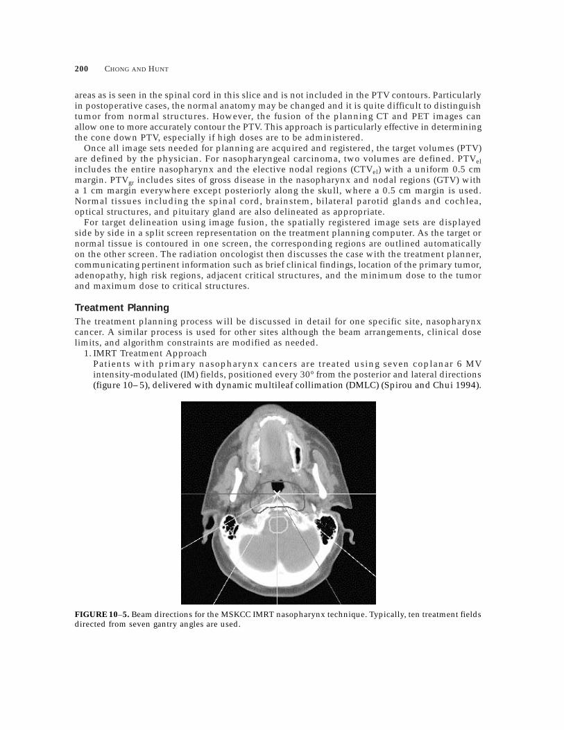
1. IMRT Treatment ApproachPatients with primary nasopharynx cancers are treated using seven coplanar 6 MVintensity-modulated (IM) fields, positioned every 30° from the posterior and lateral directions(figure 10– 5), delivered with dynamic multileaf collimation (DMLC) (Spirou and Chui 1994).
MPP02120901—IMRT–Ling—file name: 10-chong
FIGURE 10–5. Beam directions for the MSKCC IMRT nasopharynx technique. Typically, ten treatment fieldsdirected from seven gantry angles are used.
IMRT FOR HEAD AND NECK CANCER
A prescription dose of 70 Gy is delivered to gross disease (PTVgr) in the nasopharynx andneck, and 54 Gy to the elective nodal regions (PTVel). The supraclavicular nodes are treatedwith a single anterior lower neck field, the superior edge of which is matched to the IMRTfields. Patients receive 1.8 Gy per fraction for the first 20 fractions (36 Gy) and thereafter,1.8 Gy and 1.6 Gy in 2 daily fractions separated by a minimum of 6 hours for a total of 40fractions. PTVel, as defined above, is treated during the 1.8 Gy fractions, while treatment islimited to PTVgr for the 1.6 Gy fractions.
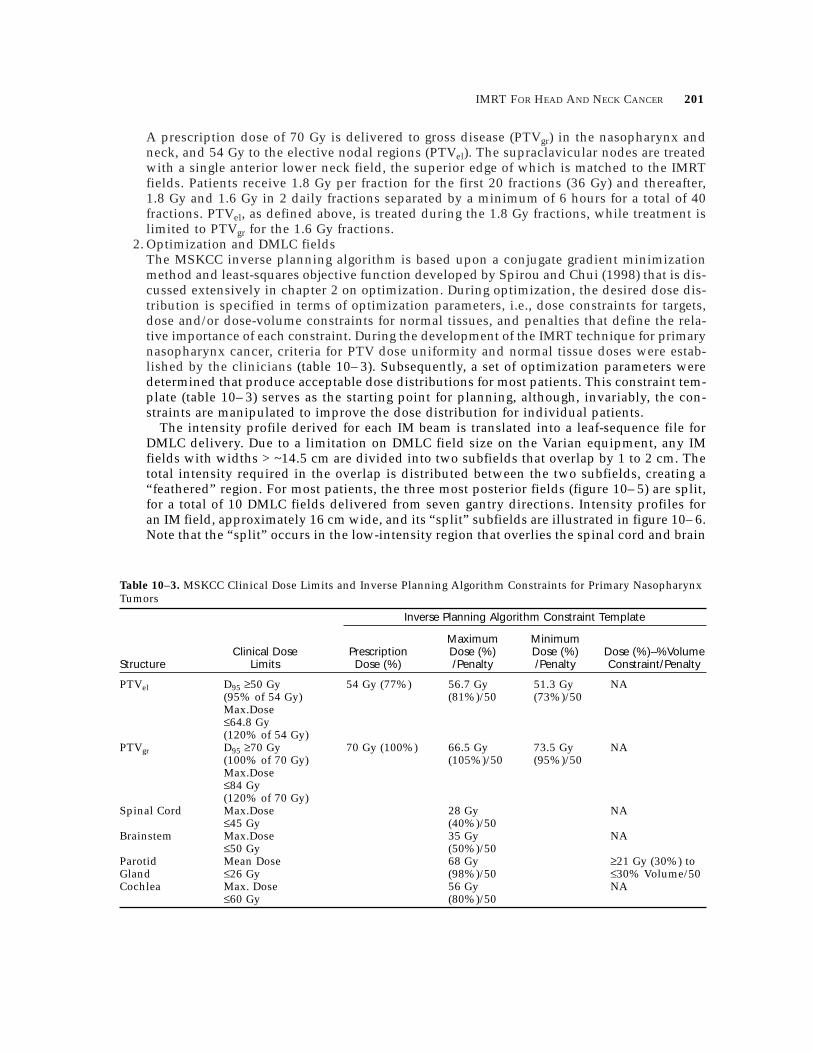
2. Optimization and DMLC fieldsThe MSKCC inverse planning algorithm is based upon a conjugate gradient minimizationmethod and least-squares objective function developed by Spirou and Chui (1998) that is dis-cussed extensively in chapter 2 on optimization. During optimization, the desired dose dis-tribution is specified in terms of optimization parameters, i.e., dose constraints for targets,dose and/or dose-volume constraints for normal tissues, and penalties that define the rela-tive importance of each constraint. During the development of the IMRT technique for primarynasopharynx cancer, criteria for PTV dose uniformity and normal tissue doses were estab-lished by the clinicians (table 10– 3). Subsequently, a set of optimization parameters weredetermined that produce acceptable dose distributions for most patients. This constraint tem-plate (table 10– 3) serves as the starting point for planning, although, invariably, the con-straints are manipulated to improve the dose distribution for individual patients.
The intensity profile derived for each IM beam is translated into a leaf-sequence file forDMLC delivery. Due to a limitation on DMLC field size on the Varian equipment, any IMfields with widths > ~14.5 cm are divided into two subfields that overlap by 1 to 2 cm. Thetotal intensity required in the overlap is distributed between the two subfields, creating a“feathered” region. For most patients, the three most posterior fields (figure 10– 5) are split,for a total of 10 DMLC fields delivered from seven gantry directions. Intensity profiles foran IM field, approximately 16 cm wide, and its “split” subfields are illustrated in figure 10– 6.Note that the “split” occurs in the low-intensity region that overlies the spinal cord and brain
201
MPP02120901—IMRT–Ling—file name: 10-chong
Table 10–3. MSKCC Clinical Dose Limits and Inverse Planning Algorithm Constraints for Primary NasopharynxTumors
Inverse Planning Algorithm Constraint Template
Maximum MinimumClinical Dose Prescription Dose (%) Dose (%) Dose (%)–%Volume
Structure Limits Dose (%) /Penalty /Penalty Constraint/Penalty
PTVel D95 ≥50 Gy 54 Gy (77%) 56.7 Gy 51.3 Gy NA(95% of 54 Gy) (81%)/50 (73%)/50Max.Dose£64.8 Gy(120% of 54 Gy)
PTVgr D95 ≥70 Gy 70 Gy (100%) 66.5 Gy 73.5 Gy NA(100% of 70 Gy) (105%)/50 (95%)/50Max.Dose£84 Gy(120% of 70 Gy)
Spinal Cord Max.Dose 28 Gy NA£45 Gy (40%)/50
Brainstem Max.Dose 35 Gy NA£50 Gy (50%)/50
Parotid Mean Dose 68 Gy ≥21 Gy (30%) toGland £26 Gy (98%)/50 £30% Volume/50Cochlea Max. Dose 56 Gy NA
£60 Gy (80%)/50

stem. This feature and the “feathering” in the overlap region help to minimize the potentialdosimetric uncertainty due to field matching.
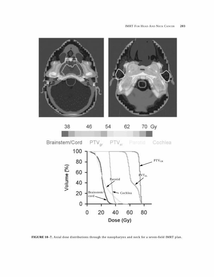
3. Plan Evaluation And Quality Assurance (QA)The dose distributions and DVHs through the center of the nasopharynx and neck nodes fora typical patient with N0 disease are shown in figure 10– 7. PTVgr and PTVel are well-coveredby the 70 and 54 Gy prescription isodose levels while the spinal cord and brain stemreceive approximately 45 Gy. The average mean dose to the parotid glands is 27 Gy and thecochleae receive an average maximum dose of 64 Gy.
For each beam of a completed IMRT plan, digitally reconstructed radiographs (DRRs) are gen-erated displaying the so-called “DMLC aperture,” corresponding to the complete irradiated area(figure 10– 8). These DRRs are compared with portal images obtained with the DMLC aperturewhen the patient comes for treatment.
Prior to treatment, all IMRT plans undergo the following QA checks. A complete review of theplan is done by a physicist, including an evaluation of the dose distributions and DVHs and areview of all data used for patient treatment including the leaf motion files. The leaf motion filesand intensity profiles are evaluated for unusual intensity peaks that might either limit treatmentdelivery or introduce dosimetric problems due to patient intra-fractional motion. An independ-ent verification of the monitor unit setting for each treatment field is performed using a com-puter program specifically designed for this purpose. Discrepancies in excess of 2% warrant furtherinvestigation including film or ionization chamber dosimetry.
Daily Treatment with IMRTPrior to the first treatment, the patient is positioned on the linear accelerator and images of eachportal are obtained. Using the VARiS Treatment and Vision software, the DMLC aperture is auto-matically created, the portal images are acquired and then carefully compared against the DRRs.
IMRT for head and neck cancer patients requires precision and care on the part of the radia-tion therapist in every phase of the patient’s daily setup and treatment procedure. Accuracy andreproducibility are vital if the tumor is to receive the proper dosage and overdosage to thenormal critical structures avoided. The patient must be motivated and capable of fully cooper-ating with the setup and treatment procedure. Body motion must be kept to a minimum. Properpatient immobilization is absolutely essential (Chao et al. 2000). Close communication betweenthe radiation oncologist and radiation therapist must be maintained throughout the course oftreatment regarding any patient setup abnormality, problems, or difficulty. Weekly port films will
202 CHONG AND HUNT
MPP02120901—IMRT–Ling—file name: 10-chong
FIGURE 10–6. Intensity profiles for a left lateral nasopharynx field before (a) and after (b and c) field splittingto overcome the DMLC maximum field width limitation.
IMRT FOR HEAD AND NECK CANCER 203
MPP02120901—IMRT–Ling—file name: 10-chong
FIGURE 10–7. Axial dose distributions through the nasopharynx and neck for a seven-field IMRT plan.
Brainstem/cord
Parotid
Cochlea
PTVGR
PVTEL

204 CHONG AND HUNT
MPP02120901—IMRT–Ling—file name: 10-chong
be compared with the DRRs throughout the treatment course. Development of facial edema andsignificant weight loss may adversely affect the snug fit of the face mask and cause problems withpatient immobilization and setup. Conservative modification of the mask, and on rare occa-sions, creation of a new mask and repeat CT simulation and treatment planning may be necessary.
Clinical Care During Radiation TherapyThe medical care of head and neck patients undergoing IMRT is the same as that required forthose treated with conventional radiation therapy. For patients treated with a multi-modalityapproach including chemotherapy with cisplatin or carboplatin and 5FU, placement of a percu-taneous endoscopic gastrostomy tube should be considered prior to initiation of treatment, par-ticularly for elderly or frail patients, those who have lost a considerable amount of weight, orthose with problems of dysphasia or odynophagia at the outset.
Weekly status evaluations are mandatory, with some patients requiring more frequent evalua-tions as treatment progresses. During these visits, an interval history is obtained reviewing thedevelopment of skin symptomology, a sore mouth or throat, xerostomia, decreased or abnormaltaste, hoarseness, or dysphagia. A pertinent examination will note the status of the portal skin;the location and size of the primary tumor; the location, size, mobility, tenderness, and textureof lymphadenopathy; the presence of mucositis and of oral Candida. Routine measurement ofthe patient’s weight and complete blood counts will be obtained.
FIGURE 10–8. Digitally-reconstructed radiograph (DRR) of a left lateral IMRT field for primary nasophar-ynx cancer. The aperture indicates the initial and final positions of the MLC leaves used for the dynamicdelivery.
IMRT FOR HEAD AND NECK CANCER
Occasionally, a patient will develop acute parotitis within the first 12 hours after commence-ment of therapy when the treatment volume includes the parotid gland. The symptoms includeswelling in the parotid regions associated with localized pain and occasionally a low-grade tem-perature. Although parotitis generally resolves on its own, we would prescribe a non-steroidalanti-inflammatory drug and reassure the patient should this occur.
Tumoritis can develop at ~20 Gy and is characterized by a mucosal inflammation that repre-sents the true extent of the tumor and may necessitate subsequent modification of the portal (Wang1997). As the primary tumor is followed during the course of treatment, those lesions that showprogression or minimal regression should be reevaluated by all the physicians on the case. Thismay be a situation where surgery is indicated.
As patients develop the acute side effects of xerostomia, decreased or abnormal taste, andmucositis, appropriate supportive medical intervention is mandatory. Narcotic analgesicsshould be considered and modified as necessary to provide adequate pain relief. This may involvethe use of long-acting morphine sulphate or a fentanyl patch as well as immediate-release mor-phine sulphate for any break-through pain. Intravenous hydration is sometimes indicated forpatients who have become dehydrated due to poor oral intake or who have difficulties withtheir percutaneous endoscopic gastrostomy tube. A fair percentage of patients may develop oralCandida which may be asymptomatic, present with acute development or exacerbation of a soremouth or throat or even perhaps an abnormal taste. Initiation of an antifungal medication willusually resolve the problem rapidly.
The head and neck cancer patients that we have treated with IMRT do not appear to haveresponses that differ from that of patients undergoing conventional treatment. Our clinicalobservation has been that these patients have similar acute reactions and can be managed quiteadequately as presented above.
Post-Treatment Follow-upImmediately upon completion of radiation therapy, routine follow-up evaluations should be sched-uled. If the patient is elderly, frail, or having a particularly difficult time with acute mucositis,esophagitis, and weight loss, we will see this patient weekly until sufficient recovery has occurredwhich generally takes 3 to 4 weeks. The patients in better condition are seen monthly for 2 monthsand every 1 to 3 months thereafter, alternating with the other physicians on the case unless weare monitoring the response of a mass. Baseline imaging studies are considered 2 to 3 monthspost treatment and may include a CT or MRI of the head and neck or and/or a PET scan.
Serial endocrine screening will be important for patients who have had irradiation of theseorgans, including the pituitary gland and thyroid gland. Thyroid function tests, including aTSH, are obtained every 6 months post treatment for up to 5 years. Clinical hypothyroidism hasbeen seen in ~5% of adults and a higher percentage in children whose thyroids have been irra-diated. There is a 20% to 25% risk of chemical hypothyroidism overall, but this increases to66% in patients who have also undergone a hemithyroidectomy. In patients who are found tohave a significant elevation of the TSH, thyroid hormone replacement therapy is initiated irre-spective of the T3 and T4 values, which oftentimes may be within normal limits. Patients whohave their pituitary gland irradiated should periodically undergo irradiated screening every 1 to2 years post irradiation. These tests should evaluate LH, FSH Serum cortisol, prolactin, TSH,free T4, and GH. For male patients, a testosterone level is also included.
Patients who have received radiation to the oral cavity or oropharynx should be seen routine-ly by the dental service for an indefinite period of time. Fluoride prophylaxis, initiated at the startof treatment, should be continued. These patients are advised that their dentist should be fullyinformed of their radiation therapy as well as the potential risk for osteoradionecrosis that mayresult from subsequent dental surgery.
205
MPP02120901—IMRT–Ling—file name: 10-chong

206 CHONG AND HUNT
Occasionally, a patient will develop Lhermitte’s syndrome, a benign, transient myelopathy pre-sumably due to radiation-induced demyelination in the cervical spinal cord. This can begin 1 to3 months post therapy and last an average of 3 to 4 months and as long as 9 to 12 months. Thisis characterized by the development of a symmetrical, instantaneous, shooting, electrical sensa-tion that radiates down the spine and extremities upon flexation of the neck, but it does notprogress and requires no treatment.
High-dose irradiation, especially when combined with chemotherapy, can lead to late effectsof the soft tissues. Particular attention should be directed towards the development of trismus aswell as the decreased range of motion of the tongue, mandible, neck, and shoulders. Physicaltherapy should be considered as it may decrease or prevent these post treatment functional deficitsresulting from fibrosis and scarring.
Some patients may develop dysphagia during treatment that could become chronic and sig-nificant. Post treatment dysphagia may be due to dysfunction of the pharyngeal muscles, thedevelopment of an esophageal stricture, or even possibly the presence of a tumor. We have observedin some patients whose pharyngeal muscles received high-dose radiation therapy, particularlyin conjunction with chemotherapy, significant swallowing problems long after completion of treat-ment. Appropriate medical evaluation must be performed for diagnosis and appropriate therapeuticintervention.
Effect Of Setup UncertaintySeveral studies evaluating setup uncertainty specifically for head and neck patients (Rabinowitzet al. 1985; Verellen et al. 1997; Hunt et al. 1993) have measured standard deviations of system-atic and random uncertainties of approximately 2 to 3 mm. Hunt et al. compared the impact ofsetup errors on target coverage, spinal cord, and brainstem dose for 3-D and parallel opposed dosedistributions. Systematic setup errors led to target underdosage and normal tissue overdosagewith both techniques, but the 3-D distributions were more susceptible to the effects of both randomand systematic errors because of the increased conformality. Although, studies evaluating theeffect of setup uncertainty specifically for IMRT head and neck distributions have not yet beendone, the impact may be even more significant because of their exquisite conformality and thepresence of steep dose gradients.
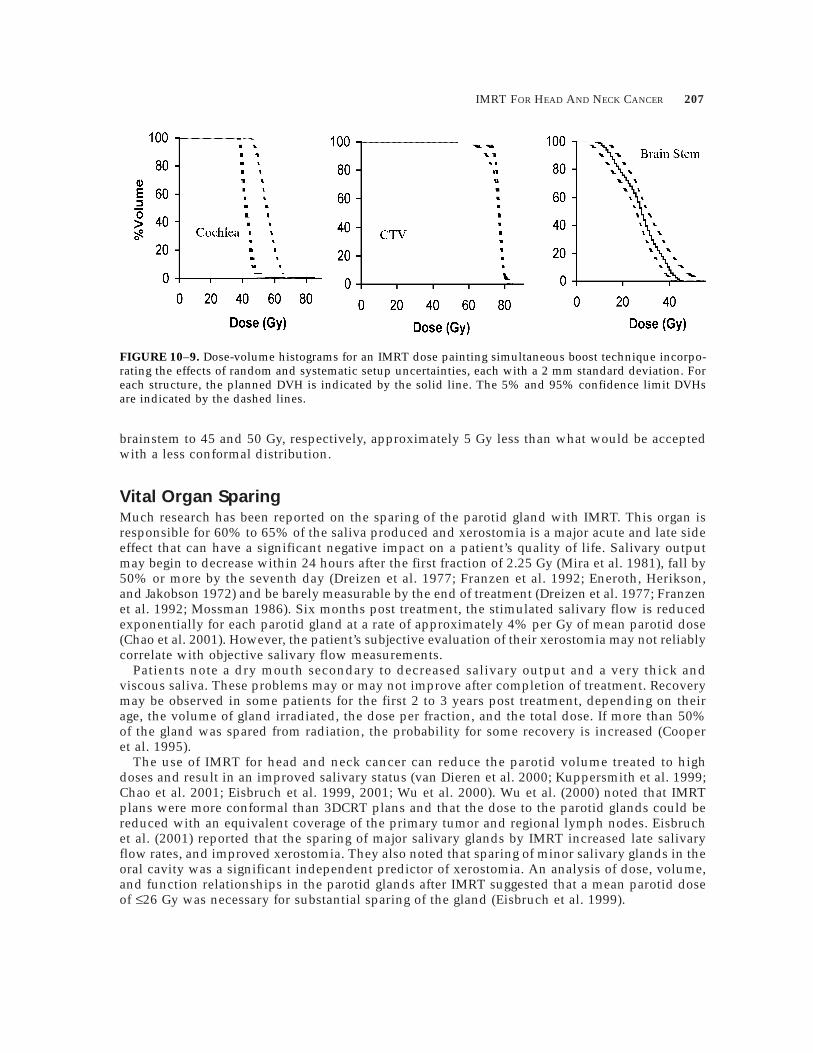
In a preliminary evaluation for nasopharynx cancer, we have modeled the effects of randomand systematic setup uncertainty on IMRT dose distributions for selected patients using the fol-lowing technique. Briefly, the random treatment uncertainty is modeled by convolving the planneddose distribution with a normal frequency distribution with a standard deviation of 2 mm, a tech-nique developed by Chui, Kutcher, and LoSasso (1992). After blurring the dose distribution toshow the effect of random uncertainty, systematic uncertainty is modeled using a Monte Carlosimulation. A normal frequency distribution with a standard deviation of 2 mm in each direc-tion is sampled 500 times. For each iteration the dose distribution corrected for random uncer-tainty is shifted according to the sampled systematic error and the doses to the targets and normaltissues are recalculated. Confidence limit DVHs, i.e., the DVHs expected with a given statisticalconfidence for a population of patients, can then be calculated and analyzed. The 2 mm stan-dard deviations of random and systematic setup uncertainty were estimated from our ownanalysis of setup for head and neck patients (Hunt et al. 1993). The planned, 95%, and 5% con-fidence limit DVHs for the PTV, brain stem, and cochlea are shown in figure 10– 9 for a patientplanned with the IMRT dose painting technique described below (IMRT Dose Painting andDose Escalation for Primary Nasopharyngeal Carcinoma). The tight conformality of the dose dis-tribution and, in particular, the manner in which the high-dose region surrounds the spinal cordand brainstem is responsible for the observed increase in normal tissue dose and degradation intarget coverage. Based on this preliminary analysis, we currently limit the spinal cord and
MPP02120901—IMRT–Ling—file name: 10-chong
IMRT FOR HEAD AND NECK CANCER
brainstem to 45 and 50 Gy, respectively, approximately 5 Gy less than what would be acceptedwith a less conformal distribution.
Vital Organ SparingMuch research has been reported on the sparing of the parotid gland with IMRT. This organ isresponsible for 60% to 65% of the saliva produced and xerostomia is a major acute and late sideeffect that can have a significant negative impact on a patient’s quality of life. Salivary outputmay begin to decrease within 24 hours after the first fraction of 2.25 Gy (Mira et al. 1981), fall by50% or more by the seventh day (Dreizen et al. 1977; Franzen et al. 1992; Eneroth, Herikson,and Jakobson 1972) and be barely measurable by the end of treatment (Dreizen et al. 1977; Franzenet al. 1992; Mossman 1986). Six months post treatment, the stimulated salivary flow is reducedexponentially for each parotid gland at a rate of approximately 4% per Gy of mean parotid dose(Chao et al. 2001). However, the patient’s subjective evaluation of their xerostomia may not reliablycorrelate with objective salivary flow measurements.
Patients note a dry mouth secondary to decreased salivary output and a very thick andviscous saliva. These problems may or may not improve after completion of treatment. Recoverymay be observed in some patients for the first 2 to 3 years post treatment, depending on theirage, the volume of gland irradiated, the dose per fraction, and the total dose. If more than 50%of the gland was spared from radiation, the probability for some recovery is increased (Cooperet al. 1995).
The use of IMRT for head and neck cancer can reduce the parotid volume treated to highdoses and result in an improved salivary status (van Dieren et al. 2000; Kuppersmith et al. 1999;Chao et al. 2001; Eisbruch et al. 1999, 2001; Wu et al. 2000). Wu et al. (2000) noted that IMRTplans were more conformal than 3DCRT plans and that the dose to the parotid glands could bereduced with an equivalent coverage of the primary tumor and regional lymph nodes. Eisbruchet al. (2001) reported that the sparing of major salivary glands by IMRT increased late salivaryflow rates, and improved xerostomia. They also noted that sparing of minor salivary glands in theoral cavity was a significant independent predictor of xerostomia. An analysis of dose, volume,and function relationships in the parotid glands after IMRT suggested that a mean parotid doseof £26 Gy was necessary for substantial sparing of the gland (Eisbruch et al. 1999).
207
MPP02120901—IMRT–Ling—file name: 10-chong
FIGURE 10–9. Dose-volume histograms for an IMRT dose painting simultaneous boost technique incorpo-rating the effects of random and systematic setup uncertainties, each with a 2 mm standard deviation. Foreach structure, the planned DVH is indicated by the solid line. The 5% and 95% confidence limit DVHsare indicated by the dashed lines.

208 CHONG AND HUNT
Butler et al. (1999) noted in a review of 20 IMRT patients with primary head and neck cancerthat the mean dose to the ipsilateral parotid gland was 23 Gy and to the contralateral gland, 21 Gy.Chao et al. (2000) reported that the mean parotid dose in their series was approximately 20 Gy.They also noted that 3%±1.4% of the primary target received less than 95% of the prescribed dosedue to proximity of the target volume to the critical structures such as the parotid gland. The steepdose gradient commonly noted in head and neck IMRT plans in which the tumors are in veryclose proximity to the parotid gland means that part of the primary target volume may beunderdosed. Further research is necessary to determine whether this is of clinical importance.
At MSKCC, we attempt to limit the mean dose to at least one parotid gland to £26 Gy, withoutcompromising target coverage. For patients with nasopharyngeal cancer, mean parotid doses of£26 Gy are usually achievable in patients with negative or unilateral neck disease for the glandon the side without neck disease. In the presence of gross adenopathy, mean parotid doses of~35 Gy are typical. To ensure adequate target coverage in the presence of a parotid sparing tech-nique, we require that at least 95% of the electively irradiated and gross disease PTVs receive 50Gy and 70 Gy, respectively. Our experience indicates that, as a consequence of parotid sparing,the dose to the oral cavity and the submandibular glands may increase and therefore should becarefully evaluated. Dose distributions and DVHs derived from inverse planning with and withoutan attempt to spare the parotid glands are compared in figure 10– 10 for a patient with a nega-tive neck. A small section of the electively irradiated PTV receives less than 54 Gy in order toachieve a mean parotid dose of 26 Gy.
Radiation therapy may also lead to sensorineural hearing loss, particularly when the radiationis delivered in combination with chemotherapy. As discussed by Choi et al. (2000), hearing lossoccurs more frequently in patients whose cochlea received ≥70 Gy. Unfortunately, the cochleaeare often within or adjacent to the high dose target in the nasopharynx and could easily receivedoses in excess of 70 Gy. Grau et al. (1991) demonstrated that doses of 50 to 70 Gy to the cochleamay lead to hearing loss within 18 months, but that the probability and severity of the loss wascorrelated with dose and the sound frequency. IMRT can be used to spare the cochlea, butsimilar to parotid sparing attempts, may lead to compromised target coverage (figure 10– 11). Aretrospective analysis of 20 of our nasopharynx patients indicated that the cochleae straddle orlie within the PTVgr in approximately three-fourths of all patients and that their position greatlyaffects the dose they receive. At MSKCC, we currently limit the dose to the cochlea fornasopharynx patients to £60 Gy when possible given the target constraints outlined in table 10– 3.To further guard against tumor underdosing, an additional constraint requiring ≥99% of theGTV to receive ≥70 Gy is used. IMRT dose painting and the simultaneous boost technique, asdiscussed in the next section, may facilitate lower cochlear doses without compromising targetcoverage. Figure 10– 12 shows dose distributions and DVHs for a patient planned with the IMRTdose painting simultaneous boost technique according to the target criteria in table 10– 3 andthe additional GTV coverage criteria. The cochleae receive a maximum dose of 30 Gy.
In addition to the structures already discussed, other important normal structures to considerwhen planning head and neck tumors with IMRT include the orbital structures, optic nerves,optic chiasm, brain, and mandible. Generally, the dose limits for these structures can easily beachieved with IMRT except in patients with extensive superior disease or cranial extension.
IMRT Dose Painting And Dose Escalation For Primary Nasopharyngeal CarcinomaThe use of IMRT to plan non-uniform dose distributions within the target volume for head andneck patients has been described recently by Wu et al. (2000). The advantages of this techniqueinclude the delivery of a biologically higher dose to the gross disease and the simplification of
MPP02120901—IMRT–Ling—file name: 10-chong

IMRT FOR HEAD AND NECK CANCER
the planning and treatment processes since only one plan is designed and used for the entiretreatment course. The study by Wu examined the potential of this concomitant or simultaneousintegrated boost technique for a variety of head and neck tumors and concluded that, using IMRT,they could achieve distributions similar to conventional fractionation in terms of target coverageand normal tissue doses. The technique being considered at MSKCC would deliver 70.2 Gy tothe nasopharynx in 30 fractions (2.34 Gy/fraction) while concomitantly treating the neck to 54Gy (1.8 Gy/fraction). Typical dose distributions and DVHs comparing this technique with ourstandard two-phase technique are shown in figure 10– 13. Coverage of the PTVgr is very similarto that achieved with a conventional treatment strategy and IMRT, although the mean dose toPTVel is slightly less.
209
MPP02120901—IMRT–Ling—file name: 10-chong
FIGURE 10–10. Axial dose distributions and DVHs for the PTVel and parotid glands with and without anIMRT parotid sparing technique for a patient with N0 disease. See COLOR PLATE 23.
PTVel

210 CHONG AND HUNT
Plans designed with a simultaneous boost technique are inherently more conformal thanthose using a two-phase technique, leading to lower doses to critical structures in very close prox-imity to the 70 Gy volume such as the cochlea. Additional dose distributions in the nasophar-ynx for the current MSKCC two-phase treatment and the simultaneous boost technique are comparedin figure 10– 14. These distributions were generated with constraints on target coverage anddose uniformity, spinal cord and brainstem maximum dose, and parotid mean dose, but no con-straint on the cochlea. The conformity index (Volume (70 Gy)/Volume (PTV)) is significantlyimproved with the simultaneous boost technique. As a result of this improved conformality, thedose to the cochlea is also less.
Treating Recurrent Head And Neck Tumors With IMRTThe management of head and neck cancer patients with recurrent disease who have previouslyreceived radical radiation therapy is a challenge. For these patients, surgery is often the treatmentof choice, provided the lesion is resectable, the patient is able to tolerate the procedure, and that
MPP02120901—IMRT–Ling—file name: 10-chong
FIGURE 10–11. Dose distributions and DVHs illustrating the effect of cochlear sparing on PTV coveragewhen the cochleae lie within PTVgr. Results are shown for three plans: unconstrained cochlear dose, 50 Gy,and 70 Gy maximum cochlear dose. See COLOR PLATE 24.

IMRT FOR HEAD AND NECK CANCER
recovery and rehabilitation are likely. For patients who are not surgical candidates, re-irradia-tion can be considered. This is a highly select group with true local-regional recurrence ratherthan persistence of the disease post radiation therapy. Evaluation of the following items is nec-essary: (1) patient condition; (2) time interval since completion of initial radiation therapy; (3) radi-ation dosage administered; (4) tolerance of treatment and any complications; (5) anatomic locationof recurrence and adjacent normal critical structures; (6) condition of previously irradiated tissues;(7) symptoms related to the recurrence; (8) life expectancy. Relative contraindications to re-irra-diation include: (1) poor condition; (2) recurrence less than 6 months from initial radiation therapy;(3) ultra-high radiation doses; (4) massive tumor recurrence equivalent to T3–T4 lesions; (5)location of recurrence in or around the central nervous system. The dose of re-irradiation willneed to be in the range of 60 to 65 Gy (De Crevoisier et al. 1998; Stevens, Britsch, and Moss1994; Wang 1994). Moderate dose re-irradiation of 45 Gy will most likely not be effective and maynot even provide sufficient palliation. The more limited the disease, the better the chances for ameaningful therapeutic intervention.
Meticulous treatment planning and careful radiation technique are necessary. At MSKCC, IMRTis often used for re-irradiation cases although brachytherapy as the primary treatment or as a boostis also considered. Conservative margins around the tumor of no more than 1 cm are appropri-ate. The central nervous system must not be directly re-irradiated by the primary beam. Onlypatients who understand the high risks involved and exhibit a willingness to accept the possiblecomplications should be considered for re-irradiation.
Re-irradiation of recurrent nasopharyngeal cancer with a stage equivalent to a T1 or T2 lesionhas frequently been reported in the literature (Teo et al. 1998; Wang 1993). PET or MR imagefusion can aid in localization of the tumor allowing for a limited treatment volume with a high
211
MPP02120901—IMRT–Ling—file name: 10-chong
FIGURE 10–12. Dose distribution and DVH of the PTVgr and cochlea for a patient planned with the 70 GyIMRT dose painting simultaneous boost technique. The cochleae receive a maximum of 30 Gy with acceptabletarget coverage and dose uniformity.

212 CHONG AND HUNT
MPP02120901—IMRT–Ling—file name: 10-chong
FIGURE 10–14. Comparison of the MSKCC two-phase (a) and simultaneous boost (b) techniques for the treat-ment of nasopharynx cancer. Using the simultaneous boost technique, the PTVgr receives 70.2 Gy in 30fractions (2.34 Gy/fraction) while the electively irradiated volume, PTVel, receives 1.8 Gy/fraction to 54 Gy.The 70 Gy conformity index is 2.4 for the two-phase plan and 1.7 for the simultaneous boost. Themaximum cochlea doses with the two-phase and simultaneous boost plans are 70 and 63 Gy, respectively.See COLOR PLATE 26.
Figure 10– 13. Comparison of IMRT nasopharynx dose distributions for a two-phase treatment technique(a) and a simultaneous boost treatment (b). See COLOR PLATE 25.
2 PhaseSim. Boost

IMRT FOR HEAD AND NECK CANCER
level of confidence that the disease is contained within the treatment region. Limited volumeIMRT with a brachytherapy boost can potentially provide good local control although brachyther-apy may not be suitable in some cases because of the size and extent of the tumor. Thesepatients must be treated with IMRT often with concurrent chemotherapy. Special care must beexercised in analyzing the IMRT plan with respect to the central nervous system, orbit, andoptic nerve and chiasm doses.
Other regions of the head and neck have been treated with re-irradiation with promising pre-liminary results. Studies have shown good palliation of symptoms and some have reported longterm control with 20% 2-year survivals and 15% to 17% 5-year survivals (DeCrevoisier et al. 1998;Stevens, Britsch, and Moss 1994). The results appear better than those obtained with the use ofchemotherapy alone.
The incidence of late toxicity is greater than that noted after primary radical radiationtherapy. Several studies have suggested however that these adverse effects were still deemedacceptable (Wang 1994).
Re-irradiation with IMRT at MSKCCAt MSKCC, IMRT is used routinely in the treatment of recurrent cancers, primarily nasopharynxalthough additional sites including paranasal sinus have been treated. Typically, doses of ~60Gy are prescribed with dose limits to the spinal cord and brainstem of 10 to 12 Gy. The dosesdelivered to other normal tissues, particularly optical pathway structures, are determined afterconsideration of the previous therapy.
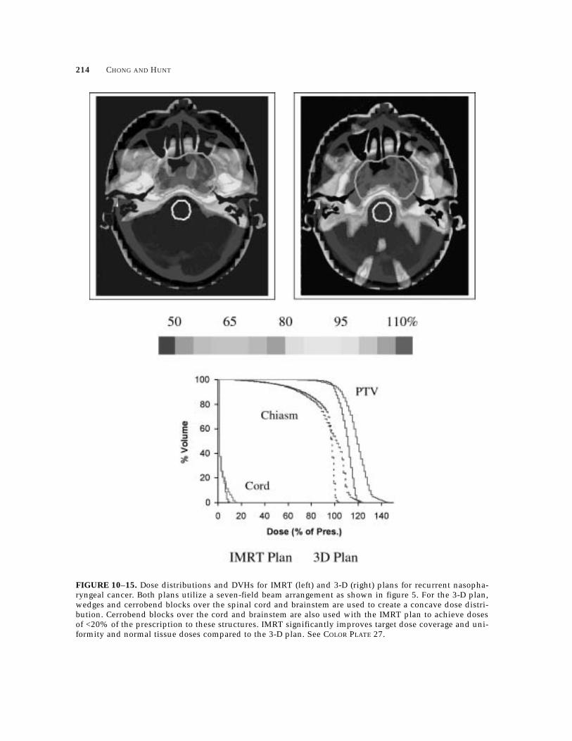
Typically, five to nine equally spaced treatment fields are used, including non-coplanar beamarrangements when beneficial. Although PTV constraints similar to those for PTVgr in table10– 3 are used, the individual needs of each patient are considered when defining the normaltissue constraints. IMRT and 3-D conformal plans for recurrent nasopharynx disease, created usingseven field beam arrangements, are compared in figure 10– 15. IMRT improved the target doseuniformity and led to lower doses to the optical structures. Our experience has been that it is gen-erally not possible to achieve the extremely low normal tissue doses required for these caseswith IMRT alone. Conventional cerrobend blocking is combined with dynamic multileaf IMRTwhen normal tissue doses must be less than approximately 30% of the prescription.
SummaryThe concave shape of the target volume and close proximity of normal tissues make head andneck tumors ideal cases for IMRT. Multiple planning studies within the past 5 years have clearlydemonstrated the ability of IMRT to improve target coverage and dose uniformity for many headand neck sites. More exciting, perhaps, is the opportunity to impact the significant normaltissue morbidity associated with head and neck radiotherapy and the ability to deliver differentfractionation schemes using the SMART technique or IMRT “dose painting.” Clinical results havealready established that IMRT can be used to decrease the morbidity associated with the irradi-ation of the salivary glands. It remains to be seen if similar improvements in hearing loss can beachieved without sacrificing local control.
Head and neck sites have always been among the most challenging, complex and time con-suming to plan. Our experience with head and neck IMRT planning has been that the completeplanning process can require 10 to 12 hours of a planner’s time, more if image fusion isrequired. Site-specific class solutions, specifying the clinical criteria for target and normal tissuedoses in as much detail as possible, the beam arrangements and constraint templates to use asstarting points for planning are mandatory for efficient head and neck IMRT planning. Despitethe increased complexity and time required to produce them, IMRT dose distributions offer
213
MPP02120901—IMRT–Ling—file name: 10-chong
214 CHONG AND HUNT
MPP02120901—IMRT–Ling—file name: 10-chong
FIGURE 10–15. Dose distributions and DVHs for IMRT (left) and 3-D (right) plans for recurrent nasopha-ryngeal cancer. Both plans utilize a seven-field beam arrangement as shown in figure 5. For the 3-D plan,wedges and cerrobend blocks over the spinal cord and brainstem are used to create a concave dose distri-bution. Cerrobend blocks over the cord and brainstem are also used with the IMRT plan to achieve dosesof <20% of the prescription to these structures. IMRT significantly improves target dose coverage and uni-formity and normal tissue doses compared to the 3-D plan. See COLOR PLATE 27.

IMRT FOR HEAD AND NECK CANCER
significant improvements over 3-D conformal plans. We believe that IMRT will become the stan-dard method of treatment for many head and neck sites.
ReferencesAnzai, Y., W. R. Carrol, D. J. Quint, C. R. Bradford, S. Minoshima, G. T. Wolf, and R. H. Wahl. (1996).
“Recurrence of head and neck cancer after surgery or irradiation: Prospective comparison of 2-deoxy-2-[F-18]fluoro-D-glucose PET and MR imaging diagnoses.” Radiol. 200:135– 141.
Blanco, A. I., and K. S. C. Chao. “Intensity-Modulated Radiation Therapy and Protection of Normal TissueFunction in Head and Neck Cancer” in Principles and Practice of Radiation Oncology: Updates. Vol. 3(No. 3). New York: Lippincott Williams & Wilkins Healthcare, 2002.
Bourhis, J., and J. P. Pignon. (1999). “Meta-analyses in head and neck squamous cell carcinoma. What isthe role of chemotherapy?” Hematol. Oncol. Clin. North Am. 13:769– 775, vii.
Boyer, A. L., P. Geis, W. Grant, and M. Carol. (1997). “Modulated beam conformal therapy for head andneck tumors.” Int. J. Radiat. Oncol. Biol. Phys. 39:227– 236.
Brizel, D. M., M. E. Albers, S. R. Fisher, R. L. Scher, W. J. Richtsmeier, V. Hors, S. L. George, A. T. Huang,and L. R. Prosnitz. (1998). “Hyperfractionated irradiation with or without concurrent chemotherapy forlocally advanced head and neck cancer.” N. Engl. J. Med. 338:1798– 1804.
Butler, E. B., B. S. Teh, W. H. Grant 3rd, B. H. Uhl, R B. Kuppersmith, J. K. Chui, D. T. Donovan, and S. Y.Woo. (1999). “Smart (simultaneous modulated accelerated radiation therapy) boost: A new acceleratedfractionation schedule for the treatment of head and neck cancer with intensity modulated radiothera-py.” Int. J. Radiat. Oncol. Biol. Phys. 45:21– 32.
Chao, K. S., D. A. Low, C. A. Perez, and J. A. Purdy. (2000). “Intensity-modulated radiation therapy in headand neck cancers: The Mallinckrodt experience.” Int. J. Cancer 90:92– 103.
Chao, K. S., J. O. Deasy, J. Markman, J. Haynie, C. A. Perez, J. A. Purdy, and D. A. Low. (2001). “A prospec-tive study of salivary function sparing in patients with head-and-neck cancers receiving intensity-mod-ulated or three-dimensional radiation therapy: Initial results.” Int. J. Radiat. Oncol. Biol. Phys. 49:907– 916.
Chao, K. S., F. J. Wippold, G. Ozyigit, B. N. Tran, and J. F. Dempsey. (2002). “Determination and delineationof nodal target volumes for head-and-neck cancer based on patterns of failure in patients receiving defin-itive and postoperative IMRT.” Int. J. Radiat. Oncol. Biol. Phys. 53(5):1174– 1184.
Chen, B. C., C. Hoh, B. Choi, et al. (1990). “Evaluation of primary head and neck tumor with PET-FDG.”(Abstract). Clin. Nucl. Med. 15:758.
Choi, S., S. Wolden, D. Pfister, A. S. Budnick, S. Levegrün, A. Jackson, M. A. Hunt, M. J. Zelefsky, B. Singh,J. O. Boyle, and D. H. Kraus. (2000). “Ototoxicity following combined modality therapy for nasopharyn-geal carcinoma.” Proceedings of the 42nd Annual ASTRO Meeting, Boston, MA, October 22-26, 2000. Int.J. Radiat. Oncol. Biol. Phys. 48:261.
Chui, C. S., G. J. Kutcher, and T. Lossaso. (1992). “A convolution method for incorporating uncertainties indose calculation.” (Abstract). Med. Phys. 19:814.
Cooper, J. S., K. Fu, J. Marks, and S. Silverman. (1995). “Late effects of radiation therapy in the head andneck region.” Int. J. Radiat. Oncol. Biol. Phys. 31:1141– 1164.
De Crevoisier, R., J. Bourhis, C. Domenge, P. Wibault, S. Koscielny, A. Lusinchi, G. Mamelle, F. Janot, M.Julieron, A. M. Leridant, P. Marandas, J. P. Armand, G. Schwaab, B. Luboinski, and F. Eschwege. (1998).“Full-dose reirradiation for unresectable head and neck carcinoma: Experience at the Gustave-RoussyInstitute in a series of 169 patients.” J. Clin. Oncol. 16:3556– 3562.
Dreizen, S., L. R. Brown, T. E. Daly, and J. B. Drane. (1977). “Prevention of xerostomia-related dental cariesin irradiated cancer patients.” J. Dent. Res. 56:99– 104.
Eisbruch, A., L. H. Marsh, M. K. Martel, J. A. Ship, R. Ten Haken, A. T. Pu, B. A. Fraass, and A. S. Lichter.(1998). “Comprehensive irradiation of head and neck cancer using conformal multisegmental fields:Assessment of target coverage and noninvolved tissue sparing.” Int. J. Radiat. Oncol. Biol. Phys. 41:559–568.
Eisbruch, A., R. K. Ten Haken, H. M. Kim, L. H. Marsh, and J. A. Ship. (1999). “Dose, volume, and functionrelationships in parotid salivary glands following conformal and intensity-modulated irradiation of headand neck cancer.” Int. J. Radiat. Oncol. Biol. Phys. 45:577– 587.
215
MPP02120901—IMRT–Ling—file name: 10-chong

216 CHONG AND HUNT
Eisbruch, A., H. M. Kim, J. E. Terrell, L. H. Marsh, L. A. Dawson, and J. A. Ship. (2001). “Xerostomia andits predictors following parotid-sparing irradiation of head-and-neck cancer.” Int. J. Radiat. Oncol. Biol.Phys. 50:695– 704.
Eneroth, C. M., C. O. Herikson, and P. A. Jakobson. (1972). “Effect of fractionated radiotherapy on salivarygland function.” Cancer 30:1142– 1153.
Franzen, L., U. Funegard, T. Ericson, and R. Henriksson. (1992). “Parotid gland function during and fol-lowing radiotherapy of malignancies in the head and neck. A consecutive study of salivary flow and patientdiscomfort.” Eur. J. Cancer 28:457– 462.
Fu, K. K. (1997). “Combined-modality therapy for head and neck cancer.” Oncology (Huntingt) 11:1781– 1790,1796; discussion 1796, 179.
Fu, K. K., T. F. Pajak, A. Trotti, C. U. Jones, S. A. Spencer, T. L. Phillips, A. S. Garden, J. A. Ridge, J. S. Cooper,and K. K. Ang. (2000). “A Radiation Therapy Oncology Group (RTOG) phase III randomized study tocompare hyperfractionation and two variants of accelerated fractionation to standard fractionationradiotherapy for head and neck squamous cell carcinomas: First report of RTOG 9003.” Int. J. Radiat.Oncol. Biol. Phys. 48:7– 16.
Grau, C., K. Moller, M. Overgaard, J. Overgaard, and O. Elbrond. (1991). “Sensori-neural hearing loss inpatients treated with irradiation for nasopharyngeal carcinoma.” Int. J. Radiat. Oncol. Biol. Phys. 21:723–728.
Happersett, L., M. Hunt, L. Chong, et al. (2000). “Intensity modulated radiation therapy for the treatment ofthyroid cancer.” Int. J. Radiat. Oncol. Biol. Phys. 48:351.
Hunt, M. A., G. J. Kutcher, C. Burman, D. Fass, L. Harrison, S, Leibel, and Z. Fuks. (1993). “The effect ofsetup uncertainties on the treatment of nasopharynx cancer.” Int. J. Radiat. Oncol. Biol. Phys. 27:437– 447.
Hunt, M. A., M. J. Zelefsky, S. Wolden, C. S. Chui, T. LoSasso, K. Rosenzweig, L. M. Chong, S. V. Spirou, L.Fromme, M. Lumley, H. A. Amols, C. C. Ling, and S. A. Leibel. (2001). “Treatment planning and deliv-ery of intensity-modulated radiation therapy for primary nasopharynx cancer.” Int. J. Radiat. Oncol.Biol. Phys. 49:623– 632.
Jabour, B. A., Y. Choi, C. K. Hoh, S. D. Rege, J. C. Soong, R. B. Lufkin, W. N. Hanafee, J. Maddahi, L.Chaiken, J. Bailet, et al. (1993). “Extracranial head and neck: PET imaging with 2-[F-18]fluoro-2-deoxy-D-glucose and MR imaging correlation.” Radiol. 186:27– 35.
Kuppersmith, R. B., S. C. Greco, B. S. Teh, D. T. Donovan, W. Grant, J. K. Chui, R. B. Cain, and E. B. Butler.(1999). “Intensity-modulated radiotherapy: First results with this new technology on neoplasms of thehead and neck.” Ear Nose Throat J. 78:238, 241– 246, 248 passim.
Leblanc, A. The Cranial Nerves. Berlin: Springer-Verlag, 1995.Lefebvre, J. L., D. Chevalier, B. Luboinski, A. Kirkpatrick, L. Collette, and T. Sahmoud. (1996). “Larynx preser-
vation in pyriform sinus cancer: preliminary results of a European Organization for Research and Treatmentof Cancer phase III trial. EORTC Head and Neck Cancer Cooperative Group.” J. Natl. Cancer Inst. 88:890–899.
Leibel, S. A., G. J. Kutcher, L. B. Harrison, D. E. Fass, C. M. Burman, M. A. Hunt, R. Mohan, L. J. Brewster,C. C. Ling, and Z. Y. Fuks. (1991). “Improved dose distributions for 3D conformal boost treatments incarcinoma of the nasopharynx.” Int. J. Radiat. Oncol. Biol. Phys. 20:823– 833.
Mira, J. G., W. B. Wescott, E. N. Starcke, and I. L. Shannon. (1981). “Some factors influencing salivary func-tion when treating with radiotherapy.” Int. J. Radiat. Oncol. Biol. Phys. 7:535– 541.
Mossman, K. L. (1986). “Gustatory tissue injury in man: radiation dose response relationships and mecha-nisms of taste loss.” Br. J. Cancer Suppl. 7:9– 11.
Nowak, P. J., O. B. Wijers, F. J. Lagerwaard, and P. C. Levendag. (1999). “A three-dimensional CT-based targetdefinition for elective irradiation of the neck.” Int. J. Radiat. Oncol. Biol. Phys. 45:33– 39.
Pfister, D. G., L. B. Harrison, E. W. Strong EW, and G. J. Bosl. (1992). “Current status of larynx preservationwith multimodality therapy.” Oncology (Huntingt) 6:33– 38, 43; discussion 44, 47.
Pignon, J. P., J. Bourhis, C. Domenge, and L. Designe. (2000). “Chemotherapy added to locoregional treat-ment for head and neck squamous-cell carcinoma: Three meta-analyses of updated individual data. MACH-NC Collaborative Group. Meta-Analysis of Chemotherapy on Head and Neck Cancer.” Lancet 355:949– 955.
Rabinowitz, I., J. Broomberg, M. Goitein, K. McCarthy, and J. Leong. (1993). “Accuracy of radiation fieldalignment in clinical practice.” Int. J. Radiat. Oncol. Biol. Phys. 11:1857– 1867.
Spirou, S. V., and C. S. Chui. (1994). “Generation of arbitrary intensity profiles by dynamic jaws or multi-leaf collimators.” Med. Phys. 21:1031– 1041.
MPP02120901—IMRT–Ling—file name: 10-chong
IMRT FOR HEAD AND NECK CANCER
Spirou, S. V., and C. S. Chui. (1998). “A gradient inverse planning algorithm with dose-volume con-straints.” Med. Phys. 25:321– 333.
Stevens, K. R., Jr., A. Britsch, and W. T. Moss. (1994). “High-dose reirradiation of head and neck cancerwith curative intent.” Int. J. Radiat. Oncol. Biol. Phys. 29:687– 698.
Sultanem, K., H. K. Shu, P. Xia, C. Akazawa, J. M. Quivey, L. J. Verhey, and K. K. Fu. (2000). “Three-dimen-sional intensity-modulated radiotherapy in the treatment of nasopharyngeal carcinoma: The Universityof California-San Francisco experience.” Int. J. Radiat. Oncol. Biol. Phys. 48:711– 722.
Teo, P. M., W. H. Kwan, A. T. Chan, W. Y. Lee, W. W. King, and C. O. Mok. (1998). “How successful is high-dose (> or = 60 Gy) reirradiation using mainly external beams in salvaging local failures of nasopharyn-geal carcinoma?” Int. J. Radiat. Oncol. Biol. Phys. 40:897– 913.
van Dieren, E. B., P. J. Nowak, O. B. Wijers, J. R. van Sornsen de Koste, H. van der Est, D. P. Binnekamp, B.J. Heijmen, and P. C. Levendag. (2000). “Beam intensity modulation using tissue compensators or dynamicmultileaf collimation in three-dimensional conformal radiotherapy of primary cancers of the orophar-ynx and larynx, including the elective neck.” Int. J. Radiat. Oncol. Biol. Phys. 47:1299– 1309.
Verellen, D., N. Linthout, D. van den Berge, A. Bel, and G. Storme. (1997). “Initial experience with intensi-ty-modulated conformal radiation therapy for treatment of the head and neck region.” Int. J. Radiat. Oncol.Biol. Phys. 39:99– 114.
Vokes, E. E., R. R. Weichselbaum, S. M. Lippman, and W. K. Hong. (1993). “Head and neck cancer.” N.Engl. J. Med. 328:184– 194.
Wang, C. C. (1993). “Decision making for re-irradiation of nasopharyngeal carcinoma.” Int. J. Radiat.Oncol. Biol. Phys. 26:903.
Wang, C. C. (1994). “To re-irradiate or not to re-irradiate.” Int. J. Radiat. Oncol. Biol. Phys. 20:913.Wang, C. C. Radiation Therapy for Head and Neck Neoplasms. New York: Wiley-Liss, 1997.Wijers, O. B., P. C. Levendag, T. Tan, E. B. van Dieren, J. van Sornsen de Koste, H. van der Est, S. Senan,
and P. J. Nowak. (1999). “A simplified CT-based definition of the lymph node levels in the node negativeneck.” Radiother. Oncol. 52:35– 42.
Withers, H. R., J. M. Taylor, and B. Maciejewski. (1988). “The hazard of accelerated tumor clonogen repop-ulation during radiotherapy.” Acta Oncol. 27:131– 146.
Wolden, S. L., M. J. Zelefsky, M. A. Hunt, K. E. Rosenzweig, L. M. Chong, D. H. Krause, D. G. Pfister, andS. A Leibel. (2001). “Failure of a 3D conformal boost to improve radiotherapy for nasopharyngeal carci-noma.” Int. J. Radiat. Oncol. Biol. Phys. 49:1229– 1234.
Wu, Q., M. Manning, R. Schmidt-Ullrich, and R. Mohan. (2000). “The potential for sparing of parotids andescalation of biologically effective dose with intensity-modulated radiation treatments of head and neckcancers: A treatment design study.” Int. J. Radiat. Oncol. Biol. Phys. 46:195– 205.
217
MPP02120901—IMRT–Ling—file name: 10-chong