hard tissue cephalometrics
TRANSCRIPT
CEPHALO
METRIC
SH A R D T
I SS U E C
E P H A LO M E T R I C L
A N D M A R K S
Made By: JUBIN BABU 4th YEAR BDSGuided By: Dr. HITEN KALRA Dr. PRAFFUL KUMAR

HISTORY OF CEPHALOMETRIC RADIOGRAPHYo In 1895, Prof. Wilhelm Conrad Roentgen
made a remarkable contribution to science with the discovery of x-rays

o On December 28, 1895 he submitted a paper “On A New Kind of Rays, A Preliminary Communication” to the Wurzburg Physical Medical Society.
o Prof. Wilhem Koening & Dr. Otto Walkhoff
simultaneously made the first dental radiograph in 1896.
o It was clear that the use of x-rays provided the means of obtaining a different perspective on the arrangement and relation of bones thus expanding the horizons of craniometry & cephalometry .
o In 1922 Paccini standardized radiogrpahic head images by positioningthe subjects against a film cassette at a distance of 2 meters from the X-ray tube.
o In 1931 Boardbent in USA and Hofrath in Germany simultaneously presented a standardized cephalometric technique using a high powered X-ray machine and a head holder called cephalostat.
o The term CEPHALOMETRICS is used to describe the analysis and measurements made on the cephalometric radiographs.
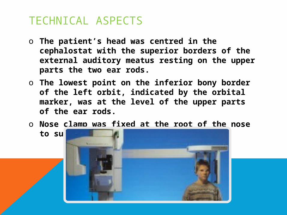
TECHNICAL ASPECTSo The patient’s head was centred in the
cephalostat with the superior borders of the external auditory meatus resting on the upper parts the two ear rods.
o The lowest point on the inferior bony border of the left orbit, indicated by the orbital marker, was at the level of the upper parts of the ear rods.
o Nose clamp was fixed at the root of the nose to support the upper face.

o The focus film distance was set at 5 feet (152.4 cm) and the subject film distance could be measured to calculate image magnification.
o With the two X ray tubes at right angles to each other in the same horizontal plane, two images (lateral & PA) could be simultaneously produced.
Hofrath’s technique differed from
Broadbent’s in that the path of
the central ray was not fixed in
relation to the head and no plan
was suggested for super
positioning subsequent x-rays.
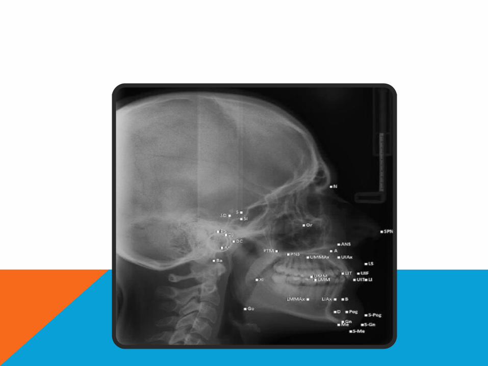
CEPHALOGRAM CAN BE OF TWO TYPES:a) LATERAL CEPHALOGRAM: this provides
a lateral view of the skull. It is taken with the head in a standardized reproducible position at a specified distance from the source of the X ray.
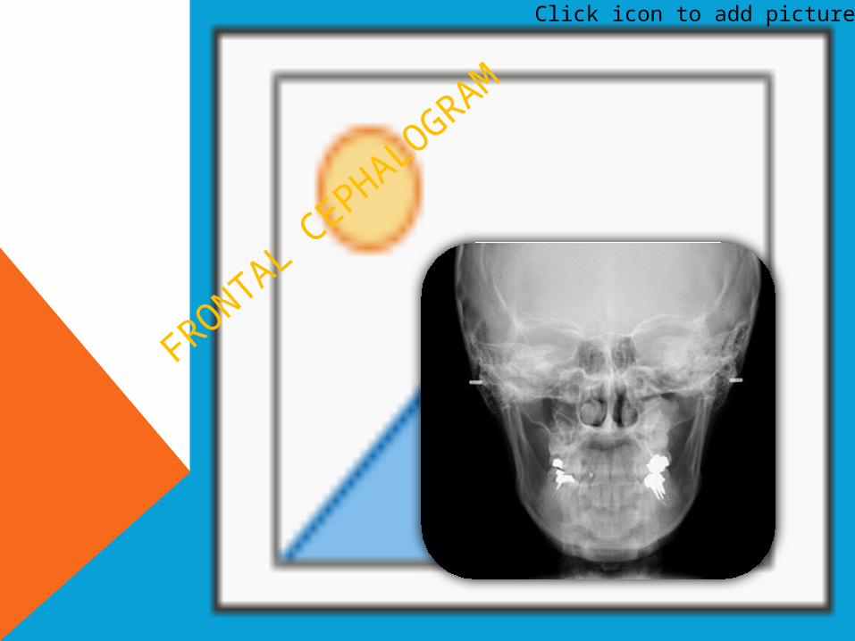
b) FRONTAL CEPHALOGRAM: this provides an antero-posterior view of the skull.

Click icon to add picture
LATERAL C
EPHALO
GRAM
Click icon to add picture
FRONTAL CEPH
ALOGRAM
CEPHALOMETRIC LANDMARKSo Cephalometric landmarks are readily
recognizable points on a cephalometric radiograph or tracing, representing certain hard or soft tissue anatomical structures (anatomical landmarks) & (derived landmarks).
o Anatomic landmarks represent actual anatomic structures of the skull.
o Derived landmarks are the landmarks obtained secondarily from anatomic structures in the cephalogram.
REQUIREMENTS o Should be easily seen on the roentgenogram, o Be uniform in out line, and easily reproducible.o Should have significant relationship to the
vectors of growth.o Should permit valid quantitative
measurements of lines and angles projected from them.
o Measurements should be amenable to statistical analyses.
HARD TISSUE LA
NDMARKS

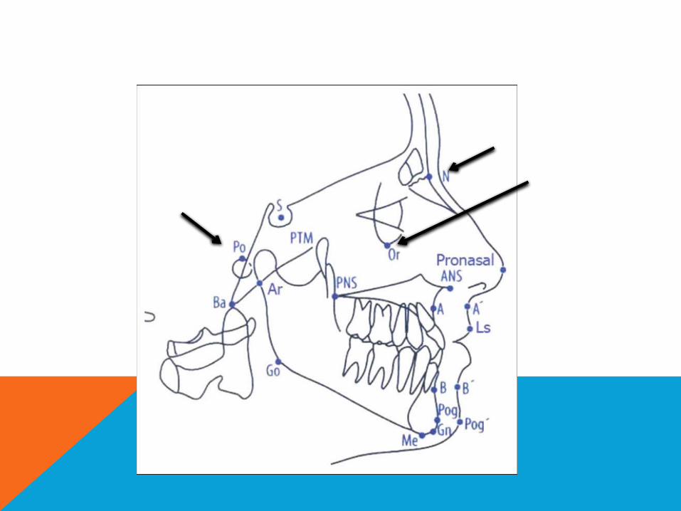
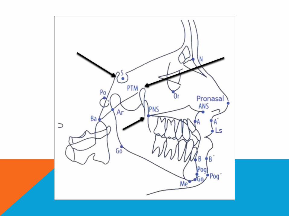
LATERAL CEPHALOMETRIC LANDMARKSNasion (N,Na) : the most anterior on the
frontonasal sutures in the midsagittal plane
Orbitale (Or) : the lowest point on the inferior margin of the orbit.
Porion (Po): the most superior point on the outline of the external auditory meatus (anatomic). The superior most point of the ear rods (machine porion) sometimes is used.
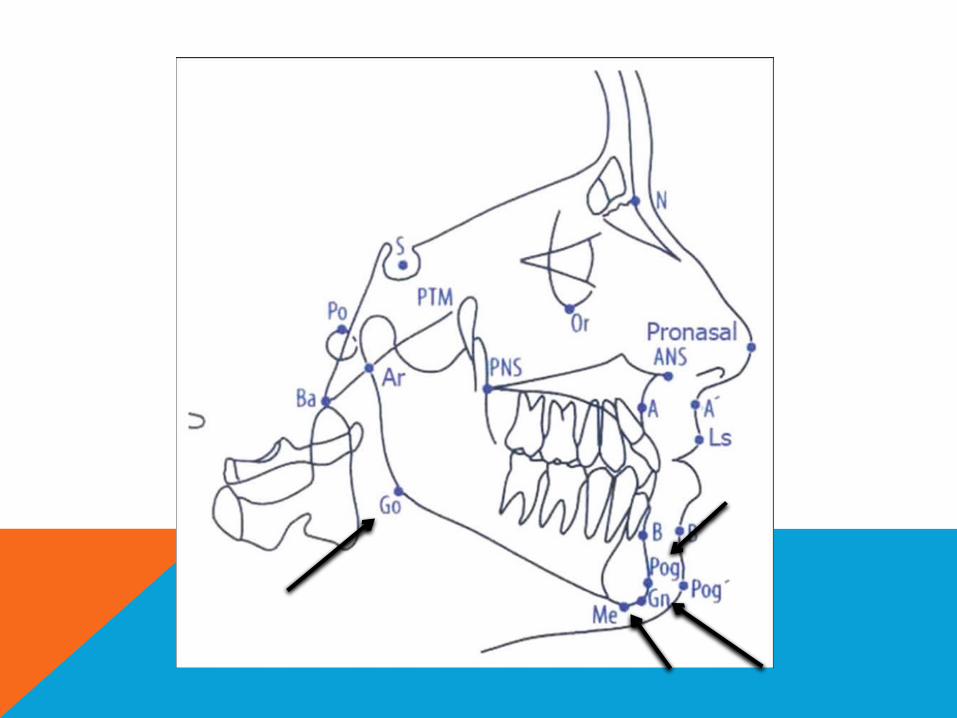
Gonion (Go): the most posterior inferior point on the outline of the angle of the mandible.
Pogonion(pog): its is the most anterior point of the boney cin in the median plane.
Gnathion (Gn) : the most anterior inferior point on the bony chin in the midsagittal plane.
Menton (Me) : the most inferior point of the mandibular symphysis in the midsagittal plane.

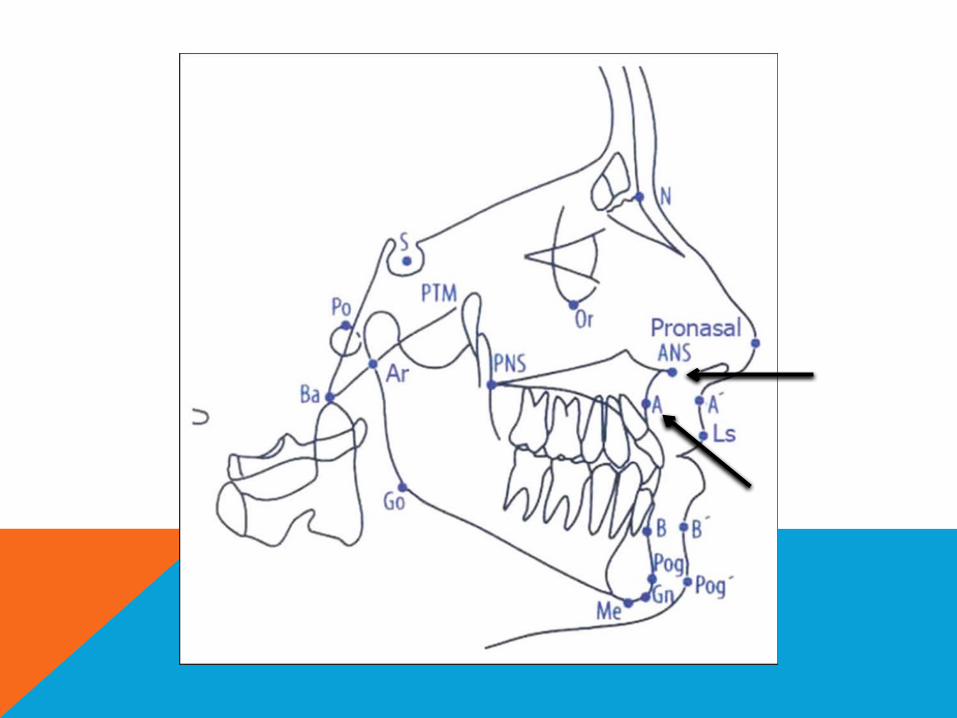
A-point (Point A, Subspinale, SS) : the most posterior midline point on the concavity between the ANS and prosthion.
Anterior nasal spine (ANS): the anterior tip of the sharp bony process of maxilla at he lower margin anterior nasal opening.

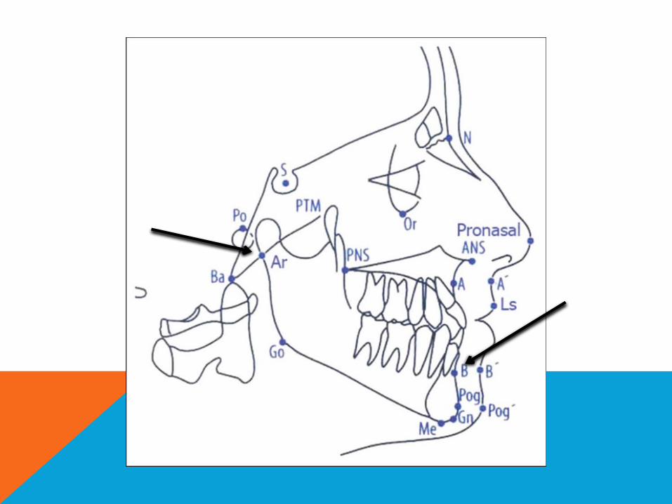
Articulare (Ar) a point at the junction of the posterior border of ramus of mandible and inferior border of posterior cranial base (occipital bone).
B-point (Point B, Supramentale, sm): the most posterior midline point in the concavity of the mandible between the most superior point on the alveolar bone overlying the mandibular incisors (infradental) and Pog.
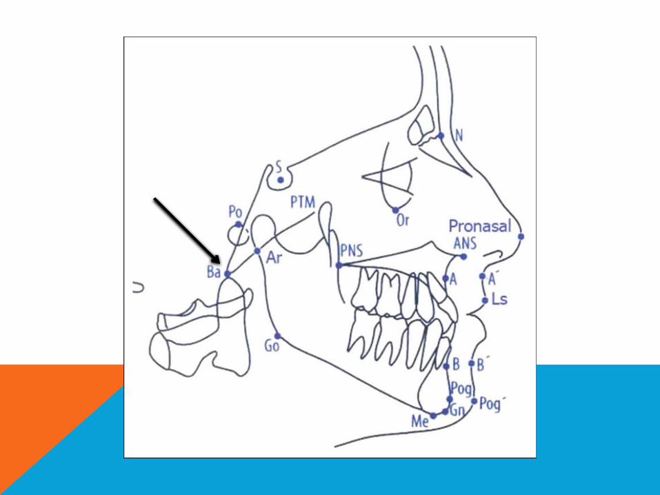
Basion (Ba): the lowest point on the anterior rim of the foramen magnum.
Bolton (Bo): the intersection of the outline of the occipital condyle and the foramen magnum at the highest point on the notch posterior to the occipital condyle.
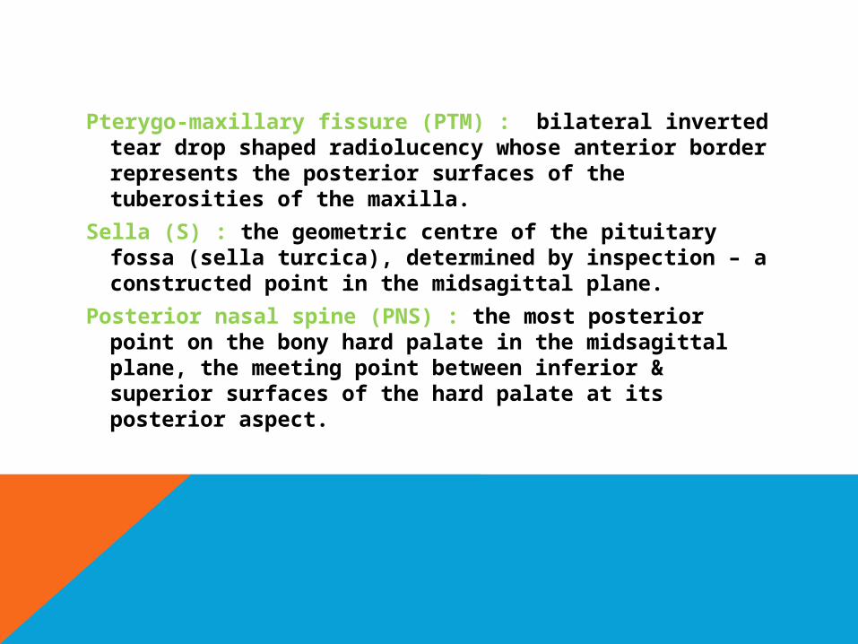
Pterygo-maxillary fissure (PTM) : bilateral inverted tear drop shaped radiolucency whose anterior border represents the posterior surfaces of the tuberosities of the maxilla.
Sella (S) : the geometric centre of the pituitary fossa (sella turcica), determined by inspection – a constructed point in the midsagittal plane.
Posterior nasal spine (PNS) : the most posterior point on the bony hard palate in the midsagittal plane, the meeting point between inferior & superior surfaces of the hard palate at its posterior aspect.
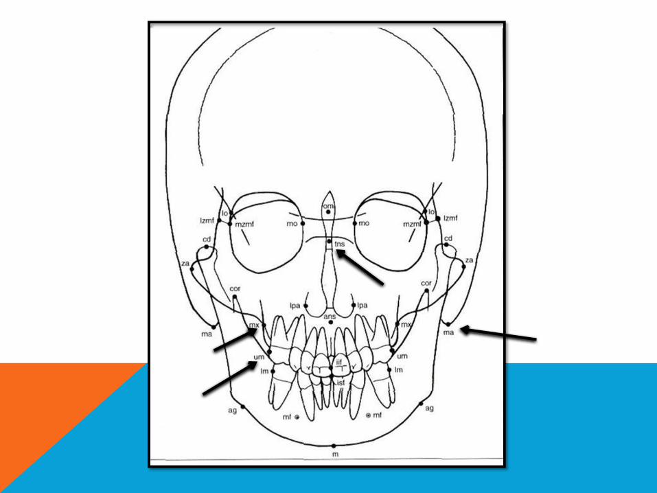
POSTERO-ANTERIOR CEPHALOMETRIC LANDMARKS
ag - antegonion - the highest point in the antegonia/ notch (left and right)
cd - condylar - the most superior point of the condylar head (left and right)
cor - coronoid — the most superior point of the coronoid process (left and right)
iif- incision inferior frontale - the midpoint between the mandibular central incisors at the level of the incisal edges
isf - incision superior frontale - the midpoint between the maxillary central incisors at the level of the incisal edges
m - mandibular midpoint - located by projecting the mental spine on the lower mandibular border, perpendicular to the line ag-ag

Ipa - lateral piriform aperture - the most lateral aspect of the piriform aperture (left and right)
lo - latero-orbitale - the intersection of the lateral orbital contour with the innominate line (left and right)
Im - mandibular molar - the most prominent lateral point on the buccal surface of the second deciduous or first permanent mandibular molar (left and right)
mo - medio-orbitale - the point on the medial orbital margin that is closest to the median plane (left and right)
mf - mental foramen - the centre of the mental foramen (left and right)
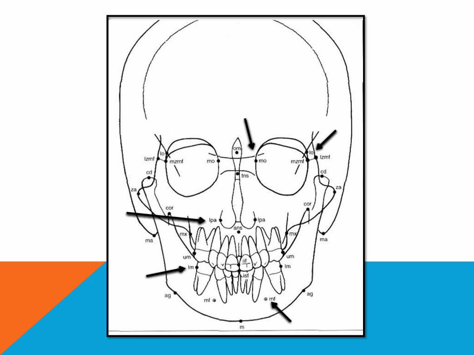

tns - top nasal septum - the highest point on the superior aspect of the nasal septum
ma - mastoid - the lowest point of the mastoid process (left and right)
mx - maxillare - the intersection of the lateral contour of the maxillary alveolar process and the lower contour of the maxillozygomatic process of the maxilla (left and right) . Aka JUGAL POINT
um - maxillary molar - the most prominent lateral point on the buccal surface of the second deciduous or first permanent maxillary molar (left and right)

THANK YO
U!!