guided us procedures.ppt us...paracentesis thoracentesis ... post‐surgical collections lymphocelle...
TRANSCRIPT

7/11/2011
1
Ultrasound Guided Ultrasound Guided ProceduresProcedures
Andrej Lyshchik, M.D., Ph.D.Interventional Radiology Fellow
Department of Radiology and Radiological SciencesVanderbilt University Medical Center
Nashville, TN
BackgroundBackground
Technique and instrumentationTechnique and instrumentation
OutlineOutline
Clinical applicationsClinical applications
Future Future developementsdevelopements
Sagittal image Sagittal image of normal liver of normal liver
Sagittal image Sagittal image of two liver metastasisof two liver metastasis
UltrasonicallyUltrasonically‐‐controlled controlled finefine‐‐needle needle aspiration is aspiration is more more
h h lh h laccurate than the usual accurate than the usual blind procedure in the blind procedure in the diagnosis of liver diagnosis of liver metastases.metastases.
BiopsyBiopsy
Fluid aspiration & drainageFluid aspiration & drainage
Vascular accessVascular access
Current clinical applicationsCurrent clinical applications
Vascular accessVascular access
Image guided treatmentsImage guided treatments
MSK applicationsMSK applications
Sonohysterography

7/11/2011
2
Focal nodules or masses anywhereFocal nodules or masses anywhere
Elevated LFT’sElevated LFT’s
Medical renal diseaseMedical renal disease
Biopsy Biopsy ‐‐ IndicationsIndications
Medical renal diseaseMedical renal disease
Transplant evaluationTransplant evaluation
LymphadenopathyLymphadenopathy
In spite of the dramatic improvement in In spite of the dramatic improvement in tumoraltumoraldiagnosis, diagnosis, percutaneouspercutaneous biopsy continues to be widely biopsy continues to be widely used in oncology.used in oncology.
Biopsy Biopsy ‐‐ IndicationsIndications
In patients with cancer detailed information on the In patients with cancer detailed information on the tumor molecular composition is important to support tumor molecular composition is important to support correct selection of an appropriate treatment.correct selection of an appropriate treatment.
The performance of the biopsy is limited by several The performance of the biopsy is limited by several factors, among which tumor characteristics such as factors, among which tumor characteristics such as tumor type, size and location.tumor type, size and location.
A physical examination should be done before the A physical examination should be done before the procedureprocedure
The preThe pre biopsy coagulation status should be knownbiopsy coagulation status should be known
Biopsy Biopsy ‐‐ TechniqueTechnique
The preThe pre‐‐biopsy coagulation status should be known.biopsy coagulation status should be known.
PT / PTTPT / PTT
PLTPLT
Local anesthesia / conscious sedation is indicatedLocal anesthesia / conscious sedation is indicated
Sterile fieldSterile field
Biopsy guide or free handBiopsy guide or free hand
22G to 25G for fine22G to 25G for fine‐‐needle aspiratesneedle aspirates
Biopsy Biopsy ‐‐ TechniqueTechnique
14G to 21G core biopsy needles14G to 21G core biopsy needles
Shortest distance/safest pathwayShortest distance/safest pathway
Keep needle in plane of beamKeep needle in plane of beam
Always keep needle in the same plane Always keep needle in the same plane as the beamas the beam
Entry point and angle for a superficial lesion: Entry point and angle for a superficial lesion: Aim needle more parallel to the skin Aim needle more parallel to the skin

7/11/2011
3
Entry point and angle for a superficial lesion: Entry point and angle for a superficial lesion: Aim needle more parallel to the skin Aim needle more parallel to the skin
BiopsyBiopsyBiopsyBiopsy
Entry point and angle for a deep lesion: Entry point and angle for a deep lesion: Aim needle more perpendicular to the skin Aim needle more perpendicular to the skin
Entry point and angle for a deep lesion: Entry point and angle for a deep lesion: Aim needle more perpendicular to the skinAim needle more perpendicular to the skin
BiopsyBiopsyBiopsyBiopsy
Targeting of the needle in the vascular, Targeting of the needle in the vascular, viable areas of several tumorsviable areas of several tumors
Avoiding necrotic / avascular areas in largerAvoiding necrotic / avascular areas in larger
CEUS assisted biopsyCEUS assisted biopsy
Avoiding necrotic / avascular areas in larger Avoiding necrotic / avascular areas in larger tumors or in those with frequent necrosis; tumors or in those with frequent necrosis;
Targeting of otherwise invisible lesions or Targeting of otherwise invisible lesions or those hardly visible (small nodules of HCC those hardly visible (small nodules of HCC on cirrhosis, adenocarcinoma’s areas in the on cirrhosis, adenocarcinoma’s areas in the prostate)prostate)
CEUS assisted biopsyCEUS assisted biopsy
Spârchez et al. Medical Ultrasonography, 2010
ParacentesisParacentesis
ThoracentesisThoracentesis
Abscess drainageAbscess drainage
Image guided drainageImage guided drainage
Abscess drainageAbscess drainage
PostPost‐‐surgical collectionssurgical collections
Lymphocelle treatmentLymphocelle treatment

7/11/2011
4
ParacentesisParacentesis ‐‐ IndicationsIndications
Symptomatic Symptomatic ascitesascites abdominal distensionabdominal distension
abdominal discomfortabdominal discomfort
h f b hh f b h shortness of breathshortness of breath
Spontaneous bacterial peritonitisSpontaneous bacterial peritonitis
Asymptomatic Asymptomatic ascitesascites of unknown of unknown etiologyetiology
Shortness of breathShortness of breath
Question of infectionQuestion of infection
Asymptomatic effusion of unknownAsymptomatic effusion of unknown
ThoracentesisThoracentesis ‐‐ IndicationsIndications
Asymptomatic effusion of unknown Asymptomatic effusion of unknown etiologyetiology
Recurrent effusionsRecurrent effusions
TechniqueTechnique TechniqueTechnique
TechniqueTechnique ImagingImaging
Amount of Amount of ascitisascitis
Fluid compositionFluid composition
Presence of locationsPresence of locationsPresence of locationsPresence of locations
Adjacent organsAdjacent organs
VacularVacular structuresstructures
Inferior Inferior epigastricepigastric vesselsvessels
IntercostalIntercostal vesselsvessels

7/11/2011
5
Inferior Inferior epigastricepigastric arteryartery Inferior Inferior epigastricepigastric arteryartery
Inferior Inferior epigastricepigastric arteryartery
Teodori et al. MMCTC 2006
Inferior Inferior epigastricepigastric arteryartery
PercutaneousPercutaneous DrainageDrainage
Before During
After
PercutaneousPercutaneous DrainageDrainage
Before During
After

7/11/2011
6
Entry for IV therapyEntry for IV therapy
Entry for diagnostic purposesEntry for diagnostic purposes
Vascular accessVascular access
Entry for diagnostic purposesEntry for diagnostic purposes
Entry for endovascular proceduresEntry for endovascular procedures
ComplicationsComplications
McGee et al. NEJM 2003
Overall success: 100% vs. 88.1%Overall success: 100% vs. 88.1%
First attempt success: 78% vs 38%First attempt success: 78% vs 38%
US vs. blind stickUS vs. blind stick
First attempt success: 78% vs. 38%First attempt success: 78% vs. 38%
Skin to vein time: 9.8 sec vs. 44.5 secSkin to vein time: 9.8 sec vs. 44.5 sec
Carotid puncture: 1.7% vs. 8.3%Carotid puncture: 1.7% vs. 8.3%
Denys et al. Circulation 1993
Vascular accessVascular access
Radiofrequency
Liquid nitrogen (cryoablation)
ImageImage‐‐guided Treatmentsguided Treatments
Liquid nitrogen (cryoablation)
Microwave
Laser
RadioRadio‐‐frequency ablationfrequency ablation
http://www.radiologyinfo.org

7/11/2011
7
RadioRadio‐‐frequency ablationfrequency ablation
Illustration courtesy of Gerald D. Dodd II, MD, University of Colorado
RadioRadio‐‐frequency ablationfrequency ablation
http://www.radiologyinfo.org
RFA vs. SurgeryRFA vs. Surgery
Kudo, Oncology 2010
CEUS assisted liver RFACEUS assisted liver RFA
Gallotti A. Radiol Med. 2009
Before treatmentBefore treatment
CEUS assisted liver RFACEUS assisted liver RFA
Gallotti A. Radiol Med. 2009
After treatmentAfter treatment
CEUS assisted liver RFACEUS assisted liver RFA
P. Ricci et al. Ultraschall in Med 2009
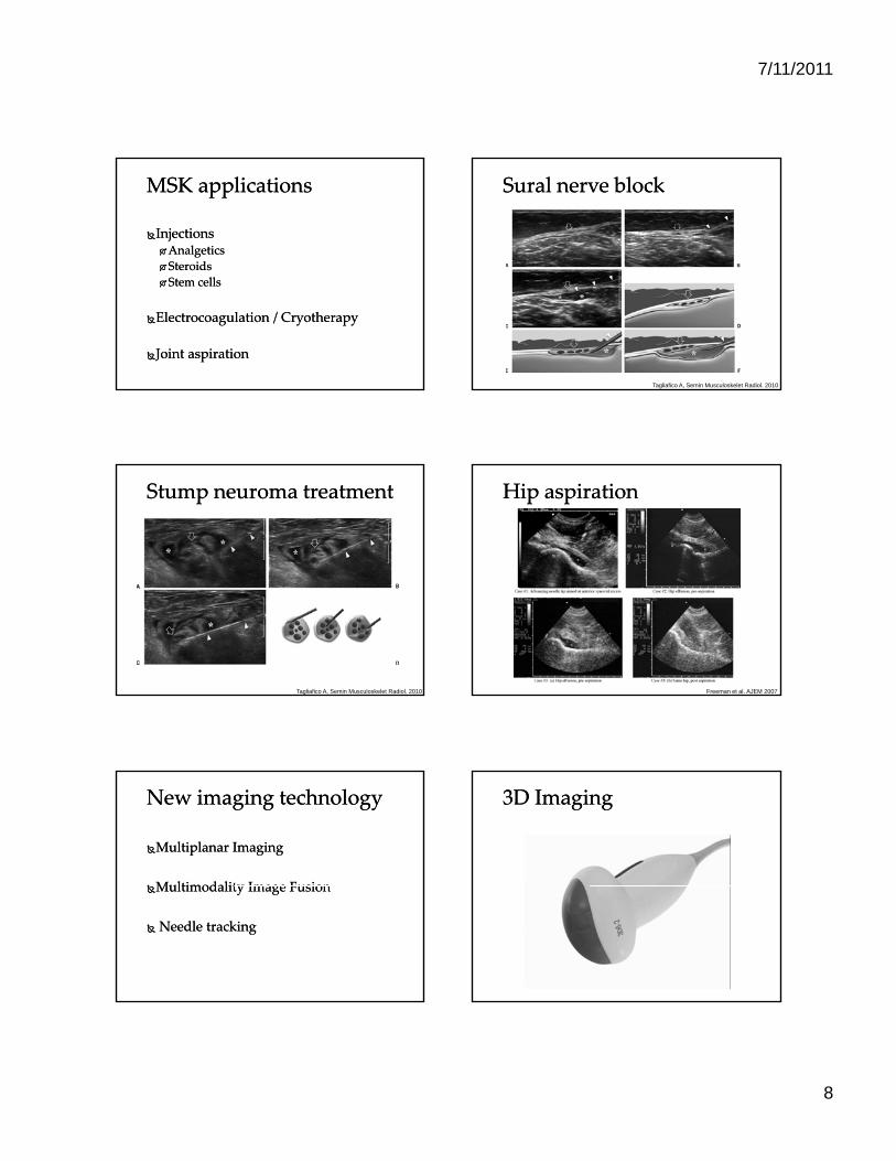
7/11/2011
8
Injections Injections AnalgeticsAnalgetics
SteroidsSteroids
MSK applicationsMSK applications
Stem cellsStem cells
ElectrocoagulationElectrocoagulation / / CryotherapyCryotherapy
Joint aspirationJoint aspiration
SuralSural nerve blocknerve block
Tagliafico A, Semin Musculoskelet Radiol. 2010
Stump Stump neuromaneuroma treatmenttreatment
Tagliafico A, Semin Musculoskelet Radiol. 2010
Hip aspirationHip aspiration
Freeman et al. AJEM 2007
MultiplanarMultiplanar ImagingImaging
Multimodality Image FusionMultimodality Image Fusion
New imaging technologyNew imaging technology
Multimodality Image FusionMultimodality Image Fusion
Needle trackingNeedle tracking
3D Imaging3D Imaging

7/11/2011
9
MultiplanarMultiplanar imagingimaging MultiplanarMultiplanar imagingimaging
Fusion ImagingFusion Imaging Needle trackingNeedle tracking
Interventional Interventional sonographysonography is an evolving is an evolving and rapidly developing technology. and rapidly developing technology.
ConclusionConclusion
It provides unique advantages of high It provides unique advantages of high resolution, real time guidance, lower cost. resolution, real time guidance, lower cost. Thus, making it ideal guiding method in Thus, making it ideal guiding method in variety of interventional applications.variety of interventional applications.
Thank you!Thank you!