guide to the 1-3 minute blood film microscopic review: why ... · guide to the 1-3 minute blood...
TRANSCRIPT

Guide to the 1-3 Minute Blood Film Microscopic Review: Why and How?
Dennis B. DeNicola, DVM, PhD, DACVP
Chief Veterinary Educator
IDEXX Laboratories, Inc.
Westbrook, ME USA
Adjunct Professor of Veterinary Clinical Pathology
Purdue University College of Veterinary Medicine
Objectives:
•Understand the value of a brief blood film microscopic review
• Validate hematology analyzer performance
• Add valuable information regarding cellular morphology
•Understand how to develop standardized logical steps for this brief blood film review
•Understand that in the majority of the times, less than a one minute review is required and that only a maximum of a three minute review should be required even for the most difficult cases

20031990s1980spre1980s
Manual Quantitative
Buffy coat.
Impedance Laser
2010+
In-house Hematology Analyzers
RBC
WBC
RBC
WBC
ProCyteDx

How frequently do you currently use any of the various cytograms generated by your hematology analyzer?
A. Never
B. Less than 25% of the time
C. Between 25 and 50% of
the time
D. Between 50 and 75% of
the time
E. Greater than 75% of the
time

20031990s1980spre1980s
Manual Quantitative
Buffy coat.
Impedance Laser
2010+
In-house Hematology Analyzers
RBC
WBC
RBC
WBC
ProCyteDx

What has been gained with hematology analyzer evolution?
•Greater amount of data – sometimes even more than the reference laboratory provides
• Improved precision and accuracy of results – more advanced analyzers perform equal to or better than reference laboratory analyzers
•Decreased technician time –
• Analyzers have minimal maintenance requirements
• Operations are generally “load and go”
• Microscopic blood film review should still be included
• Majority of time not performing leukocyte differential
• Quickly scanning slide for cellular morphology changes
Hematology—Is a blood film needed?
ABSOLUTELY YES!!
• Blood film examination is needed for all
• Low-end hematology analyzers
• IDEXX VetAutoread™ Hematology Analyzer
• Impedance-based instruments
• High-end hematology analyzers
• LaserCyte®, LaserCyte® Dx, and ProCyte Dx® Hematology
Analyzers (in-house)
• Cell-Dyn (reference laboratory)
• Advia (reference laboratory)
• Sysmex (reference laboratory

Hematology—Why is a blood film needed?
• Validate numerical data generated• RBC mass (RBC count, HCT, HGB)
• RBC indices (MCV, MCH, MCHC, RDW)
• WBC count
• WBC distribution
• Platelet count
• Add valuable/critical information that is not generated by the hematology analyzers• RBC morphology—clues to cause of anemia
• WBC morphology—characterize presence or absence of inflammatory disease
• Platelet morphology

Hematology — How long should a blood film review take?
• 1–3 minutes maximum time on scope
• What is accomplished?
•Validate data
•Provide morphology comments
•Spending more than 3 minutes results in over-interpretation of potential subtle changes
•Only prominent changes prove helpful

Peripheral Blood Film Preparation
• 30–45 degree angle
• Increased angle with low PCV
• Decreased angle with high PCV
• Fluid-controlled motion
•Results
• Body
• Monolayer
• Feathered edge

Anatomy of the Peripheral Blood Film
Pati
en
t ID
Date
Feathered Edge
Which of the following IS NOT evaluated when examining the feathered edge of a blood film?
1. Clumped platelets
2. Large cells
3. Microfilaria
4. Leukocyte distribution
5. RBC morphology

Anatomy of the Peripheral Blood Film
Pati
en
t ID
Date
Feathered Edge
• Clumped platelets
• Large cells
• Microfilaria
• Leukocyte distribution

Anatomy of the Peripheral Blood Film
Pati
en
t ID
Date
Body
• Rouleaux formation
• Agglutination
• Cell clumping

Anatomy of the Peripheral Blood Film
Monolayer
• Platelet number estimation
• Leukocyte number estimation
• Morphologic evaluation
• Data validation
Pati
en
t ID
Date

Erythrogram – Validate Data

Erythrogram – Validate Data
Measure of RBC mass -severity of anemia

Measure of RBC mass -severity of anemia
Erythrogram - Validate Data
Sample A Sample B

Which one of these samples is from a severely anemic dog?
1. Sample A
2. Sample B

Measure of RBC mass -severity of anemia
Erythrogram - Validate Data
Sample A Sample B

Erythrogram - Validate Data
Geo
Measure of RBC mass -severity of anemia
Description of RBC population
Low MCV, Low MCHC

Geo - Erythrogram
NormalNormal
MCV
Geo’s
MCV
Geo RBC run dot
plot for data
validation
Compare to
normal

Erythrogram - Validate Data
Measure of RBC mass -severity of anemia
Description of RBC population

Erythrogram - Validate Data
Measure of RBC mass -severity of anemia
Description of RBC population
Objective measure of variation in RBC size

Erythrogram - Validate Data
Measure of RBC mass -severity of anemia
Description of RBC population
Objective measure of variation in RBC size
Objective measure of erythroid response

Which of the following is the BEST support for the presence of an increased reticulocyte count?
1. Presence of nucleated red
blood cells
2. Presence of large red
blood cells
3. Presence of target cells
4. Presence of
polychromatophils
5. Presence of pale red blood
cells

Erythrogram - Validate Data
Measure of RBC mass -severity of anemia
Description of RBC population
Objective measure of variation in RBC size
Objective measure of erythroid response
Routine Stain New Methylene Blue

Erythrogram - Validate Data
Geo
Measure of RBC mass -severity of anemia
Description of RBC population
Objective measure of variation in RBC size
Objective measure of erythroid response
Normal
Dot plot review provides rapid confirmation of reticulocyte count

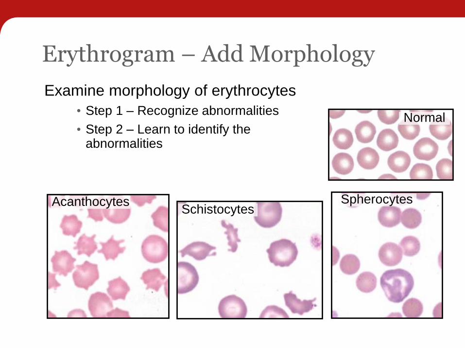
Erythrogram – Add Morphology
Examine morphology of erythrocytes
• Step 1 – Recognize abnormalitiesNormal
Erythrogram – Add Morphology
Examine morphology of erythrocytes
• Step 1 – Recognize abnormalities
• Step 2 – Learn to identify the abnormalities
Normal
AcanthocytesSchistocytes
Spherocytes

Erythrogram – Add Morphology
Examine morphology of erythrocytes
• Step 1 – Recognize abnormalities
• Step 2 – Learn to identify the abnormalities
• Step 3 – Understand what they mean
Normal
AcanthocytesSchistocytes
Spherocytes

Spherocytosis

Immune - Mediated Hemolytic Anemia
FcRMacrophage
RBC
Spherocytosis

Spherocytosis

Erythrogram – Add Morphology
Examine morphology of erythrocytes
• Step 1 – Recognize abnormalities
• Step 2 – Learn to identify the abnormalities
• Step 3 – Understand what they mean
• Step 4 – Look for RBC inclusions
Normal
AcanthocytesSchistocytes
Spherocytes

Miscellaneous Inclusions
Mycoplasma
(Haemobartonella)
Basophilic stippling
Distemper virus
inclusions
Babesia gibsoni

Leukogram - Validate Data
• Validate WBC count

Leukogram - Validate WBC Count
3,000 5,000 10,000 25,000 50,000

Which of the following is the BEST estimated WBC count based on blood film examination?
1. 3,000 / µL
2. 5,000 / µL
3. 10,000 / µL
4. 25,000 / µL
5. 50,000 / µL

Leukogram - Validate WBC Count
3,000 5,000 10,000 25,000 50,000
WBC reported
24,000

Leukogram - Validate WBC Count
3,000 5,000 10,000 25,000 50,000

Which of the following is the BEST estimated WBC count based on blood film examination?
1. 3,000 / µL
2. 5,000 / µL
3. 10,000 / µL
4. 25,000 / µL
5. 50,000 / µL
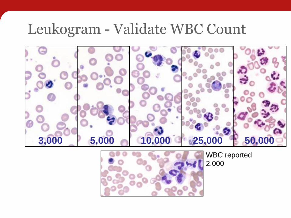
Leukogram - Validate WBC Count
WBC reported
2,000
3,000 5,000 10,000 25,000 50,000

Leukogram - Validate Data
•Validate WBC count
•Validate leukocyte distribution

Five-Part DifferentialWBC = 70.60 x10³/µL ( 5.50 - 16.90 )
NEU = 64.25 x10³/µL ( 2.00 - 12.00 )
LYM = 4.94 x10³/µL ( 0.70 - 4.90 )
MONO = 1.41 x10³/µL ( 0.30 - 2.00 )
EOS = 0.00 x10³/µL ( 0.10 - 1.49 )
BASO = 0.00 x10³/µL ( 0.00 - 0.10 )

Leukogram - Validate Leukocyte Distribution
Leukocyte Distribution
5-part differential
Normal WBC DotPlot
Lymphocytes
Monocytes
Neutrophils
Eosinophils
Right Angle Scatter
High Numerical Aperture
50-130 degrees
Low Angle
Forward Scatter
2-4 degrees
High Angle
Forward Scatter
8-12 deg
Axial
Light
Loss
Quartz Flow
Cell
Laser and Lens
Sy stem
Core Sample
Stream
Sheath Flow
Channel
WBC = 70.60 x10³/µL ( 5.50 - 16.90 )
NEU = 64.25 x10³/µL ( 2.00 - 12.00 )
LYM = 4.94 x10³/µL ( 0.70 - 4.90 )
MONO = 1.41 x10³/µL ( 0.30 - 2.00 )
EOS = 0.00 x10³/µL ( 0.10 - 1.49 )
BASO = 0.00 x10³/µL ( 0.00 - 0.10 )
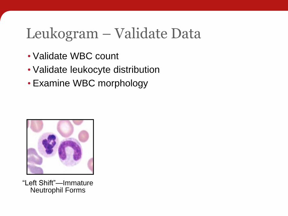
Leukogram – Validate Data
•Validate WBC count
•Validate leukocyte distribution
•Examine WBC morphology
“Left Shift”—Immature Neutrophil Forms

Blood Film—Validate Data
•Validate WBC count
•Validate leukocyte distribution
•Examine WBC morphology
Neutrophil Toxicity
“Left Shift”—Immature Neutrophil Forms
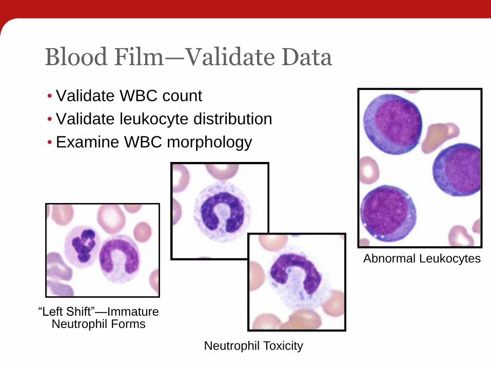
Blood Film—Validate Data
•Validate WBC count
•Validate leukocyte distribution
•Examine WBC morphology
Neutrophil Toxicity
Abnormal Leukocytes
“Left Shift”—Immature Neutrophil Forms

Belle – 4-24-13
NormalBell Immature or toxic neutrophils suspected
Monotonous population of large mononuclear cells –suspect abnormal cell population

Belle – 4-24-13
NormalBell Immature or toxic neutrophils suspected

Belle – 4-24-13
NormalBell Immature or toxic neutrophils suspected
Monotonous population of large mononuclear cells –suspect abnormal cell population

Belle – 4-24-13
PoikilocytosisAcanthocytes
2+ toxic hyposegmented neutrophil
Decreased platelets
Two large immature appearing mononuclear cells

Thrombogram - Validate Data
•Validate platelet count

Thrombogram - Validate Data
•Validate platelet count
• Never accept a low platelet count from any analyzer without confirming with a blood film

Platelet Number Evaluation
•Number of platelets per 100x oil objective field of view
• Minimum: 8–10
• Maximum: 35–40
•Potential semiquantitation
• 20,000 x number of platelets seen per 100x objective field of view
Thrombocytopenia
Normal platelet count
Thrombogram - Validate Data
•Validate platelet count
•Add platelet morphology comments
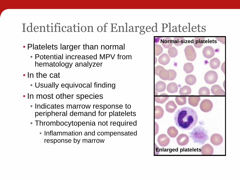
Identification of Enlarged Platelets
•Platelets larger than normal
• Potential increased MPV from hematology analyzer
• In the cat
• Usually equivocal finding
• In most other species
• Indicates marrow response to peripheral demand for platelets
• Thrombocytopenia not required
• Inflammation and compensated response by marrow
Enlarged platelets
Normal-sized platelets

Reference Laboratory Hematology —Needed?
•Experience
• Use the laboratory for complicated cases
• Potential of a “pathology review”
• Use the laboratory as a great teaching resource
•Quality Assurance
• Use the laboratory to periodically check your in-house system
• Realize that different instruments produce different results
• Realize that aged samples are not the same as fresh samples

Questions?