guide to feeder-based culture of pluripotent stem cells
TRANSCRIPT

1
GUIDE TO FEEDER-BASED CULTURE OF PLURIPOTENT STEM CELLS
Make the connection
2
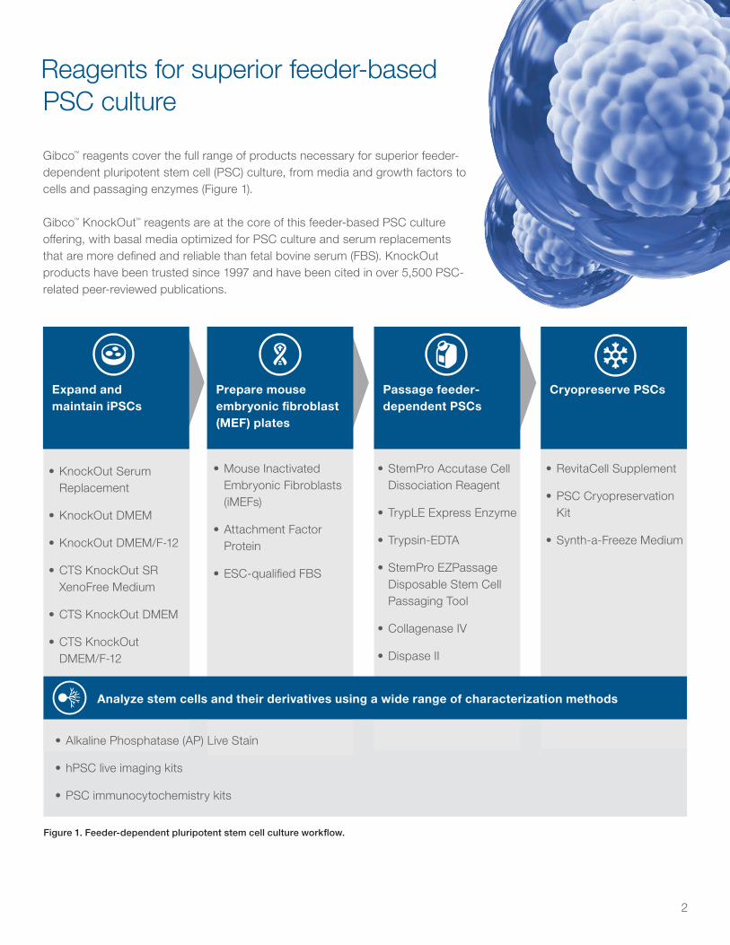
Reagents for superior feeder-based PSC culture
Gibco™ reagents cover the full range of products necessary for superior feeder-dependent pluripotent stem cell (PSC) culture, from media and growth factors to cells and passaging enzymes (Figure 1).
Gibco™ KnockOut™ reagents are at the core of this feeder-based PSC culture offering, with basal media optimized for PSC culture and serum replacements that are more defined and reliable than fetal bovine serum (FBS). KnockOut products have been trusted since 1997 and have been cited in over 5,500 PSC-related peer-reviewed publications.
Analyze stem cells and their derivatives using a wide range of characterization methods
Prepare mouse embryonic fibroblast (MEF) plates
Expand and maintain iPSCs
Passage feeder-dependent PSCs
Cryopreserve PSCs
• KnockOut Serum Replacement
• KnockOut DMEM
• KnockOut DMEM/F-12
• CTS KnockOut SR XenoFree Medium
• CTS KnockOut DMEM
• CTS KnockOut DMEM/F-12
• Mouse Inactivated Embryonic Fibroblasts (iMEFs)
• Attachment Factor Protein
• ESC-qualified FBS
• StemPro Accutase Cell Dissociation Reagent
• TrypLE Express Enzyme
• Trypsin-EDTA
• StemPro EZPassage Disposable Stem Cell Passaging Tool
• Collagenase IV
• Dispase II
• RevitaCell Supplement
• PSC Cryopreservation Kit
• Synth-a-Freeze Medium
• Alkaline Phosphatase (AP) Live Stain
• hPSC live imaging kits
• PSC immunocytochemistry kits
Figure 1. Feeder-dependent pluripotent stem cell culture workflow.
3
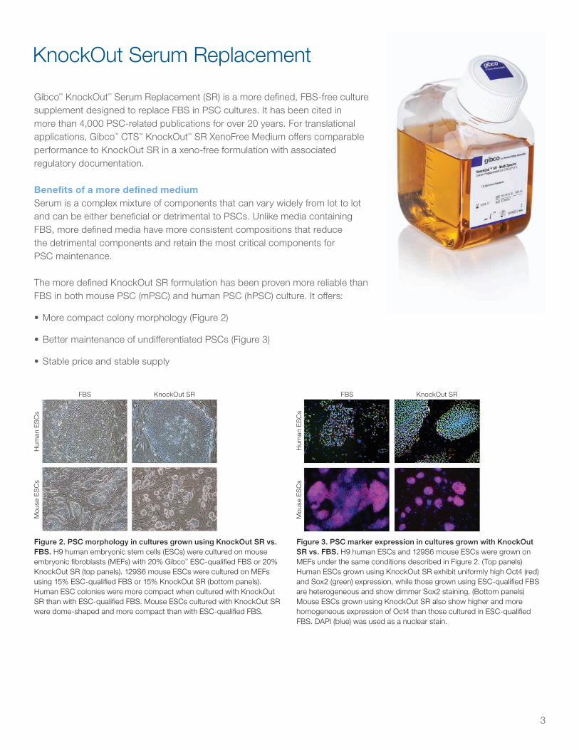
Gibco™ KnockOut™ Serum Replacement (SR) is a more defined, FBS-free culture supplement designed to replace FBS in PSC cultures. It has been cited in more than 4,000 PSC-related publications for over 20 years. For translational applications, Gibco™ CTS™ KnockOut™ SR XenoFree Medium offers comparable performance to KnockOut SR in a xeno-free formulation with associated regulatory documentation.
Benefits of a more defined mediumSerum is a complex mixture of components that can vary widely from lot to lot and can be either beneficial or detrimental to PSCs. Unlike media containing FBS, more defined media have more consistent compositions that reduce the detrimental components and retain the most critical components for PSC maintenance.
The more defined KnockOut SR formulation has been proven more reliable than FBS in both mouse PSC (mPSC) and human PSC (hPSC) culture. It offers:
• More compact colony morphology (Figure 2)
• Better maintenance of undifferentiated PSCs (Figure 3)
• Stable price and stable supply
KnockOut Serum ReplacementM
ouse
ES
Cs
FBS
Hum
an E
SC
s
KnockOut SR
Figure 2. PSC morphology in cultures grown using KnockOut SR vs. FBS. H9 human embryonic stem cells (ESCs) were cultured on mouse embryonic fibroblasts (MEFs) with 20% Gibco™ ESC-qualified FBS or 20% KnockOut SR (top panels). 129S6 mouse ESCs were cultured on MEFs using 15% ESC-qualified FBS or 15% KnockOut SR (bottom panels). Human ESC colonies were more compact when cultured with KnockOut SR than with ESC-qualified FBS. Mouse ESCs cultured with KnockOut SR were dome-shaped and more compact than with ESC-qualified FBS.
Mou
se E
SC
s
FBS
Hum
an E
SC
s
KnockOut SR
Figure 3. PSC marker expression in cultures grown with KnockOut SR vs. FBS. H9 human ESCs and 129S6 mouse ESCs were grown on MEFs under the same conditions described in Figure 2. (Top panels) Human ESCs grown using KnockOut SR exhibit uniformly high Oct4 (red) and Sox2 (green) expression, while those grown using ESC-qualified FBS are heterogeneous and show dimmer Sox2 staining. (Bottom panels) Mouse ESCs grown using KnockOut SR also show higher and more homogeneous expression of Oct4 than those cultured in ESC-qualified FBS. DAPI (blue) was used as a nuclear stain.
4
Human PSC culture for research use
Culture hPSCs confidently with KnockOut SR, a more defined medium that is proven more reliable than FBS for basic research applications.
• Supports robust growth—results in a higher growth rate compared to FBS (Figure 5)
• Maintains quality colonies—colonies are more compact and show strong expression of PSC markers compared to FBS
• Easy to transition into—supports cultures that are directly passaged from FBS-based medium
• Stable price and supply—FBS-free formulation is not subject to the same FBS price and supply fluctuations
Mouse PSC culture for research use
Maintain mPSCs with KnockOut SR and a basal medium that approximates the low osmolarity of mouse embryonic tissue.
• Minimizes differentiation—improves maintenance of undifferentiated mPSCs compared to FBS (Figure 4) and traditional DMEM
• Maintains quality colonies—colonies are more dome-shaped and show more homogeneous gene expression compared to FBS-based medium
• Easy to transition into—supports cultures that are directly thawed or passaged from FBS-based medium
Feeder-dependent culture proven more reliable than FBSChoose your KnockOut product combination
Extrapolated proliferation curve
FBS
KnockOut SR
KnockOut SRESC-qualified FBS
Traditional DMEM
KnockOut DMEM
Und
i�er
entia
ted
ESC
col
onie
s (%
)1 2 3
1,000
100
10
0
Cel
ls (x
106 )
Passage
1
100
90
80
70
60
50
40
30
20
10
0
Figure 5. Human PSC growth with KnockOut SR vs. FBS. H9 human ESCs were grown on MEFs under the same conditions as in Figure 2, and the mean viable cell numbers were plotted to growth curves for media supplemented with ESC-qualified FBS or KnockOut SR. Proliferation of human ESCs was significantly higher in KnockOut SR over 3 passages.
Figure 4. Mouse PSC culture with KnockOut SR vs. FBS in the absence of LIF. Mouse D3 ESCs were cultured at low density in either Gibco™ DMEM (with high glucose) or KnockOut DMEM, supplemented with either ESC-qualified FBS or KnockOut SR. No LIF was used. After 7 days, colonies were fixed and stained for alkaline phosphatase, a marker for undifferentiated ESCs. Undifferentiated colonies were scored based on morphology and staining.
Extrapolated proliferation curve
FBS
KnockOut SR
KnockOut SRESC-qualified FBS
Traditional DMEM
KnockOut DMEM
Und
i�er
entia
ted
ESC
col
onie
s (%
)
1 2 3
1,000
100
10
0
Cel
ls (x
106 )
Passage
1
100
90
80
70
60
50
40
30
20
10
0
Recommended products:• KnockOut SR
• Gibco™ KnockOut™ DMEM
• Gibco™ LIF Recombinant Mouse Protein
Recommended products:• KnockOut SR
• Gibco™ DMEM/F-12, GlutaMAX™ supplement
• Gibco™ bFGF Recombinant Human Protein

5
Xeno-free human PSC culture for translational use
Count on Gibco™ Cell Therapy Systems (CTS™) products for the xeno-free derivation, maintenance, and cryopreservation of human PSCs in translational applications (Figure 6).
• Xeno-free—contains only recombinant or human-derived material and is compatible with human feeder cells
• Provides extensive QC testing—helps reduce the burden of qualifying material when transitioning to clinical applications
• Provides traceability documentation—supporting documentation available for regulatory submissions
Figure 6. Xeno-free culture and gene expression of hESCs. (A) Morphology of BG01v hESCs at passage 4, cultured in CTS KnockOut SR XenoFree Medium on human foreskin fibroblast (HFF) feeder cells attached with CELLstart Substrate. (B) Maintenance of pluripotency with CTS KnockOut SR XenoFree Medium. Following 10 passages, either in KnockOut SR (left lane of the pair for each marker) or in CTS KnockOut SR XenoFree (right lane for each marker) on HFFs attached with CELLstart Substrate, undifferentiated BG01v gene expression was examined by RT-qPCR of pluripotency markers, OCT4 and NANOG (top). Gene expression of embryoid bodies generated from the same passage-10 BG01v/HFF culture (bottom). (NTC=No template control; GAPDH=reference gene).
Table 1. Comparison of FBS and the KnockOut SR formulation.
Species SerumKnockOut SR References
Unwanted differentiation
Human,mouse
Higher Lower 1–5
Efficiency of ESC derivation
Human,mouse
Lower Higher 3, 6–8
Clonal efficiency
Human,mouse
Lower Higher 7, 9
A B
Recommended products:• CTS KnockOut SR XenoFree Medium
• Gibco™ CTS™ KnockOut™ DMEM
• Gibco™ FGF-Basic Full Length Recombinant Human Protein
• Gibco™ CELLstart™ Substrate
References1. Inzunza J, Gertow K, Strömberg MA, Matilainen E, Blennow E, Skottman H, Wolbank S,
Ahrlund-Richter L, Hovatta O. "Derivation of human embryonic stem cell lines in serum replacement medium using postnatal human fibroblasts as feeder cells." Stem Cells. 2005 Apr;23(4):544-9. doi: 10.1634/stemcells.2004-0201. PMID: 15790775.
2. Skottman H, Strömberg AM, Matilainen E, Inzunza J, Hovatta O, Lahesmaa R. "Unique gene expression signature by human embryonic stem cells cultured under serum-free conditions correlates with their enhanced and prolonged growth in an undifferentiated stage." Stem Cells. 2006 Jan;24(1):151-67. doi: 10.1634/stemcells.2004-0189. Epub 2005 Aug 11. PMID: 16100004.
3. Hong-mei P, Gui-an C. "Serum-free medium cultivation to improve efficacy in establishment of human embryonic stem cell lines." Hum Reprod. 2006 Jan;21(1):217-22. doi: 10.1093/humrep/dei275. Epub 2005 Sep 2. PMID: 16143641.
4. Denning C, Allegrucci C, Priddle H, Barbadillo-Muñoz MD, Anderson D, Self T, Smith NM, Parkin CT, Young LE. "Common culture conditions for maintenance and cardiomyocyte differentiation of the human embryonic stem cell lines, BG01 and HUES-7." Int J Dev Biol. 2006;50(1):27-37. doi: 10.1387/ijdb.052107cd. PMID: 16323075.
5. Guo G, Pinello L, Han X, Lai S, Shen L, Lin TW, Zou K, Yuan GC, Orkin SH. "Serum-Based Culture Conditions Provoke Gene Expression Variability in Mouse Embryonic Stem Cells as Revealed by Single-Cell Analysis." Cell Rep. 2016 Feb 2;14(4):956-965. doi:10.1016/j.celrep.2015.12.089
6. Davies TJ, Fairchild PJ. "Optimization of protocols for derivation of mouse embryonic stem cell lines from refractory strains, including the non obese diabetic mouse." Stem Cells Dev. 2012 Jul 1;21(10):1688-700. doi: 10.1089/scd.2011.0427. Epub 2011 Nov 2. PMID: 21933027; PMCID: PMC3376457.
7. Cheng J, Dutra A, Takesono A, Garrett-Beal L, Schwartzberg PL. "Improved generation of C57BL/6J mouse embryonic stem cells in a defined serum-free media." Genesis. 2004 Jun;39(2):100-4. doi: 10.1002/gene.20031. PMID: 15170695.
8. Bryja V, Bonilla S, Arenas E. "Derivation of mouse embryonic stem cells." Nat Protoc. 2006;1(4):2082-7. doi: 10.1038/nprot.2006.355. PMID: 17487198.
9. Amit M, Carpenter MK, Inokuma MS, Chiu CP, Harris CP, Waknitz MA, Itskovitz-Eldor J, Thomson JA. "Clonally derived human embryonic stem cell lines maintain pluripotency and proliferative potential for prolonged periods of culture." Dev Biol. 2000 Nov 15;227(2):271-8. doi: 10.1006/dbio.2000.9912. PMID: 11071754.
6
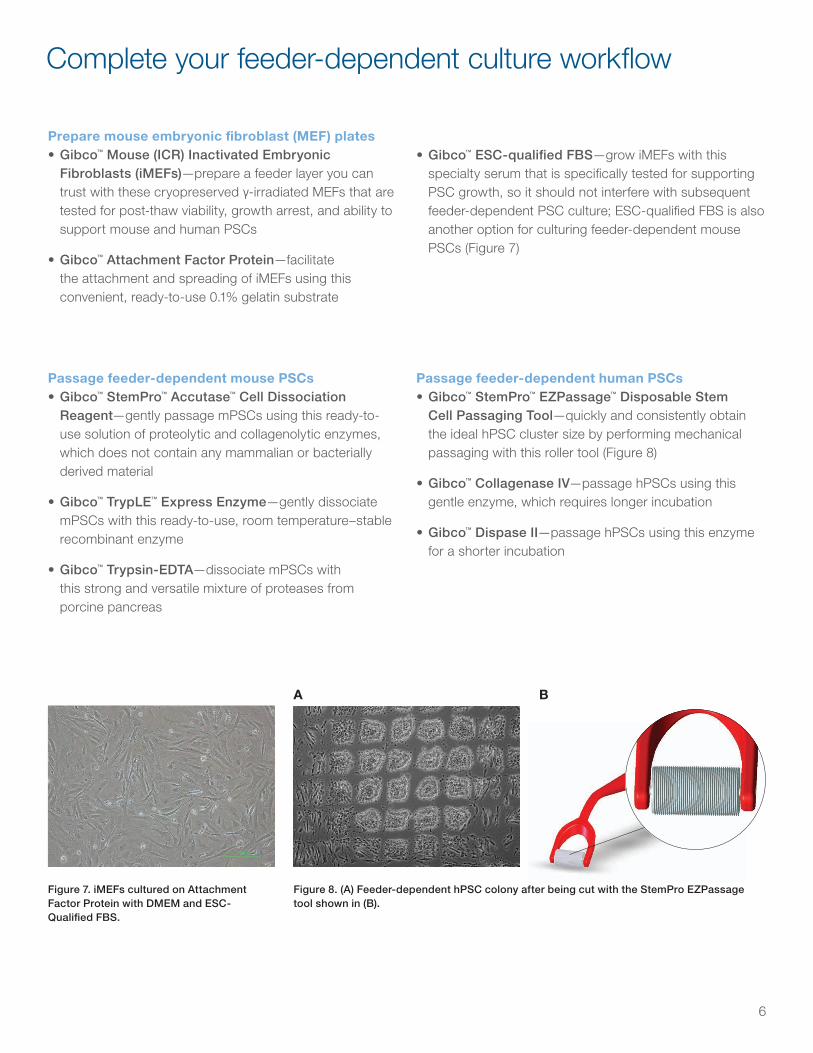
Complete your feeder-dependent culture workflow
Prepare mouse embryonic fibroblast (MEF) plates • Gibco™ Mouse (ICR) Inactivated Embryonic
Fibroblasts (iMEFs)—prepare a feeder layer you can trust with these cryopreserved γ-irradiated MEFs that are tested for post-thaw viability, growth arrest, and ability to support mouse and human PSCs
• Gibco™ Attachment Factor Protein—facilitate the attachment and spreading of iMEFs using this convenient, ready-to-use 0.1% gelatin substrate
Figure 7. iMEFs cultured on Attachment Factor Protein with DMEM and ESC-Qualified FBS.
Figure 8. (A) Feeder-dependent hPSC colony after being cut with the StemPro EZPassage tool shown in (B).
3
Table 1 Amount of inactivated MEFs needed
Culture vessel Surface area Number of MEFs Optimum volume (mL)
6-well plate 10 cm2/well 3.0 × 10⁵ 2.0 mL per well
12-well plate 4 cm2/well 1.5 × 10⁵ 1.0 mL per well
24-well plate 2 cm2/well 0.8 × 10⁵ 0.5 mL per well
35-mm dish 10 cm2 3.0 × 10⁵ 2.0 mL
60-mm dish 20 cm2 6.0 × 10⁵ 4.0 mL
100-mm dish 60 cm2 1.8 × 10⁶ 10.0 mL
Figure 1 Mitotically inactivated MEFs on an Attachment Factor-coated culture plate in MEF medium
Thaw and plate hPSCs 1. Aspirate the MEF medium from a dish containing inactivated MEFs and add pre-warmed PSC Culture
Medium to the dish, 3–4 hours before plating hPSCs.
2. Label the dish containing inactivated MEF cells with the passage number from the vial, the date, and user initials.
3. Remove the vial of hPSCs from liquid nitrogen storage using metal forceps.
Note: If the vial is going to be exposed to ambient temperatures for more than 15 seconds between removal and thawing, transfer the vial into a container containing a small amount of liquid nitrogen.
4. Roll the vial between your gloved hands until the outside is free of frost. This should take ~10–15 seconds.
5. Immerse the vial in a 37°C water bath without submerging the cap. Swirl the vial gently.
6. When only an ice crystal remains, remove the vial from the water bath.
7. Spray the outside of the vial with 70% ethanol and place it in hood.
8. Pipet cells gently into a sterile 50-mL conical tube using a 5-mL sterile pipette.
7
5. Rotate the culture dish 90°, and then repeat rolling the cell layer as described above.
6. When you are finished, discard the EZPassage™ tool and do not reuse. Use a cell scraper to lift cell clusters off the plate, if necessary.
7. Using a serological pipette, rinse the dish with medium so that the cut colonies are suspended in the medium.
8. Transfer the medium containing the colonies to a 15-mL sterile tube.
9. Seed the cell colonies on dishes plated with mitotically inactivated MEFs at an appropriate density.
10. Place the plates into a 37°C, 5% CO2 incubator. Shake the plates gently to evenly spread out cells.
Figure 5 PSC colony after being cut with the StemPro™ EZPassage™ Disposable Cell Passaging Tool
Transition FBS cultures into KnockOut™ SR – Multi-Species • Start transfer only after initial bank of material has been secured.
• For ESCs or iPSCs that are already in culture, follow the Passage PSCs section, and transfer the FBS cultures directly into medium containing KnockOut™ SR – Multi-Species.
• If cells are frozen, recover in FBS medium first. Once robust colonies have developed in the FBS-based medium, adapt the cells by simply passaging into medium containing KnockOut™ SR – Multi-Species.
Figure 6. hESCs were directly transitioned from FBS-based medium to medium containing KnockOut™ SR –Multi-Species (A) then passaged 3 times prior to characterization (B–D). Note homogeneous expression of examined pluripotent markers SSEA4 (B), Tra 1-60 (C) and Oct4 (D).
A B C D
A B
• Gibco™ ESC-qualified FBS—grow iMEFs with this specialty serum that is specifically tested for supporting PSC growth, so it should not interfere with subsequent feeder-dependent PSC culture; ESC-qualified FBS is also another option for culturing feeder-dependent mouse PSCs (Figure 7)
Passage feeder-dependent mouse PSCs• Gibco™ StemPro™ Accutase™ Cell Dissociation
Reagent—gently passage mPSCs using this ready-to-use solution of proteolytic and collagenolytic enzymes, which does not contain any mammalian or bacterially derived material
• Gibco™ TrypLE™ Express Enzyme—gently dissociate mPSCs with this ready-to-use, room temperature–stable recombinant enzyme
• Gibco™ Trypsin-EDTA—dissociate mPSCs with this strong and versatile mixture of proteases from porcine pancreas
Passage feeder-dependent human PSCs • Gibco™ StemPro™ EZPassage™ Disposable Stem
Cell Passaging Tool—quickly and consistently obtain the ideal hPSC cluster size by performing mechanical passaging with this roller tool (Figure 8)
• Gibco™ Collagenase IV—passage hPSCs using this gentle enzyme, which requires longer incubation
• Gibco™ Dispase II—passage hPSCs using this enzyme for a shorter incubation

7
Cryopreserve human PSCs• Gibco™ RevitaCell™ Supplement—improve the post-
thaw viability and growth of human PSCs with this optimized supplement containing a high-specificity ROCK inhibitor, antioxidants, and free radical scavengers (Figure 9)
• Gibco™ PSC Cryopreservation Kit—freeze and recover human PSCs with this combination of a ready-to-use, xeno-free PSC cryopreservation medium and RevitaCell Supplement to maximize post-thaw recovery and minimize unwanted differentiation
• Gibco™ Synth-a-Freeze™ Cryopreservation Medium—freeze human PSCs with this defined, ready-to-use cryopreservation medium for improved cell viability and recovery after thawing
Characterize human PSCs• Invitrogen™ Alkaline Phosphatase (AP) Live Stain—
quickly identify PSCs with this fluorescent enzyme substrate that reversibly stains AP-expressing cells while maintaining cell viability (Figure 10)
• Invitrogen™ hPSC live imaging kits—visualize the positive and negative PSC markers TRA-1-60 and CD44, respectively, in live cells using these superior kits that pair optimally labeled primary antibodies with a reduced-background imaging medium (Figure 11)
• Invitrogen™ PSC immunocytochemistry kits—analyze up to four key PSC markers (OCT4, SOX2, SSEA4, and TRA-1-60) with these high-performance kits, which include a complete set of primary and secondary antibodies, a nuclear DNA stain, and premade buffers
Figure 9. RevitaCell Supplement boosts post-thaw recovery of human PSC colonies on feeders. Whole-well images show OCT4 staining of colonies (green) 6 days post-thaw.
Figure 10. Live AP staining is reversible. AP Live Stain (green) shows robust staining of a human PSC colony. The fluorescent signal is lost from the cells within 90 minutes after removal of the dye from the medium.
Figure 11. Live-cell imaging of human PSCs using the Invitrogen™ TRA-1-60 Alexa Fluor™ 555 and Invitrogen™ CD44 Alexa Fluor™ 488 Conjugate Kits. Imaging was performed with Gibco™ FluoroBrite™ DMEM imaging medium.
Pha
se c
ontr
ast
2 ho
urs
post
-sta
inin
g
With
Rev
itaC
ell S
uppl
emen
t
TRA
-1-6
0
CD
44
AP
Liv
e S
tain
With
out R
evita
Cel
l Sup
plem
ent

Find out more at thermofisher.com/ksrmedia
Unless otherwise indicated, all products are For Research Use Only. Not for use in diagnostic procedures. © 2021 Thermo Fisher Scientific Inc. All rights reserved. All trademarks are the property of Thermo Fisher Scientific and its subsidiaries unless otherwise specified. COL34051 0321
Ordering information
Product Cat. No.
Mouse PSC culture for research use
Knockout SR Medium 10828010 or 10828028
KnockOut DMEM 10829018
Leukemia Inhibitory Factor (LIF) Recombinant Mouse Protein PMC9484
Passaging mouse PSCs
StemPro Accutase Cell Dissociation Reagent A1110501
TrypLE Express Enzyme (1X), no phenol red 12604021
Trypsin-EDTA (0.05%), phenol red 25300054
Human PSC culture for research use
Knockout SR Medium 10828010 or 10828028
DMEM/F-12, GlutaMAX Supplement 10565018
Xeno-free human PSC culture for translational use
CTS KnockOut SR XenoFree Medium* 12618012 or 12618013
CTS KnockOut DMEM* A1286101
CELLstart Substrate* A1014201
Preparing MEF plates
Mouse (ICR) Inactivated Embryonic Fibroblasts A24903
Attachment Factor Protein S006100
FBS, embryonic stem cell–qualified 10439024 or 16141061
DMEM, high glucose, GlutaMAX Supplement, pyruvate 10569010
Product Cat. No.
Passaging human PSCs
StemPro EZPassage Disposable Stem Cell Passaging Tool 23181010
Collagenase, Type IV, powder 17104019
Dispase II, powder 17105041
Cryopreserving human PSCs
RevitaCell Supplement (100X) A2644501
PSC Cryopreservation Kit A2644601
Synth-a-Freeze Cryopreservation Medium A1254201
Characterizing human PSCs
Alkaline Phosphatase Live Stain A14353
TRA-1-60 Kits for Live Cell Imaging (Alexa Fluor 488, 594, or 555)
A25618, A24882, or A24879
CD44 AlexaFluor 488 Conjugate Kit for Live Cell Imaging (rat anti-human/mouse mAb) A25528
Pluripotent Stem Cell 4-Marker Immunocytochemistry Kit A24881
Pluripotent Stem Cell Immunocytochemistry Kit (OCT4, SSEA4) A25526
* For Research Use or Manufacturing of Cell, Gene, or Tissue-Based Products. CAUTION: Not intended for direct administration into humans or animals.