germline and pluripotent stem cells
TRANSCRIPT
Germline and Pluripotent Stem Cells
Wolf Reik1,2 and M. Azim Surani2
1The Babraham Institute, Babraham Research Campus, Cambridge CB2 3EG, United Kingdom; 2Wellcome Trust CancerResearch UK Gurdon Institute & Wellcome Trust-Medical Research Council Cambridge Stem Cell Institute, Universityof
Cambridge, Cambridge CB2 1QN, United Kingdom
Correspondence: [email protected]
SUMMARY
Epigenetic mechanisms play an essential role in the germline and imprinting cycle. Germ cells showextensive epigenetic programming in preparation for the generation of the totipotent state, which in turnleads to the establishment of pluripotent cells in blastocysts. The latter are the cells from which pluripotentembryonic stem cells are derived and maintained in culture. Following blastocyst implantation, postim-plantation epiblast cells develop, which give rise to all somatic cells as well as primordial germ cells, theprecursors of sperm and eggs. Pluripotent stem cells in culture can be induced to undergo differentiationinto somatic cells and germ cells in culture. Understanding the natural cycles of epigenetic reprogram-ming that occur in the germline will allow the generation of better and more versatile stem cells for boththerapeutic and research purposes.
Outline
1 The genetic and epigenetic continuum of themammalian life cycle
2 Mechanisms regulating germ cellspecification
3 From the oocyte to the early embryo
4 From pluripotent stem cells to somatic cellsand back to germ cells
5 Perspective
References
Editors: C. David Allis, Marie-Laure Caparros, Thomas Jenuwein, Danny Reinberg, and Monika Lachner
Additional Perspectives on Epigenetics available at www.cshperspectives.org
Copyright # 2015 Cold Spring Harbor Laboratory Press; all rights reserved; doi: 10.1101/cshperspect.a019422
Cite this article as Cold Spring Harb Perspect Biol 2015;7:a019422
1
on March 21, 2022 - Published by Cold Spring Harbor Laboratory Press http://cshperspectives.cshlp.org/Downloaded from
OVERVIEW
An egg or oocyte is a most remarkable cell because it is theonly cell in the body that is potentially capable of developinginto a whole organism. William Harvey was the first to recog-nize this in 1651 when he remarked “Ex Ovo Omni” or“everything comes from an egg.” He recognized that an eggprobably develops progressively into an organism, and thisinsight was important for the concept of “epigenesis” or pro-gressive development. This eventually led to the demise of the“preformationist” view of development, a theory proposingthat individuals develop from the enlargement of tiny fullyformed organisms (the so-called homunculus) contained inthe germ cells. Conrad Waddington later depicted this con-cept in his famous illustration as an “epigenetic landscape,” asymbolic representation of sequential development from anegg (Waddington 1956; a variation of which is illustrated inTakahashi 2014). Development of an entire organism from anegg is possible in some organisms without any contributionfrom a male, which is called “parthenogenesis,” but this can-not occur in mammals because of the phenomenon of “ge-nomic imprinting” in which fertilization of an egg by sperm isobligatory for development to adulthood.
In most organisms, development commences followingfusion between sperm and eggs to generate a zygote, whichgives rise not only to a new individual but, theoretically atleast, to an endless series of generations. In this way, germcells provide the enduring link between all generations. Thenewly fertilized egg or zygote is therefore unique because noother cell has the potential to develop into an entirely neworganism. This property is referred to as “totipotency.” Germcells are unique as transmitters of both genetic and epigeneticinformation to subsequent generations, and they show manyexceptional properties that are required to fulfill this potential.The oocyte also has the striking property of conferring totipo-tency on cell nuclei from somatic cells, such as a nerve cellwhen it is transplanted into the egg, a process referred to ascloning or nuclear reprogramming.
During development from a zygote onward, there is aprogressive decline in totipotency of the newly dividing cells.In mammals, only the products of very early cell divisionsretain totipotency in which each of the cells is, in principle,separately capable of generating a new organism.
Further on in development, the mammalian embryo givesrise to a blastocyst, a structure with an outer group of trophec-toderm cells destined to form the placenta, and an inner groupof cells that will give rise to the entire fetus and, eventually, anew organism (Gardner 1985). These inner cells will thereforedifferentiate into all the known 200 or so specialized somatic
cells found in adults and they are, therefore, referred to as“pluripotent.” Under certain culture conditions, these plurip-otent cells can be “rescued” from early embryos and made togrow indefinitely in vitro while still retaining the ability todifferentiate into any specific cell type found in embryosand adults, including sperm and eggs themselves (Evans andKaufman 1981; Martin 1981). Such cells have been derivedfrom human, mouse, and rat embryos and are called pluripo-tent embryonic stem (ES) cells. The capacity to generate plu-ripotent stem cells is lost quite rapidly when the embryoimplants and commences the program of embryonic devel-opment. Our recent understanding of how pluripotency isregulated by transcription factors epigenetically has givenrise to the exciting technology of “induced pluripotent (iPS)cells” by which somatic cells can be reprogrammed to iPScells that are similar to ES cells.
Among the earliest cell types to emerge during embryonicdevelopment, after implantation, are the precursors of spermand eggs called primordial germ cells (PGCs) (McLaren2003). This early developmental event ensures that PGCsthat eventually give rise to subsequent generations are setaside from the remaining cells that form somatic tissues. Theseare highly specialized cells that eventually develop into ma-ture sperm or eggs in the adult organism, thus repeating thecycle of life, while the rest of the body’s cells eventually per-ish. PGCs are therefore very special cells. PGCs can be isolat-ed to derive pluripotent stem cells called embryonic germ(EG) cells.
Stem cells are also present in adults. For example, adultstem cells generate billions of different blood cells that arisefrom blood stem cells in the bone marrow. Similarly, our skincells or the cells in the gut are continually replaced throughdifferentiation of their appropriate stem cells. Adult stem cellsnormally only have the potential to generate cells of specifictissues and not the diverse cell types that can be made frompluripotent stem cells. One of the key research objectives is tounderstand the similarities and differences between pluripo-tent ES and adult stem cells, including the underlying epige-netic mechanisms that regulate their properties. Interestingly,our understanding of the principles behind the differentiationof adult cells has resulted in the ability to reprogram onesomatic cell type into another, often termed transdifferentia-tion, converting, for example, skin cells into cells of the pan-creas and fibroblasts into neuronal cells. Understanding theunique epigenetic properties of germ cells and pluripotentstem cells will contribute to enabling us to develop new con-cepts for therapies, particularly in regenerative medicine.
W. Reik and M.A. Surani
2 Cite this article as Cold Spring Harb Perspect Biol 2015;7:a019422
on March 21, 2022 - Published by Cold Spring Harbor Laboratory Press http://cshperspectives.cshlp.org/Downloaded from
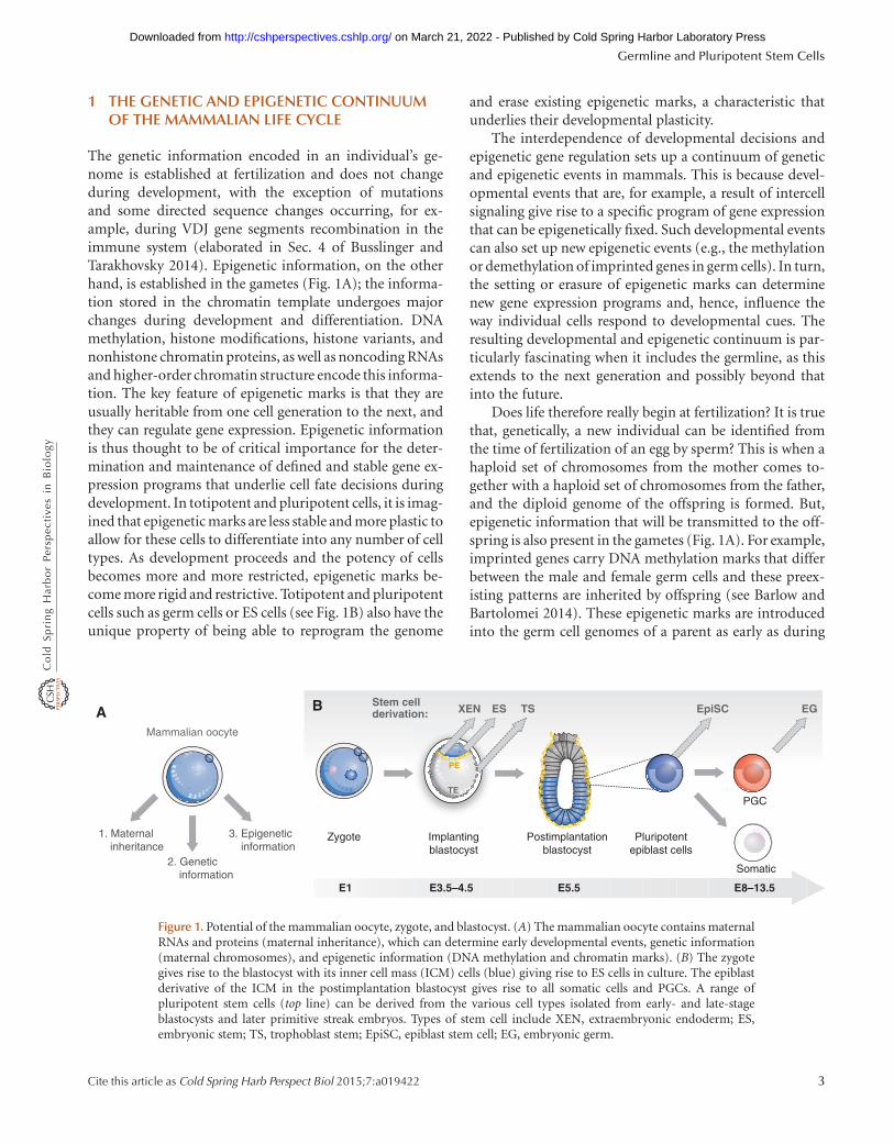
1 THE GENETIC AND EPIGENETIC CONTINUUMOF THE MAMMALIAN LIFE CYCLE
The genetic information encoded in an individual’s ge-nome is established at fertilization and does not changeduring development, with the exception of mutationsand some directed sequence changes occurring, for ex-ample, during VDJ gene segments recombination in theimmune system (elaborated in Sec. 4 of Busslinger andTarakhovsky 2014). Epigenetic information, on the otherhand, is established in the gametes (Fig. 1A); the informa-tion stored in the chromatin template undergoes majorchanges during development and differentiation. DNAmethylation, histone modifications, histone variants, andnonhistone chromatin proteins, as well as noncoding RNAsand higher-order chromatin structure encode this informa-tion. The key feature of epigenetic marks is that they areusually heritable from one cell generation to the next, andthey can regulate gene expression. Epigenetic informationis thus thought to be of critical importance for the deter-mination and maintenance of defined and stable gene ex-pression programs that underlie cell fate decisions duringdevelopment. In totipotent and pluripotent cells, it is imag-ined that epigenetic marks are less stable and more plastic toallow for these cells to differentiate into any number of celltypes. As development proceeds and the potency of cellsbecomes more and more restricted, epigenetic marks be-come more rigid and restrictive. Totipotent and pluripotentcells such as germ cells or ES cells (see Fig. 1B) also have theunique property of being able to reprogram the genome
and erase existing epigenetic marks, a characteristic thatunderlies their developmental plasticity.
The interdependence of developmental decisions andepigenetic gene regulation sets up a continuum of geneticand epigenetic events in mammals. This is because devel-opmental events that are, for example, a result of intercellsignaling give rise to a specific program of gene expressionthat can be epigenetically fixed. Such developmental eventscan also set up new epigenetic events (e.g., the methylationor demethylation of imprinted genes in germ cells). In turn,the setting or erasure of epigenetic marks can determinenew gene expression programs and, hence, influence theway individual cells respond to developmental cues. Theresulting developmental and epigenetic continuum is par-ticularly fascinating when it includes the germline, as thisextends to the next generation and possibly beyond thatinto the future.
Does life therefore really begin at fertilization? It is truethat, genetically, a new individual can be identified fromthe time of fertilization of an egg by sperm? This is when ahaploid set of chromosomes from the mother comes to-gether with a haploid set of chromosomes from the father,and the diploid genome of the offspring is formed. But,epigenetic information that will be transmitted to the off-spring is also present in the gametes (Fig. 1A). For example,imprinted genes carry DNA methylation marks that differbetween the male and female germ cells and these preex-isting patterns are inherited by offspring (see Barlow andBartolomei 2014). These epigenetic marks are introducedinto the germ cell genomes of a parent as early as during
Stem cellderivation:
Mammalian oocyte
1. Maternal inheritance
2. Genetic information
3. Epigenetic information
Zygote Postimplantationblastocyst
Implantingblastocyst
Pluripotentepiblast cells
TS EpiSCESXEN
Somatic
E1 E3.5–4.5 E5.5 E8–13.5
PGC
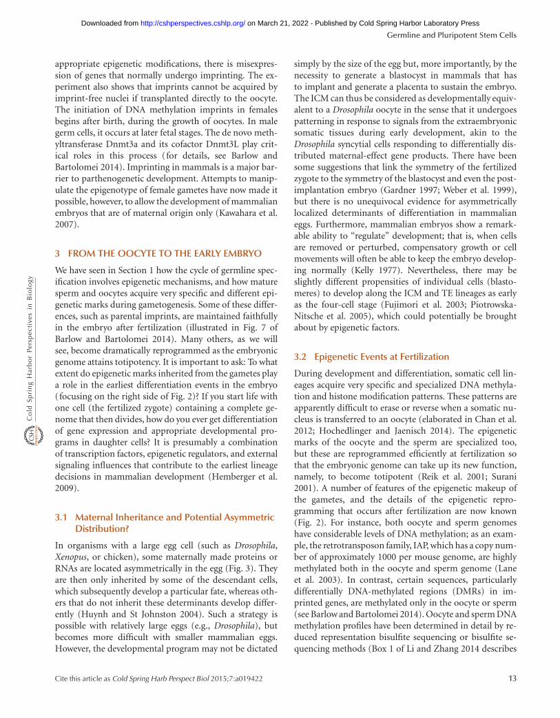
EGA B
TE
PE
Figure 1. Potential of the mammalian oocyte, zygote, and blastocyst. (A) The mammalian oocyte contains maternalRNAs and proteins (maternal inheritance), which can determine early developmental events, genetic information(maternal chromosomes), and epigenetic information (DNA methylation and chromatin marks). (B) The zygotegives rise to the blastocyst with its inner cell mass (ICM) cells (blue) giving rise to ES cells in culture. The epiblastderivative of the ICM in the postimplantation blastocyst gives rise to all somatic cells and PGCs. A range ofpluripotent stem cells (top line) can be derived from the various cell types isolated from early- and late-stageblastocysts and later primitive streak embryos. Types of stem cell include XEN, extraembryonic endoderm; ES,embryonic stem; TS, trophoblast stem; EpiSC, epiblast stem cell; EG, embryonic germ.
Germline and Pluripotent Stem Cells
Cite this article as Cold Spring Harb Perspect Biol 2015;7:a019422 3
on March 21, 2022 - Published by Cold Spring Harbor Laboratory Press http://cshperspectives.cshlp.org/Downloaded from
fetal or early postnatal development. Increasing data areshowing that epigenetic marks may also occasionally betransmitted from one generation to the next resulting intransgenerational epigenetic inheritance (discussed in Sec.2.4 of Blewitt and Whitelaw 2013).
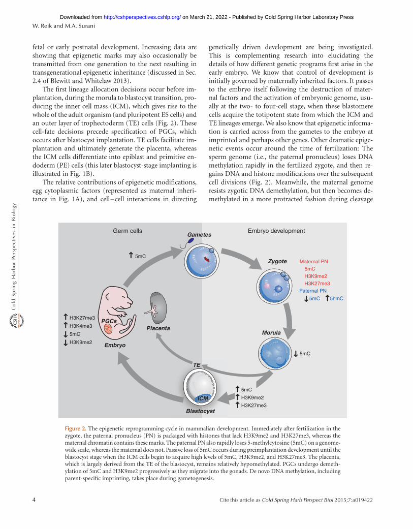
The first lineage allocation decisions occur before im-plantation, during the morula to blastocyst transition, pro-ducing the inner cell mass (ICM), which gives rise to thewhole of the adult organism (and pluripotent ES cells) andan outer layer of trophectoderm (TE) cells (Fig. 2). Thesecell-fate decisions precede specification of PGCs, whichoccurs after blastocyst implantation. TE cells facilitate im-plantation and ultimately generate the placenta, whereasthe ICM cells differentiate into epiblast and primitive en-doderm (PE) cells (this later blastocyst-stage implanting isillustrated in Fig. 1B).
The relative contributions of epigenetic modifications,egg cytoplasmic factors (represented as maternal inheri-tance in Fig. 1A), and cell–cell interactions in directing
genetically driven development are being investigated.This is complementing research into elucidating thedetails of how different genetic programs first arise in theearly embryo. We know that control of development isinitially governed by maternally inherited factors. It passesto the embryo itself following the destruction of mater-nal factors and the activation of embryonic genome, usu-ally at the two- to four-cell stage, when these blastomerecells acquire the totipotent state from which the ICM andTE lineages emerge. We also know that epigenetic informa-tion is carried across from the gametes to the embryo atimprinted and perhaps other genes. Other dramatic epige-netic events occur around the time of fertilization: Thesperm genome (i.e., the paternal pronucleus) loses DNAmethylation rapidly in the fertilized zygote, and then re-gains DNA and histone modifications over the subsequentcell divisions (Fig. 2). Meanwhile, the maternal genomeresists zygotic DNA demethylation, but then becomes de-methylated in a more protracted fashion during cleavage
Germ cellsGametes
5mC
5mC
H3K9me2
PGCs
Embryo
Placenta
Blastocyst
5mC
5mC
Paternal PN
Maternal PN5mCH3K9me2H3K27me3
Zygote
Embryo development
TE
H3K27me3
H3K4me3
5hmC
ooccystBlassttttoto
TE
PGCs
Morula
ICM
5mC
H3K9me2
H3K27me3
Figure 2. The epigenetic reprogramming cycle in mammalian development. Immediately after fertilization in thezygote, the paternal pronucleus (PN) is packaged with histones that lack H3K9me2 and H3K27me3, whereas thematernal chromatin contains these marks. The paternal PN also rapidly loses 5-methylcytosine (5mC) on a genome-wide scale, whereas the maternal does not. Passive loss of 5mC occurs during preimplantation development until theblastocyst stage when the ICM cells begin to acquire high levels of 5mC, H3K9me2, and H3K27me3. The placenta,which is largely derived from the TE of the blastocyst, remains relatively hypomethylated. PGCs undergo demeth-ylation of 5mC and H3K9me2 progressively as they migrate into the gonads. De novo DNA methylation, includingparent-specific imprinting, takes place during gametogenesis.
W. Reik and M.A. Surani
4 Cite this article as Cold Spring Harb Perspect Biol 2015;7:a019422
on March 21, 2022 - Published by Cold Spring Harbor Laboratory Press http://cshperspectives.cshlp.org/Downloaded from
divisions of the early embryo discussed later in Section 3.2(illustrated in Fig. 3 of Li and Zhang 2014). These repro-gramming events may be important to achieve totipotencyof the zygote and pluripotency of ICM cells. Yet, thesesomewhat opposing and distinct epigenetic programslead to an overall loss of gametic epigenetic information,and it is likely that these dynamic reprogramming eventsinteract with the cellular and genetic processes that deter-mine the earliest processes of cell allocation into ICM andTE lineages.
Epigenetic regulation at the later blastocyst stage differsconsiderably between the extraembryonic (TE) and embry-onic lineages (ICM). For example, the overall levels of DNAmethylation are lower in the extraembryonic tissues, andmaintenance of imprinting and imprinted X inactivationcan be different. Ultimately, however, ICM and TE cells, likethe later differentiated PGCs, are largely determined by agenetic program involving transcription factors and, whereappropriate, pluripotency genes. Yet, some of these tran-scription factor genes appear to be epigenetically regulated,which then contributes to the maintenance of cell-fatedecisions.
After implantation of blastocysts, the postimplantationepiblast cells start to undergo modifications of DNA andchromatin. These are the cells that give rise to both germcells and those that will differentiate into diverse cell typesof the body. The earliest signs for the onset of germ cell fateare seen among a small group of cells formed in the earlypostimplantation embryo at E6.25, in response to receivingsignaling molecules that come from other parts of the con-ceptus, primarily the adjacent extraembryonic lineages,which include the cells destined to form the placenta.Germ cell specification entails repression of somatic cellgene products while they begin to show expression of theunique germ-cell-specific genes. This genetically governedfunction is also responsible for initiating epigenetic repro-gramming in early germ cells, leading to the erasure andreestablishment of imprinting, chromosome recombina-tion during meiosis, and reduction divisions to form hap-loid gametes. These various stages of early developmentthus far described clearly illustrate that the epigenetic mod-ifications occur progressively and accompany changes ingenetic programs.
2 MECHANISMS REGULATING GERM CELLSPECIFICATION
The specification of germ cells in animals is one of theearliest events during development, segregating themfrom somatic cells (Surani et al. 2004). Germ cells eventuallygenerate the totipotent state. This section focuses on theprocesses of mouse-germline specification and their subse-
quent maturation as they migrate into the developinggonads.
The events and mechanisms known to direct the pro-cess of germline specification in mice occur in postimplan-tation epiblast cells that emerge from ICM cells after theblastocyst (which contains just three cell types: ICM, TE,and PE) has implanted. PGCs after specification migrateinto the developing gonads and come to reside in their finaldestination, the male or female gonads (illustrated sche-matically on the left side of Fig. 2). A description of theevents and mechanisms regulating early embryogenesis,from early cleavage and morula stages through to the earlyblastocyst, are detailed in Section 3 (corresponding to stag-es of the life cycle shown on the right side of Fig. 2).
2.1 Principles of Germline Development in DifferentAnimal Groups
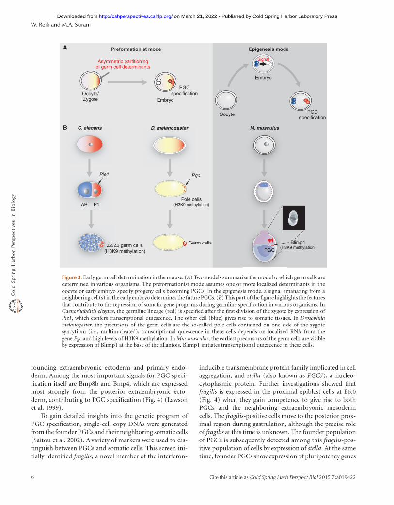
There are two key modes by which a germ cell lineage can beestablished; these are referred to as the preformation mode(this is distinct from the old usage of the word as in pre-formationism) and the epigenesis mode (Extavour andAkam 2003). The first involves the maternal inheritanceof preformed germ cell determinants by specific cells, asoccurs in Caenorhabditis elegans and Drosophila mela-nogaster (Fig. 3) (Leatherman and Jongens 2003; Blackwell2004). In contrast, the epigenesis mode of germ cell spec-ification is a process in which a group of potentially equiv-alent pluripotent cells acquire a germ cell fate in response toinductive signals, whereas the remaining cells acquire thesomatic fate (Lawson and Hage 1994; McLaren 2003). Thismechanism for germ cell specification operates in mice andprobably in other mammals, but also in some other verte-brates such as Axolotls.
2.2 Early Germline Development in Mammals
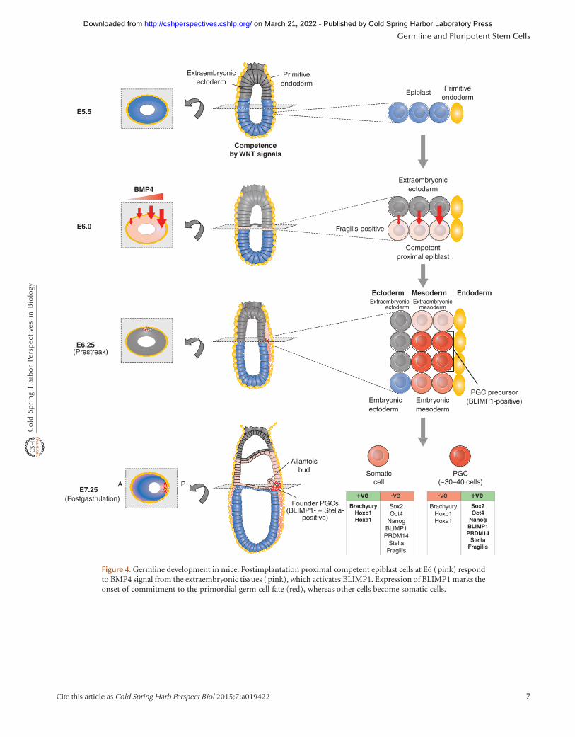
PGCs in mice are first detected at E7.5 (the early bud stage),as a cluster of approximately 30 to 40 cells that constitutethe founder population of the germ cell lineage (Lawsonand Hage 1994; McLaren 2003). They are positive for alka-line phosphatases and located within the extraembryonicmesoderm at the base of the allantois (see Fig. 4). Clonalanalysis reveals that the proximal epiblast cells located ad-jacent to the extraembryonic ectoderm at E6.0–E6.5 (pre-streak and early-streak-stage embryos) give rise to bothPGCs and tissues of the extraembryonic mesoderm (Law-son and Hage 1994). Before PGC specification, how-ever, the epiblast cells acquire a state of competence inresponse to signals that include Wnt signaling (Ohinataet al. 2009). Consequently, proximal epiblast cells formPGCs in response to signaling molecules produced by sur-
Germline and Pluripotent Stem Cells
Cite this article as Cold Spring Harb Perspect Biol 2015;7:a019422 5
on March 21, 2022 - Published by Cold Spring Harbor Laboratory Press http://cshperspectives.cshlp.org/Downloaded from
rounding extraembryonic ectoderm and primary endo-derm. Among the most important signals for PGC speci-fication itself are Bmp8b and Bmp4, which are expressedmost strongly from the posterior extraembryonic ecto-derm, contributing to PGC specification (Fig. 4) (Lawsonet al. 1999).
To gain detailed insights into the genetic program ofPGC specification, single-cell copy DNAs were generatedfrom the founder PGCs and their neighboring somatic cells(Saitou et al. 2002). A variety of markers were used to dis-tinguish between PGCs and somatic cells. This screen ini-tially identified fragilis, a novel member of the interferon-
inducible transmembrane protein family implicated in cellaggregation, and stella (also known as PGC7), a nucleo-cytoplasmic protein. Further investigations showed thatfragilis is expressed in the proximal epiblast cells at E6.0(Fig. 4) when they gain competence to give rise to bothPGCs and the neighboring extraembryonic mesodermcells. The fragilis-positive cells move to the posterior prox-imal region during gastrulation, although the precise roleof fragilis at this time is unknown. The founder populationof PGCs is subsequently detected among this fragilis-pos-itive population of cells by expression of stella. At the sametime, founder PGCs show expression of pluripotency genes
C. elegans D. melanogaster M. musculus
Epigenesis modePreformationist mode
Oocyte
Oocyte/Zygote
Embryo
SignalAsymmetric partitioningof germ cell determinants
Embryo
PGCspecification
PGCspecification
B
A
Pole cells(H3K9 methylation)P1AB
M. musculus
PP1ABAB
C. elegans D. melanogaster
PgcPie1
Z2/Z3 germ cells(H3K9 methylation) PGC
Germ cells Blimp1(H3K9 methylation)
Figure 3. Early germ cell determination in the mouse. (A) Two models summarize the mode by which germ cells aredetermined in various organisms. The preformationist mode assumes one or more localized determinants in theoocyte or early embryo specify progeny cells becoming PGCs. In the epigenesis mode, a signal emanating from aneighboring cell(s) in the early embryo determines the future PGCs. (B) This part of the figure highlights the featuresthat contribute to the repression of somatic gene programs during germline specification in various organisms. InCaenorhabditis elegans, the germline lineage (red) is specified after the first division of the zygote by expression ofPie1, which confers transcriptional quiescence. The other cell (blue) gives rise to somatic tissues. In Drosophilamelanogaster, the precursors of the germ cells are the so-called pole cells contained on one side of the zygotesyncytium (i.e., multinucleated); transcriptional quiescence in these cells depends on localized RNA from thegene Pgc and high levels of H3K9 methylation. In Mus musculus, the earliest precursors of the germ cells are visibleby expression of Blimp1 at the base of the allantois. Blimp1 initiates transcriptional quiescence in these cells.
W. Reik and M.A. Surani
6 Cite this article as Cold Spring Harb Perspect Biol 2015;7:a019422
on March 21, 2022 - Published by Cold Spring Harbor Laboratory Press http://cshperspectives.cshlp.org/Downloaded from
BrachyuryHoxb1Hoxa1
Sox2Oct4
NanogBLIMP1PRDM14
StellaFragilis
-ve+ve
Competenceby WNT signals
Competentproximal epiblast
Extraembryonicectoderm
Epiblast
BMP4
E5.5
E7.25
E6.25
E6.0
Extraembryonicectoderm
Primitiveendoderm
A P
Fragilis-positive
Ectoderm EndodermMesoderm
Primitiveendoderm
Somaticcell
PGC(~30–40 cells)
BrachyuryHoxb1Hoxa1
Sox2Oct4
NanogBLIMP1PRDM14
StellaFragilis
-ve +ve
PGC precursor(BLIMP1-positive)Embryonic
mesodermEmbryonicectoderm
Extraembryonicectoderm
Extraembryonicmesoderm
(Postgastrulation)
(Prestreak)
Founder PGCs(BLIMP1- + Stella-
positive)
Allantoisbud
Figure 4. Germline development in mice. Postimplantation proximal competent epiblast cells at E6 (pink) respondto BMP4 signal from the extraembryonic tissues (pink), which activates BLIMP1. Expression of BLIMP1 marks theonset of commitment to the primordial germ cell fate (red), whereas other cells become somatic cells.
Germline and Pluripotent Stem Cells
Cite this article as Cold Spring Harb Perspect Biol 2015;7:a019422 7
on March 21, 2022 - Published by Cold Spring Harbor Laboratory Press http://cshperspectives.cshlp.org/Downloaded from
including Sox2, Oct4, and Nanog (see Sec. 4.2 for a moredetailed description), suggesting that PGCs reacquire anunderlying pluripotency, which is lost in the neighboringsomatic cells (Fig. 4). In contrast, the founder PGCs showrepression of some genes including Brachyury, Hoxb1, andHoxa1, which are, at this time, significantly up-regulated insomatic neighbors. The repression of Hox genes amongothers is part of an important mechanism that underliesrepression of the somatic cell fate in founder PGCs (elab-orated in Sec. 2.4; Saitou et al. 2002).
Based on the analysis of the emergence of founderPGCs, it is evident that, as in other organisms, repressionof the somatic program is likely to be a key feature of PGCspecification in mice (Seydoux and Strome 1999; Blackwell2004; Surani et al. 2004). The lysine (K) methyltransferase(KMT) class of histone-modifying enzymes or “writers”(for more detail, see Allis et al. 2014; Cheng 2014) wereanalyzed for differential expression between PGCs andneighboring somatic cells. KMTs modify histone lysine res-idues through the addition of one to three methyl groups.Methylation of histones H3 and H4 at positions H3K9 andH4K20 is generally associated with repressive chromatinregions. Methylation at H3K4 and H3K36, however, gener-ally correlates with active transcription. Some of the KMTgenes, such as G9a, Pfm1, Set1, and Ezh2, were detectedboth in the founder PGCs and the somatic cells. However,one of these genes, Blimp1 (B lymphocyte maturation-in-duced protein-1 or prdm1), showed expression exclusively inthe founder PGCs and not in the neighboring somatic cellsat E7.5 (Ohinata et al. 2005). Blimp1 is a known transcrip-tional repressor with a SET/PR domain (a domain thattypically acts as a methyltransferase), a proline-rich regionthat can recruit Groucho and HDAC2, five C2H2 zinc fin-gers that can form a complex with Prmt5, and an acidic tail(Gyory et al. 2004; Sciammas and Davis 2004, Ancelin et al.2006). Blimp1 was first identified for its role during speci-fication of plasma cells following repression of the B-cellprogram in the precursor cells (Turner et al. 1994). Blimp1is indeed widely expressed during mouse development.
Detailed analysis of Blimp1 in early mouse embryosled to some unexpected findings. Among them was the dis-covery that Blimp1 expression commences in the proximalepiblast cells at E6.25 at the onset of gastrulation, initiallyin only four to six cells that are in direct contact with theextraembryonic ectoderm cells (Fig. 4) (Ohinata et al. 2005,2009). Blimp1 expression is detected at one end of the shortanterior–posterior axis in a region that is destined to formthe posterior proximal region. The number of Blimp1-pos-itive cells increase progressively so that there are approxi-mately 20 cells at the midstreak stage that are seen to form atight cluster in the posterior proximal region at E6.75. Atthe E7.5 early bud stage, the number of Blimp1-positive
cells increases to approximately 40. These cells constitutethe founder population of PGCs, and show expression ofthe classical alkaline phosphatase PGC marker and com-mence expression of stella (Fig. 4). A genetic lineage tracingexperiment confirmed that all the Blimp1-positive cellsoriginating in the epiblast from E6.25 onward are indeedlineage restricted PGC precursor cells. These data contrastwith the previous hypothesis, based on clonal analysis,which suggested that the proximal epiblast cells at E6.0–E6.5 are not lineage restricted to give rise exclusively toPGCs because clonal descendants of individual cells couldgive rise to both a somatic and germ cell (Lawson and Hage1994; McLaren and Lawson 2005). A likely explanation forthis discrepancy may be that in the clonal analysis, themarked cells may initially have been negative for Blimp1and they subsequently divided to generate a positive cellthat gave rise to PGCs, whereas the daughter cell produced asomatic descendant. The mechanism that regulates the ac-cretion of Blimp1-positive cells is currently unknown.
2.3 The Role of Blimp1 in Specification of PGCs
Analysis of the role of Blimp1 in PGC specification hasgenerated insights into the underlying mechanism ofgerm cell specification in mice. Loss of function of Blimp1showed that this is a key determinant of PGC specificationin mice (Ohinata et al. 2005; Vincent et al. 2005). At E7.5,Blimp1 mutant embryos contain an aberrant cluster of ap-proximately 20 PGC-like cells, unlike control embryos inwhich the PGCs continue to proliferate and commencemigration out of the cluster. Furthermore, the number ofaberrant PGC-like cells fails to increase when examined atE8.5 (Ohinata et al. 2005).
Single-cell analysis of mutant PGC cells revealed a lackof consistent repression of Hox genes. Therefore, it is likelythat Blimp1 has a role in the repression of the somaticprogram in founder PGCs. There was also inconsistencyin the up-regulation of PGC-specific genes such as stellaand Nanos3, and some pluripotency-specific genes such asSox2 in mutants. These findings stress that Blimp1 has acritical role as a transcriptional regulator during PGC spec-ification and in the prevention of these cells from acquiringa somatic cell fate.
Studies on B cells have revealed that Blimp1 is necessaryto induce differentiation into plasma cells through repres-sion of key molecules that maintain B-cell identity (seeSec. 3.2 of Busslinger and Tarakhovsky 2014 for details; Tur-ner et al. 1994; Sciammas and Davis 2004). It does thisthrough the formation of a Groucho and HDAC2 repressorcomplex (Ren et al. 1999). Its zinc fingers also seem impor-tant for the formation of a complex with G9a (Gyory et al.2004), a histone KMT that is required for H3K9me2. Blimp1,
W. Reik and M.A. Surani
8 Cite this article as Cold Spring Harb Perspect Biol 2015;7:a019422
on March 21, 2022 - Published by Cold Spring Harbor Laboratory Press http://cshperspectives.cshlp.org/Downloaded from
which itself contains a SET/PR domain that typically actsas a methyltransferase, has no known KMT activity, andhow it functions during PGC specification is unknown.
Blimp1 is an evolutionarily conserved gene in both ver-tebrates and invertebrates and it has a variety of functions.For example, it has a role in the development of severallineages in vertebrates such as the zebrafish and Xenopus(de Souza et al. 1999; Roy and Ng 2004; Hernandez-Lagu-nas et al. 2005), although not specifically in germ cell spec-ification. This implies that the gene has acquired a new rolein PGC specification in mice and perhaps in all mammals.For this highly conserved gene, it suggests that additionalcontrol elements must have evolved to drive its expressionin PGC precursors and founder cells.
Analysis and comparison of Blimp1 mutant versus con-trol PGC cells led to the discovery of a second key regulatorof PGC specification called Prdm14, which like Blimp1 isanother PR/SET domain family member (Yamaji et al.2008). Blimp1, together with Prdm14, is involved in therepression of the somatic program and onset of epigeneticreprogramming in PGCs, leading to global DNA demeth-ylation described in Section 2.5.
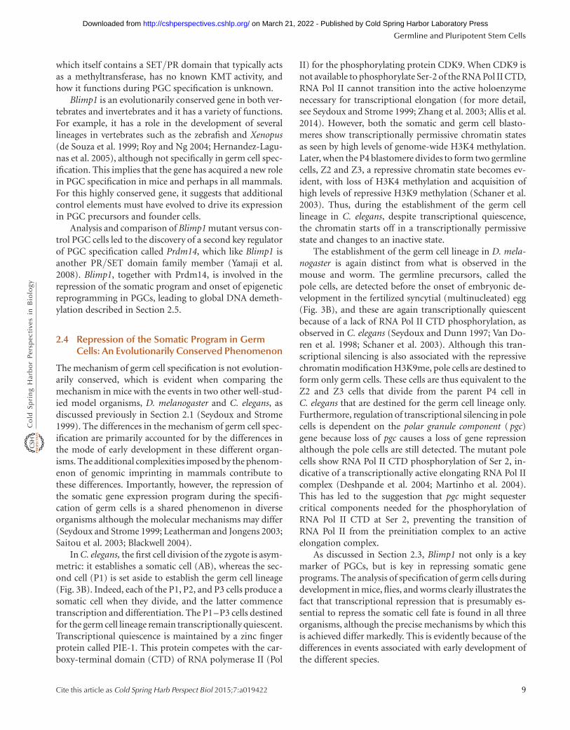
2.4 Repression of the Somatic Program in GermCells: An Evolutionarily Conserved Phenomenon
The mechanism of germ cell specification is not evolution-arily conserved, which is evident when comparing themechanism in mice with the events in two other well-stud-ied model organisms, D. melanogaster and C. elegans, asdiscussed previously in Section 2.1 (Seydoux and Strome1999). The differences in the mechanism of germ cell spec-ification are primarily accounted for by the differences inthe mode of early development in these different organ-isms. The additional complexities imposed by the phenom-enon of genomic imprinting in mammals contribute tothese differences. Importantly, however, the repression ofthe somatic gene expression program during the specifi-cation of germ cells is a shared phenomenon in diverseorganisms although the molecular mechanisms may differ(Seydoux and Strome 1999; Leatherman and Jongens 2003;Saitou et al. 2003; Blackwell 2004).
In C. elegans, the first cell division of the zygote is asym-metric: it establishes a somatic cell (AB), whereas the sec-ond cell (P1) is set aside to establish the germ cell lineage(Fig. 3B). Indeed, each of the P1, P2, and P3 cells produce asomatic cell when they divide, and the latter commencetranscription and differentiation. The P1–P3 cells destinedfor the germ cell lineage remain transcriptionally quiescent.Transcriptional quiescence is maintained by a zinc fingerprotein called PIE-1. This protein competes with the car-boxy-terminal domain (CTD) of RNA polymerase II (Pol
II) for the phosphorylating protein CDK9. When CDK9 isnot available to phosphorylate Ser-2 of the RNA Pol II CTD,RNA Pol II cannot transition into the active holoenzymenecessary for transcriptional elongation (for more detail,see Seydoux and Strome 1999; Zhang et al. 2003; Allis et al.2014). However, both the somatic and germ cell blasto-meres show transcriptionally permissive chromatin statesas seen by high levels of genome-wide H3K4 methylation.Later, when the P4 blastomere divides to form two germlinecells, Z2 and Z3, a repressive chromatin state becomes ev-ident, with loss of H3K4 methylation and acquisition ofhigh levels of repressive H3K9 methylation (Schaner et al.2003). Thus, during the establishment of the germ celllineage in C. elegans, despite transcriptional quiescence,the chromatin starts off in a transcriptionally permissivestate and changes to an inactive state.
The establishment of the germ cell lineage in D. mela-nogaster is again distinct from what is observed in themouse and worm. The germline precursors, called thepole cells, are detected before the onset of embryonic de-velopment in the fertilized syncytial (multinucleated) egg(Fig. 3B), and these are again transcriptionally quiescentbecause of a lack of RNA Pol II CTD phosphorylation, asobserved in C. elegans (Seydoux and Dunn 1997; Van Do-ren et al. 1998; Schaner et al. 2003). Although this tran-scriptional silencing is also associated with the repressivechromatin modification H3K9me, pole cells are destined toform only germ cells. These cells are thus equivalent to theZ2 and Z3 cells that divide from the parent P4 cell inC. elegans that are destined for the germ cell lineage only.Furthermore, regulation of transcriptional silencing in polecells is dependent on the polar granule component ( pgc)gene because loss of pgc causes a loss of gene repressionalthough the pole cells are still detected. The mutant polecells show RNA Pol II CTD phosphorylation of Ser 2, in-dicative of a transcriptionally active elongating RNA Pol IIcomplex (Deshpande et al. 2004; Martinho et al. 2004).This has led to the suggestion that pgc might sequestercritical components needed for the phosphorylation ofRNA Pol II CTD at Ser 2, preventing the transition ofRNA Pol II from the preinitiation complex to an activeelongation complex.
As discussed in Section 2.3, Blimp1 not only is a keymarker of PGCs, but is key in repressing somatic geneprograms. The analysis of specification of germ cells duringdevelopment in mice, flies, and worms clearly illustrates thefact that transcriptional repression that is presumably es-sential to repress the somatic cell fate is found in all threeorganisms, although the precise mechanisms by which thisis achieved differ markedly. This is evidently because of thedifferences in events associated with early development ofthe different species.
Germline and Pluripotent Stem Cells
Cite this article as Cold Spring Harb Perspect Biol 2015;7:a019422 9
on March 21, 2022 - Published by Cold Spring Harbor Laboratory Press http://cshperspectives.cshlp.org/Downloaded from
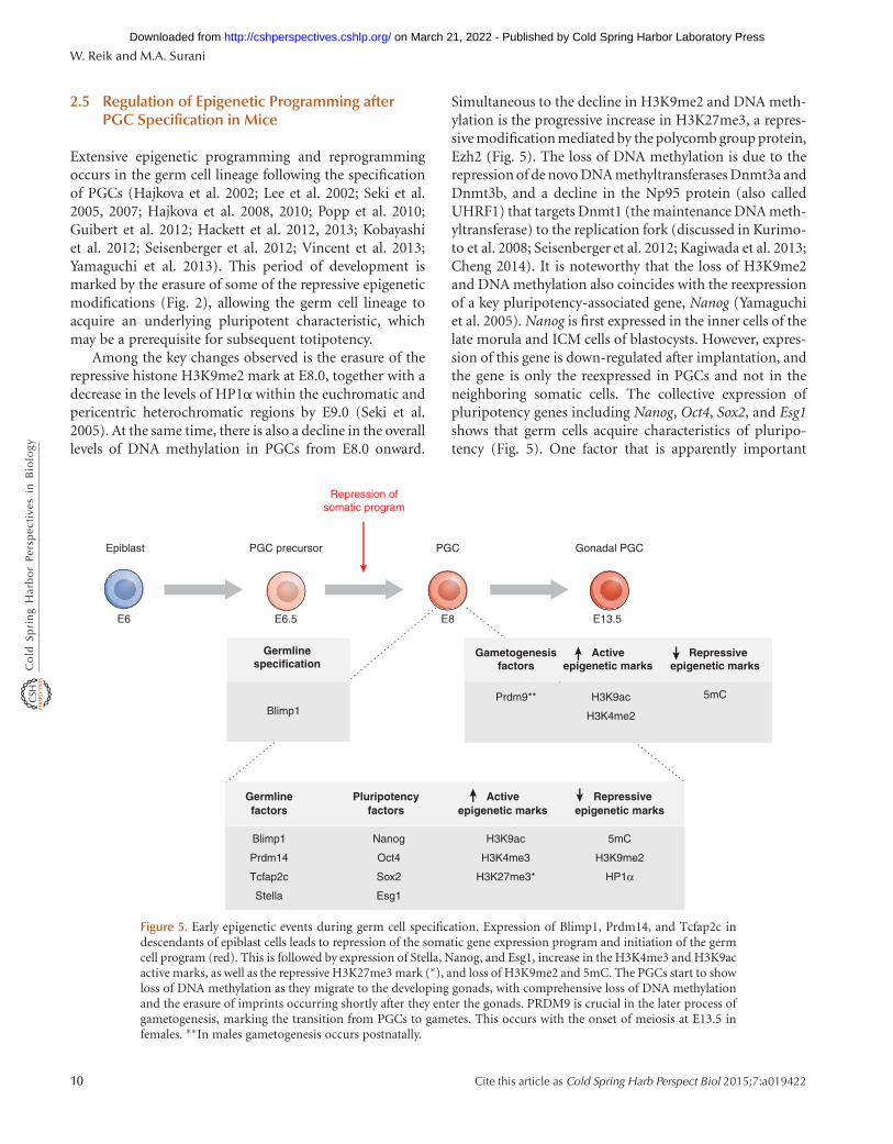
2.5 Regulation of Epigenetic Programming afterPGC Specification in Mice
Extensive epigenetic programming and reprogrammingoccurs in the germ cell lineage following the specificationof PGCs (Hajkova et al. 2002; Lee et al. 2002; Seki et al.2005, 2007; Hajkova et al. 2008, 2010; Popp et al. 2010;Guibert et al. 2012; Hackett et al. 2012, 2013; Kobayashiet al. 2012; Seisenberger et al. 2012; Vincent et al. 2013;Yamaguchi et al. 2013). This period of development ismarked by the erasure of some of the repressive epigeneticmodifications (Fig. 2), allowing the germ cell lineage toacquire an underlying pluripotent characteristic, whichmay be a prerequisite for subsequent totipotency.
Among the key changes observed is the erasure of therepressive histone H3K9me2 mark at E8.0, together with adecrease in the levels of HP1a within the euchromatic andpericentric heterochromatic regions by E9.0 (Seki et al.2005). At the same time, there is also a decline in the overalllevels of DNA methylation in PGCs from E8.0 onward.
Simultaneous to the decline in H3K9me2 and DNA meth-ylation is the progressive increase in H3K27me3, a repres-sive modification mediated by the polycomb group protein,Ezh2 (Fig. 5). The loss of DNA methylation is due to therepression of de novo DNA methyltransferases Dnmt3a andDnmt3b, and a decline in the Np95 protein (also calledUHRF1) that targets Dnmt1 (the maintenance DNA meth-yltransferase) to the replication fork (discussed in Kurimo-to et al. 2008; Seisenberger et al. 2012; Kagiwada et al. 2013;Cheng 2014). It is noteworthy that the loss of H3K9me2and DNA methylation also coincides with the reexpressionof a key pluripotency-associated gene, Nanog (Yamaguchiet al. 2005). Nanog is first expressed in the inner cells of thelate morula and ICM cells of blastocysts. However, expres-sion of this gene is down-regulated after implantation, andthe gene is only the reexpressed in PGCs and not in theneighboring somatic cells. The collective expression ofpluripotency genes including Nanog, Oct4, Sox2, and Esg1shows that germ cells acquire characteristics of pluripo-tency (Fig. 5). One factor that is apparently important
Gonadal PGCPGCPGC precursorEpiblast
Repressiveepigenetic marks
Activeepigenetic marks
Pluripotencyfactors
5mC
H3K9me2
HP1α
H3K9ac
H3K4me3
H3K27me3*
Nanog
Oct4
Sox2
Esg1
Blimp1
Prdm14
Tcfap2c
Stella
Germlinefactors
Germlinespecification
Repressiveepigenetic marks
5mC
Blimp1
Activeepigenetic marks
H3K9ac
H3K4me2
Repression ofsomatic program
E13.5E8E6.5E6
Prdm9**
Gametogenesisfactors
Figure 5. Early epigenetic events during germ cell specification. Expression of Blimp1, Prdm14, and Tcfap2c indescendants of epiblast cells leads to repression of the somatic gene expression program and initiation of the germcell program (red). This is followed by expression of Stella, Nanog, and Esg1, increase in the H3K4me3 and H3K9acactive marks, as well as the repressive H3K27me3 mark (∗), and loss of H3K9me2 and 5mC. The PGCs start to showloss of DNA methylation as they migrate to the developing gonads, with comprehensive loss of DNA methylationand the erasure of imprints occurring shortly after they enter the gonads. PRDM9 is crucial in the later process ofgametogenesis, marking the transition from PGCs to gametes. This occurs with the onset of meiosis at E13.5 infemales. ∗∗In males gametogenesis occurs postnatally.
W. Reik and M.A. Surani
10 Cite this article as Cold Spring Harb Perspect Biol 2015;7:a019422
on March 21, 2022 - Published by Cold Spring Harbor Laboratory Press http://cshperspectives.cshlp.org/Downloaded from
for expression of pluripotency genes in early PGCs is theH3K27me3 demethylase, Utx, likely counteracting the re-pressive effects of the overall increase in H3K27me3 (Man-sour et al. 2012). The expression of the pluripotencynetwork in PGCs is extensive and comparable to ES cells(Seisenberger et al. 2012).
Additional extensive epigenetic programming eventsensue when PGCs enter into the developing gonads (Suraniet al. 2004). First, there are increases in H3K4me2 methyl-ation and H3K9 acetylation, which are characteristic ofpermissive chromatin states, excluding H3K9 methylation.In addition, there is very extensive genome-wide DNA de-methylation (Fig. 5) that includes the erasure of parentalimprints and methylation in single-copy genes. In femaleembryos, the inactive X chromosome is also reactivated atthis time. Genome-wide demethylation is likely to occur bya combination of active and passive mechanisms, includ-ing down-regulation of Np95 (discussed in Kurimoto et al.2008; Cheng 2014), the activation-induced deaminase(AID) and thymine-DNA glycosylase (Popp et al. 2010;Cortellino et al. 2011), and TET1 and TET2, which arepotentially coupled to the base excision repair pathway(see Fig. 6 of Li and Zhang 2014 and Feng et al. 2010;Hajkova et al. 2010; Hackett et al. 2012; Saitou et al.2012; Dawlaty et al. 2013; Hackett and Surani 2013; Vincentet al. 2013; Yamaguchi et al. 2013).
Not all epigenetic marks are completely removed duringgerm cell development, despite effective mechanisms oper-ating to erase “acquired” epigenetic modifications. Forexample, DNA methylation of the intracisternal A particle(IAP) retrotransposon family and some 200 or so othergenomic locations is only partially reprogrammed (Laneet al. 2003; Popp et al. 2010; Guibert et al. 2012; Seisenbergeret al. 2012; Hackett and Surani 2013). When some epige-netic marks are incompletely removed during gameto-genesis, this can apparently lead to epigenetic inheritancethrough the germline, of which there are a number of ex-amples now in mammals (e.g., see Fig. 6 in Blewitt andWhitelaw 2013 and Chong and Whitelaw 2004), and thiscould potentially explain the transgenerational epigeneticinheritance of some metabolic phenotypes (Ferguson-Smith and Patti 2011). How widespread this phenome-non is and how many gene loci it involves needs to beestablished.
2.6 The Germline and Stem Cells: A ReversiblePhenotype
Pluripotent stem cells can be derived from ICM and germcells (Fig. 1B). More specifically, pluripotent EG cells can bemade from PGCs when cultured in the presence of FGF2(Matsui et al. 1992; Resnick et al. 1992). They are, in many
respects, similar to pluripotent ES cells (discussed moreextensively in Sec. 4) except that EG cells may show erasureof parental imprints during their derivation (Tada et al.1998; Leitch et al. 2010). Recent studies have shown thatEG cells can also be derived from rat PGCs (Leitch et al.2010).
As PGCs show some characteristics of pluripotencywhile retaining unipotency in that they are only able toform sperm or eggs, it is probable that mechanisms existto allow PGCs to retain their distinct lineage-specific char-acteristics. How this is achieved is as yet unclear, but it ispossible that Blimp1 may have a continuing role followingthe initial specification of PGCs. During the derivation ofEG cells, it is assumed that unipotent restriction is relievedand they acquire an overtly pluripotent character with theability to differentiate into many distinct cell types, whichseldom occurs with germ cells in vivo. It is noteworthy thatthe derivation of EG cells becomes progressively less effi-cient, however, when later-stage PGCs from E11.5 andE12.5 are used. This further suggests a change in the char-acteristics of these cells from E11.5 when they begin theirdifferentiation pathway toward definitive male and femalegerm cells.
2.6.1 Development of Germ Cells from PluripotentES Cells
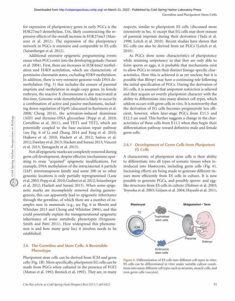
A characteristic of pluripotent stem cells is their abilityto differentiate into all types of somatic tissues when in-troduced into blastocysts, including germ cells (Fig. 6).Increasing efforts are being made to generate different tis-sues more efficiently from ES cells in culture. It is nowpossible to generate PGCs, and possibly sperm- and egg-like structures from ES cells in culture (Hubner et al. 2003;Toyooka et al. 2003; Geijsen et al. 2004; Hayashi et al. 2011;
Blastocyst Midgestation - Term
TE
Trophoblaststem cells
Embryonicstem cells
PlacentaEmbryo
ICM
Figure 6. Differentiation of ES cells into different cell types in vitro.ES cells can be differentiated in vitro under suitable culture condi-tions into many different cell types such as neurons, muscle cells, andeven germ cells (oocytes).
Germline and Pluripotent Stem Cells
Cite this article as Cold Spring Harb Perspect Biol 2015;7:a019422 11
on March 21, 2022 - Published by Cold Spring Harbor Laboratory Press http://cshperspectives.cshlp.org/Downloaded from
Vincent et al. 2011). This has opened up the possibility ofstudying the process of germ cell specification in vitro, withthe hope of being able to determine the precise mechanismsinvolved, aided by an ever-increasing knowledge base of thegenetic programs that govern PGC and gamete cell func-tion. The ability to in vitro differentiate PGCs from ES cellsmay also provide a model system to examine the regulationof epigenetic reprogramming in this lineage. Such an ap-proach could ultimately advance our understanding of thehuman germ cell lineage, which, for ethical reasons, hasbeen hard to research to date. Furthermore, if it becomespossible to direct differentiation toward human oocytesfrom cultured ES cells, it may be possible to use them for“therapeutic” cloning, circumventing the need for donoroocytes that are difficult to obtain. These oocytes couldthen be used for somatic nuclear transplantation to gener-ate blastocysts, and subsequently to derive ES cells from.This is because somatic nuclei undergo reprogramming tototipotency when transplanted into oocytes (elaborated inHochedlinger and Jaenisch 2014). This procedure is likelyto complement the currently used direct derivation of iPScells from somatic tissues.
The use of human embryos and ES cells in researchand therapy does raise many ethical issues. A variety ofguidelines and regulations exist in different countries tomonitor research in this area. Within these ethical frame-works, it is hoped that the generation of viable gametesfrom ES cells, if possible, may lead to advances in repro-ductive medicine.
2.7 From PGCs to Gametes
The next stage in the development of the germ cell lineage isthe initiation of gametogenesis and entry of germ cells intomeiosis. The gonadal somatic environment regulates thetiming of this event. In females, germ cells arrest in meioticprophase whereas male germ cells enter into mitotic arrest.A number of environmental signals dictate whether germcells enter meiosis or not. Recently, a novel gene, Prdm9(also called Meisetz), another PR/SET domain familymember, was identified and shown to play a crucial role ininitiating meiosis (Hayashi et al. 2005). The PR/SET do-main has proven H3K4me3 catalytic activity and also pos-sesses multiple zinc-fingers. Expression of Prdm9 is specificto germ cells, and is detected at the time of entry into mei-otic prophase in females at E13.5 and in postnatal testis. It islikely that Prdm9/PRDM9 in mouse and human germ cellsmay introduce epigenetic marks or “hot spots” that consti-tute narrow segments of the genome as sites for meioticrecombination. Mutation in Prdm9 results in sterility inboth males and females, demonstrating its essential role ingerm cells. The mutant germ cells show marked deficiency
in the DNA double-strand break repair pathway and pairingof homologous chromosomes during meiosis. These stud-ies suggest a significant role for epigenetic mechanismsin germ cells during meiosis (Baudat et al. 2010; Parvanovet al. 2010).
Extensive chromatin modifications continue duringspermatogenesis. Eventually, the somatic linker histonesare replaced by testis-specific variants (Kimmins andSassone-Corsi 2005), followed by the replacement of mosthistones with protamines. Studies have shown that Suv39,an H3K9 histone methyltransferase, is involved in generepression and chromosome pairing. Two such SET-do-main proteins, Suv39h1 and Suv39h2, have roles in malegerm cells, the latter being expressed preferentially in thetestis, and accumulating in the chromatin of the sex vesicle(i.e., XY chromosome pair). Mutations in both Suv39h1and Suv39h2 result in infertility because of the arrest ofspermatogenic cells (Peters et al. 2001). In addition, thereis also a chromatoid body; this is a cloud-like cytoplasmicstructure present in male germ cells. It is an RNA-process-ing body consisting of Dicer and Argonaute proteinsand microRNAs, a germ-cell-specific cytoplasmic organellethat interacts with the nucleus and contains compactedmessenger RNA.
Noncoding RNAs, the RNA interference (RNAi)machinery, and histone lysine methyltransferases are impli-cated in the process of germ cell renewal during spermato-genesis. The members of the Piwi/Argonaute (called Miwiin mice) family have been reported to play a role in RNAipheomena. Loss of Miwi-like proteins (Mili) results in ste-rility in males (Kuramochi-Miyagawa et al. 2004), causingelevated expression of retrotransposon transcripts, IAP, andLine 1. The involvement of Miwi-like proteins in their re-pression has been directly demonstrated shown through thepiRNA pathway (Siomi et al. 2011).
Interestingly, pluripotent stem cells from spermatogo-nial stem cells can even be recovered from adult mousetestis (Kanatsu-Shinohara et al. 2004; Guan et al. 2006).These cells may be maintained in culture indefinitely, butunlike ES cells, they have a paternal (androgenetic) imprint.Nonetheless, they can differentiate into a variety of somaticcell types in vitro and in vivo, and can contribute to thegermline in vivo. These cells thus provide an important toolto study many aspects of spermatogenesis, including therole of epigenetic mechanisms in regulating their stemnessand capability for differentiating into male gametes.
Erasure of imprints in early germ cells leads to epige-netically equivalent parental chromosomes for the first andonly time in the life of mammals. Transplantation of such“imprint-free” nuclei directly into oocytes leads to the de-velopment of embryos that are aberrant and die at earlyembryonic stages. This is presumably because without the
W. Reik and M.A. Surani
12 Cite this article as Cold Spring Harb Perspect Biol 2015;7:a019422
on March 21, 2022 - Published by Cold Spring Harbor Laboratory Press http://cshperspectives.cshlp.org/Downloaded from
appropriate epigenetic modifications, there is misexpres-sion of genes that normally undergo imprinting. The ex-periment also shows that imprints cannot be acquired byimprint-free nuclei if transplanted directly to the oocyte.The initiation of DNA methylation imprints in femalesbegins after birth, during the growth of oocytes. In malegerm cells, it occurs at later fetal stages. The de novo meth-yltransferase Dnmt3a and its cofactor Dnmt3L play crit-ical roles in this process (for details, see Barlow andBartolomei 2014). Imprinting in mammals is a major bar-rier to parthenogenetic development. Attempts to manip-ulate the epigenotype of female gametes have now made itpossible, however, to allow the development of mammalianembryos that are of maternal origin only (Kawahara et al.2007).
3 FROM THE OOCYTE TO THE EARLY EMBRYO
We have seen in Section 1 how the cycle of germline spec-ification involves epigenetic mechanisms, and how maturesperm and oocytes acquire very specific and different epi-genetic marks during gametogenesis. Some of these differ-ences, such as parental imprints, are maintained faithfullyin the embryo after fertilization (illustrated in Fig. 7 ofBarlow and Bartolomei 2014). Many others, as we willsee, become dramatically reprogrammed as the embryonicgenome attains totipotency. It is important to ask: To whatextent do epigenetic marks inherited from the gametes playa role in the earliest differentiation events in the embryo(focusing on the right side of Fig. 2)? If you start life withone cell (the fertilized zygote) containing a complete ge-nome that then divides, how do you ever get differentiationof gene expression and appropriate developmental pro-grams in daughter cells? It is presumably a combinationof transcription factors, epigenetic regulators, and externalsignaling influences that contribute to the earliest lineagedecisions in mammalian development (Hemberger et al.2009).
3.1 Maternal Inheritance and Potential AsymmetricDistribution?
In organisms with a large egg cell (such as Drosophila,Xenopus, or chicken), some maternally made proteins orRNAs are located asymmetrically in the egg (Fig. 3). Theyare then only inherited by some of the descendant cells,which subsequently develop a particular fate, whereas oth-ers that do not inherit these determinants develop differ-ently (Huynh and St Johnston 2004). Such a strategy ispossible with relatively large eggs (e.g., Drosophila), butbecomes more difficult with smaller mammalian eggs.However, the developmental program may not be dictated
simply by the size of the egg but, more importantly, by thenecessity to generate a blastocyst in mammals that hasto implant and generate a placenta to sustain the embryo.The ICM can thus be considered as developmentally equiv-alent to a Drosophila oocyte in the sense that it undergoespatterning in response to signals from the extraembryonicsomatic tissues during early development, akin to theDrosophila syncytial cells responding to differentially dis-tributed maternal-effect gene products. There have beensome suggestions that link the symmetry of the fertilizedzygote to the symmetry of the blastocyst and even the post-implantation embryo (Gardner 1997; Weber et al. 1999),but there is no unequivocal evidence for asymmetricallylocalized determinants of differentiation in mammalianeggs. Furthermore, mammalian embryos show a remark-able ability to “regulate” development; that is, when cellsare removed or perturbed, compensatory growth or cellmovements will often be able to keep the embryo develop-ing normally (Kelly 1977). Nevertheless, there may beslightly different propensities of individual cells (blasto-meres) to develop along the ICM and TE lineages as earlyas the four-cell stage (Fujimori et al. 2003; Piotrowska-Nitsche et al. 2005), which could potentially be broughtabout by epigenetic factors.
3.2 Epigenetic Events at Fertilization
During development and differentiation, somatic cell lin-eages acquire very specific and specialized DNA methyla-tion and histone modification patterns. These patterns areapparently difficult to erase or reverse when a somatic nu-cleus is transferred to an oocyte (elaborated in Chan et al.2012; Hochedlinger and Jaenisch 2014). The epigeneticmarks of the oocyte and the sperm are specialized too,but these are reprogrammed efficiently at fertilization sothat the embryonic genome can take up its new function,namely, to become totipotent (Reik et al. 2001; Surani2001). A number of features of the epigenetic makeup ofthe gametes, and the details of the epigenetic repro-gramming that occurs after fertilization are now known(Fig. 2). For instance, both oocyte and sperm genomeshave considerable levels of DNA methylation; as an exam-ple, the retrotransposon family, IAP, which has a copy num-ber of approximately 1000 per mouse genome, are highlymethylated both in the oocyte and sperm genome (Laneet al. 2003). In contrast, certain sequences, particularlydifferentially DNA-methylated regions (DMRs) in im-printed genes, are methylated only in the oocyte or sperm(see Barlow and Bartolomei 2014). Oocyte and sperm DNAmethylation profiles have been determined in detail by re-duced representation bisulfite sequencing or bisulfite se-quencing methods (Box 1 of Li and Zhang 2014 describes
Germline and Pluripotent Stem Cells
Cite this article as Cold Spring Harb Perspect Biol 2015;7:a019422 13
on March 21, 2022 - Published by Cold Spring Harbor Laboratory Press http://cshperspectives.cshlp.org/Downloaded from
the main DNA methylation assay techniques; Smallwoodet al. 2011; Kobayashi et al. 2012; Smith et al. 2012).
The oocyte genome has high levels of histone modifi-cations, both active ones (e.g., H3K9 acetylation and H3K4methylation) and repressive ones (e.g., H3K9 methylation,and H3K27 methylation; Morgan et al. 2005). At this pointbefore fertilization, the oocyte genome is transcriptionallyinactive, but contains maternally inherited transcripts andproteins needed during the first few cleavage divisions,including those required for important reprogrammingevents (Fig. 1A). The sperm genome, in contrast, is highlyspecialized: The majority of the histones have been replacedduring spermatogenesis by highly basic protamines, whichmay facilitate the packaging of DNA into the compactedsperm head (McLay and Clarke 2003). However, a minorityof the sperm chromatin is organized with histones thatcarry modifications such as H3K4 or H3K27 methylationthat are proposed to be connected with gene transcriptionor repression, respectively, in the early embryo (Hammoudet al. 2009; Brykczynska et al. 2010).
Shortly after fertilization, a highly regulated sequence ofreprogramming events occurs in the sperm genome. Prot-amines are rapidly removed and replaced by histones. It islikely that being DNA replication–independent involvesincorporation of the histone variant H3.3 by the histonechaperone HIRA (illustrated in Fig. 9 of Henikoff andSmith 2014; also see van der Heijden et al. 2005). At thesame time, there is genome-wide demethylation of DNA inthe male pronucleus involving single-copy and repetitivesequences, but not paternally methylated imprinted genes(Olek and Walter 1997; Oswald et al. 2000; Mayer et al.2000; Dean et al. 2001; Santos et al. 2002; Lane et al.2003; Smith et al. 2012).
Before DNA replication, histones in the paternal pro-nucleus are acetylated (H3 and H4), H3K4 methylated, andrapidly acquire H3K9me1 and H3K27me1 (Arney et al.2002; Santos et al. 2002; Lepikhov and Walter 2004; Santoset al. 2005). H3K9me2/3 and H3K27me2/3, however, onlyoccur subsequent to DNA replication, likely in conjunctionwith the incorporation of core histone H3.1 instead of H3.3(Santos et al. 2005). By the first mitosis, most histonemarks analyzed begin to be quite similar on the maternaland paternal chromosomes, at least as determined by low-resolution immunofluorescence staining (Santos et al.2005). With the advent of chromatin immunoprecipita-tion-sequencing, in the years to come it should be possibleto obtain higher resolution results with smaller startingmaterial (e.g., even single cell) at a genome-wide and in-dividual loci level.
The enzyme activities that are responsible for theseearly reprogramming steps are all likely to be present in theoocyte, either as protein or RNA molecules that can be
rapidly translated. We already mentioned HIRA, but afterDNA replication, it is CAF1 that is needed for replication-dependent incorporation of histone H3.1. The su(var)enzymes methylate H3K9, and Ezh2, together with its co-factor Eed, methylates H3K27 (Erhardt et al. 2003; Santoset al. 2005). It is likely that the dramatic DNA demethyla-tion of the paternal genome is caused by a process of “activedemethylation,” which is in part now explained by oxida-tion of 5-methylcytosine (5mC) to 5-hydroxymethylcyto-sine (5hmC) and further to 5-formylcytosine (5fC) and 5-carboxylcytosine (5caC) by the TET hydoxylase enzymefamily (Branco et al. 2011; see Figs. 3 and 6 of Li and Zhang2014). The paternal pronucleus thus rapidly loses 5mC andgains 5hmC (as well as some 5fC and 5caC; Gu et al. 2011;Inoue et al. 2011; Inoue and Zhang 2011; Iqbal et al. 2011;Wossidlo et al. 2011). Indeed, TET3 protein is highly ex-pressed in the zygote, localizes preferentially to the malepronucleus after fertilization, and is responsible for someDNA demethylation and the gain of 5hmC (Gu et al. 2011;Iqbal et al. 2011; Wossidlo et al. 2011). Base excision repair(potentially excising modified methylcytosine, illustratedin Fig. 6 of Li and Zhang 2014), the elongator complex, andDNA replication are also thought to contribute to deme-thylation of the paternal genome, the biological function ofwhich remains unknown (Hajkova et al. 2010; Okada et al.2010; Inoue and Zhang 2011; Santos et al. 2013). Why thematernal genome is not demethylated at the same time asthe paternal one is a key outstanding question in this fieldof research. Apart from preferential localization of TET3,the maternal chromatin or pronucleus also possesses a spe-cific protection mechanism in the form of H3K9me2,which is apparently recognized by Stella and protectsfrom TET3-mediated demethylation (Arney et al. 2002;Santos et al. 2002, 2005; Nakamura et al. 2007; Nakamuraet al. 2012). A number of other factors, including Trim28,Kap1, and Zfp57, are also involved in “protecting” the im-prints (Messerschmidt 2012).
Although the evidence mainly suggests that histonemodifications are acquired rather than lost at the globallevel during this period, it is possible that histone argininemethylation is more dynamic. Indeed, a candidate for eras-ing histone arginine methylation by “deimination”, Padi4,is present in the oocyte (Sarmento et al. 2004).
The main result of the rapid chromatin changes thatoccur at fertilization seems to be that, at the two-cell stage,the paternal genome is similar to the maternal one. Thisexcludes DNA methylation, which differs considerablybetween the two genomes largely as a result of the demeth-ylation of the sperm genome. Also, the level of analysisso far has not excluded identifying gene-specific differ-ences in histone modifications that may be established atthis stage.
W. Reik and M.A. Surani
14 Cite this article as Cold Spring Harb Perspect Biol 2015;7:a019422
on March 21, 2022 - Published by Cold Spring Harbor Laboratory Press http://cshperspectives.cshlp.org/Downloaded from
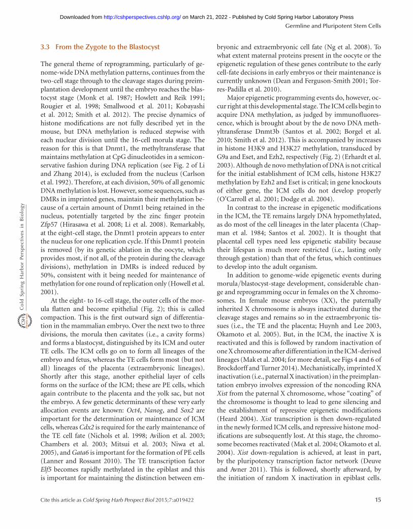
3.3 From the Zygote to the Blastocyst
The general theme of reprogramming, particularly of ge-nome-wide DNA methylation patterns, continues from thetwo-cell stage through to the cleavage stages during preim-plantation development until the embryo reaches the blas-tocyst stage (Monk et al. 1987; Howlett and Reik 1991;Rougier et al. 1998; Smallwood et al. 2011; Kobayashiet al. 2012; Smith et al. 2012). The precise dynamics ofhistone modifications are not fully described yet in themouse, but DNA methylation is reduced stepwise witheach nuclear division until the 16-cell morula stage. Thereason for this is that Dnmt1, the methyltransferase thatmaintains methylation at CpG dinucleotides in a semicon-servative fashion during DNA replication (see Fig. 2 of Liand Zhang 2014), is excluded from the nucleus (Carlsonet al. 1992). Therefore, at each division, 50% of all genomicDNA methylation is lost. However, some sequences, such asDMRs in imprinted genes, maintain their methylation be-cause of a certain amount of Dnmt1 being retained in thenucleus, potentially targeted by the zinc finger proteinZfp57 (Hirasawa et al. 2008; Li et al. 2008). Remarkably,at the eight-cell stage, the Dnmt1 protein appears to enterthe nucleus for one replication cycle. If this Dnmt1 proteinis removed (by its genetic ablation in the oocyte, whichprovides most, if not all, of the protein during the cleavagedivisions), methylation in DMRs is indeed reduced by50%, consistent with it being needed for maintenance ofmethylation for one round of replication only (Howell et al.2001).
At the eight- to 16-cell stage, the outer cells of the mor-ula flatten and become epithelial (Fig. 2); this is calledcompaction. This is the first outward sign of differentia-tion in the mammalian embryo. Over the next two to threedivisions, the morula then cavitates (i.e., a cavity forms)and forms a blastocyst, distinguished by its ICM and outerTE cells. The ICM cells go on to form all lineages of theembryo and fetus, whereas the TE cells form most (but notall) lineages of the placenta (extraembryonic lineages).Shortly after this stage, another epithelial layer of cellsforms on the surface of the ICM; these are PE cells, whichagain contribute to the placenta and the yolk sac, but notthe embryo. A few genetic determinants of these very earlyallocation events are known: Oct4, Nanog, and Sox2 areimportant for the determination or maintenance of ICMcells, whereas Cdx2 is required for the early maintenance ofthe TE cell fate (Nichols et al. 1998; Avilion et al. 2003;Chambers et al. 2003; Mitsui et al. 2003; Niwa et al.2005), and Gata6 is important for the formation of PE cells(Lanner and Rossant 2010). The TE transcription factorElf5 becomes rapidly methylated in the epiblast and thisis important for maintaining the distinction between em-
bryonic and extraembryonic cell fate (Ng et al. 2008). Towhat extent maternal proteins present in the oocyte or theepigenetic regulation of these genes contribute to the earlycell-fate decisions in early embryos or their maintenance iscurrently unknown (Dean and Ferguson-Smith 2001; Tor-res-Padilla et al. 2010).
Major epigenetic programming events do, however, oc-cur right at this developmental stage. The ICM cells begin toacquire DNA methylation, as judged by immunofluores-cence, which is brought about by the de novo DNA meth-yltransferase Dnmt3b (Santos et al. 2002; Borgel et al.2010; Smith et al. 2012). This is accompanied by increasesin histone H3K9 and H3K27 methylation, transduced byG9a and Eset, and Ezh2, respectively (Fig. 2) (Erhardt et al.2003). Although de novo methylation of DNA is not criticalfor the initial establishment of ICM cells, histone H3K27methylation by Ezh2 and Eset is critical; in gene knockoutsof either gene, the ICM cells do not develop properly(O’Carroll et al. 2001; Dodge et al. 2004).
In contrast to the increase in epigenetic modificationsin the ICM, the TE remains largely DNA hypomethylated,as do most of the cell lineages in the later placenta (Chap-man et al. 1984; Santos et al. 2002). It is thought thatplacental cell types need less epigenetic stability becausetheir lifespan is much more restricted (i.e., lasting onlythrough gestation) than that of the fetus, which continuesto develop into the adult organism.
In addition to genome-wide epigenetic events duringmorula/blastocyst-stage development, considerable chan-ge and reprogramming occur in females on the X chromo-somes. In female mouse embryos (XX), the paternallyinherited X chromosome is always inactivated during thecleavage stages and remains so in the extraembryonic tis-sues (i.e., the TE and the placenta; Huynh and Lee 2003,Okamoto et al. 2005). But, in the ICM, the inactive X isreactivated and this is followed by random inactivation ofone X chromosome after differentiation in the ICM-derivedlineages (Mak et al. 2004; for more detail, see Figs 4 and 6 ofBrockdorff and Turner 2014). Mechanistically, imprinted Xinactivation (i.e., paternal X inactivation) in the preimplan-tation embryo involves expression of the noncoding RNAXist from the paternal X chromosome, whose “coating” ofthe chromosome is thought to lead to gene silencing andthe establishment of repressive epigenetic modifications(Heard 2004). Xist transcription is then down-regulatedin the newly formed ICM cells, and repressive histone mod-ifications are subsequently lost. At this stage, the chromo-some becomes reactivated (Mak et al. 2004; Okamoto et al.2004). Xist down-regulation is achieved, at least in part,by the pluripotency transcription factor network (Deuveand Avner 2011). This is followed, shortly afterward, bythe initiation of random X inactivation in epiblast cells.
Germline and Pluripotent Stem Cells
Cite this article as Cold Spring Harb Perspect Biol 2015;7:a019422 15
on March 21, 2022 - Published by Cold Spring Harbor Laboratory Press http://cshperspectives.cshlp.org/Downloaded from
We will see in Section 4 that ES cells are “frozen” at the stageafter reactivation of the X chromosome such that female EScells contain two active X chromosomes.
4 FROM PLURIPOTENT STEM CELLS TO SOMATICCELLS AND BACK TO GERM CELLS
4.1 Derivation of Pluripotent Stem Cells
In Section 3.3, we learned that there are dramatic epigeneticreprogramming events in the zygote, cleavage-stage em-bryos, and the blastocyst, resulting in different epigeneticpatterns in the ICM and TE. We now consider the geneticand epigenetic properties of early stem cells derived intoculture from the blastocyst and later lineages (Fig. 1B), suchas ES cells (Smith 2001), trophoblast stem (TS) cells (Ros-sant 2001), extraembryonic endoderm (XEN) stem cells(Kunath et al. 2005), and EG stem cells (Matsui et al. 1992).
The feature that is common to these cell types is thatthey can be isolated or established from intact embryos andput into culture under certain culturing conditions. Onceestablished, they can be cultured for extended periods oftime and show no signs of senescence. They can also begenetically manipulated during culture and then reintro-duced into living embryos to participate in the develop-ment of the appropriate lineages.
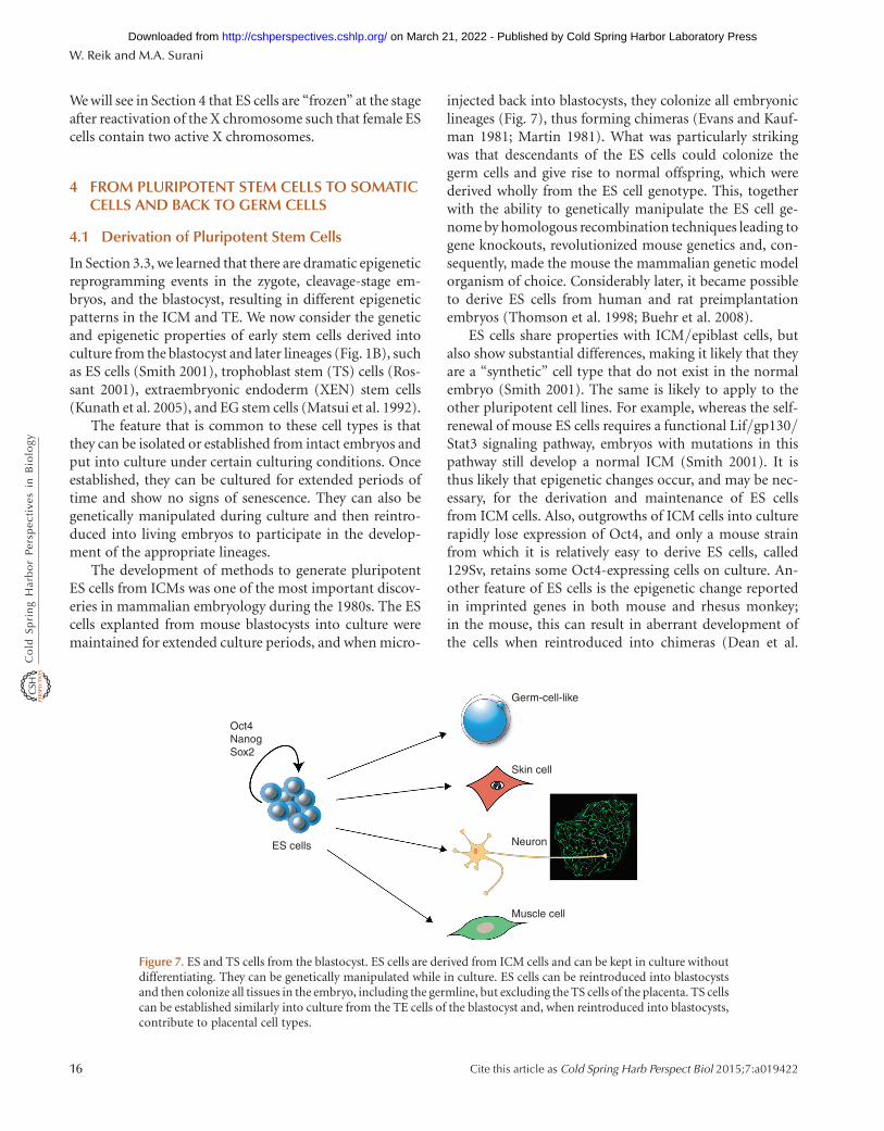
The development of methods to generate pluripotentES cells from ICMs was one of the most important discov-eries in mammalian embryology during the 1980s. The EScells explanted from mouse blastocysts into culture weremaintained for extended culture periods, and when micro-
injected back into blastocysts, they colonize all embryoniclineages (Fig. 7), thus forming chimeras (Evans and Kauf-man 1981; Martin 1981). What was particularly strikingwas that descendants of the ES cells could colonize thegerm cells and give rise to normal offspring, which werederived wholly from the ES cell genotype. This, togetherwith the ability to genetically manipulate the ES cell ge-nome by homologous recombination techniques leading togene knockouts, revolutionized mouse genetics and, con-sequently, made the mouse the mammalian genetic modelorganism of choice. Considerably later, it became possibleto derive ES cells from human and rat preimplantationembryos (Thomson et al. 1998; Buehr et al. 2008).
ES cells share properties with ICM/epiblast cells, butalso show substantial differences, making it likely that theyare a “synthetic” cell type that do not exist in the normalembryo (Smith 2001). The same is likely to apply to theother pluripotent cell lines. For example, whereas the self-renewal of mouse ES cells requires a functional Lif/gp130/Stat3 signaling pathway, embryos with mutations in thispathway still develop a normal ICM (Smith 2001). It isthus likely that epigenetic changes occur, and may be nec-essary, for the derivation and maintenance of ES cellsfrom ICM cells. Also, outgrowths of ICM cells into culturerapidly lose expression of Oct4, and only a mouse strainfrom which it is relatively easy to derive ES cells, called129Sv, retains some Oct4-expressing cells on culture. An-other feature of ES cells is the epigenetic change reportedin imprinted genes in both mouse and rhesus monkey;in the mouse, this can result in aberrant development ofthe cells when reintroduced into chimeras (Dean et al.
Oct4NanogSox2
ES cells
Germ-cell-like
Skin cell
Neuron
Muscle cell
Figure 7. ES and TS cells from the blastocyst. ES cells are derived from ICM cells and can be kept in culture withoutdifferentiating. They can be genetically manipulated while in culture. ES cells can be reintroduced into blastocystsand then colonize all tissues in the embryo, including the germline, but excluding the TS cells of the placenta. TS cellscan be established similarly into culture from the TE cells of the blastocyst and, when reintroduced into blastocysts,contribute to placental cell types.
W. Reik and M.A. Surani
16 Cite this article as Cold Spring Harb Perspect Biol 2015;7:a019422
on March 21, 2022 - Published by Cold Spring Harbor Laboratory Press http://cshperspectives.cshlp.org/Downloaded from
1998; Humpherys et al. 2001). One of the importantdeterminants of ES cells in culture is the signaling systemthey respond to; it seems that ES cells grown in serumand leukemia inhibitory factor are epigenetically and tran-scriptionally somewhat heterogeneous, whereas ES cellsgrown in the presence of inhibitors of ERK (FGF4) andGSK3 are more homogeneous (Hayashi et al. 2008; Lannerand Rossant 2010; Leitch et al. 2010; Marks et al. 2012; Ficzet al. 2013). This may reflect the natural tendency of epi-blast cells to progress from a naıve state of pluripotencyin the early ICM to a state in which they are primed fordifferentiation and thus receive increased levels of prodif-ferentiation FGF signals through ERK or GSK3 (for moredetail on epiblast stem cells, see Hochedlinger and Jaenisch2014).
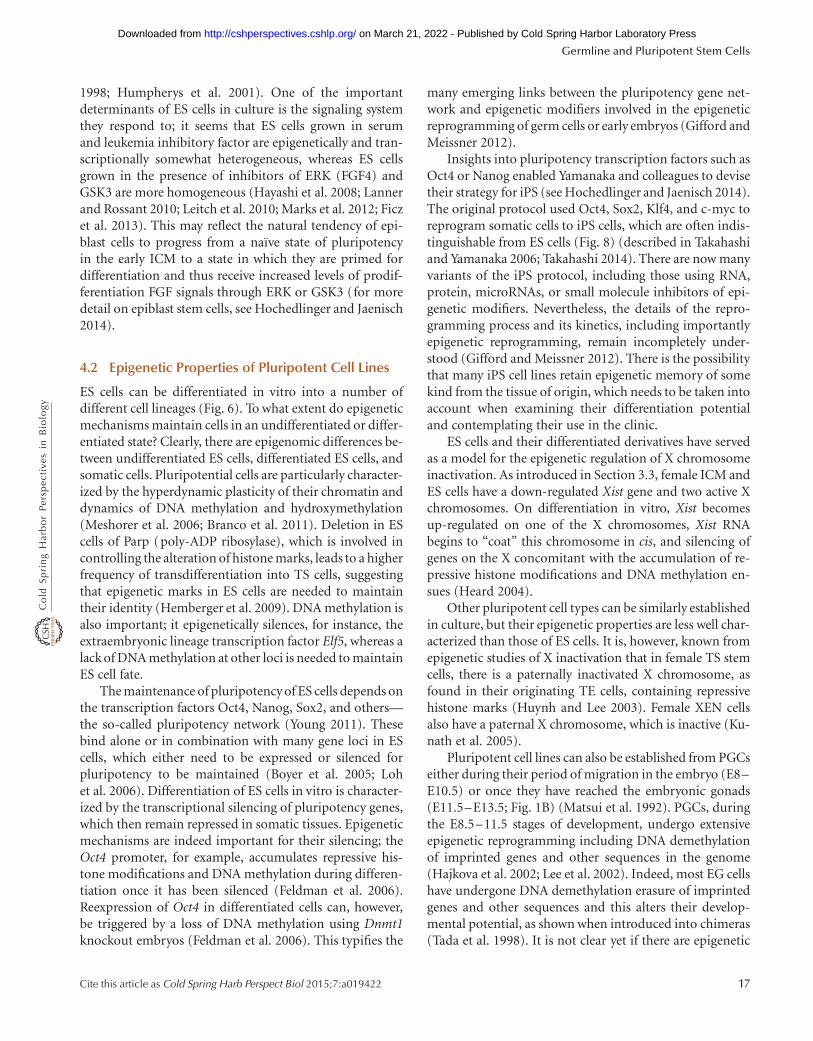
4.2 Epigenetic Properties of Pluripotent Cell Lines
ES cells can be differentiated in vitro into a number ofdifferent cell lineages (Fig. 6). To what extent do epigeneticmechanisms maintain cells in an undifferentiated or differ-entiated state? Clearly, there are epigenomic differences be-tween undifferentiated ES cells, differentiated ES cells, andsomatic cells. Pluripotential cells are particularly character-ized by the hyperdynamic plasticity of their chromatin anddynamics of DNA methylation and hydroxymethylation(Meshorer et al. 2006; Branco et al. 2011). Deletion in EScells of Parp (poly-ADP ribosylase), which is involved incontrolling the alteration of histone marks, leads to a higherfrequency of transdifferentiation into TS cells, suggestingthat epigenetic marks in ES cells are needed to maintaintheir identity (Hemberger et al. 2009). DNA methylation isalso important; it epigenetically silences, for instance, theextraembryonic lineage transcription factor Elf5, whereas alack of DNA methylation at other loci is needed to maintainES cell fate.
The maintenance of pluripotency of ES cells depends onthe transcription factors Oct4, Nanog, Sox2, and others—the so-called pluripotency network (Young 2011). Thesebind alone or in combination with many gene loci in EScells, which either need to be expressed or silenced forpluripotency to be maintained (Boyer et al. 2005; Lohet al. 2006). Differentiation of ES cells in vitro is character-ized by the transcriptional silencing of pluripotency genes,which then remain repressed in somatic tissues. Epigeneticmechanisms are indeed important for their silencing; theOct4 promoter, for example, accumulates repressive his-tone modifications and DNA methylation during differen-tiation once it has been silenced (Feldman et al. 2006).Reexpression of Oct4 in differentiated cells can, however,be triggered by a loss of DNA methylation using Dnmt1knockout embryos (Feldman et al. 2006). This typifies the
many emerging links between the pluripotency gene net-work and epigenetic modifiers involved in the epigeneticreprogramming of germ cells or early embryos (Gifford andMeissner 2012).
Insights into pluripotency transcription factors such asOct4 or Nanog enabled Yamanaka and colleagues to devisetheir strategy for iPS (see Hochedlinger and Jaenisch 2014).The original protocol used Oct4, Sox2, Klf4, and c-myc toreprogram somatic cells to iPS cells, which are often indis-tinguishable from ES cells (Fig. 8) (described in Takahashiand Yamanaka 2006; Takahashi 2014). There are now manyvariants of the iPS protocol, including those using RNA,protein, microRNAs, or small molecule inhibitors of epi-genetic modifiers. Nevertheless, the details of the repro-gramming process and its kinetics, including importantlyepigenetic reprogramming, remain incompletely under-stood (Gifford and Meissner 2012). There is the possibilitythat many iPS cell lines retain epigenetic memory of somekind from the tissue of origin, which needs to be taken intoaccount when examining their differentiation potentialand contemplating their use in the clinic.
ES cells and their differentiated derivatives have servedas a model for the epigenetic regulation of X chromosomeinactivation. As introduced in Section 3.3, female ICM andES cells have a down-regulated Xist gene and two active Xchromosomes. On differentiation in vitro, Xist becomesup-regulated on one of the X chromosomes, Xist RNAbegins to “coat” this chromosome in cis, and silencing ofgenes on the X concomitant with the accumulation of re-pressive histone modifications and DNA methylation en-sues (Heard 2004).
Other pluripotent cell types can be similarly establishedin culture, but their epigenetic properties are less well char-acterized than those of ES cells. It is, however, known fromepigenetic studies of X inactivation that in female TS stemcells, there is a paternally inactivated X chromosome, asfound in their originating TE cells, containing repressivehistone marks (Huynh and Lee 2003). Female XEN cellsalso have a paternal X chromosome, which is inactive (Ku-nath et al. 2005).
Pluripotent cell lines can also be established from PGCseither during their period of migration in the embryo (E8–E10.5) or once they have reached the embryonic gonads(E11.5–E13.5; Fig. 1B) (Matsui et al. 1992). PGCs, duringthe E8.5–11.5 stages of development, undergo extensiveepigenetic reprogramming including DNA demethylationof imprinted genes and other sequences in the genome(Hajkova et al. 2002; Lee et al. 2002). Indeed, most EG cellshave undergone DNA demethylation erasure of imprintedgenes and other sequences and this alters their develop-mental potential, as shown when introduced into chimeras(Tada et al. 1998). It is not clear yet if there are epigenetic
Germline and Pluripotent Stem Cells
Cite this article as Cold Spring Harb Perspect Biol 2015;7:a019422 17
on March 21, 2022 - Published by Cold Spring Harbor Laboratory Press http://cshperspectives.cshlp.org/Downloaded from
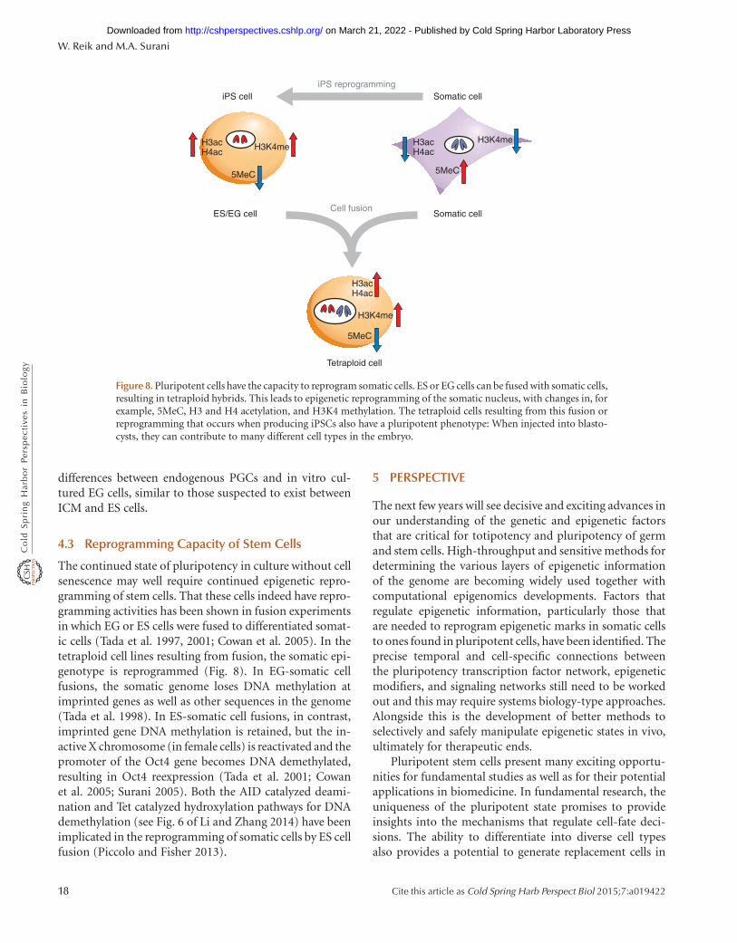
differences between endogenous PGCs and in vitro cul-tured EG cells, similar to those suspected to exist betweenICM and ES cells.
4.3 Reprogramming Capacity of Stem Cells
The continued state of pluripotency in culture without cellsenescence may well require continued epigenetic repro-gramming of stem cells. That these cells indeed have repro-gramming activities has been shown in fusion experimentsin which EG or ES cells were fused to differentiated somat-ic cells (Tada et al. 1997, 2001; Cowan et al. 2005). In thetetraploid cell lines resulting from fusion, the somatic epi-genotype is reprogrammed (Fig. 8). In EG-somatic cellfusions, the somatic genome loses DNA methylation atimprinted genes as well as other sequences in the genome(Tada et al. 1998). In ES-somatic cell fusions, in contrast,imprinted gene DNA methylation is retained, but the in-active X chromosome (in female cells) is reactivated and thepromoter of the Oct4 gene becomes DNA demethylated,resulting in Oct4 reexpression (Tada et al. 2001; Cowanet al. 2005; Surani 2005). Both the AID catalyzed deami-nation and Tet catalyzed hydroxylation pathways for DNAdemethylation (see Fig. 6 of Li and Zhang 2014) have beenimplicated in the reprogramming of somatic cells by ES cellfusion (Piccolo and Fisher 2013).
5 PERSPECTIVE
The next few years will see decisive and exciting advances inour understanding of the genetic and epigenetic factorsthat are critical for totipotency and pluripotency of germand stem cells. High-throughput and sensitive methods fordetermining the various layers of epigenetic informationof the genome are becoming widely used together withcomputational epigenomics developments. Factors thatregulate epigenetic information, particularly those thatare needed to reprogram epigenetic marks in somatic cellsto ones found in pluripotent cells, have been identified. Theprecise temporal and cell-specific connections betweenthe pluripotency transcription factor network, epigeneticmodifiers, and signaling networks still need to be workedout and this may require systems biology-type approaches.Alongside this is the development of better methods toselectively and safely manipulate epigenetic states in vivo,ultimately for therapeutic ends.
Pluripotent stem cells present many exciting opportu-nities for fundamental studies as well as for their potentialapplications in biomedicine. In fundamental research, theuniqueness of the pluripotent state promises to provideinsights into the mechanisms that regulate cell-fate deci-sions. The ability to differentiate into diverse cell typesalso provides a potential to generate replacement cells in
H3acH4ac
H3K4me
5MeC
iPS cell Somatic cell
5MeC
H3acH4ac
H3K4me
H3acH4ac
H3K4me
5MeC
Tetraploid cell
Cell fusionES/EG cell Somatic cell
iPS reprogramming
Figure 8. Pluripotent cells have the capacity to reprogram somatic cells. ES or EG cells can be fused with somatic cells,resulting in tetraploid hybrids. This leads to epigenetic reprogramming of the somatic nucleus, with changes in, forexample, 5MeC, H3 and H4 acetylation, and H3K4 methylation. The tetraploid cells resulting from this fusion orreprogramming that occurs when producing iPSCs also have a pluripotent phenotype: When injected into blasto-cysts, they can contribute to many different cell types in the embryo.
W. Reik and M.A. Surani
18 Cite this article as Cold Spring Harb Perspect Biol 2015;7:a019422
on March 21, 2022 - Published by Cold Spring Harbor Laboratory Press http://cshperspectives.cshlp.org/Downloaded from
the quest to repair diseased tissues. Pluripotent stem cellsare also being used to develop disease models to explorehow various human diseases originate from the very begin-ning as a result of specific mutations and epimutations(i.e., mutations caused by alterations of DNA methylationor the chromatin template, but not involving changes to theDNA sequence). Such disease models may, in turn, allowthe development of new drugs to cure or even preventdiseases. Transdifferentiation technologies that directlytransform one differentiated cell type into another (e.g.,skin into neurons) have recently been established and thismay offer new routes to therapy, avoiding the use of plu-ripotent cell types with the potential danger of tumor for-mation when cells are used for transplantation in patients(discussed further in Hochedlinger and Jaenisch 2014).There is also the prospect of reversing epigenetic effectsduring aging by “epigenetic rejuvenation,” which is repro-gramming cells to a younger and healthier cell state withoutgoing back to pluripotency (Rando and Chang 2012).
ACKNOWLEDGMENTS
The use of human pluripotent stem cells raises sensitiveethical issues that are being debated by the wider public.Appropriate ethical and regulatory frameworks are beingestablished and continue to be refined in step with scientificprogress for the use of stem cells in research and biomedicalapplications.
REFERENCES
∗Reference is also in this collection.
∗ Allis CD, Jenuwein T, Reinberg D. 2014. Overview and concepts. ColdSpring Harb Perspect Biol doi: 10.1101/cshperspect.a018739.
Ancelin K, Lange UC, Hajkova P, Schneider R, Bannister AJ, KouzaridesT, Surani MA. 2006. Blimp1 associates with Prmt5 and directs his-tone arginine methylation in mouse germ cells. Nat Cell Biol 8: 623–630.
Arney KL, Bao S, Bannister AJ, Kouzarides T, Surani MA. 2002. Histonemethylation defines epigenetic asymmetry in the mouse zygote. Int JDev Biol 46: 317–320.
Avilion AA, Nicolis SK, Pevny LH, Perez L, Vivian N, Lovell-Badge R.2003. Multipotent cell lineages in early mouse development dependon SOX2 function. Genes Dev 17: 126–140.
∗ Barlow DP, Bartolomei MS. 2014. Genomic imprinting in mammals.Cold Spring Harb Perspect Biol 6: a018382.
Baudat F, Buard J, Grey C, Fledel-Alon A, Ober C, Przeworski M, Coop G,de Massy B. 2010. PRDM9 is a major determinant of meiotic recom-bination hotspots in humans and mice. Science 327: 836–840.
Blackwell TK. 2004. Germ cells: Finding programs of mass repression.Curr Biol 14: R229–R230.
∗ Blewitt M, Whitelaw E. 2013. The use of mouse models to study epi-genetics. Cold Spring Harb Perspect Biol 5: a017939.
Borgel J, Guibert S, Li Y, Chiba H, Schubeler D, Sasaki H, Forne T, WeberM. 2010. Targets and dynamics of promoter DNA methylation duringearly mouse development. Nat Genet 42: 1093–1100.
Boyer LA, Lee TI, Cole MF, Johnstone SE, Levine SS, Zucker JP, GuentherMG, Kumar RM, Murray HL, Jenner RG, et al. 2005. Core transcrip-tional regulatory circuitry in human embryonic stem cells. Cell 122:947–956.
Branco MR, Ficz G, Reik W. 2011. Uncovering the role of 5-hydroxy-methylcytosine in the epigenome. Nat Rev Genet 13: 7–13.
∗ Brockdorff N, Turner BM. 2014. Dosage compensation in mammals.Cold Spring Harb Perspect Biol doi: 10.1101/cshperspect.a019406.
Brykczynska U, Hisano M, Erkek S, Ramos L, Oakeley EJ, Roloff TC,Beisel C, Schubeler D, Stadler MB, Peters AH. 2010. Repressive andactive histone methylation mark distinct promoters in human andmouse spermatozoa. Nat Struct Mol Biol 17: 679–687.
Buehr M, Meek S, Blair K, Yang J, Ure J, Silva J, McLay R, Hall J, Ying QL,Smith A. 2008. Capture of authentic embryonic stem cells from ratblastocysts. Cell 135: 1287–1298.
∗ Busslinger M, Tarakhovsky A. 2014. Epigenetic control of immunity.Cold Spring Harb Perspect Biol 6: a019307.
Carlson LL, Page AW, Bestor TH. 1992. Properties and localization ofDNA methyltransferase in preimplantation mouse embryos: Implica-tions for genomic imprinting. Genes Dev 6: 2536–2541.
Chambers I, Colby D, Robertson M, Nichols J, Lee S, Tweedie S, Smith A.2003. Functional expression cloning of Nanog, a pluripotencysustain-ing factor in embryonic stem cells. Cell 113: 643–655.
Chan MM, Smith ZD, Egli D, Regev A, Meissner A. 2012. Mouse ooplasmconfers context-specific reprogramming capacity. Nat Genet 44: 978–980.
Chapman V, Forrester L, Sanford J, Hastie N, Rossant J. 1984. Cell line-age-specific undermethylation of mouse repetitive DNA. Nature 307:284–286.
∗ Cheng X. 2014. Structural and functional coordination of DNA andhistone methylation. Cold Spring Harb Perspect Biol 6: a018747.
Chong S, Whitelaw E. 2004. Epigenetic germline inheritance. Curr OpinGenet Dev 14: 692–696.
Cortellino S, Xu J, Sannai M, Moore R, Caretti E, Cigliano A, Le Coz M,Devarajan K, Wessels A, Soprano D, et al. 2011. Thymine DNA glyco-sylase is essential for active DNA demethylation by linked deamina-tion-base excision repair. Cell 146: 67–79.
Cowan CA, Atienza J, Melton DA, Eggan K. 2005. Nuclear reprogram-ming of somatic cells after fusion with human embryonic stem cells.Science 309: 1369–1373.
Dawlaty MM, Breiling A, Le T, Raddatz G, Barrasa MI, Cheng AW, Gao Q,Powell BE, Li Z, Xu M, et al. 2013. Moation. Dev Cell 24: 310–323.
Dean W, Bowden L, Aitchison A, Klose J, Moore T, Meneses JJ, Reik W,Feil R. 1998. Altered imprinted gene methylation and expression incompletely ES cell-derived mouse fetuses: Association with aberrantphenotypes. Development 125: 2273–2282.
Dean W, Santos F, Stojkovic M, Zakhartchenko V, Walter J, Wolf E, ReikW. 2001. Conservation of methylation reprogramming in mammaliandevelopment: Aberrant reprogramming in cloned embryos. Proc NatlAcad Sci 98: 13734–13738.
Deshpande G, Calhoun G, Schedl P. 2004. Overlapping mechanismsfunction to establish transcriptional quiescence in the embryonicDrosophila germline. Development 131: 1247–1257.
de Souza FS, Gawantka V, Gomez AP, Delius H, Ang SL, Niehrs C. 1999.The zinc finger gene Xblimp1 controls anterior endomesodermal cellfate in Spemann’s organizer. EMBO J 18: 6062–6072.
Deuve JL, Avner P. 2011. The coupling of X-chromosome inactivation topluripotency. Annu Rev Cell Dev Biol 27: 611–629.
Dodge JE, Kang YK, Beppu H, Lei H, Li E. 2004. Histone H3-K9 meth-yltransferase ESET is essential for early development. Mol Cell Biol24: 2478–2486.
Erhardt S, Su IH, Schneider R, Barton S, Bannister AJ, Perez-Burgos L,Jenuwein T, Kouzarides T, Tarakhovsky A, Surani MA. 2003. Conse-quences of the depletion of zygotic and embryonic enhancer of zeste2 during preimplantation mouse development. Development 130:4235–4248.
Germline and Pluripotent Stem Cells
Cite this article as Cold Spring Harb Perspect Biol 2015;7:a019422 19
on March 21, 2022 - Published by Cold Spring Harbor Laboratory Press http://cshperspectives.cshlp.org/Downloaded from
Evans MJ, Kaufman MH. 1981. Establishment in culture of pluripoten-tial cells from mouse embryos. Nature 292: 154–156.
Extavour CG, Akam M. 2003. Mechanisms of germ cell specificationacross the metazoans: Epigenesis and preformation. Development130: 5869–5884.
Feldman N, Gerson A, Fang J, Li E, Zhang Y, Shinkai Y, Cedar H,Bergman Y. 2006. G9a-mediated irreversible epigenetic inactivationof Oct-3/4 during early embryogenesis. Nat Cell Biol 8: 188–194.
Feng S, Jacobsen SE, Reik W. 2010. Epigenetic reprogramming in plantand animal development. Science 330: 622–627.
Ferguson-Smith AC, Patti ME. 2011. You are what your dad ate. CellMetab 13: 115–117.
Ficz G, Hore TA, Santos F, Lee HJ, Dean W, Arand J, Krueger F, Oxley D,Paul YL, Walter J, et al. 2013. FGF signaling inhibition in ESCs drivesrapid genome-wide demethylation to the epigenetic ground state ofpluripotency. Cell Stem Cell 13: 351–359.
Fujimori T, Kurotaki Y, Miyazaki J, Nabeshima Y. 2003. Analysis ofcell lineage in two- and four-cell mouse embryos. Development 130:5113–5122.
Gardner RL. 1985. Clonal analysis of early mammalian development.Philos Trans R Soc Lond B Biol Sci 312: 163–178.
Gardner RL. 1997. The early blastocyst is bilaterally symmetrical and itsaxis of symmetry is aligned with the animal–vegetal axis of the zygotein the mouse. Development 124: 289–301.
Geijsen N, Horoschak M, Kim K, Gribnau J, Eggan K, Daley GQ. 2004.Derivation of embryonic germ cells and male gametes from embryonicstem cells. Nature 427: 148–154.
Gifford CA, Meissner A. 2012. Epigenetic obstacles encountered by tran-scription factors: Reprogramming against all odds. Curr Opin GenetDev 22: 409–415.
Gu TP, Guo F, Yang H, Wu HP, Xu GF, Liu W, Xie ZG, Shi L, He X, Jin SG,et al. 2011. The role of Tet3 DNA dioxygenase in epigenetic repro-gramming by oocytes. Nature 477: 606–610.
Guan K, Nayernia K, Maier LS, Wagner S, Dressel R, Lee JH, Nolte J, WolfF, Li M, Engel W, et al. 2006. Pluripotency of spermatogonial stem cellsfrom adult mouse testis. Nature 440: 1199–1203.
Guibert S, Forne T, Weber M. 2012. Global profiling of DNA methylationerasure in mouse primordial germ cells. Genome Res 22: 633–641.
Gyory I, Wu J, Fejer G, Seto E, Wright KL. 2004. PRDI-BF1 recruits thehistone H3 methyltransferase G9a in transcriptional silencing. NatImmunol 5: 299–308.
Hackett JA, Surani MA. 2013. Beyond DNA: Programming and inheri-tance of parental methylomes. Cell 172: 5427–5440.
Hackett JA, Zylicz JJ, Surani MA. 2012. Parallel mechanisms of epigeneticreprogramming in the germline. Trends Genet 28: 164–174.
Hackett JA, Sengupta R, Zylicz JJ, Murakami K, Lee C, Down TA, SuraniMA. 2013. Germline DNA demethylation dynamics and imprint era-sure through 5-hydroxymethylcytosine. Science 339: 448–452.
Hajkova P, Erhardt S, Lane N, Haaf T, El-Maarri O, Reik W, Walter J,Surani MA. 2002. Epigenetic reprogramming in mouse primordialgerm cells. Mech Dev 117: 15–23.
Hajkova P, Ancelin K, Waldmann T, Lacoste N, Lange UC, Cesari F, Lee C,Almouzni G, Schneider R, Surani MA. 2008. Chromatin dynamicsduring epigenetic reprogramming in the mouse germ line. Nature452: 877–881.
Hajkova P, Jeffries SJ, Lee C, Miller N, Jackson SP, Surani MA. 2010.Genome-wide reprogramming in the mouse germ line entails thebase excision repair pathway. Science 329: 78–82.
Hammoud SS, Nix DA, Zhang H, Purwar J, Carrell DT, Cairns BR. 2009.Distinctive chromatin in human sperm packages genes for embryodevelopment. Nature 460: 473–478.
Hayashi K, Yoshida K, Matsui Y. 2005. A histone H3 methyltransferasecontrols epigenetic events required for meiotic prophase. Nature 438:374–378.
Hayashi K, Lopes SM, Tang F, Surani MA. 2008. Dynamic equilibriumand heterogeneity of mouse pluripotent stem cells with distinct func-tional and epigenetic states. Cell Stem Cell 3: 391–401.
Hayashi K, Ohta H, Kurimoto K, Aramaki S, Saitou M. 2011. Reconsti-tution of the mouse germ cell specification pathway in culture bypluripotent stem cells. Cell 146: 519–532.
Heard E. 2004. Recent advances in X-chromosome inactivation. CurrOpin Cell Biol 16: 247–255.
Hemberger M, Dean W, Reik W. 2009. Epigenetic dynamics of stem cellsand cell lineage commitment: Digging Waddington’s canal. Nat RevMol Cell Biol 10: 526–537.
∗ Henikoff S, Smith MM. 2014. Histone variants and epigenetics. ColdSpring Harb Perspect Biol doi: 10.1101/cshperspect.a019364.
Hernandez-Lagunas L, Choi IF, Kaji T, Simpson P, Hershey C, Zhou Y,Zon L, Mercola M, Artinger KB. 2005. Zebrafish narrowminded dis-rupts the transcription factor prdm1 and is required for neural crestand sensory neuron specification. Dev Biol 278: 347–357.
Hirasawa R, Chiba H, Kaneda M, Tajima S, Li E, Jaenisch R, Sasaki H.2008. Maternal and zygotic Dnmt1 are necessary and sufficient forthe maintenance of DNA methylation imprints. Genes Dev 22: 1607–1616.
∗ Hochedlinger K, Jaenisch R. 2014. Induced pluripotency and epigeneticreprogramming. Cold Spring Harb Perspect Biol doi: 10.1101/cshperspect.a019448.
Howell CY, Bestor TH, Ding F, Latham KE, Mertineit C, Trasler JM,Chaillet JR. 2001. Genomic imprinting disrupted by a maternal effectmutation in the Dnmt1 gene. Cell 104: 829–838.
Howlett SK, Reik W. 1991. Methylation levels of maternal and paternalgenomes during preimplantation development. Development 113:119–127.
Hubner K, Fuhrmann G, Christenson LK, Kehler J, Reinbold R, De LaFuente R, Wood J, Strauss JF 3rd, Boiani M, Scholer HR. 2003. Der-ivation of oocytes from mouse embryonic stem cells. Science 300:1251–1256.
Humpherys D, Eggan K, Akutsu H, Hochedlinger K, Rideout WM 3rd,Biniszkiewicz D, Yanagimachi R, Jaenisch R. 2001. Epigenetic insta-bility in ES cells and cloned mice. Science 293: 95–97.
Huynh KD, Lee JT. 2003. Inheritance of a pre-inactivated paternal Xchromosome in early mouse embryos. Nature 426: 857–862.
Huynh JR, St Johnston D. 2004. The origin of asymmetry: Early polar-isation of the Drosophila germline cyst and oocyte. Curr Biol 14:R438–R449.
Inoue A, Zhang Y. 2011. Replication-dependent loss of 5-hydroxy-methylcytosine in mouse preimplantation embryos. Science 334: 194.
Inoue A, Shen L, Dai Q, He C, Zhang Y. 2011. Generation and replica-tion-dependent dilution of 5fC and 5caC during mouse preimplan-tation development. Cell Res 21: 1670–1676.
Iqbal K, Jin SG, Pfeifer GP, Szabo PE. 2011. Reprogramming of thepaternal genome upon fertilization involves genome-wide oxidationof 5-methylcytosine. Proc Natl Acad Sci 108: 3642–3647.
Kagiwada S, Kurimoto K, Hirota T, Yamaji M, Saitou M. 2013. Replica-tion-coupled passive DNA demethylation for the erasure of genomeimprints in mice. EMBO J 32: 340–353.
Kanatsu-Shinohara M, Inoue K, Lee J, Yoshimoto M, Ogonuki N, MikiH, Baba S, Kato T, Kazuki Y, Toyokuni S, et al. 2004. Generation ofpluripotent stem cells from neonatal mouse testis. Cell 119: 1001–1012.
Kawahara M, Wu Q, Takahashi N, Morita S, Yamada K, Ito M, Ferguson-Smith AC, Kono T. 2007. High-frequency generation of viable micefrom engineered bi-maternal embryos. Nat Biotechnol 25: 1045–1050.
Kelly SJ. 1977. Studies of the developmental potential of 4- and 8-cellstage mouse blastomeres. J Exp Zool 200: 365–376.
Kimmins S, Sassone-Corsi P. 2005. Chromatin remodelling and epige-netic features of germ cells. Nature 434: 583–589.
Kobayashi H, Sakurai T, Imai M, Takahashi N, Fukuda A, Yayoi O, Sato S,Nakabayashi K, Hata K, Sotomaru Y, et al. 2012. Contribution ofintragenic DNA methylation in mouse gametic DNA methylomes toestablish oocyte-specific heritable marks. PLoS Genet 8: e1002440.
Kunath T, Arnaud D, Uy GD, Okamoto I, Chureau C, Yamanaka Y, HeardE, Gardner RL, Avner P, Rossant J. 2005. Imprinted X-inactivation in
W. Reik and M.A. Surani
20 Cite this article as Cold Spring Harb Perspect Biol 2015;7:a019422
on March 21, 2022 - Published by Cold Spring Harbor Laboratory Press http://cshperspectives.cshlp.org/Downloaded from
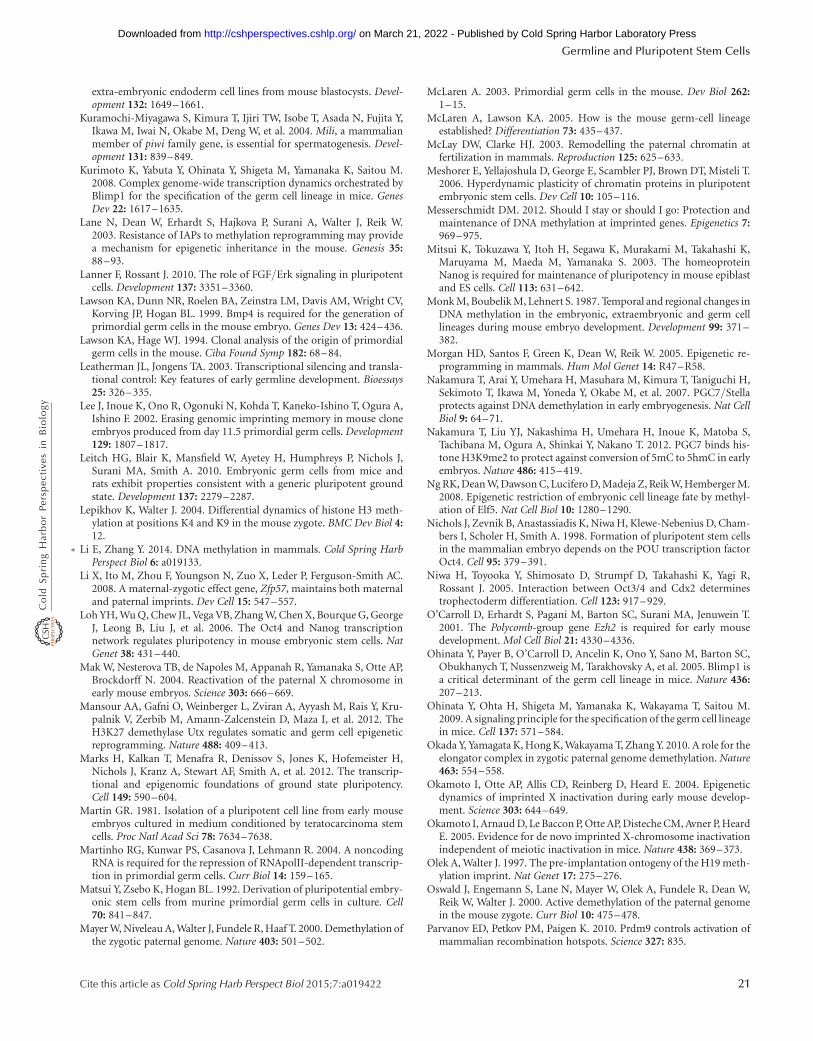
extra-embryonic endoderm cell lines from mouse blastocysts. Devel-opment 132: 1649–1661.
Kuramochi-Miyagawa S, Kimura T, Ijiri TW, Isobe T, Asada N, Fujita Y,Ikawa M, Iwai N, Okabe M, Deng W, et al. 2004. Mili, a mammalianmember of piwi family gene, is essential for spermatogenesis. Devel-opment 131: 839–849.
Kurimoto K, Yabuta Y, Ohinata Y, Shigeta M, Yamanaka K, Saitou M.2008. Complex genome-wide transcription dynamics orchestrated byBlimp1 for the specification of the germ cell lineage in mice. GenesDev 22: 1617–1635.
Lane N, Dean W, Erhardt S, Hajkova P, Surani A, Walter J, Reik W.2003. Resistance of IAPs to methylation reprogramming may providea mechanism for epigenetic inheritance in the mouse. Genesis 35:88–93.
Lanner F, Rossant J. 2010. The role of FGF/Erk signaling in pluripotentcells. Development 137: 3351–3360.
Lawson KA, Dunn NR, Roelen BA, Zeinstra LM, Davis AM, Wright CV,Korving JP, Hogan BL. 1999. Bmp4 is required for the generation ofprimordial germ cells in the mouse embryo. Genes Dev 13: 424–436.
Lawson KA, Hage WJ. 1994. Clonal analysis of the origin of primordialgerm cells in the mouse. Ciba Found Symp 182: 68–84.
Leatherman JL, Jongens TA. 2003. Transcriptional silencing and transla-tional control: Key features of early germline development. Bioessays25: 326–335.
Lee J, Inoue K, Ono R, Ogonuki N, Kohda T, Kaneko-Ishino T, Ogura A,Ishino F. 2002. Erasing genomic imprinting memory in mouse cloneembryos produced from day 11.5 primordial germ cells. Development129: 1807–1817.
Leitch HG, Blair K, Mansfield W, Ayetey H, Humphreys P, Nichols J,Surani MA, Smith A. 2010. Embryonic germ cells from mice andrats exhibit properties consistent with a generic pluripotent groundstate. Development 137: 2279–2287.
Lepikhov K, Walter J. 2004. Differential dynamics of histone H3 meth-ylation at positions K4 and K9 in the mouse zygote. BMC Dev Biol 4:12.
∗ Li E, Zhang Y. 2014. DNA methylation in mammals. Cold Spring HarbPerspect Biol 6: a019133.
Li X, Ito M, Zhou F, Youngson N, Zuo X, Leder P, Ferguson-Smith AC.2008. A maternal-zygotic effect gene, Zfp57, maintains both maternaland paternal imprints. Dev Cell 15: 547–557.
Loh YH, Wu Q, Chew JL, Vega VB, Zhang W, Chen X, Bourque G, GeorgeJ, Leong B, Liu J, et al. 2006. The Oct4 and Nanog transcriptionnetwork regulates pluripotency in mouse embryonic stem cells. NatGenet 38: 431–440.
Mak W, Nesterova TB, de Napoles M, Appanah R, Yamanaka S, Otte AP,Brockdorff N. 2004. Reactivation of the paternal X chromosome inearly mouse embryos. Science 303: 666–669.
Mansour AA, Gafni O, Weinberger L, Zviran A, Ayyash M, Rais Y, Kru-palnik V, Zerbib M, Amann-Zalcenstein D, Maza I, et al. 2012. TheH3K27 demethylase Utx regulates somatic and germ cell epigeneticreprogramming. Nature 488: 409–413.
Marks H, Kalkan T, Menafra R, Denissov S, Jones K, Hofemeister H,Nichols J, Kranz A, Stewart AF, Smith A, et al. 2012. The transcrip-tional and epigenomic foundations of ground state pluripotency.Cell 149: 590–604.
Martin GR. 1981. Isolation of a pluripotent cell line from early mouseembryos cultured in medium conditioned by teratocarcinoma stemcells. Proc Natl Acad Sci 78: 7634–7638.
Martinho RG, Kunwar PS, Casanova J, Lehmann R. 2004. A noncodingRNA is required for the repression of RNApolII-dependent transcrip-tion in primordial germ cells. Curr Biol 14: 159–165.
Matsui Y, Zsebo K, Hogan BL. 1992. Derivation of pluripotential embry-onic stem cells from murine primordial germ cells in culture. Cell70: 841–847.
Mayer W, Niveleau A, Walter J, Fundele R, Haaf T. 2000. Demethylation ofthe zygotic paternal genome. Nature 403: 501–502.
McLaren A. 2003. Primordial germ cells in the mouse. Dev Biol 262:1–15.
McLaren A, Lawson KA. 2005. How is the mouse germ-cell lineageestablished? Differentiation 73: 435–437.
McLay DW, Clarke HJ. 2003. Remodelling the paternal chromatin atfertilization in mammals. Reproduction 125: 625–633.
Meshorer E, Yellajoshula D, George E, Scambler PJ, Brown DT, Misteli T.2006. Hyperdynamic plasticity of chromatin proteins in pluripotentembryonic stem cells. Dev Cell 10: 105–116.
Messerschmidt DM. 2012. Should I stay or should I go: Protection andmaintenance of DNA methylation at imprinted genes. Epigenetics 7:969–975.
Mitsui K, Tokuzawa Y, Itoh H, Segawa K, Murakami M, Takahashi K,Maruyama M, Maeda M, Yamanaka S. 2003. The homeoproteinNanog is required for maintenance of pluripotency in mouse epiblastand ES cells. Cell 113: 631–642.
Monk M, Boubelik M, Lehnert S. 1987. Temporal and regional changes inDNA methylation in the embryonic, extraembryonic and germ celllineages during mouse embryo development. Development 99: 371–382.
Morgan HD, Santos F, Green K, Dean W, Reik W. 2005. Epigenetic re-programming in mammals. Hum Mol Genet 14: R47–R58.
Nakamura T, Arai Y, Umehara H, Masuhara M, Kimura T, Taniguchi H,Sekimoto T, Ikawa M, Yoneda Y, Okabe M, et al. 2007. PGC7/Stellaprotects against DNA demethylation in early embryogenesis. Nat CellBiol 9: 64–71.
Nakamura T, Liu YJ, Nakashima H, Umehara H, Inoue K, Matoba S,Tachibana M, Ogura A, Shinkai Y, Nakano T. 2012. PGC7 binds his-tone H3K9me2 to protect against conversion of 5mC to 5hmC in earlyembryos. Nature 486: 415–419.
Ng RK, Dean W, Dawson C, Lucifero D, Madeja Z, Reik W, Hemberger M.2008. Epigenetic restriction of embryonic cell lineage fate by methyl-ation of Elf5. Nat Cell Biol 10: 1280–1290.
Nichols J, Zevnik B, Anastassiadis K, Niwa H, Klewe-Nebenius D, Cham-bers I, Scholer H, Smith A. 1998. Formation of pluripotent stem cellsin the mammalian embryo depends on the POU transcription factorOct4. Cell 95: 379–391.
Niwa H, Toyooka Y, Shimosato D, Strumpf D, Takahashi K, Yagi R,Rossant J. 2005. Interaction between Oct3/4 and Cdx2 determinestrophectoderm differentiation. Cell 123: 917–929.
O’Carroll D, Erhardt S, Pagani M, Barton SC, Surani MA, Jenuwein T.2001. The Polycomb-group gene Ezh2 is required for early mousedevelopment. Mol Cell Biol 21: 4330–4336.
Ohinata Y, Payer B, O’Carroll D, Ancelin K, Ono Y, Sano M, Barton SC,Obukhanych T, Nussenzweig M, Tarakhovsky A, et al. 2005. Blimp1 isa critical determinant of the germ cell lineage in mice. Nature 436:207–213.
Ohinata Y, Ohta H, Shigeta M, Yamanaka K, Wakayama T, Saitou M.2009. A signaling principle for the specification of the germ cell lineagein mice. Cell 137: 571–584.
Okada Y, Yamagata K, Hong K, Wakayama T, Zhang Y. 2010. A role for theelongator complex in zygotic paternal genome demethylation. Nature463: 554–558.
Okamoto I, Otte AP, Allis CD, Reinberg D, Heard E. 2004. Epigeneticdynamics of imprinted X inactivation during early mouse develop-ment. Science 303: 644–649.
Okamoto I, Arnaud D, Le Baccon P, Otte AP, Disteche CM, Avner P, HeardE. 2005. Evidence for de novo imprinted X-chromosome inactivationindependent of meiotic inactivation in mice. Nature 438: 369–373.
Olek A, Walter J. 1997. The pre-implantation ontogeny of the H19 meth-ylation imprint. Nat Genet 17: 275–276.
Oswald J, Engemann S, Lane N, Mayer W, Olek A, Fundele R, Dean W,Reik W, Walter J. 2000. Active demethylation of the paternal genomein the mouse zygote. Curr Biol 10: 475–478.
Parvanov ED, Petkov PM, Paigen K. 2010. Prdm9 controls activation ofmammalian recombination hotspots. Science 327: 835.
Germline and Pluripotent Stem Cells
Cite this article as Cold Spring Harb Perspect Biol 2015;7:a019422 21
on March 21, 2022 - Published by Cold Spring Harbor Laboratory Press http://cshperspectives.cshlp.org/Downloaded from
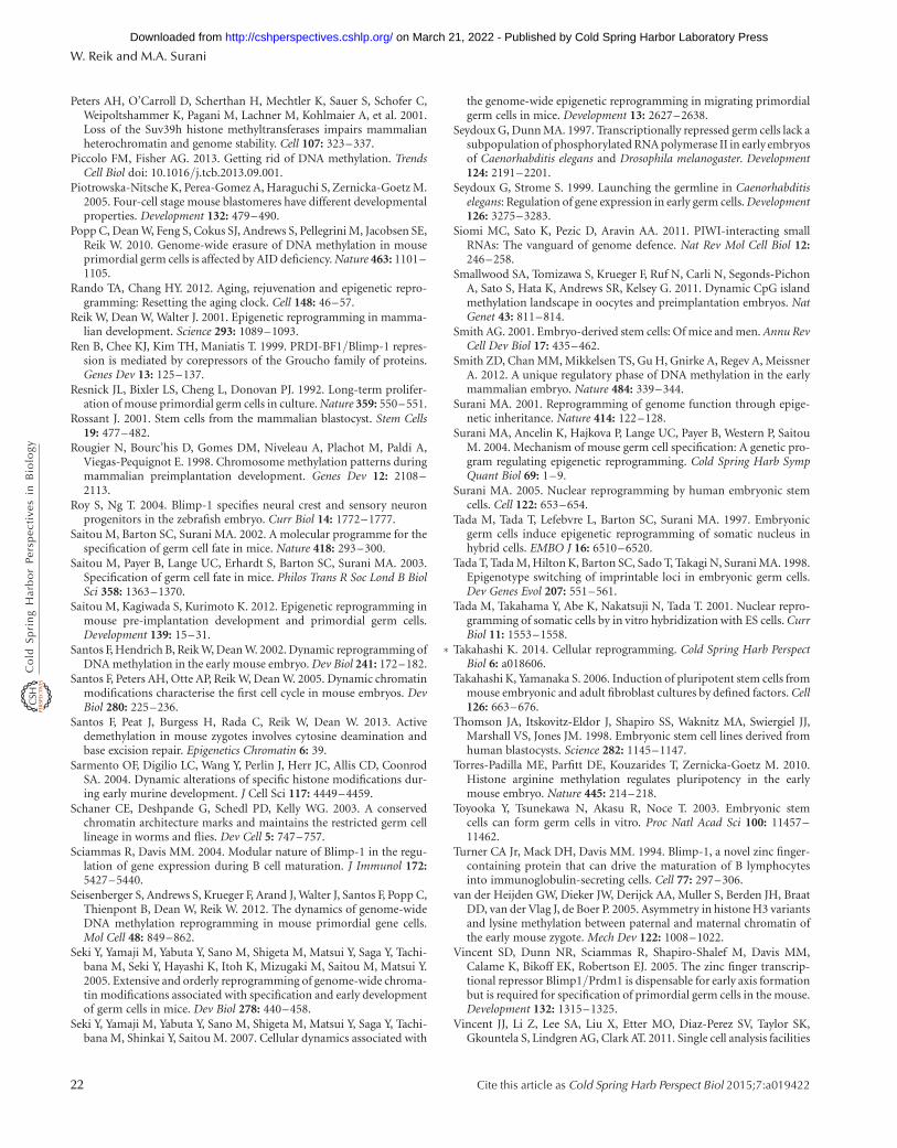
Peters AH, O’Carroll D, Scherthan H, Mechtler K, Sauer S, Schofer C,Weipoltshammer K, Pagani M, Lachner M, Kohlmaier A, et al. 2001.Loss of the Suv39h histone methyltransferases impairs mammalianheterochromatin and genome stability. Cell 107: 323–337.
Piccolo FM, Fisher AG. 2013. Getting rid of DNA methylation. TrendsCell Biol doi: 10.1016/j.tcb.2013.09.001.
Piotrowska-Nitsche K, Perea-Gomez A, Haraguchi S, Zernicka-Goetz M.2005. Four-cell stage mouse blastomeres have different developmentalproperties. Development 132: 479–490.
Popp C, Dean W, Feng S, Cokus SJ, Andrews S, Pellegrini M, Jacobsen SE,Reik W. 2010. Genome-wide erasure of DNA methylation in mouseprimordial germ cells is affected by AID deficiency. Nature 463: 1101–1105.
Rando TA, Chang HY. 2012. Aging, rejuvenation and epigenetic repro-gramming: Resetting the aging clock. Cell 148: 46–57.
Reik W, Dean W, Walter J. 2001. Epigenetic reprogramming in mamma-lian development. Science 293: 1089–1093.
Ren B, Chee KJ, Kim TH, Maniatis T. 1999. PRDI-BF1/Blimp-1 repres-sion is mediated by corepressors of the Groucho family of proteins.Genes Dev 13: 125–137.
Resnick JL, Bixler LS, Cheng L, Donovan PJ. 1992. Long-term prolifer-ation of mouse primordial germ cells in culture. Nature 359: 550–551.
Rossant J. 2001. Stem cells from the mammalian blastocyst. Stem Cells19: 477–482.
Rougier N, Bourc’his D, Gomes DM, Niveleau A, Plachot M, Paldi A,Viegas-Pequignot E. 1998. Chromosome methylation patterns duringmammalian preimplantation development. Genes Dev 12: 2108–2113.
Roy S, Ng T. 2004. Blimp-1 specifies neural crest and sensory neuronprogenitors in the zebrafish embryo. Curr Biol 14: 1772–1777.
Saitou M, Barton SC, Surani MA. 2002. A molecular programme for thespecification of germ cell fate in mice. Nature 418: 293–300.
Saitou M, Payer B, Lange UC, Erhardt S, Barton SC, Surani MA. 2003.Specification of germ cell fate in mice. Philos Trans R Soc Lond B BiolSci 358: 1363–1370.
Saitou M, Kagiwada S, Kurimoto K. 2012. Epigenetic reprogramming inmouse pre-implantation development and primordial germ cells.Development 139: 15–31.
Santos F, Hendrich B, Reik W, Dean W. 2002. Dynamic reprogramming ofDNA methylation in the early mouse embryo. Dev Biol 241: 172–182.
Santos F, Peters AH, Otte AP, Reik W, Dean W. 2005. Dynamic chromatinmodifications characterise the first cell cycle in mouse embryos. DevBiol 280: 225–236.
Santos F, Peat J, Burgess H, Rada C, Reik W, Dean W. 2013. Activedemethylation in mouse zygotes involves cytosine deamination andbase excision repair. Epigenetics Chromatin 6: 39.
Sarmento OF, Digilio LC, Wang Y, Perlin J, Herr JC, Allis CD, CoonrodSA. 2004. Dynamic alterations of specific histone modifications dur-ing early murine development. J Cell Sci 117: 4449–4459.
Schaner CE, Deshpande G, Schedl PD, Kelly WG. 2003. A conservedchromatin architecture marks and maintains the restricted germ celllineage in worms and flies. Dev Cell 5: 747–757.
Sciammas R, Davis MM. 2004. Modular nature of Blimp-1 in the regu-lation of gene expression during B cell maturation. J Immunol 172:5427–5440.
Seisenberger S, Andrews S, Krueger F, Arand J, Walter J, Santos F, Popp C,Thienpont B, Dean W, Reik W. 2012. The dynamics of genome-wideDNA methylation reprogramming in mouse primordial gene cells.Mol Cell 48: 849–862.
Seki Y, Yamaji M, Yabuta Y, Sano M, Shigeta M, Matsui Y, Saga Y, Tachi-bana M, Seki Y, Hayashi K, Itoh K, Mizugaki M, Saitou M, Matsui Y.2005. Extensive and orderly reprogramming of genome-wide chroma-tin modifications associated with specification and early developmentof germ cells in mice. Dev Biol 278: 440–458.
Seki Y, Yamaji M, Yabuta Y, Sano M, Shigeta M, Matsui Y, Saga Y, Tachi-bana M, Shinkai Y, Saitou M. 2007. Cellular dynamics associated with
the genome-wide epigenetic reprogramming in migrating primordialgerm cells in mice. Development 13: 2627–2638.
Seydoux G, Dunn MA. 1997. Transcriptionally repressed germ cells lack asubpopulation of phosphorylated RNA polymerase II in early embryosof Caenorhabditis elegans and Drosophila melanogaster. Development124: 2191–2201.
Seydoux G, Strome S. 1999. Launching the germline in Caenorhabditiselegans: Regulation of gene expression in early germ cells. Development126: 3275–3283.
Siomi MC, Sato K, Pezic D, Aravin AA. 2011. PIWI-interacting smallRNAs: The vanguard of genome defence. Nat Rev Mol Cell Biol 12:246–258.
Smallwood SA, Tomizawa S, Krueger F, Ruf N, Carli N, Segonds-PichonA, Sato S, Hata K, Andrews SR, Kelsey G. 2011. Dynamic CpG islandmethylation landscape in oocytes and preimplantation embryos. NatGenet 43: 811–814.
Smith AG. 2001. Embryo-derived stem cells: Of mice and men. Annu RevCell Dev Biol 17: 435–462.
Smith ZD, Chan MM, Mikkelsen TS, Gu H, Gnirke A, Regev A, MeissnerA. 2012. A unique regulatory phase of DNA methylation in the earlymammalian embryo. Nature 484: 339–344.
Surani MA. 2001. Reprogramming of genome function through epige-netic inheritance. Nature 414: 122–128.
Surani MA, Ancelin K, Hajkova P, Lange UC, Payer B, Western P, SaitouM. 2004. Mechanism of mouse germ cell specification: A genetic pro-gram regulating epigenetic reprogramming. Cold Spring Harb SympQuant Biol 69: 1–9.
Surani MA. 2005. Nuclear reprogramming by human embryonic stemcells. Cell 122: 653–654.
Tada M, Tada T, Lefebvre L, Barton SC, Surani MA. 1997. Embryonicgerm cells induce epigenetic reprogramming of somatic nucleus inhybrid cells. EMBO J 16: 6510–6520.
Tada T, Tada M, Hilton K, Barton SC, Sado T, Takagi N, Surani MA. 1998.Epigenotype switching of imprintable loci in embryonic germ cells.Dev Genes Evol 207: 551–561.
Tada M, Takahama Y, Abe K, Nakatsuji N, Tada T. 2001. Nuclear repro-gramming of somatic cells by in vitro hybridization with ES cells. CurrBiol 11: 1553–1558.
∗ Takahashi K. 2014. Cellular reprogramming. Cold Spring Harb PerspectBiol 6: a018606.
Takahashi K, Yamanaka S. 2006. Induction of pluripotent stem cells frommouse embryonic and adult fibroblast cultures by defined factors. Cell126: 663–676.
Thomson JA, Itskovitz-Eldor J, Shapiro SS, Waknitz MA, Swiergiel JJ,Marshall VS, Jones JM. 1998. Embryonic stem cell lines derived fromhuman blastocysts. Science 282: 1145–1147.
Torres-Padilla ME, Parfitt DE, Kouzarides T, Zernicka-Goetz M. 2010.Histone arginine methylation regulates pluripotency in the earlymouse embryo. Nature 445: 214–218.
Toyooka Y, Tsunekawa N, Akasu R, Noce T. 2003. Embryonic stemcells can form germ cells in vitro. Proc Natl Acad Sci 100: 11457–11462.
Turner CA Jr, Mack DH, Davis MM. 1994. Blimp-1, a novel zinc finger-containing protein that can drive the maturation of B lymphocytesinto immunoglobulin-secreting cells. Cell 77: 297–306.
van der Heijden GW, Dieker JW, Derijck AA, Muller S, Berden JH, BraatDD, van der Vlag J, de Boer P. 2005. Asymmetry in histone H3 variantsand lysine methylation between paternal and maternal chromatin ofthe early mouse zygote. Mech Dev 122: 1008–1022.
Vincent SD, Dunn NR, Sciammas R, Shapiro-Shalef M, Davis MM,Calame K, Bikoff EK, Robertson EJ. 2005. The zinc finger transcrip-tional repressor Blimp1/Prdm1 is dispensable for early axis formationbut is required for specification of primordial germ cells in the mouse.Development 132: 1315–1325.
Vincent JJ, Li Z, Lee SA, Liu X, Etter MO, Diaz-Perez SV, Taylor SK,Gkountela S, Lindgren AG, Clark AT. 2011. Single cell analysis facilities
W. Reik and M.A. Surani
22 Cite this article as Cold Spring Harb Perspect Biol 2015;7:a019422
on March 21, 2022 - Published by Cold Spring Harbor Laboratory Press http://cshperspectives.cshlp.org/Downloaded from
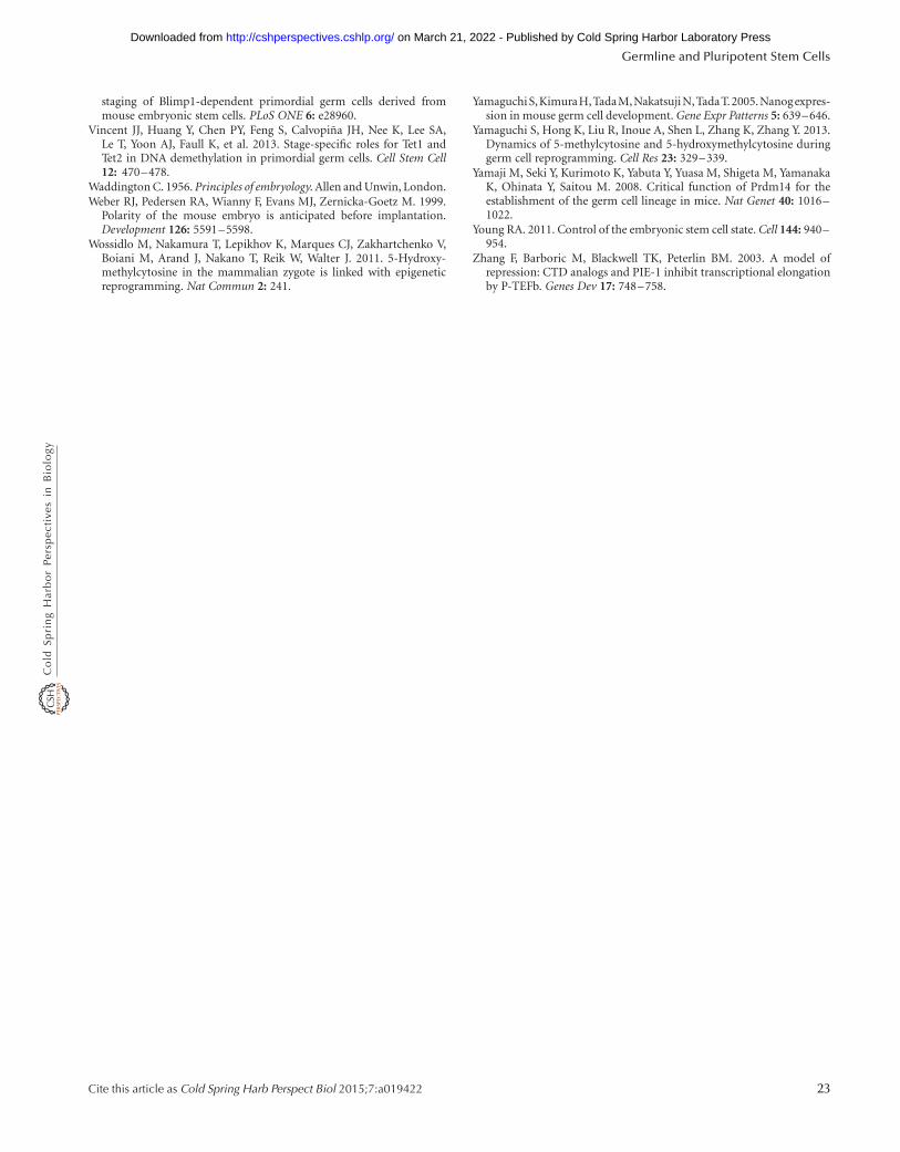
staging of Blimp1-dependent primordial germ cells derived frommouse embryonic stem cells. PLoS ONE 6: e28960.
Vincent JJ, Huang Y, Chen PY, Feng S, Calvopina JH, Nee K, Lee SA,Le T, Yoon AJ, Faull K, et al. 2013. Stage-specific roles for Tet1 andTet2 in DNA demethylation in primordial germ cells. Cell Stem Cell12: 470–478.
Waddington C. 1956. Principles of embryology. Allen and Unwin, London.Weber RJ, Pedersen RA, Wianny F, Evans MJ, Zernicka-Goetz M. 1999.
Polarity of the mouse embryo is anticipated before implantation.Development 126: 5591–5598.
Wossidlo M, Nakamura T, Lepikhov K, Marques CJ, Zakhartchenko V,Boiani M, Arand J, Nakano T, Reik W, Walter J. 2011. 5-Hydroxy-methylcytosine in the mammalian zygote is linked with epigeneticreprogramming. Nat Commun 2: 241.
Yamaguchi S,KimuraH,TadaM,NakatsujiN,TadaT.2005.Nanogexpres-sion in mouse germ cell development. Gene Expr Patterns 5: 639–646.
Yamaguchi S, Hong K, Liu R, Inoue A, Shen L, Zhang K, Zhang Y. 2013.Dynamics of 5-methylcytosine and 5-hydroxymethylcytosine duringgerm cell reprogramming. Cell Res 23: 329–339.
Yamaji M, Seki Y, Kurimoto K, Yabuta Y, Yuasa M, Shigeta M, YamanakaK, Ohinata Y, Saitou M. 2008. Critical function of Prdm14 for theestablishment of the germ cell lineage in mice. Nat Genet 40: 1016–1022.
Young RA. 2011. Control of the embryonic stem cell state. Cell 144: 940–954.
Zhang F, Barboric M, Blackwell TK, Peterlin BM. 2003. A model ofrepression: CTD analogs and PIE-1 inhibit transcriptional elongationby P-TEFb. Genes Dev 17: 748–758.
Germline and Pluripotent Stem Cells
Cite this article as Cold Spring Harb Perspect Biol 2015;7:a019422 23
on March 21, 2022 - Published by Cold Spring Harbor Laboratory Press http://cshperspectives.cshlp.org/Downloaded from
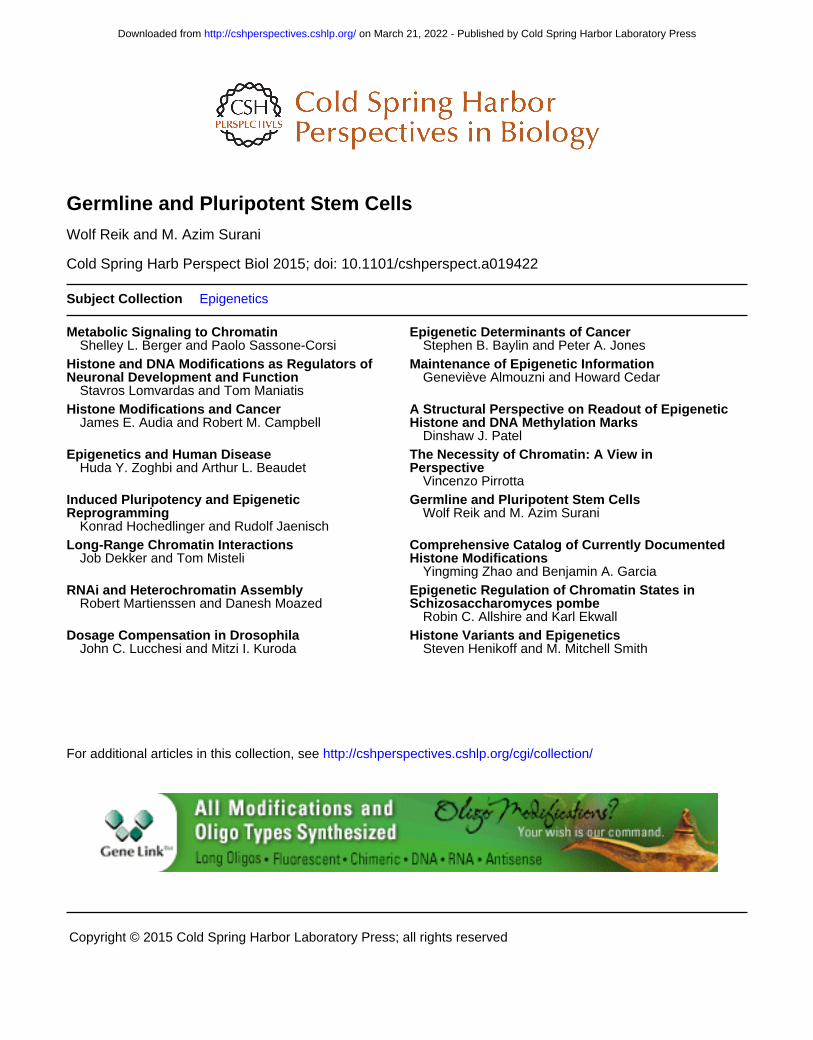
2015; doi: 10.1101/cshperspect.a019422Cold Spring Harb Perspect Biol Wolf Reik and M. Azim Surani Germline and Pluripotent Stem Cells
Subject Collection Epigenetics
Metabolic Signaling to ChromatinShelley L. Berger and Paolo Sassone-Corsi
Epigenetic Determinants of CancerStephen B. Baylin and Peter A. Jones
Neuronal Development and FunctionHistone and DNA Modifications as Regulators of
Stavros Lomvardas and Tom Maniatis
Maintenance of Epigenetic InformationGeneviève Almouzni and Howard Cedar
Histone Modifications and CancerJames E. Audia and Robert M. Campbell Histone and DNA Methylation Marks
A Structural Perspective on Readout of Epigenetic
Dinshaw J. PatelEpigenetics and Human Disease
Huda Y. Zoghbi and Arthur L. Beaudet PerspectiveThe Necessity of Chromatin: A View in
Vincenzo Pirrotta
ReprogrammingInduced Pluripotency and Epigenetic
Konrad Hochedlinger and Rudolf Jaenisch
Germline and Pluripotent Stem CellsWolf Reik and M. Azim Surani
Long-Range Chromatin InteractionsJob Dekker and Tom Misteli Histone Modifications
Comprehensive Catalog of Currently Documented
Yingming Zhao and Benjamin A. GarciaRNAi and Heterochromatin Assembly
Robert Martienssen and Danesh Moazed Schizosaccharomyces pombeEpigenetic Regulation of Chromatin States in
Robin C. Allshire and Karl EkwallDrosophilaDosage Compensation in
John C. Lucchesi and Mitzi I. KurodaHistone Variants and Epigenetics
Steven Henikoff and M. Mitchell Smith
http://cshperspectives.cshlp.org/cgi/collection/ For additional articles in this collection, see
Copyright © 2015 Cold Spring Harbor Laboratory Press; all rights reserved
on March 21, 2022 - Published by Cold Spring Harbor Laboratory Press http://cshperspectives.cshlp.org/Downloaded from
http://cshperspectives.cshlp.org/cgi/collection/ For additional articles in this collection, see
Copyright © 2015 Cold Spring Harbor Laboratory Press; all rights reserved
on March 21, 2022 - Published by Cold Spring Harbor Laboratory Press http://cshperspectives.cshlp.org/Downloaded from