grace wong gpst1. assessment of the red eye common causes of red eye painful and non painful signs...
TRANSCRIPT
Grace Wong
GPST1
Red Eye

Assessment of the red eyeCommon causes of red eye
Painful and Non PainfulSigns and symptomsManagement of each condition
Aims
Common presentation in primary care and in A+E
Most cases due to relatively trivial problemsMost common is conjunctivitisSmall proportion are serious and need urgeny
treatmentSometimes difficulty in discerning between
causes
Most practical way is; Pain or notVisual acuity
Red Eye
OnsetPainVisual ChangesPhotophobiaForeign body sensationTraumaDischarge, clear or coloredBilateral or unilateral
History
Social historyNursery school teacher
Co-morbid condition Collagen vascular disordersRheumatoid, goutTB, sarcoidosisHTN
Past Ocular HistoryE.g. Similar episodesSurgeryLazy eyeContact lenses
Visual acuityExtra ocular movementsPen light examination (reactivity, corneal
opacity, pupil shape, discharge, infection)Test for direct and consensual photophobiaSlit lamp examination – with and without
fluorescein
Anterior chamber evaluation – depth, cellsIOP meaurements
Examination
Think systemically about the structures within the eye to common to differential diagnosis
Inflammation of orbit?Lid DiseaseScleral inflammationCorneal diseaseUveal/iris inflammationOther e.g. glaucoma
Differential Diagnosis
?
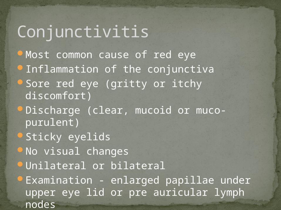
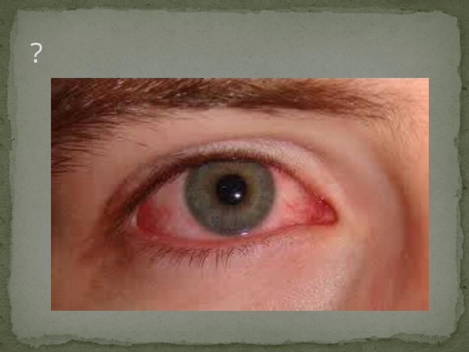
Most common cause of red eyeInflammation of the conjunctivaSore red eye (gritty or itchy discomfort)Discharge (clear, mucoid or muco-purulent)Sticky eyelidsNo visual changes Unilateral or bilateralExamination - enlarged papillae under upper
eye lid or pre auricular lymph nodes
Conjunctivitis

Allergic, viral or bacterialDifficult to distinguish between typesBoth bacterial and viral can occur after a
viral URTI
Causes
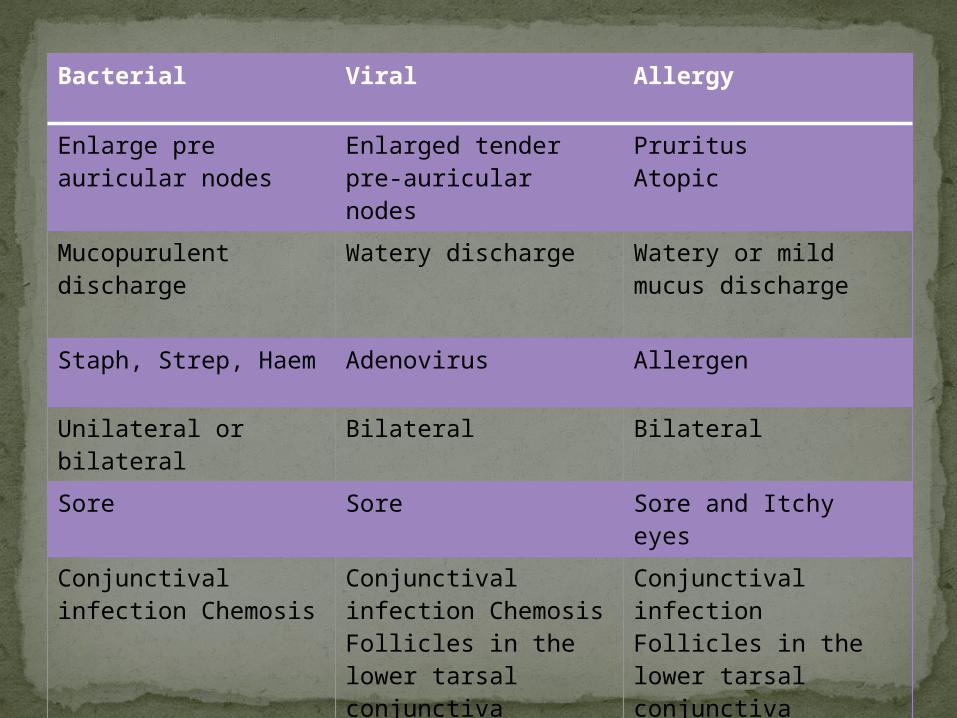
Bacterial Viral Allergy
Enlarge pre auricular nodes
Enlarged tender pre-auricular nodes
PruritusAtopic
Mucopurulent discharge
Watery discharge Watery or mild mucus discharge
Staph, Strep, Haem Adenovirus Allergen
Unilateral or bilateral Bilateral Bilateral
Sore Sore Sore and Itchy eyes
Conjunctival infection Chemosis
Conjunctival infection ChemosisFollicles in the lower tarsal conjunctiva
Conjunctival infection Follicles in the lower tarsal conjunctiva Cobblestone under the upper lid
85% of cases clear in <7 days with or without tx
Advise patients to bathe the affected eye with boiled cooled water am and pm
If symptoms not improve in >5 daysSwab for MC+STreat empirically with chloramphenicol QDSconsider alternative diagnosis e.g. allergy, dry
eyes,
Consider referral >7-10 days or if suspicion of herpetic infection
Management - Infective
Topic or systemic anti histamines e.g. sodium cromoglicate eye drops
Avoid topical steroids – long term complications e.g. cataract, glaucoma, fungal infection
Consider cold compress and wash out with cold water during acute exacerbation
Refer if symptoms are persistent despite treatment or if vision is affected
Management - Allergic
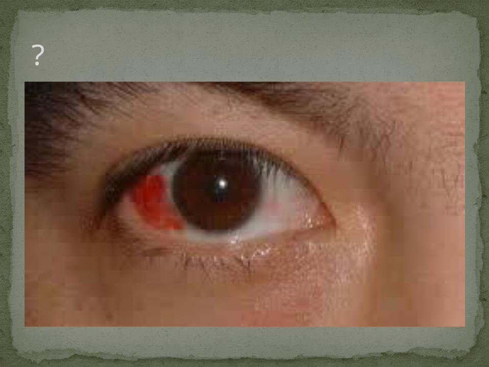
?
Spontaneous painless localised haemorrhage under the conjunctiva
Common in the elderlySpontaneous or traumaticLooks alarming but generally painless (may
cause some aching)Clear spontaneously in 1-2 weeks but may
recur
Subconjunctival Haemorrhage
HypertensionClotting disordersLeukaemiaIncreased venous pressure
Check BPIf severe/recurrent Check FBC and clotting screen
Associations

Blood under conjunctiva covering part or all of eye
Normal Visual Acuity
Consider referral if;Follows trauma More than a slight discomfortFails to settle spontaneously over 1 week
Signs
?
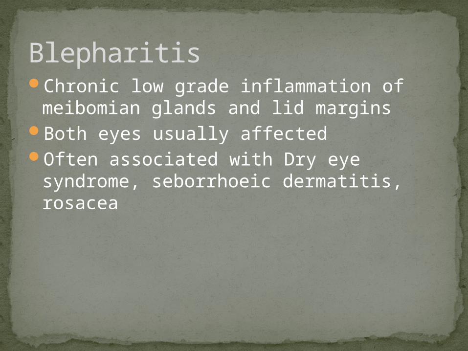
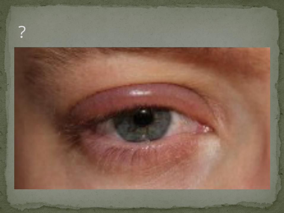
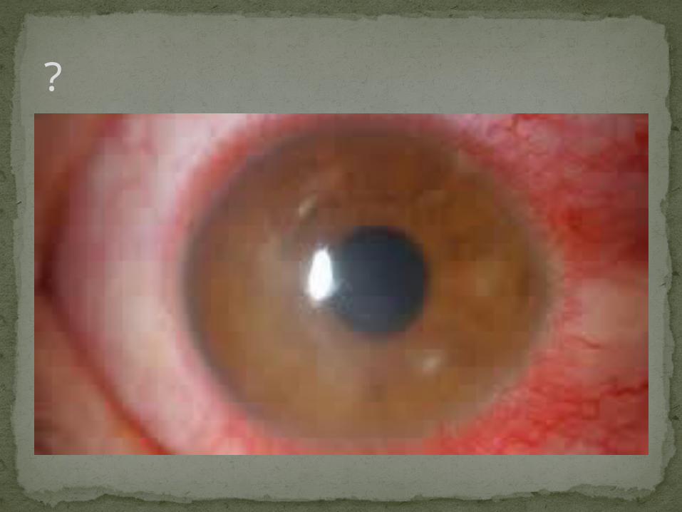
Chronic low grade inflammation of meibomian glands and lid margins
Both eyes usually affectedOften associated with Dry eye syndrome,
seborrhoeic dermatitis, rosacea
Blepharitis
Staphylococcal Seborrhoeic – associated with seborrhoeic
dermatitis. Yeast is involved and can trigger inflammatory reaction
Meibomain – gland dysfunction unable to lubricate eye
Causes
Presents with long history of irritable burning dry red eyes
Eyelids have red margins Look inflamed and greasyTiny flakes or scales on eyelidsSticky with dischargeMeibomian glands may block an fill with oily
fluid
Symptoms come and go
Symptoms
Regular eyelid hygiene – warm, massage and cleansing
Remove scales and crusts from lid marginsTreat dry eye symptoms with preservative
free tear supplements e.g. liquifilmAntibiotic eye treatment if eyelid becomes
infection e.g. fusidic acid (topical on eyelid). Can be up to 3 month course
Treatment
?
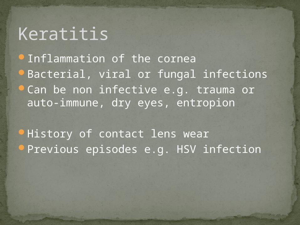
Inflammation of the corneaBacterial, viral or fungal infectionsCan be non infective e.g. trauma or auto-
immune, dry eyes, entropion
History of contact lens wearPrevious episodes e.g. HSV infection
Keratitis

Very painful red eye PhotophobiaForeign body sensationReduced visual acuity depends on nature of
problemCircumcorneal injectionConjunctiva is also inflamed – keratoconjuncivitisDischarge – water, mucoid or purulentPupil may be smallFluorescin readily demonstrates any ulceration
Signs

Significant loss of vision secondary to scarring or astigmastism
Complications can lead to blindness;Corneal perforationChoroidal detachmentEndopthalmitis
CORNEAL ULCERATION IS AN OPTHALMOLOGIC EMERGENCY
Complications

The cause must be identified prior to treatment - some therapies benefit whilst others can harm
Refer the same day for urgent ophthalmological review
Delay may result in loss of sight
Treatment

If caused by Herpes simplex infection and dendritic ulcer
AVOID topical steroids as can cause massive amoebic ulceration and blindness
Typical dendritic ulcer – delicate branching pattern
Caution
?
Severe inflammation that occurs throughout the entire thickness of the sclera
Rare Average age 52 yrsCan be unilateral or bilateralAffects more women than menCan affect anterior or posterior segmentEither nodular, diffuse or necrotizing
Scleritis
The sclera is an avascular structure50% is associated with systemic illness;
Herpes ZosterRheumatoid arthritisSLEPolyarteritis nodosumWegner’s granulomatosisTraumaInfectionSurgery
Associated

Red eyeSevere boring eye pain – may radiate to forehead,
brow or jawKey symptom; gradual onset (days or weeks)Pain worse with movement of eye and at nightWateringPhotophobiaDecreased visual acuityEye is tender to touch and may have deep purple
hueThere may be accompanying uveitis and keratitis
Presentation
Urgent referral to ophthalmologyTreated with steroids
Complications includeCataractGlaucomaRetinal detachment
Treatment

?
Most common in young/middle aged adults
Acute onset of painIncreasing pain as eye converges and pupil
constrictPhotophobiaBlurred visionDecreased visual acuityWateringCircumcorneal rednressSmall or irregular pupil + hypopyon (pus causing white fluid level line)
Iritis (anterior uveitis)

Secondary to corneal graft rejectionEye infections e.g. toxoplasmosis, herpes
virus keratitis30% are associated with seronegative
arthropathies e.g. AS
Causes
Refer urgently to ophthalmology
Complications include; Posterior synechiae (irregular pupil shape)GlaucomaCataract
Relapses are common
Management
Decreased visual acuityPain deep in the eye – not surface irritationPhotophobiaAbsent or sluggish pupil responseCorneal Damage on fluorsecein staining or
opacificationHistory of trauma
These need same day referral
Red Flag signs of a Red Eye
http://www.patient.co.uk/doctor/The-Red-Eye.htm
Questions?